Note: Descriptions are shown in the official language in which they were submitted.
CA 02745796 2011-07-08
1
METHOD AND SYSTEM FOR COLLECTING OPTICAL DATA FOR USE IN TIME
RESOLVED OPTICAL IMAGING OF A TURBID MEDIA
TECHNICAL FIELD
[0001] The present disclosure relates to the field of optical imaging of
biological tissues. More specifically, the present disclosure relates to a
method
and a system for collecting optical data for use in time resolved optical
imaging of
a turbid media.
BACKGROUND
[0002] Different types of imaging techniques such as positron emission
tomography (PET), magnetic resonance imaging (MRI) and ultrasound imaging
are available that can non-invasively gather information from within
biological
tissues as a basis for image reconstruction. More recently, another imaging
technique, namely optical imaging has been the subject of intense research and
commercial development.
[0003] Optical imaging is based on information that can be derived from
the analysis of the signal resulting from the interaction of light with matter
as it is
propagated within an object. A time domain (TD) approach, by conveying
information on the time required by photons to travel within the object, is
considered to be "time resolved" and can be used to calculate the spatial
distribution of optical characteristics of the object, such as absorption and
scatter
coefficients, via well known photon diffusion.
CA 02745796 2011-07-08
2
[0004] Optical
imaging is particularly attractive in view of its non-
invasiveness which permits the acquisition of in vivo information without
damaging
biological tissues. Furthermore the technique may be useful to monitor drug
distribution, detect the presence of abnormalities within organs, or map
physiological activities within mammals.
[0005] Optical
imaging systems rely on the presence of bioluminescent
molecules, also called biomarkers or fluorescent markers, within a biological
region of interest (ROI). For example certain endogenous molecules and some
exogenous molecules, such as exogenous chromophores as well as fluorophores,
provide useful levels of optical contrast. Recently, an explosive development
in the
variety of biomarkers and nanoprobes is revolutionizing the molecular imaging
and
generates new premises for translational medicine paradigm. Molecular imaging
using optical methods will be required to provide the means for managing the
huge
diversity of biomarkers, for their identification and classification, and for
a thorough
validation of their specificity and reliability.
[0006] In view of
the above, it would be desirable to provide a method
and an optical imaging system for imaging turbid media such as biological
tissues
that can rapidly and efficiently support a wide variety of fluorescent
markers.
SUMMARY
[0007] According to
the present disclosure, there is provided a method for
collecting optical data for use in time resolved optical imaging of a turbid
media.
An excitation wavelength of a pulsed light beam is tuned according to an
excitation
spectrum of a fluorescent marker of interest. The pulsed light beam is
directionally
CA 02745796 2011-07-08
3
propagated to illuminate a plurality of predetermined illumination points in a
region
of interest of the turbid media. Light emanating from a plurality of
predetermined
collection points in the region of interest is collected. The collected light
includes a
fluorescence signal from the fluorescent marker. The collected light is
filtered to
allow the fluorescence signal to propagate through a filter while rejecting
photons
outside a fluorescence emission spectrum of the fluorescent marker. The
filtered
light is measured at a detector to produce a time resolved optical signal for
one or
more illumination point/collection point configurations.
[0008] According to the present disclosure, there is also provided a
system for collecting optical data for use in time resolved optical imaging of
a
turbid media. The system comprises a pulsed light source providing a light
beam
at a tunable wavelength according to an excitation wavelength of a fluorescent
marker of interest. Also comprised is an illuminating optic component for
directionally propagating the pulsed light beam such that a region of interest
of the
turbid media is illuminated at a plurality of illumination points. A
collecting optic
component collects light emanating from a plurality of predetermined
collection
points in the region of interest. The collected light includes a fluorescence
signal
from the fluorescent marker. A fluorescence filter allows the fluorescence
signal to
propagate therethrough while rejecting photons outside a fluorescence emission
spectrum of the fluorescent marker. A time domain detector detects the
filtered
light and produces a time resolved optical signal for one or more illumination
point/collection point configurations.
[0009] The foregoing and other features will become more apparent upon
reading of the following non-restrictive description of illustrative
embodiments
thereof, given by way of example only with reference to the accompanying
drawings.
CA 02745796 2011-07-08
4
BRIEF DESCRIPTION OF THE DRAWINGS
[0010] Embodiments of the disclosure will be described by way of
example only with reference to the accompanying drawings, in which:
[0011] Fig. 1 is a flow chart of an exemplary method for collecting
optical data for use in time resolved optical imaging;
[0012] Fig. 2 is perspective view of an embodiment of a system for
collecting optical data for use in time resolved optical imaging;
[0013] Fig. 3 is a detailed view of some of the optic components of Fig.
2;
[0014] Fig. 4 schematically illustrates a raster scan pattern of
illumination in a region of interest at the surface of a mammal;
[0015] Fig. 5 is a block diagram of an example of imaging system; and
[0016] Figure 6 is a graphical representation of an example of laser
wavelength and fluorescence filter optimization.
[0017] It will be noted that throughout the appended drawings, like
features are identified by like reference numerals.
DETAILED DESCRIPTION
5
[0018] Various
aspects of the present disclosure generally address
one or more needs related to optical imaging systems and methods supporting a
diversity of fluorescent markers.
[0019] The present
disclosure relates to the field of optical imaging of
turbid media such as biological tissues as parts of human organs, animals, and
the like. While the following description of exemplary embodiments provides
examples that relate to imaging of small mammals such as mice, it will be
appreciated that the method can also be applied in clinical testing for the
benefit
of actual patients as well as to laboratory testing involving larger animals
and in
particular to laboratory animals such as dogs, pigs and primates. US Patent No
6,992,762 describes a system and method for collecting optical data using one
or
more fixed wavelength light sources.
[0020] The following
terminology is used throughout the present
disclosure:
[0021] Time resolved
imaging: Imaging based on time required by
photons to travel within an object being imaged.
[0022] Time domain
detector: A detector sensitive to timed
characteristics of a signal.
[0023] Turbid media:
A substantially opaque medium due to a
relatively high light scattering, for example a biological tissue.
[0024] Region of interest (ROI): Part of a turbid media to be imaged.
REPLACEMENT PAGE
CA 2745796 2018-01-12
CA 02745796 2011-07-08
6
[0026] Fluorescent marker: A molecule inserted into a turbid media,
capable of emitting light when subject to energy transfer.
[0026] Biomarker: A biological or biologically-derived marker.
[0027] Fluorescence signal: Light emitted from a fluorescent marker.
[0028] Fluorescence spectrum: A wavelength, or a wavelength range,
of light emitted by a fluorescent marker.
[0029] Excitation wavelength: Wavelength causing a fluorescent
marker to emit light, usually at another wavelength distinct from
the excitation wavelength.
[0030] Excitation spectrum: Range of excitation wavelength causing a
fluorescent marker to emit light.
[0031] Tuning: Adjusting wavelength or frequency.
[0032] Pulsed light: Light emitted intermittently, according to a duty
cycle.
[0033] Directionally propagating: Of light, emitting in a non-dispersive
(collimated) fashion,
[0034] Illuminating optic component: Optical device on the emitting
CA 02745796 2011-07-08
7
side of an illuminating system, located upstream of an
illumination target.
[0035] Collecting optic component: Optical device on the receiving side
of an illuminating system, located downstream of an illumination
target.
[0036] Maximum rejection point: Of a filter, a wavelength at which
rejection by the filter is maximized.
[0037] Fluorescence filter: A filter for a fluorescence signal.
[0038] Excitation filter: A filter located upstream of an illumination
target.
[0039] Collection filter: A filter located downstream of an
illumination
target.
[0040] Free space optics: Line-of-sight optics in which light travels
unimpeded through empty space, air, or like medium.
[0041] Controller: A device such as a computer or a processor capable
of controlling another device or component based on an input.
[0042] With reference to the drawings, Fig. 1 is a flow chart of an
exemplary method for collecting optical data for use in time resolved optical
imaging. Optical imaging of a turbid media, such as for example a biological
tissue
CA 02745796 2011-07-08
8
or a part of a human organ or animal organ, may be obtained using a sequence
100 comprising a first step 102 of tuning an excitation wavelength of a pulsed
light
beam according to an excitation spectrum of a fluorescent marker of interest
For
example, a laser wavelength could be adjusted as close as possible to a
wavelength for maximum absorption of Cy5.5 fluorescent probes (i.e. ¨675nm),
while maintaining an optimum offset compared with a cut-on of the fluorescence
filter for maximizing rejection of laser light. At step 104, the pulsed light
beam is
directionally propagated to illuminate a plurality of predetermined
illumination
points in a region of interest of the turbid media. Light emanating from a
plurality of
predetermined collection points in the region of interest is collected at step
106.
The collected light includes a fluorescence signal from the fluorescent marker
of
interest, and may also contain other components such as for example
fluorescence from tissue and light from the excitation laser beam. At step
108, the
collected light is filtered to allow the fluorescence signal of interest to
propagate
through a filter while rejecting photons outside a fluorescence emission
spectrum
of the fluorescent marker of interest. The filtered light is measured at a
detector, at
step 110, to produce a time resolved optical signal for one or more
illumination
point/collection point configurations.
[0043] The emitted light, the collected light and the filtered light may
propagate between various optical components in air (i.e. through free space
optics) or through optical components such as fiber optics
[0044] The fluorescent marker of interest, present in the region of
interest, may comprise one or more biomarkers, in which case the excitation
wavelength may be tuned to correspond to an excitation wavelength of one or
more biomarkers. As multiple fluorescent markers with different excitation
CA 02745796 2011-07-08
9
wavelengths may be used concurrently or in different areas of the turbid
medium,
the excitation wavelength of the pulsed light beam may be tuned in various
ways.
[0045] Tuning the excitation wavelength may be such that it matches
the excitation spectrum of one or several fluorescent marker(s) of interest,
or
biomarker(s) either concurrently or sequentially. Variations of the tuning may
be
made in order to allow maximum rejection of the excitation wavelength by the
filter
while at the same time obtaining a sufficient fluorescence level. The filter,
which
may be a fluorescence filter, may be adaptable for maximizing a collection
within a
spectrum of a fluorescence signal of interest, corresponding to the
fluorescent
marker, while at the same time rejecting wavelengths that are not of interest.
In
particular, the fluorescence filter may minimize interference in the
fluorescence
signal of interest stemming from the excitation light source itself. Otherwise
stated,
the filter may be adapted to minimize leaking of the light emitted from the
source,
in its original wavelength, into the detector. Tuning may be performed
manually or
automatically. Information regarding a level of rejection by the filter may be
fed
back to a source of the pulsed light via a controller such as for example a
computer (shown on a later figure). Additionally, an intensity of the pulsed
light
beam may be adjusted, for example based on one or several of the following:
specifics of the turbid media, on geometry of various optical elements (shown
on
later figures) used in collecting the collecting optical data, or on an
intensity of the
collected light. Thus both the pulsed light beam and the filter are adjustable
to
allow efficient use with various fluorescent markers and biomarkers.
[0046] Light emanating from a plurality of collection points after
diffusion through the tissue has a somewhat different, usually longer
wavelength
than that of the light emitted from the source. Some fluorescent markers,
biomarkers, or fluorophores, may differ and fluoresce at a shorter wavelength
than
CA 02745796 2011-07-08
that of the light emitted from the source. The present disclosure is not
limited to
any type of fluorescent marker or biomarker and includes fluorescent
wavelengths
that are either longer or shorter than the excitation wavelengths.
[0047] The collected light measured using the detector is used to
produce a time resolved optical signal. Light collection may be selective so
that
light emanating from points other than those being sampled may be optically
excluded from detection.
[0048] Embodiments of the system used for collecting the optical data
will now be described mostly referring to small mammals as the object to be
imaged but it will be appreciated that a wide variety of biological tissues
may be
amenable to optical imaging using the technique described herein. These can be
but are not limited to breast tissue, brain, tumors and the like.
[0049] A general schematic representation of a first embodiment of a
system used for imaging turbid media, represented as a small mammal, is shown
in Fig. 2, which is an exemplary perspective view of an embodiment of a system
for collecting optical data for use in time resolved optical imaging. The
system 200
comprises a pulsed light source 210 providing a pulsed light beam 212 at a
tunable wavelength. The wavelength of the pulsed light beam 212 may be tuned
according to an excitation wavelength of a fluorescent marker of interest
present in
the turbid media being imaged 214.
[0050] The pulsed light source 210 is capable of generating a beam of
light 212 at an adaptable or tunable wavelength. Illuminating optic
components,
comprising for example a movable reflective mirror 224, directionally
propagate
CA 02745796 2011-07-08
11
the pulsed light beam 212 such that a region of interest of the turbid media
is
illuminated at one or a plurality of predetermined illumination points.
Collecting
optic components, for example a collecting lens 234, collect light emanating
from a
plurality of predetermined collection points in the region of interest. The
collected
light includes a fluorescence signal from the fluorescent marker(s) or
biomarker(s)
of interest. A fluorescence filter allows the fluorescence signal(s) to
propagate
therethrough while rejecting photons outside a fluorescence emission spectrum
of
the fluorescent marker(s) of interest. In some embodiments, a single component
forms the fluorescence filter. In a variant, the fluorescence filter may
comprise an
excitation filter 229 in a path of the light beam 212 and a collection filter
227 in a
path of the collected light. Alternately, the collection filter 227 may
consist of a
plurality of filters to allow filtering of photons of wavelength other than
the
fluorescence emission spectrum of the fluorescence marker(s) and biomarker(s)
of
interest. A time domain detector 218 detects the collected light and if
applicable
filtered light and produces a time resolved optical signal for one or more
illumination point/collection point configurations. A fluorescence lifetime of
a
biomarker refers to the average time the biomarker molecule stays in its
excited
state before emitting a photon. In an embodiment, post-processing of the
information collected by the detector 218 may be made at the computer 219,
based on a known fluorescence lifetime of a biomarker.
[0051] As shown on
Fig. 2, illuminating optics may directionally
propagate the beam of light through free space, toward desired illumination
points
on the surface of the turbid medium 214. Likewise, collecting optics may
collect
the light 216 re-emitted from the turbid medium through free space, forwarding
the
light through a collection filter 227 and further to the detector 218. Another
embodiment of the system may use fiber optics (not shown) associated with
CA 02745796 2011-07-08
12
various optic components, rather than free space optics, for at least some or
all of
the light paths of Fig. 2.
[0052] As shown a movable supporting tray 220 is mounted on a
translational stage 222. A computer 219 is an exemplary controller that may be
used for controlling the pulsed light source 210, the various optic
components, the
detector 218 and the tray 220.
[0053] The movable reflective mirror 224 may be a mirror
galvanometer. The beam 212 may pass through the excitation filter 229 and then
be reflected by the movable mirror 224 at an angle 9 and directed towards a
thin
angled mirror 226 which reflects the beam in a direction substantially
perpendicular to the surface of the turbid medium 214 being scanned. It can be
appreciated that the partial rotation of the movable mirror 224 will modify
the angle
0 and direct the beam to a different point on the thin angled mirror 226 and,
consequently, to a different illumination point on the surface of the turbid
medium
214. Successive partial rotations of the movable mirror 224 thus produce a
line
scan substantially parallel to the thin angled mirror 226. Lens 228 is
optionally
provided and positioned between the movable mirror 224 and the thin angled
mirror 226 such that the movable mirror 224 is at the focal distance of the
lens 228
to provide telecentric imaging.
[0054] Fig. 3 is a detailed view of some of the optic components of Fig.
2, with additional components. The pulsed light source 210 emits light at a
tunable
wavelength and may do so at a variable intensity. The pulsed light source 210
may
be a tunable laser, for example a supercontinuum tunable laser having a
dispersion device (not shown) for selecting a desired wavelength or bandwidth.
CA 02745796 2011-07-08
13
Using non-linear optical effects the supercontinuum laser is able to generate
ultrashort light pulses (picoseconds) with a very broadband spectrum (from
400nm
up 2400nm). A spectral selector with fixed or adjustable bandwidth is used to
extract from this spectrum the optimum bandwidth and power required for the
excitation of any fluorophore. Alternatively, the pulsed light source 210 may
be a
xenon lamp but its pulse duration is much longer (microseconds) and power
density much lower limiting drastically the sensitivity, the range of
fluorescence
lifetimes that could be measured and the accuracy of the depth &
concentrations
(3D volumetric) evaluations by comparison with the supercontinuum laser.
[0055] The
excitation filter 229 and the collection filter 227 are
positioned, respectively, between the pulsed light source 210 and a region of
interest (ROI), and between the re-emitted light and the detector 218 (shown
on
Figure 2), which may be a time correlated single photon counting detector. The
movable mirror 224 may be a switching or dichroic mirror system and may be
used
for either sequential or simultaneous illumination of the ROI at different
wavelengths. The excitation filter 229, the movable mirror 224 and the
collection
filter 227 together may form an adaptable fluorescence filter. In an
embodiment, a
selectable fluorescence filter may alternatively be used. The fluorescence
filter
rejects wavelengths outside the spectrum of fluorescence signal(s) of
interest. The
fluorescence filter may further have spectral regions of maximum rejection
(for
wavelengths outside those specific to the fluorophore of interest) where the
collected light is rejected to a large extent (>60D) while the fluorescence
wavelengths of interest from the light reflected on the ROI is to a large
extent
unimpeded by the fluorescence filter. Of course, embodiments supporting
phenomena in which detected fluorescent light having a shorter wavelength than
an excitation wavelength are also within the scope of the present disclosure.
Information about rejection, including about the maximum rejection point, may
be
CA 02745796 2011-07-08
14
fed back from the detector 218 to the pulsed light source 210. Observation and
adaptation of this maximum rejection point may be used to adjust the tunable
wavelength of the pulsed light source 210, thereby improving the rejection of
the
pulsed light source wavelength while maintaining optimum excitation
efficiency.
[0056] In an embodiment, a user of the system may adjust the
wavelength of the pulsed light source 210 manually, via observation of impacts
of
such adjustments on a behavior of the light reflected on the ROI. In another
embodiment, the wavelength adjustment may be made automatically, under
control of the computer 219, on the basis of feedback and/or detected light
from
the detector 218. Those of ordinary skill in the art will appreciate that
feedback
from the detector 218 about rejection of the excitation wavelength may be
provided to the tunable pulsed light source 210 by the computer 219 and/or
other
types of controllers (not shown) by means of appropriate software and code.
[0067] Returning to Fig. 2, tray 220 supports the exemplary turbid
medium 214, in the current graphical representation the mammal, while it is
being
imaged. The tray can be displaced longitudinally on a translational stage 222
to
position the turbid medium such that a plurality of line scans parallel to
each other
can be generated. This stepwise process is repeated a selected number of times
to produce a raster scan of a region of interest. The raster scan can
alternatively
be achieved by longitudinally displacing the thin angled mirror 226. The
raster
scan can be also generated by using fiber optics for illumination of the
turbid
medium and collection of the signal that are installed on the arm of a robot
that
can generate 30 movements following the shape of the turbid medium or
specimen under investigation.
CA 02745796 2011-07-08
[0058] Fig. 4 schematically illustrates a raster scan pattern of
illumination in a region of interest (ROI) at the surface of a mammal. The
user
defined ROI 440 delimits the area to be scanned which comprises the
predetermined illumination points 442 according to a selected configuration.
Predetermined collection points may generally correspond to the illumination
points 442. The arrangement of the optic components also permits other
scanning
patterns to be performed. It will be appreciated that the ROI may consist of
the
whole animal.
[0059] Considering at once Figs. 2 and 3, light re-emitted from the
turbid medium is collected by the collecting optics, which may comprise
collecting
lens 234 and may additionally comprise reflective mirror 236 which may be a
mirror galvanometer, collection filter 227 and lens 238. The collecting lens
234 is
located above the ROI and above the thin angled mirror 226. The angular
position
of the mirror 236 relative to the incoming light and the detector 218
determines
which collection point is being sampled since only part of the light re-
emitted
(corresponding to a given collection point) impinging on the mirror is
reflected at
the proper angle to reach the detector 218. Selective detection of the light
re-
emitted from a given collection point may be further enhanced by optically
coupling
the mirror galvanometer with lenses and/or pinholes.
[0060] Upon impinging on the surface of the ROI, part of the excitation
light penetrates the tissue and part is reflected at the air/tissue boundary.
The
photons of the excitation light that are propagated within the ROI are
absorbed
and scattered, thereby producing a large number of photon paths. In biological
tissues, absorption may arise as a result of the presence of natural
(endogenous)
or exogenous chromophores, biomarkers or fluorescent markers, while scattering
is triggered by the presence of micro and macromolecular structures such as
cell
CA 02745796 2011-07-08
16
nucleus and organelle, proteins, lipids and the like which create refractive
index
inhomogeneities. The fraction of the excitation light that is not absorbed
ultimately
exits the ROI by diffusing through the skin barrier at various distances from
the
illumination point. It can be appreciated that photons that have traveled
deeper in
the tissue will take a longer time to exit at the surface of the ROI. This
provides the
basis for time resolved detection of the collected light signal from which
useful
information about the optical properties of a region of interest can be
extracted to
be incorporated into image reconstruction algorithms. Throughout the present
specification, time resolved and time domain (TD) are alternately used, but
refer to
the same principle. I
[0061] In TD measurements, the pulsed light source is briefly pulsed
and the collected light signal is detected as a function of time to generate a
temporal point spread function (TPSF). The light source may be a laser source
capable of generating pulses characterized by a width in the picoseconds
range.
Time domain detectors such as time gated intensified charge coupled devices
(1CCDs), time correlated single photon counting devices (TCSPC's), ultrafast
semiconductor detectors (avalanche and PIN photodiodes), photomultipliers and
streak cameras can be used. In an embodiment, a TCSPC device is used in the
optical system. TCSPC's are capable of measuring the time taken by a photon to
reach the detector as it travels through the illuminating optical path, the
tissue and
the collecting optical path. Time measurements may be provided by a "clock"
circuitry electronically coupling the light source and the detector.
[0062] While the TD imaging of turbid medium can rely on the natural
optical properties of the endogenous molecules for providing optical contrast,
exogenous molecules may be introduced in the tissue to provide additional
contrast. In this respect, exogenous chromophores as well as fluorophores and
CA 02745796 2011-07-08
17
biomarkers may be used as contrast agents. Furthermore the biodistribution of
such contrast agents can be followed using the method and system described
hereinabove. In an embodiment, the biodistribution can be followed over time
thereby producing pharmacokinetics data.
[0063] Reference is now also made to Figure 6, which depicts a
graphical representation of an example of laser wavelength and fluorescence
filter
optimization. The various optical components as well as the light source are
arranged to illuminate and detect light at a tunable wavelength, as is
described
hereinabove. This property can be exploited to follow the pharmacokinetics of
two
or more biomarkers such as fluorophores and/or chromophores. In particular,
the
tunable source may be arranged to illuminate at an excitation wavelength of a
fluorophore while the fluorescence filter maintains optimum selectivity at an
emission wavelength, or more generally in an emission spectrum, of the
fluorophore. The optical components and the light source may thus be tuned to
subsequently measure one or several fluorophore(s) and or chromophores(s), so
as to extract combined and more elaborate pharmacokinetics data.
[0064] In addition to the formerly described components, the system
may further comprise one or several of the following components. The selection
of
additional components depends on the applications, the type of turbid medium,
the
type of contrast agent(s), the pharmacokinetics data sought, etc. Thus the
following components can be added separately, as sub-combinations or
concurrently to the previously described system. Therefore, various
embodiments
of systems for collecting optical data for use in time resolved optical
imaging, as
disclosed herein, may be envisioned.
CA 02745796 2011-07-08
18
System for Visible Image Capturing
[0065] The imaging system may comprise an illumination system
having means for adjusting the illumination intensity and/or sensitivity. As
an
example, a charged coupled device (CCD) camera with adjustable sensitivity and
contrast may allow easy discrimination between a turbid medium or specimen and
a carrier used for the installation in the imaging area (background of the
imaging
area) for any expected pigmentation in order to realize an automated specimen
location identification and scan only the turbid medium or specimen.
Pro filometer & 3D Raster Scanner
[0066] A profilometer providing laser power optimization according to
optical properties of the turbid medium, may be used to allow maximum accuracy
for the profile independent of visual physical characteristics of the turbid
medium.
An automatic three dimensional (3D) scan of the turbid medium minimizing the
shape effect on the depth of field of the illumination and detection optics
may be
obtained.
Illumination Subassembly
[0061] In yet another aspect, a multi-wavelength pulsed laser source
may be used to provide a wide spectral coverage matching the requirements for
the excitation of an extended number of fluorescent markers. Such laser source
may be configured for a pulse of duration in the picoseconds range for
providing a
required temporal resolution. An option may be provided for selecting between
multiple repetition rates for the pulses for allowing optimum efficiency when
CA 02745796 2011-07-08
19
investigating fluorophores with very different lifetimes, for example from few
hundred picoseconds to microseconds. The multi-wavelength pulsed laser source
may be formed from a combination of many pulsed lasers with different
wavelengths, a pulsed tunable laser that has internal means for wavelength
selection, or a pulsed supercontinuum laser with a spectral selector that
allow the
selection of optimized narrow bandwidth. The spectral selector may be an
acousto-optical tunable filter (AOTF), a dispersive prism based filter,
interferential
filters, or narrow band selective reflection mirrors. For the AOTF, additional
spatial
filtering may be performed for improving the spectral purity of the selected
bandwidth. A pulse-picker may be included for flexible selection of the pulse
repetition rate. A combined solution of using these sources may be used to
acquire fluorescence data.
Illumination system with multi-channel software controlled attenuators
[0068] Multi-channel software (SW) controlled attenuators may be used
to provide means for independent excitation signal optimization for each of
the
selected wavelengths.
Time-multiplexing module
[0069] A time-multiplexing module may be used to provide means for
optimum and highly accurate temporal correlation between the selected
wavelengths and the time-window (time-gate) of the detection channel. Temporal
multiplexing-demultiplexing is used for separation of light signals generated
by
different wavelengths without requiring supplementary spectral demultiplexing.
It is
used for pump-probe experiments using controlled delay between the wavelengths
CA 02745796 2011-07-08
used for initiating a process and respectively interrogating the status of the
process at a certain moment after its initiation.
Spectral multiplexing module
[0070] A spectral multiplexing module may further be used to provide
means for spectral multiplexing of two or more wavelengths required for
sequential
or simultaneous excitation of multi-fluorescent markers cocktails.
Illumination-detection optical channels
[0071] Illumination-detection optics may be installed on an arm of a
robot to allow performing 3D raster scanning of the turbid medium. The
configuration is adjustable so it also generates spot sizes and optimized
separation required by constraints imposed by a mathematical model.
Signal collection module
[0072] A signal collection module of the detection channel may be
configured to perform sequential or simultaneously the collection of the re-
emitted
light from one or more well defined small areas (detection points) on the
surface of
the turbid medium.
Spectral demultiplexer
[0073] A spectral demultiplexer is used to spectrally separate the
collected light and transfer it to the detector(s). In various embodiments, it
may
CA 02745796 2011-07-08
21
comprise interference filter(s), an acousto-optical tunable filter, or
dispersive
elements such as prisms or gratings, as non-limiting examples. Sequential
transfer
may be made towards a single detector. Alternatively, parallel transfer may be
made towards multiple detectors or an array of detectors.
Detection system with multi-channel software controlled attenuators
[0074] Multi-channel SW controlled attenuators may further be used
with the present system to provide means for independent optimization of the
level
of the collected light for each of the selected spectral band to compensate
the
differences in overall efficiency of the fluorescent markers that are excited-
detected simultaneously.
3D raster scanning module
[0075] A high precision 3D raster scanning module and robot with 3
axes may be used for obtaining 3D raster scan. The 3D raster scanning module
provides means for installing an illumination and detection head in the
configuration required by a mathematical model used (optimized separation
between the illumination and detection spots). The method may further comprise
a
flexible scanning step which allows the selection of the image resolution that
matches better the needs.
Self-diagnostic module
[0076] A self-diagnostic module is used to provide means for system
diagnostic using the following sequence: validating the functionality of each
CA 02745796 2011-07-08
22
components and module, validating the illumination subsystem status, and
validating the whole collection/acquisition system status. The results of the
whole
system status are used as input for corrections of the data for compensating
changes generated by aging of some of the components or unexpected
environmental changes.
Specimen table
[0077] Various types of specimen tables from dedicated carriers may
be used for the samples/ turbid medium to be investigated. These tables may
include, as non-limiting examples, vials support, well-plates table, small
animal
supports for a single animal or for up to five (5) animals simultaneously, or
an
isolation box. Multi-modality imaging may take the form of Optical-CT, Optical-
MRI, or Optical-PET. These carriers are provided with means for animal
control,
care and monitoring that may include means for controlled temperature, for
anesthetic and oxygen delivery, and for animal positioning monitoring.
System control and data acquisition
[0078] A system control and data acquisition module may be used in an
interactive loop for analyzing the quality of the image and making required
adjustments, for optimizing the illumination level and CCD sensitivity, for
improved
contrast of the turbid medium (for example performance less dependent on its
pigmentation, easier to discriminate when using "whole specimen" option during
acquisition, possibility to implement an automated process instead of manual
drawing of the ROI, etc.). The system control and data acquisition module may
be
CA 02745796 2011-07-08
23
installed for example in combination with a carrier that imposes a certain
positioning for the installation of the turbid medium.
[0079] The system control and data acquisition module provides means
for visualizing and selecting regions of interest according to the goal of the
study,
for example based on visible image of the turbid medium/specimen, visible
image
of the specimen in conjunction with preliminary quality parameters features of
the
preliminary image or an anterior study-image.
[0080] This system control and data acquisition module further provides
means for optimizing the workflow according to the specificity of the study,
for
each of the typical phase of the process. This applies to various types of
turbid
media, such as in-vitro, in-vivo, and ex-vivo. This further applies to various
types of
experiments. Supported experiments include Single fluorescent marker, such as
using pre-defined wavelength & fluorescence filter configuration or using
search
loop for optimum excitation & collection efficiencies when using continuously
adjustable wavelength selector for the laser and spectral demultiplexer for
the
detection at optimized central wavelength, bandwidths and offset between the
cut-
off and cut-on of the two bandwidths, or optimized wavelengths for maximizing
the
brightness in the cases of multi-photon processes (as Up-converting
nanoparticles, etc.). Other supported experiments include multi-fluorescent
labels
experiments, such as excitation & detection of multiple probes/markers
attached to
same or multiple biomarkers allowing higher throughput experiments and
increased accuracy validation assay, and Pump-probe experiments performed by
generating the required combination of wavelengths for initiating the process
and
interrogation of its status.
CA 02745796 2011-07-08
24
[0081] This system control and data acquisition module is amenable to
various ROI sizes, patterns and step sizes, various levels of data quality in
terms
of intensity, including full quantitative, various acquisition sequences
including
single-scan and multiple-scans using pre-defined delay between scans.
[0082] The system control and data acquisition module provides means
for automated optimization of the quality of the data acquired based on
specific
algorithms and criteria, for example regarding ROI segment wise or pixel wise
optimization of the excitation power and integration time, in which pixels
will have
a signal to noise ratio (SNR) at least equal or superior to a minimum required
by
post-processing algorithms. The system control and data acquisition module is
further usable for monitoring and saving useful parameters required for system
monitoring and troubleshooting, and for collecting and saving parameters that
are
useful for data analysis.
Data analysis and display
[0083] A data analysis and display module is used to provide means for
data analysis in accordance with some mathematical algorithms and quality
criteria and display of the characteristic parameters defined as quality
features for
the study. The list of the parameter includes for example an intensity map,
fluorescence lifetime(s) for single or multiple fluorescent probes/markers,
depth &
concentration two dimensional (2D) image information, and 3D tomographic views
of the fluorescent probe/marker, including 3D volumetric estimations.
CA 02745796 2011-07-08
[0084] The data analysis and display module may export data for
further processing and multi-modality imaging co-registration, and provide
means
for high throughput data processing using list of user selectable tasks.
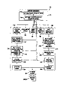
[0086] Fig. 5 is a block diagram of an example of an imaging system
500. The imaging system 500 as shown comprises optional variants of some of
the components introduced hereinbefore. The imaging system 500 comprises the
pulsed light source 210, the time domain detector 218, the computer 219, the
supporting tray 220 in combination with the translational stage 222 and the
movable mirror 224 introduced in the foregoing description of Figs. 2 and 3.
Additional modules are added, including a profilometer module 502, a visible
image module 504 and a self-diagnostic module 506.
[0086] The computer 219 as illustrated on Fig. 5 is functionally
subdivided into two main components, comprising a software module and system
for data acquisition control 219A, and a data analysis and display software
module
219B. The software module and system for data acquisition control 219A
provides
control for operation of the imaging system 500, for example by using feedback
information from the time domain detector 218 to control the pulsed light
source
210. The software module and system for data acquisition control 219A further
controls the modules 502-506. The data analysis and display software module
219B provides and displays information about optical images obtained from the
turbid media.
[0087] The light source 210 may be functionally subdivided into various
components. In an embodiment, the light source 210 comprises an illumination
channel 210A, a spectral multiplexer 210B, a time-multiplexing module 210C, a
CA 02745796 2011-07-08
26
multi-channel dynamic attenuator 210D capable of independently making power
adjustments to each of a plurality of wavelengths, and a multi-wavelength
laser
module 210E. Building the light source 210 from the modules 210A-210E provides
a laser source capable of rapidly acquiring an optical image of a turbid
media,
using a plurality of fluorescent markers subject to a variety of excitation
wavelengths. Of course, a simpler tunable light source 210 may be used in
other
embodiments. Various rearrangements and modifications may be performed to
the sub-components 210A-210E of the light source 210.
[0088] The time domain detector 218 may also be functionally
subdivided into various components. It may comprise a detection channel 218A,
a
spectral demultiplexer 218B, a multi-channel dynamic attenuation system 218C
capable of independently optimizing signals at a plurality of wavelengths, a
single
detection module 218D or a module 218D formed from an array of detectors, and
a signal processor 218E. Various embodiments of the time domain detector 218
may comprise all or a subset of the modules 218A-218D.
[0089] Arrows on Fig. 5 show paths for information signals and for
control signals. The computer 219¨ or a suitable controller ¨ is generally in
control
of the imaging system 500. It controls the tuning of the light source 210 and
may
do so using feedback from the time domain detector 218. The computer 219 also
controls angular movements of the movable mirror 224, as well as movements of
the supporting tray 220 in combination with the translational stage 222, in
order to
provide a 30 scanning of a plurality of predetermined collection points in a
region
of interest of the turbid media.
CA 02745796 2011-07-08
27
[00901 The systems
and method introduced in the present disclosure
involve a time-domain platform that offers maximum spectral coverage adapted
to
most of the known fluorescent probes/markers. High temporal resolution and low
time jitter enable maximum accuracy for fluorescence lifetime evaluation,
depth
and concentration estimation, in 3D tomographic views. Highest flexibility is
available for selecting and tuning the wavelength for maximizing the efficacy
of
excitation and detection as well as overall sensitivity for any fluorescent
probe.
Temporal and spectral multiplexing-demultiplexing are allowed for performing
experiments capable of simultaneously testing a large number of biomarkers and
design high throughput assays. Pump-probe experiments are made possible by
multiplexing both spectral and temporal the wavelengths required for
initiation of a
process and monitoring its evolution by interrogating its status at predefined
moments in time at ultra short time scales. Self-diagnostic is available in
real-time.
ROI and pixel-wise signal conditioning are capable of providing the level of
data
quality and signal to noise ratio (SNR) required for a highly accurate
estimation by
the model based algorithms used for data analysis. Data acquisition
optimization is
for best matching the assumptions and requirements of the mathematical models
used for analysis and interpretation. Structured functionalities allow maximum
flexibility in defining the optimum sequence and workflow for a given study.
[0091] Those of
ordinary skill in the art will realize that the description of
the systems and their sub-components and method for collecting optical data
for
use in time resolved optical imaging of a turbid media are illustrative only
and are
not intended to be in any way limiting. Other embodiments will readily suggest
themselves to such persons with ordinary skill in the art having the benefit
of the
present disclosure. Furthermore, the disclosed systems and methods may be
customized to offer valuable solutions to existing needs and problems of
optical
imaging.
CA 02745796 2011-07-08
28
[0092] In the
interest of clarity, not all of the routine features of the
implementations of the systems and method for collecting optical data for use
in
time resolved optical imaging of a turbid media are shown and described. It
will, of
course, be appreciated that in the development of any such actual
implementation
of the system and method described herein numerous implementation-specific
decisions may need to be made in order to achieve the developer's specific
goals,
such as compliance with application-, system - and business-related
constraints,
and that these specific goals will vary from one implementation to another and
from one developer to another. Moreover, it will be appreciated that a
development
effort might be complex and time-consuming, but would nevertheless be a
routine
undertaking of engineering for those of ordinary skill in the field of optical
imaging
having the benefit of the present disclosure.
[0093] Systems and
modules described herein may comprise software,
firmware, hardware, or any combination(s) of software, firmware, or hardware
suitable for the purposes described herein. Software and other modules may
reside on servers, workstations, personal computers, computerized tablets,
personal digital assistants (PDA), and other devices suitable for the purposes
described herein.
[0094] It is to be
understood that the present disclosure is not limited in
its application to the details of construction and parts illustrated in the
accompanying drawings and described hereinabove. The present disclosure is
capable of other embodiments and of being practiced in various ways. It is
also to
be understood that the phraseology or terminology used herein is for the
purpose
of description and not limitation. Hence, although the present disclosure has
been
described hereinabove by way of illustrative embodiments thereof, it can be
modified, without departing from the spirit, scope and nature of the present
CA 02745796 2011-07-08
29
disclosure.