Note: Descriptions are shown in the official language in which they were submitted.
ak 027463V71 2015-04-10
52723-54PPH
- 1 -
SYSTEMS AND METHODS
FOR OPTIMIZING AND MAINTAINING
VISUALIZATION OF A SURGICAL FIELD
DURING THE USE OF SURGICAL SCOPES
Field of the Invention
The invention generally relates to surgical scopes,
and, more particularly, for optimizing and maintaining
visualization of a surgical field when using a surgical scope,
such as, e.g., a laparoscope.
CA 02746371 2011-06-09
WO 2010/068265
PCT/US2009/006467
- 2 -
Background of the Invention
Minimally invasive surgical procedures utilizing
surgical scopes are desirable because they often provide
one or more of the following advantages: reduced blood
loss; reduced post-operative patient discomfort;
shortened recovery and hospitalization time; smaller
incisions; and reduced exposure of internal organs to
possible contaminants.
Generally, minimally invasive surgeries utilize
scopes, such as laparoscopes, that permit remote
visualization of a surgical site within a patient's body
while the surgical procedure is being performed. During a
laparoscopic procedure, the patient's abdominal or pelvic
cavity is acessed through two or more relatively small
incisions rather than through a single large incision
that is typical in a conventional surgery. Surgical
scopes, such as laparoscopes, usually consist in part of
a rigid or relatively rigid rod or shaft having an
objective lens at one end and an eyepiece and/or
integrated visual display at the other. The scope may
also be connected to a remote visual display device or a
video camera to record surgical procedures.
In laparoscopic surgeries, the abdomen is typically
inflated with a gas through the use of an insufflator, to
distend the abdominal space by elevating the abdominal
wall above the internal organs and thereby create a
sufficient working and viewing space for the surgeon.
Carbon dioxide is usually used for insufflation, though
other suitable gases may also be used. Conventional
insufflators are adapted to cycle on and off to maintain
a preset and suitable pressure within the patient's body
cavity.
The local environment within a patient's abdominal
space is generally rather warm and humid, and the use of
devices such as harmonic scalpels and other cutting and ,
CA 02746371 2011-06-09
W02010/068265
PCT/US2009/006467
- 3 -
coagulating devices generate mist, smoke, and other
debris that is released into the surgical field and often
becomes suspended throughout the expanded abdominal
space. Additionally, blood, bodily fluids, pieces of
tissue, fat or other bodily material may come in contact
with or even attach to the lens. As a result of these
conditions, visualization through the scope can be
significantly diminished. Typically, the only solution to
fogging and debris collection on the lens is removal of
the scope from the body cavity and defogging or cleaning
the lens by wiping it with a cloth, warming the scope
tip, or utilizing another defogging method. The need to
remove the scope to defog and remove debris from the lens
is inconvenient for the scope operator and the surgeon
and can interrupt and undesirably prolong surgical
procedures.
Summary of the Invention
One aspect of the invention provides a view
optimizing assembly having a deflector assembly with
critical physical, pneumatic, and optical characteristics
that make possible intra-operative defogging, surgical
debris deflection, and cleaning of a laparoscope lens
during minimally invasive surgery, while also maintaining
visualization of the surgical site. In use, the view
optimizing assembly makes possible the practice of a
surgical method for maintaining clear visualization of
the surgical site without removing the laparoscope 12
from the abdominal cavity for the purpose of cleaning or
de-fogging its lens.
Another aspect of the invention provides a view
optimizing assembly having a quick exchange feature. In
use, the quick exchange feature makes possible a
surgical method for maintaining clear visualization that ;
includes the ability to make a quick exchange of
laparoscopes having different operating characteristics
CA 02746371 2015-04-10
52723-54PPH
- 4 -
(e.g., laparoscopes with different tip angles, lengths, or
diameters) entirely on the sterile operating field and without
interference with the preexisting surgical set-up on the
sterile operating field. The view optimizing assembly
integrates with the existing suite of minimally invasive
instrumentation. It does not interfere with the surgical set-
up, and it requires minimal change in the process or practice
of a surgical operating room (OR) team.
Another aspect of the invention provides a view
optimizing assembly comprising a laparoscope, a sheath sized
and configured to receive the laparoscope, a first lumen in a
wall of the sheath for conveying a gas, a second lumen in the
wall of the sheath for conveying a sterile fluid, a tubing set
having a first tube and a second tube, the first tube having a
first end sized and configured to couple to a source of gas and
a second end coupled to a first part of a two part quick
exchange coupler, the first part of the two part quick exchange
coupler having a normally closed one way valve to normally
prevent flow of the gas out of the second end of the first
tube, the second tube having a first end sized and configured
to couple to a source of the sterile fluid and a second end
coupled to the first part of the two part quick exchange
coupler, a manifold carried by the sheath and communicating
with the first and second lumens, the manifold including a
second part of the two part quick exchange coupler sized and
configured to mate with the first part of the quick exchange
coupler, the second part of the quick exchange coupler
including an element that opens the normally closed one way
valve in response to mating the first and the second parts of
the quick exchange coupler to allow flow of the gas out of the
CA 02746371 2014-10-01
52723-54
-4a-
second end of the first tube and into the first lumen, wherein
the one way valve is configured to close in response to
disconnecting the first and second parts to prevent the flow of
the gas out of the second end of the first tube and into the
first lumen; and a manual squeeze burst actuator in-line with
the tubing set and configured to provide a burst of air through
the tubing set.
Brief Description of the Drawings
Fig. lA is a somewhat schematic views of a view
optimizing assembly for use with a laparoscope having a 0
shaft tip.
Fig. 1B is a section view of the sheath, showing
internal fluid flow lumens, taken generally along line 1B-1B in
Fig. 1A.
Fig. 2A is a somewhat schematic of a view optimizing
assembly for use with a laparoscope having an angled shaft tip.
Fig. 2B is a section view of the sheath, showing
internal fluid flow lumens, taken generally along line 2B-2B in
Fig. 2A.
Fig. 3A is an enlarged perspective view of a manifold
that the view optimizing assembly shown in Fig. lA or Fig. 2A
incorporates, including a quick exchange coupling, and a quick
exchange coupler that the tubing set shown in Fig. lA or
Fig. 2A incorporates, the coupling and the coupler being
disconnected.
CA 02746371 2014-10-01
52723-54
,
-4b-
Fig. 3B is a sectional view taken generally along
line 3B-3B in Fig. 3A, showing a one way check value that is
normally closed.
Fig. 4A is an enlarged perspective view of the
manifold including a quick exchange coupling and the quick
exchange coupler of the tubing set, as shown in Fig. 3A, but
now connected.
CA 02746371 2011-06-09
WO 2010/068265
PCT/US2009/006467
- 5 -
Fig . 4B is a sectional view taken generally along
line 4B-4B in Fig. 4A, showing the one way check valve
that is opened by the connection of the quick exchange
coupling and connectors.
Figs. 5A(1) and 5A(2) are enlarged, exploded views
of the deflector assembly for use with a laparoscope
having a 00 shaft tip.
Figs. 5B(1) and 5B(2) are enlarged, exploded views
of the deflector assembly for use with a laparoscope
having an angled shaft tip.
Fig. 6 is a schematic view of the critical physical,
pneumatic, and optical characteristics of the deflector
assembly shown in Figs. 5A and 5B.
Figs. 7 to 34 illustrate a representative method
including the set up and use of the view optimizing
assembly using sterile technique by technicians/operating
room personnel.
Description of the Preferred Embodiments
Although the disclosure hereof is detailed and exact
to enable those skilled in the art to practice the
invention, the physical embodiments herein disclosed
merely exemplify the invention, which may be embodied in
other specific structure. While the preferred embodiment
has been described, the details may be changed without
departing from the invention, which is defined by the
claims.
I. View Optimizing Assembly
A. Overview
Figs. 1A/1B and Fig. 2A/23 show a view optimizing
assembly 10 for use in association with a state of the
art laparoscope 12. In Figs. 1A/1B, the laparoscope 12
possesses at 00 (blunt) shaft tip In Figs. 2A/2B, the
laparoscope possess an angle shaft tip (e.g., a 30 shaft .
= tip or 45 shaft tip). The components of the view
optimizing assembly 10 may be made from plastic -
CA 02746371 2011-06-09
W02010/068265
PCT/US2009/006467
- 6 -
materials (extruded and/or molded), but other suitable
materials, such as metal or a composite material, or
combinations thereof could be used.
As will be described in greater detail, the view
optimizing assembly 10 facilitates intra-operative
defogging, surgical debris deflection, and cleaning of a
laparoscope lens during minimally invasive surgery, while
also maintaining visualization of the surgical site. The
view optimizing assembly 10 is intended to be a single-
use, disposable laparoscopic accessory. The view
optimizing assembly 10 is desirably a sterile accessory
for immediate set up and use on a sterile operating
field.
As shown in Figs. 1A and 2A, the view optimizing
assembly 10 comprises a multi-lumen sheath assembly 14,
which mounts over the shaft of the laparoscope 12. The
end of the shaft is sized and configured to match the
size and configuration of the corresponding laparoscope
12, having a blunt tip in Fig. LA and and angled tip in
Fig. 2A. The assembly 10 includes a tubing set 16 to
connect the sheath 14 to =an existing anhydrous carbon
dioxide (CO2) insufflation circuit.
In use, the view optimizing assembly 10 makes
possible the practice of a surgical method for
maintaining clear visualization of the surgical site
without removing the laparoscope 12 from the abdominal
cavity for the purpose of cleaning or de-fogging its
lens. Furthermore, the view optimizing assembly 10 also
makes possible a surgical method for maintaining clear
visualization that includes the ability to make a quick
exchange of laparoscopes having different operating
characteristics (e.g., laparoscopes with different tip
angles, lengths, or diameters) entirely on the sterile
operating field and without interference with the
preexisting surgical set-up on the sterile operating
CA 02746371 2011-06-09
WO 2010/068265
PCT/US2009/006467
- 7 -
field. The view optimizing assembly 10 integrates with
the existing suite of minimally invasive instrumentation.
It does not interfere with the surgical set-up, and it
requires minimal change in the process or practice of a
surgical operating room (OR) team.
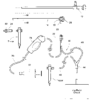
The view optimization assembly 10 desirably comes
packaged for use in sterile peel away pouches (see Fig.
7). As also shown in Figs. IA and 2A, the pouches contain
the components of the view optimization assembly 10,
including the sheath 14 and a manifold 18 that is
assembled to the sheath 14 and that includes a quick
exchange coupling 20; the tubing set 16 which includes a
quick exchange coupler 22 that mates with the quick
exchange coupling 20 on the manifold 18; and (optionally)
a vent device 24.
B. The Sheath / Manifold Assembly
As shown in Figs. 1A and 2A, the sheath 14 /
manifold 18 assembly includes a sheath 14 that is sized
and configured to receive a laparoscope 12 having a
prescribed tip angle, length, and diameter. The sheath 14
includes a stop 26 (see Figs. 5A(2) and 5B(2) formed
adjacent the distal end of the sheath 14. The stop 26
prevents advancement of the laparoscope 12 beyond the
distal end of the sheath 14, so that lens at the distal
end of the laparoscope 12 rests in a desired, generally
coterminous alignment with the distal end of the sheath
14. The sheath 14 also includes a locking collar 28 at
its proximal end to frictionally engage the laparoscope
12 and resist axial withdrawal of the laparoscope 12 from
the sheath 14.
In use, it is expected that the laparoscope 12 will
be inserted into the sheath 14 by a scrub nurse during
set-up for the operation (see Figs. 8 to 11). The
assembled laparoscopic and sheath 14 will then be handed ,
as a unit to personnel at the operating room (OR) table :
CA 02746371 2011-06-09
W02010/068265
PCT/US2009/006467
- 8 -
at the desired
time) . The 1 aparos cope 12 is then
connected by personnel at the OR table in conventional
fashion to a light cable 30 (which directs light to
illuminate the operative field) and the camera cable 32
(which takes the image from the scope and displays it on
monitors in the OR) (see Fig. 14). The sheath 14 is
sized and configured not to interfere with this normal
set-up of the laparoscope 12.
In use, the assembled laparoscopic and sheath 14 are
placed as a unit through a trocar into the body cavity
(e.g, the _abdominal cavity), for viewing the surgical
procedure as it is performed (see Fig. 16).
As shown in Figs. lA and 2A, and as further shown in
Figs. 3A, the sheath 14 / manifold 18 assembly also
includes the manifold 18 at the proximal end of the
sheath 14. The manifold 18 communicates with multiple
lumens (five 34 to 42) are shown in the illustrated
embodiment) formed within the wall of the sheath 14 (see
Figs. 1B and 2B. In use, the lumens 34 to 42 convey
anhydrous CO2 to the distal end of the sheath 14; vent or
exhaust air from the distal end of the sheath 14 through
the manifold 18; and, if desired, convey sterile fluid
and bursts of air to the distal end of the sheath 14. In
a representative embodiment (see Figs. 1B and 2B), two
lumens 34 and 36 are dedicated to the transport of CO2;
two lumens 40 and 42 are dedicated to venting; and one
lumen 38 is dedicated to the transports of sterile fluid
or air.
C. The Tubing Set
As previously described, the tubing set 16 includes
a quick exchange coupler 22 that mates with the quick
exchange coupling 20 on the manifold 18 (see Figs. 3A/3B
and 4A/4B). The tubing
set 16 includes lengths of
flexible medical grade tubing with individual end ,
couplers (best shown in Figs. lA and 2A)that connect to
CA 02746371 2011-06-09
WO 2010/068265 PCT/US2009/006467
- 9 -
an existing CO2 insufflation circuit and, if desired, a
source of sterile fluid (saline or sterile water,
preferably with a "surface active agent") on the sterile
operating field (e.g., a bag or a syringe). The tubing
set 16 includes a Y-connector 44 that divides the
anhydrous CO2 output of the insufflation circuit in a
first branch 46 for coupling to an insufflation trocar
inserted in the body cavity (as will be described later),
and a second branch 48 coupled to the quick exchange
coupler 22.
The second branch 48 diverts a small portion of the
CO2 output (e.g., 20% or less) to the quick exchange
coupler 22.
As shown in Figs. 3B and 4B, the quick exchange
coupler 22 includes a one way check valve 50 that
communicates with the second branch 48 of the tubing set
16. In the illustrated embodiment, the check valve 50 - -- -
comprises a ball valve. Insufflation pressure normally
presses the ball valve 50 against a ball valve seat 52
(as shown in Fig. 3B). A projection 54 in the manifold
18 displaces the ball valve 50 from the valve seat 52
when the quick exchange coupler 22 mates with the quick
exchange coupling 20 on the manifold 18 (as shown in
Fig.4B). Unseating the ball valve 50 opens flow
communication through the check valve 50. In the absence
of coupling the quick exchange coupler 22 on the tubing
set 16 to the quick exchange coupling 20 on the manifold
18, the check valve 50 remains closed, normally blocking
flow of CO2 through the second branch 48.
Thus, the tubing set 16 accommodates the set-up of
the supply of the entire CO2 output to a insufflation
trocar through the tubing set 16, separate and
independent of the connection of the tubing set 16 to the
manifold 18 of the sheath 14.
As Figs. 3A and 4A further show, a latch 56 carried -
CA 02746371 2011-06-09
WO 2010/068265
PCT/US2009/006467
- 10
on a spring-biased button 58 on the quick exchange
coupler 22 "clicks" into a detent 60 on the quick
exchange coupling 20 on the manifold 18 to reliably lock
the coupler 22 and coupling 20 together for use, opening
the check valve to flow CO2 through the second branch 48
(shown in Figs. 4A/4B). Depressing the button 58 allows
the quick exchange coupler 22 and coupling 20 to be
separated, and the check valve 50 will close in response
to insufflation pressure in the second branch 48 (as
shown in Figs. 3A/3B).
Connection of the quick exchange coupling 20 on the
manifold 18 to the quick exchange coupler 22 on the
tubing set 16 is intended to occur at the OR table in the
normal course, after the laparoscope 12 is connected to
the light cable 30 and the camera cable 32 (see Fig. 15).
Upon coupling, the one way check valve 50 is opened, and
the manifold 18 directs the small portion of CO2 from the
CO2 insufflation circuit. Disconnection of the of the
quick exchange coupling 20 on the manifold 18 to the
quick exchange coupler 22 on the tubing set 16 is also
intended to occur at the OR table in the normal course,
after a removal and/or exchange of a laparoscope 12 (see
Fig. 22).
D. The Vent Device
The vent device 24 (see Figs. IA and 2A) comprises a
tube with an inline membrane 62 that restricts air flow
through the tube. A proximal end of the tube is sized
and configured to couple to a stopcock valve of a
conventional trocar, as will be described later. In use,
the vent device 24 provides a controlled leak of CO2 from
the operating cavity, as will also be described in
greater detail later.
E. The Deflector Assembly
1. CO2
The sheath 14 includes at its distal end a deflector
CA 02746371 2011-06-09
WO 2010/068265
PCT/US2009/006467
- 11 -
assembly 64 (see Figs. 5A(1) and 5A(2) for a blunt shaft
tip and Figs. 53(1) and 53(2) for an angled shaft tip).
The deflector assembly 64 projects a predetermined
distance beyond the distal end of the sheath 14, and thus
also a predetermined distance beyond the lens at the
distal end of the laparoscope 12. The deflector assembly
64 communicates with the lumens in the sheath 14. The
deflector assembly 64 is sized and configured to direct
the small portion of the CO2 from the insufflation
circuit in a prescribed flow path and flow velocity
continuously across the laparoscopic lens.
The desired flow path and flow velocity of CO2
established by the deflector assembly 64 continuously
across the laparoscopic lens creates a "wind shear." The
wind shear path of anhydrous CO2 prevents fogging. The
desired flow path and flow velocity of CO2 established by
the deflector assembly 64 continuously across the
laparoscopic lens also desirably serves to deflect smoke
and surgical debris away from the laparoscopic lens
during surgery.
2. Physical, Pneumatic, and Optical
Characteristics of the Deflector Assembly
The size and configuration of the deflector assembly
64 are defined and constrained by several, sometime
overlapping considerations including (i) prescribed
physical characteristics, which are imposed due to the
need to access the operating environment in as minimally
invasive manner as possible and to be compatible with
state of the art laparoscopes and other laparoscopic
surgical instruments and techniques; (ii) prescribed
pneumatic characteristics, which are imposed due to the
need to create a particular "wind shear" effect in terms
of the flow path and flow velocity of CO2 across the :
laparoscopic lens; and (iii) prescribed optical ,
characteristics, which are imposed due to the need to :
CA 02746371 2011-06-09
W02010/068265
PCT/US2009/006467
- 12 -
prevent interference with the field of view and the
visualization of the operating field by the laparoscope
12.
3. Physical Characteristics
The size and configuration requirements for
minimally invasive access compatible with state of the
art laparoscopic instrumentation and techniques are
paramount. These requirements impose constrains upon the
minimum inside diameter of the sheath 14 as well as the
maximum outside diameter of the sheath 14. Because state
of the art laparoscopes are provided with different shaft
diameters, lengths, and lens configurations, the sheath
dimensions and configuration change for compatibility
with them. The view optimizing assembly 10 actually
includes a family of sheath 14 / manifold 18 assemblies
differently sized and configured to accommodate different -
classes of laparoscopes, to make possible compatibility
with the families of state of the art laparoscopes that
are in use.
For example, state of the art laparoscopes include
mm laparoscopes, 5 mm laparoscopes, and, within these
sizes, 0 shaft tips, 30 shaft tips, and 450 shaft tips.
Further, within these classes of laparoscopes,
manufacturing tolerances typically vary from scope to
scope, as well as from manufacturer to manufacturer. A
given sheath 14 / manifold 18 assembly for a given
laparoscope class (e.g., 10 mm or 5 mm) desirably takes
these typical manufacturing and manufacturer variances
into account, and is desirably sized and configured to
fit the largest scope variance encountered within a given
laparoscope class.
To maximize the fluid flow lumen area within the
sheath 14, the minimum inside diameter of a given sheath :
14 must closely conform to the maximum outside diameter
of the. shaft of the particular state of the class of .
CA 02746371 2011-06-09
W02010/068265
PCT/US2009/006467
- 13 -
laparoscope 12 selected for use, which the sheath 14 must
accommodate in a smooth, sliding fit. Further, a gap
between the outside diameter of the laparoscope shaft and
the inside diameter of the sheath 14 must be minimized to
avoid the transport and leakage of blood and fluids from
the operating field. Still further, minimizing the gap
also assures that the laparoscope 12 self-centers in the
sheath 14, thereby assuring faithful and accurate
visualization through the laparoscope lens.
For example, for a typical laparoscope 12 in the 10
mm class, which measures 0.392 inch, the inside diameter
of the sheath 14 is manufactured to .405 inch, providing
a gap thickness of 0.0064 inch. For a 5 mm laparoscope
12 in the 5 mm class, which measures 0.196 inch, the
inside diameter of the sheath 14 is manufactured to 0.218
inch, providing gap thickness of 0.011 inch.
The maximum outside diameter of the sheath 14 for
minimally invasive access must take into account the
minimum inside diameter of the trocar, which the maximum
outside diameter cannot exceed.
For example, for a typical 10 mm trocar that
measures 0.509 inch, the outside diameter of the sheath
14 is manufactured to 0.486 inch, providing a gap
thickness of 0.0115 inch. For a typical 5 mm trocar that
measures 0.324 inch, the outside diameter of the sheath
14 is manufactured to 0.300 inch, providing a gap
thickness of 0.012 inch.
It is desirable, given the particular size and
configuration constraints of the laparoscopic
instrumentation and techniques used, to maximize the
outside diameter to the extent possible. This is
because, together the inside and outside diameters of the
sheath 14 define the wall thickness for the sheath S.
The wall thickness Sw, together with the length of the ,
sheath 14, in turn, define the maximum area available for ,
CA 02746371 2011-06-09
W02010/068265
PCT/US2009/006467
- 14 -
the transport of the CO2 and fluids by the sheath 14.
The area of the fluid flow lumen or lumens dedicated to
the supply of CO2, in turn, defines the maximum flow rate
of the CO2 directed by the deflector assembly 64. The
flow rate should be sufficient at a minimum, given the
output of the insufflator selected for use, to supply
anhydrous CO2 across the lens of the laparoscope 12
sufficient to prevent fogging. Also affecting the
effectiveness of the CO2 to defog the lens, is the water
content of the anhydrous CO2. Given the same flow rate,
the less water that is present in the anhydrous CO2, the
greater is the defogging capacity of the assembly.
Further, the flow rate desirable should also be
sufficient to deflect smoke and surgical debris away from
the viewing field of the laparoscopic lens during
surgery, so that the anhydrous CO2 directed by the
deflector assembly 64 both defogs and deflects-debris.
Medical grade CO2 for use with conventional
insufflators is typically 99% pure, that is, no more than
1% of the gas is other than CO2, and such medical grade
anhydrous CO2 generally has a maximum moisture content of
25 parts per million by volume. Typically, a state of
the art insufflator circuit delivers anhydrous CO2 at a
max flow rate of about 20 liters per hour. Typically,
the insufflator circuit will sense pressure in the
circuit and cycle off when the sensed pressure is at or
above 15 mmHg and cycle on when the sensed pressure is
below 15 mmHg.
Given the above sheath dimensions, and given the
supply of typical medical grade anhydrous CO2, a flow
rate of at least about 1.0 liters per minute is critical
to achieving this objective. Given the above dimensions,
and the supply of typical medical grade anhydrous CO2, a
flow rate less than 0.8 liters per minute is not
sufficient to prevent significant accumulation of i
CA 02746371 2011-06-09
W02010/068265
PCT/US2009/006467
- 15 -
moisture on the laparoscope lens.
In a representative embodiment, for a sheath 14
having an inside diameter of .405 inch and an outside
diameter of .486 inch, and a length of 11.25 inch (which
accommodates passage of a typical 10 mm laparoscope and
its own passage through a conventional trocar) (i.e., Sw
= .081 inch), the total area available in the sheath wall
is 0.056 square inches. Based upon required structural
support within the wall (inside, outside, and radial) the
total available area for lumens to transport fluids is
0.027 square inch.
In a representative embodiment, the total lumen area
is occupied by five lumens 34 to 42, two for transporting
CO2 (34 and 36), one for sterile fluid (38), and two for
passive exhaust air venting (40 and 42).
The area of each lumen can be maximized by selection- -
of lumen geometry. In a representative embodiment, lumen
geometry is generally triangular or pie shaped with
rounded corners. The radial walls that separate the
lumens within the sheath 14 are sized to minimize the
spacing between the lumens.
In a representative embodiment, CO2 transport is
accomplished by two lumens 34 and 36 that extend about
175 degrees about the outer circumference of the sheath
14 and comprising a flow area of 0.013 square inches.
Sterile fluid transport is accomplished by one lumen 38
comprising a flow area of 0.003 square inches. Exhaust
air venting is accomplished by two lumens 40 and 42
comprising a flow area of 0.011 square inches. The distal
openings of the exhaust lumens 40 and 42 desirably are
spaced from the distal end of the sheath, to prevent
uptake of blood and fluids.
4. Pneumatic Characteristics.
As diagrammatically shown in Fig. 6, the deflector .
assembly 64 must overhang the laparoscopic lens by a
CA 02746371 2011-06-09
WO 2010/068265
PCT/US2009/006467
- 16 -
prescribed transverse distance, defining a deflection
width X, sufficient to change the direction of CO2
flowing axially through lumens of the sheath 14 (i.e.,
along the axis of the laparoscope shaft) into a non-
axially, transverse path across the laparoscopic lens
(i.e., at an angle relative to the axis of the
laparoscope shaft). Still, the distance of the
deflection width X should not extend to the point that is
obstructs the field of the view of the laparoscopic lens.
This is an example where a pneumatic characteristic of
the deflector assembly 64 overlaps with an optical
characteristic. Further optical characteristics will be
described in greater detail below.
The deflector assembly 64 must also project axially
beyond the distal terminus of the sheath 14 by a
prescribed axial distance, defining an air channel
distance Y, sufficient to maintain the CO2 flowing along
the path bounded by the deflection width X at a distance
sufficiently close (proximal) to the laparoscopic lens to
achieve the desired shear flow effect, but without
forming an abrupt flow bend that can lead to a reduction
in the desired CO2 flow velocity.
Together, the deflection width X and the channel
distance Y define the pneumatic characteristics of the
deflection assembly. At the desired minimum flow rate,
the pneumatic characteristics create a flow path that
conveys CO2 continuously across the laparoscopic lens at
the desired flow velocity, in shorthand called the "wind
shear." The pneumatic characteristics of the CO2 "wind
shear" across the laparoscopic lens prevent fogging, as
well as desirably deflect smoke and surgical debris away
from the viewing field of the laparoscopic lens during
surgery.
Together, the pneumatic characteristics defined by ,
the deflection width X and the channel distance Y create
CA 02746371 2011-06-09
WO 2010/068265
PCT/US2009/006467
- 17 -
an exit angle AExiT , measured between the plane of the
=laparoscopic lens and the terminal edge of the deflector
assembly 64. The exit angle AExii. must be less than a
maximum angle of 45 degrees, else the flow path of the
CO2 will not pass sufficiently both across and proximal
to the laparoscopic lens. To maintain a desired exit
angle AExIT , the channel distance Y should be at least
equal to the wall thickness of the sheath Sw and should
not exceed 1.5 times the wall thickness of the sheath S.
The deflection width X should be at least equally to two
times the channel distance Y, but not extend into the
field of view of the laparoscopic lens.
5. Optical Characteristics
The optical characteristics of the deflector
assembly 64 are selected (i) to not block or reduce the
illuminated image of the operating field provided by the
laparoscope 12; (ii) not decrease the intensity of the
illumination provided by the laparoscope 12 on the
operating field; and (iii) prevent reflection of
illumination light at the lens of the laparoscope 12.
- As discussed above, the maximum deflection width X
takes into account one of the desirable optical
characteristics; namely, the deflection width X should
not obstruct the field of the view of the laparoscopic
lens.
To prevent the decrease of the illumination, the
deflector assembly 64 is desirably made from a material
having high light transmission properties (i.e.,
transparency), to not interfere with the passage of light
through the light cable 30 onto the operating field as
well as the passage of the reflected image conveyed to
the camera cable 32 of the laparoscope 12.
Furthermore, the material and surface finish of the ,
deflector assembly 64 must pose minimal reflectively to
light. In a representative embodiment, the deflector L
CA 02746371 2011-06-09
WO 2010/068265
PCT/US2009/006467
- 18 -
assembly 64 is made from Bayer Makrolen Rx1805 with a
surface finish defined as SPI/SPE A-3.
6. Orientation
As before described, CO2 transport is accomplished
by two lumens 34 and 36 that extend about 175 degrees
about the outer circumference of the sheath 14. For a 00
shaft tip (see Fig. 5A), the orientation of the deflector
assembly 64 relative to the laparoscopic lens is not
critical. However, for angled shafts (e.g., 30 shaft
tips and 45 shaft tips) (see Fig. 5B), the orientation
of the deflector assembly 64 relative to the laparoscopic
lens is critical.
As Fig. 5B shows, the angled tip of a typical
laparoscope 12 has a high end 66 and a low end 68. The
lens slopes at the prescribed angle between the high end
66 and the low end 68. In a laparoscope 12 having a
angled tip, the illumination cable 30 (transmitting light
onto the operating field) is located at the high end 66
of the angled tip, and the camera cable 32 (transmitting
reflected light back to the camera) is located at the low
end 68 of the angled tip. To provide the desired wind
shear effect on an angled tip, it is critical that the
deflector assembly 64 be oriented relative to the sloped
laparoscopic lens such that the flow CO2 is directed
across the sloped plane of the lens from the low end 68
of the tip toward the high end 66 of the tip. In this
arrangement, the defogging and debris deflection flow
path originates proximal to the camera cable 32, which
effectively comprises the eyes of the OR team. In this
arrangement, the desired exit angle AmaT directs the flow
path of the CO2 both sufficiently across and proximal to
the sloped plane of the laparoscopic lens to achieve
optimal defogging and debris deflection.
F. Sterile Fluid Flush =
As previously explained, if desired, the tubing set i
CA 02746371 2011-06-09
WO 2010/068265
PCT/US2009/006467
- 19 -
16 can also include, connected to the quick exchange
coupler 22, a length of tubing 70 sized and configured
for connection to a source 72 of sterile fluid, such as
saline or sterile water (as shown in Figs. 1A and 2A).
Preferably, the sterile fluid includes in solution a
"surface-active agent" that stabilizes mixtures of oil
and water (e.g., fat) by reducing the surface tension at
the interface between the oil and water molecules.
The quick exchange coupling 20 on the manifold 18
(see Fig. 3A/3B and 4A/43) can also include a port to
integrally connect the sterile fluid tubing 70 to direct
the sterile fluid through the separate lumen 38 in the
sheath 14 to the distal end of the sheath 14. The
deflector assembly 64 directs the sterile fluid across
the laparoscopic lens.
As shown in Figs. 1A/2A, the sterile fluid tubing
70, if present, desirably includes an in-line pumping
device 72. The in-line pumping device 72 is sized and
configured to be operated on demand by a person at the OR
table to convey bursts of sterile fluid through the
manifold 18 through the lumen to the distal end of the
sheath 14. The in-line pumping device 72 and source can
be integrated and comprise, e.g., a 20cc syringe filled
with sterile fluid and connected by a tubing luer-lock on
the saline tubing. Alternatively, the in-line pumping
device 72 and source can be separate and comprise, e.g.,
a bag of sterile fluid, a spike connection on the saline
tubing of the tubing set 16 to open communication with
the bag in conventional fashion, and an inline squeeze
bulb or the like to pump burst of sterile fluid from the
bag to the quick exchange coupler 22.
In this arrangement, the deflector assembly 64 is
also sized and configured to direct the burst of sterile
fluid in a desired path across the laparoscopic lens.
The bursts of sterile fluid serve to flush debris off the
CA 02746371 2011-06-09
WO 2010/068265
PCT/US2009/006467
- 20 -
end of the lens that may eventually accumulate, thereby
cleaning the lens. Thereafter, bursts of air supplied
through the deflector assembly 64 by a squeeze pump 74 in
the tubing set 16 (see Figs. 1A/2A) serve to clear
residual fluid droplets off the lens and away from the
deflector assembly 64 to maintain the desired flow path
and flow velocity of CO2 established by the deflector
assembly 64 continuously across the laparoscopic lens, to
maintain an acceptable view.
In an illustrative embodiment (see Figs. 5A and 5B),
the deflector assembly 64 directs the bursts of sterile
fluid or air along a plurality of individual diverging
channels 76 (three are shown). The diverging channels 76
distribute the bursts of sterile fluid or air in a
fanning pattern across the lens of the laparoscope 12. In
the illustrative embodiment, the diverging channels 76
discharge the bursts of sterile fluid or air in a path
that is generally ninety-degrees to the path of CO2.
This orientation of the sterile fluid path relative to
the CO2 path across the lens, optimal for effective lens
cleaning, applies to both 00 shaft tips and angled tips
(e.g., 30 shaft tips and 45 shaft tips).
II. Use of the View Optimizing Assembly
The view optimizing assembly is well suited for use
as a single-use disposable laparoscopic accessory device
to facilitate intra-operative defogging and debris
deflection (due to the flow of anhydrous CO2) and
cleaning of the lens of a laparoscope 12 (due to burst of
sterile fluid, preferably including a "surface-active
agent") during minimally invasive surgery, while also
maintaining visualization of the surgical site.
Figs. 7 to 34 illustrate a representative method
including the set up and use of the _view optimizing .
assembly using sterile technique by qualified ,
technicians/operating room personnel.
CA 02746371 2011-06-09
W02010/068265
PCT/US2009/006467
- 21 -
The procedure can be incorporated into written
instructions for use that accompany the packaging. The
instructions can also be supplied separately, e.g.,
embodied in separate instruction manuals, or in video or
audio tapes, CD's, and DVD's. The instructions for use
can also be available through an internet web page.
The instructions can direct the OR set-up to peel
open the outer pouches in which the components of the
view optimizing assembly (shown in Fig. 7), and remove
the sterile contents on the sterile field. The sheath 14
/ manifold 18 assembly is removed, taking care to prevent
damage to the walls of the sheath 14 or to its distal
end, and also keeping the tubing set 16 and vent device
24 on the sterile field prior to making necessary
connections.
During set up (see Figs. 8 and 9), the sheath 14
(with the manifold 18, which is integrally connected to
the sheath 14 during manufacture, called a sheath
assembly) can be assembled to the corresponding
laparoscope 12. In this representative example, it is
contemplated that the OR team plan to use a 0-degree
laparoscope 12 (see Figs. 8 and 9) and at least one
angled laparoscope 12 (see Figs. 10 and 11), e.g., a 30-
degree and/or a 45-degree laparoscope 12. Therefore,
during set-up, a sheath assembly for each laparoscope 12
selected for use will be pre-assembled to the
corresponding laparoscope 12.
As Figs. 8 and 10 show, while gently pressing the
tip of the sheath assembly against one hand or finger-
tip, the laparoscope 12 can be inserted down into the
sheath 14. The sheath 14 is sized and configured so that
the laparoscope 12 will slide smoothly through the sheath
14. Insertion continues until the lens and distal rim of
the laparoscope 12 seat against the stop at the distal
end of the sheath 14. The laparoscope 12 will "bottom .
CA 02746371 2011-06-09
W02010/068265
PCT/US2009/006467
- 22 -
out" inside the sheath 14 against the stop 26, assuring
correct axial alignment of the lens with the deflector
assembly 64.
If the laparoscope 12 fs angled (as shown in Fig.
10), the corresponding sheath assembly will also include
an alignment fork guide 78. The light post of the scope
seats within the alignment fork guide 78, therefore
assuring correct rotational alignment between the angled
lens and the deflector assembly 64.
The laparoscope 12 (now fully inserted into the
sheath 14) the manifold 18 are supported by hand, a
member of the OR set-up team rotates the locking collar
28 on the sheath assembly in the desired direction, e.g.,
clockwise (see Figs. 9 and 11), indicated by an arrow on
the locking collar 28, until a firm stop is felt
tactilely (e.g., after approximately one-third (1/3) of a
turn). Registration of an alignment mark on the locking
collar 28 and an alignment mark on the manifold 18 serves
to visually confirm that the laparoscope 12 is secured
against axial movement relative to the sheath 14.
The insufflator is set up off the sterile field.
Once the patient is draped on the sterile field, and it
is expected that the end of the output tubing from the
insufflator (originating from the insufflator off the
sterile field) will brought onto the sterile field. It
is also expected that the light cable 30 and the camera
cable 32 for the laparoscope 12 will be brought onto the
sterile field.
As Figs. 12 and 13 generally show, the OR team makes
an incision to gain access to the laparoscopic operating
site within the body, e.g., into the abdominal cavity
through the abdominal wall. A first trocar with a
stopcock valve (which may take the form of an optical :
trocar) is inserted through the incision. Alternatively, ,
according to physician preference, the first trocar can ,
CA 02746371 2011-06-09
WO 2010/068265
PCT/US2009/006467
- 23 -
be pushed through abdominal wall with only a skin ,
incision. The obturator (the sharp inner insert of the
trocar) is removed from the first trocar once it is in
position.
The insufflator line of the tubing set 16 on the
sterile field is connected to the output tubing of the
insufflator circuit on the sterile field. The first
branch 46 of the tubing set 16 on the sterile field,
originating at the Y-connector 44, is coupled to the
stopcock valve of the first trocar (see Fig. 13). The
stopcock valve is opened, and the insufflator is turned
on. CO2 output of the insufflation circuit inflates the
abdomen through the first trocar.
During this time (see Figs. 8 and 10), the second
branch 48 of the tubing set 16 on the sterile field, also
originating at the Y-connector 44, and the quick exchange
coupler 22 integrally attached to it can remain on the
sterile field in a free, unconnected condition as the
insufflator supplies CO2 through the first branch 46. The
one-way check valve in the quick exchange coupler 22
serves to block flow of CO2 through the second branch 48,
even as the insufflator supplies CO2 through the first
branch 46. The entire CO2 pressure of the insufflator
circuit is, at the present, delivered to the first trocar
through the first branch 46.
The first laparoscope 12 selected for use, which has
been pre-inserted into the sheath 14 by the OR set-up
team as just described, is handed to personnel at the OR
table at the appropriate time. On the sterile field,
personnel at the OR table connect the light cable 30 and
the camera cable 32 to the laparoscope 12 (see Fig. 14).
On the sterile field, personnel at the OR table now
connect the quick exchange coupler 22 of the tubing set i
16 to the quick exchange coupling 20 of the manifold 18
(see Fig. 15). The one way valve opens, and a small i
CA 02746371 2011-06-09
WO 2010/068265
PCT/US2009/006467
- 24 -
portion of the output of the insufflator circuit is
routed by the second branch 48 through the manifold 18
into to the sheath 14.
The laparoscope / sheath assembly is then placed as
an integrated unit through the first trocar to get an
initial view of the abdominal cavity (see Fig. 16). Due
to the technical features of the deflector assembly 64,
CO2 flows over the lens, eliminating fogging and also
deflecting away debris. If present, the pump (e.g., the
20cc syringe) filled with sterile fluid (preferably with
a "surface-actuve agent") and connected to the tubing
luer-lock, can be operated by personnel at the OR table
to flush sterile fluid through the deflector assembly 64
of the sheath 14. The deflector assembly 64 directs the
fluid bursts across the lens in a path generally 90- -
degress offset from the CO2 path. Once this is done, the
bulb on the tubing set 16 can be pumped several times
introduce bursts of air to clear droplets off the lens
and away from the tip deflector, to maintain to the
continuous directed flow of CO2 across the laparoscopic
lens.
Once a satisfactory view is achieved, additional
ancillary trocars with stopcock valves, e.g. three to
four, or more, are also placed through incisions to
provide access for other instruments (see Fig. 17). The
trocar vent device 24 provided with the view optimizing
assembly is desirably placed in the stopcock of one of
the ancillary trocars, and the stopcock valve is opened
(see Fig. 18).
As Fig. 19 shows, a member of the OR team preferable
decouples the main insufflation line (the first branch 46
tubing of the Y-connector 44 of the tubing set 16) from
the first trocar to the stopcock valve of another
available trocar on the sterile field (except the trocar
to which the vent device 24 is coupled). This other .
CA 02746371 2011-06-09
WO 2010/068265
PCT/US2009/006467
- 25 -
_
trocar then serves as the main insufflation trocar,
separate from the first trocar, which now serves as the
main visualization trocar. In this way, the main CO2
insufflation provided for the duration of the surgery is
provided by an insufflation trocar that is also not the
visualization trocar. The controlled leak of .
insufflation pressure that the vent device 24 provides
creates a pressure gradient within the pneumo-peritoneum
that helps maintain a generally continuous flow of CO2
from the deflector assembly 64 across the lens, despite
periodic cycling of the insufflator. Lumens 40 and
42
in the sheath 14 (previously described) can also serve as
additional passive vents, to leak insufflation pressure
out through the manifold 18.
The surgery proceeds. The deflector assembly
provides intra-operative defogging and cleaning of the
laparoscope lens during the minimally invasive surgery,
while maintaining visualization of the surgical site. The
sterile fluid flush mechanism can be used, as desired, if
required to augment visualization by flushing the lens.
If this is done, the bulb on the tubing set 16 should be
pumped several times to clear droplets off the lens and
away from the deflector assembly 64 to maintain the CO2
curtain across the lens.
During the surgery, the OR team can decide, e.g.,
that one portion of the procedure is better visualized
with a different angle scope. The quick
exchange
features of the coupler of the tubing set 16 and the
coupling of the manifold 18, greatly facilitate the
exchange of one laparoscope 12 for another with minimal
interruption of the surgical procedure and without
compromising the sterile field.
To exchange one laparoscope 12 for another, a member
of the OR team withdraws the laparoscope/ sheath assembly
an integrated unit from the visualization trocar (see .
CA 02746371 2011-06-09
WO 2010/068265
PCT/US2009/006467
- 26 -
Fig. 20). ). A member of the OR team disconnects the
laparoscope 12 from the light cable 30 and camera cable
32 (see Fig. 21). A member of the OR team uncouples the
quick exchange coupler 22 from the quick exchange
coupling 20, freeing the laparoscope/ sheath assembly
from the tubing set 16 (see Fig. 22). The disconnected
laparoscope/ sheath assembly is handed as an integrated
unit to a member of the OR team, e.g., a scrub nurse (see
Fig. 23). There is no reason to remove the sheath 14
from the matching laparoscope 12 at this time. This can
be accomplished later, after the surgery is all done.
The laparoscope/ sheath assembly that includes the
second laparoscope 12 that is to be used, has already
been assembled into an integrated unit, as previously
described. This pre-assembled unit is handed to a member
of the OR team (see Fig. 24). A member of the OR team
connects the second laparoscope 12 to the light cable 30
and camera cable 32 (see Fig. 25). A member of the OR
team couples the quick exchange coupler 22 of the tubing
set 16 to the quick exchange coupling 20, connecting the
second laparoscope/ sheath assembly in flow communication
with the tubing set 16 (see Fig. 26), completing the
quick exchange. The second laparoscope/ sheath assembly
is inserted into the visualization trocar (see Fig. 27).
The quick connect feature functions with a manifold
18 associated with every sheath 14. The tubing set 16 on
the sterile field can be rapidly disconnected, but need
not, and desirably is not, exchanged with another tubing
set 16. During a given surgical procedure, the same
tubing set 16 serves every laparoscope / sheath assembly
used (unneeded tubing sets 16 that came with the
additional sheaths can be simply discarded).
The surgery proceeds using the second laparoscope /
sheath assembly.
Additional quick exchanges of laparoscopes can be :
CA 02746371 2011-06-09
W02010/068265
PCT/US2009/006467
- 27 -
accomplished as surgery proceeds in the manner just
described.
Once surgery is completed, all instruments,
including the laparoscope / sheath assembly in use are
removed from the visualization trocar (see Fig. 28). A
member of the OR.team disconnects the laparoscope 12 from
the light cable 30 and camera cable 32 (see Fig. 29). A
member of the OR team uncouples the quick exchange
coupler 22 from the quick exchange coupling 20, freeing
the laparoscope/ sheath assembly from the tubing set 16.
The laparoscope / sheath assembly is handed to a
member of the OR team (see Fig. 31), and placed alongside
previously used laparoscope/ sheath assemblies (see Fig.
32).
Access sites are closed. The insufflator is shut
off. The tubing set 16 is disconnected from the
insufflator circuit. The lock collars on the manifolds
18 are loosened, and laparoscopes are withdrawn from the
sheaths for reuse (Fig. 33). The sheaths and tubing set
16 are disposed of (Fig. 34).
Some trocars are called "optical trocars" that have
a lumen within the obturator, that is within the trocar.
If the lens of a laparoscope 12 is first placed into the
center of an optical trocar to guide the first trocar
insertion, then the sheath 14 cannot be present on the
laparoscope 12, as the combination cannot fit through the
lumen of the obturator. In this situation, the
laparoscope 12 is used without a sheath 14 is used to
place the first trocar. The laparoscope 12 is then
inserted through the sheath 14, and connection of the
tubing set 16 occurs in the manner just described. With
the obturator removed from the trocar, the laparoscope /
sheath assembly is placed through the first trocar in the ;
manner described.
=