Note: Descriptions are shown in the official language in which they were submitted.
CA 02747971 2011-06-21
WO 2010/073244
PCT/1L2009/001207
RADIAL CUTTER IMPLANT
FIELD OF THE DISCLOSED TECHNIQUE
The disclosed technique relates to system and method for treating a
prostate enlargement (e.g., as a result of benign prostatic hyperplasia), in
general, and to systems and methods for creating incisions in the muscles of
the
bladder neck, in particular.
BACKGROUND OF THE DISCLOSED TECHNIQUE
The prostate is a walnut-sized gland that forms part of the male
reproductive system. The prostate is located in front of the rectum and just
below the bladder, where urine is stored. The prostate surrounds the urethra,
the canal through which urine passes out of the body. Prostate enlargement
can result from a number of medical problems such as Benign Prostatic
Hyperplasia (BPH), prostatic Bladder Neck Obstruction (BNO) and the like. The
enlarged prostate applies pressure on the urethra and damages bladder
function.
Transurethral incision of the prostate (TUIP) is an endoscopic
procedure usually performed under general anaesthetic in which a surgeon
employs an instrument (e.g., a scalpel, a laser beam generator and an
electrical
current actuator) inserted into the urethra for making incisions in the
bladder
neck where the prostate meets the bladder (i.e., more specifically in the
midline
to the level of the verumontanum). Incising the muscles in the bladder neck
area relieves the obstructive effect of the prostate on the bladder neck and
prostatic urethra and relaxes the opening of the bladder, thus decreasing
resistance to the flow of urine out of the bladder. It is noted that, no
tissue is
removed during TUIP.
Infarction is a process resulting in a macroscopic area of necrotic
tissue in some organ caused by loss of adequate blood supply. The inadequate
blood supply can result from pressure applied to the blood vessels. Even by
-1-
CA 02747971 2011-06-21
WO 2010/073244
PCT/11,2009/001207
applying a relative small but continuous pressure on a tissue, one can block
the
tiny blood vessels within the tissue and induce infarction.
PCT patent application publication No. WO 2006/040767 Al to the
inventor, entitled "Prostate Treatment Stent" is directed at a tissue
dissecting
implant kit. The tissue dissecting implant kit includes an implant and a
sterile
package. The implant
includes a plurality of rings elastically coupled
there-between. An elastic pressure is applied on tissue caught between
adjacent rings. The sterile package encompasses the implant. The implant has
different distances between adjacent rings along its length. Alternatively,
the
implant has different material thickness or cross-section shape along its
length.
It is noted that, the tissue dissecting implant kit applies pressure on tissue
caught between adjacent rings until the tissue is cut away or until the tissue
falls
off.
US Patent No. 5,209,725 issued to Roth, and entitled "Prostatic
Urethra Dilatation Catheter System and Method", is directed to an instrument
for
performing a transurethral balloon dilatation procedure of the prostate. The
balloon dilatation instrument includes a hollow catheter and optical viewing
means. The hollow catheter includes a shaft, an inflatable optically
transparent
balloon, and at least one suitable visible marking.
The distal end portion of the shaft is made of an optically transparent
material. The inflatable optically transparent balloon is coupled with the
distal
end portion of the shaft, and is sized to dilate the prostatic urethra. The at
least
one suitable visible marking is positioned on the catheter proximally to the
balloon, such that the marking can be visualized relative to a predetermined
anatomical landmark (e.g., verumon tanum). In this manner, proper positioning
of the balloon, relative to the prostatic urethra, is performed prior to and
during
the dilation of the prostatic urethra. The optical viewing means, is slidable
within
the catheter, for visibly viewing the marking intra-luminally from within the
catheter. The balloon is correctly located relative to the prostatic urethra.
The
balloon is inflated so as to dilate the prostatic urethra without damaging the
external sphincter at the apex of the prostate.
US Patent No. 5,499,994 issued to Tihon et al., and entitled "Dilation
Device for the Urethra", is directed to a dilation device for opening a
portion of
-2-
CA 02747971 2011-06-21
WO 2010/073244
PCT/1L2009/001207
an obstructed urethra. The dilation device includes an inner hollow tubular
core
and an outer confining covering. The inner hollow tubular core defines a lumen
therein. The lumen is a conduit of sufficient diameter to permit urine to flow
freely there-through from the bladder. The core is substantially non-
collapsible.
The outer confining covering is capable of expanding radially outwardly to a
predetermined extent. The covering has a length of at least partially that of
the
obstructed portion of the urethra. The dilation device can further include
retractable spikes for anchoring the device in its intended position.
-3-
CA 02747971 2011-06-21
WO 2010/073244
PCT/1L2009/001207
SUMMARY OF THE PRESENT DISCLOSED TECHNIQUE
It is an object of the disclosed technique to provide a novel method
and system for creating incisions in the muscles of the bladder neck by
implanting a radial cutter implant which applies continuous pressure on the
muscles of the bladder neck.
In accordance with the disclosed technique, there is thus provided an
implant for creating incisions in the tissues surrounding the bladder neck and
the
urethra of a patient, for relaxing the opening of the bladder. The implant
includes a central connector and at least one wire. The wires extend radially
outwardly from the center of the central connector. The wires apply continuous
pressure on the surrounding tissues. The wires are foldable within an implant
sheath for enabling delivery and extraction thereof. The implant is implanted
within a restricted location of the urethra for a period of time for creating
incisions at the locations where the wires apply pressure on the surrounding
tissues.
In accordance with another embodiment of the disclosed technique,
there is thus provided a method for creating incisions in the tissues
surrounding
the bladder neck and the urethra of a patient for relaxing the opening of the
bladder. The method includes the procedures of delivering a radial cutter
implant, releasing the radial cutter, applying continuous pressure, and
extracting
the radial cutter implant. The radial cutter implant is delivered to a
constricted
location within the urethra by employing a delivery system. After the radial
cutter implant is delivered the delivery system is removed. The continuous
pressure is applied on the surrounding tissues by employing the radial cutter
implant. At the appearance of a predetermined condition, the radial cutter
implant is extracted from the patient.
In accordance with a further embodiment of the disclosed technique,
there is thus provided a delivery system for delivering a radial cutter
implant.
The delivery system includes a positioning tube, a balloon tube, a balloon, an
internal delivery tube, and an implant sheath. The balloon tube slidably goes
through the portioning tube. The balloon is coupled with a distal end of the
balloon tube. The balloon is inflatable via the balloon tube. The radial
cutter
implant is coupled with a distal end of the internal delivery tube. The
implant
-4-
CA 02747971 2011-06-21
WO 2010/073244
PCT/1L2009/001207
sheath is externally slidably coupled with the internal delivery tube for
holding
the radial cutter implant at a folded configuration during delivery and
extraction
thereof. A physician inserts the positioning tube and the balloon tube into a
urethra of a patient until the balloon is positioned inside a bladder of the
patient.
The physician inflates the balloon and pulls the positioning tube and the
balloon
tube in the distal direction until the balloon is blocked by a bladder neck of
the
patient. The physician deflates the balloon. The physician removes the balloon
tube while keeping the positioning tube in place. The physician inserts the
internal delivery tube including the implant sheath, having the radial cutter
io implant folded therein. The physician positions the radial cutter
implant within a
constricted location of the urethra, according to the position of the
positioning
tube. The physician pulls the implant sheath and exposes the radial cutter
implant. The radial cutter implant expands and applies pressure on surrounding
tissues. The physician removes the internal delivery tube, including the
implant
sheath.
-5-
CA 02747971 2011-06-21
WO 2010/073244
PCT/1L2009/001207
BRIEF DESCRIPTION OF THE DRAWINGS
The disclosed technique will be understood and appreciated more
fully from the following detailed description taken in conjunction with the
drawings in which:
Figure 1 is a schematic illustration of an overtube for determining the
location of a bladder neck of a patient and delivering a radial cutter implant
thereto, constructed and operative in accordance with an embodiment of the
disclosed technique;
Figure 2 is a schematic illustration of a delivery for delivering a radial
cutter implant to the bladder neck of a patient, constructed and operative in
accordance with another embodiment of the disclosed technique;
Figures 3A, 3B and 3C are schematic illustrations of a system for
delivering a radial cutter implant to the bladder neck of a patient,
constructed
and operative in accordance with a further embodiment of the disclosed
technique;
Figures 4A, 4B and 4C are schematic illustrations of a delivery for
delivering a radial cutter implant, constructed and operative in accordance
with
another embodiment of the disclosed technique;
Figure 5A is a schematic illustration of a coupler for coupling a radial
cutter implant with an internal tube of a delivery system, constructed and
operative in accordance with a further embodiment of the disclosed technique;
Figure 5B is a schematic illustration of a coupler for coupling a radial
cutter implant with an internal tube of a delivery system, constructed and
operative in accordance with another embodiment of the disclosed technique;
Figures 5C and 5D are schematic illustrations of a coupler for
coupling a radial cutter implant with an internal tube of a delivery system,
constructed and operative in accordance with a further embodiment of the
disclosed technique;
Figure 5E is a schematic illustration of a coupler for coupling a radial
cutter implant with an internal tube of a delivery system, constructed and
operative in accordance with another embodiment of the disclosed technique;
-6-
WO 2010/073244
PCT/IL2009/001207
Figures 6A and 6B are schematic illustrations of a radial cutter
implant, constructed and operative in accordance with a further embodiment of
the disclosed technique;
Figures 7A and 7B are schematic illustrations of a radial cutter
implant, constructed and operative in accordance with another embodiment of
the disclosed technique;
Figure 8 is a schematic illustration of a radial cutter implant,
constructed and operative in accordance with a further embodiment of the
disclosed technique;
Figures 9A and 9B are schematic illustrations of a radial cutter
implant, constructed and operative in accordance with another embodiment of
the disclosed technique;
Figures 10A and 10B are schematic illustrations of a radial cutter
implant, constructed and operative in accordance with a further embodiment of
the disclosed technique;
Figures 11A and 11B are schematic illustrations of a radial cutter
implant, constructed and operative in accordance with another embodiment of
the disclosed technique;
Figure 12 is a schematic illustration of a radial cutter implant,
zo constructed and operative in accordance with a further embodiment of the
disclosed technique;
Figures 13A, 13B and 13C are schematic illustrations of a radial
cutter implant, constructed and operative in accordance with another
embodiment of the disclosed technique;
Figure 14 is a schematic illustration of radial cutter implant,
constructed and operative in accordance with a further embodiment of the
disclosed technique;
Figures 15A and 15B are schematic illustrations of a radial cutter
implant, constructed and operative in accordance with another embodiment of
the disclosed technique;
Figures 16A to 16D are schematic illustrations of a radial cutter
implant, constructed and operative in accordance with a further embodiment of
the disclosed technique;
-7-
CA 2747971 2017-11-01
WO 2010/073244
PCT/IL2009/001207
Figure 17 is a schematic illustration of a radial cutter implant,
positioned within a bladder neck of a patient, constructed and operative in
accordance with another embodiment of the disclosed technique; and
Figure 18 is a schematic illustration of a method for creating incisions
in the muscles of the bladder neck by infarction, operative in accordance with
a
further embodiment of the disclosed technique.
-8-
CA 2747971 2017-11-01
CA 02747971 2011-06-21
WO 2010/073244
PCT/1L2009/001207
DETAILED DESCRIPTION OF THE EMBODIMENTS
The disclosed technique overcomes the disadvantages of the prior art
by providing an implant for applying small yet continuous pressure on the
tissues
of the bladder neck sphincter (i.e., as well as tissues of the urethra and the
prostate gland) by a plurality of wires. The pressure induces infarction in
the
tissues (i.e., tissues of the bladder neck, urethra, and prostate gland) which
creates a plurality of desired incisions (i.e., each of the wires creates an
incision). The incisions relive a prostate enlargement problem by cutting
through
the tissues and extending the urinal passage (i.e., the wires both incise and
extend the tissues in the radial direction from the urethra axis outwardly).
The
disclosed technique further includes a delivery and deployment system for the
incising implant. It is noted that, in this application, a radial cutter
implant which
applies pressure on the tissues of the bladder neck, further applies pressure
on
the tissues of the prostate and urethra unless specifically mentioned
otherwise
along the text.
The terms proximal and distal refer to directions relative to the body
of the patient. In particular, the term proximal refers to a direction facing
toward
the center of the body of the patient. The term distal refers to a direction
facing
the periphery of the body of the patient, opposite of the proximal direction.
For
example a catheter is inserted into the urethra of the patient with the
proximal
end thereof first.
Reference is now made to Figure 1, which is a schematic illustration
of an overtube, generally referenced 100, for determining the location of a
bladder neck of a patient and delivering a radial cutter implant thereto,
constructed and operative in accordance with an embodiment of the disclosed
technique. Overtube 100 includes a balloon 102, a balloon tube 104 (i.e.,
balloon Foley-catheter 104), and a positioning tube 106. Balloon 102 is
coupled
around balloon tube 104. Balloon tube 104 slidably goes through positioning
tube 106.
Overtube 100 enables a physician (not shown) to deploy a radial
cutter implant (e.g., radial cutter implant 320 of Figure 6A) at the bladder
neck of
a patient (both bladder neck and patient are not shown). The physician inserts
overtube 100 through the urethra of the patient until balloon 102 is
positioned
-9-
CA 02747971 2011-06-21
WO 2010/073244
PCT/IL2009/001207
within the bladder (e.g., bladder 152 of Figure 3A) of the patient. The
physician
inflates balloon 102 via balloon tube 104. When balloon 102 is inflated, the
physician pulls overtube 100 in the distal direction (i.e., the physician
pulls
overtube 100 back towards him) until inflated balloon 102 is blocked by the
bladder neck. Thus, the physician determines the exact position of the bladder
neck of the patient. The physician deflates balloon 102 and removes balloon
tube 104 from overtube 100 while leaving positioning tube 106 in place.
Alternatively, the physician can determine the location of the bladder neck,
and
position positioning tube 106 accordingly, by employing any method known in
lo the art, such as Ureteroscopy, Ultra-Sound imaging, fluoroscopy, and the
like.
Reference is now made to Figure 2, which is a schematic illustration
of a delivery system, generally referenced 120, for delivering a radial cutter
implant to the bladder neck of a patient, constructed and operative in
accordance with another embodiment of the disclosed technique. Delivery
system 120 includes an implant sheath 122, an external tube 124, an external
tube handle 126, an internal tube proximal end 128, an internal tube 130, and
an
internal tube handle 132. Implant sheath 122 is coupled with the proximal end
of
external tube 124. External tube handle 126 is coupled with the distal end of
external tube 124. Internal tube proximal end 128 is coupled with the proximal
end of internal tube 130. Internal tube 130 slidably goes through external
tube
124. Internal tube handle 132 is coupled with the distal end of internal tube
130.
A radial cutter implant (not shown - e.g., radial cutter implant 320 of
Figure 6A) is detachably coupled with internal tube proximal end 128 such that
the implant is covered by implant sheath 122. In particular, and relating to
the
configuration of delivery system 120, as depicted in Figure 2, internal tube
130
slides along external tube 124 in the distal direction until implant sheath
122 is
positioned adjacent internal tube proximal end 128. In this manner implant
sheath 122 covers the radial cutter implant, thereby restraining it.
The physician inserts delivery system 120 into the urethra of the
patient through positioning tube 106 of Figure 1. The physician employs
positioning tube 106 (Figure 1) for positioning the radial cutter implant at
the
location of the bladder neck (i.e., or of the restricted location of the
urethra) as
located by employing overtube 100. Once the radial cutter implant is
positioned
-10-
CA 02747971 2011-06-21
WO 2010/073244
PCT/1L2009/001207
within the bladder neck, the physician exposes the radial cutter implant, as
detailed further with reference to Figures 3A to 3C.
Reference is now made to Figures 3A, 3B and 3C which are
schematic illustrations of a system, generally referenced 150, for delivering
a
radial cutter implant to the bladder neck of a patient, constructed and
operative
in accordance with a further embodiment of the disclosed technique. With
reference to Figure 3A, delivery system 150 includes an overtube 164,
substantially similar to overtube 100 of Figure 1. Overtube 164 includes a
balloon 158, a balloon tube 160 and a positioning tube 162. Each of balloon
158, balloon tube 160 and positioning tube 162 is substantially similar to
balloon
102, balloon tube 104 and positioning tube 106 of Figure 1, respectively.
The physician inserts overtube 164 into a penis 182 of the patient and
through a urethra 154 (Figure 3B) of the patient, until balloon 158 is
positioned
within a bladder 152 of the patient. The physician inflates balloon 158 via
balloon tube 160. Once balloon 158 is inflated, the physician pulls back
overtube 164 (i.e., in the distal direction) until balloon 158 is blocked by
bladder
neck 156 of the patient. The physician deflates balloon 158 and removes
balloon tube 160 from within overtube 164 while keeping positioning tube 162
in
place. Thus, the physician locates the exact position of bladder neck 156.
With reference to Figure 3B, delivery system 150 further includes a
delivery 176, substantially similar to delivery system 120 of Figure 2.
Delivery
176 includes an implant sheath 166, an external tube 168 (located within
positioning tube 162 and is not shown in the figure), an external tube handle
170, an internal tube 172, and an internal tube handle 174. Delivery system
150
further includes a radial cutter implant 178 within implant sheath 166. Each
of
implant sheath 166, external tube 168, external tube handle 170, internal tube
172, and internal tube handle 174, is substantially similar to each of implant
sheath 122, external tube 124, external tube handle 126, internal tube 130,
and
internal tube handle 132, respectively.
After removing balloon tube 160 from overtube 164 (Figure 3A), the
physician inserts delivery 176 into positioning tube 162. The physician
positions
delivery 176 such that radial cutter implant 178 is positioned according to
the
position of positioning tube 162. The physician pulls external tube handle 170
-11-
CA 02747971 2011-06-21
WO 2010/073244
PCT/IL2009/001207
for exposing radial cutter implant 178. Radial cutter implant 178 expands
until it
is attached to the walls of bladder neck 156 (i.e., to the muscles of bladder
neck
156 and the surrounding tissues). Radial cutter implant 178 starts applying
pressure to the walls of bladder neck 156 and urethra 154 (i.e., as well as on
tissues of the prostate ¨ not shown ¨ as detailed herein above). In the
example
set forth in Figure 3B, radial cutter implant 178 is self expanding.
Alternatively,
radial cutter implant 178 is expanded manually by the physician by employing
an
expander (i.e., a device for expanding implant 178 as known in the art ¨ for
example, a balloon).
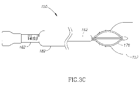
With reference to Figure 3C, radial cutter implant 178 is positioned
within urethra 154 in an expanded configuration. The physician pulls
positioning
tube 162 out of the patient and leaves radial cutter implant within urethra
154 for
a predetermined period of time (as detailed herein below - e.g., two weeks).
Radial cutter implant 178 applies pressure on the walls of the surrounding
tissues (e.g., bladder neck 156, urethra 154, and the prostate gland ¨ not
shown) incising the surrounding tissues over the predetermined period of time.
The prolonged incision of the tissue, created by continuous pressure,
decreases
the pain involved in the procedure. Furthermore, by performing the incisions
via
continuous pressure (i.e., via infarction), bleeding is avoided.
The period of time, radial cutter implant 178 is implanted in the
urethra of the patient, is determined by the physician at least according to
the
diagnosis of the patient (i.e., predetermined period of time). Alternatively,
the
time period is determined according to observations of the radial cutter
implant
effect over time (i.e., real time period determination), or any other way
known in
the art. Further alternatively, the time period ranges between one hour and
twenty nine days.
Reference is now made to Figures 4A, 4B and 4C, which are
schematic illustrations of a delivery, generally referenced 200, for
delivering a
radial cutter implant, constructed and operative in accordance with another
embodiment of the disclosed technique. With reference to Figure 4A, delivery
200 is substantially similar to delivery system 120 of Figure 2. Delivery 200
includes an implant sheath 202, an external tube 204, an external tube handle
206, an internal tube 208, and an internal tube handle 210. Each of implant
-12-
CA 02747971 2011-06-21
WO 2010/073244
PCT/1L2009/001207
sheath 202, external tube 204, external tube handle 206, internal tube 208,
and
internal tube handle 210 is substantially similar to each of implant sheath
122,
external tube 124, external tube handle 126, internal tube 130, and internal
tube
handle 132 of Figure 2, respectively.
Implant sheath 202 is coupled with the proximal end of external tube
204. External tube handle 206 is coupled with the distal end of external tube
204. A radial cutter implant 212 (Figure 4B) is coupled, at a folded
configuration
thereof, with the proximal end of internal tube 208 and is covered by implant
sheath 202. Internal tube 208 is slidably coupled with external tube 204.
Internal tube handle 210 is coupled with the distal end of internal tube 208.
With reference to Figure 4B, a physician (not shown) pulls external
tube 204 via external tube handle 206 while keeping internal tube 208 in
place.
Thus, external tube 204 slides along internal tube 208 in the distal direction
and
implant sheath 202 is removed from radial cutter implant 212.
With reference to Figure 4C, once implant sheath 202 is fully
removed from radial cutter implant 212 (i.e., radial cutter implant is fully
exposed), radial cutter implant 212 expands. In the example set forth in
Figure
4C, radial cutter implant 212 is self-expanding. Alternatively, radial cutter
implant 212 is expanded manually by the physician employing an implant
zo expander (not shown).
The physician leaves radial cutter implant 212 within the body of the
patient for a predetermined period of time. When the physician wishes to
remove radial cutter implant 212, the physician inserts delivery 200 into the
urethra (not shown) of the patient. The physician couples the proximal end of
internal tube 208 with radial cutter implant 212 by employing a coupler (not
shown - e.g., coupler 240 of Figure 5A). The physician pulls back internal
tube
208 while keeping external tube 204 in place. Thus, radial cutter implant 212
is
folded within, and is restrained by, implant sheath 202 and can be extracted
from the body (i.e., the bladder neck and the urethra) of the patient, without
damaging the tissues of the urethra. It is noted that, the delivery of radial
cutter
implant 212 and the extraction thereof are substantially reverse duplicates of
each other. In other words, the steps performed upon delivery are repeated in
a
reverse order upon extraction.
-13-
CA 02747971 2011-06-21
WO 2010/073244
PCT/1L2009/001207
Reference is now made to Figures 5A, 5B, 5C, 5D and 5E. Figure 5A
is a schematic illustration of a coupler, generally referenced 240, for
coupling a
radial cutter implant with an internal tube of a delivery system, constructed
and
operative in accordance with a further embodiment of the disclosed technique.
Figure 5B is a schematic illustration of a coupler, generally referenced 250,
for
coupling a radial cutter implant with an internal tube of a delivery system,
constructed and operative in accordance with another embodiment of the
disclosed technique. Figures 5C and 5D are schematic illustrations of a
coupler,
generally referenced 260, for coupling a radial cutter implant with an
internal
lo tube of a delivery system, constructed and operative in accordance with
a
further embodiment of the disclosed technique. Figure 5E is a schematic
illustration of a coupler, generally referenced 280, for coupling a radial
cutter
implant with an internal tube of a delivery system, constructed and operative
in
accordance with another embodiment of the disclosed technique.
With reference to Figure 5A, coupler 240 includes a female portion
246 and a male portion 248. Male portion 248 is inserted into female portion
246 and is attached to female portion by screwing mechanism. In other words,
the external circumference of male portion 248 is similar to that of a screw
and
the internal circumference of female portion 246 is similar to that of a nut.
In the
example set forth in Figure 5A, female portion 246 is coupled with the distal
end
of a radial cutter implant 242 (e.g., radial cutter implant 320 of Figure 6A),
and
male portion 248 is coupled with the proximal end of an internal tube 244 of a
delivery system (e.g., internal tube 208 of Figure 4A). Alternatively, female
portion 246 is coupled with the proximal end of internal tube 244, and male
portion 248 is coupled with the distal end of radial cutter implant 242.
With reference to Figure 5B, coupler 250 includes a loop 256 and a
hook 258. Hook 258 is inserted into loop 256 such that the physician is able
to
pull both hook 258 and loop 256 when pulling either of them. In the example
set
forth in Figure 5B, loop 256 is coupled with the distal end of a radial cutter
implant 252, and hook 258 is coupled with the proximal end of an internal tube
254. Alternatively, loop 256 is coupled with the proximal end of internal tube
254, and hook 258 is coupled with the distal end of radial cutter implant 252.
-14-
CA 02747971 2011-06-21
WO 2010/073244
PCT/1L2009/001207
With reference to Figure 5C, coupler 260 includes a dilating tip 266
and a recessed tube 262 (i.e., a tube which is sliced for forming a pair of
pincers
at the end thereof ¨ the pincers are not referenced). Recessed tube 262 is
coupled with the proximal end of a delivery system (e.g., delivery system 200
of
Figure 4A). Dilating tip 266 is coupled with recessed tube 262, such that
dilating
tip can be pulled into a recess 264 of recessed tube 262 and pushed out of
recess 264 of recessed tube 262. When dilating tip 266 is positioned within
recess 264, dilating tip 266 expands the diameter of recessed tube 262.
Recessed tube 262 is coupled with the proximal end of an internal tube 262.
With reference to Figure 5D, a distal end of a radial cutter implant 274
is coupled with a bottleneck 272. Bottleneck 272 includes an aperture 270
positioned approximately in the middle thereof. From the distal side of
aperture
270 a gradually narrowing niche 268 is culminating in aperture 270. The
diameter of aperture 270 is slightly larger than the diameter of recessed tube
260 and the diameter of dilating tip 266. The physician pushes dilating tip
266
and recessed tube 262 through aperture 270. After recessed tube 262 and
dilating tip 266 are positioned proximally to aperture 270, the physician
pulls
dilating tip 266 into recess 264 for enlarging the diameter of recessed tube
262.
When the physician pulls recessed tube 262 back in the distal direction,
radial
zo cutter implant
274 is pulled there-along (i.e., enlarged recessed tube 262 is
blocked by aperture 270 of bottle neck 272). When the physician pushes
dilating tip 266 away from recess 264, recessed tube 262 returns to the
original
diameter thereof. Thus, recessed tube 262 and dilating tip 266 can go through
bottleneck 272 (i.e., through aperture 270).
In the example set forth in Figures 5C and 5D, recessed tube 262 and
dilating tip 266 are coupled with an internal tube (not referenced) of the
delivery
system, and bottle neck 272 is coupled with a radial cutter implant 274.
Alternatively, recessed tube 262 and dilating tip 266 are coupled with radial
cutter implant 274, and bottleneck 272 is coupled with the internal tube.
With reference to Figure 5E, coupler 280 includes a rigid ball 286 and
a flexible socket 292. Flexible socket 292 includes a gradually narrowing
opening 288 and a spherical niche 290. When rigid ball 286 is pushed against
flexible socket 292, rigid ball 286 enters through gradually narrowing opening
-15-
WO 2010/073244 PCT/1
L2009/001207
288 and expands the proximal end thereof when entering spherical niche 290.
Once rigid ball 286 is positioned inside flexible socket 290 (i.e., rigid ball
286 is
securely coupled with flexible socket 292), the physician can pull flexible
socket
292, and rigid ball 286 is pulled there-along. In the example set forth in
Figure
5E, rigid ball 286 is coupled with the distal end of a radial cutter implant
282, and
flexible socket 292 is coupled with the proximal end of an internal tube 284.
Alternatively, rigid ball 286 is coupled with the proximal end of internal
tube 284,
and flexible socket 292 is coupled with the distal end of radial cutter
implant 282.
Reference is now made to Figures 6A and 6B, which are schematic
io illustrations of a radial cutter implant, generally referenced 320,
constructed and
operative in accordance with a further embodiment of the disclosed technique.
With reference to Figure 6A, radial cutter implant 320 is depicted from a side
view perspective. Radial cutter implant 320 includes a distal end 322, three
wires 326, 328 and 330, and a proximal end 324. Three wires 326, 328 and
330 are coupled between distal end 322 and proximal end 324. Distal end 322
is coupled with a coupler (e.g., coupler 240, 250, 260, and 280 of Figures 5A,
5B, 5C, and 5E, respectively).
Proximal end 324 is tapered for dilating the urethra of the patient
during delivery of radial cutter implant 320. The shape of each of wires 326,
328
and 330, is substantially a portion of a circle. Each of wires 326, 328 and
330 is
made from a Shape Memory Alloy (SMA), such as Nickel Titanium alloy
(Nitino10). Alternatively, each of wires 326, 328 and 330 is made from any
material which is flexible enough to be folded within an implant sheath and is
strong enough (e.g., 0.5 Newton) to apply pressure on the surrounding tissues
and induce infarction. Each of wires 326, 328 and 330 is flexible such that it
can
be straightened in order for radial cutter 320 to be folded within an implant
sheath
(e.g., implant sheath 202 of Figure 4A ¨ not shown). Each of wires 326, 328
and
330 springs back to the portion of the circle stance, once not limited by an
obstacle (e.g., implant sheath 202 of Figure 4A, the walls of the bladder neck
of
the patient). In this manner, when radial cutter implant 320 is positioned
within
the bladder neck of the patient, wires 326, 328 and 330, apply pressure to the
surrounding tissues (e.g., bladder neck, urethra and prostate). With reference
to
Figure 6B, radial cutter implant 320 is depicted from a bottom view
perspective.
-16-
CA 2747971 2017-11-22
CA 02747971 2011-06-21
WO 2010/073244
PCT/1L2009/001207
Alternatively, implant 320 is made of biodegradable materials, such
that there is no need to remove implant 320 from the body of the patient. In
this
manner, implant 320 is constructed such it biodegrades, ceases from
functioning
and dissolves within the patient after the predetermined period of time, or
after a
triggering event initiated by the physician, as known in the art.
Reference is now made to Figures 7A and 7B, which are schematic
illustrations of a radial cutter implant, generally referenced 350,
constructed and
operative in accordance with another embodiment of the disclosed technique.
With reference to Figure 7A, radial cutter implant 350 is depicted from a side
view perspective. Radial cutter implant 350 includes a distal end 352 and
three
wires 354, 356, and 358. Each of distal end 352 and wires 354, 356, and 358,
is
substantially similar to each of distal end 322, and wires 326, 328, and 330
of
Figure 6A, respectively. Wires 354, 356, and 358 are not coupled
there-between at the proximal end thereof. Radial cutter implant 350 operates
in a substantially similar manner to that of radial cutter implant 320. With
reference to Figure 7B, radial cutter implant 350 is depicted from a bottom
view
perspective.
Reference is now made to Figure 8, which is a schematic illustration
of a radial cutter implant, generally referenced 380, constructed and
operative in
accordance with a further embodiment of the disclosed technique. Radial cutter
implant 380 includes a distal end 382 and three wires 384, 386 and 388. Distal
end 382 is substantially similar to distal end 322 of Figure 6A. Each of wires
384, 386 and 388, is substantially similar to each of wires 326, 328 and 330
of
Figure 6A, respectively. Each of wires 384, 386 and 388 is in the shape of
half a
heart shape (i.e., the shape of a single side of a heart shape). Each of wires
384, 386 and 388 extends from the distal side of distal end 382 and U-turns to
project proximally from distal end 382. In other words, each of wires 384, 386
and 388 is bent such that it is coupled with the distal side of distal end 382
and it
extends proximally from distal end 382. Radial cutter implant 380 operates in
a
substantial similar manner to that of radial cutter implant 320 of Figure 6A.
Reference is now made to Figures 9A and 9B, which are schematic
illustrations of a radial cutter implant, generally referenced 410,
constructed and
operative in accordance with another embodiment of the disclosed technique.
-17-
CA 02747971 2011-06-21
WO 2010/073244
PCT/1L2009/001207
With reference to Figure 9A, radial cutter implant 410 is depicted from a side
view perspective. Radial cutter implant 410 includes a distal end 412, a
proximal end 414 and two wires 416 and 418. Wires 416 and 418 are coupled
between distal end 412 and proximal end 414. Distal end 412 is substantially
similar to distal end 322 of Figure 6A. Each of wires 416 and 418 is
substantially similar to wire 326 of Figure 6A. Each of wires 416 and 418 is
coupled with the distal side of distal end 412 and with the proximal side of
proximal end 414. Each of wires 416 and 418 is substantially C shaped. Radial
cutter implant 410 operates in a substantial similar manner to that of radial
cutter
implant 320 of Figure 6A. With reference to Figure 9B, radial cutter implant
410
is depicted from a bottom view perspective.
Reference is now made to Figures 10A and 10B, which are schematic
illustrations of a radial cutter implant, generally referenced 440,
constructed and
operative in accordance with a further embodiment of the disclosed technique.
With reference to Figure 10A, radial cutter implant 440 is depicted from a
side
view perspective. Radial cutter 440 includes a tube 442 and three wires 444,
446 and 448 (wire 448 is hidden behind tube 442 and is depicted in Figure
10B).
Each of wires 444, 446 and 448 is substantially similar to each of
wires 326, 328 and 330 of Figure 6A. Each of wires 444, 446 and 448 is in the
shape of a portion of a circle. The distal end of each of wires 444, 446 and
448
is coupled with the distal portion of tube 442, and the proximal end of each
of
wires 444, 446 and 448 is coupled with the proximal portion of tube 442. Tube
442 enables a clear passage of urine from the bladder of the patient through
the
bladder neck and into the urethra. The distal end of tube 442 can be coupled
with an internal tube of a delivery system (e.g., delivery system 150 of
Figures
3A to 3C) by employing a coupler (e.g., coupler 240 of Figure 5A). Radial
cutter
implant 440 operates in a substantial similar manner to that of radial cutter
implant 320. With reference to Figure 10A, radial cutter implant 440 is
depicted
from a bottom view perspective.
Reference is now made to Figures 11A and 11B, which are schematic
illustrations of a radial cutter implant, generally referenced 470,
constructed and
operative in accordance with another embodiment of the disclosed technique.
With reference to Figure 11A, radial cutter implant 470 is depicted from an
-18-
CA 02747971 2011-06-21
WO 2010/073244
PCT/1L2009/001207
isometric perspective. Radial cutter implant 470 includes a distal end 472,
four
butterfly wing shaped wires 474, 476, 478 and 480, and a proximal end 482.
Distal end 472 is substantially similar to distal end 322 of Figure 6A.
Proximal
end 482 is substantially similar to proximal end 324 of Figure 6A. Each of
butterfly wing shaped wires 474, 476, 478 and 480 is coupled between distal
end 472 and proximal end 482. Each of butterfly wing shaped wires 474, 476,
478 and 480 is flexible such that it can be folded within an implant sheath
(e.g.,
implant sheath 402 of Figure 4A).
The shape of each of butterfly wing shaped wires 474, 476, 478 and
480 enables radial cutter implant 470 to be fixed in the bladder neck of the
patient without moving. Radial cutter implant 470 is narrower at the middle
thereof than at the distal and proximal portions thereof (i.e., butterfly wing
shaped wires 474, 476, 478 and 480). In this manner, the narrow middle of
radial cutter implant 470 is positioned at the bladder neck of the patient.
The
proximal portion of radial cutter implant 470 is positioned within the bladder
of
the patient, and the distal portion of radial cutter implant 470 is positioned
within
the urethra of the patient, such that radial cutter implant 470 is fixed in
place. A
string 484 is looped around distal end 482 for enabling extraction of radial
cutter
implant 470. The physician (not shown) can employ the string 484 for guiding a
delivery system (e.g., delivery system 150 of Figures 3A to 3C) for the
extraction
of radial cutter implant 470 (i.e., string 484 is employed as a guide wire).
Radial
cutter implant 470 operates in a substantially similar manner to that of
radial
cutter implant 322 of Figure 6A. With reference to Figure 11B, radial cutter
implant 470 is depicted from a side view perspective.
Reference is now made to Figure 12, which is a schematic illustration
of a radial cutter implant, generally referenced 500, constructed and
operative in
accordance with a further embodiment of the disclosed technique. Radial cutter
implant 500 includes a right side portion 502 and a left side portion 504.
Right
side portion 502 is coupled with left side portion 504 at the proximal and
distal
ends thereof. Each of right side portion 502 and left side portion 504 is
butterfly
wing shaped. Each of right side portion 502 and left side portion 504 is
substantially similar to butterfly wing shaped wire 474 of Figure 11A. Radial
cutter implant 500 is positioned in the bladder neck and is fixed in place in
a
-19-
CA 02747971 2011-06-21
WO 2010/073244
PCT/1L2009/001207
substantial similar manner to that of radial cutter implant 470 of Figure 11A.
A
string 506 is coupled with the distal end of radial cutter implant 500 for
enabling
extraction of radial cutter implant 500, for guiding a delivery system for the
extraction procedure, or as anchoring device (i.e., string 506 is anchored
outside
-- of the body of the patient and prevents radial cutter implant 500 from
moving).
In the examples set forth in Figures 6A, 6B, 7A, 7B, 8, 9A, 9B, 10A,
10B, 11A, 11B and 12, each of the radial cutter implants includes two to four
wires. A radial cutter implant according to the disclosed technique should
include at least one wire, radially extending outwardly from the center of the
-- radial cutter implant, for applying pressure on the surrounding tissues.
The
radial cutter implant can include larger numbers of wires than four, such as
five
wires, six wires, and the like. The shape of the wires can be a portion of a
circle,
a butterfly wing shape, a polygon, and the like. The cross-section of the
wires is
round, rectangular, triangular, or any polygonal shaped.
In the examples set forth in Figures 6A, 6B, 7A, 7B, 8, 9A, 9B, 10A,
10B, 11A, 11B and 12, each of the radial cutters include either a distal end
or a
tube coupled with the distal end of the wires. It is noted that, a radial
cutter
implant according to the disclosed technique includes at least one central
connector (e.g., distal end 322 of Figure 6A or tube 442 of Figure 10A)
-- connecting the wires. It is further noted that the central connector can be
connected to the wires at the proximal end of the wires, at the distal end of
the
wires, and at any point in the middle of the wires.
Reference is now made to Figures 13A, 13B and 13C, which are
schematic illustrations of a radial cutter implant, generally referenced 520,
-- constructed and operative in accordance with another embodiment of the
disclosed technique. With reference to Figure 13A, radial cutter implant 520
is
depicted from a side view perspective. Radial cutter implant 520 is
substantially
similar to radial cutter implant 440 of Figure 10A. Radial cutter implant 520
includes a tube 522 and three wires 524, 526 and 528. Each of wires 524, 526
-- and 528 includes two barbs 530. Tube 522 is a catheter which allows urine
to
pass there-through. Each of barbs 530 penetrates into the surrounding tissues
(i.e., the tissues surrounding implant 520) for anchoring radial cutter
implant 520
in place (i.e., for preventing migration of radial cutter implant 520 into the
-20-
CA 02747971 2011-06-21
WO 2010/073244
PCT/1L2009/001207
bladder or into the urethra). When radial cutter implant 520 is extracted from
the
bladder neck (i.e., pulled back in the distal direction), barbs 530 are pulled
out of
the surrounding tissues. With reference to Figure 13B, Figure 13B depicts an
enlarged view portion 532 of radial cutter implant 520. Each of barbs 530 is
directed in the proximal direction for preventing radial cutter 520 from
moving in
the proximal direction, from the bladder neck into the bladder (i.e., keeping
radial
cutter implant 520 anchored in place). With reference to Figure 13C, radial
cutter implant 520 is depicted from a bottom view perspective.
It is noted that, the number of wires including barbs can vary.
Furthermore, the number of barbs on a wire can vary. For example, a radial
cutter implant including five wires, three of which includes two barbs each, a
fourth wire includes four barbs, and the fifth wire includes no barbs.
Reference is now made to Figure 14, which is a schematic illustration
of radial cutter implant, generally referenced 550, constructed and operative
in
accordance with a further embodiment of the disclosed technique. Radial cutter
implant 550 includes a distal end 552 and two wires 554 and 556. Radial cutter
implant 550 is substantially similar to radial cutter implant 350 of Figure
7A,
except for the number of wires (radial cutter implant 350 includes three wires
and radial cutter implant 550 includes two wires). Each of wires 554 and 556
includes two barbs 558 and 560. The direction of extension of barbs 558 is
different from the direction of extension of barbs 560 for enabling a stronger
fixation into the bladder neck (i.e., stronger than a configuration of similar
extending barbs).
Reference is now made to Figures 15A and 15B, which are schematic
illustrations of a radial cutter implant, generally referenced 580,
constructed and
operative in accordance with another embodiment of the disclosed technique.
With reference to Figure 15A, radial cutter implant 580 is depicted from an
isometric perspective. Radial cutter implant includes a tube 582, three wires
584, 586 and 588, and three wings 590, 592 and 594. Each of wires 584, 586
and 588 includes two barbs 596. Each of wires 584, 586 and 588 is coupled
with the distal end of tube 582. Each of wings 590, 592 and 594 is coupled
between tube 582 and each of wires 584, 586 and 588, respectively.
-21-
W02010/073244
PCT/1L2009/001207
Each of wires 584, 586 and 588, and each of wings 590, 592 and 594
is flexible for enabling radial cutter implant 580 to be folded within an
implant
sheath (e.g., implant sheath 202 of Figure 4A). Each of
barbs 596 is
substantially similar to each of barbs 558 of Figure 14. While radial cutter
implant 580 is implanted within the patient, tissue can grow around each of
wires 584, 586 and 588. Tissue growth around wires 584, 586 and 588
complicates the extraction of radial cutter implant 580 and prevent the
widening
effect of implant 580. Wings 590, 592 and 594 prevent tissue from growing
around wires 584, 586 and 588 and from holding radial cutter implant 580 in
place. Wings 590, 592 and 594 are made from Polyester (PET), Poly-Urethane
(PU), Nitinol foil, Silicon, and the like.
Reference is now made to Figures 16A to 160, which are schematic
illustrations of a radial cutter implant, generally referenced 620,
constructed and
operative in accordance with a further embodiment of the disclosed technique.
Implant 620 includes four wires 622, an anchoring leaflet 624, a distal end
626
and a proximal end 628. Each of wires 622 is coupled between proximal end
628 and distal end 626. Anchoring leaflet 624 extends from distal end 626, and
is positioned between two of wires 622.
Each of wires 622 is substantially similar to each of wires 474, 476,
478 and 480, all of Figure 11A. The shape of wires 622 is wider at the
proximal
end than at the distal end thereof. In this manner, wires 622 prevent implant
620 from moving from the bladder neck into the urethra. In particular, the
wider
proximal portion of wires 622 is positioned within the bladder and the
narrower
distal portion of wires 622 is positioned at the bladder neck and within the
urethra. Wires 622 (i.e., the wider proximal portion thereof) prevent implant
620
from moving out of the bladder and into the urethra by being blocked at the
bladder neck.
Anchoring leaflet 624 is constructed of similar materials to those of
wires 622. Anchoring leaflet 624 is in the shape of a tongue extending
substantially in the proximal-normal direction (i.e., the normal direction
refers to
a direction normal to the proximal-distal axis ¨ e.g., the dorsal direction).
In this
manner, anchoring leaflet 624 prevents implant 620 from moving from the
bladder neck into the bladder. In particular, anchoring leaflet 624 is blocked
by
-22-
CA 2747971 2017-11-22
WO 2010/073244 PCT/1
L2009/00 1 207
the bladder neck such that implant 620 can not move into the bladder. It is
noted that, anchoring leaflet 624 can be a wire leaflet (e.g., as depicted in
Figures 6A to 6D) or a full surface leaflet (e.g., substantially similar to
wings 590
of Figure 15A).
Alternatively, anchoring leaflet 624, extends in the distal-normal
direction and prevents radial cutter implant 620 from moving in the distal
direction towards the urethra. Further alternatively, there are at least two
leaflets 624 extending in both directions and fixing implant 620 in place.
It is noted that, wires 622, and in particular the wider proximal portion
thereof, prevent implant 620 from moving in the distal direction. Anchoring
leaflet 624 prevent implant 620 from moving in the proximal direction. Thus,
implant 620 is anchored in position within the bladder neck. It is further
noted
that, wires 622 and anchoring leaflet 624 are delivered within a sheath (e.g.,
implant sheath 202 of Figures 4A to 4C) and are expanded (e.g., self expand
upon exposure from the sheath) once positioned in the bladder neck. The
number of wire 622 of implant 620 is at least one, and can vary. The number of
anchoring leaflets 624, is at least one and can vary.
Reference is now made to Figure 17, which is a schematic illustration
of a radial cutter implant, generally referenced 650, positioned within a
bladder
neck of a patient, constructed and operative in accordance with another
embodiment of the disclosed technique. Radial cutter implant 650 includes two
wires 652, an anchoring leaflet 654, a distal end 656 and a proximal end 658.
Radial cutter implant 620 is substantially similar to implant 620 of Figures
16A to
16D.
The distal wider portion of wires 652 is positioned within a bladder
660 of a patient (not shown). The proximal narrower portion of wires 652 is
positioned within a bladder neck 662 of the patient. Anchoring leaflet 654
applies a proximal radial force against bladder neck 662 thereby producing a
niche 666 within bladder neck 662. Anchoring leaflet 654 is anchored within
niche 666. In this manner anchoring leaflet 654 anchors implant 650 within
bladder neck 662. In other words, the proximal wider portion of wires 652
prevents implant 650 from moving distally (i.e., towards a urethra 664 of the
-23-
CA 27 47 971 2 017 -11-22
CA 02747971 2011-06-21
WO 2010/073244
PCT/1L2009/001207
patient) and anchoring leaflet 654 prevents implant 650 from moving proximally
(i.e., towards bladder 660).
Alternatively, proximal end 658 is a substance release element, which
slowly releases substances into the bladder neck, urethra and prostate of the
patient, over a period of time. The released substances can include
painkillers,
anti-inflammatory materials, anti-biotic materials, and the like. Further
alternatively, implant 620 (i.e., or at least some portions thereof, such as
wires
652) is covered with such materials as detailed herein above, and releases
these materials slowly into the body of the patient.
Reference is now made to Figure 18, which is a schematic illustration
of a method for creating incisions in the muscles of the bladder neck by
infarction, operative in accordance with a further embodiment of the disclosed
technique. In procedure 700, the location of the bladder neck of a patient is
found. With reference to Figure 3A, the physician inserts overtube 164 into
the
urethra of the patient until balloon 158 is inside bladder 152. The physician
inflates balloon 158 and pulls overtube 164 back until balloon 158 is blocked
by
the bladder neck.
In procedure 702, a radial cutter implant is delivered to the constricted
location. With reference to Figure 3B, the physician inserts delivery 176 into
positioning tube 162 and delivers radial cutter implant 178 to the location of
the
bladder neck (i.e., the constricted location). Alternatively, the physician
delivers
the implant to a different constricted location within the urinal system of
the
patient for relieving the constriction. In procedure 704, the radial cutter
implant
is released and the delivery system is removed. With reference to Figure 3C,
the physician exposes radial cutter implant 178 from implant sheath 166.
Radial
cutter implant 178 expands and attaches itself to the surrounding tissues
(i.e.,
the wires of Radial cutter implant 178 are attached to the tissues surrounding
the implant and apply pressure thereon). The physician removes delivery
system 150 from urethra 154 of the patient.
In procedure 706, continuous pressure is applied on the tissues
surrounding the implant by employing the radial cutter implant. With reference
to Figure 3C, the wires of radial cutter implant 178 apply continuous pressure
on
the surrounding tissues. In procedure
708, at the appearance of a
-24-
CA 02747971 2011-06-21
WO 2010/073244
PCT/1L2009/001207
predetermined condition, the radial cutter implant is extracted from the
patient.
With reference to Figure 3C, the physician extracts radial cutter implant 178
from the patient at the appearance of a predetermined condition. The
predetermined condition can be the passage of a predetermined period of time,
the appearance of desired incisions on the surrounding tissues, the appearance
of a predetermined physiological effect, and the like.
It will be appreciated by persons skilled in the art that the disclosed
technique is not limited to what has been particularly shown and described
hereinabove. Rather the scope of the disclosed technique is defined only by
the
claims, which follow.
-25-