Language selection

Search

Patent 2748730 Summary

Third-party information liability

Some of the information on this Web page has been provided by external sources. The Government of Canada is not responsible for the accuracy, reliability or currency of the information supplied by external sources. Users wishing to rely upon this information should consult directly with the source of the information. Content provided by external sources is not subject to official languages, privacy and accessibility requirements.

Claims and Abstract availability

Any discrepancies in the text and image of the Claims and Abstract are due to differing posting times. Text of the Claims and Abstract are posted:

  • At the time the application is open to public inspection;
  • At the time of issue of the patent (grant).
(12) Patent: (11) CA 2748730
(54) English Title: DEVICE AND METHOD FOR MONITORING THE SUCCESS OF SPINAL ANESTHESIA
(54) French Title: DISPOSITIF ET PROCEDE DESTINES A CONTROLER LA REUSSITE D'ANESTHESIES RACHIDIENNES
Status: Expired and beyond the Period of Reversal
Bibliographic Data
(51) International Patent Classification (IPC):
  • A61B 05/00 (2006.01)
  • A61M 16/01 (2006.01)
(72) Inventors :
  • GROSSMANN, ULF (Germany)
  • PENNO, ANDREAS (Germany)
(73) Owners :
  • ANDREAS PENNO
(71) Applicants :
  • ANDREAS PENNO (Germany)
(74) Agent: SMART & BIGGAR LP
(74) Associate agent:
(45) Issued: 2014-09-16
(86) PCT Filing Date: 2009-12-23
(87) Open to Public Inspection: 2010-07-08
Examination requested: 2011-12-21
Availability of licence: N/A
Dedicated to the Public: N/A
(25) Language of filing: English

Patent Cooperation Treaty (PCT): Yes
(86) PCT Filing Number: PCT/EP2009/009238
(87) International Publication Number: EP2009009238
(85) National Entry: 2011-06-30

(30) Application Priority Data:
Application No. Country/Territory Date
10 2009 003 897.3 (Germany) 2009-01-03

Abstracts

English Abstract


The invention relates to a device
for monitoring the success of spinal anesthesia
ha-ving at least one electronic temperature sensor for
measuring the skin surface temperature within at
least one dermatome of a patient; an electronic
analysis device connected to the at least one
tem-perature sensor and designed so that it monitors
the measurement signal provided by the at least
one temperature sensor to determine whether the
skin surface temperature increases by about 2 to 3
°C; and a display device optically and/or
acousti-cally indicating the result of the analysis by the
electronic analysis device.


French Abstract

L'invention concerne un dispositif destiné à contrôler la réussite d'anesthésies rachidiennes comprenant au moins un capteur de température électronique pour mesurer la température de surface de la peau dans un moins un dermatome d'un patient; un dispositif d'évaluation électronique qui est relié au capteur de température et qui est conçu de manière à surveiller le signal de mesure fourni par le capteur de température pour voir si la température de surface de la peau augmente d'environ 2 à 3 °C; et un dispositif d'affichage qui affiche visuellement et/ou acoustiquement le résultat de l'évaluation effectuée par l'unité d'évaluation électronique.

Claims

Note: Claims are shown in the official language in which they were submitted.


- 9 -
Claims:
1. A device for the monitoring of the success of spinal anesthesia with:
(i) at least one electronic temperature sensor (2) for measuring the skin
surface temperature within at least one dermatome of a patient;
(ii) an electronic evaluation device (5), which is connected with the at
least one temperature sensor (2) and is designed such that it
monitors the measurement signal delivered by the at least one
temperature sensor (2) to determine whether the skin surface
temperature increases by approximately 2 to 3° C;
(iii) wherein the electronic evaluation device (5) is also designed such
that in the case of the determination of an increase in the skin
surface temperature by approx. 2 to 3° C it determines that the
analgesia has occurred in a dermatome, which is closer to the sacral
lumbar vertebrae by approximately 2 to 6 dermatomes than the
dermatome, in which the temperature sensor (2) measures the skin
surface temperature; and
(iv) a display device (7) that displays the result of the evaluation by the
electronic evaluation device either optically, acoustically, or
optically and acoustically.
2. The device according to claim 1, in which the electronic evaluation unit
(5) is designed such that in the case of the determination of an increase
in the skin surface temperature by approx. 2 to 3° C it determines that
the analgesia has occurred in a dermatome, which is closer to the sacral
lumbar vertebrae by approximately 2 to 3 dermatomes than the
dermatome, in which the temperature sensor (2) measures the skin
surface temperature.

- 10 -
3. The device according to claim 1 or 2, in which the at least one
temperature sensor (2) is an NTC resistor.
4. The device according to one of claims 1 through 2, in which at least one
temperature sensor (2) is held via spring means on means for fastening
(9) on the body of a patient.
5. The device according to one of claims 1 through 4, in which several
temperature sensors (2) are arranged on a band (9) at certain distances
from each other, which is fastenable with means for fastening on the
body of a patient.
6. The device according to one of claims 1 through 5, in which the at least
one temperature sensor is connected with a tape for fastening on the
body of a patient.
7. The device according to claim 5 or 6, in which the distances between
two neighboring temperature sensors (2) on the band (9) corresponds
with the distances between one or more dermatomes.
8. The device according to one of claims 1 through 7, in which the at least
one temperature sensor (2) is connected with a digital evaluation device
(5) via at least one analog-digital converter (4).
9. The device according to claim 8, in which the digital evaluation device
(5) is a PC.

- 11 -
10. The device according to claim 8 or 9, in which the analog-digital
converter (4) is connected with a USB port of the PC (5).
11. The device according to one of claims 1 through 10, in which the
display
device (7) shows a graphic (8) of a human body, in which the
dermatomes, in which the analgesia have occurred, and the dermatomes
in which the skin surface temperature has increased by approximately 2
to 3° C, are highlighted graphically.
12. The method for monitoring the success of spinal anesthesia, in which:
(i) at least one electronic temperature sensor measures the skin surface
temperature within at least one dermatome of a patient;
(ii) an electronic evaluation device monitors the measurement signal
delivered by the at least one temperature sensor for an increase in
the skin surface temperature by approximately 2 to 3° C;
(iii) the electronic evaluation device in the case of an increase in the
skin surface temperature by approximately 2 to 3° C determines the
analgesia in a dermatome closer to the sacral lumbar vertebrae of
the patient by 2 to 6 dermatomes than the temperature sensor; and
(iv) a display device shows the result of the evaluation by the electronic
evaluation device.
13. The method according to claim 12, in which the display device display
the analgesia in the determined dermatome.
14. The method according to claim 12 or 13, in which the electronic
evaluation device determines, in the case of an increase in the skin
surface temperature by approximately 2 to 3° C, the analgesia in a

- 12 -
dermatome closer to the sacral lumbar vertebrae of the patient by 2 to 3
dermatomes than the temperature sensor and the display device displays
the analgesia in the determined dermatome.
15. The method according to one of claims 12 through 14, in which at least
one temperature sensor measures the skin surface within different
dermatomes.
16. Method according to claim 15, in which at least one temperature sensor
is arranged on the skin surface.
17. The method according to claim 16, in which several temperature sensors
are arranged on the skin surface of the patient at a distance of at least one
dermatome.
18. The method according to claim 16 or 17, in which at least one sensor is
pressed against the skin surface via a spring means.
19. The method according to one of the claims 16 through 18, in which the
temperature sensors are placed on the skin surface of the patient by
means of a band, on which they are arranged at certain distances from
each other.
20. The method according to one of the claims 12 through 19, in which the
temperature sensors deliver an analog measurement signal, which is
converted into a digital signal by means of the analog-digital converter
and which is digitally processed by the evaluation device.

- 13 -
21. The method according to one of claims 12 through 20, in which the
display device displays in a graphically highlighted manner in a graphic
of a human body the dermatomes, in which the analgesia has occurred,
and the dermatomes in which the skin surface has increased by
approximately 2 to 3° C.
22. The method according to one of claims 12 through 21, in which the
evaluation device displays the result of the evaluation optically,
acoustically, or optically and acoustically.

Description

Note: Descriptions are shown in the official language in which they were submitted.


CA 02748730 2011-06-30
Device and Method for Monitoring the Success of Spinal Anesthesia
The object of the invention is a device and a method for monitoring the
success of spinal anesthesia in medicine.
Spinal anesthesia is a form of regional anesthesia close to the spinal cord.
Through the injection of a local anesthesia into the cerebrospinal fluid area
at
the height of the lumbar spine, the signal transfer in the nerves extending
from
the spinal cord is inhibited. A temporary, reversible blockage of the
sympathetic nervous system, the sensitivity and the motor function of the
lower half of the body is thereby achieved.
As a standard procedure in anesthesia, spinal anesthesia is used today in a
plurality of operations in the lower stomach, the pelvis, the lower
extremities
and in obstetrics and represents an alternative to other regional procedures
such as peridural anesthesia and full narcosis.
The human spinal column consists of 34 vertebrae, which are connected by
firm bands and surround the spinal cord. Spinal nerves extend out between the
vertebrae, which innervate the body segmentally and enable sensitivity and
also carry fibers of the vegetative nervous system (sympathetic /
parasympathetic nervous systems). As part of the central nervous system, the
spinal cord is surrounded by the meninges, which restrict the cerebrospinal
fluid area, in which the cerebrospinal fluid circulates. This cerebrospinal
fluid
space is punctured with a thin cannula during spinal anesthesia. Local
anesthesia is injected through the tip of the needle, which acts on the front
and

CA 02748730 2011-06-30
- 2 -
rear roots of the spinal nerves and temporarily blocks their ability to
transmit
nerve impulses.
It is known that, in spinal anesthesia, different qualities of the sensor
system
can be switched off in succession: first, the preganglionic sympathetic
nervous system of the vegetative nervous system is blocked. This results in
vessel dilation, a warming of the skin and an eventual drop in blood pressure.
Pain and temperature fibers are then blocked. Touch and pressure sensation
follows. Motor activity and sense of vibration and location is switched off
last.
The effective height of the spinal anesthesia depends on the dispersion of the
injected activate agents in the cerebrospinal fluid, which can be influenced
through dose and concentration of the local anesthesia through the positioning
of the patient.
So far, there is no device that can show the anesthetization for this form of
anesthesia. The analgesia has previously been tested by means of a cold spray
by the anesthetist on the body of the patient. The patient needed to verbally
indicate whether or not he/she still felt a corresponding cold sensation.
Based on this, the object of the invention is to provide a technique for
facilitating the monitoring of the success of spinal anesthesia.

CA 02748730 2014-01-22
- 3 -
The device according to the invention for monitoring the success of spinal
anesthesia has
¨ at least one electronic temperature sensor for measuring the skin surface
temperature within at least one dermatome of a patient,
¨ an electronic evaluation device, which is connected with the at least one
temperature sensor and is designed such that it monitors the
measurement signal delivered by the at least one temperature sensor to
determine whether the skin surface temperature increases by
approximately 2 to 3 C,
¨ wherein the electronic evaluation device is also designed such that in
the
case of the determination of an increase in the skin surface temperature
by approx. 2 to 3 C it determines that the analgesia has occurred in a
dermatome, which is closer to the sacral lumbar vertebrae by
approximately 2 to 6 dermatomes than the dermatome, in which the
temperature sensor (2) measures the skin surface temperature, and
¨ a display device that displays the result of the evaluation by the
electronic evaluation device optically and/or acoustically.
The device according to the invention assumes that, in the case of the
sympatholysis (exclusion of the sympathetic innervation) by the spinal
anesthesia, the "thinner" non-myelinated nerve fibers are blocked first and
only then the "thicker" myelinated nerve fibers. As a result, during the
spinal
anesthesia, the blockage of the sympathetic nervous system is generally two
to three segments or respectively dermatomes further away than the sensory

CA 02748730 2014-01-22
- 4 -
blockage. The blockage of the sympathetic nervous system can be up to six
segments or respectively dermatomes further away than the sensory blockage.
Furthermore, the sensory blockage is approx. two segments or respectively
dermatomes further away than the motor blockage. The blockage of the
sympathetic nervous system accompanies an increase in the skin temperature
by approximately 2 to 3 C in the associated dermatome. This temperature
increase thus indicates that a sensory blockage and thus the analgesia
(anesthetization) has occurred approximately 2 to 3 (up to 6) dermatomes
more caudally. For example, in the case of an increase in the temperature of
the skin surface by approximately 2 to 3 C in dermatome Th2, the analgesia
should be assumed approximately 2 to 3 (-6) dermatomes more caudally in
dermatomes Th4 through Th5 (-Th8). The invention takes advantage of this
knowledge in that it makes it possible, due to the measured temperature
increase of approximately 2 to 3 C at least one dermatome of a patient, to
identify the dermatome arranged 2 to 3 (-6 dermatomes) more caudally than
the one in which the analgesia already occurred. For example, through display
of the dermatome, in which the temperature increase of 2 to 3 C occurred,
and if applicable the dermatomes, in which this temperature increase already
occurred previously, the checking of the success of the spinal anesthesia will
be easier for the healthcare professional. The previous check by means of cold
spray and verbal reaction of the patient is replaced by an objective
measurement and evaluation of the measurement results.
A simple design of the device according to the invention shows as a result of
the evaluation by the evaluation device that or respectively those dermatomes,
in which the temperature increase of approximately 2 to 3 C occurred. Based

CA 02748730 2014-01-22
- 4a -
on this display, it can be assumed that the analgesia has occurred
approximately 2 to 3 (up to 6) dermatomes more caudally. The electronic
evaluation device determines that the analgesia has occurred in a dermatome,
which is closer to the sacral lumbar vertebrae by approximately 2 to 6
dermatomes than the dermatome, in which a temperature increase of the skin
surface of approximately 2 to 3 C was determined. The display device
shows the dermatome, in which

CA 02748730 2011-06-30
- 5 -
the analgesia occurred, as a result of the evaluation by the electronic
evaluation device. If applicable, the display device also shows the
dermatomes, in which the analgesia already occurred previously. The
electronic evaluation device preferably determines that the analgesia has
occurred in a dermatome, which is closer to the sacral lumbar vertebrae by
approximately 2 to 3 dermatomes than the dermatome, in which a temperature
increase of the skin surface of approximately 2 to 3 C was determined. In
accordance with another embodiment, the display device shows the
dermatomes, in which the skin surface temperature is increased by 2 to 3 C,
and the dermatomes, in which the analgesia has occurred (e.g. in different
colors).
The temperature sensor can be designed differently. For example, it is
possible to measure the skin surface temperature of the patient over a large
area with a thermal camera and to determine the temperatures in the
individual dermatomes with an automatic image evaluation procedure. In
accordance with another embodiment, individual temperature sensors are
used. In accordance with a preferred embodiment, the at least one temperature
sensor is an NTC resistor.
In accordance with one embodiment of the invention, at least one temperature
sensor is held via spring means on means for fastening the temperature sensor
on the body of a patient. The temperature sensor is pressed against the skin
surface with a constant pressing force via spring means. Measurement errors
are hereby avoided.
As a general rule, the temperature sensors can be secured individually to the
skin surface of the patient. In accordance with one embodiment, several

=
CA 02748730 2011-06-30
- 6 -
temperature sensors are arranged at certain distances from each other on a
band, which can be fastened by means for fastening on the body of a patient.
Through the specified arrangement of the temperature sensors on a band, the
placement of the temperature sensors on the body of the patient is
facilitated.
The distance between two neighboring temperature sensors on the band is
preferably the distance of one or more dermatomes. This embodiment
facilitates the attachment of the temperature sensors within the different
dermatomes of the patient. The measurement of the distance of neighboring
temperature sensors on the band can be based on an average-sized patient.
Furthermore, it is possible to provide several bands with temperature sensors
at different distances for patients of a different size.
In accordance with one embodiment, the temperature sensor is connected with
a tape for fastening on the body of a patient.
The electronic evaluation device can be analog or digital. It can be a program-
controlled electronic data-processing device or pure hardware. A
programmable, digital evaluation device is preferably used. In particular, a
PC
can be used as the evaluation device.
In accordance with one embodiment, an analog temperature sensor is
connected with a digital evaluation device via at least one analog-digital
converter. In accordance with another embodiment, the analog-digital
converter is connected to a USB port of the PC.

CA 02748730 2011-06-30
- 7 -
The display device is for example a monitor of a PC. In accordance with one
embodiment, the display device shows a graphic of a human body, in which the
dermatomes, in which the analgesia has occurred, and/or the dermatomes, in
which
the skin surface temperature has increased by approximately 2 to 3 C, are
highlighted graphically. The graphical highlighting can take place e.g. by
coloring
the concerned dermatomes a different color than the rest of the graphic.
Furthermore, the object is solved through a method with the characteristics of
claim
12. Advantageous embodiments of the method are specified in the dependent
claims
13 through 22.
The invention is explained in greater detail below based on the attached
drawings of
an exemplary embodiment. The drawings show the following:
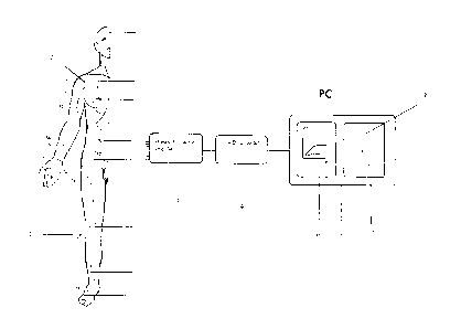
Fig. 1 a device according to the invention for monitoring the success of
spinal
anesthesia on a body of a patient in a rough, schematic block diagram;
Fig. 2 a section of a band with temperature sensors in a top view;
Fig. 3 an enlarged detail of the band from Fig. 2 in a vertical cut.
In accordance with Fig. 1, the skin surface of a patient 1 is segmented into
different
dermatomes, which are labeled with reference symbols such as Th2, L3 and C4.
Temperature sensors 2 are attached to certain dermatomes.

CA 02748730 2011-06-30
- 8 -
The temperature sensors 2 are connected with an analog-digital converter 4
via an amplifier 3 with at least 8 channels. The analog-digital converter 4
scans the output channels of the amplifier 3 and converts the amplified,
analog measurement signal into a digital signal.
The analog-digital converter 4 is attached to a PC 5. The PC 5 determines
whether the skin temperature measured by the temperature sensors 2 increases
by 2 to 3 C as shown in the temperature-time diagram 6. If the PC 5
determines an increase by 2 to 3 C, it calculates that an analgesia has
occurred in a dermatome arranged approximately 2 to 3 (-6) dermatomes
more caudally. The dermatomes, in which the analgesia was determined, are
displayed on a screen 7 with a graphic 8 of a human body.
In accordance with Fig. 2 and 3, several temperature sensors 2 are fastened on
a band 9 each under an intermediate layer of a foam cushion. The distances
between neighboring temperature sensors 2 correspond with the distances
between certain dermatomes. The band 9 is fastenable on the body 1 with
tapes which are attached transversely on the band 9.
Representative Drawing
A single figure which represents the drawing illustrating the invention.
Administrative Status

2024-08-01:As part of the Next Generation Patents (NGP) transition, the Canadian Patents Database (CPD) now contains a more detailed Event History, which replicates the Event Log of our new back-office solution.

Please note that "Inactive:" events refers to events no longer in use in our new back-office solution.

For a clearer understanding of the status of the application/patent presented on this page, the site Disclaimer , as well as the definitions for Patent , Event History , Maintenance Fee  and Payment History  should be consulted.

Event History

Description Date
Time Limit for Reversal Expired 2019-12-23
Common Representative Appointed 2019-10-30
Letter Sent 2018-12-24
Change of Address or Method of Correspondence Request Received 2018-01-12
Grant by Issuance 2014-09-16
Inactive: Cover page published 2014-09-15
Small Entity Declaration Request Received 2014-06-26
Pre-grant 2014-06-26
Inactive: Final fee received 2014-06-26
Notice of Allowance is Issued 2014-02-13
Letter Sent 2014-02-13
Notice of Allowance is Issued 2014-02-13
Inactive: Q2 passed 2014-02-04
Inactive: Approved for allowance (AFA) 2014-02-04
Amendment Received - Voluntary Amendment 2014-01-22
Inactive: S.30(2) Rules - Examiner requisition 2013-07-22
Letter Sent 2012-01-09
Request for Examination Received 2011-12-21
Request for Examination Requirements Determined Compliant 2011-12-21
All Requirements for Examination Determined Compliant 2011-12-21
Inactive: Cover page published 2011-09-07
Application Received - PCT 2011-08-24
Inactive: Notice - National entry - No RFE 2011-08-24
Inactive: IPC assigned 2011-08-24
Inactive: First IPC assigned 2011-08-24
National Entry Requirements Determined Compliant 2011-06-30
Small Entity Declaration Determined Compliant 2011-06-30
Application Published (Open to Public Inspection) 2010-07-08

Abandonment History

There is no abandonment history.

Maintenance Fee

The last payment was received on 2013-11-05

Note : If the full payment has not been received on or before the date indicated, a further fee may be required which may be one of the following

  • the reinstatement fee;
  • the late payment fee; or
  • additional fee to reverse deemed expiry.

Patent fees are adjusted on the 1st of January every year. The amounts above are the current amounts if received by December 31 of the current year.
Please refer to the CIPO Patent Fees web page to see all current fee amounts.

Fee History

Fee Type Anniversary Year Due Date Paid Date
Basic national fee - small 2011-06-30
MF (application, 2nd anniv.) - small 02 2011-12-23 2011-11-03
Request for examination - small 2011-12-21
MF (application, 3rd anniv.) - small 03 2012-12-24 2012-11-19
MF (application, 4th anniv.) - small 04 2013-12-23 2013-11-05
Final fee - small 2014-06-26
MF (patent, 5th anniv.) - small 2014-12-23 2014-11-14
MF (patent, 6th anniv.) - small 2015-12-23 2015-12-18
MF (patent, 7th anniv.) - small 2016-12-23 2016-11-15
MF (patent, 8th anniv.) - small 2017-12-27 2017-11-28
Owners on Record

Note: Records showing the ownership history in alphabetical order.

Current Owners on Record
ANDREAS PENNO
Past Owners on Record
ULF GROSSMANN
Past Owners that do not appear in the "Owners on Record" listing will appear in other documentation within the application.
Documents

To view selected files, please enter reCAPTCHA code :


To view images, click a link in the Document Description column. To download the documents, select one or more checkboxes in the first column and then click the "Download Selected in PDF format (Zip Archive)" or the "Download Selected as Single PDF" button.

List of published and non-published patent-specific documents on the CPD .

If you have any difficulty accessing content, you can call the Client Service Centre at 1-866-997-1936 or send them an e-mail at CIPO Client Service Centre.


Document
Description 
Date
(yyyy-mm-dd) 
Number of pages   Size of Image (KB) 
Abstract 2011-06-29 2 84
Claims 2011-06-29 5 133
Drawings 2011-06-29 2 21
Description 2011-06-29 9 294
Representative drawing 2011-09-06 1 12
Description 2014-01-21 9 298
Claims 2014-01-21 5 147
Drawings 2014-01-21 2 28
Representative drawing 2014-09-01 1 11
Reminder of maintenance fee due 2011-08-23 1 112
Notice of National Entry 2011-08-23 1 194
Acknowledgement of Request for Examination 2012-01-08 1 177
Commissioner's Notice - Application Found Allowable 2014-02-12 1 162
Maintenance Fee Notice 2019-02-03 1 180
PCT 2011-06-29 16 536
Correspondence 2014-06-25 1 56