Note: Descriptions are shown in the official language in which they were submitted.
CA 02748962 2011-08-15
1
CATHETER WITH THIN FILM PRESSURE SENSING DISTAL TIP
FIELD OF INVENTION
[0001] The present invention relates to an electrophysiologic
catheter useful for ablation and
sensing electrical activity of heart tissue, in particular, an
electrophysiologic catheter with contact
force sensing capabilities at its distal end.
BACKGROUND OF INVENTION
[0002] Cardiac arrythmias, and atrial fibrillation in particular,
persist as common and
dangerous medical ailments, especially in the aging population. In patients
with normal sinus
rhythm, the heart, which is comprised of atrial, ventricular, and excitatory
conduction tissue, is
electrically excited to beat in a synchronous, patterned fashion. In patients
with cardiac
arrythmias, abnormal regions of cardiac tissue do not follow the synchronous
beating cycle
associated with normally conductive tissue as in patients with normal sinus
rhythm. Instead, the
abnormal regions of cardiac tissue aberrantly conduct to adjacent tissue,
thereby disrupting the
cardiac cycle into an asynchronous cardiac rhythm. Such abnormal conduction
has been
previously known to occur at various regions of the heart, such as, for
example, in the region of
the sino-atrial (SA) node, along the conduction pathways of the
atrioventricular (AV) node and
the Bundle of His, or in the cardiac muscle tissue forming the walls of the
ventricular and atrial
cardiac chambers.
[0003] Cardiac arrhythmias, including atrial arrhythmias, may be of a
multiwavelet reentrant
type, characterized by multiple asynchronous loops of electrical impulses that
are scattered about
the atrial chamber and are often self propagating. Alternatively, or in
addition to the multiwavelet
reentrant type, cardiac arrhythmias may also have a focal origin, such as when
an isolated region
of tissue in an atrium fires autonomously in a rapid, repetitive fashion.
-1-
CA 02748962 2011-08-15
1
[0004] Ventricular tachycardia (V-tach or VT) is a tachycardia, or
fast heart rhythm that
originates in one of the ventricles of the heart. This is a potentially life-
threatening arrhythmia
because it may lead to ventricular fibrillation and sudden death.
[0005] Diagnosis and treatment of cardiac arrythmias include mapping
the electrical
properties of heart tissue, especially the endocardium and the heart volume,
and selectively
ablating cardiac tissue by application of energy. Such ablation can cease or
modify the
propagation of unwanted electrical signals from one portion of the heart to
another. The ablation
process destroys the unwanted electrical pathways by formation of non-
conducting lesions.
Various energy delivery modalities have been disclosed for forming lesions,
and include use of
microwave, laser and more commonly, radiofrequency energies to create
conduction blocks along
the cardiac tissue wall. In a two-step procedure--mapping followed by ablation-
-electrical activity
at points within the heart is typically sensed and measured by advancing a
catheter containing one
or more electrical sensors (or electrodes) into the heart, and acquiring data
at a multiplicity of
points. These data are then utilized to select the endocardial target areas at
which ablation is to be
performed.
[0006] Ablation and mapping involves contacting tissue wall with the
tip electrode of the
catheter. However, proper positioning of the tip electrode relative to tissue
wall is not always
possible. It is therefore desirable to provide catheters with contact force
sensing at a distal tip.
Recent studies have suggested that lesion depth may be dependent on contact
force of the tip
electrode against tissue wall during RF ablation.
[0007] Accordingly, it is desirable that a catheter be adapted for
mapping and ablation with
contact force sensing at the distal tip electrode. It is also desirable that
such a catheter be
equipped with a thin-film pressure sensor for detecting, measuring and/or
determining a three
dimensional contact force vector acting upon the catheter tip. Since the
catheter location is
monitored using a magnetic-based location sensor and the heart chamber walls
are mapped in 3D,
-2-
1
it is possible to determine the tip electrode contact area in relation to the
heart wall and thus
calculate the tip electrode contact pressure.
SUMMARY OF THE INVENTION
[00081
The present disclosure is directed to a mapping and ablation catheter with
contact force
sensing capabilities at a distal end. In one embodiment, the catheter includes
a catheter body, a
deflectable section, and a tip distal tip section which has a tip electrode
with a thin-film pressure
sensor that is adapted to detect a force vector applied to the tip electrode.
The thin-film pressure
sensor includes two opposing flexible and thin support members containing a
pressure-sensitive
material therebetween whose resistivity is responsive to and changes as a
result of pressure. The
thin-film pressure further includes trace electrode intersections supported on
interfacing surfaces
of the flexible and thin support members to detect the change in resistivity
of the pressure
sensitive material between the intersections and provide signals by which a
signal processor can
determine radial and axial components of the force vector.
[0008A]
In one embodiment, there is provided a catheter adapted for mapping and/or
ablation, comprising: a tubing adapted for passage through a vessel in a
patient's body; a tip
electrode distal the tubing, the tip electrode configured for contact with
body tissue for mapping
or ablation; and a thin, flexible force sensor having a first and second thin
flexible backing sheets;
at least a first flexible electrode and a second flexible electrode, the first
flexible electrode
attached to and supported by the first thin flexible backing sheet to provide
a driven electrode, the
second flexible electrode attached to and supported by the second thin
flexible backing sheet to
provide a sensed electrode; a resistive layer applied to at least one of the
sensed and driven
electrodes and made of a resistive material whose resistance changes as a
function of the pressure
applied thereto.
-3-
CA 2748962 2017-12-07
1
[0009] In one embodiment, the thin-film pressure sensor is has 2-D,
radially-symmetrical
shape, e.g., a disc or ring configuration, and is positioned on axis with the
longitudinal axis of the
distal tip section of the catheter. In a planar position perpendicular to the
longitudinal axis, the
thin-film pressure sensor is sandwiched between the tip electrode and a stop
member against
which the pressure sensor abuts to detect a contact force applied to the tip
electrode such as when
the tip electrode comes in contact with tissue wall.
[0010] In another embodiment, the thin-film pressure sensor has a 3-D,
radially-symmetrical
shape that allows it to rest between a conforming 3-D distal end of the stop
member and a
conforming 3-D proximal end of the tip electrode. The 3-D configuration
enables greater
sensitivity in the pressure sensor to detect radial and axial components of
the contact force vector
acting on the tip electrode. For example, the 3-D thin-film pressure sensor
has a conical shape
20
-3A-
CA 2748962 2017-12-07
CA 02748962 2011-08-15
1
that conforms to a concave conical distal end of a stop member and a convex
conical proximal end
of a tip electrode such that the pressure sensor is nested therebetween. The
proximal end of the
tip electrode and the distal end of the stop member may be rotatably and
pivotably coupled
through a hole in the pressure sensor such as by a ball and socket coupling
that allows relative
movement between the tip electrode and the stop member in the radial and axial
directions.
BRIEF DESCRIPTION OF THE DRAWINGS
[0011] These and other features and advantages of the present invention
will be better
understood by reference to the following detailed description when considered
in conjunction with
the accompanying drawings wherein:
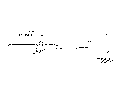
[0012] FIG. 1 is a top plan view of an embodiment of the catheter of
the present invention.
[0013] FIG. 2A is a side cross-sectional view of the catheter of FIG.
1, including a junction of
the catheter body and an intermediate section taken along a first diameter.
[0014] FIG. 2B is a side cross-sectional view of the catheter of FIG.
1, including a junction of
the catheter body and an intermediate section taken along a second diameter
generally
perpendicular to the first diameter.
[0015] FIG. 2C is an end cross-sectional view of the embodiment of
FIGS. 2A and 2C, taken
alone line C--C.
[0016] FIG. 3 is an exploded view of an embodiment of a distal tip
section of the catheter of
the present invention, including a tip electrode and a 2-D thin-film pressure
sensor.
[0017] FIG. 4A is side cross-sectional view of the distal tip section
of FIG. 3, as assembled,
taken generally along a first diameter.
[0018] FIG. 4B is a side cross-sectional view of the distal tip section of
FIG. 3, as assembled,
taken generally along a second diameter.
-4-
CA 02748962 2011-08-15
1
[0019] FIG. 4c is an end cross-sectional view of the distal tip
section of FIGS. 4A and 4B,
taken along line C--C.
[0020] FIG. 5 is an exploded view of an embodiment of a 2-D pressure
sensor, without the
pressure-sensitive material.
[0021] FIG. 6 is an isometric view of the 2-D pressure sensor of FIG.
5, as assembled.
[0022] FIG. 6A is a cross-sectional view of the 2-D pressure of FIG.
6, taken along line A--A.
[0023] FIG. 7 is top plan view of an embodiment of a 2-D pressure
sensor, with a first support
member and a second support member before assembly.
[0024] FIG. 8 is a top plan view of the 2-D pressure sensor of FIG.
7, as assembled.
[0025] FIG. 9 is an alternate embodiment of a catheter with a 2-D
pressure sensor, with a
catheter body joined to a distal tip section.
[0026] FIG. 9A is a side cross-sectional view of the catheter of FIG.
9.
[0027] FIG. 10 is an exploded view of an embodiment of a distal tip section
of the catheter of
the present invention, including a 3-D pressure sensor.
[0028] FIG. 11 is side cross-sectional view of the distal tip section
of FIG. 10, as assembled.
[0029] FIG. 12 is an exploded view of the 3-D pressure sensor,
without the pressure-sensitive
material.
DETAILED DESCRIPTION OF THE INVENTION
[0030] FIG. 1 illustrates an embodiment of a catheter 10 with force-
sensing capabilities at a
distal tip. The catheter has an elongated catheter body 12 with proximal and
distal ends, an
intermediate deflectable section 14 at the distal end of the catheter body 12,
and a distal tip section
15 adapted for mapping, ablation and detecting forces applied to a tip
electrode 17 such as when
the tip electrode is in contact with tissue wall 19. The catheter also
includes a control handle 16
at the proximal end of the catheter body 12 where the control handle is
adapted for controlling bi-
-5-
CA 02748962 2011-08-15
1
directional deflection of the intermediate section 14. The control handle 16
may further serve as a
conduit to a controller 11 adapted to send, receive and process electrical
input and output signals
to and from the distal tip section 15 for mapping, ablation and/or force-
sensing, such as by means
of a microprocessor 13 applying program algorithms with force-sensing
solutions. In accordance
with the present invention, such signals include signals from a thin-film
pressure sensor with trace
electrode intersections for directly measuring contact force, including
detecting and measuring
radial and/or axial contact forces on the tip electrode, whereby the
controller and microprocessor
are adapted to processes such signals in computing a contact force vector.
Amplifiers and data
acquisition (DAQ) equipment may be provided within the control handle to
convert measurements
from the thin film pressure sensor into usable signals. These equipment may
also be provided
externally to the catheter such as in a separate junction box or in a catheter
navigation system
designed to visualise real-time calculated position and orientation of a
catheter within the patient's
heart, such as the CARTO navigation system manufactured by Biosense Webster,
Inc.
100311 With reference to FIGS. 2A and 2B, the catheter body 12
comprises an elongated
tubular construction having a single, axial or central lumen 18. The catheter
body 12 is flexible,
i.e., bendable, but substantially non-compressible along its length. The
catheter body 12 can be of
any suitable construction and made of any suitable material. A presently
preferred construction
comprises an outer wall 20 made of polyurethane or PEBAX. The outer wall 20
comprises an
imbedded braided mesh of stainless steel or the like to increase torsional
stiffness of the catheter
body 12 so that, when the control handle 16 is rotated, the intermediate
section 14 of the
catheter10 will rotate in a corresponding manner.
100321 The outer diameter of the catheter body 12 is not critical,
but is preferably no more
than about 8 french, more preferably 7 french. Likewise the thickness of the
outer wall 20 is not
critical, but is thin enough so that the central lumen 18 can accommodate
wires, cables, tubings
and the like. If desired, the inner surface of the outer wall 20 is lined with
a stiffening tube 22 to
-6-
CA 02748962 2011-08-15
1
provide improved torsional stability. In a disclosed embodiment, the catheter
has an outer wall 20
with an outer diameter of from about 0.090 inch to about 0.100 inch and an
inner diameter of from
about 0.061 inch to about 0.065 inch. Distal ends of the stiffening tube 22
and the outer wall 20
are fixedly attached to each other by adhesive bonds therebetween near the
distal end and
proximal ends of the catheter body 12.
[00331 Components that extend between the control handle 16 and the
deflectable section 14
pass through the central lumen 18 of the catheter body 12. These components
include lead wire
40 for the tip electrode 17, lead wires 42 for the pressure sensor in the tip
section, an irrigation
tubing 38 for delivering fluids to the tip electrode 17, a cable 48 for an
electromagnetic position
location sensor 19 housed in the tip electrode 17, thermocouple wires 41, 43
for sensing
temperature of the tip electrode, and a pair of puller wires 44 for
bidirectional deflection of the
intermediate section 14.
[0034] Also illustrated in FIGS. 2A, 2B and 2C is an embodiment of the
deflectable
intermediate section 14 which comprises a shorter section of tubing 19. The
tubing also has a
braided mesh construction but with multiple off-axis lumens, for example
first, second, third and
fourth lumens 30, 31, 32 and 33. In the illustrated embodiment, each of
diametrically opposing
second and fourth lumens 31 and 33 carries one puller wire 44 for bi-
directional deflection. The
first lumen 30 carries the lead wires 40 and 42, and the thermocouple wires
41, 43. The third
lumen 32 carries the irrigation tubing 38 and the sensor cable 48 and
additional lead wires 42.
[0035] The tubing 19 of the intermediate section 14 is made of a
suitable non-toxic material
that is more flexible than the catheter body 12. A suitable material for the
tubing 19 is braided
polyurethane, i.e., polyurethane or PEBAX with an embedded mesh of braided
stainless steel or
the like. The size of each lumen is not critical so long it each lumen is
sufficiently sized to house
the respective components extending therethrough.
-7-
1
[0036] A means for attaching the catheter body 12 to the tubing 19 of
the intermediate section
14 is illustrated in FIGS. 2A and 2B. The proximal end of the intermediate
section 14 comprises
an outer circumferential notch that receives an inner surface of the outer
wall 20 of the catheter
body 12. The intermediate section 14 and catheter body 12 are attached by glue
or the like.
[0037] If desired, a spacer (not shown) can be located within the
catheter body between the
distal end of the stiffening tube (if provided) and the proximal end of the
intermediate section.
The spacer provides a transition in flexibility at the junction of the
catheter body and intermediate
section, which allows this junction to bend smoothly without folding or
kinking. A catheter
having such a spacer is described in U.S. Pat. No. 5,964,757.
[0038] Each puller wire 44 is preferably coated with Teflon® The
puller wires 44 can be
made of any suitable metal, such as stainless steel or Nitinol and the Teflon
coating imparts
lubricity to the puller wire. The puller wire preferably has a diameter
ranging from about 0.006 to
about 0.010 inch. As shown in FIGS. 2B and 2C, a portion of each puller wire
44 in the catheter
body 12 passes through a compression coil 35 in surrounding relation to its
puller wire 44. The
compression coil 35 extends from the proximal end of the catheter body 12 to
the proximal end of
the intermediate section 14. The compression coil 35 is made of any suitable
metal, preferably
stainless steel, and is tightly wound on itself to provide flexibility, i.e.,
bending, but to resist
compression. The inner diameter of the compression coil is slightly larger
than the diameter of the
puller wire 44. Within the catheter body 12, the outer surface of the
compression coil 35 is also
covered by a flexible, non-conductive sheath 39, e.g., made of polyimide
tubing.
[0039] Proximal ends of the puller wires 44 are anchored in the
control handle 16. Distal ends
of the puller wires are anchored near the distal end of the intermediate
section 14 as shown in
FIG. 4B. The distal end of each puller wire is provided with a T-shaped anchor
47 that includes a
short piece of tubular stainless steel, e.g., hypodermic stock, that is fitted
over and crimped onto
-8-
CA 2748962 2017-12-07
CA 02748962 2011-08-15
1
the distal end of the puller wire. The tubular stainless steel is fixed, e.g.,
by welding, to a cross-
piece formed of stainless steel ribbon or the like. The cross-piece is fixedly
secured to the outer
wall of the tubing 19 to anchor the distal end of each puller wire. A first
puller wire passes
through the second lumen 31 and a second puller wire passes through the fourth
lumen 33 of the
deflectable intermediate section 14. Separate and independent longitudinal
movement of the
deflection wires 44 relative to the catheter body 12, which results in
deflection of the intermediate
section 14 and hence steering of the tip section 15 is accomplished by
suitable manipulation of a
deflection member 37 (FIG. 1).
[0040] With reference to FIGS. 3, 4A and 4B, at the distal end of the
intermediate section 14
is the tip section 15 that includes the tip electrode 17, the thin film
pressure sensor 60 and a stop
member 62 between the pressure sensor and the tubing 19. In the illustrated
embodiment, the
pressure sensor has a flattened annular or ring shape that is radially
symmetrical. It has a thin
planar construction and a circular cross-section having an outer diameter D1
and an inner
diameter D2 defining a central lumen 64. The outer diameter D1 of the sensor
is similar to or
slightly less than an outer diameter of the tubing 19 and the tip electrode 17
such that a smooth
and atraumatic profile is presented between the tubing 19 and the tip
electrode 17 at or near the
distal end of the catheter. The inner diameter D2 is sufficiently large to
allow components, such
as lead wires, irrigation tubing, thermocouple wires and/or sensor cable, to
extend through it.
[0041] As shown in the embodiment of FIGS. 5 and 6, the thin-film
pressure sensor 60
includes first trace electrodes 66 and second trace electrodes 68, each of
which is formed on a
respective thin, flexible supporting sheet 63 and 65, for example a polyester
film such as MYLAR
or KAPTON. The first electrodes 66 are formed on a distal surface of the first
sheet 63 and the
second electrodes are formed on a proximal surface of the second sheet 65.
Each electrode has a
thin coating 67 of a pressure-sensitive resistive material or ink such as
molydenum disulphide that
is spread over the support sheets. The sheets 63 and 65 overlie each other
such that the first and
-9-
1
second electrodes 66 and 68 face, overlie and cross each other, preferably at
an angle, e.g., a right
angle, to create a grid pattern of "intersections" X where each electrode of a
pair is separated from
the other electrode of the pair (and any other adjacent trace electrode(s)) by
the pressure-sensitive
resistive material (see FIG. 6A). An "intersecting" configuration as used
herein describes a
configuration where electrodes of an overlying pair cross each other but are
not in contact with
each other. Instead, the electrodes of a crossing pair at their closest
proximity to each other
remain separated by a gap or space 70 that is occupied by the pressure-
sensitive material so that a
change in the electrical resistivity in the pressure-sensitive material is
detected by the crossing
electrode pair. Thus, in the absence of an external force, the pressure-
sensitive material 67
between the crossing electrodes provides a high resistance, and when an
external force is applied
and pressure on opposite sides of a crossing electrode pair changes, the
resistance of the material
changes in response thereto. With multiple pairs of crossing first and second
electrodes, one
electrode of each pair is driven and the other electrode of the pair is sensed
such that measurement
of the resistance between the driven and sensed electrodes for each crossing
pair provides an
output representative of the force applied to the pressure sensor. It is
understood that triangulation
processing can be applied by the microprocessor 13 to determine a force vector
with directional
(e.g., radial and axial) components.
[0042] The pressure sensor 60 has a thickness ranging between 0.05 mm and
0.5 mm and
preferably is about 0.1 mm, and can be adapted and configured to take 1600
measurements per square
inch ranging from 0.1 psi to 25 ksi (with forces as low as 62.5 lb). Suitable
thin-film pressure sensors
and systems are available from Tekscan, Inc. of South Boston, Mass., USA and
are described in U.S.
Patent Nos. 4,856,993 and 6,964,205. These sensors and systems can measure
static and dynamic
events and because they are extremely thin and flexible they can measure
critical surface interface
-10-
CA 2748962 2017-12-07
CA 02748962 2011-08-15
1
pressure with minimal interference and provide highly accurate pressure
readings that can be
processed by a processor and displayed on a monitor.
[0043] Each intersection or crossing pair is thus characterized by a
variable resistance which
is a function of the pressure applied thereto. It is understood that each
electrode has an exposed
terminal that is connected to a respective lead wire 42 that extends through
the central lumen 64
of the pressure sensor and proximally through the catheter 10 to a suitable
circuitry (not shown).
The circuitry operates by sequentially scanning each crossing electrode pair
of intersection to
measure the resistance of the pressure-sensitive coating at that intersection
to provide an
indication of the force applied to the pressure sensor at that intersection.
[0044] In the illustrated embodiment of the catheter in FIGS. 5 and
6, the pressure sensor has
a minimum of three electrode intersections with a load range and sensistivity
appropriately
selected for the expected range of contact forces. The minimum of three
electrode intersections
enables triangulation of the forces and/or pressures in determining force
vectors with directional
and angle information such as axial and/or radial components. As shown, each
crossing is equally
spaced from the outer and inner diameters and the crossings are equally spaced
from each other
around the longitudinal axis of the tip section, for example, at 0, 120 and
240 radial degrees. As
understood by one of ordinary skill in the art, there can be any number of
crossings as desired or
appropriate depending on the resolution desired.
[0045] In the alternate embodiment of FIGS. 7 and 8, the sensor
includes at least two parallel
driven electrodes 66 and two parallel sensed electrodes 68 that are arranged
to provide four
crossings X that are equi-distanced from each other so that the four crossings
form four corners of
a square configuration where the crossings are radially symmetrical around the
longitudinal axis
of the tip section. It is understood that any number of electrodes can be
arranged in any suitable
pattern and that electrodes can be consistently driven or consistently sensed,
or that with an
appropriate circuitry the same electrode(s) can alternate between being driven
and being sensed,
-11-
CA 02748962 2011-08-15
1
as needed. Indeed, as understood by one of ordinary skill in the art, the
present system with the
appropriate circuitry can employ any suitable number of electrodes arranged as
driven and/or
sensed electrodes.
[0046] In the illustrated embodiment of FIGS. 3, 4A and 4B, the stop
member 62 is proximal
of the pressure sensor 60 and acts as a rigid foundation for the pressure
sensor. The member is
disc shaped having a thicker planar construction and a circular cross-section
having an outer
diameter D3 and an inner diameter D4 defining a central lumen 70. The outer
diameter is similar
to or slightly less than an outer diameter of the tubing 19 and the tip
electrode 17 such that a
smooth and atraumatic profile is presented between the tubing 19 and the tip
electrode 17 at or
near the distal end of the catheter. The inner diameter is sufficiently large
to allow the
aforementioned components to extend between the tubing 19 and the tip
electrode 17. The
thickness of the stop member between a distal planar surface and a proximal
planar surface ranges
between about 0.4 mm and 0.5 mm. The material of the member can be any
material that is
sufficiently rigid under forces ranging between about 5 and 150 gf.
[0047] Distal surface of the stop member and/or proximal surface of
the tip electrode are
generally planar and perpendicular to the longitudinal axis of the distal tip
section so that the
pressure sensor sandwiched therebetween detects primarily axial forces acting
on the tip electrode,
although measurement of radial components is also possible through analysis of
differences in the
measured axial components of the force acting on the tip electrode. These
surfaces can include
raised formations equally spaced about the longitudinal axis which act to
localize forces to the
grid intersections of the pressure sensor. In the illustrated embodiment, the
proximal surface of
the tip electrode has a plurality of circular mounds 74 which plurality is
equal to the plurality of
electrode intersections X in the pressure sensor. As illustrated, each mound
is situated in axial
alignment with a respective electrode intersections for localizing forces
applied to the tip electrode
to the intersections X of the pressure sensor.
-12-
CA 02748962 2011-08-15
1
[0048] It is understood that the while the words "pressure" and
"force" have different
technical meaning and units of measurement, these terms are used
interchangeably herein as the
pressure sensor is configured with the electrode intersections at various
intervals (with pre-
determined area of coverage for each intersection) allowing for the signals to
be displayed as
pressure. Thus, the representation of any particular area depends on the
plurality of intersections
in that area, with a greater plurality providing a greater representation and
a lesser plurality
providing a lesser representation. In that regard, where the raised formations
of the stop member
and/or the proximal end of the tip electrode have known surface areas, results
of measurements
taken can be determined in terms of force values. As such, multiple electrode
junctions are
advantageously utilized to determine force components and by triangulation,
force vectors.
[0049] As best seen in FIG. 4C, distal ends of the tip electrode lead
wire 40 and the
thermocouple wires 41 and 45 are anchored in blind holes 120 and 122,
respectively, formed in
the proximal end of the tip electrode 17. The electromagnetic position sensor
21 is received in the
blind hole 124. The irrigation tubing 38 extends into irrigation passage 126
formed at the
proximal end of the tip electrode. The passage 126 is in communication with
radial transverse
branches 130 to allow fluid delivered by the irrigation tubing 38 to exit to
outside of the tip
electrode via a plurality of radial ports 132.
[0050] As described, the catheter 10 is suited for bi-directional
deflection with user control of
the puller wires via the deflection member on the control handle. However, an
embodiment of a
catheter 10a adapted for use with a guiding sheath is illustrated in FIG. 9.
The catheter 10' is
without a multi-lumen tubing deflectable by puller wires. As shown, the distal
end of the catheter
body 12 is attached to the distal tip section 15, without an intermediate
portion in between, with
the components passing between the central lumen 18 of the catheter body 12
and the central
lumens of the stop member 62 and the pressure sensor 60.
-13-
CA 02748962 2011-08-15
. .
1
[0051] In another alternate embodiment of a catheter 10b illustrated
in FIGS. 10, 11 and 12, in
which similar components have similar reference numerals, a distal tip section
15b includes a thin
film pressure sensor 60b that has a 3-D shape, e.g., a conical shape, such
that each location on
proximal and distal surface has a surface vector with an axial and radial
component. In this
embodiment, the pressure sensor includes four electrode intersections X, each
equally spaced
from the outer diameter and the inner diameter of the conical shape, and
equally spaced from each
other around conical shape, for example, at 0, 90, 180 and 270 degrees around
the longitudinal
axis of the distal tip section. First electrodes 66b are supported on a
proximal surface of a first or
distal thin, flexible supporting sheet 63b that has a conical shape. Second
electrodes 68b are
supported on a distal surface of a second or proximal thin, flexible
supporting sheet 65b that also
has a conical shape and is slightly larger than the first supporting sheet so
that the second sheet
can accommodate and surround both the first sheet and the pressure-sensitive
material 67b applied
to the distal surface of the first sheet and the proximal surface of the
second sheet to form the
pressure sensor. As illustrated, first and second electrodes are arranged on
their respective sheet
to form intersections X at radially-symmetrical locations around central lumen
64b.
Corresponding holes 90 are provided in the proximal supporting sheet 65b so
that lead wires 42b
for both the first and second electrodes can extend proximally toward stop
member 62b.
[0052] The conical shape of the pressure sensor 60b generally matches and
corresponds with a
concave conical distal surface 110 of stop member 62b and a convex conical
proximal surface 112
of tip electrode 17b such that the pressure sensor is nested between the tip
electrode and the stop
member with raised formations, e.g., raised ring-shape mounds 74b, axially
aligned with the
electrode intersections. The central lumen 64b allows components to extend
through the pressure
sensor 60b, which in the instant embodiment, includes a protrusion having a
ball member 92 and a
neck 94 that are aligned with the longitudinal axis of the tip electrode and
extend proximally from
a main body of the tip electrode 17b. The ball member is received in and
interlocks with a socket
-14-
CA 02748962 2011-08-15
1
formed 96 in the distal end of stop member. The ball and socket coupling
provides additional
axial tensile strength by providing a mechanical interference between the tip
electrode and the
stop member. In that regard, any staking interference such as with a flattened
end or post can be
used. Moreover, the ball socket coupling allows for movement of the tip
electrode 17b in more
dimensions relative to the stop member 62b for increased sensitivity and
higher resolution in
detecting applied forces with radial and axial components. The thickness of
the stop member 62b
between its distal and proximal surfaces ranges between about 1.0 mm and 6.0
mm, and
preferably about 3.0 mm. The proximal surface of the stop member is notched
with an inner
circumference 100 to receive an outer circumference 102 of a notched distal
end of tubing 19b of
the intermediate section 14. Lead wires 42b for the pressure sensor electrodes
66b and 68b extend
through axial passages 108 provided in the stop member 62b that are axially
aligned with the
electrode intersections X. Tip electrode lead wire 40b and thermocouple wires
41b and 45b pass
through a central passage 112 that connects inner notched proximal end 100 of
the stop member
with the concave distal surface of the stop member. Tip electrode lead wire 40
and thermocouple
wires 41, 45 are anchored in their respective blind holes formed in the convex
proximal end of the
tip electrode.
[0053] In any of the foregoing embodiments, the pressure sensor can
be bonded to the tip
electrode and/or the stop member with any adhesive adapted to withstanding
axial loads of a
minimum of about 15 Newtons, such as when the tip electrode comes in contact
with tissue during
mapping and ablation. Suitable adhesives, including epoxy, polyurethane and
the like, should be
rigid enough to transfer force to the pressure sensor without loss but should
be sufficiently elastic
so as not to cause plastic deformation in the intended range of forces to act
on the catheter.
[0054] A flexible protective cover, for example, a thin flexible, single-
lumen extrusion or
tubing 104, can be mounted over the pressure sensor and the stop member to
insulate these
components and their interior from patient contact.
-15-
CA 02748962 2011-08-15
1
[0055] For a catheter with N number grid intersections, the sum of
the axial components of
each measured force Fi,axiai through FN,axiai should be equal to the axial
component of the force
applied to the tip electrode. For conical shaped sensors, the sum of the
radial components of each
measured force Fi,radiai through FN,radiai should be equal to the radial
component of the force
applied to the tip electrode. In the case of a purely axial applied load, the
axial component of each
measured force would be equivalent to (UN) time the magnitude of the applied
force and the sum
of the radial components of measured forces would equal zero. As the applied
force changes from
purely axial, changes in the components of each measure force allows for
determination of the
magnitude and direction of the applied force.
[0056] The illustrated embodiment of the catheter 10b is adapted for
bi-directional deflection
by means of a pair of puller wires that extend through two opposite lumens of
the tubing 19b of
the intermediate section 14b. However, it is understood that the catheter 10b
can also be adapted
for use with a guiding sheath where a catheter body is joined to the distal
tip section without an
intermediate section.
[0057] As far as pressure vs force goes, I believe both terminologies
are correct. These
sensors typically place the junctions at various intervals (pre- determined
area of coverage for
each junction) allowing for the recorded signal to be displayed as pressure.
In the disclosed
concept, the number of junction is more limited and therefore each is less
representative of the
area that they cover. However, as mentioned in paragraph 0047, the stop member
and proximal
end of the tip electrode may have raised surfaces (ideally of know surface
areas). These surface
will allow the measurements to be taken for known areas allowing the results
to be determined in
terms of force values. Because of this, multiple pressure sensors are being
utilized to determine
force components and by triangulation, force vectors.
[0058] The preceding description has been presented with reference to
certain exemplary
embodiments of the invention. The drawings shown wherein are not necessarily
to scale.
-16-
CA 02748962 2011-08-15
1
Workers skilled in the art and technology to which this invention pertains
will appreciate that
alterations and changes to the described structure may be practiced without
meaningfully
departing from the principal, spirit and scope of this invention. It is
understood that the drawings
are not necessarily to scale. Accordingly, the foregoing description should
not be read as
pertaining only to the precise structures described and illustrated in the
accompanying drawings.
Rather, it should be read as consistent with and as support for the following
claims which are to
have their fullest and fairest scope.
15
25
-17-