Note: Descriptions are shown in the official language in which they were submitted.
CA 02749554 2016-05-20
SYNTHETIC CHORD
INTRODUCTION
The mitral valve is composed of two leaflets attached to the mitral valve
annulus, which are supported at the free edge by chordae tendinae (chords)
attached to the inside wall of the left ventricle and to the papillary
muscles. However,
sometimes one or both of the valve leaflets become loose, due to loosening or
failure of one or more of these chords. The valve then prolapses, and the seal
that it
normally provides between the left atrium and left ventricle becomes
compromised,
causing the blood to flow back into the left atrium during systole.
A variety of methods have been described for placement of artificial chordae
tendineae to correct mitral valve leaflet prolapse and treat diseased mitral
valve
chordae tendineae. However, there are many technical challenges in this
surgical
procedure, especially when performed with minimally invasive techniques. The
most
common method of repairing the valves is to create synthetic chordae tendineae
from polytetrafluoroethylene (PFTE), which are sutured into place between the
papillary muscle of the heart wall and the mitral valve leaflets. Cardiac
surgeons
usually are required to perform the time-consuming process of measuring and
cutting the necessary length of synthetic chordae tendineae material during
the
surgical procedure after they have measured the dimensions of the patient's
heart
valves. In addition, anchoring the synthetic chordae tendineae in the
papillary
muscle and securing the sutures through the leaflets is often technically
difficult in
minimally invasive procedures, because of limitations in using 2-dimensional
video
for viewing the surgical field, limited exposure of the surgical field, and
limited
degrees of freedom using standard thoracoscopic instrumentation.
CA 02749554 2011-07-13
WO 2010/083103 PCT/US2010/020464
Therefore, there is considerable interest in the development of new
techniques for use in both open and minimally invasive procedures that address
the
problems of accurately and efficiently securing the valve leaflets during
cardiac
surgery.
SUMMARY
Synthetic chord devices and methods for using the same for connecting
tissues are provided. Aspects of the synthetic chord devices of the invention
include
a flexible cord having attachment elements at both a first and second end,
wherein
each attachment element includes a piercing member coupled to a securing
io
member, where the securing member attaches the flexible cord to a first
tissue. At
least a portion of the flexible cord is configured to be secured to a second
tissue.
Aspects of the invention also include sets of the synthetic chord devices,
e.g., of
different sizes. The devices and methods of the invention find use in a
variety of
applications, such as in applications in which it is desired to repair a heart
valve.
BRIEF DESCRIPTION OF THE FIGURES
Figures 1A and B provide a view of the device in accordance with an
embodiment of the invention.
Figure 2 provides a schematic view of the normal left side of the heart.
Figure 3 provides a schematic view of the left side of the heart demonstrating
a ruptured chorda tendinea of the mitral valve.
Figures 4A and B provide a schematic view of the left side of the heart after
repair of the ruptured chorda tendinea of the mitral valve with embodiments of
the
synthetic chord device of the subject invention.
Figures 5A and 5B provide another view of the device in accordance with an
embodiment of the invention.
Figure 6 provides a schematic view of the heart after repair of both the
ruptured chordae tendineae of the mitral valve and tricuspid valves with
embodiments of the synthetic chord device of the subject invention.
2
CA 02749554 2011-07-13
WO 2010/083103 PCT/US2010/020464
DETAILED DESCRIPTION
Synthetic chord devices and methods for using the same for connecting
tissues are provided. Aspects of the synthetic chord devices include a
flexible cord
having an attachment element at both a first and a second end, wherein each
attachment element includes a piercing member coupled to a securing member
that
attaches the flexible cord to a first tissue. At least a portion of the
flexible cord is
configured to be secured to a second tissue. Aspects of the invention also
include
sets of the synthetic chord devices, e.g., of different sizes. The devices and
methods
of the invention find use in a variety of applications, such as in
applications in which
io it is desired to repair a heart valve.
Before the present invention is described in greater detail, it is to be
understood that this invention is not limited to particular embodiments
described, as
such may, of course, vary. It is also to be understood that the terminology
used
is herein is for the purpose of describing particular embodiments only, and
is not
intended to be limiting, since the scope of the present invention will be
limited only
by the appended claims.
Where a range of values is provided, it is understood that each intervening
value, to the tenth of the unit of the lower limit unless the context clearly
dictates
20 otherwise, between the upper and lower limit of that range and any other
stated or
intervening value in that stated range, is encompassed within the invention.
The
upper and lower limits of these smaller ranges may independently be included
in the
smaller ranges and are also encompassed within the invention, subject to any
specifically excluded limit in the stated range. Where the stated range
includes one
25 .. or both of the limits, ranges excluding either or both of those included
limits are also
included in the invention.
Certain ranges are presented herein with numerical values being preceded by
the term "about." The term "about" is used herein to provide literal support
for the
exact number that it precedes, as well as a number that is near to or
approximately
30 the number that the term precedes. In determining whether a number is near
to or
approximately a specifically recited number, the near or approximating
unrecited
3
CA 02749554 2016-05-20
number may be a number which, in the context in which it is presented,
provides the
substantial equivalent of the specifically recited number.
Unless defined otherwise, all technical and scientific terms used herein have
the same meaning as commonly understood by one of ordinary skill in the art to
which this invention belongs. Although any methods and materials similar or
equivalent to those described herein can also be used in the practice or
testing of
the present invention, representative illustrative methods and materials are
now
described.
15
Further, the dates of publication provided may be different from the actual
publication dates which may need to be independently confirmed.
It is noted that, as used herein and in the appended claims, the singular
forms
"a", "an", and "the" include plural referents unless the context clearly
dictates
otherwise. It is further noted that the claims may be drafted to exclude any
optional
element. As such, this statement is intended to serve as antecedent basis for
use of
such exclusive terminology as "solely," "only" and the like in connection with
the
recitation of claim elements, or use of a "negative" limitation.
As will be apparent to those of skill in the art upon reading this disclosure,
each of the individual embodiments described and illustrated herein has
discrete
components and features which may be readily separated from or combined with
the
features of any of the other several embodiments without departing from the
scope
of the present invention. Any recited method can be carried out in the order
of events recited or in any other order which is logically possible.
4
CA 02749554 2011-07-13
WO 2010/083103 PCT/US2010/020464
DEVICES
Synthetic chord devices according to certain embodiments of the invention
are devices that are configured to connect or align tissues, or connect tissue
and a
prosthesis, or a combination thereof. The subject devices and methods can be
used
in endovascular, minimally invasive surgical, open surgical, or other
interventional
procedures. As such, devices of the invention can be configured to secure a
valve
leaflet, such as a mitral valve leaflet or tricuspid valve leaflet, to a
papillary muscle.
Embodiments of the synthetic chord device include a flexible cord having an
io attachment element at both a first and a second end, wherein each
attachment
element includes a piercing member coupled to a securing member that attaches
the
flexible cord to a first tissue. At least a portion of the flexible cord can
be configured
to be secured to a second tissue.
A synthetic chord device of the subject invention is a synthetic, or
artificial,
is flexible cord which has attachment elements at both ends of the cord,
for attaching
the cord to a tissue. In some embodiments, the flexible cord is configured to
be
attached to a prosthesis, or to a device that substitutes for or supplements a
missing
or defective part of the body, e.g., a synthetic cardiac valve, or a porcine
valve. In
some embodiments, a synthetic chord device is configured to be used as a
synthetic
20 chorda tendinea for use in repair of a cardiac valve, e.g., the mitral
valve.
The flexible cord element of the subject invention is a flexible elongated
structure having a first end and a second end, constructed of a material
suitable for
use in the body that can be used in the methods of the subject invention,
e.g.,
attaching a valve leaflet to the underlying cardiac tissue. The flexible cord
element
25 has a length suitable for extending from a first tissue to a second
tissue and back to
a first tissue, such that the flexible cord provides two segments, each
segment
secured to both the first and the second tissue.
For example, in certain
embodiments, each segment of the flexible cord would be equal to half of the
total
length of the flexible cord. In some embodiments, the flexible cord element
has a
30 length suitable for extending from a first tissue (e.g., a mitral valve
leaflet) to where it
is secured to a second tissue (e.g., a papillary muscle) and back to the first
tissue.
5
CA 02749554 2011-07-13
WO 2010/083103 PCT/US2010/020464
In this embodiment the length of the flexible cord may range from 8 mm to 60
mm,
such as from 16 mm to 48 mm, or 20 mm to 32 mm. In some embodiments, the first
or second end of the flexible cord can be secured to a prosthesis, or other
device
that substitutes for or supplements a missing or defective part of the body,
e.g., a
synthetic cardiac valve, or a porcine valve.
The flexible cord can be made of a variety of biocompatible polymeric
materials or metallic materials that combine flexibility, high strength, and
high fatigue
resistance. For example, the flexible cord can be formed using materials
including,
but not limited to: polytetrafluoroethene or polytetrafluoroethylene (PFTE),
including
io expanded polytetrafluoroethylene (e-PFTE), polyester (DacronTm), nylon,
polypropylene, polyethylene, high-density polyethylene (HDPE), polyurethane,
stainless steel, titanium, a nickel-titanium alloy, a nickel-cobalt alloy,
another cobalt
alloy, tantalum, and combinations or mixtures thereof. In some embodiments, an
antithrombotic component may be included in the chemical composition of a
is polymeric filament. In other embodiments, a flexible cord may be coated
with a
polymer that releases an anticoagulant and thereby reduces the risk of
thrombus
formation. In other embodiments, additional therapeutic agents or combinations
of
agents may be used, e.g., antibiotics and anti-inflammatory agents. In some
embodiments, the flexible cord can be maneuvered through a catheter.
20 The cross-sectional configuration of the flexible cord can be any
suitable
shape, such as round, oval, rectangular, square, etc. In some embodiments, the
flexible cord may have a flattened cross-sectional shape, such as a "ribbon"
shape.
In other embodiments, the flexible cord may be a combination of shapes, such
as for
example, a flexible cord which is round on two sides with a flat surface on
the
25 .. opposing two sides. In some embodiments the entire flexible cord has the
same
shape, and in other embodiments, at least a portion of the flexible cord may
have a
different shape, e.g., a ribbon configuration, or at least a portion of the
cord which is
flattened, or has a flat surface. In some embodiments, the greatest outer
diameter
of the flexible cord may range from 0.1 mm to 0.6 mm, such as from 0.149 mm to
30 0.4 mm, or 0.15 mm to 0.2 mm. In some embodiments, the entire flexible
cord has
the same diameter. In other embodiments, at least a portion of the cord has a
6
CA 02749554 2011-07-13
WO 2010/083103 PCT/US2010/020464
different diameter, e.g., a smaller diameter. In some embodiments, at least a
portion
of the cord may have both a different configuration and a different diameter,
e.g., a
portion of the cord may have a flat surface, where the portion of the cord
having a
flat surface has a largest outer diameter larger than the remainder of the
cord. In
some embodiments, the flexible cord does not comprise a knot.
A portion of the flexible cord between the first end and second ends is
configured to be secured to tissue, such as cardiac tissue located below a
cardiac
valve leaflet. In some embodiments, a portion of the flexible cord between the
first
end and second ends can be secured to a prosthesis, or other device that
io substitutes for or supplements a missing or defective part of the body.
The portion
of the flexible cord between the first end and the second end that is
configured to be
secured to tissue can have the same shape and diameter as the remainder of the
flexible cord, or in some embodiments it may have a different shape or
diameter as
the remainder of the flexible cord, as in the embodiments discussed above. For
is example, the portion of the cord between the first end and the second
end that is
configured to be attached to a second tissue may be flattened, or have a
smaller or
larger diameter.
The portion of the flexible cord between the first end and the second end that
is configured to be secured to tissue can further include a reinforcing
member. A
20 reinforcing member is an element which disperses the force of the
securing flexible
cord over a larger surface area. In some embodiments, the reinforcing member
can
be a pledget. Pledgets are generally buttressing or cushioning pads through
which a
suture or cord can be threaded, in order to prevent the suture strand or
flexible cord
from cutting into the tissue. A reinforcing member can be made of any suitable
25 biologically compatible, needle pierceable resilient material
sufficiently soft and
flexible to effectively prevent damage to the tissue, e.g., papillary muscle.
A
reinforcing member is further made of material strong enough to resist pull-
through
by the flexible cord or suture to which it is mounted. The reinforcing member
includes a top surface and a bottom surface, and can be configured in a
variety of
30 sizes and shapes, including rectangular, circular, elliptical, etc. For
example, in
certain embodiments the length of the reinforcing member may range from 1 mm
to
7
CA 02749554 2011-07-13
WO 2010/083103 PCT/US2010/020464
mm, such as from 2 mm to 8 mm, or 3 mm to 4 mm. The width of the reinforcing
member in some cases may range from 1 mm to 10 mm, such as from 2 mm to 8
mm, or 3 mm to 4 mm. In some embodiments, the thickness of the reinforcing
member may range from 0.1 mm to 2 mm, such as from 0.2 mm to 0.5 mm, or 0.3
5 mm
to 0.4 mm. The reinforcing elements may be fabricated of fabric, or felt,
including polytetrafluoroethylene and polyester felt,
polytetrafluoroethylene(PTFE),
expanded PTFE, polyester and the like. In some embodiments, an antithrombotic
component may be included in the chemical composition of the reinforcing
member.
In other embodiments, a reinforcing member may be coated with a polymer that
io
releases an anticoagulant and thereby reduces the risk of thrombus formation.
In
other embodiments, additional therapeutic agents or combinations of agents may
be
used, e.g., antibiotics and anti-inflammatory agents.
In addition, the reinforcing element can have at least one opening wherein the
flexible cord element may pass through. In other embodiments, the flexible
cord is
is
attached to the reinforcing element without passing through an opening, e.g.,
the
flexible cord has been pulled through with a needle. In some embodiments, the
reinforcing element is mounted such that it is substantially fixed in a
position on the
flexible cord. For example, the reinforcing element can be sewn, or glued, or
fused
in any suitable manner so that it is fixed in position on the flexible cord,
e.g., fixed in
position halfway between the first and second ends of the flexible cord, such
that the
reinforcing element divides the flexible cord into two segments of equal
length. In
other embodiments, the reinforcing element is mounted such that it is slidably
mounted on a flexible cord. By "slidably" is meant that the reinforcing
element is
attached to the flexible cord so that it is secure yet it is possible to move
the
reinforcing element along at least part of the length of the cord. For
example, a
flexible cord can have a reinforcing element (e.g., a pledget) initially
positioned
halfway between the first and second ends of the flexible cord. In using the
synthetic chord device, it may be desirable to move the reinforcing element to
a
position closer to the first end before securing the reinforcing element to a
tissue.
The synthetic chord devices further include attachment elements on both the
first end and the second end of a flexible cord. The attachment elements are
8
CA 02749554 2011-07-13
WO 2010/083103 PCT/US2010/020464
configured to attach a flexible cord, such as those described above, to a
tissue, e.g.,
a cardiac valve leaflet. An attachment element is an element which includes a
tissue
piercing member and a securing member. The attachment element may be
configured such that one or both of the tissue piercing members is attached to
the
securing member with a flexible member such as a suture. The attachment
element
may also be configured such that the tissue piercing member is directly
attached to
the securing member. One or both of the tissue piercing members may in some
embodiments be releaseably coupled to a securing member. In other embodiments,
the attachment element may be configured such that one or both of the tissue
io
piercing members is attached to a flexible member, such as a suture, which in
turn is
releaseably coupled to the securing member. The coupling between the flexible
member (and, thus, the tissue piercing member) and the securing member may be
configured to actuate closure of the securing member upon release of the
flexible
member (or piercing member), as discussed below. For example, the coupling may
is
hold a compression spring (which is positioned around a securing member) in a
compressed state to brace the securing member open and releaseably lock or
secure the securing member to the flexible member (or piercing member). In
some
embodiments, the attachment element can be secured to a prosthesis, or other
device that substitutes for or supplements a missing or defective part of the
body.
20 A
flexible member as discussed above, such as a suture or a wire, can be
formed from any suitable biocompatible material such as cotton, nylon,
polyester,
polypropylene, polyglycolic acid, polylactide, lactic acid, trimethlylene
carbonate,
polycaprolactone, or polydiaxanone or copolymers or homopolymers thereof, or a
metal alloy, such as nitinol or stainless steel, a polymeric material, or any
other
25
suitable material and equivalents thereof. The material may be non-stretchable
or
stretchable, and have various cross-sectional diameters. In some embodiments,
the
flexible member does not comprise a knot. The flexible members may have a
cross-
sectional diameter of 0.003 inch, for example. The diameter and length of the
flexible
member will vary depending on the specific application. The flexible members,
e.g.,
30 sutures, may be attached to the piercing members by crimping or swaging or
otherwise attaching the piercing member or needle onto the flexible member,
gluing
9
CA 02749554 2011-07-13
WO 2010/083103 PCT/US2010/020464
the flexible member to the piercing member or needle, or any other suitable
attachment method. Flexible members can also have various cross-sectional
shapes, such as round, oval, etc.
A piercing member, or penetrating member is any device that can be used in
a surgical, endovascular, or other interventional procedure that can be used
to
pierce through tissue, e.g., a needle. In some embodiments, the piercing
member
can also be used to pierce a prosthesis, e.g., synthetic valve. Piercing
members
that can be used in the subject devices include, but are not limited to, a
conventional
surgical needle, etc. The surgical needles useful in the devices of the
present
io invention include conventional cardiac surgical needles and equivalents
thereof.
Suitable surgical needles can be manufactured from stainless steel, a
stainless steel
alloy, or any other suitable material, such as a polymeric material. The
material can
also have special coatings and sharpening methods that facilitate atraumatic
tissue
penetration. The shapes and sizes of the surgical needles can vary with the
type and
is design of the needle. In some embodiments, the surgical needles have a
curved or
arced shape. In some embodiments, the needles may be permanently "swaged" or
attached to the suture material. In some embodiments, the suture may be
designed
to come off the suture with a sharp straight tug (e.g., "pop-offs").
Suitable lengths for the piercing members that are in the form of a needle can
20 range in some embodiments from 4 mm to 70 mm, such as from 9 mm to 65
mm, or
20 mm to 40 mm. The diameter of the piercing member may range in certain
embodiments from 0.05 mm to 0.6 mm, such as from 0.07 mm to 0.5 mm, or 0.1 mm
to 0.4 mm. In some embodiments, the diameter of at least a portion of a
piercing
member is greater than the diameter of an attached flexible member or attached
25 securing member, coupled so that the attached flexible member or
attached
securing member can easily be pulled through an opening formed in a tissue (or
other material) by the piercing member, e.g., needle. The distal end or tip of
the
piercing member can be rigid to facilitate penetration of tissue. The
remaining length
of the piercing member can be rigid or flexible to facilitate movement of the
piercing
30 member through the tissue or other material. The tips can have various
configurations and can, for example, have a piercing point, tapered point, or
have a
CA 02749554 2011-07-13
WO 2010/083103 PCT/US2010/020464
cutting or reverse cutting configuration for example, and have a shape such as
conical, tapered, or grounded to attain a three or four facet tip. Piercing
members
can have any suitable shape or radius of curvature. Piercing members can have
any suitable cross-sectional shape which may vary in different sections of the
.. needle, e.g., round, rectangular, etc. In some embodiments, the piercing
member
can also be integrally formed with the flexible member (e.g., both piercing
member
and flexible member formed of the same material).
The attachment elements of the subject devices also include a securing
member. A securing member is any device that can be used in a surgical,
io endovascular, or other interventional procedure that can be used to
secure a flexible
cord, or suture, e.g., an artificial mitral valve chorda tendinea. Suitable
material for
securing members can include shape memory materials, which are materials that
have a temperature induced phase change, e.g., a material that if deformed
when
cool, returns to its "undeformed", or original, shape when warmed. Suitable
material
is includes but is not limited to metals such as a nickel-titanium (NiTi)
alloy (e.g.,
nitinol), a nickel-cobalt alloy, another cobalt alloy, alloys of CuZnAl, a
thermoset
plastic, stainless steel, a suitable biocompatible shape-memory material, a
suitable
biocompatible superelastic material, combinations thereof, and any suitable
biocompatible shape memory alloy that can return to its undeformed, or
original
20 shape when warmed to body temperature, e.g., human body temperature. A
securing member can have any suitable configuration. In some embodiments, for
example, a securing member can have an anchor configuration, such that the arm
segments of the anchoring members are constructed of a biocompatible material
capable of being preset into an anchor shape. In another embodiment, a
securing
25 member can have a loop shape, such that the securing member is
constructed of a
biocompatible material capable of being preset into a loop shape. In some
embodiments, a securing member can have an umbrella configuration, such that
the
arm segments of the anchoring members are constructed of a biocompatible
material capable of being preset into an umbrella shape. The securing member
may
30 .. in other embodiments have various undeformed or deformed configurations
such as
11
CA 02749554 2011-07-13
WO 2010/083103 PCT/US2010/020464
a "parachute" configuration, an ellipse, a triangle, a square, a rectangle,
spiral,
conical, or other geometric shape, etc.
As discussed above, in some embodiments, the securing member may be
releaseably coupled to a tissue piercing member. In some embodiments, a
flexible
s member, such as a suture, may be provided between at least one of the tissue
piercing members and the securing member to facilitate threading the securing
member.
In some embodiments, the securing member may secure the flexible cord
without piercing the adjacent tissue, e.g., in the same manner as a surgical
knot
io prevents a suture from pulling back through a tissue. In other
embodiments, the
securing member may secure the flexible cord by at least partially piercing
the
adjacent tissue. In some embodiments, the securing member may do both.
In some embodiments, the securing member is a self-closing fastener, which
is any device that can be used in a surgical, endovascular, or other
interventional
is procedure that can secure a flexible cord to tissue or other material
(e.g., secure a
flexible cord to a cardiac valve leaflet). In some embodiments, the self-
closing
fasteners can be made of a shape memory material is meant a material that
exhibits
the shape memory effect, as discussed above. The shape memory alloy is
preferably selected with a transformation temperature suitable for use with a
stopped
20 heart condition where cold cardioplegia has been injected for temporary
paralysis of
the heart tissue (e.g., temperatures as low as 8-10 degrees Celsius). The
shape
memory alloy may also be heat activated, or a combination of heat activation
and
pseudoelastic properties may be used. Self-closing fasteners that can be used
in
the subject devices include, but are not limited to, nitinol clips, such as
the V60 U-
25 clip deviceTM (Medtronic Inc.) or any other preconfigured attachment
device, etc.
A self-closing fastener can be held in an "open" configuration by a delivery
mechanism that holds and retains the fastener in an open configuration. In
some
embodiments, a locking element can be included to connect the ends of the
securing
member when the securing member is in its closed position to prevent possible
30 opening of the securing member over time. The locking element can in some
embodiments be integrally formed with the securing member. In some
embodiments,
12
CA 02749554 2011-07-13
WO 2010/083103 PCT/US2010/020464
the self-closing fastener can include a release mechanism. Further details of
self-
closing fasteners that can be adapted for use with the present devices can be
found
in US Patent Nos. 6,913,607, 6,641,593, 6,613,059, 6,607,541, and 6,514,265.
As discussed above, a self-closing fastener can have any suitable
configuration, including but not limited to an anchor configuration, a loop
configuration, an "umbrella" or "parachute" configuration, an ellipse, a
triangle, a
square, a rectangle, spiral, conical, or other geometric shape, etc.
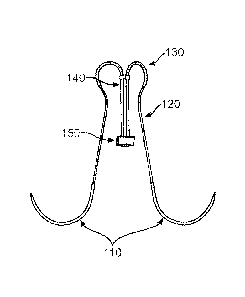
Figs. 1A and B provide a view of the device in accordance with an
embodiment of the invention. In Figure 1A, the synthetic chord device of the
subject
io invention is shown in an un-deployed state. The piercing member (e.g., a
needle) is
shown as element 110. The un-deployed self-closing fastener 130 is attached to
the
needle by flexible member (e.g., suture) 120. Flexible cord 140 is shown with
a
reinforcing member 150 (e.g., a pledget).
In Figure 1B, the synthetic chord device of the subject invention is shown in
a
is deployed state. The needle has been removed, and the self-closing
fastener has
been deployed, shown as element 135.
Figs. 5A and B provide a view of the device in accordance with another
embodiment of the invention, in which the self-closing fastener has an
"umbrella"
configuration. In Figure 5A, the synthetic chord device of the subject
invention is
20 shown in an un-deployed state. The un-deployed self-closing fastener 530
is
attached to a needle by flexible member (not shown). Flexible cord 540 is
shown
with reinforcing member 550.
In Figure 5B, the synthetic chord device of the subject invention is shown in
a
deployed state. The self-closing fastener has been deployed, shown as element
25 535.
METHODS
30 The subject devices find use in methods for fastening a tissue, such
as a
cardiac valve leaflet, to a second tissue, such as a papillary muscle, with a
flexible
13
CA 02749554 2011-07-13
WO 2010/083103 PCT/US2010/020464
cord (e.g., a synthetic mitral valve chorda tendinea). The subject devices
therefore
find use in methods in which a prolapsed cardiac valve leaflet, such as a
mitral valve
leaflet, is repaired. The subject devices can be used in an open surgical
procedure,
a minimally invasive surgical procedure, an endovascular procedure, or other
interventional procedure.
Methods for repair of a cardiac valve, such as a mitral valve, are discussed
below. When performing a conventional heart valve repair procedure, the
surgeon
makes incisions into the thoracic cavity and pericardium, and then into aorta
or
myocardium in order to have access to the damaged heart valve. The procedure
io may be an open procedure in which the sternum is opened and the ribs are
spread
with a conventional retractor, or a minimally invasive procedure wherein the
heart
and heart valve are accessed through minimally invasive openings in the
thoracic
cavity, such as through trocar cannulas or small incisions in the intercostal
spaces.
The heart may also be accessed through the lumen of an artery. The minimally
is invasive procedures can be viewed remotely using a camera and monitor,
or in
some cases directly.
Figure 2 depicts a schematic drawing of the left side of the heart. The aortic
arch 210, left atrium 215, and left ventricle 220 are shown, with the mitral
valve 250
located between the left ventricle and the left atrium. The chordae tendineae
are
20 shown as elements 240, attached to the leaflets of the mitral valve on
one end, and
the papillary muscle 230 in the left ventricle on the other end.
After exposure of the mitral valve and the subvalvular area, the desired
length
of the neochord, or flexible cord, is determined by measuring the distance
between
the prolapsed leaflet and the cardiac tissue located below the prolapsed
mitral valve
25 leaflet using methods that are well known in the art. The desired length
for the
flexible cord can be determined using any suitable measuring device, such as a
caliper, or a Mohr Suture Ruler DeviceTM (Geister, Tuttlingen, Germany). For
example, a caliper or sterile disposable flexible tape measure can be used to
assess
the correct length for the synthetic mitral valve chordae by measuring the
distance
30 between the tip of the papillary muscle and the edge of a non-prolapsing
segment of
14
CA 02749554 2011-07-13
WO 2010/083103 PCT/US2010/020464
the mitral valve leaflet. The measurement can also be confirmed by comparison
with pre-operative transesophageal echocardiography (TEE).
An illustration of a rupture, or breakage of one of the chorda tendinea which
can be repaired using the methods and devices of the subject invention is
shown in
Figure 3. The ruptured, or broken chorda tendinea is shown as element 350. The
leaflets of the mitral valve now no longer coapt, or close, and during
systole, blood
can flow from the left ventricle back into the left atrium, i.e., mitral
regurgitation.
The synthetic chord device having a flexible cord with the desired length, or
the closest to the desired length, is then selected from among a set of
synthetic
io chord devices. The set of synthetic chord devices can include two or
more flexible
cords of the same or of different lengths, such as three cords, or four cords,
etc.
The piercing member on the first end, e.g., a needle, is first advanced
through
the cardiac tissue below the prolapsed mitral valve leaflet, e.g., a papillary
muscle,
and pulled through until the reinforcing element, e.g., a pledget, is in
substantial
is contact with a surface of the papillary muscle. The needle is then
advanced through
the leaflet of the prolapsed mitral valve until the securing member, e.g., a
self-
closing fastener such as a nitinol clip, has passed through the leaflet.
The position of the prolapsed valve leaflet may be adjusted by coordinating
the tension of the cord and the location of the leaflet. For example, a
practitioner
20 (e.g., a doctor, surgeon, technician, etc.) may move the prolapsed valve
into a
correct (e.g., non-prolapsed) position by adjusting the position of the valve
leaflet
directly by pushing against the anchor attached to the valve leaflet (e.g.,
using the
fastener to push against the anchor and applying tension to the cord). The
valve
leaflet position may be adjusted in real-time in a beating heart (e.g., using
25 echocardiography). For example, the valve leaflet may be repositioned
while
monitoring mitral regurgitation (MR). Once any MR is reduced or eliminated,
the
valve leaflet is in the correct position.
Once the valve leaflet is positioned correctly, the attachment member can
then be deployed (e.g., the self-closing fastener deploys, or closes, for
example, as
30 shown in Figs. 1B and 5B). The piercing member on the second end, e.g., a
needle, is then advanced through the papillary muscle below the prolapsed
mitral
CA 02749554 2011-07-13
WO 2010/083103 PCT/US2010/020464
valve leaflet, thereby securing the pledget against the papillary muscle. The
second
tissue piercing member, e.g., needle, is then advanced through the same
prolapsed
mitral valve leaflet, until the second securing member has passed through the
leaflet. The second attachment element is then deployed, as discussed above.
It
should be noted that the number of synthetic chord devices required to secure
the
connecting tissues together may vary depending on the procedure and the
anatomy.
Figure 4A shows an embodiment of a repair of the ruptured chorda tendinea
with a synthetic chord device 470 of the subject invention. The flexible cord
460 is
attached to the mitral valve leaflet at both ends with securing members 490,
which in
io this embodiment have a ring shape. Flexible cord 460 is shown secured to
the
tissue below the mitral valve leaflet (e.g., the papillary muscle) with
reinforcing
member 480. After repair, the leaflets of the mitral valve 250 now coapt, or
close,
and blood can no longer flow from the left ventricle back into the left atrium
during
systole.
Figure 4B shows another embodiment of a repair of the ruptured chorda
tendinea with a synthetic chord device 470 of the subject invention. The
flexible
cord 460 is attached to the mitral valve leaflet at both ends with securing
members
495, which in this embodiment have a four-pronged "umbrella" shape, similar to
the
embodiment shown in Figures 5A and B. In this embodiment, the surface area of
the mitral valve leaflet which is contacted by the securing member is
increased.
Flexible cord 460 is again shown secured to the tissue below the mitral valve
leaflet
(e.g., the papillary muscle) with reinforcing member 480.
Figure 6 shows an embodiment of a repair of ruptured chordae tendineae of
both the mitral and tricuspid valves with synthetic chord devices of the
subject
invention. In this view, the left atrium is shown as element 605, the left
ventricle is
element 610; the right atrium is element 615, and the right ventricle is shown
as
element 620. The flexible cords 660 are attached to the mitral valve 650 or
tricuspid
valve 655 leaflet at both ends with securing members 690. Flexible cord 660 is
shown secured to the tissue below the valve leaflets (e.g., papillary muscle,
630)
with reinforcing members 680. After repair, the leaflets of the mitral valve
650 and
16
CA 02749554 2011-07-13
WO 2010/083103 PCT/US2010/020464
tricuspid valve 655 now coapt, or close, and blood can no longer flow from the
ventricles back into the atria during systole.
By this method, a prolapsed mitral valve leaflet can be repaired by securing
the leaflet to the papillary muscle below. Using the methods and devices of
the
subject invention, a mitral valve repair procedure can be successfully
completed
without the need for the time-consuming step of cutting the desired length of
synthetic cord while the patient is on the operating table, thereby decreasing
the
amount of time needed to place a patient on cardio-pulmonary bypass. In
addition,
the subject methods and devices obviate the need for tying sutures and
ensuring
io that the suture material does not become tangled, difficulties which are
exacerbated
by the small size of the tissues involved and the often limited field of the
operation.
Any appropriate prolapsed valve leaflet may be treated as described herein,
including mitral valve leaflets and tricuspid valve leaflets. Further, these
methods
may be performed using one or more catheters or using non-catheter surgical
is methods, or using a combination of catheter-type surgical methods and
non-catheter
type surgical methods. The methods of the subject invention may also be used
in
combination with other surgical procedures, e.g. replacement of a mitral valve
annulus, etc.
In some variations, the flexible cord may be advanced via one or more
20 catheters to the proximity of the prolapsed valve leaflet in an
anterograde approach
(e.g., from above the mitral valve). Alternatively, the flexible cord may be
advanced
via a retrograde approach (e.g., from below the mitral valve). In all of the
methods
described herein, the cardiac tissue located below the prolapsed valve (to
which one
of the anchors is secured) may be selected from the group consisting of a
papillary
25 muscle and a ventricular wall.
The subject methods also include the step of diagnosing a patient in need of
cardiac valve repair, e.g., mitral valve repair. Primary mitral regurgitation
is due to
any disease process that affects the mitral valve device itself. The causes of
primary
mitral regurgitation include myxomatous degeneration of the mitral valve,
infective
30 endocarditis, collagen vascular diseases (ie: SLE, Marfan's syndrome),
rheumatic
heart disease, ischemic heart disease/coronary artery disease, trauma. balloon
17
CA 02749554 2011-07-13
WO 2010/083103 PCT/US2010/020464
valvulotomy of the mitral valve, certain drugs (e.g. fenfluramine). If valve
leaflets are
prevented from fully coapting (i.e., closing) when the valve is closed, the
valve
leaflets will prolapse into the left atrium, which allows blood to flow from
the left
ventricle back into the left atrium, thereby causing mitral regurgitation.
The signs and symptoms associated with mitral regurgitation can include
symptoms of decompensated congestive heart failure (ie: shortness of breath,
pulmonary edema, orthopnea, paroxysmal nocturnal dyspnea), as well as symptoms
of low cardiac output (i.e., decreased exercise tolerance). Cardiovascular
collapse
with shock (cardiogenic shock) may be seen in individuals with acute mitral
io
regurgitation due to papillary muscle rupture or rupture of a chorda tendinea.
Individuals with chronic compensated mitral regurgitation may be asymptomatic,
with
a normal exercise tolerance and no evidence of heart failure. These
individuals
however may be sensitive to small shifts in their intravascular volume status,
and are
prone to develop volume overload (congestive heart failure).
Findings on clinical examination depend of the severity and duration of mitral
regurgitation. The mitral component of the first heart sound is usually soft
and is
followed by a pansystolic murmur which is high pitched and may radiate to the
axilla.
Patients may also have a third heart sound. Patients with mitral valve
prolapse often
have a mid-to-late systolic click and a late systolic murmur.
Diagnostic tests include an electrocardiogram (EKG), which may show
evidence of left atrial enlargement and left ventricular hypertrophy. Atrial
fibrillation
may also be noted on the EKG in individuals with chronic mitral regurgitation.
The
quantification of mitral regurgitation usually employs imaging studies such as
echocardiography or magnetic resonance angiography of the heart. The chest x-
ray
in patients with chronic mitral regurgitation is characterized by enlargement
of the
left atrium and the left ventricle. The pulmonary vascular markings are
typically
normal, since pulmonary venous pressures are usually not significantly
elevated.
An echocardiogram, or ultrasound, is commonly used to confirm the diagnosis of
mitral regurgitation. Color doppler flow on the transthoracic echocardiogram
(TTE)
will reveal a jet of blood flowing from the left ventricle into the left
atrium during
ventricular systole. Because of the difficulty in getting accurate images of
the left
18
CA 02749554 2011-07-13
WO 2010/083103 PCT/US2010/020464
atrium and the pulmonary veins on the transthoracic echocardiogram, a
transesophageal echocardiogram (TEE) may be necessary to determine the
severity
of the mitral regurgitation in some cases. The severity of mitral
regurgitation can be
quantified by the percentage of the left ventricular stroke volume that
regurgitates
into the left atrium (the regurgitant fraction). Other methods that can be
used to
assess the regurgitant fraction in mitral regurgitation include cardiac
catheterization,
fast CT scan, and cardiac MRI.
Indications for surgery for chronic mitral regurgitation include signs of left
ventricular dysfunction. These include an ejection fraction of less than 60
percent
io and a left ventricular end systolic dimension (LVESD) of greater than 45
mm.
The description of the present invention is provided herein in certain
instances with reference to a subject or patient. As used herein, the terms
"subject"
and "patient" refer to a living entity such as an animal. In certain
embodiments, the
animals are "mammals" or "mammalian," where these terms are used broadly to
is describe organisms which are within the class mammalia, including the
orders
carnivore (e.g., dogs and cats), rodentia (e.g., mice, guinea pigs, and rats),
lagomorpha (e.g., rabbits) and primates (e.g., humans, chimpanzees, and
monkeys).
In certain embodiments, the subjects, e.g., patients, are humans.
20 KITS
Also provided are kits that at least include the subject devices. The subject
kits at least include a synthetic chord device of the subject invention and
instructions
for how to use the synthetic chord device in a procedure.
In some embodiments, the kits can include a set of two or more synthetic
25 chord devices. In other embodiments, a set of synthetic chord devices
can include
at least three synthetic chord devices, e.g., four or more, five or more, six
or more,
etc.
In some embodiments, a set of synthetic chord devices includes two or more
synthetic chord devices in which at least two of the synthetic chord devices
have
30 flexible cords of different lengths. In other embodiments, the flexible
cord portions of
the synthetic chord devices are all of differing lengths. In some embodiments,
a set
19
CA 02749554 2016-05-20
of synthetic chord devices can have two or more synthetic chord devices in
which
the flexible cords are of the same length. A set of synthetic chord devices
can
therefore have two or more some synthetic chord devices in which some are of
the
same length, and some are of a different length. For example, in one
embodiment a
set of six synthetic chord devices can have two synthetic chord devices in
which the
flexible cord portion is 16 mm in length, which can provide two segments with
a
length of 8 mm; two synthetic chord devices in which the flexible cord portion
is 20
mm in length, which can provide two segments with a length of 10 mm; and two
synthetic chord devices in which the flexible cord portion is 24 mm in length,
which
can provide two segments with a length of 12 mm. In another embodiment, a set
of
synthetic chord devices can have four synthetic chord devices in which the
flexible
cord portion in all of them is 20 mm in length, such that each flexible cord
portion
can provide two segments with a length of 10 mm.
In addition, in some embodiments, the synthetic chord devices can be color-
is
coded, such that a desired length of the synthetic mitral valve chord, or
flexible cord
element, can be easily determined. For example, a package with multiple
synthetic
chord devices can have sutures of two different colors arranged in an
alternating
pattern to allow a medical practitioner (e.g., scrub nurse) to readily
distinguish one
synthetic chord device from another. For example, a set of ten synthetic chord
devices in a kit can be arranged in two horizontal rows of five in each row.
An
exemplary arrangement of associated suture colors would be, in the top row:
white,
green, white, green, white, and in the bottom row: green, white, green, white,
green.
(further details of packaging that can be adapted for use with the synthetic
chord
devices of the subject invention are disclosed in U.S. Patent 6,029,806.
In this manner, a scrub nurse can readily associate each pair
of tissue-piercing members (e.g., needles) with the synthetic chord device
containing
the correct length of synthetic mitral valve chord, or flexible cord. By color
coding the
synthetic chord devices with alternating, contrasting suture colors, more
synthetic
chord devices can be stored in a package of a given size without causing
confusion.
The two needles associated with each synthetic chord device can be
sufficiently
CA 02749554 2011-07-13
WO 2010/083103 PCT/US2010/020464
separated to allow grasping of each needle with a needle holder, while
maintaining
identification of the pair of needles as belonging to the same synthetic chord
device.
The kit can also include a measuring tool, which can be disposable, for
determining a desired length of a synthetic chord by measuring a desired
distance,
such as the distance between a prolapsed cardiac valve leaflet and cardiac
tissue
located below the prolapsed cardiac valve leaflet., including but not limited
to any
suitable measuring device, such as a caliper, a Mohr Suture Ruler DeviceTM
(Geister, Tuttlingen, Germany), or sterile disposable flexible tape measure.
The instructions for using the devices as discussed above are generally
io
recorded on a suitable recording medium. For example, the instructions may be
printed on a substrate, such as paper or plastic, etc. As such, the
instructions may
be present in the kits as a package insert, in the labeling of the container
of the kit or
components thereof (i.e. associated with the packaging or subpackaging) etc.
In
other embodiments, the instructions are present as an electronic storage data
file
is
present on a suitable computer readable storage medium, e.g., CD-ROM,
diskette,
etc. The instructions may take any form, including complete instructions for
how to
use the device or as a website address with which instructions posted on the
world
wide web may be accessed.
20 The
following example is offered by way of illustration and not by way of
limitation.
EXPERIMENTAL
25 A
patient is prepared for a mitral valve prolapse repair procedure in a
conventional manner. The patient is anesthetized using conventional anesthesia
and
anesthesiology procedures.
The patient undergoes an intraoperative transesophageal echocardiography
to determine the mechanism of the mitral regurgitation (MR), and to estimate
the
30
required length for the synthetic mitral valve neochordae. The intraoperative
21
CA 02749554 2011-07-13
WO 2010/083103 PCT/US2010/020464
transesophageal echocardiography also serves as a baseline evaluation for
assessing the quality of the repair, and for follow-up evaluation.
The patient's skin overlying the sternum and surrounding areas is swabbed
with a conventional disinfecting solution. Next, the surgeon accesses the
patient's
thoracic cavity via a right anterolateral mini-thoracotomy, through a 3 cm
incision.
Three additional small 1 Omm ports are made for video camera, a left atrial
retractor,
and a transthoracic aortic clamp.
The heart is then accessed by opening the pericardium. Next, the patient is
placed on cardiopulmonary bypass in a conventional manner and the patient's
heart
io is stopped from beating in a conventional manner. The surgeon then
performs the
mitral valve repair in the following manner: The valve is accessed through an
incision in the left atrium or across the atrial septum if bi-caval
cannulation is utilized
for cardiopulmonary bypass. After exposure of the mitral valve and the
subvalvular
area, the desired length of the neochord, or flexible cord, is determined by
is measuring the distance between the tip of the papillary muscle and the
edge of a
non-prolapsing segment of the mitral valve leaflet.
A synthetic chord device as depicted in Fig. 1 is selected from a set of
synthetic chord devices of the present invention based on the measurement. The
needle on the first end is advanced through the papillary muscle located below
the
20 mitral valve leaflet, and pulled through until the pledget is in
substantial contact with
a surface of the papillary muscle. The needle is then advanced through the
leaflet of
the prolapsed mitral valve until the un-deployed Nitinol U-clip has passed
through
the leaflet.
Once the length of the synthetic mitral valve chord and the function of the
25 mitral valve has been assessed, the Nitinol U-clip is deployed. The
needle on the
second end is advanced through the papillary muscle below the prolapsed mitral
valve leaflet, adjacent to the site of the first end of the flexible cord,
thereby securing
the pledget against the papillary muscle. The second needle with the un-
deployed
Nitinol U-clip is then advanced through the same prolapsed mitral valve
leaflet until
30 the Nitinol U-clip has been pulled through the leaflet.
22
CA 02749554 2016-05-20
Once the length of the synthetic mitral valve chord and the function of the
mitral valve has been assessed, the second Nitinol U-clip is deployed.
Post-repair valve competency can be assessed by filling and pressurizing the
left ventricle with saline and observing the valve. The incisions are then
closed and
the patient weaned, or removed, from cardiopulmonary bypass. After weaning the
patient from cardiopulmonary bypass, valve function is examined with
transesophageal echocardiography or like means. The chest and skin incisions
are
then closed to complete the procedure.
15
Although the foregoing invention has been described in some detail by way of
illustration and example for purposes of clarity of understanding, it is
readily
apparent to those of ordinary skill in the art in light of the teachings of
this invention
that certain changes and modifications may be made thereto without departing
from
the scope of the appended claims.
23