Language selection

Search

Patent 2749653 Summary

Third-party information liability

Some of the information on this Web page has been provided by external sources. The Government of Canada is not responsible for the accuracy, reliability or currency of the information supplied by external sources. Users wishing to rely upon this information should consult directly with the source of the information. Content provided by external sources is not subject to official languages, privacy and accessibility requirements.

Claims and Abstract availability

Any discrepancies in the text and image of the Claims and Abstract are due to differing posting times. Text of the Claims and Abstract are posted:

  • At the time the application is open to public inspection;
  • At the time of issue of the patent (grant).
(12) Patent: (11) CA 2749653
(54) English Title: MULTIPLE LUMEN VENTRICULAR DRAINAGE CATHETER
(54) French Title: CATHETER DE DRAINAGE VENTRICULAIRE MULTILUMIERES
Status: Granted and Issued
Bibliographic Data
(51) International Patent Classification (IPC):
  • A61M 27/00 (2006.01)
  • A61M 25/00 (2006.01)
(72) Inventors :
  • WILSON, STEPHEN F. (United States of America)
  • NEUKOM, EMILIE (Switzerland)
(73) Owners :
  • INTEGRA LIFESCIENCES SWITZERLAND SARL
(71) Applicants :
  • CODMAN & SHURTLEFF, INC. (United States of America)
(74) Agent: NORTON ROSE FULBRIGHT CANADA LLP/S.E.N.C.R.L., S.R.L.
(74) Associate agent:
(45) Issued: 2019-12-10
(22) Filed Date: 2011-08-19
(41) Open to Public Inspection: 2012-03-29
Examination requested: 2016-08-18
Availability of licence: N/A
Dedicated to the Public: N/A
(25) Language of filing: English

Patent Cooperation Treaty (PCT): No

(30) Application Priority Data:
Application No. Country/Territory Date
12/894,111 (United States of America) 2010-09-29

Abstracts

English Abstract

A shunt includes a housing having an inlet, an outlet and a flow control mechanism disposed within the housing. A ventricular catheter is connected to the inlet of the housing. The catheter has a longitudinal length, a proximal end, a distal end, and an inner lumen extending therethrough. The inner lumen of the catheter includes at least two lumens at the distal end and has only one lumen at the proximal end. The catheter has one slit and aperture corresponding to each of the at least two lumens located at the distal end of the catheter.


French Abstract

La présente invention concerne un shunt qui comprend un boîtier ayant une entrée, une sortie et un mécanisme de réglage de débit à lintérieur du boîtier. Un cathéter ventriculaire est relié à lentrée du boîtier. Le cathéter a une longueur longitudinale, une extrémité proximale, une extrémité distale et une lumière intérieure qui le traverse. La lumière interne du cathéter comprend au moins deux lumières à lextrémité distale et une seule lumière à lextrémité proximale. Le cathéter comporte une fente et une ouverture correspondant à chacune des deux lumières, au moins, situées à lextrémité distale du cathéter.

Claims

Note: Claims are shown in the official language in which they were submitted.

What is claimed is:
1. A shunt comprising:
a housing having an inlet, an outlet and a flow control mechanism disposed
therein;
a catheter connected to the inlet of the housing, the catheter having a
longitudinal
length, a proximal end, a distal end, and an inner lumen extending
therethrough, the inner
lumen of the catheter being comprised of at least two lumens at the distal end
and being
comprised of only one lumen at the proximal end, each of the at least two
lumens having,
respectively, an aperture adjacent to the distal end, wherein each aperture
has an enlarged
opening at the distal end and transitions from the enlarged opening to a
tapering slit only
at the proximal end of the aperture.
2. The shunt according to claim 1, wherein a portion of each aperture
between the
enlarged opening and the tapering slit is a slit of constant thickness.
3. The shunt according to claim 2, wherein the enlarged opening of each
aperture has
a smooth concave inner surface.
4. The shunt according to claim 1, wherein the enlarged opening of each
aperture has
a smooth concave inner surface.
5. The shunt according to any one of claims 1 to 4, wherein the inner lumen
of the
catheter is comprised of at least three lumens at the distal end.
6. The shunt according to claim 5, wherein the inner lumen of the catheter
is
comprised of at least four lumens at the distal end.
7. The shunt according to any one of claims 1 to 4, wherein each of the at
least two
lumens has only one aperture adjacent to the distal end.
8. A ventricular catheter comprising:
the ventricular catheter having a longitudinal length, a proximal end, a
distal end, and an inner lumen extending therethrough, the inner lumen of the
catheter
being comprised of at least two lumens at the distal end and being comprised
of only one
lumen at the proximal end, each of the at least two lumens having,
respectively, only one
aperture adjacent to the distal end corresponding to each of the at least two
lumens
wherein each aperture has an enlarged opening at the distal end and
transitions from the
enlarged opening to a tapering slit only at the proximal end of the aperture.
7

9. The ventricular catheter according to claim 8, wherein a portion of each
aperture
between the enlarged opening and the tapering slit is a slit of constant
thickness.
10. The ventricular catheter according to claim 8, wherein the enlarged
opening of
each aperture has a smooth concave inner surface.
11. The ventricular catheter according to claim 8, wherein the enlarged
opening of
each aperture has a smooth concave inner surface.
12. The ventricular catheter according to any one of claims 8 to 11,
wherein the inner
lumen of the catheter being comprised of at least three lumens at the distal
end.
13. The ventricular catheter according to claim 12, wherein the inner lumen
of the
catheter being comprised of at least four lumens at the distal end.
14. The shunt according to any one of claims 1 to 7, wherein the transition
from the at
least two lumens to the one lumen occurs from about 0.5 to about 3 cm from the
distal
end.
15. The ventricular catheter according to any one of claims 8 to 13,
wherein the
transition from the at least two lumens to the one lumen occurs from about 0.5
to about 3
cm front the distal end.
8

Description

Note: Descriptions are shown in the official language in which they were submitted.


CA 02749653 2011-08-19
MULTIPLE LUMEN VENTRICULAR DRAINAGE CATHETER
FIELD OF THE INVENTION
The present invention relates generally to a shunt and a catheter having a
system
.. for reducing the risk of blockage or obstruction of the catheter apertures
and also increases
the ease of revision surgery if the catheter is removed.
BACKGROUND OF THE INVENTION
Hydrocephalus is a neurological condition that is caused by the abnormal
accumulation of cerebrospinal fluid (CSF) within the ventricles, or cavities,
of the brain.
CSF is a clear, colorless fluid that is primarily produced by the choroid
plexus and
surrounds the brain and spinal cord. CSF constantly circulates through the
ventricular
system of the brain and is ultimately absorbed into the bloodstream. CSF aids
in the
protection of the brain and spinal cord. Because CSF keeps the brain and
spinal cord
buoyant, it acts as a protective cushion or "shock absorber" to prevent
injuries to the
central nervous system.
Hydrocephalus, which affects children and adults, arises when the normal
drainage
of CSF in the brain is blocked in some way. Such blockage can be caused by a
number of
factors, including, for example, genetic predisposition, intraventricular or
intracranial
hemorrhage, infections such as meningitis, head trauma, or the like. Blockage
of the flow
of CSF consequently creates an imbalance between the amount of CSF produced by
the
choroid plexus and the rate at which CSF is absorbed into the bloodstream,
thereby
increasing pressure on the brain, which causes the ventricles to enlarge.
Hydrocephalus is most often treated by surgically inserting a shunt system
that
diverts the flow of CSF from the ventricle to another area of the body where
the CSF can
be absorbed as part of the circulatory system. Shunt systems come in a variety
of models,
and typically share similar functional components. These components include a
ventricular catheter which is introduced through a burr hole in the skull and
implanted in
the patient's ventricle, a drainage catheter that carries the CSF to its
ultimate drainage site,
and optionally a flow-control mechanism, e.g., shunt valve, that regulates the
one-way
.. flow of CSF from the ventricle to the drainage site to maintain normal
pressure within the
1

CA 02749653 2011-08-19
ventricles. The ventricular catheter typically contains multiple holes or
apertures
positioned along the length of the ventricular catheter to allow the CSF to
enter into the
shunt system, as shown in Figures 6 and 7.
Shunting is considered one of the basic neurosurgical procedures, yet it has
the
highest complication rate. The most common complication with shunting is
obstruction of
the system. Although obstruction or clogging may occur at any point along the
shunt
system, it most frequently occurs at the ventricular end of the shunt system.
While there
are several ways that the ventricular catheter may become blocked or clogged,
obstruction
is typically caused by growth of tissue, such as the choroid plexus, around
the catheter and
into the apertures, as shown in Figure 7. The apertures of the ventricular
catheter can also
be obstructed by debris, bacteria, or coagulated blood.
Some of these problems can be treated by backflushing, which is a process that
uses the CSF present in the shunt system to remove the obstructing matter.
This process
can be ineffective, however, due to the small size of the apertures of the
ventricular
catheter and due to the small amount of flushing liquid available in the shunt
system.
Other shunt systems have been designed to include a mechanism for flushing the
shunt
system. For example, some shunt systems include a pumping device within the
system
which causes fluid in the system to flow with considerable pressure and
velocity, thereby
flushing the system. As with the process of backflushing, using a built-in
mechanism to
flush the shunt system can also fail to remove the obstruction due to factors
such as the
size of the apertures and the degree and extent to which the apertures have
been clogged.
Occluded ventricular catheters can also be repaired by cauterizing the
catheter to
remove blocking tissue, thereby reopening existing apertures that have become
occluded.
Alternatively, new apertures can be created in the catheter. These repairs,
however, may
be incapable of removing obstructions from the ventricular catheter depending
on the
location of the clogged apertures. Additionally, the extent of tissue growth
into and
around the catheter can also preclude the creation of additional apertures,
for example, in
situations where the tissue growth covers a substantial portion of the
ventricular catheter.
Another disadvantage of creating new apertures to repair an occluded
ventricular catheter
is that this method fails to prevent or reduce the risk of repeated
obstructions.
2

Because attempts at flushing or repairing a blocked ventricular catheter are
often
futile and ineffective, occlusion is more often treated by replacing the
catheter. Although
this can be accomplished by removing the obstructed catheter from the
ventricle, the
growth of the choroid plexus and other tissues around the catheter and into
the apertures
can hinder removal and replacement of the catheter. Care must be exercised to
avoid
damage to the choroid plexus, which can cause severe injury to the patient,
such as, for
example, hemorrhaging. Not only do these procedures pose a significant risk of
injury to
the patient, they can also be very costly, especially when shunt obstruction
is a recurring
problem.
Accordingly, there exists a need for a shunt and a ventricular catheter that
minimizes or eliminates the risk of blockage or obstruction of the catheter
apertures, that
increases the ease of revision surgery if the catheter is removed and reduces
the need for
repeated repair and/or replacement.
SUMMARY OF THE INVENTION
The present invention provides a shunt that includes a housing having an
inlet, an outlet and a flow control mechanism disposed within the housing. A
ventricular
catheter is connected to the inlet of the housing. The catheter has a
longitudinal length, a
proximal end, a distal end, and an inner lumen extending therethrough. The
inner lumen
of the catheter includes at least two lumens at the distal end and has only
one lumen at the
proximal end. The catheter has one aperture corresponding to each of the at
least two
lumens located at the distal end of the catheter. Each aperture has an
enlarged opening at
the distal end and transitions from the enlarged opening to a tapering slit
only at the
proximal end of the aperture
BRIEF DESCRIPTION OF THE DRAWING FIGURES
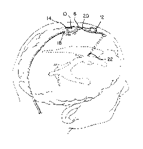
Figure I is a top perspective view of the shunt and ventricular catheter
according to the
present invention;
Figure 2 is a partial perspective view, with parts broken away, showing the
interior of the
ventricular catheter according to the present invention;
Figure 3 is a cross-sectional view taken along lines 3-3 of Figure 2 and
looking in the
direction of the arrows;
CAN_DMS, \125708583\1 3
CA 2749653 2019-02-27

CA 02749653 2011-11-22
Figure 4 is a cross-sectional view taken along lines 4-4 of Figure 3 and
looking in the
direction of the arrows;
Figure 4A is a cross-sectional view taken along lines 4A-4A of Figure 3 and
looking in the
direction of the arrows;
.. Figure 5 is a cross-sectional view taken along lines 5-5 of Figure 3 and
looking in the
direction of the arrows;
Figure 6 is a partial top view of a prior art ventricular catheter;
Figure 7 is cross-sectional view taken along lines 7-7 of Figure 6 and looking
in the
direction of the arrows;
.. Figure 8A is a partial top view of the ventricular catheter according to
the present
invention; and
Figure 8B is a partial top view of the ventricular catheter according to the
present
invention.
Figure 9 is a partial perspective view, with parts broken away, showing the
interior
of the ventricular catheter according to the present invention.
4

CA 02749653 2011-08-19
DETAILED DESCRIPTION OF THE PRESENT INVENTION
Referring now to Figures 1-5, 8A, 8B and 9 a shunt 10 and a ventricular
catheter
12 in accordance with the present invention is illustrated.
As illustrated in Figure 1, shunt 10 has a housing 14, which has an inlet 16,
an
outlet 18 and a flow control mechanism disposed therein. Ventricular catheter
12 is
connected to inlet 16 of the housing. Catheter 12 has a longitudinal length, a
proximal end
20, a distal end 22, and an inner lumen 24 extending therethrough. Inner lumen
24 is a
single lumen at proximal end 20 of the catheter and is comprised of two or
more lumens
241, 24", 241H, 24", etc. at the distal end 22 of catheter 12. Catheter 12 is
preferably
made of silicone. In addition, catheter 12 can be impregnated with
antimicrobial
antibiotics, such as the CODMANO BACTISEALO catheter, which is commercially
sold
by Codman & Shurtleff, Inc. of Raynham, MA.
Ventricular catheter 12 has only one aperture 26 at distal end 22 of catheter
12
corresponding to each of the lumens 241, 24111, 241111, etc. There are
preferably between 2
and 7 lumens, with only three and four lumens being shown in the drawing
Figures for the
sake of brevity. Of course, one skilled in the art would readily know how to
make the
ventricular catheter of the present invention with multiple lumens based on
the present
disclosure. Each tapering slit 30 receives cerebrospinal fluid (CSF) when in
use. Each
aperture 26 has an enlarged opening 28 at the distal end and transitions from
the enlarged
opening to a tapering slit 30 at the proximal end of the aperture. In some
examples of the
present invention, for each aperture 26, a portion 32 between the enlarged
opening 28 and
the tapering slit 30 is a slit 32 of constant thickness. Likewise, in other
examples of the
present invention, for each aperture 26, there may be no portion 32 between
the enlarged
opening 28 and the tapering slit 30. In addition, as illustrated in Figure 9,
for each
aperture 26, a slit 42 of constant thickness can be disposed between the
enlarged opening
28 and the proximal end of the aperture with no tapering slit being utilized.
The entire
aperture, from the enlarged opening 28 to the proximal end of tapering slit 30
is in fluid
communication with its respective lumen 241, 24111, 2411115
etc. The enlarged opening 28
of each aperture has a smooth concave inner surface 34, similar to a spoon
shape.
The use of multiple lumens 241, 241H, 241111, etc. in accordance with the
present
invention help prevent complete occlusion of ventricular catheter 12. In the
present
invention, ventricular catheter 12 can only be completely occluded if all the
lumens
become blocked. In addition, because the transition from multiple lumens to a
single
lumen 24 occurs from about 0.5 to about 3.0 centimeters from the distal end of
the
5

ventricular catheter, any ingrowth of choroid plexus or ependymal tissue must
extend
beyond this junction to cause complete occlusion and interconnection of tissue
from
multiple apertures, which is unlikely to happen because of the length the
tissue has to
grow. The only other way that the choroid plexus or ependymal tissue would
cause a
complete occlusion is for the tissue to block each of the multiple lumens 24
beyond the slit
30, or to occlude the entire slit 30 and the aperture 26.
The slit lumen geometry is preferably tapered or purposely shaped to provide
resistance to fluid flow through the slit that corresponds to the size of the
pathway
provided by the slit. The size of the slit opening distributes the pressure
gradient over a
larger distance and surface area than conventional ventricular catheters.
Diffusing the
pressure gradient diminishes the attractive fluid forces and diminishes areas
of high fluid
flow, thereby lessens the propensity for tissue ingrowth.
Referring now to Figures 8A and 8B of the present invention, tissue ingrowth
36 is
illustrated. As can be seen, tissue ingrowth into aperture 26 will not
interconnect with
tissue ingrowth from another lumen. Thus, should the ventricular catheter 12
need to be
removed, catheter 12 will be pulled back out and tissue ingrowth 36 can be
removed from
the lumen at the wider end of the taper or at the enlarged opening 28, as
illustrated in
Figure 8B. In contrast, in the prior art, tissue ingrowth 36 can be rather
difficult to remove
from the ventricular catheter should the ventricular catheter 12 need to be
removed, as
discussed above.
Referring now to Figures 2 and 3, ventricular catheter 12, has a blunt distal
end 38
to permit the catheter to be introduced into the brain without damaging brain
tissue. In
addition, an inner concave surface 40 is sized to receive a stylet for use in
introducing the
catheter.
It will be understood that the foregoing is only illustrative of the
principles of the
invention, and that various modifications can be made by those skilled in the
art without
departing from the scope and spirit of the invention.
6
CA 2749653 2018-01-10

Representative Drawing
A single figure which represents the drawing illustrating the invention.
Administrative Status

2024-08-01:As part of the Next Generation Patents (NGP) transition, the Canadian Patents Database (CPD) now contains a more detailed Event History, which replicates the Event Log of our new back-office solution.

Please note that "Inactive:" events refers to events no longer in use in our new back-office solution.

For a clearer understanding of the status of the application/patent presented on this page, the site Disclaimer , as well as the definitions for Patent , Event History , Maintenance Fee  and Payment History  should be consulted.

Event History

Description Date
Common Representative Appointed 2020-11-07
Grant by Issuance 2019-12-10
Inactive: Cover page published 2019-12-09
Common Representative Appointed 2019-10-30
Inactive: Final fee received 2019-10-17
Pre-grant 2019-10-17
Notice of Allowance is Issued 2019-05-01
Letter Sent 2019-05-01
Notice of Allowance is Issued 2019-05-01
Inactive: Filing certificate - RFE (bilingual) 2019-04-30
Inactive: Approved for allowance (AFA) 2019-04-18
Inactive: Q2 passed 2019-04-18
Amendment Received - Voluntary Amendment 2019-02-27
Inactive: S.30(2) Rules - Examiner requisition 2018-10-17
Inactive: Report - No QC 2018-10-15
Amendment Received - Voluntary Amendment 2018-09-05
Inactive: S.30(2) Rules - Examiner requisition 2018-03-07
Inactive: Report - No QC 2018-03-05
Letter Sent 2018-02-02
Inactive: Multiple transfers 2018-01-12
Amendment Received - Voluntary Amendment 2018-01-10
Inactive: S.30(2) Rules - Examiner requisition 2017-07-10
Inactive: Report - No QC 2017-07-10
Letter Sent 2016-08-25
All Requirements for Examination Determined Compliant 2016-08-18
Request for Examination Requirements Determined Compliant 2016-08-18
Request for Examination Received 2016-08-18
Letter Sent 2013-10-25
Amendment Received - Voluntary Amendment 2012-12-27
Application Published (Open to Public Inspection) 2012-03-29
Inactive: Cover page published 2012-03-28
Amendment Received - Voluntary Amendment 2011-11-30
Amendment Received - Voluntary Amendment 2011-11-22
Inactive: IPC assigned 2011-10-25
Inactive: First IPC assigned 2011-10-25
Inactive: IPC assigned 2011-10-25
Filing Requirements Determined Compliant 2011-09-01
Application Received - Regular National 2011-09-01
Inactive: Filing certificate - No RFE (English) 2011-09-01

Abandonment History

There is no abandonment history.

Maintenance Fee

The last payment was received on 2019-07-23

Note : If the full payment has not been received on or before the date indicated, a further fee may be required which may be one of the following

  • the reinstatement fee;
  • the late payment fee; or
  • additional fee to reverse deemed expiry.

Please refer to the CIPO Patent Fees web page to see all current fee amounts.

Owners on Record

Note: Records showing the ownership history in alphabetical order.

Current Owners on Record
INTEGRA LIFESCIENCES SWITZERLAND SARL
Past Owners on Record
EMILIE NEUKOM
STEPHEN F. WILSON
Past Owners that do not appear in the "Owners on Record" listing will appear in other documentation within the application.
Documents

To view selected files, please enter reCAPTCHA code :


To view images, click a link in the Document Description column. To download the documents, select one or more checkboxes in the first column and then click the "Download Selected in PDF format (Zip Archive)" or the "Download Selected as Single PDF" button.

List of published and non-published patent-specific documents on the CPD .

If you have any difficulty accessing content, you can call the Client Service Centre at 1-866-997-1936 or send them an e-mail at CIPO Client Service Centre.


Document
Description 
Date
(yyyy-mm-dd) 
Number of pages   Size of Image (KB) 
Drawings 2011-08-19 3 173
Description 2011-08-19 6 269
Abstract 2011-08-19 1 13
Claims 2011-08-19 3 116
Representative drawing 2011-10-31 1 12
Cover Page 2012-03-22 2 43
Description 2011-11-22 6 272
Description 2018-01-10 6 250
Claims 2018-09-05 2 65
Description 2019-02-27 6 258
Claims 2019-02-27 2 68
Cover Page 2019-11-07 1 38
Representative drawing 2019-11-07 1 10
Maintenance fee payment 2024-06-25 35 1,429
Filing Certificate (English) 2011-09-01 1 156
Reminder of maintenance fee due 2013-04-22 1 114
Courtesy - Certificate of registration (related document(s)) 2013-10-25 1 127
Reminder - Request for Examination 2016-04-20 1 126
Acknowledgement of Request for Examination 2016-08-25 1 177
Filing Certificate 2019-04-30 1 207
Courtesy - Certificate of registration (related document(s)) 2013-10-25 1 107
Commissioner's Notice - Application Found Allowable 2019-05-01 1 163
Examiner Requisition 2018-10-17 3 176
Amendment / response to report 2018-09-05 8 295
Request for examination 2016-08-18 2 69
Examiner Requisition 2017-07-10 3 194
Amendment / response to report 2018-01-10 4 161
Examiner Requisition 2018-03-07 3 173
Amendment / response to report 2019-02-27 7 256
Prosecution correspondence 2011-11-30 1 55
Final fee 2019-10-17 3 89