Note: Descriptions are shown in the official language in which they were submitted.
CA 02750178 2011-06-28
WO 2010/078063 PCT/US2009/068613
IN-SITU REFILLABLE OPHTHALMIC IMPLANT
Cross Reference to Related Application
This application claims priority under 35 U.S.C. 119 to U.S. Provisional
Patent Application No. 61/142,242, filed January 2, 2009, the entire contents
of
which are incorporated herein by reference.
Technical Field of the Invention
The present invention is related to an in-situ refillable ophthalmic implant
having a refill port and a release control mechanism. The present invention
also
is relates to methods of forming and using the ophthalmic implant.
Background of the Invention
For many ocular conditions such as glaucoma, age related macular
degeneration, secondary cataracts or others, it is often desirable to provide
therapeutic agent to particular locations within the eye and to provide those
agents
over an extended time period (e.g., weeks, month or even years). Ophthalmic
implants provide at least one mechanism for providing therapeutic agents in
this
manner. As such, the pharmaceutical industry has dedicated significant
resources
in the development of such implants.
U.S. Patent No. 5,466,233 to Weiner et al. describes a tack shaped device
having a post and head. The post can include a permeable membrane that forms a
chamber, the chamber being filled with liquid drug that is delivered to the
eye by
passing through the membrane.
U.S. Patent No. 5,707,643 to Ogura et al. describes a scleral plug having at
least a portion thereof formed of a lactic acid copolymer of lactic acid units
and
glycolic acid units and containing a drug. The material of the plug is
biodegradable
3s for allowing drug to be released gradually over time.
-1-
CA 02750178 2011-06-28
WO 2010/078063 PCT/US2009/068613
U.S. Patent No. 6,976,982 to Santini, Jr et al. describe flexible microchip
devices suitable for application to the surface of an eye and designed to
controllably release therapeutic agents to the eye.
While many advances have made in the arena of ophthalmic implants, there
are still many drawbacks that plague conventional extended release ophthalmic
implants. As one example of these drawbacks, many conventional ophthalmic
implants have only a particular amount of therapeutic agent upon implantation
within an eye and must be replaced once that amount of agent has been
delivered.
As another example of these drawbacks, many conventional ophthalmic implants
lack a reliable mechanism for controlling the amount of drug released over
time or
the conventional implants can include overly complex mechanisms for
controlling
drug release. As still another example of these drawbacks, many conventional
ophthalmic implants lack the ability to deliver therapeutic agents into
locations
substantially below the surface of the eye.
In view of the above, the present invention provides an ophthalmic implant
and a method of applying and/or using the implant where the implant and/or
method overcome one or more of the aforementioned drawbacks or other
drawbacks commonly associated with conventional ophthalmic implants.
Summary of the Invention
The present invention is directed to an in-situ refillable ophthalmic implant.
The implant typically includes a body portion, a fill portion and a release
control
mechanism. The body portion defines a reservoir suitable for receipt of a
pharmaceutical composition that includes a therapeutic agent. The fill portion
defines a fill port in fluid communication with the reservoir for allowing the
pharmaceutical composition to be repeatedly located within the reservoir. The
release control mechanism includes at least one opening suitable for providing
a
controlled passive release of the pharmaceutical composition into the eye over
an
extended time period. Upon application of the implant to the eye, the release
control mechanism is typically located within the eye (e.g., the vitreous of
the eye)
and the fill portion is located adjacent the sclera or cornea of the eye such
that the
fill port remains accessible outside of the vitreous of the eye and also
possibly
outside of the sclera, cornea or both.
-2-
CA 02750178 2011-06-28
WO 2010/078063 PCT/US2009/068613
The implant can include various additional or alternative components or
features and can be characterized by various additional or alternative
configurations. The body portion, the fill portion or a combination thereof
can
define a contact surface that is disposed over the sclera upon application of
the
implant to the eye. The at least one opening of the release control mechanism
can
include multiple openings wherein the multiple openings are sized to
effectuate the
controlled release of the therapeutic agent. The release control mechanism can
include a door that can be opened and closed remotely to provide release of
therapeutic agent to the vitreous. The control release mechanism can be
comprised
of a silicon disc through which the at least one or multiple opening[s]
extend. The
body portion can be overmolded onto the control release mechanism. The fill
portion can include a diaphragm associated with the port, the diaphragm being
penetrable by a needle or other elongated injection device for allowing
filling of the
reservoir through such injection device, the diaphragm also being capable of
self
sealing after removal of the needle. The fill portion can include a cap
portion that,
upon implantation of the implant, is below the conjunctiva and resides upon
the
sclera.
Brief Description of the Drawings
FIG. 1 is a side view of an exemplary ophthalmic implant in accordance
with the present invention; and
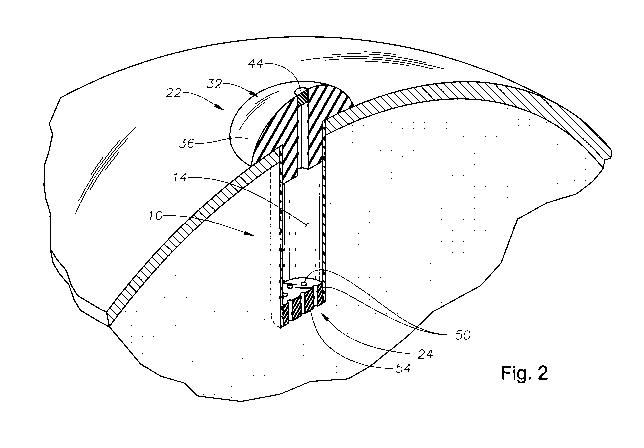
FIG. 2 is a perspective view of the exemplary implant of FIG. 1 applied to
an eye of an individual.
Detailed Description of the Invention
The present invention is predicated upon the provision of an ophthalmic
implant and a method of implanting and/or using that implant. The implant will
typically include a body portion defining a reservoir suitable for the receipt
of a
pharmaceutical composition. The implant will also typically include a fill
portion
that will allow the implant reservoir to be initially filled with the
pharmaceutical
composition and will typically also allow the implant reservoir to be refilled
after
the implants has been implanted in an eye. The implant will also typically
include
-3-
CA 02750178 2011-06-28
WO 2010/078063 PCT/US2009/068613
a release control mechanism that can reliably control the amount of
pharmaceutical
composition release to the eye.
With reference to Figs. 1 and 2, there is illustrated an exemplary in-situ
s refillable ophthalmic implant 10 in accordance with the present invention.
The
implant 10 is illustrated as including a body portion 12, which defines a
reservoir
14 within the implant 10. In the embodiment illustrated, the implant 10 is
generally
symmetrical about an axis 18, which extends along a length (L) of the implant
10,
the body portion 12 or both.
A fill portion 22 is included at one end of the length (L) of the implant 10
and a release control mechanism 24 is included at an opposite end of the
length (L)
of the implant 10. The fill portion 22 is illustrated as having a port 28
suitable for
aiding in the receipt of a pharmaceutical composition into the reservoir 14 of
the
is implant 10. The fill portion 22 is also illustrated as including a cap 32
from which
the body portion 12 extends.
The cap 32 may be formed integrally with and of the same material as the
body portion 12. However, in the illustrated embodiment, the cap 32 is formed
of a
separate material from the body portion 12 and is attached to the body portion
12.
The cap 32 may be attached to body portion 12 using any of a variety of
fastening
mechanisms, but preferably involves an interference fit with a portion of the
cap 32
extending partially into the body 12 or a portion of the cap 32 extending
about the
body 12 externally.
In a preferred embodiment, the cap 32 is formed of a relatively soft material
(e.g., a polymeric material) that is biocompatible with the human eye.
Examples of
preferred materials include, without limitation, silicone, parylene, an
acrylic
material or the like. In the illustrated embodiment, the port 28 extends
centrally
through the cap 32 and the cap 32 is annular about the port 28.
The cap 32 of the fill portion 22 includes an external surface 36 that is
designed to be external of and face outwardly away from the eyeball including
the
vitreous of the eye, the sclera of the eye or both after the implant 10 is
surgically
applied to the eye. The external surface 36 is illustrated as being generally
convex.
Advantageously, when used, the convex surface and material of the cap 32 can
aid
-4-
CA 02750178 2011-06-28
WO 2010/078063 PCT/US2009/068613
in allowing for the implant 10 to reside in its intended location within the
eye
without causing significant irritation or discomfort.
The cap 32 of the fill portion 22 is also shown to include a contacting
surface 40 that is designed to contact the sclera or conjunctiva after the
implant 10
has been applied to the eye. In a preferred embodiment, the contacting surface
40
can be slight convex for better accommodation of the sclera or conjunctiva. In
one
particular embodiment, the cap 32, the fill portion 22 or both are disposed
within or
over the pars plana of the eye over or near the limbus.
An access element 44 will typically be associated with the port 28 for
selectively restricting movement of fluid through the port 28. The access
element
44 can be a removable plug, a door, a valve or other such element. In one
preferred
embodiment, the access element 44 is a diaphragm, which can be opened through
penetration by a needle or other delivery device but will also close to again
restrict
fluid flow after removal of the needle or other delivery device from the
diaphragm.
In such an embodiment, it is contemplated that the cap 32 and the access
element
44 (i.e., the diaphragm) could be integrally formed as a singular part of the
same
material. In such an embodiment, silicone (e.g., a non-coring silicone),
parylene or
another material could ideally be used and a thin portion of the cap 32 that
acts as a
diaphragm 44. Advantageously, a needle or other device can be extended through
these materials and any opening made by the needle will typically self close
and/or
seal after removal of the needle or other device.
The body portion 12 is illustrated as being annular, and more particularly
cylindrical, for defining the reservoir 14. The body portion 12 may be formed
of a
variety of materials (e.g., polymer or metal materials) that are biologically
compatible with the human eye. Exemplary suitable materials include, without
limitation, parylene, polyetheretherketone (PEEK), polyethylene, polyimide,
ethylene vinyl acetate, acrylic polymers, combinations thereof or the like.
The control release mechanism 24 will typically include one or more
opening[s] 50 through which material (e.g., fluid that contains therapeutic
agent)
can pass. The use of multiple openings 50 is generally preferable and there is
3s typically at least 3, more typically at least 6 and even more typically at
least 10
openings and there is typically no greater than 1000, more typically no
greater than
200 and even more typically no greater than 50 openings.
-5-
CA 02750178 2011-06-28
WO 2010/078063 PCT/US2009/068613
It is preferable that the control release mechanism 24 may be configured for
passive passage of material through the opening[s] 50. Thus, flow through the
opening[s] 50 is generated or driven through natural diffusion and/or
equilibrium
mechanisms. The control release mechanism may consist or consist essentially
of
the opening[s] 50 and the material through which the openings extend.
Alternatively, the control release mechanism 24 can include mechanical
mechanisms for selectively inhibiting or allowing the passive passage of
material
through the openings 50. Examples of such mechanism include valves or doors,
which can be selectively and even remotely (e.g., through radio frequency
signaling) opened and closed to respectively allow and inhibit passage of
material
through the opening[s] 50. As used herein, the terms opened and closed as they
refer to the control release mechanism include partial and full opening or
close.
Moreover, it is contemplated that partial opening or closing of the mechanism
may
be employed to further control the amount of diffusion or movement of fluid
through the opening[s] 50 thereby further controlling the deliver of the
pharmaceutical composition to the eye.
Material, particularly ophthalmic pharmaceutical composition and aqueous
humor fluid, is typically allowed to freely flow and/or diffuse into and out
of the
reservoir 14 with the size of the opening[s] 50 assisting in controlling the
rate of
flow and/or diffusion into and out of the reservoir 14. The opening[s] 50,
particularly for a passive system, have a cross-sectional area that controls
the rate at
which material, particularly therapeutic agent, flows out of the reservoir and
into
the eye. That cross-sectional area is typically at least 8 microns2, more
typically at
least 15 microns2 and even more typically at least 50 microns2. That same
cross-
sectional area is also typically no greater than 4000 microns2, more typically
no
greater than 2000 microns2 and still more typically no greater than 500
microns2.
The cross-sectional area of the opening, as used herein, is any sectional area
of the
opening wherein the outer perimeter of the opening is fully defined by the
material
of the control release mechanism and wherein, for fluid to pass through the
opening
into or out of the reservoir 14, it must also pass through the cross-sectional
area.
In the illustrated embodiment, the control release mechanism 24 is a plate 54
through which the opening[s] 50 extend. The plate 54 has opposing
substantially
parallel surfaces through with the opening[s] 50 extend. In the embodiment
shown,
the opening[s] 50 or cylindrical in shape although they may be shaped
otherwise as
-6-
CA 02750178 2011-06-28
WO 2010/078063 PCT/US2009/068613
well. The opening[s] 50 typically have a diameter of at least about 0.2
microns,
more typically at least about 2 microns and even more typically at least about
8
microns. The diameter of the opening[s] illustrated is also typically no
greater than
about 100 microns, even more typically no greater than 40 microns and even
more
typically no greater than about 25 microns. While it is understood that a
generally
uniform distribution of the opening[s] 50 over the surface of the plate 54 is
desirable other non-uniform distribution of opening[s] 50 are also possible. A
suitable thickness for the plate will typically be at least about 0.05 mm,
more
typically at least about 0.08 mm and will typically no greater than 0.5 mm and
more typically no greater than 0.3 mm.
In the illustrated embodiment, the length (L) of the implant 10 will typically
be less than about 15 mm, more typically less than 10 mm and even more
typically
less than 8 mm. Also in the illustrated embodiment, the outer diameter of the
body
portion 12 of the implant 10 will typically be less than 7 mm more typically
less
than 4 mm and even more typically less than 2.5 mm. The length of the implant
is
typically sufficiently small such that it does not interfere with the vision
or field of
view of the eye.
The control release mechanism 24, and particularly the plate 54, may be
formed of a variety of materials such as metals or polymeric materials. In a
preferred embodiment, however, it is formed of an etchable material such as
silicon, which allows the opening[s] 50 to be etched into the material.
The control release mechanism 24, and particularly the plate 54, can be
attached to the body portion 12 of the implant 10 using an interference fit or
other
fastening technique. In one preferred embodiment, the body portion 12 is
overmolded onto the plate 54 for attaching the plate 54 to the body portion
12.
Other suitable fastening techniques could involve the use of sealing members,
adhesive, fasteners, specially designed attachment members or the like. It is
further
contemplated that the body portion 12 and the control release mechanism 24
could
be integrally formed of the same material.
For implantation, the implant 10 is typically inserted into a surgical
incision
in the eye. Once implanted, the implant 10 may be held in place with sutures
or
other mechanisms. Additionally or alternatively, it is contemplated that the
body
portion 12 or other portion of the implant 10 may be shaped to assist in
maintaining
-7-
CA 02750178 2011-06-28
WO 2010/078063 PCT/US2009/068613
the implant 10 in place within the eye. As one example, the body portion 12
may
have a spiral configuration such that the body portion 12 itself substantially
maintains the implant 10 in place in the eye. An example of such a spiral
configuration is illustrated in U.S. Patent No. 6,719,750 to Varner et al,
which is
fully incorporated herein by reference for all purposes.
Generally, the implant 10 may be located in a variety of locations within the
eye. In one preferred embodiment, the implant 10 is surgically positioned such
that
the body portion 12 extends into the vitreous of the eye and the fill portion
22,
particularly the cap, is located between the conjunctiva of the eye and the
vitreous
of the eye. In a highly preferred embodiment, the cap 22 is beneath the
conjunctiva
of the eye, the surface 40 of the cap 22 contacts the sclera of the eye and
the body
portion 12 extends through the sclera into the eye.
The pharmaceutical composition that is provided within the implant 10 will
typically include a therapeutic agent and that agent may or may not be
provided
within a pharmaceutical vehicle. The therapeutic agent of the present
invention
may be provided in various forms within the implant and, when used, could be
provided with various different pharmaceutical vehicles (e.g., water alone or
combined with additional ingredients). The agent could be a solid, semi-solid
or
liquid within the implant. As one example, the therapeutic agent could be
provided
as a solid within a liquid (e.g., aqueous) suspension. As another example, the
therapeutic agent could be provided as an oil without any vehicle at all.
It is generally preferable that the pharmaceutical composition be injectable
with a syringe. Thus, it is preferable that the pharmaceutical composition be
liquid
or semi-solid even when the therapeutic agent may be entirely or substantially
entirely solid (e.g., a suspended solid). Such liquid or semi-solid
compositions can
be injected into the implant 10 with a syringe prior to insertion of the
implant 10
within an eye and/or after insertion of the implant 10 within an eye. Thus,
the
implant 10 may be filled and then re-filled one or multiple times.
Non-limiting examples of potential ophthalmic therapeutic agents for the
present invention include: anti-glaucoma agents, anti-angiogenesis agents;
anti-
3s infective agents; anti-inflammatory agents; growth factors;
immunosuppressant
agents; and anti-allergic agents. Anti-glaucoma agents include beta-blockers,
such
as betaxolol and levobetaxolol; carbonic anhydrase inhibitors, such as
brinzolamide
-8-
CA 02750178 2011-06-28
WO 2010/078063 PCT/US2009/068613
and dorzolamide; prostaglandins, such as travoprost, bimatoprost, and
latanoprost;
seretonergics; muscarinics; dopaminergic agonists. Anti-angiogenesis agents
include anecortave acetate (RETAANETM, AlconTM Laboratories, Inc. of Fort
Worth, Tex.) and receptor tyrosine kinase inhibitors (RTKi). Anti-inflammatory
agents include non-steroidal and steroidal anti-inflammatory agents, such as
triameinolone actinide, suprofen, diclofenac, ketorolac, nepafenac,
rimexolone, and
tetrahydrocortisol. Growth factors include EGF or VEGF. Anti-allergic agents
include olopatadine and epinastine. The ophthalmic drug may be present in the
form of a pharmaceutically acceptable salt.
Advantageously, the opening[s] 50 of the implant 10 can act as a simple
mechanism for controlling the release of the pharmaceutical composition,
particularly the therapeutic agent, over time. In a preferred embodiment, the
implant 10 includes opening[s] 50 sized to include the cross-sectional areas
is discussed above. In such an embodiment, the opening[s] 50 can operate to
release
at least 50%, more typically at least 80% and even more typically at least 90%
of
an amount of therapeutic agent located within the implant 10 over a period of
time
that is at least 48 hours, more typically at least 7 days and even more
typically at
least 60 days but is no greater than 5 years, more typically no greater than
one year
and still more typically no greater than 6 months.
The initial amount of pharmaceutical composition including therapeutic
agent can be disposed within the reservoir 14 during assembly of the implant
10 or
thereafter. For refilling the implant 10, a device such as a syringe is used
to extend
a needle through the access element 44, the port 28 or both and push
pharmaceutical composition into the reservoir 14. For aiding in refill, it may
desirable to use one device (e.g., syringe) to aspirate material (e.g.,
aqueous humor
liquid) from the reservoir 14 and another device (e.g., syringe) to push
pharmaceutical composition into the reservoir 14 thereafter. Alternatively, a
single
syringe device can be created to concurrently aspirate fluid from the
reservoir 14
while replacing that fluid with pharmaceutical ophthalmic composition.
The entire contents of all cited references are specifically incorporated by
reference into this disclosure for all purposes. Further, when an amount,
concentration, or other value or parameter is given as either a range,
preferred
range, or a list of upper preferable values and lower preferable values, this
is to be
understood as specifically disclosing all ranges formed from any pair of any
upper
-9-
CA 02750178 2011-06-28
WO 2010/078063 PCT/US2009/068613
range limit or preferred value and any lower range limit or preferred value,
regardless of whether ranges are separately disclosed. Where a range of
numerical
values is recited herein, unless otherwise stated, the range is intended to
include the
endpoints thereof, and all integers and fractions within the range. It is not
intended
that the scope of the invention be limited to the specific values recited when
defining a range.
Other embodiments of the present invention will be apparent to those skilled
in the art from consideration of the present specification and practice of the
present
invention disclosed herein. It is intended that the present specification and
examples be considered as exemplary only with a true scope and spirit of the
invention being indicated by the following claims and equivalents thereof.
-10-