Note: Descriptions are shown in the official language in which they were submitted.
CA 02751273 2011-08-01
WO 2010/107424 PCT/US2009/037289
1
BIOPSY DEVICE HAVING ROTATIONAL CUTTING
BACKGROUND OF THE INVENTION
1. Field of the Invention
[0001] The present invention relates to medical devices, and, more
particularly, to a biopsy
device having rotational cutting.
2. Description of the Related Art
[0002] A typical biopsy device includes a probe assembly having a cannula
configured
with a sample notch and a tissue sampling chamber and associated tissue
cutting mechanism.
During a biopsy procedure, vacuum assistance may be used to help draw tissue
through the
sample notch and into the sampling chamber and maximize the amount of tissue
obtained
with each sample. Some biopsy devices, commonly referred to as single
insertion, multiple
samples, or S1MS devices, utilize sample acquisition and delivery mechanisms
that allow
multiple samples to be acquired from a given lesion without removing and
reinserting the
needle after each sample. One type of cutting mechanism used in a vacuum
assisted S1MS
biopsy device uses rotational and linear motion of a cutter with respect to
the sample notch to
sever the tissue drawn through the sample notch into the tissue sampling
chamber. Vacuum is
applied to transport the tissue from the sampling chamber to a sample
collection basket. This
process may be repeated until the desired amount of tissue has been obtained.
[0003] In one common SIMS biopsy device, it is necessary for an operator to
manually
rotate the probe assembly to different orientations after each sample in order
to obtain tissue
samples at different radial orientations within the target site. However, in
some situations,
such manual rotation may be inconvenient.
SUMMARY OF THE INVENTION
[0004] The present invention provides a biopsy device and method for obtaining
biopsy
samples, wherein the biopsy device is configured to periodically form a
virtual tissue sample
aperture at a plurality of angular radial positions.
[0005] In the description of the invention that follows, the terms "first" and
"second"
preceding an element name are used for identification purposes to distinguish
between similar
or related elements, results or concepts, and are not intended to necessarily
imply order, nor
CA 02751273 2011-08-01
WO 2010/107424 PCT/US2009/037289
2
are the terms "first" and "second" intended to preclude the inclusion of
additional similar or
related elements, results or concepts, unless otherwise indicated.
[0006] The invention, in one form thereof, is directed to a biopsy device
including a probe
assembly and a driver unit. The probe assembly includes a first cannula having
a first side
wall defining a first lumen. The first cannula has a first proximal end and a
first distal end.
The first cannula has a first aperture extending through the first side wall
to the first lumen
proximal to the first distal end. The first cannula has a longitudinal axis. A
second cannula
has a second side wall defining a second lumen. The second cannula has a
second proximal
end and a second distal end. The second cannula has a second aperture
extending through the
second side wall to the second lumen proximal to the second distal end. The
second cannula
is disposed co-axially with the first cannula. A least one of the first
aperture and the second
aperture has a cutting edge. The driver unit is configured for releasably
mounting the probe
assembly. The driver unit is operatively configured to simultaneously rotate
the first cannula
and the second cannula in opposite rotational directions at different
rotational velocities so
that the first aperture and the second aperture periodically come into
alignment to form a
virtual tissue sample aperture.
[0007] The invention, in another form thereof, is directed to a biopsy device
including a
probe assembly and a driver unit. The probe assembly includes a first cannula
having a first
side wall defining a first lumen. The first cannula has a first proximal end
and a first distal
end. The first cannula has a first aperture extending through the first side
wall to the first
lumen proximal to the first distal end. The first cannula has a longitudinal
axis. A second
cannula has a second side wall defining a second lumen. The second cannula has
a second
proximal end and a second distal end. The second cannula has a second aperture
extending
through the second side wall to the second lumen proximal to the second distal
end. The
second cannula is disposed co-axially with the first cannula. At least one of
the first aperture
and the second aperture has a cutting edge. The driver unit is configured for
releasably
mounting the probe assembly. The driver unit is operatively configured to
rotate the first
cannula in accordance with a first velocity profile and the second cannula in
accordance with
a second velocity profile to periodically align the first aperture and the
second aperture to
form a virtual tissue sample aperture at a plurality of angular radial
positions relative to the
longitudinal axis during a biopsy procedure by continuous simultaneous
rotation of both of
the first cannula and the second cannula.
CA 02751273 2011-08-01
WO 2010/107424 PCT/US2009/037289
3
[0008] The invention, in another form thereof, is directed to a method for
controlling a
biopsy device during a biopsy procedure, the biopsy device having a probe
assembly with an
outer cannula having a distal needle tip and an inner cannula arranged coaxial
with the outer
cannula with respect to a longitudinal axis, the outer cannula having a first
side aperture and
the inner cannula having a second side aperture with at least one of the first
side aperture and
the second side aperture having a cutting edge, and a vacuum source connected
in fluid
communication with a lumen of the inner cannula and with a tissue sample
receptacle. The
method includes positioning each of the outer cannula and the inner cannula at
a respective
initial rotational position; inserting the probe assembly in a region of a
patient to be biopsied;
establishing continuous simultaneous rotation of the outer cannula in
accordance with a first
velocity profile and the inner cannula in accordance with a second velocity
profile to
periodically align the first side aperture and the second side aperture to
form a virtual tissue
sample aperture at a plurality of angular radial positions relative to the
longitudinal axis;
establishing a supply of negative pressure in the lumen of the inner cannula,
such that each
time the virtual tissue sample aperture is formed tissue is pulled through the
virtual tissue
sample aperture into the lumen of the inner cannula, and thereafter the first
side aperture and
the second side aperture cooperate to sever the tissue that is pulled into the
inner cannula as
the virtual tissue sample aperture is closed by the continuous simultaneous
rotation of the
outer cannula and the inner cannula, each tissue sample so severed being
transported through
the lumen of the inner cannula by the negative pressure to a tissue sample
receptacle; and
ceasing the continuous simultaneous rotation of the outer cannula and the
inner cannula after
all desired tissue samples have been harvested.
BRIEF DESCRIPTION OF THE DRAWINGS
[0009] The above-mentioned and other features and advantages of this
invention, and the
manner of attaining them, will become more apparent and the invention will be
better
understood by reference to the following description of embodiments of the
invention taken
in conjunction with the accompanying drawings, wherein:
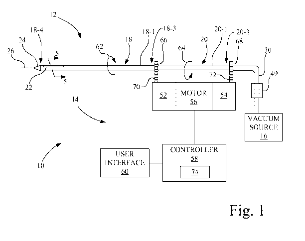
[0010] Fig. 1 is a pictorial illustration of a biopsy device including a probe
assembly and
driver unit, configured in accordance with an embodiment of the present
invention.
[0011] Fig. 2A is an exploded view of the probe assembly of Fig. 1.
[0012] Fig. 2B is a cross-section view of the outer cannula of Fig. 2A taken
along line 2B-
2B.
CA 02751273 2011-08-01
WO 2010/107424 PCT/US2009/037289
4
[0013] Fig. 2C is a cross-section view of the inner cannula of Fig. 2A taken
along line 2C-
2C.
[0014] Fig. 3 is an assembled view of the probe assembly of Fig. 2A having the
respective
apertures of the outer cannula and inner cannula in alignment.
[0015] Fig. 4 is a cross-section view of the probe assembly of Fig. 3 taken
along line 4-4,
showing tissue being drawn through a virtual tissue sample aperture.
[0016] Fig. 5 is a cross-section view of the probe assembly of Fig. 1 taken
along line 5-5.
[0017] Fig. 6A is an exploded view of another embodiment for a probe assembly
suitable
for use in the biopsy device of Fig. 1.
[0018] Fig. 6B is a cross-section view of the outer cannula of Fig. 6A taken
along line 6B-
6B.
[0019] Fig. 6C is a cross-section view of the inner cannula of Fig. 6A taken
along line 6C-
6C.
[0020] Fig. 7 is an assembled view of the probe assembly of Fig. 6A having the
respective
apertures of the outer cannula and inner cannula in alignment.
[0021] Fig. 8 is a graphical representation of exemplary velocity profiles for
the outer
cannula and the inner cannula of Fig. 1.
[0022] Fig. 9 is a graphical representation of the formation of a virtual
tissue sample
aperture at each of a plurality of angular radial positions.
[0023] Fig. 10 is a pictorial illustration of another embodiment of a biopsy
device including
a probe assembly and driver unit, configured in accordance with an embodiment
of the
present invention.
[0024] Fig. 11 is a graphical representation of exemplary velocity profiles
for the outer
cannula and the inner cannula in the embodiment of Fig. 10.
[0025] Fig. 12 is a flowchart of a method for controlling a biopsy device,
such as the
biopsy device of Fig. 1.
[0026] Corresponding reference characters indicate corresponding parts
throughout the
several views. The exemplifications set out herein illustrate embodiments of
the invention,
and such exemplifications are not to be construed as limiting the scope of the
invention in any
manner.
CA 02751273 2011-08-01
WO 2010/107424 PCT/US2009/037289
DETAILED DESCRIPTION OF THE INVENTION
[0027] Referring now to the drawings and particularly to Fig. 1, there is
shown a biopsy
device 10 configured in accordance with an embodiment of the present
invention. Biopsy
device 10 includes a probe assembly 12, a driver unit 14, and a vacuum source
16.
[0028] Referring also to Figs. 2A-2C, 3, 4, and 5, probe assembly 12 includes
an outer
cannula 18 and an inner cannula 20.
[0029] Outer cannula 18 has a first side wall 18-1 defining a first lumen 18-
2. Outer
cannula 18 has a first proximal end 18-3, a first distal end 18-4, and a first
aperture 22
extending through first side wall 18-1 to the first lumen 18-2 at a location
proximal to first
distal end 18-4. A needle tip 24 is located at first distal end 18-4 of outer
cannula 18. A
longitudinal axis 26 of probe assembly 12 passes centrally through first lumen
18-2 of outer
cannula 18 parallel to a longitudinal extent 18-5 of outer cannula 18.
[0030] Inner cannula 20 is disposed co-axially with outer cannula 18 with
respect to
longitudinal axis 26. Inner cannula 20 has a second side wall 20-1 defining a
second lumen
20-2. Inner cannula 20 has a second proximal end 20-3, a second distal end 20-
4, and a
second aperture 28 extending through second side wall 20-1 to second lumen 20-
2 at a
location proximal to second distal end 20-4. Longitudinal axis 26 of probe
assembly 12
passes centrally through second lumen 20-2 of inner cannula 20 parallel to a
longitudinal
extent 20-5 of inner cannula 20.
[0031] Vacuum source 16 is in fluid communication with inner cannula 20 via a
fluid
conduit 30, and may establish a continuous or intermittent negative pressure
in second lumen
20-2 of inner cannula 20.
[0032] In the present embodiment as shown in Figs. 1 and 2, first aperture 22
has a
longitudinal edge 22-1 spaced apart from a longitudinal edge 22-2, with a
longitudinal extent
22-3 of first aperture 22 being parallel to longitudinal axis 26. Second
aperture 28 has a
longitudinal edge 28-1 spaced apart from a longitudinal edge 28-2, with a
longitudinal extent
28-3 of second aperture 28 being parallel to longitudinal axis 26. At least
one of first
aperture 22 of outer cannula 18 and second aperture 28 of inner cannula 20 has
a cutting edge
32 that is sharpened to razor sharpness. For example, cutting edge 32 may be
formed on one
or more of longitudinal edges 22-1, 22-2, 28-1 and 28-2. Also, for example,
the one or more
of longitudinal edges 22-1, 22-2, 28-1 and 28-2 having cutting edge 32 may
have an elliptical
shape so that cutting edge 32 is correspondingly elliptical to aid in severing
tissue.
CA 02751273 2011-08-01
WO 2010/107424 PCT/US2009/037289
6
[0033] Figs. 6A-6C and 7 show another exemplary embodiment for a probe
assembly 34
that may be substituted for probe assembly 12. Probe assembly 34 has an outer
cannula 36
and an inner cannula 38.
[0034] Outer cannula 36 has a first side wall 36-1 defining a first lumen 36-
2. Outer
cannula 36 has a first proximal end 36-3, a first distal end 36-4, and a first
aperture 40
extending through first side wall 36-1 to the first lumen 36-2 at a location
proximal to first
distal end 36-4. Needle tip 24 is located at first distal end 36-4 of outer
cannula 36.
Longitudinal axis 26 of probe assembly 34 passes centrally through first lumen
36-2 of outer
cannula 36.
[0035] Inner cannula 38 is disposed co-axially with outer cannula 36 with
respect to
longitudinal axis 26. Inner cannula 38 has a second side wall 38-1 defining a
second lumen
38-2. Inner cannula 38 has a second proximal end 38-3, a second distal end 38-
4, and a
second aperture 42 extending through second side wall 38-1 to second lumen 38-
2 at a
location proximal to second distal end 38-4. Longitudinal axis 26 of probe
assembly 34
passes centrally through second lumen 38-2 of inner cannula 38.
[0036] Probe assembly 34 differs from probe assembly 12 only in the shape of
apertures 40
and 42 relative to apertures 22, 28. Aperture 40 of outer cannula 36 has a
longitudinal edge
40-1 spaced apart from a longitudinal edge 40-2, with a longitudinal extent 40-
3 of aperture
40 being non-parallel, i.e., angled, with respect to longitudinal axis 26 at a
first direction 40-
4. Aperture 42 of inner cannula 38 has a longitudinal edge 42-1 spaced apart
from a
longitudinal edge 42-2, with a longitudinal extent 42-3 of aperture 42 being
non-parallel, i.e.,
angled, with respect to longitudinal axis 26 in a second direction 42-4 that
intersects first
direction 40-4 of aperture 40.
[0037] At least one of first aperture 40 of outer cannula 36 and second
aperture 42 of inner
cannula 38 has a cutting edge 44 that is sharpened to razor sharpness. For
example, cutting
edge 44 may be formed on one or more of longitudinal edges 40-1, 40-2, 42-1
and 42-2. The
angled extent of the one or more of longitudinal edges 40-1, 40-2, 42-1 and 42-
2 having
cutting edge 44 aids in severing tissue.
[0038] Referring again to Figs. 1, 2A and 6A, driver unit 14 is configured for
releasably
mounting probe assembly 12 or probe assembly 34. For brevity, unless otherwise
indicated,
the discussions that follow will describe the invention with reference to the
components of
probe assembly 12. However, it is to be understood that the discussion as
applied to probe
CA 02751273 2011-08-01
WO 2010/107424 PCT/US2009/037289
7
assembly 12 may be easily applied to the use of probe assembly 34 as a
substitute for probe
assembly 12, and thus for brevity will not be repeated.
[0039] Referring to Figs. 1-5, driver unit 14 is operatively configured to
simultaneously
rotate outer cannula 18 and inner cannula 20, which in one exemplary
implementation are
rotated in opposite rotational directions at different rotational velocities
so that first aperture
22 and second aperture 28 periodically come into alignment to form a virtual
tissue sample
aperture 46, as illustrated in Figs. 3 and 4. As more fully described below,
virtual tissue
sample aperture 46 may be formed at a plurality of angular radial positions
relative to
longitudinal axis 26 during a biopsy procedure by continuous simultaneous
rotation of both
of outer cannula 18 and inner cannula 20.
[0040] In the present embodiment, as shown in Figs. 3 and 4, a maximum opening
size of
virtual tissue sample aperture 46 is equal to the smaller of a respective
opening size for each
of first aperture 22 of outer cannula 18 and second aperture 28 of inner
cannula 20. In some
implementations, it may be desirable for first aperture 22 and second aperture
28 to be of
substantially the same size.
[0041] Each time a virtual tissue sample aperture 46 is formed, negative
pressure
established in second lumen 20-2 of inner cannula 20 by vacuum source 16 pulls
surrounding
tissue 48 that is adjacent to virtual tissue sample aperture 46 into inner
cannula 20. Grooves
or channels (not shown) may be placed in inner cannula 20 to allow vacuum to
reach both
sides of the tissue collection area in second lumen 20-2. Thereafter, the
first aperture 22 of
outer cannula 18 and second aperture 28 of inner cannula 20 cooperate to sever
tissue 48 that
is pulled into inner cannula 20 as virtual tissue sample aperture 46 is closed
by the continued
simultaneous rotation of outer cannula 18 and inner cannula 20. Each tissue
sample so
severed is transported through the second lumen 20-2 of inner cannula 20 by
the negative
pressure to a tissue sample receptacle 49.
[0042] In the embodiment shown in Figs. 6A-6C, with further reference to Fig.
7, probe
assembly 34 including outer cannula 36 and inner cannula 38 may be installed
on driver unit
14, and in a one implementation outer cannula 36 and inner cannula 38 may be
rotated in
opposite rotational directions at different rotational velocities so aperture
40 and aperture 42
periodically come into alignment to form a virtual tissue sample aperture 50,
as illustrated in
Fig. 7. In this embodiment as shown in Figs. 6A-7, however, a maximum opening
size of
virtual tissue sample aperture 50 is less than an opening size of either of
aperture 40 of outer
cannula 36 and aperture 42 of inner cannula 38.
CA 02751273 2011-08-01
WO 2010/107424 PCT/US2009/037289
8
[0043] It is contemplated that other shapes may be used for the respective
apertures, such
as polygonal, circles, ellipses or combinations thereof.
[0044] Referring again to Figs. 1-5, driver unit 14 includes a first drive
mechanism 52, a
second drive mechanism 54, a motor 56, a controller 58 and a user interface
60. First drive
mechanism 52 is configured for drivable engagement with outer cannula 18 to
rotate outer
cannula 18 of probe assembly 12 at a first rotational velocity in a first
rotational direction 62.
Second drive mechanism 54 is configured for drivable engagement with the inner
cannula 20
of probe assembly 12 to rotate inner cannula 20 at a second rotational
velocity different from
the first rotational velocity in a second rotational direction 64, opposite to
the first rotational
direction 62, simultaneously with the rotation of outer cannula 18.
[0045] More particularly, in the present embodiment as shown in Figs. 1-3, a
first gear 66
is fixedly attached to outer cannula 18 for rotation about longitudinal axis
26. A second gear
68 is fixedly attached to inner cannula 20 for rotation about longitudinal
axis 26. First drive
mechanism 52 may be in the form of a first gear drive mechanism 70 engaged
first gear 66.
Second drive mechanism 54 may be in the form of a second gear drive mechanism
72
engaged with second gear 68. Motor 56, such as a D.C. motor, is drivably
coupled to each of
first drive mechanism 52 (and in turn first gear drive mechanism 70) and
second drive
mechanism 54 (and in turn second gear drive mechanism 72).
[0046] In the present embodiment having a single motor 56 common to first
drive
mechanism 52 and second drive mechanism 54, the rotational velocity
differences and
rotational directions associated with outer cannula 18 and inner cannula 20,
and in turn the
angular radial positions of the formation of virtual tissue sample aperture 46
for harvesting
the tissue samples, are predefined by the gearing in the gear drive mechanisms
70, 72
respectively of first drive mechanism 52 and second drive mechanism 54.
[0047] Controller 58 is communicatively coupled to user interface 60, such as
a keypad,
touch screen, foot-pedal, etc., and may be used to receive user input, such as
the desired
number of tissue samples to be taken, and to display status. Also, controller
58 is
communicatively coupled to motor 56 and controls the speed of motor 56 in
accordance with
a motor velocity profile 74. As such, referring now also to Fig. 8, controller
58 is configured
to control motor 56 to effect rotation of outer cannula 18 in accordance with
a first velocity
profile 76 and to effect rotation of inner cannula 20 in accordance with a
second velocity
profile 78.
CA 02751273 2011-08-01
WO 2010/107424 PCT/US2009/037289
9
[0048] In the present example, as illustrated in Fig. 8, the velocity
magnitude of inner
cannula 20 subject to velocity profile 78 is three times the velocity
magnitude of outer
cannula 18 subject to velocity profile 76, with outer cannula 18 and inner
cannula 20 rotating
in opposite directions. Accordingly, as illustrated in Fig. 9, a complete
continuous rotation of
outer cannula 18 as illustrated by waveform 79 from an initial position 80
(see also Fig. 5) to
a final position 82 (see also Fig. 5), and a simultaneous counter rotation of
inner cannula 20
at three times the velocity of that of outer cannula 18 as illustrated by
waveform 83 from
initial position 80 to a final position 82, results in the formation of a
plurality of virtual tissue
sample apertures 46 (see Figs. 3 and 4), which in the present example virtual
tissue sample
apertures 46-1, 46-2, 46-3 and 46-4 are formed at angular radial positions
relative to
longitudinal axis 26 offset from one another at 90 degrees of rotation of
outer cannula 18,
resulting in four samples being harvested within one rotation of outer cannula
18. More
particularly, in the example shown in Fig. 9, a virtual tissue sample aperture
46-1 is formed at
0 degrees, a virtual tissue sample aperture 46-2 is formed at 90 degrees, a
virtual tissue
sample aperture 46-3 is formed at 180 degrees and a virtual tissue sample
aperture 46-4 is
formed at 270 degrees.
[0049] Referring again to Fig. 8, first velocity profile 76 and second
velocity profile 78
include an acceleration 84 of outer cannula 18 and an acceleration 86 of inner
cannula 20, in
their respective directions of rotation 62, 64, to facilitate an increase in
rotational velocity
during the onset of tissue cutting, e.g., immediately following the formation
of each
respective virtual tissue sample aperture 46, to enhance the start of tissue
cutting.
[0050] Thus, controller 58 may be configured to execute a velocity profile,
e.g., motor
velocity profile 74, first velocity profile 76 and/or second velocity profile
78, that provides a
variable rotational velocity for at least one of outer cannula 18 and inner
cannula 20 during
continuous simultaneous rotation of outer cannula 18 and inner cannula 20. The
velocity
profile provides an increase in velocity of at least one of outer cannula 18
and inner cannula
20 as virtual tissue sample aperture 46 begins to close to sever the tissue.
[0051] Fig. 10 shows an alternative embodiment for the driver unit 14 of Fig.
1, and is
referenced as driver unit 88. Driver unit 88 differs from driver unit 14 in
that first drive
mechanism 52 is driven by a first motor 90-1 and second drive mechanism 54 is
driven by a
second motor 90-2. Each motor 90-1 and 90-2 is separately coupled to
controller 58 for
independent control thereof, thus facilitating more design options with
respect to the velocity
profiles used in controlling the rotation of outer cannula 18 and inner
cannula 20.
CA 02751273 2011-08-01
WO 2010/107424 PCT/US2009/037289
[0052] For example, referring also to Fig. 11, controller 58 is configured to
control motor
90-1 to effect rotation of outer cannula 18 in accordance with a first
velocity profile 92 and is
configured to control motor 90-2 to effect rotation of inner cannula 20 in
accordance with a
second velocity profile 94. On average, as shown in Fig. 11, the velocity
magnitude of inner
cannula 20 subject to velocity profile 94 is three times the velocity
magnitude of outer
cannula 18 subject to velocity profile 92, with outer cannula 18 and inner
cannula 20 rotating
in opposite directions.
[0053] In the present example, however, first velocity profile 92 provides for
the rotation
of outer cannula 18 at a constant velocity. Second velocity profile 94
provides for both
acceleration 96, and offsetting deceleration 98, to maintain on average the
velocity magnitude
of inner cannula 20 at three times the velocity magnitude of outer cannula 18.
Accordingly,
as illustrated in Figs. 10 and 11, with further reference to Fig. 8, a
complete continuous
rotation of outer cannula 18 from initial position 80 to final position 82,
and a simultaneous
counter rotation of inner cannula at an average of three times the velocity of
that of outer
cannula 18 from initial position 80 to a final position 82, results in the
formation of a plurality
of virtual tissue sample apertures 46, which in the present example virtual
tissue sample
apertures 46-1, 46-2, 46-3 and 46-4 are formed at angular radial positions
relative to
longitudinal axis 26 offset from one another at 90 degrees of rotation of
outer cannula 18,
resulting in four samples being harvested within one rotation of outer cannula
18. Thus, in
the present example a virtual tissue sample aperture 46-1 is formed at 0
degrees, a virtual
tissue sample aperture 46-2 is formed at 90 degrees, a virtual tissue sample
aperture 46-3 is
formed at 180 degrees and a virtual tissue sample aperture 46-4 is formed at
270 degrees.
[0054] Since each motor 90-1 and 90-2 is separately coupled to controller 58
for
independent control thereof, and in turn providing independent control of
outer cannula 18
and inner cannula 20, the flexibility exists such that the respective velocity
profiles for outer
cannula 18 and inner cannula 20 may be modified to provide an equal magnitude
of velocity
for outer cannula 18 and inner cannula 20 as virtual tissue sample aperture 46
begins to close
to sever the tissue, if desired.
[0055] Also, the flexibility exists such that the respective velocity profiles
for outer cannula
18 and inner cannula 20 may be modified to provide a change in rotational
velocity of at least
one of outer cannula 18 and inner cannula 20 to define a next angular radial
position of a next
formation of virtual tissue sample aperture 46. For example, changes to the
rotational
velocities of outer cannula 18 and inner cannula 20 during the absence of a
virtual tissue
CA 02751273 2011-08-01
WO 2010/107424 PCT/US2009/037289
11
sample aperture, i.e., while the virtual tissue sample aperture is closed, can
orient outer
cannula 18 and inner cannula 20 to effect a new desired angular radial
position of the virtual
tissue sample aperture.
[0056] Accordingly, in view of the above, those skilled in the art will
recognize that by
varying the rotational velocity differences between the rotational velocity of
outer cannula 18
and the rotational velocity of inner cannula 20, more or less samples may be
taken than in the
example above. Further, while the example above provides for multiple samples
within one
revolution of outer cannula 18, velocity profiles may be generated to provide
for the
harvesting of samples over multiple rotations of outer cannula 18. Also, while
in the
examples discussed above outer cannula 18 rotates at a slower velocity than
inner cannula 20,
it is possible to harvest samples using the opposite approach, i.e., with the
outer cannula 18
having the higher rotational velocity than inner cannula 20. Still further,
while the examples
provided above provide for sequential sampling, it is contemplated that more
complex
velocity profiles may be generated to facilitate non-sequential sampling
during one or more
rotations of the cannula that has the slower rotational velocity.
[0057] Fig. 12 is a flowchart of a method for controlling a biopsy device,
such as biopsy
device 10, during a biopsy procedure, with reference to the embodiment of
Figs. 1-5.
[0058] At act 5100, each of outer cannula 18 and inner cannula 20 is
positioned at a
respective initial rotational position 80 (see Figs. 5 and 9). The respective
initial rotational
position of outer cannula 18 and inner cannula 20 is selected such that first
aperture 22 and
second aperture 28 are not in alignment such that the virtual tissue sample
aperture is not
formed prior to insertion of said probe assembly into the patient.
[0059] At act 5102, probe assembly 12, e.g., the distal ends of outer cannula
18 and inner
cannula 20, is inserted in a region of a patient to be biopsied. The region
may be, for
example, breast tissue.
[0060] At act 5104, continuous simultaneous rotation of outer cannula 18 in
accordance
with a first velocity profile and inner cannula 20 in accordance with a second
velocity profile
is established to periodically align first side aperture 22 and second side
aperture 28 to form a
virtual tissue sample aperture 46 at a plurality of angular radial positions
relative to
longitudinal axis 26 (see Figs. 4 and 9). In the present embodiment, for
example, outer
cannula 18 and inner cannula 20 are rotated in opposite rotational directions
62, 64.
[0061] At act 5106, a supply of negative pressure is established in lumen 20-2
of inner
cannula 20, such that each time the virtual tissue sample aperture 46 is
formed, tissue 48 is
CA 02751273 2011-08-01
WO 2010/107424 PCT/US2009/037289
12
pulled through virtual tissue sample aperture 46 into lumen 20-2 of inner
cannula 20, as
illustrated in Fig. 4, and thereafter first side aperture 22 and second side
aperture 28
cooperate to sever tissue 48 that is pulled into inner cannula 20 as virtual
tissue sample
aperture 46 is closed by the continuous simultaneous rotation of the outer
cannula 18 and
inner cannula 20 (see, e.g., Fig. 5 depicting a closed orientation). The
supply of negative
pressure may be continuous or intermittent. Thus, advantageously, biopsy
device 10 severs
the tissue sample during the tissue sample acquisition process. Each tissue
sample so severed
is transported through lumen 20-2 of inner cannula 20 by the negative pressure
provided by
vacuum source 16 to tissue sample receptacle 49.
[0062] At act 5108, the continuous simultaneous rotation of outer cannula 18
and inner
cannula 20 is ceased after all desired tissue samples have been harvested. The
end of the
continuous simultaneous rotation of outer cannula 18 and inner cannula 20 is
selected to
coincide with a final position 82 (see Figs. 5 and 9) wherein first side
aperture 22 and second
side aperture 28 are not in alignment, such that prior to removal of probe
assembly 12 from
the patient the virtual tissue sample aperture 46 is not again formed.
[0063] While this invention has been described with respect to embodiments of
the
invention, the present invention may be further modified within the spirit and
scope of this
disclosure. This application is therefore intended to cover any variations,
uses, or adaptations
of the invention using its general principles. Further, this application is
intended to cover
such departures from the present disclosure as come within known or customary
practice in
the art to which this invention pertains and which fall within the limits of
the appended
claims.