Note: Descriptions are shown in the official language in which they were submitted.
CA 02752785 2011-08-17
WO 2010/097633 PCT/GB2010/050333
-1-
SUPPORT STRUCTURE IMPLANT FOR A BONE CAVITY
This invention relates to a support structure implant which can be located
within a bone
cavity to support the bone which defines the cavity. It also relates to an
assembly for
deploying a stranded support structure implant in a bone cavity.
A cavity might be formed in a bone as a result of disease, or as a result of
trauma, or as a
result of a surgical procedure. Treatment of the condition can involve
supporting the cavity
while bone tissue regenerates within the cavity. A filler material can be
provided in the
cavity. This can be a curable material, for example an acrylate material
similar to those
used as bone cements to fix joint prosthesis components. It can be a material
which
stimulates regeneration of bone tissue, for example morcellised bone tissue.
Avascular necrosis (AVN), which is also known as osteonecrosis (ON), ischemic
bone
necrosis, or aseptic necrosis, results from the temporary or permanent loss of
circulation to
the bone tissue, and gives rise to localized death of the bone tissue. The
loss of proper
blood flow can result from trauma, or compromising conditions such as
prolonged steroid
use, alcohol use, gout diabetes, pancreatitis, venous occlusion, decompression
disease,
radiation therapy, chemotherapy, and Gaucher's disease.
Osteoporosis is an example of a condition in which bone tissue becomes
weakened through
a reduction in bone mineral density. Bone microarchitecture becomes disrupted,
and the
amount and variety of non-collagenous proteins in bone is altered. It can lead
to collapse
of vertebral structures. It can lead to hip fractures.
Conditions in which a bone is weakened can give rise to severe pain and
limitation of
movement within a short period, with a 70 to 80% chance of complete collapse
of the bone,
and of surrounding articulating surfaces when present, after only a few years
if the
condition is left untreated. In the case of avascular necrosis in the femoral
head, it can be
necessary for a patient to have joint replacement surgery. In the case of
vertebral
structures, it can be necessary for the structures to be reinforced to reduce
the likelihood of
collapse.
CA 02752785 2011-08-17
WO 2010/097633 PCT/GB2010/050333
-2-
Treatments for AVN which focus on salvaging the head of the femur or other
bone or joint
include core decompression, osteomy, bone grafting, and vascularized fibular
grafting.
Wang et al's paper entitled "Superelastic Cage Implantation: A New Technique
for
Treating Osteonecrosis of the Femoral Head with Middle-Term Follow-ups",
published
online in The Journal of Arthroplasty on 10 October 2008, discloses a cage
which is
formed from 0.5 mm diameter wires. The wires are made from a nickel titanium
alloy.
The cage is formed from the wires by weaving wires manually. Loops of wire at
the poles
are held together by lacing a fine wire through the loops. The cage has a 4 mm
diameter
hole at each pole to allow bone chips to be positioned in the cage. The cage
can be
positioned in a femoral head through a bore in the femoral neck using an
implantation tube.
The present invention provides a stranded support structure implant for
implantation in a
bone cavity in which the spacing of looped braided wires is controlled by
means of a clip
having a plurality of fingers.
In one aspect, the invention provides a support structure implant for location
within a bone
cavity to support the bone which defines the cavity, in which the structure is
formed from
interlaced wires which extend from a first end of the structure towards an
opposite second
end and in which the wires are formed into loops at the first end of the
structure, the
structure including a clip having a plurality of fingers which extend through
the loops to
control the spacing between the loops.
Preferably, the structure is formed from wires by braiding in a machine
direction from a
first end of the structure towards an opposite second end. Such braided wires
extend
helically from the first end of the implant towards the second end. Adjacent
wires are
wound in alternative senses, clockwise and anti-clockwise respectively, to
form a tubular
structure. Wires are interwoven as they extend helically around the implant at
each
crossing point. The implant is formed from an even number of wires. The
transverse
dimension of the tubular structure of the implant can be varied by varying the
braid angle
(which is the angle at which wires cross) along the length of the implant and
the braid feed
rate. The braid is formed from separate lengths of wire which extend to the
second end so
CA 02752785 2011-08-17
WO 2010/097633 PCT/GB2010/050333
-3-
that the number of lengths of wire from which the braid is formed is equal to
the number of
free wire ends at the second end of the implant. Note however that each wire
can be folded
to form a loop at the first end of the implant so that the number of loops at
the first end of
the implant is equal to one half of the number of free ends at the second end
of the implant.
Forming the implant of the invention by braiding has the advantage that a
throat can be
formed at its second end conveniently by manipulating the braided structure to
reduce its
diameter. This can be more convenient with a braided structure than with a
structure which
is formed from wires using other techniques such as interweaving, knotting and
knitting. A
further advantages of the use of braiding is that the size of the implant can
be changed
conveniently by appropriate selection of the length of the braid.
The implant of the invention can include a ring clamp at the second end to
retain the wires
in the throat portion, which includes an internal support ring. Preferably the
ratio of (a) the
distance between the internal support ring and the interface between the
tapering portion
and the throat portion to (b) the internal diameter of the throat portion is
not more than
about 1.0, more preferably not more than about 0.7, especially not more than
about 0.5, for
example not more than about 0.3 or not more than about 0.2 or in particular
not more than
about 0.1. Preferably, the distance between the internal support ring and the
interface
between the tapering portion and the throat portion is not more than about 10
mm, more
preferably not more than about 5 mm, for example not more than about 0.3 mm. A
support
structure implant with a short throat portion is disclosed in the
international patent
application filed with the present application which claims priority from UK
patent
application no. 0903250.9 (agents' ref: P211638). Subject matter which is
disclosed in the
specification of that application is incorporated in the specification of the
present
application by this reference.
The implant of the present invention has the advantage that it can be made
using
conventional braiding equipment. This facilitates efficient manufacture of the
implant of
the present invention. It has the further advantage that the resulting implant
can be made
reproducibly so that its mechanical properties can be controlled. This can be
important to
CA 02752785 2011-08-17
WO 2010/097633 PCT/GB2010/050333
-4-
ensure that appropriate support is provided to surrounding bone tissue when
the support
structure is implanted.
Each of the loops in the wires can be formed from two strands which are joined
to form the
loops. It can be preferred however that each of the loops in the wires is
formed from a
continuous looped strand. This has the advantage of ease of assembly and
reliability. It
can also help to reduce undesirable sharp points which might otherwise be
provided by the
ends of the wires.
A support structure implant which is formed by braiding wires from loops at a
first end of
the implant towards a second end is disclosed in the international patent
application filed
with the present application which claims priority from UK patent application
no.
0903247.5 (agents'ref: P211636). Subject matter which is disclosed in the
specification of
that application is incorporated in the specification of the present
application by this
reference.
Preferably, the implant includes a retainer for controlling the spacing
between the loops at
the first end of the implant. This can help to control the rigidity of the
implant and its
shape. The retainer can comprise a clip having a plurality of fingers which
extend through
the loops. For example, the clip can comprise a central hub and a plurality of
fingers
extending radially from the hub.
A retainer clip can provide one of a spigot and a socket. It can be used with
an insertion
tool which includes a probe end which carries the other of a spigot and a
socket, so that the
probe end and the clip can engage one another by means of the cooperating
spigot and
socket.
Accordingly, a retainer can have a socket formed in it which is aligned with
the braiding
axis. The socket will generally be open to the inside of the implant. The
socket can extend
through the retainer or it can be a blind opening in the form of a recess. A
blind opening
can be open on the face which faces the inside of the support structure. For
example, the
CA 02752785 2011-08-17
WO 2010/097633 PCT/GB2010/050333
-5-
hub of a retainer clip can have a recess formed in it which is open to the
inside of the
implant.
When a retainer clip comprises a plurality of fingers, each of the fingers can
be passed
through at least one loop and folded back on itself. The folded finger can
allow a loop in
each such wire to pivot about the line on which the finger is folded, in a
similar way to the
flexing of a hinge. The extent of such movement of the wires relative to the
retainer clip
can vary around the clip, allowing asymmetric deformation of the implant prior
to and
during implantation, and when implanted. The clip can provide adequate control
over the
shape of the support structure during such implantation. For example, a clip
can help to
reduce the tendency for the implant to fold at the pole, instead ensuring that
the shape of
implant remains at least partly curved.
A retainer clip should be formed from a material which can withstand forces to
which it is
exposed during manufacture of the support structure implant, and during and
after
implantation. When the clip includes fingers which are folded, the material of
the clip
should be capable of being folded without breaking, and of retaining the
folded shape. It
will generally be preferred that the retainer clip be formed from a metal.
Examples of
suitable metals include certain stainless steels, for example such as are
commonly used in
the manufacture of implantable medical devices, especially clip devices.
The support structure implant can flare outwardly from the first end.
Preferably, the
implant flares outwardly from the first end to a maximum transverse dimension
at a wide
point between the first and second ends and tapers inwardly between the wide
point and the
second end. Preferably, the shape of the implant is generally rounded when
viewed from
one side without any deforming forces. It will often be preferred that the
implant is
approximately circular when viewed in cross-section on a plane which is
perpendicular to
its axis. When the length of the implant is approximately equal to the
diameter of the
implant at its widest point, the support structure will be approximately
spherical over most
of its surface.
CA 02752785 2011-08-17
WO 2010/097633 PCT/GB2010/050333
-6-
When the support structure implant flares outwardly from the first end, and it
includes a
retainer clip with fingers which extend through the wire loops, each finger
can fit through
two or more adjacent loops.
The support structure implant can be made by braiding wires over a form. Pins
can be
provided at the top of the form in an array which extends around the braiding
axis. The
loops in the braided wires can be formed by wrapping the wires around the
pins. The pins
will generally be spaced equidistantly around the form. The number of pins
will generally
be equal to one half of the number of wires which are braided to form the
implant.
The shape of the form should be selected having regard to the desired shape of
the support
structure implant. For example, when the implant is required to have a
generally rounded
shape, the form will have a correspondingly rounded shape. The material of the
wires and
the processing of that material are selected so that the shape of the implant
can be set by the
application of heat. Heat treatments which can be used to set the shape of
appropriately
selected metallic materials will be known to appropriately skilled persons.
When the support structure implant flares outwardly from the first end to a
maximum
transverse dimension at a wide point between the first and second ends and
tapers inwardly
between the wide point and the second end, it can be formed in its intended
shape using a
combination of two or more forms. One form can be used to control the shape of
the
implant between one end and an adjacent wide point, and another form can be
used to
control the shape of the implant beyond that wide point.
For example, when the support structure implant has a constant diameter throat
portion and
a spherical portion, a first form can be used to create the part of the
implant which includes
the throat portion and one half of the spherical portion, and a second form
can be used to
create the other half of the spherical portion. The braided wires can be heat
set over the
first form before it is removed from within the wires and before the second
form is placed
within the wires. The braided wires can be heat set over the second form
before it is
removed from within the wires.
CA 02752785 2011-08-17
WO 2010/097633 PCT/GB2010/050333
-7-
The apparatus for forming the support structure implant can be provided with
features by
which the wires can be held in place relative to the or each form. For
example, a clamp can
be used to fasten wires against a cylindrical form. Pins can be used to fasten
looped ends
of wires against a form.
The invention provides a method of making a stranded support structure implant
for
location within a bone cavity to support the bone which defines the cavity,
which
comprises:
a. forming loops in a plurality of wires so that two lengths of each wire
extend from each loop and capturing the loops,
b. braiding the two lengths of each of the wires over a first form,
c. heat setting the wires over the form,
d. removing the form from within the wires.
In another aspect, the invention provides a method of making a stranded
support structure
implant for location within a bone cavity to support the bone which defines
the cavity,
which comprises:
a. forming loops in a plurality of wires so that two lengths of each wire
extend from each loop and fastening the loops against a support,
b. braiding the two lengths of each of the wires to form the support structure
having a first end provided by the loops in the wires and an opposite second
end,
c. clamping each of the wires at the second end of the support structure so
as to retain the braided structure.
The loops can be captured using a set of pins, in which each loop is fitted
around a
respective one of the pins. The lengths of each wire should cross after the
wire has passed
around the pin. The lengths should cross symmetrically around the apparatus in
the sense
that each left hand length of a looped wire should pass over the right hand
length, or
alternatively each right hand length should pass over the left hand length.
The form can have a cylindrical portion and a flared portion. The method can
include the
step of clamping the two lengths of each of the wires on to the cylindrical
portion of the
CA 02752785 2011-08-17
WO 2010/097633 PCT/GB2010/050333
-8-
form after the braiding step and before the heat setting step. The method can
include a step
of gathering the loops after the heat setting step. The gathering step will
generally be
preceded by a step of removing the first form. Preferably, the method includes
a step of
placing a second form within the braided wires after removing the first form.
The second
form can have a cylindrical portion and a spherical portion. The cylindrical
portion of the
second form can be fitted within the cylindrical portion of the braided wires
resulting from
the first heat setting step and clamped therein. The loops can then be
gathered over the
spherical portion of the second form. The gathered loops can be retained in
place on the
second form using pins which the loops can be fitted over. The loops can be
provided on
the surface of the spherical portion of the second form, preferably on the
axis of the form.
It can be preferred to fit more than two (or more) adjacent loops over each
pin in order to
form the tapered shape of the support structure implant.
In this technique for forming the support structure implant, the position of
the braided
wires between the cylindrical portion of the first form and the clamp
determines the length
of the braided wires which fits over the spherical portion of second form. The
length of
the wires between the clamp and the loops in the wires should be measured
carefully so
that the wires fit appropriately over the pins or another retainer for the
looped wires.
The wires can be braided using commercially available braiding apparatus as
used
conventionally to form tubular articles from wire by braiding. The mechanical
characteristics of the support structure can be controlled by varying the
number of wires
that are braided, and the braid angles, as is known.
The wires should be selected according to the desired mechanical properties of
the support
structure implant. Relevant variables include the material of the wires, the
dimensions of
the wires, the structure of the wires, and the processing of the wires.
Preferably, the wires are formed from a metal. Examples of suitable metals
include certain
stainless steels such as are commonly used in the manufacture of medical
implants. It can
be particularly preferred to use a shape memory alloy to form the wires of the
support
structure implant. Articles formed from shape memory alloys can exhibit shape
memory
CA 02752785 2011-08-17
WO 2010/097633 PCT/GB2010/050333
-9-
properties associated with transformations between martensite and austenite
phases of the
alloys. These properties include thermally induced changes in configuration in
which an
article is first deformed from a heat-stable configuration to a heat-unstable
configuration
while the alloy is in its martensite phase. Subsequent exposure to increased
temperature
results in a change in configuration from the heat-unstable configuration
towards the
original heat-stable configuration as the alloy reverts from its martensite
phase to its
austenite phase. It is possible to treat certain shape memory alloys so that
they exhibit
enhanced elastic properties. The enhanced elastic properties of shape memory
alloys are
well known in general, and are discussed in "Engineering Aspects of Shape
Memory
Alloys", by T W Duerig et al, Butterworth-Heinemann (1990). It is particularly
preferred
to use a shape memory alloy in the support structure of the present invention
which has
been treated so that it exhibits enhanced elastic properties. Examples of such
alloys
include nickel titanium based alloys, for example a nickel titanium binary
alloy which
contains 50.8 wt.% nickel. Techniques for treating a shape memory alloy so
that it exhibits
enhanced elastic properties, and to select desirable elastic properties, are
known.
Each wire strand can be provided by a single filament. Each wire strand can be
provided
by a plurality of filaments. The use of wires provided by single filaments
will generally be
preferred because of the mechanical support characteristics that they can
provide.
The number of loops will be equal to one half of the number of wires which are
manipulated by the braiding machine to form the support structure implant. For
example,
the implant can be formed with 12 wires or 24 wires or 48 wires or 96 wires or
192 wires.
The number of loops as the first ends of the support structure will then be 6,
12, 24, 48 and
96, respectively.
The transverse dimension of each wire strand (which will be its diameter when
the wire has
a circular cross-section) will generally be not more than about 1.0 mm,
preferably not more
than about 0.7 mm, for example about 0.5 or about 0.6 mm. The transverse
dimension will
generally be at least about 0.1 mm.
CA 02752785 2011-08-17
WO 2010/097633 PCT/GB2010/050333
-10-
The support structure implant can have a throat portion at its second end, and
include a ring
clamp at its second end to retain the wires in the throat portion. The ring
clamp can
include an internal support ring. Preferably the ring clamp includes an outer
ring, so that
the wires can be fitted between the inner support ring and the outer ring.
This can be
achieved by use of an outer ring which can contract on to the inner support
ring. The outer
ring can include a mechanical arrangement by which it can be made to contract,
for
example in the form of a crimp. Preferably, the outer ring is formed from a
shape memory
alloy which has been treated so that it shrinks from a heat-unstable expanded
configuration
towards a heat-stable contracted configuration as the alloy reverts from its
martensite phase
to its austenite phase. Such behaviour of shape memory alloys is discussed in
an article by
L McDonald Schetky in the Encyclopedia of Chemical Technology (edited by Kirk-
Othmer), volume 20 pages 726 to 736. Techniques for treating a shape memory
alloy so
that it exhibits thermally induced shape memory properties, and to select
appropriate
mechanical properties and transition temperatures for the alloy, are known.
It can be preferred to form the outer ring from a NiTiNb alloy such as
disclosed in
US-4770725. Such alloys can be fabricated with transition temperatures in an
appropriate
range for a device which is to be implanted in a patient.
The transition temperatures of a shape memory alloy are affected by the
composition of the
alloy and the techniques which are used to process it. Preferably, the alloy
is fabricated so
that its characteristic AS and Af transition temperatures are 65 and 165
respectively. An
alloy which has been treated in this way can maintain adequate clamping forces
when
exposed to temperatures in the range -60 to +300 C. The clamping forces can be
released
by exposing the alloy to a temperature which is less than -120 C.
The invention also provides an assembly which comprises the support structure
implant of
the invention and an insertion tool. The insertion tool can be used to place
the support
structure in the location in which it is to be implanted. It will usually be
elongate. It will
usually be relatively rigid. It can then be used to insert the support
structure implant into a
bone cavity through a bore in the bone that is prepared for this purpose.
CA 02752785 2011-08-17
WO 2010/097633 PCT/GB2010/050333
-11-
The insertion tool can include (a) a probe end which can be used to engage a
retainer clip at
the first end of the implant, and (b) an engagement portion which can
cooperate with
engagement formations on the ring clamp. The probe end and the engagement
portion can
be moved relative to one another, in a direction which is aligned with the
axis of the tool.
The insertion tool can include an actuator which can cause relative movement
between the
probe end and the engagement portion. When the probe end is engaged with a
retainer clip
at the first end of the implant and the tool engagement portion is engaged
with a clamp
engagement portion at the second end of the implant, the actuator can be used
to change the
length of the implant and, as a consequence, its width. For example the
actuator can be
used to cause the length of the implant to increase and its width to decrease
so that the
implant can then be implanted in a patient through a bore which is formed in a
bone.
When the implant has been placed in its intended location, the actuator can be
released so
that the implant can recover towards its undeformed configuration and so that
it can then
provide a support for surrounding tissue.
An assembly of a support structure implant and an insertion tool is disclosed
in the
international patent application filed with the present application which
claims priority
from UK patent application no. 0903251.7 (agents' ref: P211639). Subject
matter which is
disclosed in the specification of that application is incorporated in the
specification of the
present application by this reference.
In another arrangement, the implant might be fitted into a delivery device in
which it is
constrained for delivery through a bore in the patient's bone. The implant can
be released
from the delivery device and allowed to expand, towards the surfaces of the
bone which
defines a cavity in which the support structure is implanted. Such expansion
can rely on
the elasticity of the material of the implant, for example the enhanced
elasticity that is
available from certain shape memory alloys.
Preferably, the ring clamp has engagement formations by which the implant can
be
connected to an insertion tool. Examples of suitable engagement formations can
include
screw threads and a bayonet fitting. These and other suitable engagement
arrangements are
known from other implants and instruments for implanting them.
CA 02752785 2011-08-17
WO 2010/097633 PCT/GB2010/050333
-12-
Preferably, the engagement formations are provided on an extension of the ring
clamp, for
example on an extension of the internal support ring. The extension will
usually extend
beyond the ends of the braided wires. Preferably, the extension and the
internal support
ring are formed from a single body of material, especially a metal, for
example a stainless
steel.
Preferably, a bore extends through the ring clamp, including any extension, so
that material
can be passed through it into the cavity within the implant. This can be used
to place
morcellised bone tissue within the cavity.
Preferably, the length of the throat portion of the support structure implant
between the ring
clamp and the portion of the structure which tapers toward the throat portion
(which might
be the polar extremity of a spherical portion) is short. This has the
advantage that the
throat portion can be supported against compression when a wider portion of
the implant is
deformed inwardly. For example, it can be preferred that the ratio of (a) the
distance
between the internal support ring and the interface between the tapering
portion and the
throat portion to (b) the diameter of the throat portion is not more than
about 0.7, more
preferably not more than about 0.5, especially not more than about 0.4, for
example not
more than about 0.3, in particular about 0.1.
Preferably, the distance between the internal support ring and the interface
between the
tapering portion and the throat portion is not more than about 10 mm, more
preferably not
more than about 7 mm, especially not more than about 5 mm.
The dimensions of the support structure implant can be varied when it is
manufactured
according to the size of the bone cavity in which it is to be implanted. When
the implant is
to be used in the treatment of a patient with AVN, for example in the femoral
head, the
cavity might have a transverse dimension (which approximates to a diameter of
a spherical
cavity) of 15 to 35 mm. Accordingly, the transverse dimension of the implant
will
preferably be at least about 15 mm, more preferably at least about 20 mm,
especially at
least about 30 mm. The transverse dimension of the implant will generally be
not more
than about 40 mm, preferably not more than about 35 mm. The implant should be
a tight
CA 02752785 2011-08-17
WO 2010/097633 PCT/GB2010/050333
-13-
fit in the bone cavity, possibly so that it has some retained compression at
least in some
dimensions when implanted.
Factors affecting the appropriate transverse dimension of the throat portion
of the implant
can include the ability to pass material which stimulates regeneration of bone
tissue (for
example morcellised bone tissue) along its length, engagement between the
implant and an
insertion tool, and passage of the implant along a bore in a bone into the
prepared bone
cavity in which it is to be implanted. The internal transverse dimension of
the throat
portion defined by the braided wires is preferably not more than about 12 mm,
more prefer-
ably not more than about 10 mm, for example not more than about 9 mm. The
internal
transverse dimension of the throat portion defined by the braided wires is
preferably at least
about 4 mm, more preferably at least about 6 mm, for example at least about 7
mm. The
wall thickness of an internal support ring should be kept to a minimum,
subject to it
providing adequate support for the ring clamp, for example against compressive
forces
which are applied by means of an outer ring.
The implant can be implanted in a cavity within a bone to support the bone.
The implant
can be used to treat avascular necrosis, for example in the head of the femur.
The implant
can be used to treat degradation of vertebral structures, for example in the
treatment of
osteoporosis. The implant can be used to treat a bone structure which is
weakened as a
result of removal of tissue, for example in the treatment of a bone which has
been affected
by a tumour.
The support structure implant can be deployed within a cavity in a bone
through a bore in
the bone. The bore can be prepared using a drill or another cutting tool such
as a reamer.
When the implant is used in the treatment of AVN, for example in the femoral
head, the
bore can extend through the lateral femoral cortex and along the femoral neck.
The bore
can be straight for simplicity. It can be advantageous for the bore to be
curved, in
particular to locate the implant in the superior region of the femoral head.
The bore will
usually be circular in cross-section. Preferably, the diameter of the bore is
at least about
3 mm, more preferably at least about 5 mm, for example at least about 7 mm.
Preferably,
CA 02752785 2011-08-17
WO 2010/097633 PCT/GB2010/050333
-14-
the diameter of the bore is not more than about 20 mm, more preferably not
more than
about 15 mm, for example not more than about 10 mm.
An instrument for forming a curved bore in a bone for use in a surgical
procedure to treat
AVN is disclosed in US-A-2005/0203508 and WO-A-2008/099176.
Embodiments of the invention will now be described by way of example with
reference to
the accompanying drawings, in which:
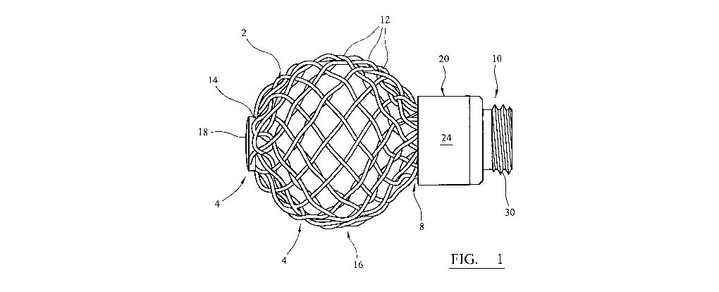
Figure 1 is a side view of a stranded support structure implant for location
within
a bone cavity to support the bone which defines the cavity.
Figure 2 is a sectional elevation through a part of the implant shown in
Figure 1,
on the line 11-11.
Figure 3 is an isometric view from below of a retainer clip which can be used
in
the implant of the invention.
Figure 4 is a top view of the stranded support structure implant showing its
first
end, with the retainer clip removed.
Figure 5a and 5b are isometric views of main and detachable parts of a first
mandrel which can be used to make a braided support structure implant.
Figure 6 is a side view of the first mandrel shown in Figure 5.
Figure 7 is an isometric view of a second mandrel which can be used to make a
braided support structure implant.
Figure 8 shows the braided support structure implant positioned on the second
mandrel during the manufacture of the implant.
Figure 9 shows an instrument which can be used to implant an implant.
Figure 10 shows the implant of the invention assembled on an instrument as
shown in Figure 9.
Figure 11 shows the implant and instrument which are shown in Figure 10, with
the implant deformed for implantation by means of the instrument.
Referring to the drawings, Figure 1 shows a stranded support structure implant
2 which can
be implanted in a cavity in a bone, to support the bone which defines the
cavity. The
CA 02752785 2011-08-17
WO 2010/097633 PCT/GB2010/050333
-15-
implant has a spherical portion 4 which is rounded at a first end 6 of the
implant, and a
cylindrical throat portion 8 at a second end 10 of the implant.
The implant 2 is formed from twelve wires 12 which are formed from a nickel
titanium
shape memory alloy which has been treated so that it exhibits enhanced elastic
properties.
The wires have a diameter of 0.5 mm.
Each of the wires is formed into a loop 14. The loops are gathered together at
the first end
6 of the implant so that two lengths of each wire extend from the first end.
There are
therefore 24 lengths of the wires extending from the first end of the implant,
which are
braided. The configuration of the spherical portion 4 is such that the implant
flares
outwardly from the first end 6 towards a wide point 16, and tapers inwardly
from the wide
point towards the throat portion 8.
The implant includes a retainer clip 18 at its first end which engages the
twelve loops 14
formed in the wires 12 to control their spacing. The retainer clip is
described in more
detail below with reference to Figure 3.
The implant includes a ring clamp 20. Details of the ring clamp are shown in
Figure 2.
The ring clamp 20 comprises an internal support ring 22 and an outer ring 24.
The internal
support ring is formed from stainless steel. It defines a cylindrical support
surface 26
which extends axially along the ring from a first end, up to a step 28. The
internal support
ring has an externally threaded collar 30 at its second end, beyond the step
28.
The outer ring 24 is formed from a nickel titanium based shape memory alloy
which is
treated so that it can be heated to a temperature which is above the
characteristic Af
temperature of the alloy to cause the ring to contract radially.
The ring clamp can be used to fasten the ends of the braided wires 12 at the
second end of
the device. The external diameter of the cylindrical support surface 26 of the
internal
support ring 22 is approximately equal to the internal diameter of the braided
wires in the
throat portion 8. The braided wires are trimmed to fit on the support surface
26, with their
CA 02752785 2011-08-17
WO 2010/097633 PCT/GB2010/050333
-16-
free ends abutting the step 28. The outer ring 24 is shrunk on to the wires so
that they are
clamped firmly between the outer ring and the support surface. The internal
diameter of
the outer ring if allowed to shrink without any restraint is slightly less
than the external
diameter of the wires when fitted over the cylindrical support surface of the
internal
support ring.
Figure 3 shows the retainer clip 18 which is used at the first end of the
support structure
implant to retain the loops 14 in their gathered configuration. The clip
comprises a central
hub 30 and six fingers 32 which extend radially from the hub. The hub has a
hole 34
extending through it. The clip is formed from stainless steel sheet by
pressing.
Figure 4 shows the aligned loops 14 which are formed in the wires 12. As can
be seen, the
loops are aligned in pairs. Within each pair of aligned loops, one loop can be
considered to
be displaced in a clockwise direction relative to the other loop. On this
basis, it can be seen
in Figure 4 that the clockwise loop of each pair is positioned under the
anticlockwise loop.
The reverse arrangement can be used in the alternative.
Each of the fingers 32 of the retainer clip 18 can be passed through two
aligned loops 14
which are formed in the wires 12. Each finger can be folded back on itself and
then retains
its folded shape.
Use of the retainer clip at the first end of the implant means that, when the
implant is
subjected to a transverse compressive force, it tends to have a flatter, more
rounded shape
at the first end when subjected to a transverse compressive force compared
with an implant
which does not include a retainer clip, which tends to fold at the first end.
Figures 5a, 5b and 6 show a first mandrel 102 which can be used in a braiding
machine to
in a first step of manufacturing a support structure implant according to the
invention. The
first mandrel has a main part 103 (Figure 5a) and a detachable part 104
(Figure 5b).
The main part 103 of the mandrel extends from a first end 106 to a second end
114. It has
a constant diameter wide portion 110 at the first end, and a constant diameter
narrow
CA 02752785 2011-08-17
WO 2010/097633 PCT/GB2010/050333
-17-
portion 112 at the second end 114. The mandrel includes a hemispherical
transition
portion 116 between the wide portion 110 and the narrow portion 112.
The main part of the mandrel has a blind socket 111 formed in it at the first
end.
The detachable part 104 of the mandrel can be mounted end-to-end with the main
part at
the end of the wide portion 110. It has a spigot 1 l5which can fit in the
socket 111 in the
main part of the mandrel. Its diameter where it is mounted end-to-end with the
wide
portion is the same as that of the wide portion. The detachable part has a
frustoconical
shape, tapering inwardly in a direction away from the main part.
The detachable part 104 of the mandrel has twelve bores 120 formed in its on
its outer
cylindrical surface. The pins are spaced apart equally around the periphery of
the mandrel,
close to the wide portion of the main part of the mandrel. As shown in Figure
6, a pin 122
can be fitted into each of the bores 120 Twelve lengths of wire are used in
the braiding
machine to fabricate the implant. In use, each length of wire is arranged so
that it extends
from one bobbin, around one of the pins on the mandrel, and back to another
bobbin. Each
wire is wound around a pin so that the two lengths of the wire cross between
the pin and
the bobbins. Each wire passes around its respective pin in the same direction
(clockwise or
anticlockwise).
The main part of the mandrel has a socket formed in it at the first end. The
detachable part
of the mandrel has a spigot formed on it. The main part and the detachable
part can be
fitted together by locating the spigot on the detachable part in the socket in
the main part.
Alternatively, the first mandrel can be made as a single component instead of
having
separable main and detachable parts. The first mandrel is shown ready for use
in Figure 6,
with the spigot 115 on the detachable part 104 received in the socket 111 on
the main part
103.
CA 02752785 2011-08-17
WO 2010/097633 PCT/GB2010/050333
-18-
The dimensions of an embodiment of the first mandrel 102 are as follows:
Diameter of narrow portion 112 7 mm
Radius of hemispherical portion 116 10 mm
Diameter of wide portion 110 20 mm
Length of wide portion 110 9.4 mm
Included angle of the frustoconical portion of detachable part 104 200
Distance from wide portion to pins 122 2.5 mm
Figure 7 shows a second mandrel 130. It has a constant diameter narrow portion
narrow
132 which has the same dimensions as the constant diameter narrow portion 122
of the first
mandrel. The constant diameter narrow portion is joined to a spherical portion
134. The
diameter of the spherical portion of the second mandrel is the same as the
diameter of the
hemispherical transition portion 116 of the first mandrel. The second mandrel
has six
holes 136 at its first end which are spaced apart equally around the pole of
the mandrel.
Pins can be fitted into the holes.
A support structure implant according to the invention can be made from a wire
made from
a binary nickel titanium alloy containing 50.8 wt.% nickel. The alloy is
treated so that the
wire exhibits enhanced elastic properties at temperatures in the range 20 to
45 O C.
The first mandrel is used in a braiding machine which has a plurality of
bobbins with
respective drives and mounts as used conventionally to form braided articles
from wire.
The braiding machine is operated conventionally to construct a tubular braid
over the
mandrel from the wires which are laid up between the bobbins and the pins,
with each wire
extending from a first bobbin, around a pin and back to a second bobbin.
The mandrel with the braided wires is removed from within the braiding machine
after the
wire has been braided over the wide portion 110, the hemispherical transition
portion 116
and on to the narrow portion 112. First and second clamps are applied to the
wires to
clamp them to the narrow cylindrical portion 112. The first clamp is
positioned as close as
CA 02752785 2011-08-17
WO 2010/097633 PCT/GB2010/050333
-19-
possible to the hemispherical transition portion 116. The second clamp is
positioned so
that the length of the braided tubular sleeve between the clamps is long
enough to form the
throat portion of the implant, and is not subject to any unravelling of the
braid. The clamps
should be capable of being tightened around the wires and the mandrel. Clamp
designs
might include for example hose clamps. A suitable clamp might make use of a
screw
thread actuator in the manner of a worm drive.
The first mandrel with the braided tubular sleeve is then placed in an oven at
500 C for
minutes to heat set the wires so that they follow the shape of the mandrel.
The first and second clamps are then removed and the first mandrel 102 is
removed from
10 within the braided tubular sleeve. It is replaced with a second mandrel
130. As shown in
Figure 8, third and fourth clamps 138, 140 are fitted to the sleeve to clamp
the wires to the
narrow cylindrical portion 132. The third clamp is positioned as close as
possible to the -
spherical transition portion 134. The fourth clamp is positioned so that the
length of the
braided tubular sleeve between the clamps is long enough to form the throat
portion of the
15 implant, and is not subject to any unravelling of the braid.
The distance between the holes 120 on the first mandrel for receiving pins and
the
transition between the hemispherical portion 116 and the constant diameter
wide portion
110 of the first mandrel is the same as the distance measured on the spherical
surface of the
spherical portion 134 of the second mandrel between its equator and the holes
136 for
receiving pins. Accordingly, the straight portion of the sleeve can be
contracted around the
spherical portion 134 of the mandrel and the loops 14 in the wires fitted over
the holes 136
and held there by means of pins. Two loops are held in place by each pin. The
mandrel
with the braided tubular sleeve are then placed in an oven at 500 C for 15
minutes to heat
set the wires so that they follow the spherical shape of the second mandrel,
and the second
mandrel is then removed from within the sleeve.
The retainer clip is fitted at the first end of the braided sleeve, as
discussed above with
reference to Figure 3.
CA 02752785 2011-08-17
WO 2010/097633 PCT/GB2010/050333
-20-
A ring clamp 150 is fitted at the second end of the sleeve. The ring clamp is
described
above with reference to Figures 1 and 2. The braided wires are cut so that the
length of the
wires in the throat portion allows the cylindrical support surface 26 of the
internal support
ring 22 to fit within the throat portion with the wires sitting on the support
surface, abutting
the step 28. The outer ring 24 is shrunk on to the wires so that they are
clamped firmly
between the outer ring and the support surface. The internal diameter of the
outer ring if
allowed to shrink without any restraint is slightly less than the external
diameter of the
wires when fitted over the cylindrical support surface of the internal support
ring.
The support structure implant can be implanted in a bone cavity to support the
bone which
defines the cavity, for example in the treatment of AVN in the femoral head.
A first step involves forming a tubular bore extending from the lateral cortex
along the
femoral neck, communicating with the affected region of the femoral head. This
can be
done with a bore cutting tool such as a drill.
A second step involves cutting away necrotic tissue. This can be achieved
using a cutter
which can be deployed in the vicinity of the necrotic tissue, such as those
disclosed in
US-A-2005/0240193 and WO-A-2008/0099187. The bone is then ready to receive the
implant of the invention.
Figure 9 shows an insertion tool 200 can be used to implant which comprises an
hollow
sheath 202 having a shaft 204 arranged to slide within it. The sheath has a
connector 206
at its remote end which is internally threaded so that it can engage the
threads 30 on the
internal support ring 22 of the ring clamp 20.
The shaft 204 has a tip 208 which can fit into the hole 34 in the hub 30 of
the retainer clip
18.
Accordingly, the support structure implant 2 can be fitted to the insertion
tool 200 by
inserting the tip 208 of shaft 204 through the throat of the implant and
advancing sheath
202 is until the threads on the connector 206 can be engaged with the threads
30 on the
CA 02752785 2011-08-17
WO 2010/097633 PCT/GB2010/050333
-21-
internal support ring 22 of the ring clamp 20. Figure 10 shows the implant and
the
insertion tool assembled in this way, with the wires 12 extending between the
internal
support ring 22 and the retainer clip 18 shown schematically.
The tip 208 of the shaft 204 can be advanced relative to the sheath 202 until
it is received
in the hole 34 in the hub of the retainer clip. Advancing the shaft 204
further relative to the
sheath 202 causes the implant 2 to elongate and a consequent reduction in the
width of the
implant. In this way, by application of a force of, for example about 300 to
400 N, the
length of the implant (measured from the end of the ring clamp to the first
end of the
implant) can be increased from 22 mm to about 30 mm, and its maximum width can
be
reduced from 22 mm to about 12 mm. The implant is shown in its elongated
configuration
in Figure 11. Deformation of the implant in this way allows it to pass along a
bore in the
patient's bone, into the cavity in the bone. The folded fingers 32 of the clip
allow the loops
in each of the wires to pivot about the line on which the finger is folded, in
a similar way to
the flexing of a hinge. The extent of such movement of the wires relative to
the retainer
clip can vary around the clip, allowing asymmetric deformation of the implant
prior to and
during implantation, and when implanted. The clip can provide control over the
shape of
the support structure when it is deformed for such implantation. For example,
a clip can
help to reduce the tendency for the implant to fold at the pole, instead
ensuring that the
shape of implant remains at least partly curved.
The insertion tool 200 can then be disengaged from the implant by unscrewing
the threads
on the connector 206 from the threads 30 on the internal support ring 22 of
the ring clamp
20, and removed from within the patient's bone. Bone chips can then be placed
within the
cavity through the bore in the ring clamp and the throat portion of the
implant.
It is an advantage of the implant of the invention that the screw threads on
the ring clamp
can be used to engage a tool, which might be similar to the insertion tool
described above,
in a procedure to remove the implant from within the bone cavity.