Note: Descriptions are shown in the official language in which they were submitted.
CA 02754128 2011-08-31
WO 2010/102119
PCT/US2010/026227
COMBINED FIELD LOCATION AND MRI TRACKING
FIELD OF THE INVENTION
[0001] The invention relates to the tracking of medical devices used in
diagnostic and therapeutic procedures and in particular to a system and method
for
combining field location tracking with magnetic resonance imaging tracking of
a
medical device.
BACKGROUND OF THE INVENTION
[0002] MRI has achieved prominence as a diagnostic imaging modality, and
increasingly as an interventional imaging modality. The primary benefits of
MRI
over other imaging modalities, such as X-ray, include superior soft tissue
imaging
and avoiding patient exposure to ionizing radiation produced by X-rays. MRI's
superior soft tissue imaging capabilities have offered great clinical benefit
with
respect to diagnostic imaging. Similarly, interventional procedures, which
have
traditionally used X-ray imaging for guidance, stand to benefit greatly from
MRI's
soft tissue imaging capabilities. In addition, the significant patient
exposure to
ionizing radiation associated with traditional X-ray guided interventional
procedures is eliminated with MRI guidance.
[0003] MRI uses three fields to image patient anatomy: a large static
magnetic field, a time-varying magnetic gradient field, and a radiofrequency
(RF)
electromagnetic field. The static magnetic field and time-varying magnetic
gradient field work in concert to establish both proton alignment with the
static
magnetic field and also spatially dependent proton spin frequencies (resonant
frequencies) within the patient. The RF field, applied at the resonance
frequencies, disturbs the initial alignment, such that when the protons relax
back
to their initial alignment, the RF emitted from the relaxation event may be
detected and processed to create an image.
[0004] For imaging of soft tissue of patients with implanted medical
devices,
such as catheters, guidewires, stents, cardiac defibrillators (ICDs),
pacemakers,
- 1 -
CA 02754128 2011-08-31
WO 2010/102119
PCT/US2010/026227
neurostimulators, cochlear implants, and the like, MRI is preferable to other
modalities including X-ray, computer tomography, ultrasound and positron
emission tomography (PET).
[0005] Localization of medical devices during use is desirable and often
required for medical procedures. For example, as a medical device is advanced
through the patient's body during an interventional procedure, its progress
may be
tracked so that the device can be delivered properly to a target site. Once
delivered
to the target site, the device can be monitored to determine whether it has
been
placed properly and/or is functioning properly. Providing the ability to track
the
location of medical devices is useful in interventional procedures such as
cardiac
electrophysiology procedures including diagnostic procedures for diagnosing
arrhythmias and ablation procedures such as atrial fibrillation ablation,
ventricular
tachycardia ablation, atrial flutter ablation, Wolfe Parkinson White Syndrome
ablation, AV node ablation, SVT ablations and the like. Tracking the location
of
.. medical devices using MRI is also useful in oncological procedures such as
breast,
liver and prostate tumor ablations; and urological procedures such as uterine
fibroid and enlarged prostate ablations.
[00061 Currently, several methods of locating position(s) of a medical
device
during a medical procedure exist. One exemplary method is a magnetic field
method. In this method, a magnetic field is transmitted that permeates all non-
metallic surfaces. A miniaturized sensor designed for medical applications is
placed on the instrument that is inserted into the body. The location of the
sensor
may be determined based upon magnetic field strength and/or orientation.
Another exemplary method is an impedance based method. In this method, an
electric field is transmitted through the body and the bioimpedance is
measured
between locations. The location of a medical device or instrument may then be
determined based upon the impedance variance. Another exemplary method
utilizes an ultrasound transducer to provide an image of a medical device and
procedural tissue used in positioning. Yet another exemplary method uses
optical
trackers that emit or reflect a light source that is in turn sensed by one or
more
- 2 -
CA 02754128 2011-08-31
WO 2010/102119
PCT/US2010/026227
detectors. The light source is typically infrared, but may alternatively
operate in
another frequency range as will be appreciated by those skilled in the art.
[0007] This non-exhaustive list of exemplary methods may be tenned
"field
location" techniques. Each of these field location techniques provides spatial
coordinates (i.e. x, y, z) relative to a point external to the patient. The
spatial
coordinates are provided in what is commonly referred to as "absolute" space.
As
appreciated by those skilled in the art, providing spatial coordinates in
absolute
space requires registration of the external point relative to the patient.
Thus, one
disadvantage of such field location techniques arises from the fact that if
the
position of the patient changes during a procedure, re-registration with
respect to
the external reference location is required. Another disadvantage of field
location
techniques is their inherent accuracy limitations due to non-ideal and/or non-
homogeneous field behavior in the body.
[0008] In an attempt to overcome the disadvantages inherent in field
location
techniques, it is possible to utilize the MR scanner to determine the location
of a
tracking coil embedded in or attached to the medical device or instrument.
Thus,
tracking position using the MR scanner is an alternative to using field
location
techniques such as those previously described. MR tracking has the advantage
of
requiring no registration with respect to any external point or reference
images
generated by the MRI, as images created with MRI are referenced to so-called
"patient" space. However, when MRI is utilized for both tracking and imaging,
there may be a decrease in the imaging performance because tracking sequences
must be time multiplexed with imaging sequences.
[0009] When used in combination with MRI, field location techniques will
suffer from being referenced to absolute space rather than patient space.
Patient
space is a coordinate system that includes spatial warping caused by non-ideal
gradient fields. For instance, assume that at some absolute point (x = y = z =
0),
patient and absolute space may be perfectly aligned. However, as one moves
away from that point, patient space may be nonlinear or increase with a
different
scale as compared to absolute space. As such, circular objects imaged with MRI
- 3 -
CA 02754128 2015-11-12
= 74105-47
may appear somewhat oblong. Correction software in the MRI may be used to
compensate
for this effect. Such compensation is, in general, dynamic, in that different
compensation is
required for different images, depending upon several variables. In addition,
absolute space
may be offset from patent space such that registration of the two spaces is
required (for
example, (x = y = z = 0) for absolute space may not be (x = y = z = 0) for
patient space, and/or
the two spaces may be rotated with respect to one another.
[0010] As will be understood based on the foregoing, current
technologies for tracking
a medical device are inadequate. Thus, what is needed is a system and method
that combines
the benefits of both field location and MRI techniques to provide an improved
means for
locating and tracking a medical device.
BRIEF SUMMARY OF THE INVENTION
[0011] The present invention solves the foregoing needs by
providing a composite
tracking system for a medical device that includes a field location tracking
system having at
least one field location sensor structured to be coupled to a medical device,
a magnetic
resonance tracking system having at least one tracking coil structured to be
coupled to a
medical device, and a composite tracking processor operably coupled to the
field location
tracking system and the magnetic resonance tracking system. The composite
tracking
processor is operable to receive and process field location parameters from
the field location
tracking system and positional coordinates from the magnetic resonance
tracking system to
register a field location coordinate system to a magnetic resonance coordinate
system.
[0011a] According to another aspect, there is provided a composite
tracking system for
tracking a medical device, the tracking system configured to be used in an MR
environment
and comprising: an MR compatible field location tracking system including at
least one field
location sensor structured to be coupled to an outside of the medical device,
said medical
device movably positioned within a patient; a magnetic resonance tracking
system including
at least one MR tracking coil structured to be coupled to the outside of the
medical device;
and a composite tracking processor in electrical communication with the field
location
tracking system and the magnetic resonance tracking system, the composite
tracking
processor operable to (a) receive and process a plurality of positional
coordinates from the
- 4 -
81704992
magnetic resonance tracking system and calculate a plurality of magnetic
resonance tracking
locations, (b) receive and process a plurality of field location parameters
from the field
location tracking system and determine a plurality of field location
parameters that correspond
to the plurality of magnetic resonance tracking locations, (c) generate a
transfer function for
.. mapping the field location parameters from the field location tracking
system to the
corresponding positional coordinates of the magnetic resonance tracking system
to register the
field location coordinate system to the magnetic resonance coordinate system,
and (d)
determine a present location of the field location sensor by applying the
transfer function,
wherein said at least one field location sensor and said at least one tracking
coil are configured
to move relative to the patient.
[0012] In accordance with another aspect of the present invention, a
method of
calibrating field location tracking to magnetic resonance tracking is provided
that generally
includes the steps of moving a medical device throughout a plurality of points
within a patient
volume, tracking the medical device with a field location tracking system and
a magnetic
.. resonance tracking system, calculating a plurality of magnetic resonance
tracking locations,
determining a plurality of field location parameters that correspond to the
plurality of magnetic
resonance tracking locations, and creating a transfer function that maps the
field location
parameters to the magnetic resonance tracking locations, wherein the transfer
function registers
a field location coordinate system to a magnetic resonance coordinate system.
BRIEF DESCRIPTION OF THE DRAWINGS
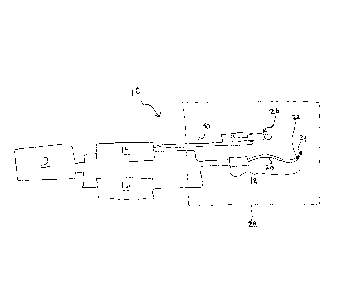
[0013] FIG. 1 is a diagram illustrating one exemplary embodiment of a
composite
tracking system in accordance with the present invention.
[0014] FIG. 2 is a flow diagram broadly illustrating an exemplary
composite tracking
method in accordance with the present invention.
100151 FIG. 3 is a flow diagram illustrating exemplary steps in a
calibration process in
accordance with one embodiment of the present invention.
- 5 -
CA 2754128 2018-04-19
81704992
[0016] FIG. 4 is a table showing a plurality of recorded data sets in
monotonically
increasing order.
[0017] FIG. 5 is a graph illustrating an exemplary mapping of
calculated x-location
values in patient space and corresponding field location parameter values.
[0018] FIG. 6 is a graph illustrating an exemplary mapping of calculated y-
location
values in patient space and corresponding field location parameter values.
[0019] FIG. 7 is a graph illustrating an exemplary mapping of
calculated z-location
values in patient space and corresponding field location parameter values.
[0020] FIG. 8 is a flow diagram illustrating exemplary steps in a
locating process in
accordance with one embodiment of the present invention.
[0021] FIG. 9 is a table showing an exemplary multidimensional
coordinate set that
may be used in accordance with one alternative method of the present
invention.
- 5a -
CA 2754128 2018-04-19
CA 02754128 2011-08-31
WO 2010/102119
PCT/US2010/026227
DETAILED DESCRIPTION OF THE INVENTION
[0022] The present invention is a system and method for combining field
location techniques with MRI tracking to provide an improved means for
locating
and tracking the position of a medical device. Generally speaking, both MRI
tracking and field location are performed simultaneously to collect position
or
location data. The MRI tracking location is then used to calibrate and
register the
field location, effectively compensating for gradient warping effects and
minimizing the field location inaccuracies. The calibration may be performed
with software, and may be accomplished with or without user input. Once this
calibration is performed, the MRI tracking may be turned off and the field
location
may be used to locate and track the medical device in patient space while
retaining
the accuracy benefits provided by MRI tracking. During a medical procedure
using this technique, periodic calibration sequences may be performed to
ensure
that the field location does not lose registration or calibration.
[0023] MR tracking is a well-known technique wherein an MR tracking coil
is embedded in a medical device such that the location of the MR tracking coil
may be determined. This is typically accomplished by applying a pulse sequence
in which only one of the three gradients is applied at a given time, and MR k-
space data is recorded from the signal received by the MR tracking coil. By
calculating the Fourier Transform of each of the three received k-space
signals,
the location of the MR tracking coil in each of the gradient directions may be
determined (i.e. x-, y-, and z-directions).
[0024] A key advantage of MR tracking when used with MR imaging is that
the location of the MR tracking coil is relative to "patient" space. Patient
space is
an image space which may be distorted from real or "absolute" space. Since the
distortion or warping of a given MR image will give rise to an identical
distortion
or warping of the MR tracking location, the two are matched and the MR
tracking
coil location can be precisely determined relative to the tissue in which it
resides.
For example, if a circular object is scanned with MRI and the resultant image
is an
oval, guiding an MR tracking coil around the circumference of the circle and
- 6 -
CA 02754128 2011-08-31
WO 2010/102119
PCT/US2010/026227
tracking its location would result in the recorded location points also
defining an
oval shape. However, whether the resultant image (i.e. an oval) accurately
represents the actual shape (i.e. a circle) is irrelevant. It is only
important that the
relative location of the tracking coil to the circle is consistent and
repeatable. One
drawback to MR tracking is that the tracking sequences must be interweaved
between imaging sequences. As a result, imaging speed performance may suffer.
This may be particularly problematic when imaging in real time or near real
time,
where fast image formation and frame rates are desirable.
[0025] As previously discussed, numerous field location techniques exist
including, but not limited to, impedance based field location, magnetic field
location, electromagnetic field location, optical field location, and
ultrasonic field
location. Each of these techniques involves the measurement of a different
electromagnetic or mechanical field parameter such as impedance, voltage,
current, time delay, sound intensity, or the like. Regardless of the type of
field
parameter measured, each of the various field location techniques may be used
to
determine positional locations in absolute space. By way of example, consider
basic impedance based field location. For this technique, three sets of
external
electrode patches are typically placed on the patient in predetermined
locations. A
first set of patches creates an electric field in the x-direction, a second
set of
patches creates an electric field in the y-direction, and a third set of
patches
creates an electric field in the z-direction. Voltage measured by the field
location
system at a sensor on the medical device, such as an electrode in the case of
an
impedance based system, may be used to determine the location of the device in
each of the three dimensions, one at a time. The three measurements may be
acquired quicicly enough to be considered coincident in time for all practical
purposes.
[0026] The error associated with field location technologies varies, and
processes have been developed in an attempt to minimize the error. These
processes are specific to the various field location technologies, and the
applicable
process therefore depends upon the technique that is being used. One major
drawback is that field location techniques estimate the location of a device
in
- 7 -
CA 02754128 2011-08-31
WO 2010/102119
PCT/US2010/026227
absolute space, which may not correspond well to patient space, as described
above. Additionally, non-homogenous characteristics of patient tissue produce
errors in many field location techniques.
[0027] This invention broadly combines field location techniques with MR
tracking such that the key advantage of MR tracking, i.e. precise location of
the
MR tracking coil relative to tissue, can be used to calibrate a field location
technique. This allows the field location technique to precisely locate a
medical
device in the patient with similar performance to MR tracking, but without
having
to interrupt MR imaging pulse sequences to run MR tracking pulse sequences.
Thus, the present invention combines the "accuracy" benefits of MR tracking
with
the "time performance" benefits of field location techniques.
[0028] FIG. 1 is a diagram illustrating one exemplary composite tracking
system 10 in accordance with the present invention. As illustrated in FIG. 1,
the
composite tracking system 10 generally includes a composite tracking processor
12, a field location system 14, and a MR tracking system 16. The field
location
system 14 and the MR tracking system 16 are operably coupled to the composite
tracking processor 12 to provide field location and MR tracking information,
respectively, to the composite tracking processor 12 for processing. The
composite tracking system 10 further includes a medical device 18 such as a
catheter having a body 20 with at least one field location sensor 22 and at
least
one MR tracking coil 24. The medical device 18 is represented generically
herein
as a catheter merely for purposes of example and not limitation. However, the
system and method of the present invention may be utilized with any type of
medical device that necessitates tracking as will be appreciated by those
skilled in
the art.
[0029] Although the field location sensor 22 and the MR tracking coil 24
may be offset from one another, they are preferably in close proximity, such
as
separated by an offset distance of less than about 5 mm in one exemplary
embodiment. As will be appreciated by those skilled in the art, if the field
location sensor 22 and the MR tracking coil 24 are separated by a large
distance
- 8 -
CA 02754128 2011-08-31
WO 2010/102119
PCT/US2010/026227
and the portion of the medical device upon which they are attached experiences
bending or deformation, the relative distance between the two elements may
change significantly which could in turn impact the accuracy of the
calibration
process. In alternative embodiments where multiple field location sensors 22
and/or MR tracking coils 24 are utilized, the need for the sensors and/or
tracking
coils to be in close proximity may be eliminated, as long as the distance
between
the field location sensors and the MR tracking coils is both fixed and known.
[0030] As further illustrated in FIG. 1, a patient 26 is positioned
within an
MR scanner 28. A plurality of field location sources/receivers 30 are
positioned
external to the patient 26. In one exemplary embodiment, the field location
sources/receivers 30 may comprise electrode patches as described above for the
example of impedance based field location. Particularly, three sets of field
location sources/receivers 30 would be placed on the patient in predetelinined
locations to create an electric field in each of the x-, y-, and z-directions.
The
structure, function, and number of the field location sources/receivers 30
will be
dependent upon the class of field location technology being used. Thus,
impedance based techniques are described herein merely for purposes of example
and not limitation.
[0031] The field location system 14 is positioned outside of the MR
scanner
28, and is operably coupled to the external field location sources/receivers
30 as
well as to the field location sensor 22 on the medical device 18. The MR
tracking
system 16 is operably coupled to the MR tracking coil 24 on the medical device
18. The MR tracking system 16 may be structured as part of the MR scanner 28,
but may also include an external MR processor for determining the location of
the
MR tracking coil 24 based on raw k-space data received from the MR scanner 28.
The composite tracking processor 12 is operably coupled so as to receive MR
tracking coil location data from the MR tracking system 16 and parameter data
from the field location system 14. Once again, the nature of the parameter
data
will vary depending on the class of field location technique being used, but
may
comprise voltage, impedance, current, time delay, sound intensity, or the
like.
With regard to impedance base field location, for example, the field location
- 9 -
CA 02754128 2011-08-31
WO 2010/102119
PCT/US2010/026227
system 14 may measure voltages at the field location sensor 22 on the medical
device 18, which may be processed by the field location system 14 or the
composite tracking processor 12 to determine the positional coordinates of the
device in the x-, y-, and z-directions.
[0032] Now that one exemplary embodiment of a composite tracking system
has been described, an exemplary method of operating the composite tracking
system to allow a field location system to precisely locate a medical device
in
patient space with similar performance to MR tracking will be described. The
exemplary method of the present invention may generally be separated into two
processes, including a calibration process 200 and a locating process 300.
FIG. 2
is a flow diagram broadly illustrating the exemplary method 100 of the present
invention. The calibration process 200 and the locating process 300 are
described
in greater detail with reference to FIGS. 3-9.
[0033] As illustrated in FIG. 2, the exemplary method 100 begins at step
102
with the calibration process 200. The calibration process 200 is operable to
register field location space (i.e. absolute space) to MR space (i.e. patient
space).
Once the calibration process is complete and field location space is
registered to
MR space, the method 100 continues at step 104 where MR tracking is
discontinued and field location may be used to locate a medical device in MR
space. Optionally, at step 106, the calibration process 200 may be repeated
periodically to ensure that the field location space does not lose
registration with
the MR space.
[0034] FIG. 3 is a flow diagram illustrating exemplary steps in the
calibration
process 200 in accordance with one embodiment of the present invention.
Beginning with step 202, a medical device is provided having at least one
field
location sensor and at least one MR tracking coil operably coupled thereto.
The
medical device may be any type of medical device that necessitates tracking.
The
structure and function of the field location sensor will depend upon the field
parameter being measured, such as voltage, impedance, current, time delay,
sound
intensity, or the like.
-10-
CA 02754128 2011-08-31
WO 2010/102119
PCT/US2010/026227
[0035] The process continues at step 204 where the medical device is
positioned within the region of interest in the patient. This region
corresponds to
the range of locations wherein the medical device will later be tracked by
field
location alone. As the medical device is being positioned within the region of
interest, the location of the medical device is tracked simultaneously with
the field
location system and the MR tracking system. Next, in step 206, an MR tracking
location in three-dimensional space is calculated. The MR tracking location
includes an x-location value, a y-location value, and a z-location value that
together provide the three-dimensional location of the MR tracking coil at
that
particular instant in time. The field location parameter values that
correspond to
the MR tracking location values are simultaneously recorded in step 208. Thus,
the recorded data set will include an x-location parameter value, a y-location
parameter value, and a z-location parameter value.
[0036] Once the three-dimensional MR tracking location in patient space
is
calculated and the corresponding field location parameter values recorded, the
process continues at step 210 wherein the system will determine whether the
requisite number of sets N of field location parameter values (px, py, pz) and
MR
tracking location values (x, y, z) have been collected. As will be appreciated
by
those skilled in the art, the number of data sets N that must be collected may
be
any number greater than or equal to two, and may depend upon the tissue volume
or range in which tracking is needed. As will be appreciated by those skilled
in
the art, two data sets will provide only a linear mapping. Thus, a larger
number of
data sets may be used in order to generate a polynomial mapping, which will
improve the precision of the calibration process. The data sets may preferably
include points along the outer boundary of the patient volume as well as
points
within the boundary.
[0037] If the system determines that the requisite number of data sets N
has
not been collected, the process 200 enters a loop 211 where steps 204-208 are
repeated for additional positions of the medical device within the patient
region of
interest. Once the system determines that the requisite number of data sets N
has
-11 -
CA 02754128 2011-08-31
WO 2010/102119
PCT/US2010/026227
been collected, this portion of the process is complete and the process moves
on to
step 212
[0038] In step 212, the N data sets of (px, py, pz)/(x, y, z) values are
ordered in
monotonically increasing order and stored in memory. This process is
illustrated
in the table shown in FIG. 4. After ordering the data sets in step 212, the
process
continues at step 214 where mappings of the N data sets are generated. In one
exemplary embodiment, three separate mappings may be generated including a
(px, x) mapping as illustrated in FIG. 5, a (py, y) mapping as illustrated in
FIG. 6,
and a (pt, z) mapping as illustrated in FIG. 7. These "mappings" represent
transfer functions that may be used during field location tracking to map a
measured field location parameter value to an observed point in patient space
as
will be discussed in further detail to follow. It is important to note that
the (IN, x),
(py, y), and (pz, z) coordinates do not need to be uniformly distributed along
either
of the axes.
[0039] Once the x-, y-, and z-location mappings are generated, the MR
tracking may be discontinued in step 216. The calibration process is now
complete, and field location space (i.e. absolute space) is registered to MR
space
(i.e. patient space). The surgeon may continue with the locating process 300
as
indicated at step 218.
[0040] FIG. 8 is a flow diagram illustrating exemplary steps in the
locating
process 300 in accordance with one embodiment of the present invention.
Beginning in step 302, with the field location system turned on and
operational,
the medical device may be positioned at or moved to a first patient location
within
the patient region where calibration was performed. Using the field location
system, the field location parameter values at the first patient location are
then
measured in step 304. The measured field location parameter values include an
x-
location parameter value (i,measured-x), a y-location parameter value
(Pmeasured-y), and
a z-location parameter value (Pmeasured-z)-
- 12 -
CA 02754128 2011-08-31
WO 2010/102119
PCT/US2010/026227
[0041] Turning next to step 306, the (px, x) mapping may be utilized to
determine an x-coordinate of the measured x-location parameter value in
patient
space. With reference to FIG. 5, the measured x-location parameter value is
first
plotted on the field location parameter axis. If the measured x-location
parameter
value happens to exactly match one of the field location parameter values
recorded during the calibration process 200, then the x-coordinate of the
measured
x-location parameter value in patient space will in turn be the corresponding
x-
location value calculated during the calibration process. However, because
there
are an almost infinite number of field location parameter values that could be
measured in the patient region of interest, it is unlikely that the measured x-
location parameter value will exactly match one of the field location
parameter
values recorded during the calibration process. In this instance, nearby (IN,
x) data
points that "surround" the measured x-location parameter value are determined
and an interpolation is performed between these (px, x) data points to
calculate an
estimated x-coordinate in patient space that corresponds with the measured x-
location parameter value in absolute space. This interpolation step may use
linear
interpolation or any suitable higher order interpolation as will be
appreciated by
those skilled in the art, such as polynomial interpolation. In the example
shown in
FIG. 5, the measured x-location parameter value is labeled n
"/ measured-x)" the closest
corresponding data points are (px5, x5) and (p,6, x6), and the interpolated x-
coordinate is labeled "Xinterp." This interpolated x-coordinate value
represents the
current patient space location of the medical device in the x-direction.
[0042] Turning next to step 308, the (py, y) mapping may be utilized to
determine a y-coordinate of the measured y-location parameter value in patient
space. With reference to FIG. 6, the measured y-location parameter value is
first
plotted on the field location parameter axis. If the measured y-location
parameter
value happens to exactly match one of the field location parameter values
recorded during the calibration process 200, then the y-coordinate of the
measured
y-location parameter value in patient space will in turn be the corresponding
y-
location value calculated during the calibration process. However, because
there
are an almost infinite number of field location parameter values that could be
- 13 -
CA 02754128 2011-08-31
WO 2010/102119
PCT/US2010/026227
measured in the patient region of interest, it is unlikely that the measured y-
location parameter value will exactly match one of the field location
parameter
values recorded during the calibration process. In this instance, nearby (py,
y) data
points that "surround" the measured y-location parameter value are determined
and an interpolation is performed between these (py, y) data points to
calculate an
estimated y-coordinate in patient space that corresponds with the measured y-
location parameter value in absolute space. In the example shown in FIG. 6,
the
measured y-location parameter value is labeled ". n measured-y3" the closest
corresponding data points are (py3, y3) and (py4, y4), and the interpolated y-
coordinate is labeled "Yinterp." This interpolated y-coordinate value
represents the
current patient space location of the medical device in the y-direction.
[0043] The process 300 continues with step 310, where the (pz, z)
mapping
may be utilized to determine a z-coordinate of the measured z-location
parameter
value in patient space. With reference to FIG. 7, the measured z-location
parameter value is first plotted on the field location parameter axis. As
discussed
above with regard to steps 306 and 308, if the measured z-location parameter
value happens to exactly match one of the field location parameter values
recorded during the calibration process 200, then the z-coordinate of the
measured
z-location parameter value in patient space will in turn be the corresponding
z-
location value calculated during the calibration process. However, it is
unlikely
that the measured z-location parameter value will exactly match one of the
field
location parameter values recorded during the calibration process. In this
instance, nearby (pz, z) data points that "surround" the measured z-location
parameter value are determined and an interpolation is performed between these
(pz, z) data points to calculate an estimated z-coordinate in patient space
that
corresponds with the measured z- location parameter value in absolute space.
In
the example shown in FIG. 7, the measured z-location parameter value is
labeled
"Pmeasured-z)" the closest corresponding data points are (p,3, z3) and (Pz4,
z4), and the
interpolated z-coordinate is labeled "zintõp." This interpolated z-coordinate
value
represents the current patient space location of the medical device in the z-
direction.
- 14-
CA 02754128 2011-08-31
WO 2010/102119
PCT/US2010/026227
[0044] Steps 306-310 have been described with reference to calculating
the
current x-, y-, and z-coordinate values by interpolation merely for pm-poses
of
example and not limitation. As will be appreciated by those skilled in the
art, the
x-, y-, and z-coordinate values may be calculated using any suitable
calculation
means, such as extrapolation. For example, if the measured x-, y-, or z-
location
parameter value is outside the range of data points previously recorded, steps
306-
310 may alternatively utilize extrapolation to estimate the current coordinate
values in patient space. As will be appreciated by those skilled in the art,
the
extrapolation step may use linear extrapolation or any suitable higher order
extrapolation, such as polynomial extrapolation.
[0045] As will be appreciated by those skilled in the art, the result of
steps
306-310 is a three-dimensional coordinate position that represents the current
location of the medical device in patient space that was determined using
field
location tracking. Once the current location of the medical device in three-
dimensional patient space has been determined, the location may be displayed
or
recorded in any suitable manner as recited in step 312. As will further be
appreciated by those skilled in the art, the medical device may be moved or
repositioned and the foregoing process repeated to determine the new three-
dimensional patient space position as recited in step 314.
[0046] Based upon the foregoing discussion, those skilled in the art will
appreciate that once a single field location sensor has been calibrated with
respect
to a single MR tracking coil, the same calibration data sets may be used to
determine the positions of additional field location sensors on the device.
Thus,
when a plurality of field location sensors is present on a device, it is not
necessary
to provide a corresponding plurality of MR tracking coils.
[0047] The calibration process 200 and locating process 300 were
described
with reference to coordinate sets that include separate (13,,, x), (py, 37),
and (pz, z)
coordinate values "mapped" into three separate mappings as illustrated in
FIGS. 5,
6, and 7 merely for purposes of example and not limitation. In one alternative
method in accordance with the present invention, a multidimensional coordinate
- 15-
CA 02754128 2011-08-31
WO 2010/102119
PCT/US2010/026227
set (põN, xN, pyN, yN, p,N, zN) may be formed, such as the exemplary
coordinate
set illustrated in FIG. 9. When a multidimensional coordinate set is generated
in
the calibration process, each measured parameter value (i.e. n
"-measured -x, Pmeasured-y,
and Pmeasured-z) that is measured during the locating process may be
interpolated in
3-space using mathematics well known to those skilled in the art to determine
the
current (x, y, z) location of the medical device in patient space. As will
further be
appreciated by those skilled in the art, although the mathematics differ when
a
multidimensional coordinate set is utilized, the general principles of the
present
invention previously discussed with reference to the calibration process 200
and
.. the locating process 300 are still applicable.
[0048] Although several exemplary steps were described with reference to
the calibration and locating processes, those skilled in the art will
appreciate that
the order and number of steps may be modified without departing from the
intended scope of the present invention. Thus, the exemplary steps were
provided
merely for purposes of example and not limitation.
[0049] Throughout the disclosure, reference was made to "current"
locations,
coordinate values, and the like. In this context the term "current" is used to
reference a point in time, a point in space, etc., and could be replaced by
any
synonymous term, such as "present."
[0050] As will further be appreciated by those skilled in the art, the
processes
previously described may be embodied as a system, method, or computer program
product. Accordingly, the present invention may take the form of an entirely
hardware embodiment, an entirely software embodiment (including firmware,
resident software, micro-code, etc.), or an embodiment combining software and
hardware aspects that may all generally be referred to as a "circuit,"
"module," or
"system." Furthermore, the present invention may take the form of a computer
program product embodied in any tangible medium of expression having
computer usable program code embodied in the medium.
-16-
CA 02754128 2013-03-04
74105-47
[0051] The processes comprising the method of the present invention
have
been described with reference to flow diagrams illustrating exemplary steps.
It
will be understood that each block of the flowchart diagrams, and combinations
of
blocks in the flowchart diagrams, can be implemented by computer program
instructions. These computer program instructions may be provided to a
processor of a general purpose computer, special purpose computer, or other
programmable data processing apparatus to produce a machine, such that the
instructions, which execute via the processor of the computer or other
programmable data processing apparatus, create means for implementing the
functions/acts specified in the flowchart diagram block or blocks.
[0052] These computer program instructions may also be stored in a
computer-readable medium that can direct a computer or other programmable data
processing apparatus to function in a particular manner, such that the
instructions
stored in the computer-readable medium produce an article of manufacture
including instruction means which implement the function/act specified in the
flowchart block or blocks.
100531 The computer program instructions may also be loaded onto a
computer or other programmable data processing apparatus to cause a series of
operational steps to be performed on the computer or other programmable
apparatus to produce a computer implemented process such that the instructions
which execute on the computer or other programmable apparatus provide
processes for implementing the functions/acts specified in the flowchart
diagram
block or blocks.
[00541 Although the present invention has been described with
reference to
preferred embodiments, workers skilled in the art will recognize that changes
may
be made in form and detail without departing from the scope of the invention
as claimed.
- 17-