Note: Descriptions are shown in the official language in which they were submitted.
CA 02754793 2011-10-03
System and method for restoring body parts
BACKGROUND
Field
[0001 ] The present application relates to restoration of body parts.
Description of the Related Art
[0002] Medical implants are used in various applications for the restoration
or the
replacement of body parts. For instance, such implants may be used to attach a
prosthesis to a bone. These implants can be installed prior to the design of
the
prosthesis, as it may be requested by the type of restoration or replacement
to be
performed on the body part.
[0003] Known methods applied for the design of prosthesis after installation
of
medical implants in a body part may involve, for example, the use of two-
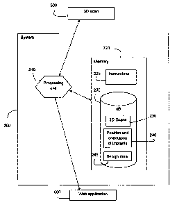
dimensional radiographic (X-ray) images to measure the distance between
implants. Other methods may require the use of a camera like in laparoscopic
interventions to retrieve the position of installed medical implants.
[0004] In the field of dental restoration, implant location data is often
obtained by
creating a model of the dental structure using a conforming material applied
to
the structure to retrieve imprints that are used afterwards to obtain a
casting for
the fabrication of a medical model and/or to obtain implant location data by
measurement. Also, the position and orientation of the implants is sometimes
estimated from the specifications of surgical guides used for implants
installation.
[0005] However, those methods are invasive and can be time ineffective for a
patient, as they lack accuracy and need further adjustments of the prosthesis
to
be precisely adapted to the installed implants. Therefore, there is a need to
address at least those issues.
CA 02754793 2011-10-03
SUMMARY
[0006] It is a broad aspect of an embodiment to provide a system for
determining
the position and orientation of implants located in a body part of a patient,
the
system comprising: a processing unit configured to operate the system, the
processing unit determines implant voxels having an intensity value
representing
the implants in a three-dimensional radiographic representation of the body
part,
selects a region to be searched for implants located in the body part,
generates
in the selected region multiple random virtual implants until obtaining a
score that
identifies implants located in the body part, the score being based on a
number
of implant voxels contained in each generated virtual implant, determines the
position and orientation of implants and generates data to be used to design a
medical model adapted to the implants located in the body part. The system
further comprises a memory unit configured to store instructions to be
executed
by the processing unit to determine the position and orientation of implants
located in the body part.
[0007] It is another broad aspect of an embodiment to provide a method for
determining the position and orientation of implants located in a body part of
a
patient, the method comprising: uploading a three-dimensional radiographic
representation of the body part, the three-dimensional radiographic
representation having voxels of different intensity values; determining
implant
voxel, based on an intensity value representing the implants in the
radiographic
representation; selecting a region to be searched for implants in the
radiographic
representation; generating multiple random virtual implants in the selected
region
until obtaining a score that identifies implants located in the body part, the
score
being based on the number of implant voxels contained in each generated
virtual
implant; determining, based on the score of virtual implants, the position and
orientation of the implants; and generating data to be used to design a
medical
model adapted to the implants located in the body part.
-2-
CA 02754793 2011-10-03
BRIEF DESCRIPTION OF THE DRAWINGS
[0008] Figure 1A illustrates a perspective view of a body part of a patient in
accordance with an embodiment;
[0009] Figure 1 B illustrates a perspective view of a body part that comprises
implants in accordance with an embodiment;
[0010] Figure 1C shows multiple views of three-dimensional X-ray
representations in accordance with an embodiment;
[0011] Figure 2 illustrates a schematic diagram of a system for determining
the
position and orientation of implants located in a body part in accordance with
an
embodiment; and
[0012] Figure 3 shows a block diagram of a method for determining the position
and orientation of implants located in a body part in accordance with an
embodiment.
DETAILED DESCRIPTION
[0013] In the following description, for purposes of explanation and not
limitation,
specific details are set forth such as particular architectures or techniques.
It will
be apparent to those skilled in the art that the system and method described
hereinafter may be practiced in other embodiments that depart from these
specific details.
[0014] The present application relates to restoration of body parts of a
patient. A
body part can be any human part such as a femur, a hip bone, a jawbone or the
like. Reference is now made to Figure 1A, which illustrates a front view of a
combination of a lower jawbone and gingiva 105 on which a dental prosthesis
can be installed to provide restoration of the dental structure. Implants 115
can
also have the shape and application of an anchor, a fixture or the like. In
Figure
-3-
CA 02754793 2011-10-03
1 B, the implants support a bar allowing a dental prosthesis to rest on the
jawbone and gingiva 105.
[0015] The bar can be designed, prototyped or manufactured providing that
exact
position and orientation of each of the implants 115 are known, so that a snug
and accurate fit of the bar on top of the implants is enabled. Pursuant to the
present disclosure, this can be accomplished without the use of a cast and
other
extensive steps according to known methods. The exact position and orientation
of each implant 115 can be determined by retrieving the position of each
implant
relative to XYZ reference axis in a three-dimensional radiographic
representation
such as a three-dimensional X-ray representation of the jawbone and gingiva
105. The three-dimensional X-ray representation of the jawbone and gingiva 105
including implants 115 can be generated with a minimally invasive technique,
such as a cone beam computer tomographic scanner (CBCT scan). A suitable
three-dimensional radiographic representation may also be generated by
positron emission tomography (PET SCAN), magnetic resonance imagery (MRI),
or like techniques usable for generating three-dimensional representations of
visible and hidden parts of the body and implanted structures. The
radiographic
representations such as DICOM files of X-ray images or representations can
then be analyzed with an appropriate computing system. An example of a three-
dimensional X-ray representation is provided in Figure IC, where sliced images
of the X-ray representation illustrate different views of a scanned body part.
[0016] A three-dimensional X-Ray representation is made of voxels, which is a
cubic picture element. The resolution corresponds to the voxel's side length.
The
resolution of voxels may be determined prior to the use of a three-dimensional
scanner, as a function of the desired accuracy in determining the position and
orientation of the implants. For example, the three-dimensional scanner may
generate a three-dimensional representation with voxels having a resolution of
less than a millimeter.
-4-
CA 02754793 2011-10-03
[0017]A voxel may have various properties. One of those properties is
indicative
of the opacity to radiation (or transitivity) of material in a generated three-
dimensional X-ray representation. For example, as a body part is made of
different compositions such as bones and tissues, they are represented with a
different opacity when they are scanned with the three-dimensional scanner.
This
is defined as the intensity value of a voxel. Similarly, an implant is made
from
high density material such as a metal and has a high opacity to which
corresponds a specific voxel intensity value.
[0018] Reference is now made to Figure 2, which illustrates a schematic
diagram
of a system 200. The system 200 comprises a processing unit 210. The
processing unit 210 can be any combination of software, hardware device that
can perform operations on data and instructions to and from other devices in
the
system 200. The system 200 also comprises a memory 220. The processing 210
unit can also interpret software programs and access the memory 220 to operate
the system 200. The processor unit 210 is configured to execute instructions
225
and to identify voxels in a three-dimensional X-ray representation of a body
part,
having an intensity value that corresponds with the material in which an
implant
is made.
[0019] The memory unit 220 also stores files of three-dimensional
representation
235 obtained from a three-dimensional radiographic scanner 500 in a database
(dB) 230 and transmitted to the system 200. The memory unit 220 can be for
example any combination of software, hardware device that can store data to be
written or accessed by the processing unit 210. The memory unit 220 is
configured to store instructions 230 to be executed by the processing unit 210
to
obtain the position and orientation (spatial coordinates) 240 of implants
located in
a body part of a patient. The orientation is the vertical vector that passes
through
-5-
CA 02754793 2011-10-03
the center axis of an implant and the position is an XYZ coordinate of the
point
located in the center of the top part of the implant.
[0020] Reference is now made to Figure 3, which shows a block diagram of a
method for determining the position and orientation of implants located in a
body
part of a patient. Steps described in the method may be executed sequentially
and repeatedly until the position and orientation of implants are retrieved.
The
system 200 may automatically detect a number of implants for which position
and
orientation are required to design an appropriate prosthesis or medical model.
Alternatively, the system 200 may ask a user to input the number of implants
for
which position and orientation are required to design an appropriate bar,
prosthesis or medical model.
[0021]At 310, the system uploads a three-dimensional radiographic (X-ray)
image file 235 from the memory 220. A user may upload a three-dimensional
representation received from the three-dimensional scanner 500 and may
observe the view of each sliced image as a bitmap. The user or alternatively
the
system 200 may determine the coordinates (Xmin, Xmax, Ymin, Ymax, Zmin,
Zmax) where the implants of interests are located based on the intensity value
of
the voxels.
[0022] At 315, the processing unit 210 identifies implant voxels, based on the
intensity value characterizing implants. Implant voxels are voxel that
represent a
portion of an implant in the three-dimensional X-ray representation. A three-
dimensional X-ray representation may comprise different voxel intensity values
or
colors such as white, grey or black voxels. A scanned implant made of metal
material can appear to be white, the bone can appear to be light grey, gingiva
can appear to be dark gray and a void space can appear to be black. This
allows
defining an intensity value based on the opacity of material similar to the
opacity
observed in 2D X-ray images. Therefore, this enables separating and
-6-
CA 02754793 2011-10-03
discriminating different scanned materials, such as implants, bones or
tissues.
The skilled reader would understand that implants can be made of different
material, which can be a non-metal material, such as a ceramic or composite
material like zirconia or hydroxypatite, which can generate a different
characteristic color or intensity when scanned with the three-dimensional
radiographic scanner 500.
[0023] At 320, the processing unit 210 selects a region (subset of voxels) to
be
searched for implants in the three-dimensional X-ray representation file. The
system 200 determines a bounded region, which can be aligned to form a box or
a different volumetric shape that contains the total sum of implant voxels in
the
three-dimensional X-ray representation. The system 200 may ask the user to
select from a virtual library the model of implants to be searched.
Alternatively,
the system may detect the model of implants while searching the position and
orientation of the implants by referring to an implant shapes library in
database
230 of memory 220 and executing instructions according to a shape recognition
algorithm.
[0024] At 325, the processing unit 210 generates virtual implants in the
selected
region. A virtual implant is a virtual replica of an implant model as selected
or
detected at 320, or an approximated shape such as a cylinder. A virtual
implant
has its own plain volume revolution with a position and an orientation. The
system 200 randomly generates within the selected region a large number of
different virtual implants, each having their own virtual position and an
orientation. The system 200 determines a matching score for each virtual
implant, which is based on the number of implant voxels contained inside each
generated virtual implant.
[0025] At 330, the processing unit 210 sorts each virtual implant according to
their matching score. The processing unit 210 repeats step 325 until the
highest
-7-
CA 02754793 2011-10-03
matching score obtained for a virtual implant stops increasing after a number
of
iterations. This determines the position and the orientation of an actual
implant.
The processing unit 210 then stores this position and orientation set 240 in
the
memory unit 220. The processing unit 210 eliminates each located implant from
the searched sets of voxels prior to repeat step 325. This is done to avoid
looking
for an implant for which a position and orientation have already been
determined.
[0026]At 335, when the processing unit 210 determines that the position and
orientation 240 of all implants are identified, it generates design data 245
to be
used to design a medical model, such as a bar, adapted to fit the located
implants. Design data 245 can be generated and stored as a file, such as an
STL
file, in the memory 220. The processing unit 210 may use spatial coordinates
of
the implants to determine the virtual model of a prosthesis to be adapted to
the
located implants.
[0027] The system 200 may use a world wide web (WWW) application 600 for
transmitting design data 245 of the virtual model to be adapted to the located
implants, for example, from a medical clinic to a location where the
prosthesis
can be prototyped or manufactured.
[0028] According to another embodiment, the scanner 500 may be remotely
located from the system 200 and three-dimensional radiographic files obtained
from the scanner, for example in a dentistry or radiological clinic, may be
transmitted to a remotely located system 200 through another Web application.
The system 200 may for example be located in a prosthesis fabrication
facility,
which may use the Web application 600 to subcontract the fabrication of
specific
parts of the medical model or prosthesis to a specialized facility, such as a
rapid
prototyping facility or an advanced machining facility.
-8-
CA 02754793 2011-10-03
[0029] In conclusion, the system and method are not to be limited to those
examples described above or the drawings shown. Although the system and
method have been described and illustrated in the accompanying Figures and
described in the foregoing Detailed Description, it will be understood that
the
system and method are not limited to the embodiments disclosed, but is capable
of numerous rearrangements, modifications and substitutions, without departing
from the scope of the claims.
-9-