Note: Descriptions are shown in the official language in which they were submitted.
CA 02759445 2011-10-19
WO 2010/123859 PCT/US2010/031697
SYSTEM AND METHOD FOR SELF FILLING BONE SCREWS
CROSS-REFERENCE TO RELATED APPLICATIONS
This application claims the benefit of U.S. Provisional Application No.
61/170,688, filed on April 20, 2009, the entire contents of which is
incorporated herein
by reference.
TECHNICAL FIELD
The invention relates to systems and methods for spinal stabilization and
fusion,
and more particularly, to systems and methods for stabilizing and fusing facet
joints
within a body of a patient.
BACKGROUND INFORMATION
The individual vertebrae in the spine of a body of a patient are joined to
each
other at three sites: the intervertebral disc and two facet joints. Each
vertebra has an
articulating surface (facet) on the left and right sides when joined with the
articulating
surfaces (facets) of the adjacent vertebrae. These articulating surfaces form
facet joints.
Each facet joint is a true synovial joint comprised of cartilaginous surfaces
surrounded by
a capsule of connective tissue. These joints contain synovial fluid which
lubricates and
nourishes the joints. The cartilaginous surfaces and synovial fluid allow the
joints to
move or articulate with each other.
Unfortunately, facet joints and intervertebral discs are commonly diseased,
degenerated, or arthritic which can result in significant pain. This pain can
be treated by
stopping motion and stabilizing the diseased vertebral segment(s). Such
treatment is
typically known as fusion. Fusion involves fusing all three sites of
articulation: the
intervertebral disc space and the facet joints. Posterior (or facet joint)
fusion can be
accomplished by placement of pedicle screws and posterior rods or by direct
facet joint
fusion. These fusion procedures have traditionally involved open surgery, and
more
recently the trend has been toward minimally invasive and percutaneous
procedures.
Surgical procedures have been hampered by prolonged postoperative recovery, as
well as
1
CA 02759445 2011-10-19
WO 2010/123859 PCT/US2010/031697
considerable peri- and postoperative morbidity and mortality. Currently
available screws
are limited by screw "loosening" or "backing out", particularly in
osteoporotic bone.
Thus, there is a need for improved percutaneous instrumentation and techniques
that result in safe, effective fusion and stabilization of facet joints as
well as placement of
pedicle screws with screw retention features. Also, there is a need for
improved bone
screws with screw retention features for other orthopedic/neurosurgical
applications such
as intramedullary rods, bone plating, and artificial joint placement requiring
screws.
SUMMARY OF THE INVENTION
According to one aspect, the invention relates to a self-filling autograft
bone
screw comprised of an elongated body member, a lumen disposed within the
elongated
body member, a plurality of external threads, a cutting section, and at least
one opening
disposed along the length of the elongated body member. The elongated body
member
has a proximal portion and a distal portion. The lumen extends from a proximal
end of
the elongated body member to a distal end of the elongated body member. A
plurality of
external threads extends from the proximal portion of the elongated body
member to the
distal portion of the elongated body member. The plurality of external threads
are
adapted for anchoring the elongated body member within an internal portion of
a bone
within a patient's body. The cutting section is disposed at the distal end of
the elongated
body member. The cutting section is adapted to enable penetration of the bone
screw into
the internal portion of the bone and facilitate the insertion of fragments
into the lumen
resulting from the penetration of the bone screw into the internal portion of
the bone. At
least one opening is disposed along the length of the elongated body member.
In
addition, at least one opening is adapted for facilitating the re-growth of
the fragments
within the internal portion of the bone and anchoring of the elongated body
member
within the internal portion of the bone.
According to a second aspect, the invention relates to a screw system for
inserting
a bone screw into a bone of a patient's body comprised of an external
fastening member,
a self filling bone screw comprised of an elongated body member, a lumen
disposed
within the elongated body member, a plurality of external threads, a cutting
section, and
2
CA 02759445 2011-10-19
WO 2010/123859 PCT/US2010/031697
at least one opening disposed along the length of the elongated body member.
The
external fastening member is used to facilitate insertion of the self filling
bone screw into
the bone within the patient's body. The external fastening member includes at
least one
flute member disposed along the length of the external fastening member. The
elongated
body member of the self filling bone screw has a proximal portion and a distal
portion. A
lumen is disposed within the elongated body member. The lumen extends from a
proximal end of the elongated body member to a distal end of the elongated
body
member. A plurality of external threads extend from the proximal portion to
the distal
portion. The plurality of external threads are adapted for anchoring the
elongated body
member within an internal portion of a bone within a patient's body. The
cutting section
is disposed at the distal end of the elongated body member. The cutting
section is
adapted to enable penetration of the bone screw into the internal portion of
the bone and
facilitate the insertion of fragments into the lumen resulting from the
penetration of the
bone screw into the internal portion of the bone. At least one opening is
disposed along
the length of the elongated body member. In addition, at least one opening is
adapted for
facilitating the re-growth of the fragments within the internal portion of the
bone and
anchoring of the elongated body member within the internal portion of the
bone.
According to a third aspect, the invention relates to a method for inserting a
bone
screw into a bone of a patient's body. The method includes providing a self
filling bone
screw, such as one of the self filling bone screws described above, forming a
hole within
the bone, advancing the bone screw into the hole within the bone, and
positioning the
bone screw into the hole within the bone.
BRIEF DESCRIPTION OF THE DRAWINGS
In the drawings, like reference characters generally refer to the same or
similar
parts throughout the different views. Also, the drawings are not necessarily
to scale,
emphasis instead generally being placed upon illustrating the principles of
the invention.
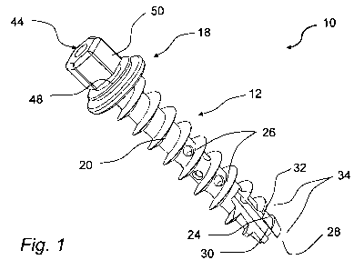
FIG. 1 is a perspective view of the embodiment of a self filling autograft
bone
screw;
3
CA 02759445 2011-10-19
WO 2010/123859 PCT/US2010/031697
FIG. 2 is a perspective view of the self filling autograft bone screw
including a
washer member;
FIG. 3 is a perspective view of the self filling autograft bone screw
including a
wiper member;
FIG. 4 is a cross sectional view of the self filling autograft bone screw of
FIG. 1;
FIG. 5 is an exploded perspective view of an external fastening member in use
with the self filling autograft bone screw of FIG. 4;
FIG. 6 is a perspective view of an external fastening member in use with the
self
filling autograft bone screw of FIG. 1;
FIG. 7 is an exploded perspective view of the self filling autograft bone
screw of
FIG. 6;
FIG. 8 is perspective view of another embodiment of the self filling bone
screw of
FIG. 1;
FIG. 9 is perspective view of another embodiment of the self filling bone
screw of
FIG. 1;
FIG. 10 is perspective view of another embodiment of the self filling bone
screw
of FIG. 1;
FIG. 11 is perspective view of another embodiment of the self filling bone
screw
of FIG. 1;
FIG. 12 is cross sectional view of another embodiment of the self filling bone
screw of FIG. 11;
FIG. 13 is perspective view of another embodiment of the self filling bone
screw
of FIG. 1;
4
CA 02759445 2011-10-19
WO 2010/123859 PCT/US2010/031697
FIG. 14 is perspective view of another embodiment of the self filling bone
screw
of FIG. 1;
FIG. 15 is perspective view of another embodiment of the self filling bone
screw
of FIG. 1;
FIG. 16 is cross sectional view of another embodiment of the self filling bone
screw of FIG. 15;
FIG. 17 is a perspective view of another embodiment of the external fastening
member and the self filling bone screw of FIG. 1;
FIG. 18 is a perspective view of the external fastening member attached to the
self filling bone screw of FIG. 1;
FIG. 19 is a perspective view of the external fastening member attached to the
self
filling bone screw of FIG. 1;
FIG. 20 is a perspective view of the external fastening member attached to the
self
filling bone screw of FIG. 1;
FIG. 21 is a perspective view of another embodiment of the external fastening
member attached to the self filling bone screw of FIG. 17;
FIG. 22 is an exploded perspective view of the external fastening member
attached to the self filling bone screw of FIG. 21;
FIG. 23 is an exploded perspective view of the external fastening member and
the self filling bone screw of FIG. 22;
FIG. 24 is a cross sectional view of the external fastening member attached to
the
self filling bone screw of FIG. 21;
FIG. 25 is an exploded cross sectional view of the external fastening member
attached to the self filling bone screw of FIG. 21;
5
CA 02759445 2011-10-19
WO 2010/123859 PCT/US2010/031697
FIG. 26 is a perspective view of another embodiment of the external fastening
member and the self filling bone screw of FIG. 1;
FIG. 27 is a perspective view of the external fastening member attached to the
self
filling bone screw of FIG. 26;
FIG. 28 is another perspective view of the external fastening member attached
to
the self filling bone screw of FIG. 26; and
FIG. 29 is an exploded perspective view of the external fastening member of
FIG.
26.
DESCRIPTION
In general, the invention relates to self filling bone screws which may be
used for
applications such as spinal stabilization and fusion, intramedullary (IM)
rods, joint
implants, plating, or any other orthopedic/neurosurgical application where
such screws
would be desirable. Such bone screws may be used with a radio-lucent, off-
angle,
motorized drill system. The bone screws may also be placed with an insertion
tool and
manual drive handle or a standard power drill.
The self filling autograft bone screw is designed with distal cutting edges
which
direct native cortical and cancellous bone and marrow into a lumen of the
hollow screw.
At least one opening is provided along the screw shaft to allow bone to grow
into and out
of the screw in order to form a lattice-work of bone criss-crossing through
the screw and
in order have the screw incorporated into the bone. Thus, the screw forms its
own
internal bone graft (autograft) with its resultant osteogenesis,
osteoconduction, and
osteoinduction properties. This allows the autograft screw to provide
immediate fixation
(due to the screw) as well as long-term fixation (due to the screw and bony
fusion). It
may also be used with bone morphogenetic protein to facilitate bone growth.
The self filling autograft bone screw also has a self-tapping and thread
cutting
design. A floating washer member is provided to allow the washer to pivot on
the
spherical undersurface of the proximal end of the screw to make better contact
with
6
CA 02759445 2011-10-19
WO 2010/123859 PCT/US2010/031697
angled bony surfaces with resultant improved holding power. A wiper member is
also
provided in the screw head which strips bone material from an external
fastening
member, such as a guide drill, when the guide drill is removed to maximize the
amount of
autograft in the screw. A stepped shank design, (smaller diameter at the
distal end in
contrast to a greater diameter at the proximal end), may be used to provide
greater grip
and strength in the endosteal (undersurface) area of the bone. A one-step
delivery device
and system is also provided which facilitates placement of the screw. This one-
step
delivery device may be driven manually or with a powered driver.
Percutaneous insertion of the self-filling bone screw may be accomplished with
an insertion tool and manual drive handle. The bone screw may be positioned in
an
insertion tool which has spring catches designed to hold the screw securely in
the
insertion tool. A long drill bit may be placed through the insertion tool and
screw. The
assembly may then placed through a small incision to the bone (e.g. pedicle,
facet joint,
etc). The guide drill and screw are then advanced into the bone - this may be
done with a
manual turning handle, standard drill, or radio-lucent, off-angle drill. Once
the guide drill
has entered the bone and provided small initial pilot hole, the handle
releases the guide
drill and engages the screw drive. The screw is then advanced to its final
depth. The drive
handle (or drill) is removed, the sleeve retracted up along the insertion tool
body
releasing the spring catches holding the screw in the insertion tool. Once the
catches are
released, the screw is left in place and the insertion tool is removed.
Percutaneous placement of the self-filling bone screw may also utilize bone
morphogenetic protein (BMP) to facilitate bone growth. The BMP wafer or putty
may be
placed inside the screw prior to placement.
Referring to FIGS. 1 through 7, in one embodiment according to the invention,
a
self filling autograft screw 10 includes an elongated screw body member 12
having a
proximal portion and a distal portion. The proximal portion of the elongated
screw body
member includes a washer 18. The elongated screw body member 12 includes a
lumen
passage 22 which extends from a proximal end of the elongated body member 12
to a
distal end of the elongated body member 12. The screw 10 further includes a
plurality of
7
CA 02759445 2011-10-19
WO 2010/123859 PCT/US2010/031697
external threads 20 which extend from the proximal portion of the elongated
body
member 12 to the distal portion of the elongated body member 12. The plurality
of
external threads 20 are adapted for anchoring the elongated body member 12
within an
internal portion of a bone within a patient's body. The bone screw 10 further
includes a
cutting section 24 disposed at the distal end of the elongated body member 12.
The
cutting section 24 is adapted to enable penetration of the bone screw 10 into
the internal
portion of the bone and facilitate the insertion of fragments into the lumen
22 resulting
from the penetration of the bone screw into the internal portion of the bone.
At least one
opening 26 is provided along the length of the elongated body member 12. The
opening
26 is adapted for facilitating the re-growth of the fragments within the
internal portion of
the bone and anchoring of the elongated body member 12 within the internal
portion of
the bone.
The bone screw 10 may also consist of an elongated body member 12, a wiper
member 14, and a washer member 18. The bone screw 10 may be inserted into and
left
in a bone to hold or affix objects. This can be either bone to bone or bone to
an artificial
construct. All parts of the bone screw 10 may be made from implant compatible
materials.
The external threads 20 can be of various shapes, sizes, or pitches. The
external
threads 20 anchor the bone screw 10 into the bone. The bone screw 10 self
fills the
lumen passage 22 with fragments such as chips of bone, bone marrow, and blood
as it is
inserted into the bone by directing the fragments cut by the external
fastening member,
such as a guide drill 16, and the thread cutting section 24 at the distal tip
of the elongated
body member 12.
As the bone heals, bone re-growth occurs between the bone material inside the
bone screw 10 and the bone material which is inserted through the various
openings 26 in
the elongated body member 12. The openings 26 can be of various shapes, sizes,
quantities, and locations around the elongated body member 12. It is
contemplated that
the bone incorporated into the bone screw 10 will increase pullout force,
resistance to
loosening, and enhance the overall structural integrity of the surrounding
bone.
8
CA 02759445 2011-10-19
WO 2010/123859 PCT/US2010/031697
The cutting section 24 consists of helical formed cutting surface at the
distal end
of the elongated body member 12 and a slot 30 that forms a cutting edge 32.
The cutting
edge 32 has an acute angle relative to the longitudinal axis of the elongated
body member
12 and feeds cut bone fragments into the lumen passage 22. The slot 30 extends
proximal through the threads 34 to a first full thread. This configuration
allows the
cutting edge 32 to cut threads into the bone as the bone screw 10 is advanced.
The lumen passage 22 is formed by a cylindrical bore with a diameter equal to
or
slightly larger than the diameter of the guide drill 16. The lumen passage 22
thus follows
the guide drill 16 as the bone screw 10 is threaded into the bone. The
openings 26 pass
through the elongated body member 12 and allow new bone growth to connect the
surrounding bone with the lumen passage 22. Fragments are deposited into the
lumen
passage 22 by scraping the material captured in flutes 36 of the guide drill
16 with the
wiper 14.
The wiper 14 may be cylindrical in shape and have an inner diameter that is
equal
to or greater that the outer diameter of the guide drill 16. The wiper 14 has
formed
wiping features 38 that approximate the cross sectional shape of the flutes
36. The wiper
14 has a slit 40 along one side parallel to the center axis. The slit 40
allows the wiper 14
to be compressed and inserted into the lumen passage 22 of the elongated body
member
12 to a position where the secondary cavity 42 has been formed to receive it.
The
secondary cavity 42 has an inner diameter that is larger than the outer
diameter of the
wiper 14 allowing it to spin freely within the elongated body member 12. In
another
embodiment, the wiper 14 may be disposed within a distal portion of a drive
shaft of the
guide drill 16. This configuration can also facilitate the removal of
fragments to be
deposited into the lumen passage 22.
As the guide drill 16 is rotated and extended into the bone forming a pilot
hole, it
collects the bone chips along the flutes 36. As the bone screw 10 is
translated towards
the distal tip of the guide drill 16, by either threading the bone screw 10
over the guide
drill 16 or by retracting the guide drill 16 after the bone screw 10 has
threaded into the
bone, the wiping features 38 of the wiper 14 prevent fragments, such as bone
chips, from
9
CA 02759445 2011-10-19
WO 2010/123859 PCT/US2010/031697
leaving the lumen passage 22 or the elongated body member 12. When the guide
drill 16
is fully retracted from the elongated body member 12, a significant portion of
the bone
chips will be left in the elongated body member 12.
Various coatings, such as hydrophobic, hydrophilic, or BMP, may be affixed to
different surfaces of the elongated body member 12, wiper 14, and guide drill
16 to
facilitate the translation of the bone fragment / blood products into the
elongated body
member 12 and to aid in promoting regenerative bone growth.
The proximal end of the screw body 12 contains a head section 44 which
consists
of a contact surface 46, a capture feature 48, and a drive structure 50. The
contact surface
46 mates with the outer surface of the bone that the bone screw 10 is driven
into. The
surface can be flat, for example, relative to the axis of the bone screw 10,
concave or
convex. The outer diameter of the contact surface 46 defines the surface area
supporting
the axial loading experienced by the bone screw 10 in use. Alternatively, the
washer 18
can be installed between the contact surface 46 and the outer surface of the
bone.
In an embodiment, the washer 18 has a spherical depression 52 on the side that
mates to the contact surface 46 of the bone screw 10 with a radius that
matches that of the
convex contact surface 46. The hole through the center of the washer 18 is
larger that the
diameter of the elongated body member 12 where it is located. This, along with
the
mating spherical contact surface 46 and depression 52, allows the washer 18 to
float
angularly about the head section 44. In addition, the ability for floating
facilitates the
washer 18 in making contact with the outer surface of the bone when that outer
surface is
angled relative to the axis of the elongated body member 12.
The capture feature 48 may be undercut in the drive structure 50.
Alternatively,
the capture feature 48 could be a depression in the wall of the drive
structure 50. The
capture feature 48 facilitates the installation of the bone screw 10 by
providing a means
to securely retain the bone screw 10 in the external fastening member until
its desired
release.
CA 02759445 2011-10-19
WO 2010/123859 PCT/US2010/031697
The drive structure 50 may be a standard external hex-shape that can transmit
driving torque required to thread the bone screw 10 into the bone. This
external hex-
shape design aids in removing the screw in a subsequent surgical procedure. It
is
contemplated that alternate external and internal configurations of the drive
structure 50
are possible.
The guide drill 16 consists of a cutting tip 54, flutes 36, body 56, and
optionally a
drive structure 58. The cutting tip 54 can take many shapes. In one
embodiment, the
cutting tip 54 has a sharp brad point 60 in the center and a flat cutting edge
62 radiating
out towards the outer diameter. The flutes 36 can be designed in different
shapes, such as
circular, parabolic, or other. The flutes 36 may extend partway up the body
56. The
proximal end of the guide drill 16 can also remain cylindrical allowing the
use of a
conventional Jacob's style chuck or ratchet, or have a drive structure 58 that
has the same
external hex shape as on the proximal end of the elongated body member 12.
Referring to FIG. 8, an alternative embodiment of the self filling autograft
bone
screw 10 of FIG. 1 is presented. A bone screw 100 is provided which includes
an
elongated body member 112 with a shank or thread root having a diameter that
is larger
at a proximal portion 102 than at a distal portion 104. The difference in
diameter
provides for a stronger elongated body member 112 that will bear greater
stress while
maintaining a smaller outer diameter where the bone may be smaller in cross
section.
Additional openings 108 and a second thread cutting section 106 may be added
to
accommodate a larger thread 110 that is incorporated into the elongated body
member
112.
Referring to FIG. 9, an alternative embodiment of the self filling autograft
bone
screw 10 of FIG. 1 is presented. A bone screw 120 is provided in which the
proximal
portion 102 has a larger diameter relative to the distal portion 104 of the
elongated body
member 112. The distal portion 104 of the elongated body member 112 is free of
additional openings 108 and the secondary thread cutting section 106. This
design can
improve the strength of proximal portion 102 of the elongated body member 112.
11
CA 02759445 2011-10-19
WO 2010/123859 PCT/US2010/031697
Referring to FIG. 10, an alternative embodiment of the self filling autograft
bone
screw 10 of FIG. 1 is presented. A bone screw 130 is provided in which an
elongated
body member 132 has a taper shank or root diameter 134 and tapered threads
136. This
design can provide greater strength at the proximal portion of the shank and a
smaller,
less invasive cross section at a distal portion 140.
Referring to FIGS. 11 through 14, alternative embodiments of the self filling
bone
screw 10 of FIG. 1 are presented. Each of the alternative embodiments may be
used
without an external fastening member, such as guide drill 16.
A bone screw 150 is provided which has a small diameter cylindrical cut 154
through a drill point 152. This design allows the bone screw 150 to follow a
standard K-
wire into a pilot hole previously formed in the bone. A bone screw 160 is
provided
having a drill point 162 added to the distal end of an elongated body member
164. The
drill point 162 has a standard cutting edge 168 and double flutes 166. In
addition, the
drill point 162 allows the bone screw 160 to self drill into a bone, thereby
eliminating the
additional step of a pilot hole. The double flutes 166 extend to the cutting
section edges
172 and into the opening of the lumen passage 170. Bone chips cut by the drill
point 162
and thread cutting edges 172 are directed into the lumen passage 170 to fill
the elongated
body member 164. Additionally, a bone screw 180 is provided having an
alternate head
design 182, such as a socket head cap screw.
Referring to FIGS. 15 and 16, an alternative embodiment of the self filling
autograft bone screw 10 of FIG. 1 is presented. A bone screw 190 is provided
which
includes a lumen passage 192 starting at a distal end 194 of an elongated body
member
196 and ending before reaching a proximal end of the elongated body member
196. In
this tulip head pedicle screw style, the proximal end of the elongated body
member 196
maintains maximum structural integrity while the distal end 194 incorporates
the
advantages of the autograft integration of the screw into the bone and the
subsequent
strength improvement of the surrounding bone structure.
Referring to FIGS. 17 through 25, embodiments of an external fastening member
and embodiments of the self filling autograft bone screw 10 of FIG. 1 are
presented. In
12
CA 02759445 2011-10-19
WO 2010/123859 PCT/US2010/031697
one embodiment, a system 200 is presented that includes an external fastening
member
220, a drive handle 250, and a bone screw 10. The system 200 may also include
a guide
drill 16, although the guide drill 16 may not be required when using a self
drilling
embodiment of the bone screw 10.
In another embodiment, a system 210 is presented which includes the bone screw
positioned within the external fastening member 220 with the guide drill 16
inserted
through the cannulated center of the external fastening member 220 until the
tip of the
guide drill 16 extends past the end 212 of the bone screw 10. Once assembled,
the
external fastening member 220, bone screw 10, and guide drill 16 may be placed
through
10 a small incision to the bone (e.g. pedicle, facet joint, etc). The guide
drill may then be
advanced into the bone until a depth stop 214 on the guide drill 16 contacts
the mating
surface on the external fastening member 220. The guide drill 16 may be
advanced by
applying torque and axial force through the drive structure 58 of the guide
drill 16. This
can be accomplished using the manual drive handle 250 or some form of powered
drill
type device.
In another embodiment, the drive handle 250 is engaged with the external
fastening member 220. The drive handle 250 advances the bone screw 10 into the
bone
while the guide drill 16 remains stationary, thereby providing a guide for the
proper
positioning of the bone screw 10. Once inserted, the bone screw 10 is released
from the
external fastening member 220 by sliding a sleeve 218 axially along the
external
fastening member 220 towards the proximal end of the bone screw 10. The act of
sliding
the sleeve 218 also uncovers spring catches 222 and allows the spring catches
222 to
deflect outward to their normal position. The ends of the spring catches 222
are held into
the undercut features 48 of the bone screw 10 by the sleeve 218, which
prevents the
spring catches 222 from opening outward. When the sleeve 218 is slid axially
away, the
spring catches 222 are free to deflect out of the undercuts 48. The cannulated
opening
226 on the drive shaft 228 allows the guide drill 16 to pass through it. The
external
fastening member 220 may now be removed from the patient's body.
13
CA 02759445 2011-10-19
WO 2010/123859 PCT/US2010/031697
Referring to FIGS. 26 through 29, an alternative embodiment of an external
fastening member and the self filling autograft bone screw 10 of FIG. 1 are
presented.
An external fastening member 300 is presented which can be used in place of
the manual
drive handle 250 and fastening member 220.
In one embodiment, a radio-lucent, off-angle drill 310 is used to provide
percutaneous insertion of the bone screw 10 using image guidance. A sleeve 302
with a
handle 304 is provided as a means to stabilize the assembly while keeping an
operator's
hands out of the fluoroscope radiation beam during the image guidance.
The drill 310 captures the drive structure 58 of the guide drill 16. The drill
310
may be powered such that pressure is applied to advance the guide drill 16
into the bone.
The drill 310 engages the drive structure 216 of the insertion tool 220. The
drill310 can
advance the bone screw 10 under power into the bone. The guide drill 16 is
removed by
pulling it fully through the drill upper surface 314. The sleeve 302 may be
slid axially to
release the bone screw 10. Once the bone screw 10 is advanced into the bone,
the guide
drill 300 may release the drive structure 58 of the guide drill 16 by pressing
the release
button 306.
It will be understood that various modifications may be made to the
embodiments
disclosed herein. Therefore, the above description should not be construed as
limiting,
but merely as illustrative of some embodiments according to the invention.
14