Note: Descriptions are shown in the official language in which they were submitted.
CA 02759506 2011-10-20
WO 2010/124163 PCT/US2010/032170
GRANULOCYTE-MACROPHAGE COLONY-STIMULATING FACTOR (GM-CSF)
NEUTRALIZING ANTIBODIES
RELATED APPLICATIONS
[0100] This application claims the benefit of provisional applications USSN
61/172,120, filed
April 23, 2009 and USSN 61/234,946, filed August 18, 2009, the contents of
which are each
herein incorporated by reference in their entirety.
FIELD OF THE INVENTION
[0101] The invention relates generally to the fields of immunology and
medicine. Specifically,
the invention relates to compositions containing the GM-CSF neutralizing human
monoclonal
antibody 1783J22, as well as methods of making and using this antibody.
BACKGROUND OF THE INVENTION
[0102] Much of the control of blood-cell formation is mediated by a group of
interacting
glycoproteins termed colony stimulating factors (CSFs). Granulocyte macrophage-
colony
stimulating factor ("GM-CSF"), a soluble secreted glycoprotein, is a potent
immunomodulatory
cytokine known to facilitate development and prolongation of both humoral and
cellular
mediated immunity.
[0103] GM-CSF also plays a role in the genesis and progression of a plurality
of human diseases,
such as cancer, inflammatory and autoimmune diseases, and degenerative
diseases.
[0104] Therefore a long-felt need exists in the field for therapeutic
compositions and methods
capable of antagonizing or inhibiting the activity of GM-CSF. Despite multiple
attempts to
generate antibodies specific for GM-CSF, for instance, through the creation of
polyclonal and
monoclonal antibodies, no one has succeeded in creating a therapeutically-
effective human
antibody composition that inhibits the activity of GM-CSF. The invention
provides compositions
and methods for inhibiting, or neutralizing, the activity of GM-CSF, and,
therefore, succeeds in
addressing the long-felt need in the art.
1
CA 02759506 2011-10-20
WO 2010/124163 PCT/US2010/032170
SUMMARY OF THE INVENTION
[0105] The invention provides an isolated human monoclonal antibody that
specifically binds
and neutralizes GM-CSF. Anti-GM-CSF monoclonal antibodies of the invention are
obtained by
a process including (a) screening memory B cell cultures from a donor
Peripheral Blood
Mononuclear Cell (PBMC) sample for neutralization activity against GM-CSF and
(b) rescuing
the monoclonal antibody from a memory B cell culture that neutralizes GM-CSF.
Optionally, the
method further includes culturing an immortalized B cell clone expressing an
antibody and
isolating antibodies from said B cell.
[0106] The invention provides an isolated fully human monoclonal antibody,
wherein the
monoclonal antibody has the following characteristics: (a) specifically binds
to an epitope of a
GM-CSF protein; and (b) neutralizes GM-CSF bioactivity in vitro. In some
embodiments, this
antibody is isolated from a B cell from a human donor. In some embodiments,
wherein this
antibody is operably-linked to a therapeutic agent or a detectable label.
[0107] In some embodiments, the epitope is linear, non-linear, or
discontinuous. For example,
the epitope is a linear amino acid polypeptide or a folded polypeptide that
reflects that native
configuration of a GM-CSF protein. Alternatively, the epitope is a
conformational or
discontinuous epitope that is recognizable by the antibody only when the GM-
CSF antibody is
folded, arranged as a homodimer, or a discontinuous portion of the GM-CSF
amino acid
sequence is maintained in a particular three-dimensional form using an
accessory structure to
mimic the native surface (for instance, by use of a CLIP). The epitope is an
immunogenic
polypeptide or a glycopeptide that is bound an antibody of the invention.
[0108] In a preferred embodiment, the antibody is 1783J22. Alternatively, or
in addition, the
antibody is a sister clone of the 1783J22 antibody. For instance, the sister
clone contains a
distinct heavy or light chain nucleic acid sequence that results in a heavy or
light chain amino
acid sequence that is identical to the 1783J22 antibody. In other aspects of
the invention, the
amino acid sequences of the heavy and light chains of the sister clones are
70%, 75%, 80%,
85%, 90%, 95%, 100% or any percentage in between identical to the amino acid
sequences of
the heavy and light chains of the 1783J22 antibody.
[0109] The invention provides an antibody that binds the same epitope as
1783J22.
[0110] The invention provides an isolated fully human monoclonal anti-GM-CSF
antibody or
fragment thereof, wherein said antibody includes a variable heavy chain (VH)
region containing a
CDR1 and a CDR2, wherein said region is encoded by a human IGHV3-23 VH
germline
2
CA 02759506 2011-10-20
WO 2010/124163 PCT/US2010/032170
sequence, or a nucleic acid sequence that is homologous to the IGHV3-23 VH
germline gene
sequence. In some embodiments, the nucleic acid sequence that is homologous to
the IGHV3-23
VH germline sequence is at least 90% homologous to the IGHV3-23 VH germline
sequence. This
antibody further includes a variable light chain (VL) region encoded by a
human IGKV1-16 VL
germline gene sequence, or a nucleic acid sequence that is homologous to the
said VL germline
gene sequence. In some embodiments, the nucleic acid sequence that is
homologous to the
IGKV1-16 VL germline sequence is at least 90% homologous to the said IGKV1-16
VL germline
sequence.
[0111] The invention provides an isolated anti-GM-CSF antibody, wherein the
antibody has a
heavy chain with three CDRs including an amino acid sequence selected from the
group
consisting of the amino acid sequences of FPFHKYTMT (SEQ ID NO: 8),
VSGVNGKTYYSPSVRG (SEQ ID NO: 9), and GPGGHLHYYYGLDV (SEQ ID NO: 10), and
a light chain with three CDRs that include an amino acid sequence selected
from the group
consisting of the amino acid sequences of RASQAINNYVA (SEQ ID NO: 14), GASNLQP
(SEQ ID NO: 15), and QNYFGYPLT (SEQ ID NO: 16).
[0112] The invention also provides an isolated anti-GM-CSF antibody, wherein
the antibody has
a heavy chain with three CDRs including an amino acid sequence selected from
the group
consisting of the amino acid sequences of GFPFHKYTMT (SEQ ID NO: 11),
VSGVNGKTY
(SEQ ID NO: 12), and GPGGHLHYYYGLDV (SEQ ID NO: 10), and a light chain with
three
CDRs that include an amino acid sequence selected from the group consisting of
the amino acid
sequences of RASQAINNYVA (SEQ ID NO: 14), GASNLQP (SEQ ID NO: 15), and
QNYFGYPLT (SEQ ID NO: 16).
[0113] The invention provides an isolated anti-GM-CSF antibody, wherein the
antibody has a
heavy chain with three CDRs including an amino acid sequence selected from the
group
consisting of the amino acid sequences of FPFHKYTMT (SEQ ID NO: 8),
VSGVNGKTYYSPSVRG (SEQ ID NO: 9), and GPGGHLHYYYGLDV (SEQ ID NO: 10),
GFPFHKYTMT (SEQ ID NO: 11), VSGVNGKTY (SEQ ID NO: 12), wherein said antibody
binds GM-CSF.
[0114] The invention provides an isolated anti-GM-CSF antibody, wherein the
antibody has a
light chain with three CDRs including an amino acid sequence selected from the
group
consisting of the amino acid sequences of RASQAINNYVA (SEQ ID NO: 14), GASNLQP
(SEQ ID NO: 15), and QNYFGYPLT (SEQ ID NO: 16), wherein said antibody binds GM-
CSF.
3
CA 02759506 2011-10-20
WO 2010/124163 PCT/US2010/032170
[0115] The invention provides an isolated anti-GM-CSF antibody, wherein the
antibody includes
a VH CDR1 region containing the amino acid sequence of FPFHKYTMT (SEQ ID NO:
8); a VH
CDR2 region containing the amino acid sequence of VSGVNGKTYYSPSVRG (SEQ ID NO:
9); a VH CDR3 region containing the amino acid sequence of GPGGHLHYYYGLDV (SEQ
ID
NO: 10); a VL CDR1 region containing the amino acid sequence of RASQAINNYVA
(SEQ ID
NO: 14); a VL CDR2 region containing the amino acid sequence of GASNLQP (SEQ
ID NO:
15); and a VL CDR3region containing the amino acid sequence of QNYFGYPLT (SEQ
ID NO:
16).
[0116] The invention provides an isolated anti-GM-CSF antibody, wherein the
antibody includes
a VH CDR1 region containing the amino acid sequence of GFPFHKYTMT (SEQ ID NO:
11); a
VH CDR2 region containing the amino acid sequence of VSGVNGKTY (SEQ ID NO:
12); a VH
CDR3 region containing the amino acid sequence of GPGGHLHYYYGLDV (SEQ ID NO:
10);
a VL CDR1 region containing the amino acid sequence of RASQAINNYVA (SEQ ID NO:
14); a
VL CDR2 region containing the amino acid sequence of GASNLQP (SEQ ID NO: 15);
and a VL
CDR3region containing the amino acid sequence of QNYFGYPLT (SEQ ID NO: 16).
[0117] The invention provides an isolated anti-GM-CSF antibody or fragment
thereof, wherein
the antibody includes: (a) a VH CDR1 region containing the amino acid sequence
of
FPFHKYTMT (SEQ ID NO: 8) or GFPFHKYTMT (SEQ ID NO: 11); (b) a VH CDR2 region
containing the amino acid sequence of VSGVNGKTYYSPSVRG (SEQ ID NO: 9) or
VSGVNGKTY (SEQ ID NO: 12); and (c) a VH CDR3 region containing the amino acid
sequence of GPGGHLHYYYGLDV (SEQ ID NO: 10), wherein the antibody binds GM-CSF.
In
some embodiments, this antibody further includes: (a) a VL CDR1 region
containing the amino
acid sequence of RASQAINNYVA (SEQ ID NO: 14); (b) a VL CDR2 region containing
the
amino acid sequence of GASNLQP (SEQ ID NO: 15); and (c) a VL CDR3 region
containing the
amino acid sequence of QNYFGYPLT (SEQ ID NO: 16).
[0118] The invention provides an isolated fully human monoclonal anti-GM-CSF
antibody
including a heavy chain sequence containing the amino acid sequence SEQ ID NO:
2 and a light
chain sequence containing amino acid sequence SEQ ID NO: 5.
[0119] The invention provides a composition including an antibody described
herein and a
pharmaceutically acceptable carrier. Preferably, the antibody is an isolated
fully human
monoclonal antibody with the following characteristics: (a) specifically binds
to an epitope of a
GM-CSF protein; and (b) neutralizes GM-CSF bioactivity in vitro. In some
embodiments, this
4
CA 02759506 2011-10-20
WO 2010/124163 PCT/US2010/032170
antibody is isolated from a B cell from a human donor. In some embodiments of
this
composition, the antibody is operably-linked to a therapeutic agent or a
detectable label. In some
embodiments, the composition further includes a second anti-GM-CSF antibody.
In some
embodiments, an antibody or composition of the invention is administered in
combination with
other therapies. In some embodiments, an antibody or composition of the
invention is
manufactured for use as an adjuvant formulation.
[0120] The invention provides a fragment of an antibody described herein.
Preferably, the
antibody is an isolated fully human monoclonal antibody with the following
characteristics: (a)
specifically binds to an epitope of a GM-CSF protein; and (b) neutralizes GM-
CSF bioactivity in
vitro. In some embodiments, this antibody is isolated from a B cell from a
human donor. In some
embodiments, the fragment is selected from the group consisting of Fab, Fab',
F(ab')2, Fv, single
chain Fv, diabody and domain antibody (dAb) fragments.
[0121] The invention provides a vector including the nucleic acid sequence of
SEQ ID NOs: 1 or
4. Alternatively, or in addition, the vector includes a nucleic acid encoding
a heavy or light chain
of an antibody described herein. In other aspects, the vector includes a
nucleic acid encoding a
heavy or light chain of an anti-GM-CSF antibody administered simultaneously or
sequentially
with respect to an antibody described herein. The invention provides a cell
including this vector.
[0122] The invention further provides a B cell clone expressing an antibody
described herein.
Preferably, the antibody is an isolated fully human monoclonal antibody with
the following
characteristics: (a) specifically binds to an epitope of a GM-CSF protein; and
(b) neutralizes
GM-CSF bioactivity in vitro. In some embodiments, this antibody is isolated
from a B cell from
a human donor. In some embodiments, the antibody is recombinant.
[0123] The invention provides a method of stimulating an immune response in a
subject,
comprising administering to a patient a composition of the invention. In some
embodiments, the
method further comprises administering a second anti-GM-CSF antibody. In one
aspect, the
second antibody is administered simultaneously or sequentially with respect to
the composition.
[0124] The invention provides a method for the treatment or prevention of a GM-
CSF-mediated
disease in a subject including administering to the subject a composition of
the invention. In
some embodiments of this method, an antibody of the composition binds to GM-
CSF and
inhibits the biological activity of GM-CSF in the patient. In some
embodiments, the CM-CSF
mediated disease is an infectious disease, an inflammatory disease, an
autoimmune disorder,
Alzheimer's Disease, or cancer.
CA 02759506 2011-10-20
WO 2010/124163 PCT/US2010/032170
[0125] In a specific embodiment, the GM-CSF-mediated disease is Alzheimer's
disease (AD) or
vascular dementia (VAD). Adminstration of a composition of the invention to a
subject with
Alzheimer's disease (AD) or vascular dementia (VAD) down-regulates expression,
translation, or
activity of autologous beta-amyloid (A13) protein or autologous amyloid
precursor protein (APP),
thereby treating or preventing the GM-CSF-mediated disease.
[0126] In a specific embodiment, the GM-CSF-mediated disease is an
inflammatory disease.
Preferably, the inflammatory disease is selected from the group consisting of
asthma, acute
inflammation, chronic inflammation, type I diabetes, type II diabetes and all
of the related
pathologies, rheumatoid arthritis, autoimmune disease, inflammatory renal
disease, inflammatory
lung disorders such as asthma and chronic obstructive pulmonary disease
(COPD), multiple
sclerosis, and autoimmune encephalomyelitis.
[0127] In another specific embodiment, the GM-CSF-mediated disease is cancer.
Although all
forms are cancer are contemplated, in preferred embodiments of this method,
the cancer is
selected from the group consisting of colon cancer, lung cancer, breast
cancer, pancreatic cancer,
leukemia, and juvenile myelomonocytic leukemia. Some embodiments of this
method further
include administering a second anti-GM-CSF antibody. The second antibody is
administered
simultaneously or sequentially with respect to the composition.
[0128] The invention provides a method of inhibiting the biological activity
of human GM-CSF
in a patient with an infectious disease comprising administering to the
patient a composition of
the invention. In some embodiments of this method, the infectious disease is
selected from the
group consisting of sepsis, severe acute respiratory syndrome (SARS; caused by
SARS-
associated coronavirus), hepatitis type B or type C, influenza, varicella,
adenovirus, herpes
simplex virus type I or type II, rinderpest, rhinovirus, echovirus, rotavirus,
respiratory syncytial
virus, papilloma virus, papova virus, cytomegalovirus, echinovirus, arbovirus,
hantavirus,
coxsachie virus, mumps virus, measles virus, rubella virus, polio virus, human
immunodeficiency virus (HIV) type I or type II, Meningitis, Septic arthritis,
Peritonitis,
Pneumonia, Epiglottitis, E. coli, Hemolytic uremic syndrome, thrombocytopenia,
to, Ebola,
Staphylococcus A-E, Plasmodium, Malaria, Dengue, hemorrhagic fever,
Leishmaniasis,
Leprosy, Toxic shock syndrome, Streptococcal myositis, Gas gangrene,
Mycobacterium,
Pneumocystis, Pelvic inflammatory disease, Orchitis/epidydimitis, Legionella,
Lyme disease
Influenza A, Epstein-Barr Virus, Viral associated hemiaphagocytic syndrome,
viral encephalitis,
aseptic meningitis, mycoplasma, Neisseria, Legionella, Rickettsia,
andChlamydia.
6
CA 02759506 2011-10-20
WO 2010/124163 PCT/US2010/032170
[0129] The invention further provides a vaccine including an epitope of an
antibody of the
invention. Preferably, the antibody is an isolated fully human monoclonal
antibody with the
following characteristics: (a) specifically binds to an epitope of a GM-CSF
protein; and (b)
neutralizes GM-CSF bioactivity in vitro. In some embodiments, this antibody is
isolated from a
B cell from a human donor.
[0130] The invention provides a vaccine including an epitope of the 1783J22
antibody.
[0131] The invention provides a therapeutic kit including an antibody of the
invention.
Preferably, the antibody is an isolated fully human monoclonal antibody with
the following
characteristics: (a) specifically binds to an epitope of a GM-CSF protein; and
(b) neutralizes
GM-CSF bioactivity in vitro. In some embodiments, this antibody is isolated
from a B cell from
a human donor.
[0132] The invention provides a therapeutic kit including a composition of the
invention.
[0133] The invention provides a prophylactic kit including a vaccine
containing an epitope of an
antibody of the invention.
[0134] The invention provides a prophylactic kit including the vaccine
containing an epitope of
the 1783J22 antibody.
[0135] The invention provides a method for inhibiting a GM-CSF activity in a
rabbit, the method
including: (a) administering to a rabbit a monoclonal antibody according to
the invention; and (b)
determining the inhibition of a GM-CSF induced activity in the rabbit. In one
embodiment, the
method further includes: (c) determining the binding of GM-CSF to the
monoclonal antibody in
the rabbit.
[0136] In some aspects, the GM-CSF induced activity is a cell proliferative
activity, or
stimulation of early- and late-phase granulocyte and macrophage progenitor
cells, or activation
of mature neutrophils and macrophages; or enhanced peripheral anti-infection
activity; or
activation of mature neutrophils, macrophages, eosinophils and basophils; or
stimulation of stem
cells to produce granulocytes (neutrophils, eosinophils, and basophils) and
monocytes.
[0137] Other features and advantages of the invention will be apparent from
and are
encompassed by the following detailed description and claims.
BRIEF DESCRIPTION OF THE DRAWINGS
[0138] Figure 1 is a graph depicting the bioactivity of GM-CSF as the amount
of relative
luminescence units (RLU) per B Cell Culture Well for both replicate and
screening plates
7
CA 02759506 2011-10-20
WO 2010/124163 PCT/US2010/032170
challenged with human anti-GM-CSF. As shown in the graph, clone 1783J22 is
identified as
inhibiting GM-CSF bioactivity in both replicate and screening plates, and,
therefore, having GM-
CSF-neutralizing activity.
[0139] Figure 2 is a graph depicting the binding activity of 1783J22 to human
GM-CSF as the
amount of relative luminescence units (RLU) per B Cell Culture Well
Identification (ID). The
assay confirms that 1783J22 binds human GM-CSF.
[0140] Figure 3 is a graph depicting the recovery of human GM-CSF binding
reactivity of the
1783J22 recombinant antibody from a pool transfectant supernatants derived
from the
combination of heavy chain (gamma, y) and light chain (kappa, x) PCR products.
GM-CSF
binding reactivity was measured as the relative luminescence units (RLU) per
heavy and light (H
& L) chain pool combinations. The human monoclonal 1783J22 antibody was
reconstituted from
the combination of y3 and xl heavy and light chains, respectively.
[0141] Figure 4A is a graph depicting the recovery of human GM-CSF binding
reactivity of the
1783J22 recombinant monoclonal antibody from transfectant supernatants derived
from
monoclonal heavy chain (gamma, y) and light chain (kappa, x) combinations. GM-
CSF binding
reactivity was measured as the relative luminescence units (RLU) per
monoclonal heavy and
light (H & L) chain combinations. Three y3 sequences, when combined with one
xl sequence,
produced 1783J22 monoclonal antibodies in transfectant supernatants that bind
human GM-CSF.
[0142] Figure 4B is a graph depicting the human GM-CSF binding reactivity of
the recovered
1783J22 recombinant monoclonal antibodies from Figure 4A, measured as the
relative
luminescence units (RLU) per reciprocal dilution of supernatants. Of the 3
reconstituted
monoclonal antibodies, those antibodies containing the G3-005 and G3-007 heavy
chains,
exhibited neutralizing activity in TF1 proliferation assay. Sequence and
reactivity data from this
graph indicate that G3005 is the authentic heavy chain for the 1783J22
recombinant
monoclonal antibody.
[0143] Figure 5 is a graph depicting the relative potencies of the human anti-
GM-CSF
monoclonal antibody 1783J22 and the control G9 antibody for neutralizing human
GM-CSF
derived from yeast. Neutralization potency was measured as the percent (%)
inhibition of TF1
proliferation per increasing monoclonal antibody (mAb) concentration (provided
in picomoles,
pM). The results of the assay were used to determine the half maximal
inhibitory concentration,
8
CA 02759506 2011-10-20
WO 2010/124163 PCT/US2010/032170
or IC50. 1783J22 exhibited a lower IC50 value than G9, which indicated a
greater neutralization
potency of 178J22 than of G9.
[0144] Figure 6 is a graph depicting the results of a competitive binding
assay between 1783J22
(complete antibody), 1783J22 Fab (positive control), G9 (complete antibody),
and G9 Fab with
respect to human GM-CSF prepared in yeast. The percent (%) cross-competition
was measured
as a function of increasing monoclonal antibody (mAb) concentration (provided
in grams per
milliliter, or g/ml). The results demonstrated that the 1783J22 Fab does not
compete with G9
whole antibody binding to human GM-CSF, and that the G9 Fab does not compete
with 1783J22
whole antibody binding to human GM-CSF. As positive controls, the Fab of
1783J22 competed
with its whole antibody in a dose dependent manner, and the Fab of G9 also
competes with its
whole antibody in a dose dependent manner.
[0145] Figure 7 is a series of graphs depicting the potential cross-reactivity
of the 1782J22
antibody, the control antibody G9, and the anti-V5 tag alone with rabbit,
human, and Rhesus
GM-CSF. The 1782J22 antibody bound to rabbit, human and rhesus GM-CSF, whereas
the G9
control antibody bound to only human and rhesus GM-CSF, but not rabbit GM-CSF.
[0146] Figure 8 is a series of photographs of Western Blot assays depicting
the cross-reactivity
of the 1783J22 antibody with rabbit GM-CSF, when secreted as a His-tagged
protein from
HEK293 transfectants.
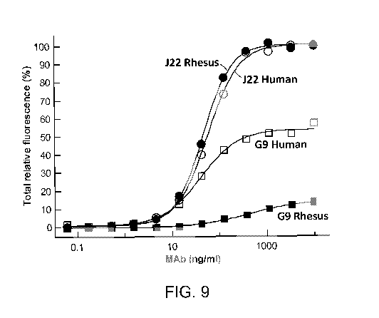
[0147] Figure 9 is a graph depicting the human and rhesus GM-CSF binding
reactivity of the
1783J22 and G9 antibodies, measured as the percent total relative fluorescence
(%) as a function
of monoclonal antibody (MAb) concentration (provided as nanograms per
milliliter, or ng/ml).
The results indicated that 1783J22 binds equally well to Rhesus GM-CSF and
Human GM-CSF,
when these proteins are expressed on the surface of HEK293 cells.
DETAILED DESCRIPTION OF THE INVENTION
GM-CSF
[0148] Blood cells in circulation are constantly replaced by newly developed
cells. Replacement
blood cells are formed in a process termed hematopoiesis which involves the
production of at
least eight mature blood cell types within two major lineages: (1) the myeloid
lineage which
includes red blood cells (erythrocytes), macrophages (monocytes), eosinophilic
granulocytes,
megakaryocytes (platelets), neutrophilic granulocytes, basophilic granulocytes
(mast cells); and
(2) the lymphoid lineage which includes T lymphocytes and B lymphocytes
(Burgess and Nicola,
9
CA 02759506 2011-10-20
WO 2010/124163 PCT/US2010/032170
Growth Factors and Stem Cells (Academic Press, New York, 1983)). Much of the
control of
blood-cell formation is mediated by a group of interacting glycoproteins
termed colony
stimulating factors (CSFs). The role of CSFs in hematopoiesis is the subject
of many reviews,
and is of great interest to clinical investigators who must treat blood
diseases or deficiencies; e.g.
Metcalf, The Hemopoietic Colony Stimulating Factors (Elsevier, N.Y., 1984);
Clark and Kamen,
Science, Vol. 236, pgs. 1229-1237 (1987); Sachs, Science, Vol. 238, pgs. 1374-
1379 (1987);
Dexter et al., eds., Colony Stimulating Factors (Dekker, N.Y., 1990); and
Morstyn et al., Cancer
Investigation, Vol. 7, pgs. 443-456 (1989).
[0149] Granulocyte macrophage-colony stimulating factor ("GM-CSF"), a soluble
secreted
glycoprotein, is a potent immunomodulatory cytokine known to facilitate
development and
prolongation of both humoral and cellular mediated immunity. GM-CSF was
originally
discovered as a protein with the capacity to generate both granulocyte and
macrophage colonies
from precursor cells in mouse bone marrow, and was accordingly named (Burgess
et al. (1980)
Blood 56:947-58.). GM-CSF stimulates stem cells to produce granulocytes
(neutrophils,
eosinophils, and basophils) and monocytes. Activities of GM-CSF include
activation and
enhanced maturation of antigen presenting cells, increasing the expression of
MHC class II
antigens, activation of mature granulocytes, macrophages and monocytes, and
proliferation and
differentiation of hematopoietic progenitor cells. The functions of GM-CSF are
mediated by
binding to CD 116, the granulocyte-macrophage colony stimulating factor
receptor, also known
as colony stimulating factor 2 receptor alpha that binds GM-CSF with low
affinity. The GM-CSF
receptors are found on myeloid progenitors and mature myeloid cells including
neutrophils,
eosinophils, mononuclear phagocytes, and monocytes. In addition, GM-CSF
receptor subunits
have been shown to be present in normal, non-hematopoietic tissues such as
human placenta,
endothelium, and oligodendrocytes of the central nervous system.
[0150] Human granulocyte macrophage colony-stimulating factor (GM-CSF) is a
glycoprotein
with a molecular weight of about 23,000 daltons. The cDNA sequence and the
expression of the
glycoprotein in mammalian cells have already been disclosed (G. G. Wong et
al., Science 228
(1985), 810-815, D. Metcalf, Science 229 (1985), 16-22). The active form of
the protein is found
extracellularly as a homodimer. The gene has been localized to a cluster of
related genes at
chromosome region 5g31, which is known to be associated with interstitial
deletions in the 5q-
syndrome and acute myelogenous leukemia. GM-CSF is also known as molgramostim
or, when
the protein is expressed in yeast cells, sargramostim (Leukine ; Berlex
Laboratories).
CA 02759506 2011-10-20
WO 2010/124163 PCT/US2010/032170
[0151] GM-CSF stimulates the production of white blood cells. GM-CSF holds
great promise as
a biopharmaceutical for use in association with cancer treatment to aid in the
restoration of white
blood cells. Naturally occurring GM-CSF is a glycoprotein containing 127 amino
acids and two
disulphide bonds. GM-CSF is present in only trace quantities in the natural
human source. GM-
CSF holds great promise as a biopharmaceutical for use in association with
cancer treatment to
aid in the restoration of white blood cells. The diverse immunomodulatory
activities of GM-CSF
have made it an attractive investigational cytokine for use as a vaccine
adjuvant for improving
the immune response to vaccines, including those used for the treatment of
cancer and HIV.
[0152] GM-CSF plays a role in the genesis and progression of leukemias, such
as juvenile
myelomonocytic leukemia (JMML). (Emanuel P D (2004) Curr. Hematol. Rep. 3:203-
209).
JMML is characterized by disruption of normal hemopoiesis resulting in
excessive, inappropriate
proliferation of immature myeloid cells in the bone marrow. Patients with JMML
are
hypersensitive to GM-CSF and exhibit pathologic features similar to those in
transgenic mice
that over-express GM-CSF (Lang et al. (1987) 51:675-86). Furthermore, GM-CSF
has been
shown to promote JMML cell growth and survival (Emanuel et al (1991) Blood
77:925-9).
[0153] There is recent evidence for a key role for GM-CSF in inflammatory and
autoimmune
diseases, therefore making it worthy of consideration for targeting. Such
evidence includes
disease exacerbation following its administration and amelioration of disease
in animal models
by GM-CSF gene targeting or by anti-GM-CSF antibody blockade. Hamilton JA,
Trends in
Immunology 23(8): 403-408 (2002). GM-CSF has been shown to play a role in
potentiating the
function of mature macrophages and granulocytes (Handman and Burgess (1979) J.
Immunol.
122:1134-1137; Hamilton et al. (1980) J. Cell Physiol. 103:435-445; Gamble et
al. (1985) Proc.
Natl. Acad. Sci. USA 82:8667-867 1), suggesting a role for GM-CSF in
inflammatory responses
(Hamilton et al. (1980) J. Cell Physiol. 103:435-445). In a clinical setting,
administration of GM-
CSF into peritoneal dialysis patients resulted in a marked recruitment of
macrophages (Selgas et
al., 1996, Kidney Int. 50:2070-2078).
[0154] GM-CSF may play a role in constitutional predisposition towards a
multitude of human
inflammatory pathologies, such as rheumatoid arthritis, autoimmune
pathologies, inflammatory
renal disease and inflammatory lung disorders such as asthma and chronic
obstructive pulmonary
disease (COPD).Patients with rheumatoid arthritis treated with GM-CSF had
their arthritis was
exacerbated (Hazenberg et al., 1991, Blood 74:2769-2770). Following cancer
chemotherapy,
11
CA 02759506 2011-10-20
WO 2010/124163 PCT/US2010/032170
GM-CSF treatment made rheumatoid arthritis worse (de Vries et al., (1991) J.
Immunol. 163:
4985-4993).
[0155] GM-CSF is a lymphokine (stimulator of the immune system) that exhibits
a broad
spectrum of immune cell stimulation as described in Burgess and Metcalf,
Blood, 56:947 (1980)
and Metcalf, Blood 67:257 (1986). GM-CSF has been shown to increase the
leukocyte count in
patients with acquired immunodeficiency syndrome (Brandt et al., N. Engl. J.
Med., 318:869
(1988)) and people suffering from chemotherapy-induced myelosuppression
(Antman et al., New
Engl. J. Med., 319:593 (1988)). It has been suggested that various colony
stimulating factors
alone or in combination with erythropoietin and/or an antiviral agent and/or
interleukin-2 (IL-2)
may be useful for the treatment of patients suffering from AIDS.
[0156] In addition to its ability to stimulate proliferation of hematopoietic
precursor cells, GM-
CSF is also able to stimulate a number of functional aspects of mature
granulocytes and
macrophages. These effects include synthesis of biologically active molecules
such as
prostaglandin E (Hancock et al., J. Immunol., 140:3021 (1988) and Kurland et
al., Proc. Natl.
Acad. Sci. USA, 76:2326 (1979)); increased phagocytic activity (Weisbart et
al., Nature, 332:647
(1988)); expression and/or affinity of various membrane markers such as the IL-
2 receptor
(Hancock et al., J. Immunol., 140:3021 (1988)) and receptors on neutrophils
which elicit the
production of superoxide anions (Atkinson et al., Immunology, 64:519 (1988));
and the
regulation of enzyme activity such as the stimulation of guanylate cyclase and
the inhibition of
adenylate cyclase (Coffey et al., J. Immunol., 140:2695 (1988)).
[0157] There may be a link between multiple sclerosis and GM-CSF (McQualter et
al. (2001) J.
Exp. Med., 194:873-88 1). In an experimental model of autoimmune
encephalomyelitis, a model
for multiple sclerosis, GM-CSF was found to be involved in the autoimmune-
mediated
demyelination.
[0158] It has been shown that GM-CSF can "prime" cells to respond in a
synergistic manner to a
second stimulus, such as LPS or interferon-gamma (Hart et al., 1988, J.
Immunol. 141:1516-
1521).
[0159] Aberrant expression of GM-CSF is associated with disease of the lung in
humans. Up-
regulation of GM-CSF in the lung by minor irritants, endotoxins or infections
predisposes
towards TH2 immune deviation and asthma (Eisenbarth et al. (2002) J. Exp. Med.
196:1645-
1651). A role for GM-CSF in asthma has been suggested. The use of neutralizing
antibodies in a
mouse model of asthma has demonstrated the ability to suppress asthmatic
phenotypes
12
CA 02759506 2011-10-20
WO 2010/124163 PCT/US2010/032170
(Yamashita (2002) Cell Immunol. 219:92). Allergens, alone or in combination
with other factors,
can spontaneously induce GM-CSF production in the airway thus present a
compelling
etiological argument for the role of GM-CSF in allergic sensitization.
(Gajewska (2003) Curr
Drug Targets Inflamm Allergy 2:279).
[0160] Adult human pulmonary alveolar proteinosis (PAP) is a rare disease
characterized by the
accumulation of phospholipids and surfactant proteins in the alveoli. GM-CSF
null mice have
impaired surfactant clearance that leads to murine pulmonary alveolar
proteinosis (PAP), which
closely mimics the human condition as described herein. Moreover, the PAP
phenotype can be
corrected by lung-specific delivery of the GM-CSF gene (Zsengaller et al.
(1998) Hum. Gene
Ther. 9:2101-2109). Patients with PAP have been shown to have circulating,
neutralizing
antibodies to GM-CSF, thereby implicating this cytokine as causative of the
disease. (Latzin P.,
et al., Thorax. (2005) 60(1):39-44).
[0161] GM-CSF has been used for lowering levels of lipoprotein cholesterol,
serum cholesterol
and other lipids. (US Pat. No. 5,019,381). Profound decreases in serum
cholesterol
concentrations were observed during GM-CSF therapy in patients with aplastic
anemia. (Nimer
SD, et al. JAMA 260(22): 3297-3300 (1988).
[0162] Local and systemic GM-CSF release in patients with Alzheimer's disease
(AD) and
vascular dementia (VAD) has been reported. (Tarkowski, E. et al., Acta Neurol
Scand. (2001)
103(3):166-174.) One of the hallmarks of AD is the accumulation of amyloid
beta plaques in the
brain parenchyma. Neutralization of GM-CSF has been shown to decrease amyloid-
beta and
suppress microglial activity in mouse models of AD. (Manczak M. et al., Hum.
Mol. Genet.
(2009, Jul. 19) Epub.) GM-CSF neutralizing antibodies have been shown to
mitigate CD40L
induced production of amyloid beta. (Volmar CH, et al., Cytokine (2008)
42(3):336-344.)
[0163] GM-CSF inhibits osteoclast differentiation by converting precursors
into dendritic cells
(see, e.g., Khapli et al., J. Immunol. 171:142-151, 2003; Miyamoto et al.,
Blood 98:2544-2554,
2001; Myint et al., Am. J. Pathol. 154:553-566, 1999; Shuto et al.,
Endocrinology 134:1121-
1126, 1994; and Kim et al., J. Biol. Chem. 280:16163-16169, 2005). There have
also been
reports that under certain conditions, GM-CSF may promote the formation of
osteoclastic cells in
vitro (e.g., U.S. Pat. No. 6,331,562) and that colony stimulating factors may
be therapeutic
targets in particular circumstances (U.S. Patent Application Publication No.
20020141994).
[0164] Therefore it is desirable to antagonize the activity of GM-CSF by
developing an antibody
to the cytokine. Such a compound may be a valuable human therapeutic. Several
polyclonal and
13
CA 02759506 2011-10-20
WO 2010/124163 PCT/US2010/032170
monoclonal antibodies have been generated to recombinant GM-CSF. For example,
Beffy et al.
((1994), Hybridoma 13:457-468), generated polyclonal antibodies to recombinant
human GM-
CSF in New Zealand White rabbits and monoclonal antibodies in Balb/c mice.
These rabbit and
some of the murine monoclonal antibodies were capable of neutralizing the
activity of GM-CSF
in an in vitro cell proliferation assay with M07c cells. Three murine
antibodies to human GM-
CSF were generated by Dempsey et al. (1990, Hybridoma 9, 545-558) that
neutralized GM-CSF
in an in vitro assay system. While these antibodies are useful reagents for
the detection of GM-
CSF in human serum as well as for in vitro assays to inhibit GM-CSF signaling,
they have little
value as therapeutics due to the fact that they are derived from either a
murine or rabbit system.
Attempts have been made to generate chimeric antibodies from murine
counterparts by
subcloning the variable domain from the murine anti-GM-CSF antibody into a
human backbone.
(WO 03/068924 A2). A human monoclonal antibody, i.e. G9, that specifically
binds to GM-CSF
has been reported. (Li J, et al, 2006, PNAS, 103:3557-62; WO 2007/092939); US
Pat. App. Pub.
No. 20080292641A1)
[0165] There is a need for therapeutic human antibodies for the treatment of
inflammation
associated with infectious diseases, inflammatory diseases, autoimmune
disorders, and other
diseases such as cancer associated with GM-CSF. It is further desired that
such antibodies would
elicit immune effector functions, as well as be well-tolerated in human
patients. There is
therefore a need for the efficient identification and production of
neutralizing antibodies effective
against GM-CSF as well as the elucidation of the target and antigenic
determinants to which
such antibodies bind. The invention addresses these and other long felt needs.
Anti-GM-CSF Antibodies
[0166] The anti-GM-CSF antibodies of the present invention are isolated by an
In-Situ
Therapeutic Antibody Rescue method (I-STARTM; Theraclone Sciences, Seattle WA)
which
involves discovery and synthesis of human therapeutic monoclonal antibodies
directly from
human memory B cells. B cells are screened for neutralization activity prior
to rescue of
antibodies. Novel neutralizing antibodies are obtained by emphasizing
neutralization as the
initial screen.
[0167] Peripheral Blood Mononuclear Cells (PBMCs) are obtained from a human
donor selected
for GM-CSF neutralizing activity in the plasma. Memory B cells are isolated
and B cell culture
supernatants are subjected to a primary screen of neutralization assay in a
high throughput
format. Optionally, GM-CSF antigen binding assays using ELISA or like methods
are also used
14
CA 02759506 2011-10-20
WO 2010/124163 PCT/US2010/032170
as a screen. B cell lysates corresponding to supernatants exhibiting
neutralizing activity are
selected for rescue of monoclonal antibodies by standard recombinant methods.
[0168] In one embodiment, the recombinant rescue of the monoclonal antibodies
involves use of
a B cell culture system as described in Weitcamp J-H et al., J. Immunol.
171:4680-4688 (2003).
Any other method for rescue of single B cells clones known in the art also may
be employed
such as EBV immortalization of B cells (Traggiai E., et al., Nat. Med.
10(8):871-875 (2004)),
electrofusion (Buchacher, A., et al., 1994. AIDS Res. Hum. Retroviruses 10:359-
369), and B cell
hybridoma (Karpas A. et al., Proc. Natl. Acad. Sci. USA 98:1799-1804 (2001).
[0169] In some embodiments, monoclonal antibodies were rescued from the B cell
cultures
using variable chain gene-specific RT-PCR, and transfectant with combinations
of H and L chain
clones were screened again for neutralization and GM-CSF antigen binding
activities. mAbs
with neutralization properties were selected for further characterization.
[0170] A human monoclonal antibody, 1783J22, identified according to these
methods is
disclosed herein. The antibody has been shown to neutralize GM-CSF in vitro.
[0171] The monoclonal antibody 1783J22 exhibits strong binding to human GM-CSF
among a
panel of B cell supernatants, most of which have no GM-CSF neutralizing
activity, as shown in
Figure 2 and Example 2 below. 1783J22 also exhibits neutralization activity in
TF1 proliferation
assays as shown in Examples 4 and 5 and Figures 4B and 5 below.
[0172] The binding and neutralization characteristics of 1783J22 were compared
to those of a
known human monoclonal GM-CSF antibody, G9. (Li J, et al, 2006, PNAS, 103:3557-
62; WO
2007/092939); US Pat. App. Pub. No. 20080292641A1). 1783J22 displays a higher
potency for
neutralizing GM-CSF derived from yeast as compared to G9. (See Figure 5 and
Example 5).
[0173] 1783J22 and G9 bind to different epitopes on GM-CSF. 1783J22 Fab does
not compete
with G9 whole antibody binding to human GM-CSF and G9 Fab does not compete
with 1783J22
whole antibody binding to human GM-CSF. (See Figure 6 and Example 6). It was
also observed
that 1782J22 bound to rabbit, human and rhesus GM-CSF, whereas G9 bound to
only human and
rhesus GM-CSF but not rabbit GM-CSF. (See Figure 7 and Example 8). Therefore,
it is
postulated that the MAbs 1783J22 and G9 also can have differences in
biological and therapeutic
activities.
[0174] The invention is based on novel monoclonal antibodies and antibody
fragments that
neutralize GM-CSF. In some embodiments, these monoclonal antibodies and
antibody fragments
have a particularly high potency in neutralizing GM-CSF in vitro. Such
antibodies are desirable,
CA 02759506 2011-10-20
WO 2010/124163 PCT/US2010/032170
as only low concentrations are required in order to neutralize a given amount
of GM-CSF. This
facilitates higher levels of therapeutic potency while administering lower
amounts of antibody.
Human monoclonal antibodies and the immortalized B cell clones that secrete
such antibodies
are also included within the scope of the invention.
[0175] Antibodies of the invention also include antibody fragments. A
"fragment" refers to
polypeptide sequences which are at least about 10, 15, 20, 30, 40, 50, 60, 70,
80 90 or about 100
amino acids in length, and which retain some biological activity or
immunological activity of the
full-length sequence, for example, binding affinity or avidity and immune
effector activity.
[0176] The invention also relates to the characterization of the epitope to
which the antibodies
bind and the use of that epitope in raising an immune response.
[0177] The invention also relates to various methods and uses involving the
antibodies of the
invention and the epitopes to which they bind.
[0178] The invention provides novel monoclonal or recombinant antibodies
having particularly
high potency in neutralizing GM-CSF. The invention also provides fragments of
these
recombinant or monoclonal antibodies, particularly fragments that retain the
antigen-binding
activity of the antibodies, for example which retain at least one
complementarity determining
region (CDR) specific for GM-CSF. In this specification, by "high potency in
neutralizing GM-
CSF " is meant that an antibody molecule of the invention neutralizes GM-CSF
in a standard
assay at a concentration (IC50) lower than that required by antibodies known
in the art.
[0179] Preferably, the antibody molecule of the present invention can
neutralize at a
concentration of 0.16 gg/ml or lower (i.e. 0.15, 0.125, 0.1, 0.075, 0.05,
0.025, 0.02, 0.016, 0.015,
0.0125, 0.01, 0.0075, 0.005, 0.004 or lower), preferably 0.016 gg/ml or lower
(an antibody
concentration of 10-8 or lower, preferably 10-9 M or lower, preferably 10-10 M
or lower, i.e. 10-11
M, 10-12 M, 10-13 M or lower). This means that only very low concentrations of
antibody are
required for 50% neutralization of GM-CSF in vitro. Potency can be measured
using a standard
neutralization assay as described in the art.
[0180] The antibodies of the invention are able to neutralize GM-CSF.
Monoclonal antibodies
can be produced by known procedures, e.g., as described by R. Kennet et al. in
"Monoclonal
Antibodies and Functional Cell Lines; Progress and Applications". Plenum Press
(New York),
1984. Further materials and methods applied are based on known procedures,
e.g., such as
described in J. Virol. 67:6642-6647, 1993.
16
CA 02759506 2011-10-20
WO 2010/124163 PCT/US2010/032170
[0181] These antibodies can be used as prophylactic or therapeutic agents upon
appropriate
formulation, or as a diagnostic tool.
[0182] A "neutralizing antibody" is one that can neutralize an activity of
that antigen in vivo or
in vitro. The invention provides a neutralizing monoclonal human antibody,
wherein the
antibody recognizes an antigen from GM-CSF.
[0183] The CDRs of the antibody heavy chains are referred to as CDRH1, CDRH2
and CDRH3,
respectively. Similarly, the CDRs of the antibody light chains are referred to
as CDRL1, CDRL2
and CDRL3, respectively. The positions of the CDR amino acids are defined
according to the
IMGT numbering system as: CDR1--IMGT positions 27 to 38, CDR2--IMGT positions
56 to 65
and CDR3--IMGT positions 105 to 117. (Lefranc, M P. et al. 2003 IMGT unique
numbering for
immunoglobulin and T cell receptor variable domains and Ig superfamily V-like
domains. Dev
Comp Immunol. 27(1):55-77; Lefranc, M P. 1997. Unique database numbering
system for
immunogenetic analysis. Immunology Today, 18:509; Lefranc, M P. 1999. The IMGT
unique
numbering for Immunoglobulins, T cell receptors and Ig-like domains. The
Immunologist,
7:132-136.)
[0184] A phylogram is a branching diagram (tree) assumed to be an estimate of
phylogeny,
branch lengths are proportional to the amount of inferred evolutionary change.
Tree diagrams of
the five heavy chains and the five light chains were prepared using ClustalW
(Larkin M.A.,
Blackshields G., Brown N.P., Chenna R., McGettigan P.A., McWilliam H.,
Valentin F., Wallace
I.M., Wilm A., Lopez R., Thompson J.D., Gibson T.J. and Higgins D.G.
Bioinformatics 23(21):
2947-2948 (2007); Higgins DG et al. Nucleic Acids Research 22: 4673-4680.
(1994)) and are
shown in Figures 3A and 3B respectively.
[0185] Preferably an antibody according to the invention is a novel monoclonal
antibody
referred to herein as 1783J22.
[0186] The 1783J22 antibody includes a heavy chain variable region (SEQ ID NO:
3), encoded
by the nucleic acid sequence shown below in SEQ ID NO: 7, and a light chain
variable region
(SEQ ID NO: 6) encoded by the nucleic acid sequence shown in SEQ ID NO: 13.
[0187] The amino acids encompassing the CDRs as defined by Chothia, C. et al.
(1989, Nature,
342: 877-883) are underlined and those defined by Kabat E.A. et al.(1991,
Sequences of Proteins
of Immunological Interest, 5th edit., NIH Publication no. 91-3242 U.S.
Department of Health and
Human Services.) are highlighted in bold in the sequences below.
17
CA 02759506 2011-10-20
WO 2010/124163 PCT/US2010/032170
[0188] The heavy chain CDRs of the 1783J22 antibody have the following
sequences per Kabat
definition: FPFHKYTMT (SEQ ID NO: 8), VSGVNGKTYYSPSVRG (SEQ ID NO: 9), and
GPGGHLHYYYGLDV (SEQ ID NO: 10). The light chain CDRs of the 1783J22 antibody
have
the following sequences per Kabat definition: RASQAINNYVA (SEQ ID NO: 14),
GASNLQP
(SEQ ID NO: 15), and QNYFGYPLT (SEQ ID NO: 16).
[0189] The heavy chain CDRs of the 1783J22 antibody have the following
sequences per
Chothia definition: GFPFHKYTMT (SEQ ID NO: 11), VSGVNGKTY (SEQ ID NO: 12), and
GPGGHLHYYYGLDV (SEQ ID NO: 10). The light chain CDRs of the 1783J22 antibody
have
the following sequences per Chothia definition: RASQAINNYVA (SEQ ID NO: 14),
GASNLQP (SEQ ID NO: 15), and QNYFGYPLT (SEQ ID NO: 16).
[0190] 1783J22 gamma heavy chain nucleotide sequence (variable region in
bold):
ATGGAGTTTGGGCTGAGCTGGCTTTTTCTTGTGACTGTTCTAAAAGGTGTCCACTGTGAGGTCC
AATTATTGCAGTCGGGGGGGGGCCTGACACATCCTGGGGGGTCCCTGAGACTCTCATGTGCGGC
GTCTGGCTTCCCCTTTCACAAATATACCATGACTTGGGTCCGCCAGCCTCCAGGGAAGGGCCTG
GAGTGGGTCTCAAGTGTTAGTGGTGTCAACGGCAAGACATATTATAGTCCCTCCGTGAGGGGCC
GCGCCATCGTCTCCAGAGACGACTCCAACAGTATGTTGTTTTTGGAAATCAAGAACATGACAGC
CGGGGACACGGCCCTCTACTTCTGCGCCAAAGGGCCGGGTGGCCATCTTCATTATTACTATGGT
CTAGACGTCTGGGGCCATGGGACCTCGGTCACCGTCTCGAGCGCCTCCACCAAGGGCCCATCGG
TCTTCCCCCTGGCACCCTCCTCCAAGAGCACCTCTGGGGGCACAGCGGCCCTGGGCTGCCTGGT
CAAGGACTACTTCCCCGAACCGGTGACGGTGTCGTGGAACTCAGGCGCCCTGACCAGCGGCGTG
CACACCTTCCCGGCTGTCCTACAGTCCTCAGGACTCTACTCCCTCAGCAGCGTGGTGACCGTGC
CCTCCAGCAGCTTGGGCACCCAGACCTACATCTGCAACGTGAATCACAAGCCCAGCAACACCAA
GGTGGACAAGAGAGTTGAGCCCAAATCTTGTGACAAAACTCACACATGCCCACCGTGCCCAGCA
CCTGAACTCCTGGGGGGACCGTCAGTCTTCCTCTTCCCCCCAAAACCCAAGGACACCCTCATGA
TCTCCCGGACCCCTGAGGTCACATGCGTGGTGGTGGACGTGAGCCACGAAGACCCTGAGGTCAA
GTTCAACTGGTACGTGGACGGCGTGGAGGTGCATAATGCCAAGACAAAGCCGCGGGAGGAGCAG
TACAACAGCACGTACCGTGTGGTCAGCGTCCTCACCGTCCTGCACCAGGACTGGCTGAATGGCA
AGGAGTACAAGTGCAAGGTCTCCAACAAAGCCCTCCCAGCCCCCATCGAGAAAACCATCTCCAA
AGCCAAAGGGCAGCCCCGAGAACCACAGGTGTACACCCTGCCCCCATCCCGGGAGGAGATGACC
AAGAACCAGGTCAGCCTGACCTGCCTGGTCAAAGGCTTCTATCCCAGCGACATCGCCGTGGAGT
GGGAGAGCAATGGGCAGCCGGAGAACAACTACAAGACCACGCCTCCCGTGCTGGACTCCGACGG
CTCCTTCTTCCTCTATAGCAAGCTCACCGTGGACAAGAGCAGGTGGCAGCAGGGGAACGTCTTC
TCATGCTCCGTGATGCATGAGGCTCTGCACAACCACTACACGCAGAAGAGCCTCTCCCTGTCTC
CGGGTAAATGA (SEQ ID NO: 1)
[0191] 1783J22 gamma heavy chain variable region nucleotide sequence:
GAGGTCCAATTATTGCAGTCGGGGGGGGGCCTGACACATCCTGGGGGGTCCCTGAGACTCTCAT
GTGCGGCGTCTGGCTTCCCCTTTCACAAATATACCATGACTTGGGTCCGCCAGCCTCCAGGGAA
GGGCCTGGAGTGGGTCTCAAGTGTTAGTGGTGTCAACGGCAAGACATATTATAGTCCCTCCGTG
AGGGGCCGCGCCATCGTCTCCAGAGACGACTCCAACAGTATGTTGTTTTTGGAAATCAAGAACA
18
CA 02759506 2011-10-20
WO 2010/124163 PCT/US2010/032170
TGACAGCCGGGGACACGGCCCTCTACTTCTGCGCCAAAGGGCCGGGTGGCCATCTTCATTATTA
CTATGGTCTAGACGTCTGGGGCCATGGGACCTCGGTCACCGTCTCGAGC (SEQ ID NO: 7)
[0192] 1783J22 gamma heavy chain amino acid sequence (variable region in
bold):
EVQLLQSGGGLTHPGGSLRLSCAASGFPFHKYTMTWVRQPPGKGLEWVSSVSGVN
GKTYYSP SVRGRAIV SRDD SN SMLFLEIKNMTAGDTALYFCAKGPGGHLHYYYGL
DVWGHGTSVTVSSASTKGPSVFPLAPS SKSTSGGTAALGCLVKDYFPEPVTVS WNSGA
LTSGVHTFPAVLQSSGLYSLS SVVTVPSSSLGTQTYICNVNHKPSNTKVDKRVEPKSCDK
THTCPPCPAPELLGGPSVFLFPPKPKDTLMISRTPEVTCVVVDVSHEDPEVKFNWYVDG
VEVHNAKTKPREEQYNSTYRVV SVLTVLHQD WLNGKEYKCKV SNKALPAPIEKTISKA
KGQPREPQVYTLPPSREEMTKNQVSLTCLVKGFYPSDIAVEWESNGQPENNYKTTPPVL
DSDGSFFLYSKLTVDKSRWQQGNVFSCSVMHEALHNHYTQKSLSLSPGK (SEQ ID
NO: 2)
[0193] 1783J22 gamma heavy chain variable region amino acid sequence (Kabat
CDRs
underlined, Chothia CDRs in bold italics):
EV QLLQSGGGLTHPGGSLRLSCAAS GFPFHKYTMT WVRQPPGKGLEWV S S VSGVNGKT
YYSPSVRGRAIVSRDDSNSMLFLEIKNMTAGDTALYFCAKGPGGHLHYYYGLD VWGHG
TSVTVSS (SEQ ID NO: 3)
[0194] 1783J22 gamma heavy chain Kabat CDRs:
CDR1: FPFHKYTMT (SEQ ID NO: 8)
CDR2: VSGVNGKTYYSPSVRG (SEQ ID NO: 9)
CDR3: GPGGHLHYYYGLDV (SEQ ID NO: 10)
[0195] 1783J22 gamma heavy chain Chothia CDRs:
CDR1: GFPFHKYTMT (SEQ ID NO: 11)
CDR2: VSGVNGKTY (SEQ ID NO: 12)
CDR3: GPGGHLHYYYGLDV (SEQ ID NO: 10)
[0196] 1783J22 kappa light chain nucleic acid sequence (variable region in
bold):
ATGNNCATGAGAGTCCTCGCTCAGCTCCTGGGGCTCCTGCTGCTCTGTTTCCCAGGTGCCAGAT
GTGACATCCAGATGACCCAATCCCCATCCTCACTGTCTGCATCTATTGGAGATAGAGTCACCAT
CTCTTGTCGGGCGAGTCAGGCCATCAACAATTATGTTGCCTGGTTTCAGCAGTCTGCAGGAAAA
GCCCCTAAGTCTCTCATCTATGGTGCGTCGAATTTGCAACCTGGTGTCCCACCAAGGTTCAGCG
GCAGTGTATCTGGGACAAATTTCTCTCTCACCATCGACGGTCTGCAGTCCGAAGACTTTGCAAC
TTATTTCTGTCAAAATTACTTTGGTTATCCCCTCACTTTCGGCGGTGGGACCACACTGGAGATC
AAACGTACGGTGGCTGCACCATCTGTCTTCATCTTCCCGCCATCTGATGAGCAGTTGAAATCTG
GAACTGCCTCTGTTGTGTGCCTGCTGAATAACTTCTATCCCAGAGAGGCCAAAGTACAGTGGAA
GGTGGATAACGCCCTCCAATCGGGTAACTCCCAGGAGAGTGTCACAGAGCAGGACAGCAAGGAC
AGCACCTACAGCCTCAGCAGCACCCTGACGCTGAGCAAAGCAGACTACGAGAAACACAAAGTCT
ACGCCTGCGAAGTCACCCATCAGGGCCTGAGCTCGCCCGTCACAAAGAGCTTCAACAGGGGAGA
GTGTTAG (SEQ ID NO: 4)
19
CA 02759506 2011-10-20
WO 2010/124163 PCT/US2010/032170
[0197] 1783J22 kappa light chain variable region nucleic acid sequence:
GACATCCAGATGACCCAATCCCCATCCTCACTGTCTGCATCTATTGGAGATAGAGTCACCATCT
CTTGTCGGGCGAGTCAGGCCATCAACAATTATGTTGCCTGGTTTCAGCAGTCTGCAGGAAAAGC
CCCTAAGTCTCTCATCTATGGTGCGTCGAATTTGCAACCTGGTGTCCCACCAAGGTTCAGCGGC
AGTGTATCTGGGACAAATTTCTCTCTCACCATCGACGGTCTGCAGTCCGAAGACTTTGCAACTT
ATTTCTGTCAAAATTACTTTGGTTATCCCCTCACTTTCGGCGGTGGGACCACACTGGAGATCAA
AC (SEQ ID NO: 13)
[0198] 1783J22 kappa light chain amino acid sequence (variable region in
bold):
DIQMTQSPSSLSASIGDRVTISCRASQAINNYVAWFQQSAGKAPKSLIYGASNLQPG
VPPRFSGSVSGTNFSLTIDGLQSEDFATYFCQNYFGYPLTFGGGTTLEIKRTVAAPSV
FIFPPSDEQLKSGTASVVCLLNNFYPREAKVQWKVDNALQSGNSQESVTEQDSKDSTYS
LSSTLTLSKADYEKHKVYACEVTHQGLSSPVTKSFNRGEC (SEQ ID NO:5)
[0199] 1783J22 kappa light chain variable region amino acid sequence (Kabat
CDRs underlined,
Chothia CDRs in bold italics):
DIQMTQSPSSLSASIGDRVTISCRASOAINNYVAWFQQSAGKAPKSLIYGASNLOPGVPPR
FSGSVSGTNFSLTIDGLQSEDFATYFCONYFGYPLTFGGGTTLEIK (SEQ ID NO: 6)
[0200] 1783J22 kappa light chain Kabat CDRs:
CDR1: RASQAINNYVA (SEQ ID NO: 14)
CDR2: GASNLQP (SEQ ID NO: 15)
CDR3: QNYFGYPLT (SEQ ID NO: 16)
[0201] 1783J22 kappa light chain Chothia CDRs:
CDR1: RASQAINNYVA (SEQ ID NO: 14)
CDR2: GASNLQP (SEQ ID NO: 15)
CDR3: QNYFGYPLT (SEQ ID NO: 16)
[0202] In one aspect, an antibody according to the invention contains a heavy
chain having the
amino acid sequence of SEQ ID NOs: 2 or 3 and a light chain having the amino
acid sequence of
SEQ ID NOs: 5 or 6. Alternatively, an antibody according to the invention
contains a heavy
chain variable region having the amino acid sequence of SEQ ID NO: 3 and a
light chain
variable region having the amino acid sequence of SEQ ID NO: 6.
[0203] In another aspect, an antibody according to the invention contains a
heavy chain having
the amino acid sequence encoded by the nucleic acid sequence of SEQ ID NOs: 1
or 7 and a
light chain having the amino acid sequence encoded by the nucleic acid
sequence of SEQ ID
NOs: 4 or 13. Alternatively, an antibody according to the invention contains a
heavy chain
variable region having the amino acid sequence encoded by the nucleic acid
sequence of SEQ ID
CA 02759506 2011-10-20
WO 2010/124163 PCT/US2010/032170
NO: 7 and a light chain variable region having the amino acid sequence encoded
by the nucleic
acid sequence of SEQ ID NO: 13. Furthermore, an antibody according to the
invention contains
a heavy chain having the amino acid sequence encoded by a nucleic acid
sequence of SEQ ID
NO: 1, which contains a silent or degenerate mutation, and a light chain
having the amino acid
sequence encoded by the nucleic acid sequence of SEQ ID NO: 4, which contains
a silent or
degenerate mutation. Silent and degenerate mutations alter the nucleic acid
sequence, but do not
alter the resultant amino acid sequence.
[0204] Preferably the three heavy chain CDRs include an amino acid sequence of
at least 90%,
92%, 95%, 97%, 98%, 99%, or more identical to the amino acid sequence of
FPFHKYTMT
(SEQ ID NO: 8), VSGVNGKTYYSPSVRG (SEQ ID NO: 9), or GPGGHLHYYYGLDV (SEQ
ID NO: 10) (as determined by the Kabat method) or GFPFHKYTMT (SEQ ID NO: 11),
VSGVNGKTY (SEQ ID NO: 12), and GPGGHLHYYYGLDV (SEQ ID NO: 10) (as determined
by the Chothia method) and a light chain with three CDRs that include an amino
acid sequence
of at least 90%, 92%, 95%, 97%, 98%, 99%, or more identical to the amino acid
sequence of
RASQAINNYVA (SEQ ID NO: 14), GASNLQP (SEQ ID NO: 15), and QNYFGYPLT (SEQ
ID NO: 16) (as determined by the Kabat method) or RASQAINNYVA (SEQ ID NO: 14),
GASNLQP (SEQ ID NO: 15), and QNYFGYPLT (SEQ ID NO: 16) (as determined by the
Chothia method).
[0205] The heavy chain of the anti-GM-CSF monoclonal antibody is derived from
a germ line
variable (V) gene such as, for example, the IGHV3 germline gene.
[0206] The anti-GM-CSF antibodies of the invention include a variable heavy
chain (VH) region
encoded by human IGHV3-23 germline gene sequences. Preferably, the anti-GM-CSF
antibodies
of the invention include a variable heavy chain (VH) region encoded by human
IGHV3-23
germline gene sequences having the IGHV3-23*02 allele. The anti-GM-CSF
antibodies of the
invention also include constant regions encoded by human IGHJ6 and IGHD3-22
germline gene
sequences, and preferably, having the IGHJ6*02 and IGHD3-22*01 alleles,
respectively. A
human IGHV3-23 germline gene sequences is shown, e.g., in Accession number
AY998715. A
human IGHJ6 germline gene sequences is shown, e.g., in Accession number
AY998715. The
anti-GM-CSF antibodies of the invention include a VH region that is encoded by
a nucleic acid
sequence that is at least 75% homologous to the IGHV3-23 germline gene
sequence. Preferably,
the nucleic acid sequence is at least 90%, 95%, 96%, 97% homologous to the
IGHV3-23
germline gene sequence, and more preferably, at least 98%, 99% homologous to
the IGHV3-23
21
CA 02759506 2011-10-20
WO 2010/124163 PCT/US2010/032170
germline gene sequence. The VH region of the anti-GM-CSF antibody is at least
75%
homologous to the amino acid sequence of the VH region encoded by the IGHV3-23
VH germline
gene sequence. Preferably, the amino acid sequence of VH region of the anti-GM-
CSF antibody
is at least 90%, 95%, 96%, 97% homologous to the amino acid sequence encoded
by the IGHV3-
23 germline gene sequence, and more preferably, at least 98%, 99% homologous
to the sequence
encoded by the IGHV3-23 germline gene sequence.
[0207] The light chain of the anti-GM-CSF monoclonal antibody is derived from
a germ line
variable (V) gene such as, for example, the IGKV 1 germline gene.
[0208] The anti-GM-CSF antibodies of the invention include a variable light
chain (VL) region
encoded by human IGKV1-16 germline gene sequences. Preferably, the anti-GM-CSF
antibodies
of the invention include a variable light chain (VL) region encoded by human
IGKV1-16
germline gene sequences having the IGKV1-16*01 allele. The anti-GM-CSF
antibodies of the
invention also include constant regions encoded by human IGKJ4 germline gene
sequences, and
preferably, having the IGKJ4*01 allele. A human IGKV1-16 VL germline gene
sequence is
shown, e.g., Accession numbers EU599329, EF589394, EF589555, EF589492,
EF589439,
EF589569, and EF589393. A human IGKJ4 germline gene sequence is shown, e.g.,
Accession
numbers AY998691, AY998685, AY998683, AF168801, EF589383, EF589502, EF589488,
EF589481, EF589472, EF589464, EF589441, EF589477, and EF589385. The anti-GM-
CSF
antibodies include a VL region that is encoded by a nucleic acid sequence that
is at least 80%
homologous to the IGKV1-16 germline gene sequence. Preferably, the nucleic
acid sequence is
at least 90%, 95%, 96%, 97% homologous to the IGKV1-16 germline gene sequence,
and more
preferably, at least 98%, 99% homologous to the IGKV1-16 germline gene
sequence. The VL
region of the anti-GM-CSF antibody is at least 80% homologous to the amino
acid sequence of
the VL region encoded the IGKV1-16 germline gene sequence. Preferably, the
amino acid
sequence of VL region of the anti-GM-CSF antibody is at least 90%, 95%, 96%,
97%
homologous to the amino acid sequence encoded by the IGKV1-16 germline gene
sequence, and
more preferably, at least 98%, 99% homologous to the sequence encoded by the
IGKV1-16
germline gene sequence.
[0209] It is to be understood that because of the natural sequence variation
likely to exist among
heavy and light chains and the genes encoding them, one skilled in the art
would expect to find
some level of variation within the amino acid sequences or the genes encoding
them, while still
maintaining the unique binding properties (e.g., specificity and affinity) of
the antibodies of the
22
CA 02759506 2011-10-20
WO 2010/124163 PCT/US2010/032170
present invention. Accordingly, such variants and homologs are considered
substantially the
same as one another and are included within the scope of the present
invention.
[0210] Monoclonal and recombinant antibodies are particularly useful in
identification and
purification of the individual polypeptides or other antigens against which
they are directed. The
antibodies of the invention have additional utility in that they may be
employed as reagents in
immunoassays, radioimmunoassays (RIA) or enzyme-linked immunosorbent assays
(ELISA). In
these applications, the antibodies can be labeled with an analytically-
detectable reagent such as a
radioisotope, a fluorescent molecule or an enzyme. The antibodies may also be
used for the
molecular identification and characterization (epitope mapping) of antigens.
[0211] As mentioned above, the antibodies of the invention can be used to map
the epitopes to
which they bind. Applicants have discovered that the antibody 1783J22
neutralizes GM-CSF.
Although the Applicant does not wish to be bound by this theory, it is
postulated that the
1783J22 antibody may bind to one or more conformational epitopes formed by GM-
CSF.
[0212] The epitopes recognized by these antibodies may have a number of uses.
The epitopes
and mimotopes in purified or synthetic form can be used to raise immune
responses (i.e. as a
vaccine, or for the production of antibodies for other uses) or for screening
patient serum for
antibodies that immunoreact with the epitopes or mimotopes. Preferably, such
an epitope or
mimotope, or antigen comprising such an epitope or mimotope is used as a
vaccine for raising an
immune response. The antibodies of the invention can also be used in a method
to monitor the
quality of vaccines in particular to check that the antigen in a vaccine
contains the correct
immunogenic epitope in the correct conformation.
[0213] The epitopes may also be useful in screening for ligands that bind to
said epitopes. Such
ligands preferably block the epitopes and thus prevent infection. Such ligands
are encompassed
within the scope of the invention.
Methods of Making and Using Anti-GM-CSF Antibodies
[0214] As will be understood by the skilled artisan, general description of
antibodies herein and
methods of preparing and using the same also apply to individual antibody
polypeptide
constituents and antibody fragments.
[0215] Standard techniques of molecular biology may be used to prepare DNA
sequences coding
for the antibodies or fragments of the antibodies of the present invention.
Desired DNA
sequences may be synthesized completely or in part using oligonucleotide
synthesis techniques.
23
CA 02759506 2011-10-20
WO 2010/124163 PCT/US2010/032170
Site-directed mutagenesis and polymerase chain reaction (PCR) techniques may
be used as
appropriate.
[0216] Any suitable host cell/vector system may be used for expression of the
DNA sequences
encoding the antibody molecules of the present invention or fragments thereof.
Bacterial, for
example E. coli, and other microbial systems may be used, in part, for
expression of antibody
fragments such as Fab and F(ab')2 fragments, and especially Fv fragments and
single chain
antibody fragments, for example, single chain Fvs. Eukaryotic, e.g. mammalian,
host cell
expression systems may be used for production of larger antibody molecules,
including complete
antibody molecules. Suitable mammalian host cells include CHO, HEK293T,
PER.C6, myeloma
or hybridoma cells.
[0217] The present invention also provides a process for the production of an
antibody molecule
according to the present invention comprising culturing a host cell comprising
a vector of the
present invention under conditions suitable for leading to expression of
protein from DNA
encoding the antibody molecule of the present invention, and isolating the
antibody molecule.
[0218] The antibody molecule may comprise only a heavy or light chain
polypeptide, in which
case only a heavy chain or light chain polypeptide coding sequence needs to be
used to transfect
the host cells. For production of products comprising both heavy and light
chains, the cell line
may be transfected with two vectors, a first vector encoding a light chain
polypeptide and a
second vector encoding a heavy chain polypeptide. Alternatively, a single
vector may be used,
the vector including sequences encoding light chain and heavy chain
polypeptides.
[0219] Alternatively, antibodies according to the invention may be produced by
i) expressing a
nucleic acid sequence according to the invention in a cell, and ii) isolating
the expressed
antibody product. Additionally, the method may include iii) purifying the
antibody.
[0220] Transformed B cells are screened for those producing antibodies of the
desired antigen
specificity, and individual B cell clones can then be produced from the
positive cells. The
screening step may be carried out by ELISA, by staining of tissues or cells
(including transfected
cells), a neutralization assay or one of a number of other methods known in
the art for identifying
desired antigen specificity. The assay may select on the basis of simple
antigen recognition, or
may select on the additional basis of a desired function e.g. to select
neutralizing antibodies
rather than just antigen-binding antibodies, to select antibodies that can
change characteristics of
targeted cells, such as their signaling cascades, their shape, their growth
rate, their capability of
24
CA 02759506 2011-10-20
WO 2010/124163 PCT/US2010/032170
influencing other cells, their response to the influence by other cells or by
other reagents or by a
change in conditions, their differentiation status, etc.
[0221] The cloning step for separating individual clones from the mixture of
positive cells may
be carried out using limiting dilution, micromanipulation, single cell
deposition by cell sorting or
another method known in the art. Preferably the cloning is carried out using
limiting dilution.
[0222] The immortalized B cell clones of the invention can be used in various
ways e.g. as a
source of monoclonal antibodies, as a source of nucleic acid (DNA or mRNA)
encoding a
monoclonal antibody of interest, for research, etc.
[0223] The antibodies of the present invention may be polyclonal or monoclonal
antibodies.
However, in preferred embodiments, they are monoclonal. In particular
embodiments, antibodies
of the present invention are human antibodies. Methods of producing polyclonal
and monoclonal
antibodies are known in the art and described generally, e.g., in U.S. Patent
No. 6,824,780.
Typically, the antibodies of the present invention are produced recombinantly,
using vectors and
methods available in the art, as described further below. Human antibodies may
also be
generated by in vitro activated B cells (see U.S. Pat. Nos. 5,567,610 and
5,229,275).
[0224] Human antibodies may also be produced in transgenic animals (e.g.,
mice) that are
capable of producing a full repertoire of human antibodies in the absence of
endogenous
immunoglobulin production. For example, it has been described that the
homozygous deletion of
the antibody heavy-chain joining region (JH) gene in chimeric and germ-line
mutant mice results
in complete inhibition of endogenous antibody production. Transfer of the
human germ-line
immunoglobulin gene array into such germ-line mutant mice results in the
production of human
antibodies upon antigen challenge. See, e.g., Jakobovits et al., Proc. Natl.
Acad. Sci. USA,
90:2551 (1993); Jakobovits et al., Nature, 362:255-258 (1993); Bruggemann et
al., Year in
Immuno., 7:33 (1993); U.S. Pat. Nos. 5,545,806, 5,569,825, 5,591,669 (all of
GenPharm); U.S.
Pat. No. 5,545,807; and WO 97/17852. Such animals may be genetically
engineered to produce
human antibodies comprising a polypeptide of the present invention.
[0225] In certain embodiments, antibodies of the present invention are
chimeric antibodies that
comprise sequences derived from both human and non-human sources. In
particular
embodiments, these chimeric antibodies are humanized or primatizedTM. In
practice, humanized
antibodies are typically human antibodies in which some hypervariable region
residues and
possibly some FR residues are substituted by residues from analogous sites in
rodent antibodies.
CA 02759506 2011-10-20
WO 2010/124163 PCT/US2010/032170
[0226] In the context of the present invention, chimeric antibodies also
include human antibodies
wherein the human hypervariable region or one or more CDRs are retained, but
one or more
other regions of sequence have been replaced by corresponding sequences from a
non-human
animal.
[0227] The choice of non-human sequences, both light and heavy, to be used in
making the
chimeric antibodies is important to reduce antigenicity and human anti-non-
human antibody
responses when the antibody is intended for human therapeutic use. It is
further important that
chimeric antibodies retain high binding affinity for the antigen and other
favorable biological
properties. To achieve this goal, according to a preferred method, chimeric
antibodies are
prepared by a process of analysis of the parental sequences and various
conceptual chimeric
products using three-dimensional models of the parental human and non-human
sequences.
Three-dimensional immunoglobulin models are commonly available and are
familiar to those
skilled in the art. Computer programs are available which illustrate and
display probable three-
dimensional conformational structures of selected candidate immunoglobulin
sequences.
Inspection of these displays permits analysis of the likely role of the
residues in the functioning
of the candidate immunoglobulin sequence, i.e., the analysis of residues that
influence the ability
of the candidate immunoglobulin to bind its antigen. In this way, FR residues
can be selected and
combined from the recipient and import sequences so that the desired antibody
characteristic,
such as increased affinity for the target antigen(s), is achieved. In general,
the hypervariable
region residues are directly and most substantially involved in influencing
antigen binding.
[0228] As noted above, antibodies (or immunoglobulins) can be divided into
five different
classes, based on differences in the amino acid sequences in the constant
region of the heavy
chains. All immunoglobulins within a given class have very similar heavy chain
constant
regions. These differences can be detected by sequence studies or more
commonly by serological
means (i.e. by the use of antibodies directed to these differences).
Antibodies, or fragments
thereof, of the present invention may be any class, and may, therefore, have a
gamma, mu, alpha,
delta, or epsilon heavy chain. A gamma chain may be gamma 1, gamma 2, gamma 3,
or gamma
4; and an alpha chain may be alpha 1 or alpha 2.
[0229] In a preferred embodiment, an antibody of the present invention, or
fragment thereof, is
an IgG. IgG is considered the most versatile immunoglobulin, because it is
capable of carrying
out all of the functions of immunoglobulin molecules. IgG is the major Ig in
serum, and the only
class of Ig that crosses the placenta. IgG also fixes complement, although the
IgG4 subclass does
26
CA 02759506 2011-10-20
WO 2010/124163 PCT/US2010/032170
not. Macrophages, monocytes, PMN's and some lymphocytes have Fc receptors for
the Fc region
of IgG. Not all subclasses bind equally well; IgG2 and IgG4 do not bind to Fc
receptors. A
consequence of binding to the Fc receptors on PMN's, monocytes and macrophages
is that the
cell can now internalize the antigen better. IgG is an opsonin that enhances
phagocytosis.
Binding of IgG to Fc receptors on other types of cells results in the
activation of other functions.
Antibodies of the present invention may be of any IgG subclass.
[0230] In another preferred embodiment, an antibody, or fragment thereof, of
the present
invention is an IgE. IgE is the least common serum Ig since it binds very
tightly to Fc receptors
on basophils and mast cells even before interacting with antigen. As a
consequence of its binding
to basophils and mast cells, IgE is involved in allergic reactions. Binding of
the allergen to the
IgE on the cells results in the release of various pharmacological mediators
that result in allergic
symptoms. IgE also plays a role in parasitic helminth diseases. Eosinophils
have Fc receptors for
IgE and binding of eosinophils to IgE-coated helminths results in killing of
the parasite. IgE does
not fix complement.
[0231] In various embodiments, antibodies of the present invention, and
fragments thereof,
comprise a variable light chain that is either kappa or lambda. The lamba
chain may be any of
subtype, including, e.g., lambda 1, lambda 2, lambda 3, and lambda 4.
[0232] As noted above, the present invention further provides antibody
fragments comprising a
polypeptide of the present invention. In certain circumstances there are
advantages of using
antibody fragments, rather than whole antibodies. For example, the smaller
size of the fragments
allows for rapid clearance, and may lead to improved access to certain
tissues, such as solid
tumors. Examples of antibody fragments include: Fab, Fab', F(ab')2 and Fv
fragments;
diabodies; linear antibodies; single-chain antibodies; and multispecific
antibodies formed from
antibody fragments.
[0233] Various techniques have been developed for the production of antibody
fragments.
Traditionally, these fragments were derived via proteolytic digestion of
intact antibodies (see,
e.g., Morimoto et al., Journal of Biochemical and Biophysical Methods 24:107-
117 (1992); and
Brennan et al., Science, 229:81 (1985)). However, these fragments can now be
produced directly
by recombinant host cells. Fab, Fv and ScFv antibody fragments can all be
expressed in and
secreted from E. coli, thus allowing the facile production of large amounts of
these fragments.
Fab'-SH fragments can be directly recovered from E. coli and chemically
coupled to form F(ab')2
fragments (Carter et al., Bio/Technology 10:163-167 (1992)). According to
another approach,
27
CA 02759506 2011-10-20
WO 2010/124163 PCT/US2010/032170
F(ab')2 fragments can be isolated directly from recombinant host cell culture.
Fab and F(ab')2
fragment with increased in vivo half-life comprising a salvage receptor
binding epitope residues
are described in U.S. Pat. No. 5,869,046. Other techniques for the production
of antibody
fragments will be apparent to the skilled practitioner.
[0234] In other embodiments, the antibody of choice is a single chain Fv
fragment (scFv). See
WO 93/16185; U.S. Pat. Nos. 5,571,894; and 5,587,458. Fv and sFv are the only
species with
intact combining sites that are devoid of constant regions. Thus, they are
suitable for reduced
nonspecific binding during in vivo use. sFv fusion proteins may be constructed
to yield fusion of
an effector protein at either the amino or the carboxy terminus of an sFv. See
Antibody
Engineering, ed. Borrebaeck, supra. The antibody fragment may also be a
"linear antibody", e.g.,
as described in U.S. Pat. No. 5,641,870 for example. Such linear antibody
fragments may be
monospecific or bispecific.
[0235] In certain embodiments, antibodies of the present invention are
bispecific or multi-
specific. Bispecific antibodies are antibodies that have binding specificities
for at least two
different epitopes. Exemplary bispecific antibodies may bind to two different
epitopes of a single
antigen. Other such antibodies may combine a first antigen binding site with a
binding site for a
second antigen. Alternatively, an anti-GM-CSF arm may be combined with an arm
that binds to
Fc receptors for IgG (FcyR), such as FcyRI (CD64), FcyRII (CD32) and FcyRIII
(CD16), so as to
focus and localize cellular defense mechanisms to the target cell. Bispecific
antibodies may also
be used to localize cytotoxic agents to infected cells. These antibodies
possess an GM-CSF-
binding arm and an arm that binds the cytotoxic agent (e.g., saporin, anti-
interferon-a, vinca
alkaloid, ricin A chain, methotrexate or radioactive isotope hapten).
Bispecific antibodies can be
prepared as full length antibodies or antibody fragments (e.g., F(ab')2
bispecific antibodies). WO
96/16673 describes a bispecific anti-ErbB2/anti-FcyRIII antibody and U.S. Pat.
No. 5,837,234
discloses a bispecific anti-ErbB2/anti-FcyRI antibody. A bispecific anti-
ErbB2/Fca antibody is
shown in W098/02463. U.S. Pat. No. 5,821,337 teaches a bispecific anti-
ErbB2/anti-CD3
antibody.
[0236] Methods for making bispecific antibodies are known in the art.
Traditional production of
full length bispecific antibodies is based on the co-expression of two
immunoglobulin heavy
chain-light chain pairs, where the two chains have different specificities
(Millstein et al., Nature,
305:537-539 (1983)). Because of the random assortment of immunoglobulin heavy
and light
28
CA 02759506 2011-10-20
WO 2010/124163 PCT/US2010/032170
chains, these hybridomas (quadromas) produce a potential mixture of ten
different antibody
molecules, of which only one has the correct bispecific structure.
Purification of the correct
molecule, which is usually done by affinity chromatography steps, is rather
cumbersome, and the
product yields are low. Similar procedures are disclosed in WO 93/08829, and
in Traunecker et
al., EMBO J., 10:3655-3659 (1991).
[0237] According to a different approach, antibody variable domains with the
desired binding
specificities (antibody-antigen combining sites) are fused to immunoglobulin
constant domain
sequences. Preferably, the fusion is with an Ig heavy chain constant domain,
comprising at least
part of the hinge, CH2, and CH3 regions. It is preferred to have the first
heavy-chain constant
region (CH1) containing the site necessary for light chain bonding, present in
at least one of the
fusions. DNAs encoding the immunoglobulin heavy chain fusions and, if desired,
the
immunoglobulin light chain, are inserted into separate expression vectors, and
are co-transfected
into a suitable host cell. This provides for greater flexibility in adjusting
the mutual proportions
of the three polypeptide fragments in embodiments when unequal ratios of the
three polypeptide
chains used in the construction provide the optimum yield of the desired
bispecific antibody. It
is, however, possible to insert the coding sequences for two or all three
polypeptide chains into a
single expression vector when the expression of at least two polypeptide
chains in equal ratios
results in high yields or when the ratios have no significant affect on the
yield of the desired
chain combination.
[0238] In a preferred embodiment of this approach, the bispecific antibodies
are composed of a
hybrid immunoglobulin heavy chain with a first binding specificity in one arm,
and a hybrid
immunoglobulin heavy chain-light chain pair (providing a second binding
specificity) in the
other arm. It was found that this asymmetric structure facilitates the
separation of the desired
bispecific compound from unwanted immunoglobulin chain combinations, as the
presence of an
immunoglobulin light chain in only one half of the bispecific molecule
provides for a facile way
of separation. This approach is disclosed in WO 94/04690. For further details
of generating
bispecific antibodies see, for example, Suresh et al., Methods in Enzymology,
121:210 (1986).
[0239] According to another approach described in U.S. Pat. No. 5,731,168, the
interface
between a pair of antibody molecules can be engineered to maximize the
percentage of
heterodimers that are recovered from recombinant cell culture. The preferred
interface comprises
at least a part of the CH3 domain. In this method, one or more small amino
acid side chains from
the interface of the first antibody molecule are replaced with larger side
chains (e.g., tyrosine or
29
CA 02759506 2011-10-20
WO 2010/124163 PCT/US2010/032170
tryptophan). Compensatory "cavities" of identical or similar size to the large
side chain(s) are
created on the interface of the second antibody molecule by replacing large
amino acid side
chains with smaller ones (e.g., alanine or threonine). This provides a
mechanism for increasing
the yield of the heterodimer over other unwanted end-products such as
homodimers.
[0240] Bispecific antibodies include cross-linked or "heteroconjugate"
antibodies. For example,
one of the antibodies in the heteroconjugate can be coupled to avidin, the
other to biotin. Such
antibodies have, for example, been proposed to target immune system cells to
unwanted cells
(U.S. Pat. No. 4,676,980), and for treatment of HIV infection (WO 91/00360, WO
92/200373,
and EP 03089). Heteroconjugate antibodies may be made using any convenient
cross-linking
methods. Suitable cross-linking agents are well known in the art, and are
disclosed in U.S. Pat.
No. 4,676,980, along with a number of cross-linking techniques.
[0241] Techniques for generating bispecific antibodies from antibody fragments
have also been
described in the literature. For example, bispecific antibodies can be
prepared using chemical
linkage. Brennan et al., Science, 229: 81 (1985) describe a procedure wherein
intact antibodies
are proteolytically cleaved to generate F(ab')2 fragments. These fragments are
reduced in the
presence of the dithiol complexing agent, sodium arsenite, to stabilize
vicinal dithiols and
prevent intermolecular disulfide formation. The Fab' fragments generated are
then converted to
thionitrobenzoate (TNB) derivatives. One of the Fab'-TNB derivatives is then
reconverted to the
Fab'-thiol by reduction with mercaptoethylamine and is mixed with an equimolar
amount of the
other Fab'-TNB derivative to form the bispecific antibody. The bispecific
antibodies produced
can be used as agents for the selective immobilization of enzymes.
[0242] Recent progress has facilitated the direct recovery of Fab'-SH
fragments from E. coli,
which can be chemically coupled to form bispecific antibodies. Shalaby et al.,
J. Exp. Med., 175:
217-225 (1992) describe the production of a humanized bispecific antibody
F(ab')2 molecule.
Each Fab' fragment was separately secreted from E. coli and subjected to
directed chemical
coupling in vitro to form the bispecific antibody. The bispecific antibody
thus formed was able to
bind to cells overexpressing the ErbB2 receptor and normal human T cells, as
well as trigger the
lytic activity of human cytotoxic lymphocytes against human breast tumor
targets.
[0243] Various techniques for making and isolating bispecific antibody
fragments directly from
recombinant cell culture have also been described. For example, bispecific
antibodies have been
produced using leucine zippers. Kostelny et al., J. Immunol., 148(5):1547-1553
(1992). The
leucine zipper peptides from the Fos and Jun proteins were linked to the Fab'
portions of two
CA 02759506 2011-10-20
WO 2010/124163 PCT/US2010/032170
different antibodies by gene fusion. The antibody homodimers were reduced at
the hinge region
to form monomers and then re-oxidized to form the antibody heterodimers. This
method can also
be utilized for the production of antibody homodimers. The "diabody"
technology described by
Hollinger et al., Proc. Natl. Acad. Sci. USA, 90:6444-6448 (1993) has provided
an alternative
mechanism for making bispecific antibody fragments. The fragments comprise a
VH connected
to a VL by a linker that is too short to allow pairing between the two domains
on the same chain.
Accordingly, the VH and VL domains of one fragment are forced to pair with the
complementary
VL and VH domains of another fragment, thereby forming two antigen-binding
sites. Another
strategy for making bispecific antibody fragments by the use of single-chain
Fv (sFv) dimers has
also been reported. See Gruber et al., J. Immunol., 152:5368 (1994).
[0244] Antibodies with more than two valencies are contemplated. For example,
trispecific
antibodies can be prepared. Tutt et al., J. Immunol. 147: 60 (1991). A
multivalent antibody may
be internalized (and/or catabolized) faster than a bivalent antibody by a cell
expressing an
antigen to which the antibodies bind. The antibodies of the present invention
can be multivalent
antibodies with three or more antigen binding sites (e.g., tetravalent
antibodies), which can be
readily produced by recombinant expression of nucleic acid encoding the
polypeptide chains of
the antibody. The multivalent antibody can comprise a dimerization domain and
three or more
antigen binding sites. The preferred dimerization domain comprises (or
consists of) an Fc region
or a hinge region. In this scenario, the antibody will comprise an Fc region
and three or more
antigen binding sites amino-terminal to the Fc region. The preferred
multivalent antibody herein
comprises (or consists of) three to about eight, but preferably four, antigen
binding sites. The
multivalent antibody comprises at least one polypeptide chain (and preferably
two polypeptide
chains), wherein the polypeptide chain(s) comprise two or more variable
domains. For instance,
the polypeptide chain(s) may comprise VD1-(X1),, -VD2-(X2),, -Fc, wherein VD1
is a first
variable domain, VD2 is a second variable domain, Fc is one polypeptide chain
of an Fc region,
X1 and X2 represent an amino acid or polypeptide, and n is 0 or 1. For
instance, the polypeptide
chain(s) may comprise: VH-CH1-flexible linker-VH-CH1-Fc region chain; or VH-
CHI-VH-
CH1-Fc region chain. The multivalent antibody herein preferably further
comprises at least two
(and preferably four) light chain variable domain polypeptides. The
multivalent antibody herein
may, for instance, comprise from about two to about eight light chain variable
domain
polypeptides. The light chain variable domain polypeptides contemplated here
comprise a light
chain variable domain and, optionally, further comprise a CL domain.
31
CA 02759506 2011-10-20
WO 2010/124163 PCT/US2010/032170
[0245] Antibodies of the present invention further include single chain
antibodies.
[0246] In particular embodiments, antibodies of the present invention are
internalizing
antibodies.
[0247] Amino acid sequence modification(s) of the antibodies described herein
are
contemplated. For example, it may be desirable to improve the binding affinity
and/or other
biological properties of the antibody. Amino acid sequence variants of the
antibody may be
prepared by introducing appropriate nucleotide changes into a polynucleotide
that encodes the
antibody, or a chain thereof, or by peptide synthesis. Such modifications
include, for example,
deletions from, and/or insertions into and/or substitutions of, residues
within the amino acid
sequences of the antibody. Any combination of deletion, insertion, and
substitution may be made
to arrive at the final antibody, provided that the final construct possesses
the desired
characteristics. The amino acid changes also may alter post-translational
processes of the
antibody, such as changing the number or position of glycosylation sites. Any
of the variations
and modifications described above for polypeptides of the present invention
may be included in
antibodies of the present invention.
[0248] A useful method for identification of certain residues or regions of an
antibody that are
preferred locations for mutagenesis is called "alanine scanning mutagenesis"
as described by
Cunningham and Wells in Science, 244:1081-1085 (1989). Here, a residue or
group of target
residues are identified (e.g., charged residues such as arg, asp, his, lys,
and glu) and replaced by a
neutral or negatively charged amino acid (most preferably alanine or
polyalanine) to affect the
interaction of the amino acids with PSCA antigen. Those amino acid locations
demonstrating
functional sensitivity to the substitutions then are refined by introducing
further or other variants
at, or for, the sites of substitution. Thus, while the site for introducing an
amino acid sequence
variation is predetermined, the nature of the mutation per se need not be
predetermined. For
example, to analyze the performance of a mutation at a given site, ala
scanning or random
mutagenesis is conducted at the target codon or region and the expressed anti-
antibody variants
are screened for the desired activity.
[0249] Amino acid sequence insertions include amino- and/or carboxyl-terminal
fusions ranging
in length from one residue to polypeptides containing a hundred or more
residues, as well as
intrasequence insertions of single or multiple amino acid residues. Examples
of terminal
insertions include an antibody with an N-terminal methionyl residue or the
antibody fused to a
cytotoxic polypeptide. Other insertional variants of an antibody include the
fusion to the N- or C-
32
CA 02759506 2011-10-20
WO 2010/124163 PCT/US2010/032170
terminus of the antibody to an enzyme (e.g., for ADEPT) or a polypeptide that
increases the
serum half-life of the antibody.
[0250] Another type of variant is an amino acid substitution variant. These
variants have at least
one amino acid residue in the antibody molecule replaced by a different
residue. The sites of
greatest interest for substitutional mutagenesis include the hypervariable
regions, but FR
alterations are also contemplated. Conservative and non-conservative
substitutions are
contemplated.
[0251] Substantial modifications in the biological properties of the antibody
are accomplished by
selecting substitutions that differ significantly in their effect on
maintaining (a) the structure of
the polypeptide backbone in the area of the substitution, for example, as a
sheet or helical
conformation, (b) the charge or hydrophobicity of the molecule at the target
site, or (c) the bulk
of the side chain.
[0252] Any cysteine residue not involved in maintaining the proper
conformation of the antibody
also may be substituted, generally with serine, to improve the oxidative
stability of the molecule
and prevent aberrant crosslinking. Conversely, cysteine bond(s) may be added
to the antibody to
improve its stability (particularly where the antibody is an antibody fragment
such as an Fv
fragment).
[0253] One type of substitutional variant involves substituting one or more
hypervariable region
residues of a parent antibody. Generally, the resulting variant(s) selected
for further development
will have improved biological properties relative to the parent antibody from
which they are
generated. A convenient way for generating such substitutional variants
involves affinity
maturation using phage display. Briefly, several hypervariable region sites
(e.g., 6-7 sites) are
mutated to generate all possible amino substitutions at each site. The
antibody variants thus
generated are displayed in a monovalent fashion from filamentous phage
particles as fusions to
the gene III product of M13 packaged within each particle. The phage-displayed
variants are
then screened for their biological activity (e.g., binding affinity) as herein
disclosed. In order to
identify candidate hypervariable region sites for modification, alanine
scanning mutagenesis can
be performed to identify hypervariable region residues contributing
significantly to antigen
binding. Alternatively, or additionally, it may be beneficial to analyze a
crystal structure of the
antigen-antibody complex to identify contact points between the antibody and
an antigen or
infected cell. Such contact residues and neighboring residues are candidates
for substitution
according to the techniques elaborated herein. Once such variants are
generated, the panel of
33
CA 02759506 2011-10-20
WO 2010/124163 PCT/US2010/032170
variants is subjected to screening as described herein and antibodies with
superior properties in
one or more relevant assays may be selected for further development.
[0254] Another type of amino acid variant of the antibody alters the original
glycosylation
pattern of the antibody. By altering is meant deleting one or more
carbohydrate moieties found in
the antibody, and/or adding one or more glycosylation sites that are not
present in the antibody.
[0255] Glycosylation of antibodies is typically either N-linked or O-linked. N-
linked refers to
the attachment of the carbohydrate moiety to the side chain of an asparagine
residue. The
tripeptide sequences asparagine-X-serine and asparagine-X-threonine, where X
is any amino
acid except proline, are the recognition sequences for enzymatic attachment of
the carbohydrate
moiety to the asparagine side chain. Thus, the presence of either of these
tripeptide sequences in
a polypeptide creates a potential glycosylation site. O-linked glycosylation
refers to the
attachment of one of the sugars N-aceylgalactosamine, galactose, or xylose to
a hydroxyamino
acid, most commonly serine or threonine, although 5-hydroxyproline or 5-
hydroxylysine may
also be used.
[0256] Addition of glycosylation sites to the antibody is conveniently
accomplished by altering
the amino acid sequence such that it contains one or more of the above-
described tripeptide
sequences (for N-linked glycosylation sites). The alteration may also be made
by the addition of,
or substitution by, one or more serine or threonine residues to the sequence
of the original
antibody (for O-linked glycosylation sites).
[0257] The antibody of the invention is modified with respect to effector
function, e.g., so as to
enhance antigen-dependent cell-mediated cyotoxicity (ADCC) and/or complement
dependent
cytotoxicity (CDC) of the antibody. This may be achieved by introducing one or
more amino
acid substitutions in an Fc region of the antibody. Alternatively or
additionally, cysteine
residue(s) may be introduced in the Fc region, thereby allowing interchain
disulfide bond
formation in this region. The homodimeric antibody thus generated may have
improved
internalization capability and/or increased complement-mediated cell killing
and antibody-
dependent cellular cytotoxicity (ADCC). See Caron et al., J. Exp Med. 176:1191-
1195 (1992)
and Shopes, B. J. Immunol. 148:2918-2922 (1992). Homodimeric antibodies with
enhanced anti-
infection activity may also be prepared using heterobifunctional cross-linkers
as described in
Wolff et al., Cancer Research 53:2560-2565 (1993). Alternatively, an antibody
can be
engineered which has dual Fc regions and may thereby have enhanced complement
lysis and
ADCC capabilities. See Stevenson et al., Anti-Cancer Drug Design 3:219-230
(1989).
34
CA 02759506 2011-10-20
WO 2010/124163 PCT/US2010/032170
[0258] To increase the serum half-life of the antibody, one may incorporate a
salvage receptor
binding epitope into the antibody (especially an antibody fragment) as
described in U.S. Pat. No.
5,739,277, for example. As used herein, the term "salvage receptor binding
epitope" refers to an
epitope of the Fc region of an IgG molecule (e.g., IgG1, IgG2, IgG3, or IgG4)
that is responsible
for increasing the in vivo serum half-life of the IgG molecule.
[0259] Antibodies of the present invention may also be modified to include an
epitope tag or
label, e.g., for use in purification or diagnostic applications. The invention
also pertains to
therapy with immunoconjugates comprising an antibody conjugated to an anti-
cancer agent such
as a cytotoxic agent or a growth inhibitory agent. Chemotherapeutic agents
useful in the
generation of such immunoconjugates have been described above.
[0260] Conjugates of an antibody and one or more small molecule toxins, such
as a
calicheamicin, maytansinoids, a trichothene, and CC 1065, and the derivatives
of these toxins that
have toxin activity, are also contemplated herein.
[0261] In one preferred embodiment, an antibody (full length or fragments) of
the invention is
conjugated to one or more maytansinoid molecules. Maytansinoids are mitototic
inhibitors that
act by inhibiting tubulin polymerization. Maytansine was first isolated from
the east African
shrub Maytenus serrata (U.S. Pat. No. 3,896,111). Subsequently, it was
discovered that certain
microbes also produce maytansinoids, such as maytansinol and C-3 maytansinol
esters (U.S. Pat.
No. 4,151,042). Synthetic maytansinol and derivatives and analogues thereof
are disclosed, for
example, in U.S. Pat. Nos. 4,137,230; 4,248,870; 4,256,746; 4,260,608;
4,265,814; 4,294,757;
4,307,016; 4,308,268; 4,308,269; 4,309,428; 4,313,946; 4,315,929; 4,317,821;
4,322,348;
4,331,598; 4,361,650; 4,364,866; 4,424,219; 4,450,254; 4,362,663; and
4,371,533.
[0262] In an attempt to improve their therapeutic index, maytansine and
maytansinoids have
been conjugated to antibodies specifically binding to tumor cell antigens.
Immunoconjugates
containing maytansinoids and their therapeutic use are disclosed, for example,
in U.S. Pat. Nos.
5,208,020, 5,416,064 and European Patent EP 0 425 235 B1. Liu et al., Proc.
Natl. Acad. Sci.
USA 93:8618-8623 (1996) described immunoconjugates comprising a maytansinoid
designated
DM1 linked to the monoclonal antibody C242 directed against human colorectal
cancer. The
conjugate was found to be highly cytotoxic towards cultured colon cancer
cells, and showed
antitumor activity in an in vivo tumor growth assay.
[0263] Antibody-maytansinoid conjugates are prepared by chemically linking an
antibody to a
maytansinoid molecule without significantly diminishing the biological
activity of either the
CA 02759506 2011-10-20
WO 2010/124163 PCT/US2010/032170
antibody or the maytansinoid molecule. An average of 3-4 maytansinoid
molecules conjugated
per antibody molecule has shown efficacy in enhancing cytotoxicity of target
cells without
negatively affecting the function or solubility of the antibody, although even
one molecule of
toxin/antibody would be expected to enhance cytotoxicity over the use of naked
antibody.
Maytansinoids are well known in the art and can be synthesized by known
techniques or isolated
from natural sources. Suitable maytansinoids are disclosed, for example, in
U.S. Pat. No.
5,208,020 and in the other patents and nonpatent publications referred to
hereinabove. Preferred
maytansinoids are maytansinol and maytansinol analogues modified in the
aromatic ring or at
other positions of the maytansinol molecule, such as various maytansinol
esters.
[0264] There are many linking groups known in the art for making antibody
conjugates,
including, for example, those disclosed in U.S. Pat. No. 5,208,020 or EP
Patent 0 425 235 B1,
and Chari et al., Cancer Research 52: 127-131 (1992). The linking groups
include disufide
groups, thioether groups, acid labile groups, photolabile groups, peptidase
labile groups, or
esterase labile groups, as disclosed in the above-identified patents,
disulfide and thioether groups
being preferred.
[0265] Immunoconjugates may be made using a variety of bifunctional protein
coupling agents
such as N-succinimidyl-3-(2-pyridyldithio)propionate (SPDP), succinimidyl-4-(N-
maleimidomethyl)cyclohexane-1-carboxylate, iminothiolane (IT), bifunctional
derivatives of
imidoesters (such as dimethyl adipimidate HCL), active esters (such as
disuccinimidyl suberate),
aldehydes (such as glutareldehyde), bis-azido compounds (such as his (p-
azidobenzoyl)hexanediamine), bis-diazonium derivatives (such as bis-(p-
diazoniumbenzoyl)-
ethylenediamine), diisocyanates (such as toluene 2,6-diisocyanate), and bis-
active fluorine
compounds (such as 1,5-difluoro-2,4-dinitrobenzene). Particularly preferred
coupling agents
include N-succinimidyl-3-(2-pyridyldithio)propionate (SPDP) (Carlsson et al.,
Biochem. J.
173:723-737 [1978]) and N-succinimidyl-4-(2-pyridylthio)pentanoate (SPP) to
provide for a
disulfide linkage. For example, a ricin immunotoxin can be prepared as
described in Vitetta et
al., Science 238: 1098 (1987). Carbon-14-labeled 1-isothiocyanatobenzyl-3-
methyldiethylene
triaminepentaacetic acid (MX-DTPA) is an exemplary chelating agent for
conjugation of
radionucleotide to the antibody. See W094/11026. The linker may be a
"cleavable linker"
facilitating release of the cytotoxic drug in the cell. For example, an acid-
labile linker, Cancer
Research 52: 127-131 (1992); U.S. Pat. No. 5,208,020) may be used.
36
CA 02759506 2011-10-20
WO 2010/124163 PCT/US2010/032170
[0266] Another immunoconjugate of interest comprises an antibody conjugated to
one or more
calicheamicin molecules. The calicheamicin family of antibiotics is capable of
producing double-
stranded DNA breaks at sub-picomolar concentrations. For the preparation of
conjugates of the
calicheamicin family, see U.S. Pat. Nos. 5,712,374, 5,714,586, 5,739,116,
5,767,285, 5,770,701,
5,770,710, 5,773,001, 5,877,296 (all to American Cyanamid Company). Another
drug that the
antibody can be conjugated is QFA which is an antifolate. Both calicheamicin
and QFA have
intracellular sites of action and do not readily cross the plasma membrane.
Therefore, cellular
uptake of these agents through antibody mediated internalization greatly
enhances their cytotoxic
effects.
[0267] Examples of other agents that can be conjugated to the antibodies of
the invention include
BCNU, streptozoicin, vincristine and 5-fluorouracil, the family of agents
known collectively LL-
E33288 complex described in U.S. Pat. Nos. 5,053,394, 5,770,710, as well as
esperamicins (U.S.
Pat. No. 5,877,296).
[0268] Enzymatically active toxins and fragments thereof that can be used
include, e.g.,
diphtheria A chain, nonbinding active fragments of diphtheria toxin, exotoxin
A chain (from
Pseudomonas aeruginosa), ricin A chain, abrin A chain, modeccin A chain, alpha-
sarcin,
Aleurites fordii proteins, dianthin proteins, Phytolaca americana proteins
(PAPI, PAPII, and
PAP-S), momordica charantia inhibitor, curcin, crotin, sapaonaria officinalis
inhibitor, gelonin,
mitogellin, restrictocin, phenomycin, enomycin and the tricothecenes. See, for
example, WO
93/21232.
[0269] The present invention further includes an immunoconjugate formed
between an antibody
and a compound with nucleolytic activity (e.g., a ribonuclease or a DNA
endonuclease such as a
deoxyribonuclease (DNase)).
[0270] For selective destruction of infected cells, the antibody includes a
highly radioactive
atom. A variety of radioactive isotopes are available for the production of
radioconjugated anti-
86 188 212
PSCA antibodies. Examples include At2i i I131 IiaS Y90 Re'
, Re , SM153 Bi2i2, P32, Ph and
radioactive isotopes of Lu. When the conjugate is used for diagnosis, it may
comprise a
radioactive atom for scintigraphic studies, for example tc99ixi or I123, or a
spin label for nuclear
magnetic resonance (NMR) imaging (also known as magnetic resonance imaging,
MRI), such as
iodine-123, iodine-131, indium-111, fluorine-19, carbon-13, nitrogen-15,
oxygen-17,
gadolinium, manganese or iron.
37
CA 02759506 2011-10-20
WO 2010/124163 PCT/US2010/032170
[0271] The radio- or other label is incorporated in the conjugate in known
ways. For example,
the peptide may be biosynthesized or may be synthesized by chemical amino acid
synthesis
using suitable amino acid precursors involving, for example, fluorine- 19 in
place of hydrogen.
Labels such as tc99ixi or I123, Re186, Rei88 and Iniii can be attached via a
cysteine residue in the
peptide. Yttrium-90 can be attached via a lysine residue. The IODOGEN method
(Fraker et al.
(1978) Biochem. Biophys. Res. Commun. 80: 49-57 can be used to incorporate
iodine-123.
"Monoclonal Antibodies in Immunoscintigraphy" (Chatal,CRC Press 1989)
describes other
methods in detail.
[0272] Alternatively, a fusion protein comprising the antibody and cytotoxic
agent is made, e.g.,
by recombinant techniques or peptide synthesis. The length of DNA may comprise
respective
regions encoding the two portions of the conjugate either adjacent one another
or separated by a
region encoding a linker peptide which does not destroy the desired properties
of the conjugate.
[0273] The antibodies of the present invention are also used in antibody
dependent enzyme
mediated prodrug therapy (ADET) by conjugating the antibody to a prodrug-
activating enzyme
which converts a prodrug (e.g., a peptidyl chemotherapeutic agent, see
W081/01145) to an
active anti-cancer drug (see, e.g., WO 88/07378 and U.S. Pat. No. 4,975,278).
[0274] The enzyme component of the immunoconjugate useful for ADEPT includes
any enzyme
capable of acting on a prodrug in such a way so as to convert it into its more
active, cytotoxic
form. Enzymes that are useful in the method of this invention include, but are
not limited to,
alkaline phosphatase useful for converting phosphate-containing prodrugs into
free drugs;
arylsulfatase useful for converting sulfate-containing prodrugs into free
drugs; cytosine
deaminase useful for converting non-toxic 5-fluorocytosine into the anti-
cancer drug, 5-
fluorouracil; proteases, such as serratia protease, thermolysin, subtilisin,
carboxypeptidases and
cathepsins (such as cathepsins B and L), that are useful for converting
peptide-containing
prodrugs into free drugs; D-alanylcarboxypeptidases, useful for converting
prodrugs that contain
D-amino acid substituents; carbohydrate-cleaving enzymes such as (3-
galactosidase and
neuraminidase useful for converting glycosylated prodrugs into free drugs; (3-
lactamase useful
for converting drugs derivatized with (3-lactams into free drugs; and
penicillin amidases, such as
penicillin V amidase or penicillin G amidase, useful for converting drugs
derivatized at their
amine nitrogens with phenoxyacetyl or phenylacetyl groups, respectively, into
free drugs.
Alternatively, antibodies with enzymatic activity, also known in the art as
"abzymes", can be
38
CA 02759506 2011-10-20
WO 2010/124163 PCT/US2010/032170
used to convert the prodrugs of the invention into free active drugs (see,
e.g., Massey, Nature
328: 457-458 (1987)). Antibody-abzyme conjugates can be prepared as described
herein for
delivery of the abzyme to an infected cell population.
[0275] The enzymes of this invention can be covalently bound to the antibodies
by techniques
well known in the art such as the use of the heterobifunctional crosslinking
reagents discussed
above. Alternatively, fusion proteins comprising at least the antigen binding
region of an
antibody of the invention linked to at least a functionally active portion of
an enzyme of the
invention can be constructed using recombinant DNA techniques well known in
the art (see, e.g.,
Neuberger et al., Nature, 312: 604-608 (1984).
[0276] Other modifications of the antibody are contemplated herein. For
example, the antibody
may be linked to one of a variety of nonproteinaceous polymers, e.g.,
polyethylene glycol,
polypropylene glycol, polyoxyalkylenes, or copolymers of polyethylene glycol
and
polypropylene glycol. The antibody also may be entrapped in microcapsules
prepared, for
example, by coacervation techniques or by interfacial polymerization (for
example,
hydroxymethylcellulose or gelatin-microcapsules and poly-(methylmethacylate)
microcapsules,
respectively), in colloidal drug delivery systems (for example, liposomes,
albumin microspheres,
microemulsions, nano-particles and nanocapsules), or in macroemulsions. Such
techniques are
disclosed in Remington's Pharmaceutical Sciences, 16th edition, Oslo, A., Ed.,
(1980).
[0277] The antibodies disclosed herein are also formulated as immunoliposomes.
A "liposome"
is a small vesicle composed of various types of lipids, phospholipids and/or
surfactant that is
useful for delivery of a drug to a mammal. The components of the liposome are
commonly
arranged in a bilayer formation, similar to the lipid arrangement of
biological membranes.
Liposomes containing the antibody are prepared by methods known in the art,
such as described
in Epstein et al., Proc. Natl. Acad. Sci. USA, 82:3688 (1985); Hwang et al.,
Proc. Natl Acad.
Sci. USA, 77:4030 (1980); U.S. Pat. Nos. 4,485,045 and 4,544,545; and
W097/38731 published
Oct. 23, 1997. Liposomes with enhanced circulation time are disclosed in U.S.
Pat. No.
5,013,556.
[0278] Particularly useful liposomes can be generated by the reverse phase
evaporation method
with a lipid composition comprising phosphatidylcholine, cholesterol and PEG-
derivatized
phosphatidylethanolamine (PEG-PE). Liposomes are extruded through filters of
defined pore
size to yield liposomes with the desired a diameter. Fab' fragments of the
antibody of the present
invention can be conjugated to the liposomes as described in Martin et al., J.
Biol. Chem. 257:
39
CA 02759506 2011-10-20
WO 2010/124163 PCT/US2010/032170
286-288 (1982) via a disulfide interchange reaction. A chemotherapeutic agent
is optionally
contained within the liposome. See Gabizon et al., J. National Cancer Inst.
81(19)1484 (1989).
[0279] In particular embodiments, an antibody of the present invention is an
antagonist antibody,
which partially or fully blocks or inhibits a biological activity of a
polypeptide or cell to which it
specifically or preferentially binds. In other embodiments, an antibody of the
present invention is
a growth inhibitory antibody, which partially or fully blocks or inhibits the
growth of an infected
cell to which it binds. In another embodiment, an antibody of the present
invention induces
apoptosis. In yet another embodiment, an antibody of the present invention
induces or promotes
antibody-dependent cell-mediated cytotoxicity or complement dependent
cytotoxicity.
[0280] GM-CSF-expressing cells described above are used to screen the
biological sample
obtained from a patient for the presence of antibodies that preferentially
bind to the cell
expressing GM-CSF using standard biological techniques. For example, in
certain embodiments,
the antibodies may be labeled, and the presence of label associated with the
cell detected, e.g.,
using FMAT or FACs analysis. In particular embodiments, the biological sample
is blood,
serum, plasma, bronchial lavage, or saliva. Methods of the present invention
may be practiced
using high throughput techniques.
[0281] Identified human antibodies may then be characterized further. For
example the particular
conformational epitopes with in the GM-CSF polypeptide that are necessary or
sufficient for
binding of the antibody may be determined, e.g., using site-directed
mutagenesis of expressed
GM-CSF polypeptide. These methods may be readily adapted to identify human
antibodies that
bind any protein expressed on a cell surface.
[0282] Polynucleotide sequences encoding the antibodies, variable regions
thereof, or antigen-
binding fragments thereof may be subcloned into expression vectors for the
recombinant
production of human anti- GM-CSF antibodies. In one embodiment, this is
accomplished by
obtaining mononuclear cells from the patient from the serum containing the
identified GM-CSF
antibody was obtained; producing B cell clones from the mononuclear cells;
inducing the B cells
to become antibody-producing plasma cells; and screening the supernatants
produced by the
plasma cells to determine if it contains the GM-CSF antibody. Once a B cell
clone that produces
a GM-CSF antibody is identified, reverse-transcription polymerase chain
reaction (RT-PCR) is
performed to clone the DNAs encoding the variable regions or portions thereof
of the GM-CSF
antibody. These sequences are then subcloned into expression vectors suitable
for the
recombinant production of human GM-CSF antibodies.
CA 02759506 2011-10-20
WO 2010/124163 PCT/US2010/032170
[0283] In particular embodiments of the methods described herein, B cells
isolated from
peripheral blood or lymph nodes are sorted, e.g., based on their being CD19
positive, and plated,
e.g., as low as a single cell specificity per well, e.g., in 96, 384, or 1536
well configurations. The
cells are induced to differentiate into antibody-producing cells, e.g., plasma
cells, and the culture
supernatants are harvested and tested for binding to cells expressing the
infectious agent
polypeptide on their surface using, e.g., FMAT or FACS analysis. Positive
wells are then
subjected to whole well RT-PCR to amplify heavy and light chain variable
regions of the IgG
molecule expressed by the clonal daughter plasma cells. The resulting PCR
products encoding
the heavy and light chain variable regions, or portions thereof, are subcloned
into human
antibody expression vectors for recombinant expression. The resulting
recombinant antibodies
are then tested to confirm their original binding specificity and may be
further tested for pan-
specificity across various strains of isolates of the infectious agent.
[0284] Thus, in one embodiment, a method of identifying GM-CSF antibodies is
practiced as
follows. First, full length or approximately full length GM-CSF cDNA is
transfected into a cell
line for expression of GM-CSF polypeptide. Secondly, individual human plasma
or sera samples
are tested for antibodies that bind the cell-expressed GM-CSF polypeptide. And
lastly, mAbs
derived from plasma- or serum-positive individuals are characterized for
binding to the same
cell-expressed GM-CSF polypeptide. Further definition of the fine
specificities of the mAbs can
be performed at this point.
[0285] Polynucleotides that encode GM-CSF antibodies or portions thereof of
the present
invention may be isolated from cells expressing GM-CSF antibodies, according
to methods
available in the art and described herein, including amplification by
polymerase chain reaction
using primers specific for conserved regions of human antibody polypeptides.
For example, light
chain and heavy chain variable regions may be cloned from the B cell according
to molecular
biology techniques described in WO 92/02551; U.S. Patent No. 5,627,052; or
Babcook et al.,
Proc. Natl. Acad. Sci. USA 93:7843-48 (1996). In certain embodiments,
polynucleotides
encoding all or a region of both the heavy and light chain variable regions of
the IgG molecule
expressed by the clonal daughter plasma cells expressing GM-CSF antibody are
subcloned and
sequenced. The sequence of the encoded polypeptide may be readily determined
from the
polynucleotide sequence.
41
CA 02759506 2011-10-20
WO 2010/124163 PCT/US2010/032170
[0286] Isolated polynucleotides encoding a polypeptide of the present
invention may be
subcloned into an expression vector to recombinantly produce antibodies and
polypeptides of the
present invention, using procedures known in the art and described herein.
[0287] Binding properties of an antibody (or fragment thereof) to GM-CSF
polypeptide may
generally be determined and assessed using immunodetection methods including,
for example,
immunofluorescence-based assays, such as immuno-histochemistry (IHC) and/or
fluorescence-
activated cell sorting (FACS). Immunoassay methods may include controls and
procedures to
determine whether antibodies bind specifically to GM-CSF, and do not recognize
or cross-react
with normal control cells.
[0288] Following pre-screening of serum to identify patients that produce
antibodies to GM-
CSF, the methods of the present invention typically include the isolation or
purification of B
cells from a biological sample previously obtained from a patient or subject.
The patient or
subject may be currently or previously diagnosed with or suspect or having a
particular disease
or infection, or the patient or subject may be considered free or a particular
disease or infection.
Typically, the patient or subject is a mammal and, in particular embodiments,
a human. The
biological sample may be any sample that contains B cells, including but not
limited to, lymph
node or lymph node tissue, pleural effusions, peripheral blood, ascites, tumor
tissue, or
cerebrospinal fluid (CSF). In various embodiments, B cells are isolated from
different types of
biological samples, such as a biological sample affected by a particular
disease or infection.
However, it is understood that any biological sample comprising B cells may be
used for any of
the embodiments of the present invention.
[0289] Once isolated, the B cells are induced to produce antibodies, e.g., by
culturing the B cells
under conditions that support B cell proliferation or development into a
plasmacyte, plasmablast,
or plasma cell. The antibodies are then screened, typically using high
throughput techniques, to
identify an antibody that specifically binds to a target antigen, e.g., a
particular tissue, cell,
infectious agent, or polypeptide. In certain embodiments, the specific
antigen, e.g., cell surface
polypeptide bound by the antibody is not known, while in other embodiments,
the antigen
specifically bound by the antibody is known.
[0290] According to the present invention, B cells may be isolated from a
biological sample,
e.g., a tumor, tissue, peripheral blood or lymph node sample, by any means
known and available
in the art. B cells are typically sorted by FACS based on the presence on
their surface of a B cell-
specific marker, e.g., CD 19, CD 138, and/or surface IgG. However, other
methods known in the
42
CA 02759506 2011-10-20
WO 2010/124163 PCT/US2010/032170
art may be employed, such as, e.g., column purification using CD19 magnetic
beads or IgG-
specific magnetic beads, followed by elution from the column. However,
magnetic isolation of B
cells utilizing any marker may result in loss of certain B cells. Therefore,
in certain
embodiments, the isolated cells are not sorted but, instead, phicol-purified
mononuclear cells
isolated from tumor are directly plated to the appropriate or desired number
of specificities per
well.
[0291] In order to identify B cells that produce an infectious agent-specific
antibody, the B cells
are typically plated at low density (e.g., a single cell specificity per well,
1-10 cells per well, 10-
100 cells per well, 1-100 cells per well, less than 10 cells per well, or less
than 100 cells per
well) in multi-well or microtiter plates, e.g., in 96, 384, or 1536 well
configurations. When the B
cells are initially plated at a density greater than one cell per well, then
the methods of the
present invention may include the step of subsequently diluting cells in a
well identified as
producing an antigen-specific antibody, until a single cell specificity per
well is achieved,
thereby facilitating the identification of the B cell that produces the
antigen-specific antibody.
Cell supernatants or a portion thereof and/or cells may be frozen and stored
for future testing and
later recovery of antibody polynucleotides.
[0292] In certain embodiments, the B cells are cultured under conditions that
favor the
production of antibodies by the B cells. For example, the B cells may be
cultured under
conditions favorable for B cell proliferation and differentiation to yield
antibody-producing
plasmablast, plasmacytes, or plasma cells. In particular embodiments, the B
cells are cultured in
the presence of a B cell mitogen, such as lipopolysaccharide (LPS) or CD40
ligand. In one
specific embodiment, B cells are differentiated to antibody-producing cells by
culturing them
with feed cells and/or other B cell activators, such as CD40 ligand.
[0293] Cell culture supernatants or antibodies obtained therefrom may be
tested for their ability
to bind to a target antigen, using routine methods available in the art,
including those described
herein. In particular embodiments, culture supernatants are tested for the
presence of antibodies
that bind to a target antigen using high- throughput methods. For example, B
cells may be
cultured in multi-well microtiter dishes, such that robotic plate handlers may
be used to
simultaneously sample multiple cell supernatants and test for the presence of
antibodies that bind
to a target antigen. In particular embodiments, antigens are bound to beads,
e.g., paramagnetic or
latex beads) to facilitate the capture of antibody/antigen complexes. In other
embodiments,
antigens and antibodies are fluorescently labeled (with different labels) and
FACS analysis is
43
CA 02759506 2011-10-20
WO 2010/124163 PCT/US2010/032170
performed to identify the presence of antibodies that bind to target antigen.
In one embodiment,
antibody binding is determined using FMATTM analysis and instrumentation
(Applied
Biosystems, Foster City, CA). FMATTM is a fluorescence macro-confocal platform
for high-
throughput screening, which mix-and-read, non-radioactive assays using live
cells or beads.
[0294] In the context of comparing the binding of an antibody to a particular
target antigen (e.g.,
a biological sample such as infected tissue or cells, or infectious agents) as
compared to a control
sample (e.g., a biological sample such as uninfected cells, or a different
infectious agent), in
various embodiments, the antibody is considered to preferentially bind a
particular target antigen
if at least two-fold, at least three-fold, at least five-fold, or at least ten-
fold more antibody binds
to the particular target antigen as compared to the amount that binds a
control sample.
[0295] Polynucleotides encoding antibody chains, variable regions thereof, or
fragments thereof,
may be isolated from cells utilizing any means available in the art. In one
embodiment,
polynucleotides are isolated using polymerase chain reaction (PCR), e.g.,
reverse transcription-
PCR (RT-PCR) using oligonucleotide primers that specifically bind to heavy or
light chain
encoding polynucleotide sequences or complements thereof using routine
procedures available in
the art. In one embodiment, positive wells are subjected to whole well RT-PCR
to amplify the
heavy and light chain variable regions of the IgG molecule expressed by the
clonal daughter
plasma cells. These PCR products may be sequenced.
[0296] The resulting PCR products encoding the heavy and light chain variable
regions or
portions thereof are then subcloned into human antibody expression vectors and
recombinantly
expressed according to routine procedures in the art (see, e.g., US Patent No.
7,112,439). The
nucleic acid molecules encoding a tumor-specific antibody or fragment thereof,
as described
herein, may be propagated and expressed according to any of a variety of well-
known procedures
for nucleic acid excision, ligation, transformation, and transfection. Thus,
in certain
embodiments expression of an antibody fragment may be preferred in a
prokaryotic host cell,
such as Escherichia coli (see, e.g., Pluckthun et al., Methods Enzymol.
178:497-515 (1989)). In
certain other embodiments, expression of the antibody or an antigen-binding
fragment thereof
may be preferred in a eukaryotic host cell, including yeast (e.g.,
Saccharomyces cerevisiae,
Schizosaccharomyces pombe, and Pichia pastoris); animal cells (including
mammalian cells); or
plant cells. Examples of suitable animal cells include, but are not limited
to, myeloma, COS,
CHO, or hybridoma cells. Examples of plant cells include tobacco, corn,
soybean, and rice cells.
By methods known to those having ordinary skill in the art and based on the
present disclosure, a
44
CA 02759506 2011-10-20
WO 2010/124163 PCT/US2010/032170
nucleic acid vector may be designed for expressing foreign sequences in a
particular host system,
and then polynucleotide sequences encoding the tumor-specific antibody (or
fragment thereof)
may be inserted. The regulatory elements will vary according to the particular
host.
[0297] One or more replicable expression vectors containing a polynucleotide
encoding a variable
and/or constant region may be prepared and used to transform an appropriate
cell line, for example,
a non-producing myeloma cell line, such as a mouse NSO line or a bacteria,
such as E.coli, in which
production of the antibody will occur. In order to obtain efficient
transcription and translation, the
polynucleotide sequence in each vector should include appropriate regulatory
sequences,
particularly a promoter and leader sequence operatively linked to the variable
domain sequence.
Particular methods for producing antibodies in this way are generally well
known and routinely
used. For example, molecular biology procedures are described by Sambrook et
al. (Molecular
Cloning, A Laboratory Manual, 2nd ed., Cold Spring Harbor Laboratory, New
York, 1989; see also
Sambrook et al., 3rd ed., Cold Spring Harbor Laboratory, New York, (2001)).
While not required,
in certain embodiments, regions of polynucleotides encoding the recombinant
antibodies may be
sequenced. DNA sequencing can be performed as described in Sanger et al.
(Proc. Natl. Acad. Sci.
USA 74:5463 (1977)) and the Amersham International plc sequencing handbook and
including
improvements thereto.
[0298] In particular embodiments, the resulting recombinant antibodies or
fragments thereof are
then tested to confirm their original specificity and may be further tested
for pan-specificity, e.g.,
with related infectious agents. In particular embodiments, an antibody
identified or produced
according to methods described herein is tested for cell killing via antibody
dependent cellular
cytotoxicity (ADCC) or apoptosis, and/or well as its ability to internalize.
[0299] The present invention, in other aspects, provides polynucleotide
compositions. In
preferred embodiments, these polynucleotides encode a polypeptide of the
invention, e.g., a
region of a variable chain of an antibody that binds to GM-CSF.
Polynucleotides of the invention
are single-stranded (coding or antisense) or double-stranded DNA (genomic,
cDNA or synthetic)
or RNA molecules. RNA molecules include, but are not limited to, HnRNA
molecules, which
contain introns and correspond to a DNA molecule in a one-to-one manner, and
mRNA
molecules, which do not contain introns. Alternatively, or in addition, coding
or non-coding
sequences are present within a polynucleotide of the present invention. Also
alternatively, or in
addition, a polynucleotide is linked to other molecules and/or support
materials of the invention.
Polynucleotides of the invention are used, e.g., in hybridization assays to
detect the presence of a
CA 02759506 2011-10-20
WO 2010/124163 PCT/US2010/032170
GM-CSF antibody in a biological sample, and in the recombinant production of
polypeptides of
the invention. Further, the invention includes all polynucleotides that encode
any polypeptide of
the present invention.
[0300] In other related embodiments, the invention provides polynucleotide
variants having
substantial identity to the sequences to 1783J22, for example those comprising
at least 70%
sequence identity, preferably at least 75%, 80%, 85%, 90%, 95%, 96%, 97%, 98%,
or 99% or
higher, sequence identity compared to a polynucleotide sequence of this
invention, as determined
using the methods described herein, (e.g., BLAST analysis using standard
parameters). One
skilled in this art will recognize that these values can be appropriately
adjusted to determine
corresponding identity of proteins encoded by two nucleotide sequences by
taking into account
codon degeneracy, amino acid similarity, reading frame positioning, and the
like.
[0301] Typically, polynucleotide variants contain one or more substitutions,
additions, deletions
and/or insertions, preferably such that the immunogenic binding properties of
the polypeptide
encoded by the variant polynucleotide is not substantially diminished relative
to a polypeptide
encoded by a polynucleotide sequence specifically set forth herein.
[0302] In additional embodiments, the present invention provides
polynucleotide fragments
comprising various lengths of contiguous stretches of sequence identical to or
complementary to
one or more of the sequences disclosed herein. For example, polynucleotides
are provided by this
invention that comprise at least about 10, 15, 20, 30, 40, 50, 75, 100, 150,
200, 300, 400, 500 or
1000 or more contiguous nucleotides of one or more of the sequences disclosed
herein as well as
all intermediate lengths there between. As used herein, the term "intermediate
lengths" is meant
to describe any length between the quoted values, such as 16, 17, 18, 19,
etc.; 21, 22, 23, etc.; 30,
31, 32, etc.; 50, 51, 52, 53, etc.; 100, 101, 102, 103, etc.; 150, 151, 152,
153, etc.; including all
integers through 200-500; 500-1,000, and the like.
[0303] In another embodiment of the invention, polynucleotide compositions are
provided that
are capable of hybridizing under moderate to high stringency conditions to a
polynucleotide
sequence provided herein, or a fragment thereof, or a complementary sequence
thereof.
Hybridization techniques are well known in the art of molecular biology. For
purposes of
illustration, suitable moderately stringent conditions for testing the
hybridization of a
polynucleotide of this invention with other polynucleotides include prewashing
in a solution of 5
X SSC, 0.5% SDS, 1.0 mM EDTA (pH 8.0); hybridizing at 50 C-60 C, 5 X SSC,
overnight;
followed by washing twice at 65 C for 20 minutes with each of 2X, 0.5X and
0.2X SSC
46
CA 02759506 2011-10-20
WO 2010/124163 PCT/US2010/032170
containing 0.1% SDS. One skilled in the art will understand that the
stringency of hybridization
can be readily manipulated, such as by altering the salt content of the
hybridization solution
and/or the temperature at which the hybridization is performed. For example,
in another
embodiment, suitable highly stringent hybridization conditions include those
described above,
with the exception that the temperature of hybridization is increased, e.g.,
to 60-65 C or 65-70 C.
[0304] In preferred embodiments, the polypeptide encoded by the polynucleotide
variant or
fragment has the same binding specificity (i.e., specifically or
preferentially binds to the same
GM-CSF epitope) as the polypeptide encoded by the native polynucleotide. In
certain preferred
embodiments, the polynucleotides described above, e.g., polynucleotide
variants, fragments and
hybridizing sequences, encode polypeptides that have a level of binding
activity of at least about
50%, preferably at least about 70%, and more preferably at least about 90% of
that for a
polypeptide sequence specifically set forth herein.
[0305] The polynucleotides of the present invention, or fragments thereof,
regardless of the
length of the coding sequence itself, may be combined with other DNA
sequences, such as
promoters, polyadenylation signals, additional restriction enzyme sites,
multiple cloning sites,
other coding segments, and the like, such that their overall length may vary
considerably. A
nucleic acid fragment of almost any length is employed, with the total length
preferably being
limited by the ease of preparation and use in the intended recombinant DNA
protocol. For
example, illustrative polynucleotide segments with total lengths of about
10,000, about 5000,
about 3000, about 2,000, about 1,000, about 500, about 200, about 100, about
50 base pairs in
length, and the like, (including all intermediate lengths) are included in
many implementations of
this invention.
[0306] It will be appreciated by those of ordinary skill in the art that, as a
result of the
degeneracy of the genetic code, there are multiple nucleotide sequences that
encode a
polypeptide as described herein. Some of these polynucleotides bear minimal
homology to the
nucleotide sequence of any native gene. Nonetheless, polynucleotides that
encode a polypeptide
of the present invention but which vary due to differences in codon usage are
specifically
contemplated by the invention. Further, alleles of the genes including the
polynucleotide
sequences provided herein are within the scope of the invention. Alleles are
endogenous genes
that are altered as a result of one or more mutations, such as deletions,
additions and/or
substitutions of nucleotides. The resulting mRNA and protein may, but need
not, have an altered
47
CA 02759506 2011-10-20
WO 2010/124163 PCT/US2010/032170
structure or function. Alleles may be identified using standard techniques
(such as hybridization,
amplification and/or database sequence comparison).
[0307] In certain embodiments of the present invention, mutagenesis of the
disclosed
polynucleotide sequences is performed in order to alter one or more properties
of the encoded
polypeptide, such as its binding specificity or binding strength. Techniques
for mutagenesis are
well-known in the art, and are widely used to create variants of both
polypeptides and
polynucleotides. A mutagenesis approach, such as site-specific mutagenesis, is
employed for the
preparation of variants and/or derivatives of the polypeptides described
herein. By this approach,
specific modifications in a polypeptide sequence are made through mutagenesis
of the
underlying polynucleotides that encode them. These techniques provides a
straightforward
approach to prepare and test sequence variants, for example, incorporating one
or more of the
foregoing considerations, by introducing one or more nucleotide sequence
changes into the
polynucleotide.
[0308] Site-specific mutagenesis allows the production of mutants through the
use of specific
oligonucleotide sequences include the nucleotide sequence of the desired
mutation, as well as a
sufficient number of adjacent nucleotides, to provide a primer sequence of
sufficient size and
sequence complexity to form a stable duplex on both sides of the deletion
junction being
traversed. Mutations are employed in a selected polynucleotide sequence to
improve, alter,
decrease, modify, or otherwise change the properties of the polynucleotide
itself, and/or alter the
properties, activity, composition, stability, or primary sequence of the
encoded polypeptide.
[0309] In other embodiments of the present invention, the polynucleotide
sequences provided
herein are used as probes or primers for nucleic acid hybridization, e.g., as
PCR primers. The
ability of such nucleic acid probes to specifically hybridize to a sequence of
interest enables
them to detect the presence of complementary sequences in a given sample.
However, other uses
are also encompassed by the invention, such as the use of the sequence
information for the
preparation of mutant species primers, or primers for use in preparing other
genetic
constructions. As such, nucleic acid segments of the invention that include a
sequence region of
at least about 15 nucleotide long contiguous sequences that has the same
sequence as, or is
complementary to, a 15 nucleotide long contiguous sequence disclosed herein is
particularly
useful. Longer contiguous identical or complementary sequences, e.g., those of
about 20, 30, 40,
50, 100, 200, 500, 1000 (including all intermediate lengths) including full
length sequences, and
all lengths in between, are also used in certain embodiments.
48
CA 02759506 2011-10-20
WO 2010/124163 PCT/US2010/032170
[0310] Polynucleotide molecules having sequence regions consisting of
contiguous nucleotide
stretches of 10-14, 15-20, 30, 50, or even of 100-200 nucleotides or so
(including intermediate
lengths as well), identical or complementary to a polynucleotide sequence
disclosed herein, are
particularly contemplated as hybridization probes for use in, e.g., Southern
and Northern
blotting, and/or primers for use in, e.g., polymerase chain reaction (PCR).
The total size of
fragment, as well as the size of the complementary stretch(es), ultimately
depends on the
intended use or application of the particular nucleic acid segment. Smaller
fragments are
generally used in hybridization embodiments, wherein the length of the
contiguous
complementary region may be varied, such as between about 15 and about 100
nucleotides, but
larger contiguous complementarity stretches may be used, according to the
length
complementary sequences one wishes to detect.
[0311] The use of a hybridization probe of about 15-25 nucleotides in length
allows the
formation of a duplex molecule that is both stable and selective. Molecules
having contiguous
complementary sequences over stretches greater than 12 bases in length are
generally preferred,
though, in order to increase stability and selectivity of the hybrid, and
thereby improve the
quality and degree of specific hybrid molecules obtained. Nucleic acid
molecules having gene-
complementary stretches of 15 to 25 contiguous nucleotides, or even longer
where desired, are
generally preferred.
[0312] Hybridization probes are selected from any portion of any of the
sequences disclosed
herein. All that is required is to review the sequences set forth herein, or
to any continuous
portion of the sequences, from about 15-25 nucleotides in length up to and
including the full
length sequence, that one wishes to utilize as a probe or primer. The choice
of probe and primer
sequences is governed by various factors. For example, one may wish to employ
primers from
towards the termini of the total sequence.
[0313] Polynucleotide of the present invention, or fragments or variants
thereof, are readily
prepared by, for example, directly synthesizing the fragment by chemical
means, as is commonly
practiced using an automated oligonucleotide synthesizer. Also, fragments are
obtained by
application of nucleic acid reproduction technology, such as the PCRTM
technology of U. S.
Patent 4,683,202, by introducing selected sequences into recombinant vectors
for recombinant
production, and by other recombinant DNA techniques generally known to those
of skill in the
art of molecular biology.
49
CA 02759506 2011-10-20
WO 2010/124163 PCT/US2010/032170
[0314] The invention provides vectors and host cells comprising a nucleic acid
of the present
invention, as well as recombinant techniques for the production of a
polypeptide of the present
invention. Vectors of the invention include those capable of replication in
any type of cell or
organism, including, e.g., plasmids, phage, cosmids, and mini chromosomes. In
various
embodiments, vectors comprising a polynucleotide of the present invention are
vectors suitable
for propagation or replication of the polynucleotide, or vectors suitable for
expressing a
polypeptide of the present invention. Such vectors are known in the art and
commercially
available.
[0315] Polynucleotides of the present invention are synthesized, whole or in
parts that are then
combined, and inserted into a vector using routine molecular and cell biology
techniques,
including, e.g., subcloning the polynucleotide into a linearized vector using
appropriate
restriction sites and restriction enzymes. Polynucleotides of the present
invention are amplified
by polymerase chain reaction using oligonucleotide primers complementary to
each strand of the
polynucleotide. These primers also include restriction enzyme cleavage sites
to facilitate
subcloning into a vector. The replicable vector components generally include,
but are not limited
to, one or more of the following: a signal sequence, an origin of replication,
and one or more
marker or selectable genes.
[0316] In order to express a polypeptide of the present invention, the
nucleotide sequences
encoding the polypeptide, or functional equivalents, are inserted into an
appropriate expression
vector, i.e., a vector that contains the necessary elements for the
transcription and translation of
the inserted coding sequence. Methods well known to those skilled in the art
are used to
construct expression vectors containing sequences encoding a polypeptide of
interest and
appropriate transcriptional and translational control elements. These methods
include in vitro
recombinant DNA techniques, synthetic techniques, and in vivo genetic
recombination. Such
techniques are described, for example, in Sambrook, J., et al. (1989)
Molecular Cloning, A
Laboratory Manual, Cold Spring Harbor Press, Plainview, N.Y., and Ausubel, F.
M. et al. (1989)
Current Protocols in Molecular Biology, John Wiley & Sons, New York. N.Y.
[0317] A variety of expression vector/host systems are utilized to contain and
express
polynucleotide sequences. These include, but are not limited to,
microorganisms such as bacteria
transformed with recombinant bacteriophage, plasmid, or cosmid DNA expression
vectors; yeast
transformed with yeast expression vectors; insect cell systems infected with
virus expression
vectors (e.g., baculovirus); plant cell systems transformed with virus
expression vectors (e.g.,
CA 02759506 2011-10-20
WO 2010/124163 PCT/US2010/032170
cauliflower mosaic virus, CaMV; tobacco mosaic virus, TMV) or with bacterial
expression
vectors (e.g., Ti or pBR322 plasmids); or animal cell systems.
[0318] Within one embodiment, the variable regions of a gene expressing a
monoclonal antibody
of interest are amplified from a hybridoma cell using nucleotide primers.
These primers are
synthesized by one of ordinary skill in the art, or may be purchased from
commercially available
sources (see, e.g., Stratagene (La Jolla, California), which sells primers for
amplifying mouse
and human variable regions. The primers are used to amplify heavy or light
chain variable
regions, which are then inserted into vectors such as ImmunoZAPTM H or
ImmunoZAPTM L
(Stratagene), respectively. These vectors are then introduced into E. coli,
yeast, or mammalian-
based systems for expression. Large amounts of a single-chain protein
containing a fusion of the
VH and VL domains are produced using these methods (see Bird et al., Science
242:423-426
(1988)).
[0319] The "control elements" or "regulatory sequences" present in an
expression vector are
those non-translated regions of the vector, e.g., enhancers, promoters, 5' and
3' untranslated
regions, that interact with host cellular proteins to carry out transcription
and translation. Such
elements may vary in their strength and specificity. Depending on the vector
system and host
utilized, any number of suitable transcription and translation elements,
including constitutive and
inducible promoters, is used.
[0320] Examples of promoters suitable for use with prokaryotic hosts include
the phoa promoter,
(3-lactamase and lactose promoter systems, alkaline phosphatase promoter, a
tryptophan (trp)
promoter system, and hybrid promoters such as the tac promoter. However, other
known
bacterial promoters are suitable. Promoters for use in bacterial systems also
usually contain a
Shine-Dalgarno sequence operably linked to the DNA encoding the polypeptide.
Inducible
promoters such as the hybrid lacZ promoter of the PBLUESCRIPT phagemid
(Stratagene, La
Jolla, Calif.) or PSPORTI plasmid (Gibco BRL, Gaithersburg, MD) and the like
are used.
[0321] A variety of promoter sequences are known for eukaryotes and any are
used according to
the present invention. Virtually all eukaryotic genes have an AT-rich region
located
approximately 25 to 30 bases upstream from the site where transcription is
initiated. Another
sequence found 70 to 80 bases upstream from the start of transcription of many
genes is a
CNCAAT region where N may be any nucleotide. At the 3' end of most eukaryotic
genes is an
51
CA 02759506 2011-10-20
WO 2010/124163 PCT/US2010/032170
AATAAA sequence that may be the signal for addition of the poly A tail to the
3' end of the
coding sequence. All of these sequences are suitably inserted into eukaryotic
expression vectors.
[0322] In mammalian cell systems, promoters from mammalian genes or from
mammalian
viruses are generally preferred. Polypeptide expression from vectors in
mammalian host cells are
controlled, for example, by promoters obtained from the genomes of viruses
such as polyoma
virus, fowlpox virus, adenovirus (e.g., Adenovirus 2), bovine papilloma virus,
avian sarcoma
virus, cytomegalovirus (CMV), a retrovirus, hepatitis-B virus and most
preferably Simian Virus
40 (SV40), from heterologous mammalian promoters, e.g., the actin promoter or
an
immunoglobulin promoter, and from heat-shock promoters, provided such
promoters are
compatible with the host cell systems. If it is necessary to generate a cell
line that contains
multiple copies of the sequence encoding a polypeptide, vectors based on SV40
or EBV may be
advantageously used with an appropriate selectable marker. One example of a
suitable
expression vector is pcDNA-3.1 (Invitrogen, Carlsbad, CA), which includes a
CMV promoter.
[0323] A number of viral-based expression systems are available for mammalian
expression of
polypeptides. For example, in cases where an adenovirus is used as an
expression vector,
sequences encoding a polypeptide of interest may be ligated into an adenovirus
transcription/translation complex consisting of the late promoter and
tripartite leader sequence.
Insertion in a non-essential E1 or E3 region of the viral genome may be used
to obtain a viable
virus that is capable of expressing the polypeptide in infected host cells
(Logan, J. and Shenk, T.
(1984) Proc. Natl. Acad. Sci. 81:3655-3659). In addition, transcription
enhancers, such as the
Rous sarcoma virus (RSV) enhancer, may be used to increase expression in
mammalian host
cells.
[0324] In bacterial systems, any of a number of expression vectors are
selected depending upon
the use intended for the expressed polypeptide. For example, when large
quantities are desired,
vectors that direct high level expression of fusion proteins that are readily
purified are used. Such
vectors include, but are not limited to, the multifunctional E. coli cloning
and expression vectors
such as BLUESCRIPT (Stratagene), in which the sequence encoding the
polypeptide of interest
may be ligated into the vector in frame with sequences for the amino-terminal
Met and the
subsequent 7 residues of (3-galactosidase, so that a hybrid protein is
produced; pIN vectors (Van
Heeke, G. and S. M. Schuster (1989) J. Biol. Chem. 264:5503-5509); and the
like. pGEX
Vectors (Promega, Madison, WI) are also used to express foreign polypeptides
as fusion proteins
with glutathione S-transferase (GST). In general, such fusion proteins are
soluble and can easily
52
CA 02759506 2011-10-20
WO 2010/124163 PCT/US2010/032170
be purified from lysed cells by adsorption to glutathione-agarose beads
followed by elution in the
presence of free glutathione. Proteins made in such systems are designed to
include heparin,
thrombin, or factor XA protease cleavage sites so that the cloned polypeptide
of interest can be
released from the GST moiety at will.
[0325] In the yeast, Saccharomyces cerevisiae, a number of vectors containing
constitutive or
inducible promoters such as alpha factor, alcohol oxidase, and PGH are used.
Examples of other
suitable promoter sequences for use with yeast hosts include the promoters for
3-
phosphoglycerate kinase or other glycolytic enzymes, such as enolase,
glyceraldehyde-3-
phosphate dehydrogcnase, hexokinase, pyruvate decarboxylase,
phosphofructokinase, glucose-6-
phosphate isomerase, 3-phosphoglycerate mutase, pyruvate kinase,
triosephosphate isomerase,
phosphoglucose isomerase, and glucokinase. For reviews, see Ausubel et al.
(supra) and Grant et
al. (1987) Methods Enzymol. 153:516-544. Other yeast promoters that are
inducible promoters
having the additional advantage of transcription controlled by growth
conditions include the
promoter regions for alcohol dehydrogenase 2, isocytochrome C, acid
phosphatase, degradative
enzymes associated with nitrogen metabolism, metallothionein, glyceraldehyde-3-
phosphate
dehydrogenase, and enzymes responsible for maltose and galactose utilization.
Suitable vectors
and promoters for use in yeast expression are further described in EP 73,657.
Yeast enhancers
also are advantageously used with yeast promoters.
[0326] In cases where plant expression vectors are used, the expression of
sequences encoding
polypeptides are driven by any of a number of promoters. For example, viral
promoters such as
the 35S and 19S promoters of CaMV are used alone or in combination with the
omega leader
sequence from TMV (Takamatsu, N. (1987) EMBO J. 6:307-311. Alternatively,
plant promoters
such as the small subunit of RUBISCO or heat shock promoters are used
(Coruzzi, G. et al.
(1984) EMBO J. 3:1671-1680; Broglie, R. et al. (1984) Science 224:838-843; and
Winter, J., et
al. (1991) Results Probl. Cell Differ. 17:85-105). These constructs can be
introduced into plant
cells by direct DNA transformation or pathogen-mediated transfection. Such
techniques are
described in a number of generally available reviews (see, e.g., Hobbs, S. or
Murry, L. E. in
McGraw Hill Yearbook of Science and Technology (1992) McGraw Hill, New York,
N.Y.; pp.
191-196).
[0327] An insect system is also used to express a polypeptide of interest. For
example, in one
such system, Autographa californica nuclear polyhedrosis virus (AcNPV) is used
as a vector to
express foreign genes in Spodopterafrugiperda cells or in Trichoplusia larvae.
The sequences
53
CA 02759506 2011-10-20
WO 2010/124163 PCT/US2010/032170
encoding the polypeptide are cloned into a non-essential region of the virus,
such as the
polyhedrin gene, and placed under control of the polyhedrin promoter.
Successful insertion of
the polypeptide-encoding sequence renders the polyhedrin gene inactive and
produce
recombinant virus lacking coat protein. The recombinant viruses are then used
to infect, for
example, S. frugiperda cells or Trichoplusia larvae, in which the polypeptide
of interest is
expressed (Engelhard, E. K. et al. (1994) Proc. Natl. Acad. Sci. 91 :3224-
3227).
[0328] Specific initiation signals are also used to achieve more efficient
translation of sequences
encoding a polypeptide of interest. Such signals include the ATG initiation
codon and adjacent
sequences. In cases where sequences encoding the polypeptide, its initiation
codon, and upstream
sequences are inserted into the appropriate expression vector, no additional
transcriptional or
translational control signals may be needed. However, in cases where only
coding sequence, or a
portion thereof, is inserted, exogenous translational control signals
including the ATG initiation
codon are provided. Furthermore, the initiation codon is in the correct
reading frame to ensure
correct translation of the inserted polynucleotide. Exogenous translational
elements and initiation
codons are of various origins, both natural and synthetic.
[0329] Transcription of a DNA encoding a polypeptide of the invention is often
increased by
inserting an enhancer sequence into the vector. Many enhancer sequences are
known, including,
e.g., those identified in genes encoding globin, elastase, albumin, a-
fetoprotein, and insulin.
Typically, however, an enhancer from a eukaryotic cell virus is used. Examples
include the
SV40 enhancer on the late side of the replication origin (bp 100-270), the
cytomegalovirus early
promoter enhancer, the polyoma enhancer on the late side of the replication
origin, and
adenovirus enhancers. See also Yaniv, Nature 297:17-18 (1982) on enhancing
elements for
activation of eukaryotic promoters. The enhancer is spliced into the vector at
a position 5' or 3' to
the polypeptide-encoding sequence, but is preferably located at a site 5' from
the promoter.
[0330] Expression vectors used in eukaryotic host cells (yeast, fungi, insect,
plant, animal,
human, or nucleated cells from other multicellular organisms) typically also
contain sequences
necessary for the termination of transcription and for stabilizing the mRNA.
Such sequences are
commonly available from the 5' and, occasionally 3', untranslated regions of
eukaryotic or viral
DNAs or cDNAs. These regions contain nucleotide segments transcribed as
polyadenylated
fragments in the untranslated portion of the mRNA encoding anti-PSCA antibody.
One useful
transcription termination component is the bovine growth hormone
polyadenylation region. See
W094/11026 and the expression vector disclosed therein.
54
CA 02759506 2011-10-20
WO 2010/124163 PCT/US2010/032170
[0331] Suitable host cells for cloning or expressing the DNA in the vectors
herein are the
prokaryote, yeast, plant or higher eukaryote cells described above. Examples
of suitable
prokaryotes for this purpose include eubacteria, such as Gram-negative or Gram-
positive
organisms, for example, Enterobacteriaceae such as Escherichia, e.g., E. coli,
Enterobacter,
Erwinia, Klebsiella, Proteus, Salmonella, e.g., Salmonella typhimurium,
Serratia, e.g., Serratia
marcescans, and Shigella, as well as Bacilli such as B. subtilis and B.
licheniformis (e.g., B.
licheniformis 41P disclosed in DD 266,710 published 12 Apr. 1989), Pseudomonas
such as P.
aeruginosa, and Streptoimyces. One preferred E. coli cloning host is E. coli
294 (ATCC 31,446),
although other strains such as E. coli B, E. coli X1776 (ATCC 31,537), and E.
coli W3110
(ATCC 27,325) are suitable. These examples are illustrative rather than
limiting.
[0332] Saccharomyces cerevisiae, or common baker's yeast, is the most commonly
used among
lower eukaryotic host microorganisms. However, a number of other genera,
species, and strains
are commonly available and used herein, such as Schizosaccharomyces pombe;
Kluyveromyces
hosts such as, e.g., K lactis, K. fragilis (ATCC 12,424), K. bulgaricus (ATCC
16,045), K
wickeramii (ATCC 24,178), K. waltii (ATCC 56,500), K. drosophilarum (ATCC
36,906), K.
thermotolerans, and K. marxianus; yarrowia (EP 402,226); Pichia pastoris. (EP
183,070);
Candida; Trichoderma reesia (EP 244,234); Neurospora crassa; Schwanniomyces
such as
Schwanniomyces occidentalis; and filamentous fungi such as, e.g., Neurospora,
Penicillium,
Tolypocladium, and Aspergillus hosts such as A. nidulans and A. niger.
[0333] In certain embodiments, a host cell strain is chosen for its ability to
modulate the
expression of the inserted sequences or to process the expressed protein in
the desired fashion.
Such modifications of the polypeptide include, but are not limited to,
acetylation, carboxylation.
glycosylation, phosphorylation, lipidation, and acylation. Post-translational
processing that
cleaves a "prepro" form of the protein is also used to facilitate correct
insertion, folding and/or
function. Different host cells such as CHO, COS, HeLa, MDCK, HEK293, and W138,
which
have specific cellular machinery and characteristic mechanisms for such post-
translational
activities, are chosen to ensure the correct modification and processing of
the foreign protein.
[0334] Methods and reagents specifically adapted for the expression of
antibodies or fragments
thereof are also known and available in the art, including those described,
e.g., in U.S. Patent
Nos. 4816567 and 6331415. In various embodiments, antibody heavy and light
chains, or
fragments thereof, are expressed from the same or separate expression vectors.
In one
CA 02759506 2011-10-20
WO 2010/124163 PCT/US2010/032170
embodiment, both chains are expressed in the same cell, thereby facilitating
the formation of a
functional antibody or fragment thereof.
[0335] Full length antibody, antibody fragments, and antibody fusion proteins
are produced in
bacteria, in particular when glycosylation and Fc effector function are not
needed, such as when
the therapeutic antibody is conjugated to a cytotoxic agent (e.g., a toxin)
and the
immunoconjugate by itself shows effectiveness in infected cell destruction.
For expression of
antibody fragments and polypeptides in bacteria, see, e.g., U.S. Pat. Nos.
5,648,237, 5,789,199 ,
and 5,840,523, which describes translation initiation region (TIR) and signal
sequences for
optimizing expression and secretion. After expression, the antibody is
isolated from the E. coli
cell paste in a soluble fraction and can be purified through, e.g., a protein
A or G column
depending on the isotype. Final purification can be carried out using a
process similar to that
used for purifying antibody expressed e.g., in CHO cells.
[0336] Suitable host cells for the expression of glycosylated polypeptides and
antibodies are
derived from multicellular organisms. Examples of invertebrate cells include
plant and insect
cells. Numerous baculoviral strains and variants and corresponding permissive
insect host cells
from hosts such as Spodopterafrugiperda (caterpillar), Aedes aegypti
(mosquito), Aedes
albopicius (mosquito), Drosophila melanogaster (fruitfly), and Boinbyx mori
have been
identified. A variety of viral strains for transfection are publicly
available, e.g., the L-1 variant of
Autographa californica NPV and the Bm-5 strain of Bombyx mori NPV, and such
viruses are
used as the virus herein according to the present invention, particularly for
transfection of
Spodoptera frugiperda cells. Plant cell cultures of cotton, corn, potato,
soybean, petunia, tomato,
and tobacco are also utilized as hosts.
[0337] Methods of propagation of antibody polypeptides and fragments thereof
in vertebrate
cells in culture (tissue culture) are encompassed by the invention. Examples
of mammalian host
cell lines used in the methods of the invention are monkey kidney CV I line
transformed by
SV40 (COS-7, ATCC CRL 1651); human embryonic kidney line (293 or 293 cells
subcloned for
growth in suspension culture, Graham et al., J. Gen Virol. 36:59 (1977)); baby
hamster kidney
cells (BHK, ATCC CCL 10); Chinese hamster ovary cells/-DHFR (CHO, Urlaub et
al., Proc.
Natl. Acad. Sci. USA 77:4216 (1980)); mouse sertoli cells (TM4, Mather, Biol.
Reprod. 23:243-
251 (1980)); monkey kidney cells (CV1 ATCC CCL 70); African green monkey
kidney cells
(VERO-76, ATCC CRL-1587); human cervical carcinoma cells (HELA, ATCC CCL 2);
canine
kidney cells (MDCK, ATCC CCL 34); buffalo rat liver cells (BRL 3A, ATCC CRL
1442);
56
CA 02759506 2011-10-20
WO 2010/124163 PCT/US2010/032170
human lung cells (W138, ATCC CCL 75); human liver cells (Hep G2, HB 8065);
mouse
mammary tumor (MMT 060562, ATCC CCL51); TR1 cells (Mather et al., Annals N.Y.
Acad.
Sci. 383:44-68 (1982)); MRC 5 cells; FS4 cells; and a human hepatoma line (Hep
G2).
[0338] Host cells are transformed with the above-described expression or
cloning vectors for
polypeptide production and cultured in conventional nutrient media modified as
appropriate for
inducing promoters, selecting transformants, or amplifying the genes encoding
the desired
sequences.
[0339] For long-term, high-yield production of recombinant proteins, stable
expression is
generally preferred. For example, cell lines that stably express a
polynucleotide of interest are
transformed using expression vectors that contain viral origins of replication
and/or endogenous
expression elements and a selectable marker gene on the same or on a separate
vector. Following
the introduction of the vector, cells are allowed to grow for 1-2 days in an
enriched media before
they are switched to selective media. The purpose of the selectable marker is
to confer resistance
to selection, and its presence allows growth and recovery of cells that
successfully express the
introduced sequences. Resistant clones of stably transformed cells are
proliferated using tissue
culture techniques appropriate to the cell type.
[0340] A plurality of selection systems are used to recover transformed cell
lines. These include,
but are not limited to, the herpes simplex virus thymidine kinase (Wigler, M.
et al. (1977) Cell
11:223-32) and adenine phosphoribosyltransferase (Lowy, I. et al. (1990) Cell
22:817-23) genes
that are employed in tk- or aprt- cells, respectively. Also, antimetabolite,
antibiotic or herbicide
resistance is used as the basis for selection; for example, dhfr, which
confers resistance to
methotrexate (Wigler, M. et al. (1980) Proc. Natl. Acad. Sci. 77:3567-70);
npt, which confers
resistance to the aminoglycosides, neomycin and G-418 (Colbere-Garapin, F. et
al.(1981) J. Mol.
Biol. 150:1-14); and als or pat, which confer resistance to chlorsulfuron and
phosphinotricin
acetyltransferase, respectively (Murry, supra). Additional selectable genes
have been described.
For example, trpB allows cells to utilize indole in place of tryptophan, and
hisD allows cells to
utilize histinol in place of histidine (Hartman, S. C. and R. C. Mulligan
(1988) Proc. Natl. Acad.
Sci. 85:8047-5 1). The use of visible markers has gained popularity with such
markers as
anthocyanins, beta-glucuronidase and its substrate GUS, and luciferase and its
substrate luciferin,
being widely used not only to identify transformants, but also to quantify the
amount of transient
or stable protein expression attributable to a specific vector system (Rhodes,
C. A. et al. (1995)
Methods Mol. Biol. 55:121-131).
57
CA 02759506 2011-10-20
WO 2010/124163 PCT/US2010/032170
[0341] Although the presence/absence of marker gene expression suggests that
the gene of
interest is also present, its presence and expression is confirmed. For
example, if the sequence
encoding a polypeptide is inserted within a marker gene sequence, recombinant
cells containing
sequences are identified by the absence of marker gene function.
Alternatively, a marker gene is
placed in tandem with a polypeptide-encoding sequence under the control of a
single promoter.
Expression of the marker gene in response to induction or selection usually
indicates expression
of the tandem gene as well.
[0342] Alternatively, host cells that contain and express a desired
polynucleotide sequence are
identified by a variety of procedures known to those of skill in the art.
These procedures include,
but are not limited to, DNA-DNA or DNA-RNA hybridizations and protein bioassay
or
immunoassay techniques which include, for example, membrane, solution, or chip
based
technologies for the detection and/or quantification of nucleic acid or
protein.
[0343] A variety of protocols for detecting and measuring the expression of
polynucleotide-
encoded products, using either polyclonal or monoclonal antibodies specific
for the product are
known in the art. Nonlimiting examples include enzyme-linked immunosorbent
assay (ELISA),
radioimmunoassay (RIA), and fluorescence activated cell sorting (FACS). A two-
site,
monoclonal-based immunoassay utilizing monoclonal antibodies reactive to two
non-interfering
epitopes on a given polypeptide is preferred for some applications, but a
competitive binding
assay may also be employed. These and other assays are described, among other
places, in
Hampton, R. et al. (1990; Serological Methods, a Laboratory Manual, APS Press,
St Paul.
Minn.) and Maddox, D. E. et al. (1983; J. Exp. Med. 158:1211-1216).
[0344] Various labels and conjugation techniques are known by those skilled in
the art and are
used in various nucleic acid and amino acid assays. Means for producing
labeled hybridization or
PCR probes for detecting sequences related to polynucleotides include
oligolabeling, nick
translation, end-labeling or PCR amplification using a labeled nucleotide.
Alternatively, the
sequences, or any portions thereof are cloned into a vector for the production
of an mRNA probe.
Such vectors are known in the art, are commercially available, and are used to
synthesize RNA
probes in vitro by addition of an appropriate RNA polymerase such as T7, T3,
or SP6 and
labeled nucleotides. These procedures are conducted using a variety of
commercially available
kits. Suitable reporter molecules or labels, which are used include, but are
not limited to,
radionuclides, enzymes, fluorescent, chemiluminescent, or chromogenic agents
as well as
substrates, cofactors, inhibitors, magnetic particles, and the like.
58
CA 02759506 2011-10-20
WO 2010/124163 PCT/US2010/032170
[0345] The polypeptide produced by a recombinant cell is secreted or contained
intracellularly
depending on the sequence and/or the vector used. Expression vectors
containing
polynucleotides of the invention are designed to contain signal sequences that
direct secretion of
the encoded polypeptide through a prokaryotic or eukaryotic cell membrane.
[0346] In certain embodiments, a polypeptide of the invention is produced as a
fusion
polypeptide further including a polypeptide domain that facilitates
purification of soluble
proteins. Such purification-facilitating domains include, but are not limited
to, metal chelating
peptides such as histidine-tryptophan modules that allow purification on
immobilized metals,
protein A domains that allow purification on immobilized immunoglobulin, and
the domain
utilized in the FLAGS extension/affinity purification system (Amgen, Seattle,
WA). The
inclusion of cleavable linker sequences such as those specific for Factor XA
or enterokinase
(Invitrogen. San Diego, CA) between the purification domain and the encoded
polypeptide are
used to facilitate purification. An exemplary expression vector provides for
expression of a
fusion protein containing a polypeptide of interest and a nucleic acid
encoding 6 histidine
residues preceding a thioredoxin or an enterokinase cleavage site. The
histidine residues
facilitate purification on IMIAC (immobilized metal ion affinity
chromatography) as described
in Porath, J. et al. (1992, Prot. Exp. Purif. 3:263-28 1) while the
enterokinase cleavage site
provides a means for purifying the desired polypeptide from the fusion
protein. A discussion of
vectors used for producing fusion proteins is provided in Kroll, D. J. et al.
(1993; DNA Cell Biol.
12:441-453).
[0347] In certain embodiments, a polypeptide of the present invention is fused
with a
heterologous polypeptide, which may be a signal sequence or other polypeptide
having a specific
cleavage site at the N-terminus of the mature protein or polypeptide. The
heterologous signal
sequence selected preferably is one that is recognized and processed (i.e.,
cleaved by a signal
peptidase) by the host cell. For prokaryotic host cells, the signal sequence
is selected, for
example, from the group of the alkaline phosphatase, penicillinase, lpp, or
heat-stable
enterotoxin II leaders. For yeast secretion, the signal sequence is selected
from, e.g., the yeast
invertase leader, a factor leader (including Saccharomyces and Kluyveromyces a
factor leaders),
or acid phosphatase leader, the C. albicans glucoamylase leader, or the signal
described in WO
90/13646. In mammalian cell expression, mammalian signal sequences as well as
viral secretory
leaders, for example, the herpes simplex gD signal, are available.
59
CA 02759506 2011-10-20
WO 2010/124163 PCT/US2010/032170
[0348] When using recombinant techniques, the polypeptide or antibody is
produced
intracellularly, in the periplasmic space, or directly secreted into the
medium. If the polypeptide
or antibody is produced intracellularly, as a first step, the particulate
debris, either host cells or
lysed fragments, are removed, for example, by centrifugation or
ultrafiltration. Carter et al.,
Bio/Technology 10:163-167 (1992) describe a procedure for isolating antibodies
that are
secreted to the periplasmic space of E. coli. Briefly, cell paste is thawed in
the presence of
sodium acetate (pH 3.5), EDTA, and phenylmethylsulfonylfluoride (PMSF) over
about 30 min.
Cell debris is removed by centrifugation. Where the polypeptide or antibody is
secreted into the
medium, supernatants from such expression systems are generally first
concentrated using a
commercially available protein concentration filter, for example, an Amicon or
Millipore
Pellicon ultrafiltration unit. Optionally, a protease inhibitor such as PMSF
is included in any of
the foregoing steps to inhibit proteolysis and antibiotics are included to
prevent the growth of
adventitious contaminants.
[0349] The polypeptide or antibody composition prepared from the cells are
purified using, for
example, hydroxylapatite chromatography, gel electrophoresis, dialysis, and
affinity
chromatography, with affinity chromatography being the preferred purification
technique. The
suitability of protein A as an affinity ligand depends on the species and
isotype of any
immunoglobulin Fc domain that is present in the polypeptide or antibody.
Protein A is used to
purify antibodies or fragments thereof that are based on human yi, 72, or y4
heavy chains
(Lindmark et al., J. Immunol. Meth. 62:1-13 (1983)). Protein G is recommended
for all mouse
isotypes and for human y3 (Guss et al., EMBO J. 5:15671575 (1986)). The matrix
to which the
affinity ligand is attached is most often agarose, but other matrices are
available. Mechanically
stable matrices such as controlled pore glass or poly(styrenedivinyl)benzene
allow for faster flow
rates and shorter processing times than can be achieved with agarose. Where
the polypeptide or
antibody comprises a CH 3 domain, the Bakerbond ABXTM resin (J. T. Baker,
Phillipsburg, N.J.)
is useful for purification. Other techniques for protein purification such as
fractionation on an
ion-exchange column, ethanol precipitation, Reverse Phase HPLC, chromatography
on silica,
chromatography on heparin SEPHAROSETM chromatography on an anion or cation
exchange
resin (such as a polyaspartic acid column), chromatofocusing, SDS-PAGE, and
ammonium
sulfate precipitation are also available depending on the polypeptide or
antibody to be recovered.
[0350] Following any preliminary purification step(s), the mixture comprising
the polypeptide or
antibody of interest and contaminants are subjected to low pH hydrophobic
interaction
CA 02759506 2011-10-20
WO 2010/124163 PCT/US2010/032170
chromatography using an elution buffer at a pH between about 2.5-4.5,
preferably performed at
low salt concentrations (e.g., from about 0-0.25M salt).
[0351] Compositions of the invention further includes pharmaceutical
formulations including a
polypeptide, antibody, or modulator of the present invention, at a desired
degree of purity, and a
pharmaceutically acceptable carrier, excipient, or stabilizer (Remingion's
Pharmaceutical
Sciences 16th edition, Osol, A. Ed. (1980)). In certain embodiments,
pharmaceutical
formulations are prepared to enhance the stability of the polypeptide or
antibody during storage,
e.g., in the form of lyophilized formulations or aqueous solutions.
[0352] The pharmaceutical compositions of antibodies of the invention may be
used to treat a
disease, for example, cancer, an infectious disease, or an inflammatory
disease in a patient.
[0353] In prophylactic applications, pharmaceutical compositions are
administered to a patient
susceptible to, or otherwise at risk of a disease or condition (e.g., cancer,
an infectious disease, or
an inflammatory disease) in a prophylactically effective amount. At-risk
individuals include, but
are not limited to, individuals with a family history of cancer, an infectious
disease, or an
inflammatory disease, individuals who have previously been treated for cancer,
an infectious
disease, or an inflammatory disease, and individuals presenting any other
clinical indicia
suggesting that they have an increased likelihood of developing cancer, an
infectious disease, or
an inflammatory disease. Alternatively stated, an at-risk individual is any
individual who is
believed to be at a higher risk than the general population for developing
cancer, an infectious
disease, or an inflammatory disease. The term "prophylactically effective
amount" is meant to
refer to an amount of a formulation which produces an effect observed as the
prevention of the
onset or recurrence of cancer, an infectious disease, or an inflammatory
disease. Prophylactically
effective amounts of a formulation are typically determined by the effect they
have compared to
the effect observed when a second formulation lacking the active agent is
administered to a
similarly situated individual.
[0354] In therapeutic applications, compositions are administered to a patient
suspected of, or
already suffering from such a disease in a therapeutically effective amount
sufficient to cure, or
at least partially arrest, the symptoms of the disease (biochemical and/or
histological), including
its complications and intermediate pathological phenotypes in development of
the disease.
[0355] In both prophylactic and therapeutic regimes, agents are usually
administered in several
dosages until a sufficient response has been achieved. Typically, the response
is monitored and
repeated dosages are given if the response starts to wane. Effective doses of
a monoclonal
61
CA 02759506 2011-10-20
WO 2010/124163 PCT/US2010/032170
antibody for the treatment of disease, e.g., cancer, an infectious disease, or
an inflammatory
disease, as described herein, vary depending upon many different factors,
including means of
administration, target site, physiological state of the patient, whether the
patient is human or an
animal, other medications administered, and whether treatment is prophylactic
or therapeutic.
Usually, the patient is a human but nonhuman mammals can also be treated.
[0356] The invention provides pharmaceutical compositions comprising one or
more MAbs for
the treatment of disease, such as but not limited to cancer, an infectious
disease, or an
inflammatory disease, formulated together with a pharmaceutically acceptable
carrier.
Acceptable carriers, excipients, or stabilizers are nontoxic to recipients at
the dosages and
concentrations employed, and include, e.g., buffers such as acetate, Tris,
phosphate, citrate, and
other organic acids; antioxidants including ascorbic acid and methionine;
preservatives (such as
octadecyldimethylbenzyl ammonium chloride; hexamethonium chloride;
benzalkonium chloride,
benzethonium chloride; phenol, butyl or benzyl alcohol; alkyl parabens such as
methyl or propyl
paraben; catechol; resorcinol; cyclohexanol; 3-pentanol; and m-cresol); low
molecular weight
(less than about 10 residues) polypeptides; proteins, such as serum albumin,
gelatin, or
immunoglobulins; hydrophilic polymers such as polyvinylpyrrolidone; amino
acids such as
glycine, glutamine, asparagine, histidine, arginine, or lysine;
monosaccharides, disaccharides,
and other carbohydrates including glucose, mannose, or dextrins; chelating
agents such as
EDTA; tonicifiers such as trehalose and sodium chloride; sugars such as
sucrose, mannitol,
trehalose or sorbitol; surfactant such as polysorbate; salt-forming counter-
ions such as sodium;
metal complexes (e.g. Zn-protein complexes); and/or non-ionic surfactants such
as TWEENTM,
PLURONICSTM or polyethylene glycol (PEG). In certain embodiments, the
therapeutic
formulation preferably comprises the polypeptide or antibody at a
concentration of between 5-
200 mg/ml, preferably between 10-100 mg/ml.
[0357] The compositions of the invention also contain one or more additional
therapeutic agents
suitable for the treatment of the particular indication, e.g., infection being
treated, or to prevent
undesired side-effects. Preferably, the additional therapeutic agent has an
activity complementary
to the polypeptide or antibody of the resent invention, and the two do not
adversely affect each
other. For example, in addition to the polypeptide or antibody of the
invention, an additional or
second antibody, anti-viral agent, anti-infective agent and/or
cardioprotectant is added to the
formulation. Such molecules are suitably present in the pharmaceutical
formulation in amounts
that are effective for the purpose intended.
62
CA 02759506 2011-10-20
WO 2010/124163 PCT/US2010/032170
[0358] The active ingredients, e.g., polypeptides and antibodies of the
invention and other
therapeutic agents, are also entrapped in microcapsules prepared, for example,
by coacervation
techniques or by interfacial polymerization, for example,
hydroxymethylcellulose or gelatin-
microcapsules and polymethylmethacylate) microcapsules, respectively, in
colloidal drug
delivery systems (for example, liposomes, albumin microspheres,
microemulsions, nano-
particles and nanocapsules) or in macroemulsions. Such techniques are
disclosed in Remingion's
Pharmaceutical Sciences 16th edition, Osol, A. Ed. (1980).
[0359] Sustained-release preparations are prepared. Suitable examples of
sustained-release
preparations include, but are not limited to, semi-permeable matrices of solid
hydrophobic
polymers containing the antibody, which matrices are in the form of shaped
articles, e.g., films,
or microcapsules. Non-limiting examples of sustained-release matrices include
polyesters,
hydrogels (for example, poly(2-hydroxyethyl-methacrylate), or
poly(vinylalcohol)), polylactides
(U.S. Pat. No. 3,773,919), copolymers of L-glutamic acid and y ethyl-L-
glutamate, non-
degradable ethylene-vinyl acetate, degradable lactic acid-glycolic acid
copolymers such as the
LUPRON DEPOTTM (injectable microspheres composed of lactic acid-glycolic acid
copolymer
and leuprolide acetate), and poly-D-(-)-3-hydroxyburyric acid.
[0360] Compositions or formulations to be used for in vivo administration are
preferably sterile.
This is readily accomplished by filtration through sterile filtration
membranes.
[0361] Antibodies of the invention can be coupled to a drug for delivery to a
treatment site or
coupled to a detectable label to facilitate imaging of a site comprising cells
of interest. Methods
for coupling antibodies to drugs and detectable labels are well known in the
art, as are methods
for imaging using detectable labels. Labeled antibodies may be employed in a
wide variety of
assays, employing a wide variety of labels. Detection of the formation of an
antibody-antigen
complex between an antibody of the invention and an epitope of interest (GM-
CSF epitope) can
be facilitated by attaching a detectable substance to the antibody. Suitable
detection means
include the use of labels such as radionucleotides, enzymes, coenzymes,
fluorescers,
chemiluminescers, chromogens, enzyme substrates or co-factors, enzyme
inhibitors, prosthetic
group complexes, free radicals, particles, dyes, and the like. Examples of
suitable enzymes
include horseradish peroxidase, alkaline phosphatase, B-galactosidase, or
acetylcholinesterase;
examples of suitable prosthetic group complexes include streptavidin/biotin
and avidin/biotin;
examples of suitable fluorescent materials include umbelliferone, fluorescein,
fluorescein
isothiocyanate, rhodamine, dichlorotriazinylamine fluorescein, dansyl chloride
or phycoerythrin;
63
CA 02759506 2011-10-20
WO 2010/124163 PCT/US2010/032170
an example of a luminescent material is luminol; examples of bioluminescent
materials include
luciferase, luciferin, and aequorin; and examples of suitable radioactive
material include 1251 1311
355, or 3H. Such labeled reagents may be used in a variety of well-known
assays, such as
radioimmunoassays, enzyme immunoassays, e.g., ELISA, fluorescent immunoassays,
and the
like.
[0362] The antibodies are tagged with such labels by known methods. For
instance, coupling
agents such as aldehydes, carbodiimides, dimaleimide, imidates, succinimides,
bid-diazotized
benzadine and the like are used to tag the antibodies with the above-described
fluorescent,
chemiluminescent, and enzyme labels. An enzyme is typically combined with an
antibody using
bridging molecules such as carbodiimides, periodate, diisocyanates,
glutaraldehyde and the like.
Various labeling techniques are described in Morrison, Methods in Enzymology
32b, 103
(1974), Syvanen et al., J. Biol. Chem. 284, 3762 (1973) and Bolton and Hunter,
Biochem J. 133,
529(1973).
[0363] An antibody according to the invention may be conjugated to a
therapeutic moiety such
as a cytotoxin, a therapeutic agent, or a radioactive metal ion or
radioisotope. Examples of
radioisotopes include, but are not limited to, I-131, 1-123, 1-125, Y-90, Re-
188, Re-186, At-21 1,
Cu-67, Bi-212, Bi-213, Pd-109, Tc-99, In-111, and the like. Such antibody
conjugates can be
used for modifying a given biological response; the drug moiety is not to be
construed as limited
to classical chemical therapeutic agents. For example, the drug moiety may be
a protein or
polypeptide possessing a desired biological activity. Such proteins may
include, for example, a
toxin such as abrin, ricin A, pseudomonas exotoxin, or diphtheria toxin.
[0364] Techniques for conjugating such therapeutic moiety to antibodies are
well known. See,
for example, Arnon et al. (1985) "Monoclonal Antibodies for Immunotargeting of
Drugs in
Cancer Therapy," in Monoclonal Antibodies and Cancer Therapy, ed. Reisfeld et
al. (Alan R.
Liss, Inc.), pp. 243-256; ed. Hellstrom et al. (1987) "Antibodies for Drug
Delivery," in
Controlled Drug Delivery, ed. Robinson et al. (2d ed; Marcel Dekker, Inc.),
pp. 623-653; Thorpe
(1985) "Antibody Carriers of Cytotoxic Agents in Cancer Therapy: A Review," in
Monoclonal
Antibodies '84: Biological and Clinical Applications, ed. Pinchera et al. pp.
475-506 (Editrice
Kurtis, Milano, Italy, 1985); "Analysis, Results, and Future Prospective of
the Therapeutic Use
of Radiolabeled Antibody in Cancer Therapy," in Monoclonal Antibodies for
Cancer Detection
and Therapy, ed. Baldwin et al. (Academic Press, New York, 1985), pp. 303-316;
and Thorpe et
al. (1982) Immunol. Rev. 62:119-158.
64
CA 02759506 2011-10-20
WO 2010/124163 PCT/US2010/032170
[0365] Diagnostic methods generally involve contacting a biological sample
obtained from a
patient, such as, e.g., blood, serum, saliva, urine, sputum, a cell swab
sample, or a tissue biopsy,
with a GM-CSF antibody and determining whether the antibody preferentially
binds to the
sample as compared to a control sample or predetermined cut-off value, thereby
indicating the
level of GM-CSF in the cells. In particular embodiments, at least two-fold,
three-fold, or five-
fold more GM-CSF antibody binds to a target cell as compared to an appropriate
control normal
cell or tissue sample. A pre-determined cut-off value is determined, e.g., by
averaging the
amount of GM-CSF antibody that binds to several different appropriate control
samples under
the same conditions used to perform the diagnostic assay of the biological
sample being tested.
[0366] Bound antibody is detected using procedures described herein and known
in the art. In
certain embodiments, diagnostic methods of the invention are practiced using
GM-CSF
antibodies that are conjugated to a detectable label, e.g., a fluorophore, to
facilitate detection of
bound antibody. However, they are also practiced using methods of secondary
detection of the
GM-CSF antibody. These include, for example, RIA, ELISA, precipitation,
agglutination,
complement fixation and immunofluorescence.
[0367] The present invention also includes kits useful for the treatment of
cancer, an infectious
disease, an autoimmune disease, or an inflammatory disease in performing
diagnostic and
prognostic assays using the antibodies of the present invention.. The kits of
the invention
comprise antibody or an antibody composition of the invention and instructions
for using the kit
in a method for treating cancer, an infectious disease, or an inflammatory
disease in a patient or
for inhibiting the biological activity of target antigen (e.g., GM-CSF). The
kit may comprise at
least one supplemental compound. Kits of the invention include a suitable
container comprising a
GM-CSF antibody of the invention in either labeled or unlabeled form. In
addition, when the
antibody is supplied in a labeled form suitable for an indirect binding assay,
the kit further
includes reagents for performing the appropriate indirect assay. For example,
the kit includes one
or more suitable containers including enzyme substrates or derivatizing
agents, depending on the
nature of the label. Control samples and/or instructions and/or means for
administering the
antibody or antibody composition are also included.
[0368] In various embodiments, antibodies of the invention are intrinsically
therapeutically
active. Alternatively, or in addition, antibodies of the invention are
conjugated to a cytotoxic
agent or growth inhibitory agent, e.g., a radioisotope or toxin, that is used
in treating infected
cells bound or contacted by the antibody. Therapeutic methods of the invention
include methods
CA 02759506 2011-10-20
WO 2010/124163 PCT/US2010/032170
of inhibiting the biological activity of a target antigen, for example, GM-CSF
and methods of
treating a disease such as but not limited to cancer, an infectious disease,
an autoimmune disease
or an inflammatory disease by administering the pharmaceutical compositions of
the antibodies
of the invention to a patient or subject in need thereof. Biological activity
of GM-CSF includes
but is not limited to binding to the GM-CSF receptor. The methods may be
employed, for
example, to effect prophylactic or therapeutic treatment of a disease.
[0369] For in vivo treatment of human and non-human patients, the patient is
usually
administered or provided a pharmaceutical formulation including a GM-CSF
antibody of the
invention. When used for in vivo therapy, the antibodies of the invention are
administered to the
patient in therapeutically effective amounts (i.e., amounts that eliminate or
reduce the patient's
viral burden). The antibodies are administered to a human patient, in accord
with known
methods, such as intravenous administration, e.g., as a bolus or by continuous
infusion over a
period of time, by intramuscular, intraperitoneal, intracerobrospinal,
subcutaneous, intra-
articular, intrasynovial, intrathecal, oral, topical, or inhalation routes.
The antibodies may be
administered parenterally, when possible, at the target cell site, or
intravenously. Intravenous or
subcutaneous administration of the antibody is preferred in certain
embodiments. Therapeutic
compositions of the invention are administered to a patient or subject
systemically, parenterally,
or locally.
[0370] For parenteral administration, the antibodies are formulated in a unit
dosage injectable
form (solution, suspension, emulsion) in association with a pharmaceutically
acceptable,
parenteral vehicle. Examples of such vehicles are water, saline, Ringer's
solution, dextrose
solution, and 5% human serum albumin. Nonaqueous vehicles such as fixed oils
and ethyl oleate
are also used. Liposomes are used as carriers. The vehicle contains minor
amounts of additives
such as substances that enhance isotonicity and chemical stability, e.g.,
buffers and preservatives.
The antibodies are typically formulated in such vehicles at concentrations of
about 1 mg/ml to 10
mg/ml.
[0371] Effective doses of a monoclonal antibody for the treatment of disease,
e.g., cancer, an
infectious disease, or an inflammatory disease, or an autoimmune disease as
described herein,
vary depending upon many different factors, including means of administration,
target site,
physiological state of the patient, whether the patient is human or an animal,
other medications
administered, and whether treatment is prophylactic or therapeutic. The dose
and dosage regimen
depends upon a variety of factors readily determined by a physician, such as
the nature of the
66
CA 02759506 2011-10-20
WO 2010/124163 PCT/US2010/032170
infection and the characteristics of the particular cytotoxic agent or growth
inhibitory agent
conjugated to the antibody (when used), e.g., its therapeutic index, the
patient, and the patient's
history. Generally, a therapeutically effective amount of an antibody is
administered to a patient.
In general, dosage is from 0.01 gg to 100 g per kg of body weight and can be
given once or more
daily, weekly, monthly or yearly. In particular embodiments, the amount of
antibody
administered is in the range of about 0.1 mg/kg to about 50 mg/kg of patient
body weight.
Depending on the type and severity of the infection, about 0.1 mg/kg to about
50 mg/kg body
weight (e.g., about 0.1-15 mg/kg/dose) of antibody is an initial candidate
dosage for
administration to the patient, whether, for example, by one or more separate
administrations, or
by continuous infusion. Optimum dosages can vary depending on the relative
potency of
individual antibodies and, in the case of concomitant administration, the
relative potency of
known drugs used in the treatment of disease. Optimum dosages can generally be
estimated
based on EC50 found to be effective in in vitro and in vivo animal models. The
progress of this
therapy is readily monitored by conventional methods and assays and based on
criteria known to
the physician or other persons of skill in the art.
[0372] In one particular embodiment, an immunoconjugate including the antibody
conjugated
with a cytotoxic agent is administered to the patient. Preferably, the
immunoconjugate is
internalized by the cell, resulting in increased therapeutic efficacy of the
immunoconjugate in
killing the cell to which it binds. In one embodiment, the cytotoxic agent
targets or interferes
with the nucleic acid in the infected cell. Examples of such cytotoxic agents
are described above
and include, but are not limited to, maytansinoids, calicheamicins,
ribonucleases and DNA
endonucleases.
[0373] Other therapeutic regimens are combined with the administration of the
GM-CSF
antibody of the present invention. The combined administration includes co-
administration,
using separate formulations or a single pharmaceutical formulation, and
consecutive
administration in either order, wherein preferably there is a time period
while both (or all) active
agents simultaneously exert their biological activities. Preferably such
combined therapy results
in a synergistic therapeutic effect.
[0374] In certain embodiments, it is desirable to combine administration of an
antibody of the
invention with another antibody directed against another antigen associated
with the infectious
agent.
67
CA 02759506 2011-10-20
WO 2010/124163 PCT/US2010/032170
[0375] Aside from administration of the antibody protein to the patient, the
invention provides
methods of administration of the antibody by gene therapy. Such administration
of nucleic acid
encoding the antibody is encompassed by the expression "administering a
therapeutically
effective amount of an antibody". See, for example, PCT Patent Application
Publication
W096/07321 concerning the use of gene therapy to generate intracellular
antibodies.
[0376] In another embodiment, anti-GM-CSF antibodies of the invention are used
to determine
the structure of bound antigen, e.g., conformational epitopes, the structure
of which is then used
to develop a vaccine having or mimicking this structure, e.g., through
chemical modeling and
SAR methods. Such a vaccine could then be used for prevention or prophylaxis
of GM-CSF
related diseases and conditions.
Definitions
[0377] Unless otherwise defined, scientific and technical terms used in
connection with the
present invention shall have the meanings that are commonly understood by
those of ordinary
skill in the art. Further, unless otherwise required by context, singular
terms shall include
pluralities and plural terms shall include the singular. Generally,
nomenclatures utilized in
connection with, and techniques of, cell and tissue culture, molecular
biology, and protein and
oligo- or polynucleotide chemistry and hybridization described herein are
those well known and
commonly used in the art. Standard techniques are used for recombinant DNA,
oligonucleotide
synthesis, and tissue culture and transformation (e.g., electroporation,
lipofection). Enzymatic
reactions and purification techniques are performed according to
manufacturer's specifications or
as commonly accomplished in the art or as described herein. The practice of
the present
invention will employ, unless indicated specifically to the contrary,
conventional methods of
virology, immunology, microbiology, molecular biology and recombinant DNA
techniques
within the skill of the art, many of which are described below for the purpose
of illustration.
Such techniques are explained fully in the literature. See, e.g., Sambrook, et
al. Molecular
Cloning: A Laboratory Manual (2nd Edition, 1989); Maniatis et al. Molecular
Cloning: A
Laboratory Manual (1982); DNA Cloning: A Practical Approach, vol. I & II (D.
Glover, ed.);
Oligonucleotide Synthesis (N. Gait, ed., 1984); Nucleic Acid Hybridization (B.
Hames & S.
Higgins, eds., 1985); Transcription and Translation (B. Hames & S. Higgins,
eds., 1984); Animal
Cell Culture (R. Freshney, ed., 1986); Perbal, A Practical Guide to Molecular
Cloning (1984).
[0378] The nomenclatures utilized in connection with, and the laboratory
procedures and
techniques of, analytical chemistry, synthetic organic chemistry, and
medicinal and
68
CA 02759506 2011-10-20
WO 2010/124163 PCT/US2010/032170
pharmaceutical chemistry described herein are those well known and commonly
used in the art.
Standard techniques are used for chemical syntheses, chemical analyses,
pharmaceutical
preparation, formulation, and delivery, and treatment of patients.
[0379] The following definitions are useful in understanding the present
invention:
[0380] It is to be understood that this invention is not limited to particular
methods, reagents,
compounds, compositions or biological systems, which can, of course, vary. It
is also to be
understood that the terminology used herein is for the purpose of describing
particular
embodiments only, and is not intended to be limiting. As used in this
specification and the
appended claims, the singular forms "a", "an" and "the" include plural
referents unless the
content clearly dictates otherwise. Thus, for example, reference to "a cell"
includes a
combination of two or more cells, and the like.
[0381] Each range recited herein includes all combinations and sub-
combinations of ranges, as
well as specific numerals contained therein. The term "about" as used herein
when referring to a
measurable value such as an amount, a temporal duration, and the like, is
meant to encompass
variations of 20% or 10%, or 5%, or %, or 0.1% from the specified value,
as such
variations are appropriate to perform the disclosed methods.
[0382] "GM-CSF" comprises a family of glycoprotein growth factors that control
the
production, differentiation and function of granulocytes and monocytes-
macrophages. GM-CSF
encompasses any protein encoded by a nucleic acid that codes for GM-CSF.
(Cantrell et al. (Sep.
1985) Proc. Natl. Acad. Sci., USA 82: 6250-6254; Wong et al. (May 1985)
Science 228:810-
815.)
[0383] "Infectious disease" includes, but is not limited to, infection with a
pathogen, a virus, a
bacterium, a fungus or a parasite. Infectious diseases include, or are caused
by infection with,
sepsis, severe acute respiratory syndrome (SARS; caused by SARS-associated
coronavirus),
hepatitis type B or type C, influenza, varicella, adenovirus, herpes simplex
virus type I or type II,
rinderpest, rhinovirus, echovirus, rotavirus, respiratory syncytial virus,
papilloma virus, papova
virus, cytomegalovirus, echinovirus, arbovirus, hantavirus, coxsachie virus,
mumps virus,
measles virus, rubella virus, polio virus, human immunodeficiency virus (HIV)
type I or type II,
Meningitis, Septic arthritis, Peritonitis, Pneumonia, Epiglottitis, E. coli,
Hemolytic uremic
syndrome, thrombocytopenia, to, Ebola, Staphylococcus A-E, Plasmodium,
Malaria, Dengue,
hemorrhagic fever, Leishmaniasis, Leprosy, Toxic shock syndrome, Streptococcal
myositis, Gas
gangrene, Mycobacterium, Pneumocystis, Pelvic inflammatory disease,
Orchitis/epidydimitis,
69
CA 02759506 2011-10-20
WO 2010/124163 PCT/US2010/032170
Legionella, Lyme disease Influenza A, Epstein-Barr Virus, Viral associated
hemiaphagocytic
syndrome, viral encephalitis, aseptic meningitis, mycoplasma, neisseria,
legionella, rickettsia or
Chlamydia.
[0384] "Inflammatory diseases" include, but are not limited to, inflammatory-
mediated
conditions or diseases such as, asthma, acute inflammation, chronic
inflammation, type I diabetes
or type II diabetes and all of the related pathologies, rheumatoid arthritis,
autoimmune disease,
inflammatory renal disease and inflammatory lung disorders such as asthma and
chronic
obstructive pulmonary disease (COPD), multiple sclerosis, and autoimmune
encephalomyelitis.
An inflammatory disease may also be a cancer including, but not limited to,
colon cancer, lung
cancer, breast cancer, pancreatic cancer, leukemia, or juvenile myelomonocytic
leukemia (JML).
[0385] An "autoimmune disease" includes, but is not limited to, rheumatoid
arthritis,
osteoarthritis, juvenile chronic arthritis, Lyme arthritis, psoriatic
arthritis, reactive arthritis,
spondyloarthropathy, systemic lupus erythematosus, Crohn's disease, ulcerative
colitis,
inflammatory bowel disease, insulin dependent diabetes mellitus, thyroiditis,
asthma, allergic
diseases, psoriasis, dermatitis scleroderma, atopic dermatitis, graft versus
host disease, organ
transplant rejection, acute or chronic immune disease associated with organ
transplantation,
sarcoidosis, atherosclerosis, disseminated intravascular coagulation,
Kawasaki's disease, Grave's
disease, nephrotic syndrome, chronic fatigue syndrome, Wegener's
granulomatosis, Henoch-
Schoenlein purpurea, microscopic vasculitis of the kidneys, chronic active
hepatitis, uveitis,
septic shock, toxic shock syndrome, sepsis syndrome, cachexia, infectious
diseases, parasitic
diseases, acquired immunodeficiency syndrome, acute transverse myelitis,
Huntington's chorea,
Parkinson's disease, Alzheimer's disease, stroke, primary biliary cirrhosis,
hemolytic anemia,
malignancies, heart failure, myocardial infarction, Addison's disease,
sporadic, polyglandular
deficiency type I and polyglandular deficiency type II, Schmidt's syndrome,
adult (acute)
respiratory distress syndrome, alopecia, alopecia greata, seronegative
arthopathy, arthropathy,
Reiter's disease, psoriatic arthropathy, ulcerative colitic arthropathy,
enteropathic synovitis,
chlamydia, yersinia and salmonella associated arthropathy, spondyloarthopathy,
atheromatous
disease/arteriosclerosis, atopic allergy, autoimmune bullous disease,
pemphigus vulgaris,
pemphigus foliaceus, pemphigoid, linear IgA disease, autoimmune haemolytic
anaemia, Coombs
positive haemolytic anaemia, acquired pernicious anaemia, juvenile pernicious
anaemia, myalgic
encephalitis/Royal Free Disease, chronic mucocutaneous candidiasis, giant cell
arteritis, primary
sclerosing hepatitis, cryptogenic autoimmune hepatitis, Acquired
Immunodeficiency Disease
CA 02759506 2011-10-20
WO 2010/124163 PCT/US2010/032170
Syndrome, Acquired Immunodeficiency Related Diseases, Hepatitis C, common
varied
immunodeficiency (common variable hypogammaglobulinaemia), dilated
cardiomyopathy,
female infertility, ovarian failure, premature ovarian failure, fibrotic lung
disease, cryptogenic
fibrosing alveolitis, post-inflammatory interstitial lung disease,
interstitial pneumonitis,
connective tissue disease associated interstitial lung disease, mixed
connective tissue disease
associated lung disease, systemic sclerosis associated interstitial lung
disease, rheumatoid
arthritis associated interstitial lung disease, systemic lupus erythematosus
associated lung
disease, dermatomyositis/polymyositis associated lung disease, Sjogren's
disease associated lung
disease, ankylosing spondylitis associated lung disease, vasculitic diffuse
lung disease,
haemosiderosis associated lung disease, drug-induced interstitial lung
disease, radiation fibrosis,
bronchiolitis obliterans, chronic eosinophilic pneumonia, lymphocytic
infiltrative lung disease,
postinfectious interstitial lung disease, gouty arthritis, autoimmune
hepatitis, type-1 autoimmune
hepatitis (classical autoimmune or lupoid hepatitis), type-2 autoimmune
hepatitis (anti-LKM
antibody hepatitis), autoimmune mediated hypoglycemia, type B insulin
resistance with
acanthosis nigricans, hypoparathyroidism, acute immune disease associated with
organ
transplantation, chronic immune disease associated with organ transplantation,
osteoarthrosis,
primary sclerosing cholangitis, idiopathic leucopenia, autoimmune neutropenia,
renal disease
NOS, glomerulonephritides, microscopic vasulitis of the kidneys, lyme disease,
discoid lupus
erythematosus, male infertility idiopathic or NOS, sperm autoimmunity,
multiple sclerosis (all
subtypes), insulin-dependent diabetes mellitus, sympathetic ophthalmia,
pulmonary hypertension
secondary to connective tissue disease, Goodpasture's syndrome, pulmonary
manifestation of
polyarteritis nodosa, acute rheumatic fever, rheumatoid spondylitis, Still's
disease, systemic
sclerosis, Takayasu's disease/arteritis, autoimmune thrombocytopenia,
idiopathic
thrombocytopenia, autoimmune thyroid disease, hyperthyroidism, goitrous
autoimmune
hypothyroidism (Hashimoto's disease), atrophic autoimmune hypothyroidism,
primary
myxoedema, phacogenic uveitis, primary vasculitis and vitiligo. The human
antibodies, and
antibody portions of the invention can be used to treat autoimmune diseases,
in particular those
associated with inflammation, including, rheumatoid spondylitis, allergy,
autoimmune diabetes,
autoimmune uveitis.
[0386] Preferably, the antibodies of the invention or antigen-binding portions
thereof, are used to
treat rheumatoid arthritis, Crohn's disease, multiple sclerosis, insulin
dependent diabetes mellitus
and psoriasis. A human antibody, or antibody portion, of the invention also
can be administered
71
CA 02759506 2011-10-20
WO 2010/124163 PCT/US2010/032170
with one or more additional therapeutic agents useful in the treatment of
autoimmune and
inflammatory diseases.
[0387] The term "antibody" (Ab) as used herein includes monoclonal antibodies,
polyclonal
antibodies, multispecific antibodies (e.g., bispecific antibodies), and
antibody fragments, so long
as they exhibit the desired biological activity. The term "immunoglobulin"
(Ig) is used
interchangeably with "antibody" herein.
[0388] A "neutralizing antibody" may inhibit the activity of GM-CSF with a
neutralization index
>1.5 or >2Ø The inhibitory concentration of the monoclonal antibody may be
less than about
25 mg/ml to neutralize about 50% of the input GM-CSF in the neutralization
assay.
[0389] An "isolated antibody" is one that has been separated and/or recovered
from a component
of its natural environment. Contaminant components of its natural environment
are materials that
would interfere with diagnostic or therapeutic uses for the antibody, and may
include enzymes,
hormones, and other proteinaceous or nonproteinaceous solutes. In preferred
embodiments, the
antibody is purified: (1) to greater than 95% by weight of antibody as
determined by the Lowry
method, and most preferably more than 99% by weight; (2) to a degree
sufficient to obtain at
least 15 residues of N-terminal or internal amino acid sequence by use of a
spinning cup
sequenator; or (3) to homogeneity by SDS-PAGE under reducing or non-reducing
conditions
using Coomassie blue or, preferably, silver stain. Isolated antibody includes
the antibody in situ
within recombinant cells since at least one component of the antibody's
natural environment will
not be present. Ordinarily, however, isolated antibody will be prepared by at
least one
purification step.
[0390] The basic four-chain antibody unit is a heterotetrameric glycoprotein
composed of two
identical light (L) chains and two identical heavy (H) chains. An IgM antibody
consists of 5
basic heterotetramer units along with an additional polypeptide called J
chain, and therefore
contain 10 antigen binding sites, while secreted IgA antibodies can polymerize
to form
polyvalent assemblages comprising 2-5 of the basic 4-chain units along with J
chain. In the case
of IgGs, the 4-chain unit is generally about 150,000 daltons. Each L chain is
linked to an H chain
by one covalent disulfide bond, while the two H chains are linked to each
other by one or more
disulfide bonds depending on the H chain isotype. Each H and L chain also has
regularly spaced
intrachain disulfide bridges. Each H chain has at the N-terminus, a variable
domain (VH)
followed by three constant domains (CH) for each of the a and y chains and
four CH domains for
ands isotypes. Each L chain has at the N-terminus, a variable domain (VL)
followed by a
72
CA 02759506 2011-10-20
WO 2010/124163 PCT/US2010/032170
constant domain (CL) at its other end. The VL is aligned with the VH and the
CL is aligned with
the first constant domain of the heavy chain (CH1). Particular amino acid
residues are believed to
form an interface between the light chain and heavy chain variable domains.
The pairing of a VH
and VL together forms a single antigen-binding site. For the structure and
properties of the
different classes of antibodies, see, e.g., Basic and Clinical Immunology, 8th
edition, Daniel P.
Stites, Abba I. Terr and Tristram G. Parslow (eds.), Appleton & Lange,
Norwalk, Conn., 1994,
page 71, and Chapter 6.
[0391] The L chain from any vertebrate species can be assigned to one of two
clearly distinct
types, called kappa (x) and lambda (k), based on the amino acid sequences of
their constant
domains (CL). Depending on the amino acid sequence of the constant domain of
their heavy
chains (CH), immunoglobulins can be assigned to different classes or isotypes.
There are five
classes of immunoglobulins: IgA, IgD, IgE, IgG, and IgM, having heavy chains
designated alpha
(a), delta (S), epsilon (c), gamma (y and mu ( ), respectively. They and a
classes are further
divided into subclasses on the basis of relatively minor differences in CH
sequence and function,
e.g., humans express the following subclasses: IgGi, IgG2, IgG3, IgG4, IgAl,
and IgA2.
[0392] The term "variable" refers to the fact that certain segments of the V
domains differ
extensively in sequence among antibodies. The V domain mediates antigen
binding and defines
specificity of a particular antibody for its particular antigen. However, the
variability is not
evenly distributed across the 110-amino acid span of the variable domains.
Instead, the V regions
consist of relatively invariant stretches called framework regions (FRs) of 15-
30 amino acids
separated by shorter regions of extreme variability called "hypervariable
regions" that are each
9-12 amino acids long. The variable domains of native heavy and light chains
each comprise four
FRs, largely adopting a R-sheet configuration, connected by three
hypervariable regions, which
form loops connecting, and in some cases forming part of, the (3-sheet
structure. The
hypervariable regions in each chain are held together in close proximity by
the FRs and, with the
hypervariable regions from the other chain, contribute to the formation of the
antigen-binding
site of antibodies (see Kabat et al., Sequences of Proteins of Immunological
Interest, 5th Ed.
Public Health Service, National Institutes of Health, Bethesda, Md. (1991)).
The constant
domains are not involved directly in binding an antibody to an antigen, but
exhibit various
effector functions, such as participation of the antibody in antibody
dependent cellular
cytotoxicity (ADCC).
73
CA 02759506 2011-10-20
WO 2010/124163 PCT/US2010/032170
[0393] The term "hypervariable region" when used herein refers to the amino
acid residues of an
antibody that are responsible for antigen binding. The hypervariable region
generally comprises
amino acid residues from a "complementarity determining region" or "CDR"
(e.g., around about
residues 24-34 (LI), 50-56 (L2) and 89-97 (L3) in the VL, and around about 31-
35 (H1), 50-65
(H2) and 95-102 (H3) in the VH when numbered in accordance with the Kabat
numbering
system; Kabat et al., Sequences of Proteins of Immunological Interest, 5th Ed.
Public Health
Service, National Institutes of Health, Bethesda, Md. (1991)); and/or those
residues from a
"hypervariable loop" (e.g., residues 24-34 (LI), 50-56 (L2) and 89-97 (L3) in
the VL, and 26-32
(H1), 52-56 (H2) and 95-101 (H3) in the VH when numbered in accordance with
the Chothia
numbering system; Chothia and Lesk, J. Mol. Biol. 196:901-917 (1987)); and/or
those residues
from a "hypervariable loop"/CDR (e.g., residues 27-38 (L1), 56-65 (L2) and 105-
120 (L3) in the
VL, and 27-38 (H1), 56-65 (H2) and 105-120 (H3) in the VHwhen numbered in
accordance with
the IMGT numbering system; Lefranc, M.P. et al. Nucl. Acids Res. 27:209-212
(1999), Ruiz, M.
e al. Nucl. Acids Res. 28:219-221 (2000)). Optionally the antibody has
symmetrical insertions at
one or more of the following points 28, 36 (LI), 63, 74-75 (L2) and 123 (L3)
in the VL, and 28,
36 (H1), 63, 74-75 (H2) and 123 (H3) in the VH when numbered in accordance
with AHo;
Honneger, A. and Plunkthun, A. J. Mol. Biol. 309:657-670 (2001)).
[0394] By "germline nucleic acid residue" is meant the nucleic acid residue
that naturally occurs
in a germline gene encoding a constant or variable region. "Germline gene" is
the DNA found in
a germ cell (i.e., a cell destined to become an egg or in the sperm). A
"germline mutation" refers
to a heritable change in a particular DNA that has occurred in a germ cell or
the zygote at the
single-cell stage, and when transmitted to offspring, such a mutation is
incorporated in every cell
of the body. A germline mutation is in contrast to a somatic mutation which is
acquired in a
single body cell. In some cases, nucleotides in a germline DNA sequence
encoding for a variable
region are mutated (i.e., a somatic mutation) and replaced with a different
nucleotide.
[0395] The term "monoclonal antibody" as used herein refers to an antibody
obtained from a
population of substantially homogeneous antibodies, i.e., the individual
antibodies comprising
the population are identical except for possible naturally occurring mutations
that may be present
in minor amounts. Monoclonal antibodies are highly specific, being directed
against a single
antigenic site. Furthermore, in contrast to polyclonal antibody preparations
that include different
antibodies directed against different determinants (epitopes), each monoclonal
antibody is
directed against a single determinant on the antigen. In addition to their
specificity, the
74
CA 02759506 2011-10-20
WO 2010/124163 PCT/US2010/032170
monoclonal antibodies are advantageous in that they may be synthesized
uncontaminated by
other antibodies. The modifier "monoclonal" is not to be construed as
requiring production of the
antibody by any particular method. For example, the monoclonal antibodies
useful in the present
invention may be prepared by the hybridoma methodology first described by
Kohler et al.,
Nature, 256:495 (1975), or may be made using recombinant DNA methods in
bacterial,
eukaryotic animal or plant cells (see, e.g., U.S. Pat. No. 4,816,567). The
"monoclonal
antibodies" may also be isolated from phage antibody libraries using the
techniques described in
Clackson et al., Nature, 352:624-628 (1991) and Marks et al., J. Mol. Biol.,
222:581-597 (1991),
for example.
[0396] In some aspects, the alternative EBV immortalization method described
in
W02004/076677 is used. Using this method, B-cells producing the antibody of
the invention can
be transformed with EBV in the presence of a polyclonal B cell activator.
Transformation with
EBV is a standard technique and can easily be adapted to include polyclonal B
cell activators.
Additional stimulants of cellular growth and differentiation may be added
during the
transformation step to further enhance the efficiency. These stimulants may be
cytokines such as
IL-2 and IL-15. In a particularly preferred aspect, IL-2 is added during the
immortalization step
to further improve the efficiency of immortalization, but its use is not
essential.
[0397] The monoclonal antibodies herein include "chimeric" antibodies in which
a portion of the
heavy and/or light chain is identical with or homologous to corresponding
sequences in
antibodies derived from a particular species or belonging to a particular
antibody class or
subclass, while the remainder of the chain(s) is identical with or homologous
to corresponding
sequences in antibodies derived from another species or belonging to another
antibody class or
subclass, as well as fragments of such antibodies, so long as they exhibit the
desired biological
activity (see U.S. Pat. No. 4,816,567; and Morrison et al., Proc. Natl. Acad.
Sci. USA, 81:6851-
6855 (1984)). The present invention provides variable domain antigen-binding
sequences
derived from human antibodies. Accordingly, chimeric antibodies of primary
interest herein
include antibodies having one or more human antigen binding sequences (e.g.,
CDRs) and
containing one or more sequences derived from a non-human antibody, e.g., an
FR or C region
sequence. In addition, chimeric antibodies of primary interest herein include
those comprising a
human variable domain antigen binding sequence of one antibody class or
subclass and another
sequence, e.g., FR or C region sequence, derived from another antibody class
or subclass.
Chimeric antibodies of interest herein also include those containing variable
domain antigen-
CA 02759506 2011-10-20
WO 2010/124163 PCT/US2010/032170
binding sequences related to those described herein or derived from a
different species, such as a
non-human primate (e.g., Old World Monkey, Ape, etc). Chimeric antibodies also
include
primatized and humanized antibodies.
[0398] Furthermore, chimeric antibodies may contain residues that are not
found in the recipient
antibody or in the donor antibody. These modifications are made to further
refine antibody
performance. For further details, see Jones et al., Nature 321:522-525 (1986);
Riechmann et al.,
Nature 332:323-329 (1988); and Presta, Curr. Op. Struct. Biol. 2:593-596
(1992).
[0399] A "humanized antibody" is generally considered to be a human antibody
that has one or
more amino acid residues introduced into it from a source that is non-human.
These non-human
amino acid residues are often referred to as "import" residues, which are
typically taken from an
"import" variable domain. Humanization is traditionally performed following
the method of
Winter and co-workers (Jones et al., Nature, 321:522-525 (1986); Reichmann et
al., Nature,
332:323-327 (1988); Verhoeyen et al., Science, 239:1534-1536 (1988)), by
substituting import
hypervariable region sequences for the corresponding sequences of a human
antibody.
Accordingly, such "humanized" antibodies are chimeric antibodies (U.S. Pat.
No. 4,816,567)
wherein substantially less than an intact human variable domain has been
substituted by the
corresponding sequence from a non-human species. In some embodiments a
humanized antibody
is an immunoglobulin, wherein the amino acids directly involved in antigen
binding, the
complementarity determining regions (CDR), of the heavy and light chains are
not of human
origin, while the rest of the immunoglobulin molecule, the framework regions
of the variable
heavy and light chains and the constant regions of the heavy and light chains,
are of human
origin.
[0400] A "human antibody" is an antibody containing only sequences present in
an antibody
naturally produced by a human. However, as used herein, human antibodies may
contain
residues or modifications not found in a naturally occurring human antibody,
including those
modifications and variant sequences described herein. These are typically made
to further refine
or enhance antibody performance.
[0401] "Fully human antibody" refers to an immunoglobulin, such as an
antibody, where the
whole molecule is of human origin or consists of an amino acid sequence
identical to a human
form of the antibody.
[0402] An "intact" antibody is one that contains an antigen-binding site as
well as a CL and at
least heavy chain constant domains, CH 1, CH 2 and CH 3. The constant domains
may be native
76
CA 02759506 2011-10-20
WO 2010/124163 PCT/US2010/032170
sequence constant domains (e.g., human native sequence constant domains) or
amino acid
sequence variant thereof. Preferably, the intact antibody has one or more
effector functions.
[0403] An "antibody fragment" comprises a portion of an intact antibody,
preferably the antigen
binding or variable region of the intact antibody. Examples of antibody
fragments include Fab,
Fab', F(ab')2, and Fv fragments; diabodies; linear antibodies (see U.S. Pat.
No. 5,641,870; Zapata
et al., Protein Eng. 8(10): 1057-1062 [1995]); single-chain antibody
molecules; and multispecific
antibodies formed from antibody fragments.
[0404] The phrase "functional fragment or analog" of an antibody is a compound
having
qualitative biological activity in common with a full-length antibody. For
example, a functional
fragment or analog of an anti-IgE antibody is one that can bind to an IgE
immunoglobulin in
such a manner so as to prevent or substantially reduce the ability of such
molecule from having
the ability to bind to the high affinity receptor, FcERI.
[0405] Papain digestion of antibodies produces two identical antigen-binding
fragments, called
"Fab" fragments, and a residual "Fc" fragment, a designation reflecting the
ability to crystallize
readily. The Fab fragment consists of an entire L chain along with the
variable region domain of
the H chain (VH), and the first constant domain of one heavy chain (CH 1).
Each Fab fragment is
monovalent with respect to antigen binding, i.e., it has a single antigen-
binding site. Pepsin
treatment of an antibody yields a single large F(ab')2 fragment that roughly
corresponds to two
disulfide linked Fab fragments having divalent antigen-binding activity and is
still capable of
cross-linking antigen. Fab' fragments differ from Fab fragments by having
additional few
residues at the carboxy terminus of the CH1 domain including one or more
cysteines from the
antibody hinge region. Fab'-SH is the designation herein for Fab' in which the
cysteine residue(s)
of the constant domains bear a free thiol group. F(ab')2 antibody fragments
originally were
produced as pairs of Fab' fragments that have hinge cysteines between them.
Other chemical
couplings of antibody fragments are also known.
[0406] The "Fc" fragment comprises the carboxy-terminal portions of both H
chains held
together by disulfides. The effector functions of antibodies are determined by
sequences in the
Fc region, which region is also the part recognized by Fc receptors (FcR)
found on certain types
of cells.
[0407] "Fv" is the minimum antibody fragment that contains a complete antigen-
recognition and
-binding site. This fragment consists of a dimer of one heavy- and one light-
chain variable region
domain in tight, non-covalent association. From the folding of these two
domains emanate six
77
CA 02759506 2011-10-20
WO 2010/124163 PCT/US2010/032170
hypervariable loops (three loops each from the H and L chain) that contribute
the amino acid
residues for antigen binding and confer antigen binding specificity to the
antibody. However,
even a single variable domain (or half of an Fv comprising only three CDRs
specific for an
antigen) has the ability to recognize and bind antigen, although at a lower
affinity than the entire
binding site.
[0408] "Single-chain Fv" also abbreviated as "sFv" or "scFv" are antibody
fragments that
comprise the VH and VL antibody domains connected into a single polypeptide
chain. Preferably,
the sFv polypeptide further comprises a polypeptide linker between the VH and
VL domains that
enables the sFv to form the desired structure for antigen binding. (For a
review of sFv, see
Pluckthun in The Pharmacology of Monoclonal Antibodies, vol. 113, Rosenburg
and Moore
eds., Springer-Verlag, New York, pp. 269-315 (1994); Borrebaeck 1995, infra.)
[0409] The term "diabodies" refers to small antibody fragments prepared by
constructing sFv
fragments (see preceding paragraph) with short linkers (about 5-10 residues)
between the VH and
VL domains such that inter-chain but not intra-chain pairing of the V domains
is achieved,
resulting in a bivalent fragment, i.e., fragment having two antigen-binding
sites. Bispecific
diabodies are heterodimers of two "crossover" sFv fragments in which the VH
and VL domains of
the two antibodies are present on different polypeptide chains. Diabodies are
described more
fully in, for example, EP 404,097; WO 93/11161; and Hollinger et al., Proc.
Natl. Acad. Sci.
USA, 90:6444-6448 (1993).
[0410] Domain antibodies (dAbs), which can be produced in fully human form,
are the smallest
known antigen-binding fragments of antibodies, ranging from 11 kDa to 15 kDa.
dAbs are the
robust variable regions of the heavy and light chains of immunoglobulins (VH
and VL
respectively). They are highly expressed in microbial cell culture, show
favourable biophysical
properties including solubility and temperature stability, and are well suited
to selection and
affinity maturation by in vitro selection systems such as phage display. dAbs
are bioactive as
monomers and, owing to their small size and inherent stability, can be
formatted into larger
molecules to create drugs with prolonged serum half-lives or other
pharmacological activities.
Examples of this technology have been described in W09425591 for antibodies
derived from
Camelidae heavy chain Ig, as well in US20030130496 describing the isolation of
single domain
fully human antibodies from phage libraries.
[0411] As used herein, an antibody that "internalizes" is one that is taken up
by (i.e., enters) the
cell upon binding to an antigen on a mammalian cell (e.g., a cell surface
polypeptide or receptor).
78
CA 02759506 2011-10-20
WO 2010/124163 PCT/US2010/032170
The internalizing antibody will of course include antibody fragments, human or
chimeric
antibody, and antibody conjugates. For certain therapeutic applications,
internalization in vivo is
contemplated. The number of antibody molecules internalized will be sufficient
or adequate to
kill a cell or inhibit its growth, especially an infected cell. Depending on
the potency of the
antibody or antibody conjugate, in some instances, the uptake of a single
antibody molecule into
the cell is sufficient to kill the target cell to which the antibody binds.
For example, certain toxins
are highly potent in killing such that internalization of one molecule of the
toxin conjugated to
the antibody is sufficient to kill the infected cell.
[0412] As used herein, an antibody is said to be "immunospecific," "specific
for" or to
"specifically bind" an antigen if it reacts at a detectable level with the
antigen, preferably with an
affinity constant, Ka, of greater than or equal to about 104 M-1, or greater
than or equal to about
105 M-1, greater than or equal to about 106 M-1, greater than or equal to
about 107 M-1, or
greater than or equal to 108 M-i. Affinity of an antibody for its cognate
antigen is also commonly
expressed as a dissociation constant KD, and in certain embodiments, a GM-CSF
antibody
specifically binds to GM-CSF, an epitope thereof, or a GM-CSF polypeptide
fragment, if it binds
with a KD of less than or equal to 10-4 M, less than or equal to about 10-5 M,
less than or equal to
about 10-6 M, less than or equal to 10-7 M, or less than or equal to 10-8 M.
Affinities of
antibodies can be readily determined using conventional techniques, for
example, those
described by Scatchard et al. (Ann. N.Y. Acad. Sci. USA 51:660 (1949)).
[0413] Binding properties of an antibody to antigens, cells or tissues thereof
may generally be
determined and assessed using immunodetection methods including, for example,
immunofluorescence-based assays, such as immuno-histochemistry (IHC) and/or
fluorescence-
activated cell sorting (FACS).
[0414] An antibody having a "biological characteristic" of a designated
antibody is one that
possesses one or more of the biological characteristics of that antibody which
distinguish it from
other antibodies. For example, in certain embodiments, an antibody with a
biological
characteristic of a designated antibody will bind the same epitope as that
bound by the
designated antibody and/or have a common effector function as the designated
antibody.
[0415] The term "antagonist" antibody is used in the broadest sense, and
includes an antibody
that partially or fully blocks, inhibits, or neutralizes a biological activity
of an epitope,
polypeptide, or cell that it specifically binds. Methods for identifying
antagonist antibodies may
79
CA 02759506 2011-10-20
WO 2010/124163 PCT/US2010/032170
comprise contacting a polypeptide or cell specifically bound by a candidate
antagonist antibody
with the candidate antagonist antibody and measuring a detectable change in
one or more
biological activities normally associated with the polypeptide or cell.
[0416] An antibody that "induces apoptosis" is one which induces programmed
cell death as
determined by binding of annexin V, fragmentation of DNA, cell shrinkage,
dilation of
endoplasmic reticulum, cell fragmentation, and/or formation of membrane
vesicles (called
apoptotic bodies). Preferably the cell is an infected cell. Various methods
are available for
evaluating the cellular events associated with apoptosis. For example,
phosphatidyl serine (PS)
translocation can be measured by annexin binding; DNA fragmentation can be
evaluated through
DNA laddering; and nuclear/chromatin condensation along with DNA fragmentation
can be
evaluated by any increase in hypodiploid cells. Preferably, the antibody that
induces apoptosis is
one that results in about 2 to 50 fold, preferably about 5 to 50 fold, and
most preferably about 10
to 50 fold, induction of annexin binding relative to untreated cell in an
annexin binding assay.
[0417] Antibody "effector functions" refer to those biological activities
attributable to the Fc
region (a native sequence Fc region or amino acid sequence variant Fc region)
of an antibody,
and vary with the antibody isotype. Examples of antibody effector functions
include: Clq
binding and complement dependent cytotoxicity; Fc receptor binding; antibody-
dependent cell-
mediated cytotoxicity (ADCC); phagocytosis; down regulation of cell surface
receptors (e.g., B
cell receptor); and B cell activation.
[0418] "Antibody-dependent cell-mediated cytotoxicity" or "ADCC" refers to a
form of
cytotoxicity in which secreted Ig bound to Fc receptors (FcRs) present on
certain cytotoxic cells
(e.g., Natural Killer (NK) cells, neutrophils, and macrophages) enable these
cytotoxic effector
cells to bind specifically to an antigen-bearing target cell and subsequently
kill the target cell
with cytotoxins. The antibodies "arm" the cytotoxic cells and are required for
such killing. The
primary cells for mediating ADCC, NK cells, express FcyRIII only, whereas
monocytes express
FcyRI, FcyRII and FcyRIII. FcR expression on hematopoietic cells is summarized
in Table 3 on
page 464 of Ravetch and Kinet, Annu. Rev. Immunol 9:457-92 (1991). To assess
ADCC activity
of a molecule of interest, an in vitro ADCC assay, such as that described in
U.S. Pat. No.
5,500,362 or U.S. Pat. No. 5,821,337 may be performed. Useful effector cells
for such assays
include peripheral blood mononuclear cells (PBMC) and Natural Killer (NK)
cells.
Alternatively, or additionally, ADCC activity of the molecule of interest may
be assessed in vivo,
CA 02759506 2011-10-20
WO 2010/124163 PCT/US2010/032170
e.g., in a animal model such as that disclosed in Clynes et al., Proc. Natl.
Acad. Sci. (USA)
95:652-656 (1998).
[0419] "Fc receptor" or "FcR" describes a receptor that binds to the Fc region
of an antibody. In
certain embodiments, the FcR is a native sequence human FcR. Moreover, a
preferred FcR is one
that binds an IgG antibody (a gamma receptor) and includes receptors of the
FcyRI, Fc7RII, and
FcyRIII subclasses, including allelic variants and alternatively spliced forms
of these receptors.
FC7RII receptors include Fc7RIIA (an "activating receptor") and FcyRIIB (an
"inhibiting
receptor"), which have similar amino acid sequences that differ primarily in
the cytoplasmic
domains thereof. Activating receptor FcyRIIA contains an immunoreceptor
tyrosine-based
activation motif (ITAM) in its cytoplasmic domain. Inhibiting receptor FcyRIIB
contains an
immunoreceptor tyrosine-based inhibition motif (ITIM) in its cytoplasmic
domain. (see review
M. in Dacron, Annu. Rev. Immunol. 15:203-234 (1997)). FcRs are reviewed in
Ravetch and
Kinet, Annu. Rev. Immunol 9:457-92 (1991); Capel et al., Immunomethods 4:25-34
(1994); and
de Haas et al., J. Lab. Clin. Med. 126:330-41 (1995). Other FcRs, including
those to be identified
in the future, are encompassed by the term "FcR" herein. The term also
includes the neonatal
receptor, FcRn, which is responsible for the transfer of maternal IgGs to the
fetus (Guyer et al., J.
Immunol. 117:587 (1976) and Kim et al., J. Immunol. 24:249 (1994)).
[0420] "Human effector cells" are leukocytes that express one or more FcRs and
perform
effector functions. Preferably, the cells express at least Fc7RIII and perform
ADCC effector
function. Examples of human leukocytes that mediate ADCC include PBMC, NK
cells,
monocytes, cytotoxic T cells and neutrophils; with PBMCs and NK cells being
preferred. The
effector cells may be isolated from a native source, e.g., from blood.
[0421] "Complement dependent cytotoxicity" or "CDC" refers to the lysis of a
target cell in the
presence of complement. Activation of the classical complement pathway is
initiated by the
binding of the first component of the complement system (C1q) to antibodies
(of the appropriate
subclass) that are bound to their cognate antigen. To assess complement
activation, a CDC assay,
e.g., as described in Gazzano-Santoro et al., J. Immunol. Methods 202:163
(1996), maybe
performed.
[0422] As used herein the term "biomolecule" refers to any molecule that can
be conjugated to,
coadministered with, administered before or after administering an antibody,
or otherwise used
in association with an antibody of the invention. Biomolecules include, but
are not limited to,
81
CA 02759506 2011-10-20
WO 2010/124163 PCT/US2010/032170
enzymes, proteins, peptides, amino acids, nucleic acids, lipids,
carbohydrates, and fragments,
homologs, analogs, or derivatives, and combinations thereof. Examples of
biomolecules include
but are not limited to interleukin-2, interferon alpha, interferon beta,
interferon gamma, rituxan,
zevalin, herceptin, erbitux, and avastin. The biomolecules can be native,
recombinant, or
synthesized, and may be modified from their native form with, for example,
glycosylations,
acetylations, phosphorylations, myristylations, and the like. The term
biomolecule as used herein
includes naturally occurring molecules and synthetic molecules having no
biological origin.
[0423] A "mammal" for purposes of treating an infection, refers to any mammal,
including
humans, domestic and farm animals, and zoo, sports, or pet animals, such as
dogs, cats, cattle,
horses, sheep, pigs, goats, rabbits, etc. Preferably, the mammal is human.
[0424] "Treating" or "treatment" or "alleviation" refers to both therapeutic
treatment and
prophylactic or preventative measures; wherein the object is to prevent or
slow down (lessen) the
targeted pathologic condition or disorder. Those in need of treatment include
those already with
the disorder as well as those prone to have the disorder or those in whom the
disorder is to be
prevented. A subject or mammal is successfully "treated" for an infection if,
after receiving a
therapeutic amount of an antibody according to the methods of the present
invention, the patient
shows observable and/or measurable reduction in or absence of one or more of
the following:
reduction in the number of infected cells or absence of the infected cells;
reduction in the percent
of total cells that are infected; and/or relief to some extent, one or more of
the symptoms
associated with the specific infection; reduced morbidity and mortality, and
improvement in
quality of life issues. The above parameters for assessing successful
treatment and improvement
in the disease are readily measurable by routine procedures familiar to a
physician.
[0425] The term "therapeutically effective amount" refers to an amount of an
antibody or a drug
effective to "treat" a disease or disorder in a subject or mammal. See
preceding definition of
"treating."
[0426] "Chronic" administration refers to administration of the agent(s) in a
continuous mode as
opposed to an acute mode, so as to maintain the initial therapeutic effect
(activity) for an
extended period of time. "Intermittent" administration is treatment that is
not consecutively done
without interruption, but rather is cyclic in nature.
[0427] Administration "in combination with" one or more further therapeutic
agents includes
simultaneous (concurrent) and consecutive administration in any order.
82
CA 02759506 2011-10-20
WO 2010/124163 PCT/US2010/032170
[0428] "Carriers" as used herein include pharmaceutically acceptable carriers,
excipients, or
stabilizers that are nontoxic to the cell or mammal being exposed thereto at
the dosages and
concentrations employed. Often the physiologically acceptable carrier is an
aqueous pH buffered
solution. Examples of physiologically acceptable carriers include buffers such
as phosphate,
citrate, and other organic acids; antioxidants including ascorbic acid; low
molecular weight (less
than about 10 residues) polypeptide; proteins, such as serum albumin, gelatin,
or
immunoglobulins; hydrophilic polymers such as polyvinylpyrrolidone; amino
acids such as
glycine, glutamine, asparagine, arginine or lysine; monosaccharides,
disaccharides, and other
carbohydrates including glucose, mannose, or dextrins; chelating agents such
as EDTA; sugar
alcohols such as mannitol or sorbitol; salt-forming counterions such as
sodium; and/or nonionic
surfactants such as TWEENTM polyethylene glycol (PEG), and PLURONICSTM.
[0429] The term "cytotoxic agent" as used herein refers to a substance that
inhibits or prevents
the function of cells and/or causes destruction of cells. The term is intended
to include
radioactive isotopes (e.g., At211 I131 I125 Y90 Re186 Re188 Sm153 Bi212, P32
and radioactive
isotopes of Lu), chemotherapeutic agents e.g., methotrexate, adriamicin, vinca
alkaloids
(vincristine, vinblastine, etoposide), doxorubicin, melphalan, mitomycin C,
chlorambucil,
daunorubicin or other intercalating agents, enzymes and fragments thereof such
as nucleolytic
enzymes, antibiotics, and toxins such as small molecule toxins or
enzymatically active toxins of
bacterial, fungal, plant or animal origin, including fragments and/or variants
thereof, and the
various antitumor or anticancer agents disclosed below. Other cytotoxic agents
are described
below.
[0430] A "growth inhibitory agent" when used herein refers to a compound or
composition
which inhibits growth of a cell, either in vitro or in vivo. Examples of
growth inhibitory agents
include agents that block cell cycle progression, such as agents that induce
G1 arrest and M-
phase arrest. Classical M-phase blockers include the vinca alkaloids
(vincristine, vinorelbine and
vinblastine), taxanes, and topoisomerase II inhibitors such as doxorubicin,
epirubicin,
daunorubicin, etoposide, and bleomycin. Those agents that arrest G1 also spill
over into S-phase
arrest, for example, DNA alkylating agents such as tamoxifen, prednisone,
dacarbazine,
mechlorethamine, cisplatin, methotrexate, 5-fluorouracil, and ara-C. Further
information can be
found in The Molecular Basis of Cancer, Mendelsohn and Israel, eds., Chapter
1, entitled "Cell
cycle regulation, oncogenes, and antineoplastic drugs" by Murakami et al. (W B
Saunders:
Philadelphia, 1995), especially p. 13. The taxanes (paclitaxel and docetaxel)
are anticancer drugs
83
CA 02759506 2011-10-20
WO 2010/124163 PCT/US2010/032170
both derived from the yew tree. Docetaxel (TAXOTERETM, Rhone-Poulenc Rorer),
derived from
the European yew, is a semisynthetic analogue of paclitaxel (TAXOL , Bristol-
Myers Squibb).
Paclitaxel and docetaxel promote the assembly of microtubules from tubulin
dimers and stabilize
microtubules by preventing depolymerization, which results in the inhibition
of mitosis in cells.
[0431] "Label" as used herein refers to a detectable compound or composition
that is conjugated
directly or indirectly to the antibody so as to generate a "labeled" antibody.
The label may be
detectable by itself (e.g., radioisotope labels or fluorescent labels) or, in
the case of an enzymatic
label, may catalyze chemical alteration of a substrate compound or composition
that is
detectable.
[0432] The term "epitope tagged" as used herein refers to a chimeric
polypeptide comprising a
polypeptide fused to a "tag polypeptide." The tag polypeptide has enough
residues to provide an
epitope against which an antibody can be made, yet is short enough such that
it does not interfere
with activity of the polypeptide to which it is fused. The tag polypeptide is
also preferably fairly
unique so that the antibody does not substantially cross-react with other
epitopes. Suitable tag
polypeptides generally have at least six amino acid residues and usually
between about 8 and 50
amino acid residues (preferably, between about 10 and 20 amino acid residues).
[0433] A "small molecule" is defined herein to have a molecular weight below
about 500
Daltons.
[0434] The terms "nucleic acid" and "polynucleotide" are used interchangeably
herein to refer to
single- or double-stranded RNA, DNA, or mixed polymers. Polynucleotides may
include
genomic sequences, extra-genomic and plasmid sequences, and smaller engineered
gene
segments that express, or may be adapted to express polypeptides.
[0435] An "isolated nucleic acid" is a nucleic acid that is substantially
separated from other
genome DNA sequences as well as proteins or complexes such as ribosomes and
polymerases,
which naturally accompany a native sequence. The term embraces a nucleic acid
sequence that
has been removed from its naturally occurring environment, and includes
recombinant or cloned
DNA isolates and chemically synthesized analogues or analogues biologically
synthesized by
heterologous systems. A substantially pure nucleic acid includes isolated
forms of the nucleic
acid. Of course, this refers to the nucleic acid as originally isolated and
does not exclude genes or
sequences later added to the isolated nucleic acid by the hand of man.
[0436] The term "polypeptide" is used in its conventional meaning, i.e., as a
sequence of amino
acids. The polypeptides are not limited to a specific length of the product.
Peptides,
84
CA 02759506 2011-10-20
WO 2010/124163 PCT/US2010/032170
oligopeptides, and proteins are included within the definition of polypeptide,
and such terms may
be used interchangeably herein unless specifically indicated otherwise. This
term also does not
refer to or exclude post-expression modifications of the polypeptide, for
example, glycosylations,
acetylations, phosphorylations and the like, as well as other modifications
known in the art, both
naturally occurring and non-naturally occurring. A polypeptide may be an
entire protein, or a
subsequence thereof. Particular polypeptides of interest in the context of
this invention are amino
acid subsequences comprising CDRs and being capable of binding a GM-CSF
antigen.
[0437] An "isolated polypeptide" is one that has been identified and separated
and/or recovered
from a component of its natural environment. In preferred embodiments, the
isolated polypeptide
will be purified (1) to greater than 95% by weight of polypeptide as
determined by the Lowry
method, and most preferably more than 99% by weight, (2) to a degree
sufficient to obtain at
least 15 residues of N-terminal or internal amino acid sequence by use of a
spinning cup
sequenator, or (3) to homogeneity by SDS-PAGE under reducing or non-reducing
conditions
using Coomassie blue or, preferably, silver stain. Isolated polypeptide
includes the polypeptide
in situ within recombinant cells since at least one component of the
polypeptide's natural
environment will not be present. Ordinarily, however, isolated polypeptide
will be prepared by at
least one purification step.
[0438] A "native sequence" polynucleotide is one that has the same nucleotide
sequence as a
polynucleotide derived from nature. A "native sequence" polypeptide is one
that has the same
amino acid sequence as a polypeptide (e.g., antibody) derived from nature
(e.g., from any
species). Such native sequence polynucleotides and polypeptides can be
isolated from nature or
can be produced by recombinant or synthetic means.
[0439] A polynucleotide "variant," as the term is used herein, is a
polynucleotide that typically
differs from a polynucleotide specifically disclosed herein in one or more
substitutions,
deletions, additions and/or insertions. Such variants may be naturally
occurring or may be
synthetically generated, for example, by modifying one or more of the
polynucleotide sequences
of the invention and evaluating one or more biological activities of the
encoded polypeptide as
described herein and/or using any of a number of techniques well known in the
art.
[0440] A polypeptide "variant," as the term is used herein, is a polypeptide
that typically differs
from a polypeptide specifically disclosed herein in one or more substitutions,
deletions, additions
and/or insertions. Such variants may be naturally occurring or may be
synthetically generated,
for example, by modifying one or more of the above polypeptide sequences of
the invention and
CA 02759506 2011-10-20
WO 2010/124163 PCT/US2010/032170
evaluating one or more biological activities of the polypeptide as described
herein and/or using
any of a number of techniques well known in the art.
[0441] Modifications may be made in the structure of the polynucleotides and
polypeptides of
the present invention and still obtain a functional molecule that encodes a
variant or derivative
polypeptide with desirable characteristics. When it is desired to alter the
amino acid sequence of
a polypeptide to create an equivalent, or even an improved, variant or portion
of a polypeptide of
the invention, one skilled in the art will typically change one or more of the
codons of the
encoding DNA sequence.
[0442] For example, certain amino acids may be substituted for other amino
acids in a protein
structure without appreciable loss of its ability to bind other polypeptides
(e.g., antigens) or cells.
Since it is the binding capacity and nature of a protein that defines that
protein's biological
functional activity, certain amino acid sequence substitutions can be made in
a protein sequence,
and, of course, its underlying DNA coding sequence, and nevertheless obtain a
protein with like
properties. It is thus contemplated that various changes may be made in the
peptide sequences of
the disclosed compositions, or corresponding DNA sequences that encode said
peptides without
appreciable loss of their biological utility or activity.
[0443] In many instances, a polypeptide variant will contain one or more
conservative
substitutions. A "conservative substitution" is one in which an amino acid is
substituted for
another amino acid that has similar properties, such that one skilled in the
art of peptide
chemistry would expect the secondary structure and hydropathic nature of the
polypeptide to be
substantially unchanged.
[0444] In making such changes, the hydropathic index of amino acids may be
considered. The
importance of the hydropathic amino acid index in conferring interactive
biologic function on a
protein is generally understood in the art (Kyte and Doolittle, 1982). It is
accepted that the
relative hydropathic character of the amino acid contributes to the secondary
structure of the
resultant protein, which in turn defines the interaction of the protein with
other molecules, for
example, enzymes, substrates, receptors, DNA, antibodies, antigens, and the
like. Each amino
acid has been assigned a hydropathic index on the basis of its hydrophobicity
and charge
characteristics (Kyte and Doolittle, 1982). These values are: isoleucine
(+4.5); valine (+4.2);
leucine (+3.8); phenylalanine (+2.8); cysteine/cystine (+2.5); methionine
(+1.9); alanine (+1.8);
glycine (-0.4); threonine (-0.7); serine (-0.8); tryptophan (-0.9); tyrosine (-
1.3); proline (-1.6);
86
CA 02759506 2011-10-20
WO 2010/124163 PCT/US2010/032170
histidine (-3.2); glutamate (-3.5); glutamine (-3.5); aspartate (-3.5);
asparagine (-3.5); lysine (-
3.9); and arginine (-4.5).
[0445] It is known in the art that certain amino acids may be substituted by
other amino acids
having a similar hydropathic index or score and still result in a protein with
similar biological
activity, i.e. still obtain a biological functionally equivalent protein. In
making such changes, the
substitution of amino acids whose hydropathic indices are within 2 is
preferred, those within 1
are particularly preferred, and those within 0.5 are even more particularly
preferred. It is also
understood in the art that the substitution of like amino acids can be made
effectively on the
basis of hydrophilicity. U. S. Patent 4,554,101 states that the greatest local
average
hydrophilicity of a protein, as governed by the hydrophilicity of its adjacent
amino acids,
correlates with a biological property of the protein.
[0446] As detailed in U. S. Patent 4,554,101, the following hydrophilicity
values have been
assigned to amino acid residues: arginine (+3.0); lysine (+3.0); aspartate
(+3.0 1); glutamate
(+3.0 1); serine (+0.3); asparagine (+0.2); glutamine (+0.2); glycine (0);
threonine (-0.4);
proline (-0.5 1); alanine (-0.5); histidine (-0.5); cysteine (-1.0);
methionine (-1.3); valine (-
1.5); leucine (-1.8); isoleucine (-1.8); tyrosine (-2.3); phenylalanine (-
2.5); tryptophan (-3.4). It
is understood that an amino acid can be substituted for another having a
similar hydrophilicity
value and still obtain a biologically equivalent, and in particular, an
immunologically equivalent
protein. In such changes, the substitution of amino acids whose hydrophilicity
values are within
2 is preferred, those within 1 are particularly preferred, and those within
0.5 are even more
particularly preferred.
[0447] As outlined above, amino acid substitutions are generally therefore
based on the relative
similarity of the amino acid side-chain substituents, for example, their
hydrophobicity,
hydrophilicity, charge, size, and the like. Exemplary substitutions that take
various of the
foregoing characteristics into consideration are well known to those of skill
in the art and
include: arginine and lysine; glutamate and aspartate; serine and threonine;
glutamine and
asparagine; and valine, leucine and isoleucine.
[0448] Amino acid substitutions may further be made on the basis of similarity
in polarity,
charge, solubility, hydrophobicity, hydrophilicity and/or the amphipathic
nature of the residues.
For example, negatively charged amino acids include aspartic acid and glutamic
acid; positively
charged amino acids include lysine and arginine; and amino acids with
uncharged polar head
87
CA 02759506 2011-10-20
WO 2010/124163 PCT/US2010/032170
groups having similar hydrophilicity values include leucine, isoleucine and
valine; glycine and
alanine; asparagine and glutamine; and serine, threonine, phenylalanine and
tyrosine. Other
groups of amino acids that may represent conservative changes include: (1)
ala, pro, gly, glu,
asp, gln, asn, ser, thr; (2) cys, ser, tyr, thr; (3) val, ile, leu, met, ala,
phe; (4) lys, arg, his; and
(5) phe, tyr, trp, his. A variant may also, or alternatively, contain
nonconservative changes. In a
preferred embodiment, variant polypeptides differ from a native sequence by
substitution,
deletion or addition of five amino acids or fewer. Variants may also (or
alternatively) be
modified by, for example, the deletion or addition of amino acids that have
minimal influence on
the immunogenicity, secondary structure and hydropathic nature of the
polypeptide.
[0449] Polypeptides may comprise a signal (or leader) sequence at the N-
terminal end of the
protein, which co-translationally or post-translationally directs transfer of
the protein. The
polypeptide may also be conjugated to a linker or other sequence for ease of
synthesis,
purification or identification of the polypeptide (e.g., poly-His), or to
enhance binding of the
polypeptide to a solid support. For example, a polypeptide may be conjugated
to an
immunoglobulin Fc region.
[0450] When comparing polynucleotide and polypeptide sequences, two sequences
are said to be
"identical" if the sequence of nucleotides or amino acids in the two sequences
is the same when
aligned for maximum correspondence, as described below. Comparisons between
two sequences
are typically performed by comparing the sequences over a comparison window to
identify and
compare local regions of sequence similarity. A "comparison window" as used
herein, refers to a
segment of at least about 20 contiguous positions, usually 30 to about 75, 40
to about 50, in
which a sequence may be compared to a reference sequence of the same number of
contiguous
positions after the two sequences are optimally aligned.
[0451] Optimal alignment of sequences for comparison may be conducted using
the Megalign
program in the Lasergene suite of bioinformatics software (DNASTAR, Inc.,
Madison, WI),
using default parameters. This program embodies several alignment schemes
described in the
following references: Dayhoff, M.O. (1978) A model of evolutionary change in
proteins -
Matrices for detecting distant relationships. In Dayhoff, M.O. (ed.) Atlas of
Protein Sequence
and Structure, National Biomedical Research Foundation, Washington DC Vol. 5,
Suppl. 3, pp.
345-358; Hein J. (1990) Unified Approach to Alignment and Phylogenes pp. 626-
645 Methods
in Enzymology vol. 183, Academic Press, Inc., San Diego, CA; Higgins, D.G. and
Sharp, P.M.
(1989) CABIOS 5:151-153; Myers, E.W. and Muller W. (1988) CABIOS 4:11-17;
Robinson,
88
CA 02759506 2011-10-20
WO 2010/124163 PCT/US2010/032170
E.D. (1971) Comb. Theor 11:105; Santou, N. Nes, M. (1987) Mol. Biol. Evol.
4:406-425; Sneath,
P.H.A. and Sokal, R.R. (1973) Numerical Taxonomy - the Principles and Practice
of Numerical
Taxonomy, Freeman Press, San Francisco, CA; Wilbur, W.J. and Lipman, D.J.
(1983) Proc. Natl.
Acad., Sci. USA 80:726-730.
[0452] Alternatively, optimal alignment of sequences for comparison may be
conducted by the
local identity algorithm of Smith and Waterman (1981) Add. APL. Math 2:482, by
the identity
alignment algorithm of Needleman and Wunsch (1970) J. Mol. Biol. 48:443, by
the search for
similarity methods of Pearson and Lipman (1988) Proc. Natl. Acad. Sci. USA 85:
2444, by
computerized implementations of these algorithms (GAP, BESTFIT, BLAST, FASTA,
and
TFASTA in the Wisconsin Genetics Software Package, Genetics Computer Group
(GCG), 575
Science Dr., Madison, WI), or by inspection.
[0453] One preferred example of algorithms that are suitable for determining
percent sequence
identity and sequence similarity are the BLAST and BLAST 2.0 algorithms, which
are described
in Altschul et al. (1977) Nucl. Acids Res. 25:3389-3402 and Altschul et al.
(1990) J. Mol. Biol.
215:403-410, respectively. BLAST and BLAST 2.0 can be used, for example with
the
parameters described herein, to determine percent sequence identity for the
polynucleotides and
polypeptides of the invention. Software for performing BLAST analyses is
publicly available
through the National Center for Biotechnology Information.
[0454] In one illustrative example, cumulative scores can be calculated using,
for nucleotide
sequences, the parameters M (reward score for a pair of matching residues;
always >0) and N
(penalty score for mismatching residues; always <0). Extension of the word
hits in each direction
are halted when: the cumulative alignment score falls off by the quantity X
from its maximum
achieved value; the cumulative score goes to zero or below, due to the
accumulation of one or
more negative-scoring residue alignments; or the end of either sequence is
reached. The BLAST
algorithm parameters W, T and X determine the sensitivity and speed of the
alignment. The
BLASTN program (for nucleotide sequences) uses as defaults a wordlength (W) of
11, and
expectation (E) of 10, and the BLOSUM62 scoring matrix (see Henikoff and
Henikoff (1989)
Proc. Natl. Acad. Sci. USA 89:10915) alignments, (B) of 50, expectation (E) of
10, M=5, N=-4
and a comparison of both strands.
[0455] For amino acid sequences, a scoring matrix can be used to calculate the
cumulative score.
Extension of the word hits in each direction are halted when: the cumulative
alignment score
falls off by the quantity X from its maximum achieved value; the cumulative
score goes to zero
89
CA 02759506 2011-10-20
WO 2010/124163 PCT/US2010/032170
or below, due to the accumulation of one or more negative-scoring residue
alignments; or the end
of either sequence is reached. The BLAST algorithm parameters W, T and X
determine the
sensitivity and speed of the alignment.
[0456] In one approach, the "percentage of sequence identity" is determined by
comparing two
optimally aligned sequences over a window of comparison of at least 20
positions, wherein the
portion of the polynucleotide or polypeptide sequence in the comparison window
may comprise
additions or deletions (i.e., gaps) of 20 percent or less, usually 5 to 15
percent, or 10 to 12
percent, as compared to the reference sequences (which does not comprise
additions or deletions)
for optimal alignment of the two sequences. The percentage is calculated by
determining the
number of positions at which the identical nucleic acid bases or amino acid
residues occur in
both sequences to yield the number of matched positions, dividing the number
of matched
positions by the total number of positions in the reference sequence (i.e.,
the window size) and
multiplying the results by 100 to yield the percentage of sequence identity.
[0457] "Homology" refers to the percentage of residues in the polynucleotide
or polypeptide
sequence variant that are identical to the non-variant sequence after aligning
the sequences and
introducing gaps, if necessary, to achieve the maximum percent homology. In
particular
embodiments, polynucleotide and polypeptide variants have at least 70%, at
least 75%, at least
80%, at least 90%, at least 95%, at least 98%, or at least 99% polynucleotide
or polypeptide
homology with a polynucleotide or polypeptide described herein.
[0458] "Vector" includes shuttle and expression vectors. Typically, the
plasmid construct will
also include an origin of replication (e.g., the ColEI origin of replication)
and a selectable
marker (e.g., ampicillin or tetracycline resistance), for replication and
selection, respectively, of
the plasmids in bacteria. An "expression vector" refers to a vector that
contains the necessary
control sequences or regulatory elements for expression of the antibodies
including antibody
fragment of the invention, in bacterial or eukaryotic cells. Suitable vectors
are disclosed below.
[0459] As used in this specification and the appended claims, the singular
forms "a," "an" and
"the" include plural references unless the content clearly dictates otherwise.
[0460] The invention also includes nucleic acid sequences encoding part or all
of the light and
heavy chains and CDRs of the present invention. Due to redundancy of the
genetic code, variants
of these sequences will exist that encode the same amino acid sequences.
[0461] Variant antibodies are also included within the scope of the invention.
Thus, variants of
the sequences recited in the application are also included within the scope of
the invention.
CA 02759506 2011-10-20
WO 2010/124163 PCT/US2010/032170
Further variants of the antibody sequences having improved affinity may be
obtained using
methods known in the art and are included within the scope of the invention.
For example, amino
acid substitutions may be used to obtain antibodies with further improved
affinity. Alternatively,
codon optimization of the nucleotide sequence may be used to improve the
efficiency of
translation in expression systems for the production of the antibody.
[0462] Preferably, such variant antibody sequences will share 70% or more
(i.e. 80, 85, 90, 95,
97, 98, 99% or more) sequence identity with the sequences recited in the
application. Preferably
such sequence identity is calculated with regard to the full length of the
reference sequence (i.e.
the sequence recited in the application). Preferably, percentage identity, as
referred to herein, is
as determined using BLAST version 2.1.3 using the default parameters specified
by the NCBI
(the National Center for Biotechnology Information;
http://www.ncbi.nlm.nih.gov/) [Blosum 62
matrix; gap open penalty=11 and gap extension penalty=1].
[0463] Further included within the scope of the invention are vectors such as
expression vectors,
comprising a nucleic acid sequence according to the invention. Cells
transformed with such
vectors are also included within the scope of the invention.
[0464] The following examples are included to demonstrate preferred
embodiments of the
invention. It should be appreciated by those of skill in the art that the
techniques disclosed in the
examples which follow represent techniques discovered by the inventor to
function well in the
practice of the invention, and thus can be considered to constitute preferred
modes for its
practice. However, those of skill in the art should, in light of the present
disclosure, appreciate
that many changes can be made in the specific embodiments which are disclosed
and still obtain
a like or similar result without departing from the spirit and scope of the
invention.
EXAMPLES
Example 1: Identification of 1783J22 that Neutralizes GM-CSF Bioactivity in
TF1 Proliferation
Assay.
[0465] IgG expressing Memory B cells were isolated from an idiotypic pulmonary
alveolar
proteinosis (iPAP) patient using negative depletion of other peripheral blood
mononuclear cells
(PBMC) on magnetic beads. Memory B cells were activated for 7 days at seeding
density of
around 3 memory B cells/well in the presence of cytokines and feeder cells
that promote
polyclonal B cell activation. Supernatants of B cell culture wells containing
secreted antibodies
91
CA 02759506 2011-10-20
WO 2010/124163 PCT/US2010/032170
were screened for GM-CSF neutralization in TF1 (human erthyleukemic cell line)
proliferation
assay. TF1 cell growth is GM-CSF dependent.
[0466] TF1 proliferation screening assay was conducted in duplicate plates.
TF1 cells were
starved in serum-free culture media containing 0.1% BSA overnight followed by
seeding at 1500
TF1 cells/well in 25 gl culture media containing 10% FCS in 384-well plates. B
cell culture
supernatants were added at 2.5 gl/well. Human GM-CSF intrinsically present in
the B cell
culture supernatants at around 50 pM supported the growth TF 1 cells.
Neutralizing anti-GM-
CSF, if present in the B cell culture supernatants, inhibited TF1
proliferation. TF1 cells were
cultured in the presence of B cell supernatants for 4 days. Cell Titer-Glo
luminescent reagent
(Promega, Catalog G7571) was added at 25 gl/well and relative luminescence
units (RLU) was
measured according to manufacturer's instruction.
[0467] Figure 1 shows that the screening result of the duplicate assay plates
containing the
neutralizing antibody of interest, 1783J22. The hit was identified based on
the reduced RLU
compared to the rest of the culture wells on the same plates, indicative of GM-
CSF neutralizing
activity. Control monoclonal antibody G9 (Li J, et al, 2006, PNAS, 103:3557-
62; Sass PM, et al,
WO 2007/092939), added to B cell culture supernatants derived from a healthy
donor, was used
as positive control at 100 ng/ml. G9 monoclonal antibody was generated by
grafting the
published G9 variable region gene into the same human IgGI sequence used in
reconstructing
recombinant 1783J22 (see example 4 below).
Example 2: Confirmation of Binding of 1783J22 to Human GM-CSF.
[0468] B cell culture supernatant of 1783J22 was tested for binding reactivity
to E coli-derived
human GM-CSF in the homogeneous proximity-based Alphascreen assay (Perkin
Elmer). In
brief, the supernatant was pre-incubated with biotinylated human GM-CSF in the
presence of
protein A-coated acceptor beads overnight at 4 C. Streptavidin-coated donor
beads were added
to the mixture and luminescence was measured following incubation at ambient
temperature in
the dark for 2 hours.
[0469] Figure 2 shows the binding activity of 1783J22 to human GM-CSF among a
panel of B
cell supernatants, most of which with no GM-CSF neutralizing activity.
Example 3: Recovery of GM-CSF Binding Activity in 1783J22 Recombinant Antibody
Pool in
Transfectant Supernatants.
[0470] The variable region genes for H & L chains were isolated from lysate of
the B cell culture
corresponding to 1783J22 by RT-PCR amplification using family-specific primer
sets. From
92
CA 02759506 2011-10-20
WO 2010/124163 PCT/US2010/032170
positive family-specific PCR reactions, pools of the VH or VL-region clones
were cloned into an
expression vector upstream to human IgGI constant domain sequence. Minipreps
of these DNA
pools, derived from bacterial cultures I suspension, were combined in all
possible VH and VL
family-specific pairs and used to transiently transfect 293 cells. All
transfectant supernatants
containing secreted recombinant antibodies were screened in Alphascreen assay
as described in
example 2.
[0471] Figure 3 shows the human GM-CSF binding reactivity of transfectant
supernatants
derived from the combination of y3 and i 1 PCR products.
Example 4: Recovery of Neutralizing Activity in 1783J22 Recombinant Monoclonal
Antibody in
Transfectant Supernatants.
[0472] To reconstitute the neutralizing mAbs, the miniprep DNA pool of VH or
VL clones
corresponding to the 1783J22 transfectant hit in example 3 was subjected to a
deconvolution
process. Multiple bacterial colonies were isolated from each VH or VL miniprep
DNA pool and
individual sequences were determined. Paired combinations of individual VH and
VL sequences
in all permutations were transfected in 293 cells. Each transfectant
supernatant was screened for
human GM-CSF binding reactivity in Alphascreen assay as well as for
neutralization activity in
TF1 proliferation assay.
[0473] Figure 4A shows that three y3 sequences, when combined to one ICI
sequence, produced
monoclonal antibodies in transfectant supernatants that bound to human GM-CSF.
The
transfectant supernatants with binding reactivity were further tested for
neutralizing activity in
TF1 proliferation assay. The TF1 proliferation assay was conducted in similar
fashion as
described in example 1, except that 2 pM exogenous human GM-CSF derived from
yeast
(Leukine or Sargramostim , Berlex) was used as stimulator and the cells were
incubated in the
presence of the transfectant supernatants for 3 days.
[0474] Figure 4B shows that 2 of the 3 reconstituted monoclonal antibodies, G3-
005 and G3-
007, exhibited neutralizing activity in TF1 proliferation assay. The remaining
one monoclonal
antibody had very weak neutralization activity. Sequencing analysis indicated
that the heavy
chain clone G3-007 was contaminated with light chain clones. Only the
reconstitution of G3-
005 heavy chain clone combined with K1-005 light chain clone as monoclonal
antibody in
purified form yielded binding and neutralization activities, therefore the G3-
005 and K1-005
combination was considered as the authentic constituent of monoclonal antibody
of 1783J22.
93
CA 02759506 2011-10-20
WO 2010/124163 PCT/US2010/032170
Example 5: Neutralization Potency of 1783J22 for GM-CSF-Dependent TF1
Proliferation
Compared to G9.
[0475] 1783J22 monoclonal antibody was purified from transient transfectant
supernatants of
293 cells and compared to purified G9 monoclonal antibodies (Li J, et al,
2006, PNAS,
103:3557-62; WO 2007/092939); US Pat. App. Pub. No. 20080292641A1) in
neutralizing TF1
cell proliferation. The TF1 proliferation was conducted in similar fashion as
described in
Example 4. Figure 5 shows the relative potency of 1783J22 and G9 in
neutralizing human GM-
CSF derived from yeast. MAb 1783J22 exhibited a lower IC50 value, indicative
of higher
potency than G9.
Example 6: 1783J22 Does Not Compete with G9 in Binding to Human GM-CSF
Prepared in
Yeast.
[0476] Purified Fab proteins for 1783J22 and G9 were generated from the
corresponding whole
IgG antibodies by enzymatic digestion. They were used to cross-compete the
binding of whole
IgG antibodies of each other to human GM-CSF in Alphascreen assay. In brief,
various
concentrations of Fab was pre-incubated with 10 nM human GM-CSF derived from E
coli at 4
degree C for 2 hours, followed by incubation with whole IgG antibody at 6
ng/ml and protein A-
coated acceptor beads at 4 degree C for 2 hours. Streptavidin-coated donor
beads were then
added to the mixture to incubate at ambient temperature for 2 hours before
luminescence was
measured.
[0477] Figure 6 shows that 1783J22 Fab did not compete with G9 whole antibody
binding to
human GM-CSF, and vice versa, G9 Fab did not compete with 1783J22 whole
antibody binding
to human GM-CSF. As positive controls, Fab of 1783J22 competed with its whole
antibody in a
dose dependent manner, and Fab of G9 also competed with its whole antibody in
a dose
dependent manner.
Example 7: Affinity of 1783J22 Fab Binding to Human GM-CSF Prepared in Yeast
via
BIACORE Analysis.
[0478] 1783J22 Fab was used to determine the affinity of binding to human GM-
CSF derived
from yeast via BIACORE9 analysis (Biosensor Tools, Salt Lake City, UT).
1783J22 exhibited
binding affinity of 38.9 pM (Table 1). In contrast, G9 exhibited binding
characteristic of 3
independent sites with affinities of 5.1 nM, 611 pM, 58.2 pM (Table 1). Since
the human GM-
CSF derived from yeast contained 3 glycoforms of apparent molecular size of
15.5, 16.8 and
19.5 kD, it is possible that the 3 affinities of G9 corresponded to binding to
the 3 glycoforms.
94
CA 02759506 2011-10-20
WO 2010/124163 PCT/US2010/032170
[0479] Table 1. Affinity of 1783J22 Fab Binding to Human GM-CSF Prepared in
Yeast via
BIACORE Analysis
Anti-GM-CSF Affinity O (D) K1 (M-is-') Kd (s-)
1783J22 38.9 pM 3.15x10⁵ 1.23x10⁻⁵
5.1 nM (59%) 4.90x10⁵ 2.50x10⁻³
G9 (Morphotek) 611 pM (17%) 1.80x10⁵ 1.10x10⁻⁴
58.2 pM (24%) 5.50x10⁶ 3.20x10⁻⁴
Example 8: Cross-Reactivity of 1783J22 with Rabbit GM-CSF.
[0480] 1783J22 was tested for cross-species reactivity to rabbit and rhesus GM-
CSF. GM-CSF
of human, rhesus and rabbit origins were recombinantly expressed as anchored
to the surface of
transient transfected cells via a glycophosphatidylinositol (GPI) moiety. The
amino acid
sequence that enabled the addition of the GPI moiety during post-translational
protein
modification was derived from LFA-3 and engineered at the C-terminus of GM-
CSF. A V5 tag
was also included at the C-terminus of expressed GPI-linked GM-CSF and was
used to monitor
the recombinant GPI-linked GM-CSF protein expression on the 293 transfectant
cell surface.
Figure 7 demonstrates that 1782J22 binds to rabbit, human and rhesus GM-CSF,
whereas G9
binds to only human and rhesus GM-CSF but not rabbit GM-CSF. No binding of
either antibody
was detected for Tetanus toxoid that was GPI-linked to the transfectant cell
surface in similar
manner.
Example 9: Cross-reactivity of 1783J22 with Recombinant Rabbit GM-CSF-His
Proteins.
[0481] The cross-reactivity of 1783J22 was further confirmed by Western blot
analysis of
recombinant rabbit GM-CSF expressed as a 6xHis-tagged soluble protein.
Clarified culture
supernatants containing recombinant rabbit GM-CSF-His (His-rGMCSF) secreted by
HEK293
transfectants were separated in 4-20% SDS-PAGE gel under non-reducing
conditions and
western-transferred for staining with 1783J22 or anti-His. Figure 8 (right
panel) shows that
1783J22 binds to the 20 kD and 40 kD proteins (marked by red arrows) that are
also recognized
by anti-His. Supernatants derived from HEK293 cells transfected with untagged
rabbit GM-CSF
(rGMCSF) contained much lower levels of 1783J22 binding. Human GM-CSF
(Leukine) was
used as the positive binding control for 1783J22. Figure 8 (left panel) shows
that the 20 kD and
40 kD proteins can be purified from the clarified culture supernatants using
Nickel chelate
affinity chromatography specifically recognizing the 6-His tag. The 40 kD
protein band is likely
CA 02759506 2011-10-20
WO 2010/124163 PCT/US2010/032170
a disulfide-linked dimer of the 20 kD rabbit GM-CSF-His as it is not
detectable under reducing
conditions of SDS-PAGE analysis. The overall results demonstrate that 1783J22
can cross-react
with soluble recombinant rabbit GM-CSF.
Example 10: Affinity of 1783J22 Fab Binding to Rabbit GM-CSF Prepared in Human
HEK293
Cells via BIACORE Analysis.
[0482] 1783J22 Fab was used to determine the affinity of binding to soluble
recombinant rabbit
GM-CSF-His purified from human HEK293 transfectants via BIACORE analysis
(Biosensor
Tools, Salt Lake City, UT). 1783J22 exhibited binding affinity of 900 200 pM
to rabbit GM-
CSF (Table 2). The affinity for rabbit GM-CSF is about 25-fold lower than that
for human GM-
CSF.
[0483] Table 2. Affinity of 1783J22 Fab Binding to Rabbit GM-CSF
G 1- 1~F Affinity (Kr,) K_ (M's (
Hurry rr (t_eukine) 38.9 k 0.9 pM 3.1, 0.07 x1 1.23x1.0'
Rabbit (His tagged) 900 200 CAM 1.06 O.03 x1.05 LO 0.2 x10
Example 11: 1783J22 Binds to Rhesus GM-CSF Expressed on HEK293 Cells Similarly
Well as
to Human GM-CSF.
[0484] Rhesus monkeys provide clinically relevant inflammatory disease models
for studying
the effects of neutralizing anti-GM-CSF. As shown in Figure 7 and described in
Example 8,
1783J22 binds to both human and rhesus GM-CSF. To further evaluate the
relative affinity of
1783J22 binding to rhesus GM-CSF compared to human GM-CSF, the binding
intensity of
1783J22 whole antibody to HEK293 cells transiently transfected with
recombinant GPI-linked
rhesus or human GM-CSF was determined. Figure 9 illustrates the dose response
of the 1783J22
binding analysis by FACS. Results demonstrate that 1783J22 binds to rhesus and
human GM-
CSF with similar relative affinity. In comparison, anti-GM-CSF G9 binds rhesus
GM-CSF more
weakly than it binds human GM-CSF.
OTHER EMBODIMENTS
[0485] Although specific embodiments of the invention have been described
herein for purposes
of illustration, various modifications may be made without deviating from the
spirit and scope of
the invention. Accordingly, the invention is not limited except as by the
appended claims.
96
CA 02759506 2011-10-20
WO 2010/124163 PCT/US2010/032170
[0486] While the invention has been described in conjunction with the detailed
description
thereof, the foregoing description is intended to illustrate and not limit the
scope of the invention,
which is defined by the scope of the appended claims. Other aspects,
advantages, and
modifications are within the scope of the following claims.
[0487] The patent and scientific literature referred to herein establishes the
knowledge that is
available to those with skill in the art. All United States patents and
published or unpublished
United States patent applications cited herein are incorporated by reference.
All published
foreign patents and patent applications cited herein are hereby incorporated
by reference.
GenBank and NCBI submissions indicated by accession number cited herein are
hereby
incorporated by reference. All other published references, documents,
manuscripts and scientific
literature cited herein are hereby incorporated by reference.
[0488] While this invention has been particularly shown and described with
references to
preferred embodiments thereof, it will be understood by those skilled in the
art that various
changes in form and details may be made therein without departing from the
scope of the
invention encompassed by the appended claims.
97