Note: Descriptions are shown in the official language in which they were submitted.
CA 02761121 2011-11-04
WO 2010/144231 PCT/US2010/035698
1
TITLE
COMPOSTIONS AND METHODS FOR TREATING AIDS OR CANCER BY
INHIBITING THE SECRETION OF MICROPARTICLES
[0011 This application claims priority from U.S. Provisional Application
Serial No.
61/213,471, filed June 12, 2009 and U.S. Application Serial No. 12/783,829,
filed May 20,
2010. The entirety of all of the aforementioned applications is incorporated
herein by
reference.
FIELD
[0021 The present invention generally relates to medical treatment and, in
particular,
to a method for treating AIDS or tumors by inhibiting the secretion of
microparticles.
BACKGROUND
[003] Membrane vesicles are spherical membrane microparticles, generally less
than
200 nm in diameter. The microparticles are composed of a lipid bilayer
containing a cytosolic
fraction. Particular membrane vesicles are more specifically produced by
cells, from
intracellular compartments through fusion with the cytoplasmic membrane of a
cell, resulting
in their release into the extracellular biological fluids of an organism or
into the supernatant of
cells in culture. These vesicles/microparticles may be released in a number of
ways. The
classical secretory pathway processes mainly traditional membrane signals
bearing receptors
through the Endoplasmic Reticulum (ER) membrane (Lee et al., (2004)
Annu.Rev.Cell
Dev.Biol. 20, 87-123).
[0041 The secretory proteins are packaged into transport vesicles, delivered
to the
Golgi apparatus, and eventually released of into the extracellular space.
[0051 Alternatively, nonclassical secretory pathways exist and mediate
translocation
of cytosolic, nonsignal bearing molecules into the extracellular space
(Lippincott-Schwartz et
al., (1989) Cell 56, 801-813; and Misumi et al., (1986) J Biol.Chem. 261,
11398-11403).
Two of these involve intracellular vesicles of the endocytie membrane system,
such as
secretory lysosomes (Muesch et al., (1990) Trends Biochem.Sci. 15, 86-88) and
exosomes
(Johnstone et al., (1987) J.Biol.Chem. 262, 9412-9420), the latter ones being
internal vesicles
of late endosomes or multivesicular bodies (MVB). Lysosomal contents gain
access to the
CA 02761121 2011-11-04
WO 2010/144231 PCT/US2010/035698
2
exterior of cells when specialized endocytic structures such as secretory
lysosomes of
cytotoxic T lymphocytes fuse with the plasma membrane. Lumenal contents of
late endocytic
structures are released into the extracellular space when MVBs fuse with the
plasma
membrane resulting in release of the internal multivesicular endosomes into
the extracellular
space (called exosomes) along with their cargo molecules. Other nonclassical
pathways
involve direct translocation of cytosolic factors across the plasma membrane
using protein
conducting channels or a process called membrane blebbing (Nickel, W. (2005)
Traffic. 6,
607-614). Membrane blebbing is characterized by shedding of plasma membrane-
derived
microvesicles into the extracellular space.
[006) Microparticle release has been demonstrated from different cell types in
varied
physiological contexts. It has been demonstrated that tumor cells secrete
microparticles, such
as exosornes; texosomes, Tex or tumor exosomes (Yu et al., (2007) J.Immunol.
178, 6867-
6875) in a regulated manner, carrying tumor antigens and capable of presenting
these antigens
or transmitting them to antigen presenting cells (patent application No.
W099/03499). These
microparticles are released by tumor cells and cause immune suppression
through immune cell
killing or deregulation allowing tumor growth. Release of these FasL or TNF
containing
exosomes has been found to be one mechanism by which the tumor promotes a
state of
immune privilege/immune suppression. Alternatively, it has shown that HIV
infected cells
release Nef containing vesicles (Guy et al., (1990) Virology 176, 413-425; and
Campbell et al.,
(2008) Ethn.Dis. 18, S2-S9). We postulate that these vesicles are similarly
used by HIV to
dysregulate the immune system allowing HIV to survive. Finally, the endosomal
trafficking
pathway has been suggested to also be involved in virion release from infected
cells
(Sanfridson et al., (1997) Proc.Natl.Acad.Sci. U.S.A 94, 873-878; and Esser et
al., (2001) J
Viral. 75, 6173-6182). Thus, during the HIV infection, the endosomal pathway,
involved in
several vesicle release pathways, serves a dual function in both regulation of
the immune
system and in virion release of infected cells. It would be of particular
interest to have an
effective method that could be used to dampen or inhibit microparticle/vesicle
release.
CA 02761121 2011-11-04
WO 2010/144231 PCT/US2010/035698
3
SUMMARY
[007] One aspect of the present invention relates to a novel peptide that
inhibits the
release of microparticles from cells. The peptide has a length of 10-100 amino
acids and
contains (1) at least one VGFPV (SEQ ID NO: 1) motif at the N-terminal, or (2)
at least one
VGFPV (SEQ ID NO: 1) motif at the C-terminal, or (3) at least two VGFPV (SEQ
ID NO: 1)
motifs.
[0081 In one embodiment, the peptide contains at least one VGFPV (SEQ ID NO:
1)
motif at the N-terminal. In another embodiment, the peptide contains at least
one VGFPV
(SEQ ID NO: 1) motif at the C-terminal. In another embodiment, the peptide
contains at least
two VGFPV (SEQ ID NO: 1) motifs. In another embodiment, the peptide contains
the amino
acid sequence VGFPVAAVGFPV (SEQ ID NO: 2). In yet another embodiment, the
peptide
has the sequence of H2N-VGFPVAAVGFPVDYKDDDDK-OH (SEQ ID NO: 3).
[009] Another aspect of the present invention relates to a polynucleotide
encoding
the novel peptide of the present invention and an expression vector carrying a
polynucleotide
encoding the novel peptide of the present invention.
[010] Another aspect of the present invention relates to a pharmaceutical
composition
for treating AIDS or tumors. The pharmaceutical composition comprises (1) a
peptide has a
length of 10-100 amino acids and contains (a) at least one VGFPV (SEQ ID NO:
1) motif at
the N-terminal, or (b) at least one VGFPV (SEQ ID NO: 1) motif at the C-
terminal, or (c) at
least two VGFPV (SEQ ID NO: 1) motifs or an expression vector encoding such a
peptide,
and (2) a pharmaceutically acceptable carrier.
[011[ Another aspect of the present invention relates to a method for treating
AIDS.
The method comprises administering to a subject in need of such treatment an
effective
amount of a peptide containing at least one SEQ ID NO: 1 motif and having a
length of 10-100
amino acids.
[0121 Another aspect of the present invention relates to a method for treating
tumors.
The method comprises administering to a subject in need of such treatment an
effective
amount of a peptide containing at least one SEQ ID NO: 1 motif and having a
length of 10-100
amino acids.
CA 02761121 2011-11-04
WO 2010/144231 PCT/US2010/035698
4
BRIEF DESCRIPTION OF THE DRAWINGS
[0131 Figure 1 is a composite of diagrams showing the synthetic HIV-1 NefSMRwt
peptide (antagonist) and HIV-i NefSMRmt peptide (negative control) (panel A);
the vector
constructs expressing HIV-1 Ne1SMRwt peptide fused with GFP or HIV-1 NefSMRmt
peptide
fused with GFP (panel B); and the amount of acetyicholinesterase, a marker for
exosomes, in
MDA-MB-231 cells transfected with either HIV-1 NefSMRwt peptide (panelC) or
SMRmt
peptide (panel D). Untransfected MDA-MB -231 cells were used as negative
controls. The
cells were cultured for 48 hours in serum-free medium. One ml of supernatant
was spun at
400,000xg. Supernatant pellets or set volume of cell lysate were run on PAGE,
blotted, and
probed with anti-AchE mAb (Acetylcholinesterase - 1:1000 dilution; marker for
exosomes).
The cell lyasates were reprobed with anti-Tubulin mAb (1:4000). Bands were
measured by
densitometry, normalized against intracellular tubulin. Data shown here as
percent relative to
the untransfected control.
[0141 Figure 2 is a diagram showing that the HN-1 NefSMRwt peptide antagonizes
the release of NefGFP in HEK293 cells.
[0151 Figures 3A and 3B are diagrams showing that the HIV-1 NefSMRwt peptide
antagonizes the release of NefGFP in Jurkat cells.
[016] Figure 4A is a composite of diagrams showing ELISA analysis of p24
concentration in Jurkat cells (panel A), HEK293 cells (panel B), THP-1
Monocytes (panel C)
and U937 monocytes (panel D). Figure 4B is a composite of confocal microscope
pictures
showing blocking of p24 release in Jurkat cells by SMRwt peptide (panel A) but
not by
SMRmt peptide (panel B). Figure 4C is a composite of confocal and electron
microscope
pictures showing viral particle distribution in Jurkat cells at day 6 post-
transfection with R7
and SMRwt peptide (panel A-1) or with R7 and SMRmt peptide (panel B-1). Figure
4D is a
composite of confocal and electron microscope pictures showing viral particle
distribution in
Jurkat cells at day 14 post-transfection with R7 and SMRwt peptide (panel A-2)
or with R7
and SMRmt peptide (panel B-2). Figure 4E is a composite of confocal and
electron
microscope pictures showing viral particle distribution in subcellular
structures in Jurkat cells
at day 6 post-transfection with R7 and SMRwt peptide (panel A-1) or with R7
and SMRmt
peptide (panel B-1) in Jurkat cells and at day 14 post-trransfection with R7
and SMRwt peptide
(panel A-2) or with R7 and SMRmt peptide (panel B-2) in Jurkat cells.
CA 02761121 2011-11-04
WO 2010/144231 PCT/US2010/035698
[017] Figure 5 is a composite of pictures showing the Western blot analysis of
Nef
and p24 in Jurkat cells transfected with R7/SMRwt (panel A) or R7/SMRmt (panel
B).
[0181 Figure 6 is a composite of pictures showing the Western blot analysis of
Nef
and p24 in HEK293 cells transfected with R7/SMRwt (panel A) or R7/SMRmt (panel
B).
[019] Figure 7 is a composite of pictures showing the Western blot analysis of
Nef
and p24 in THP-1 monocyte transfected with R7/SMRwt (panel A) or R7/SMRmt
(panel B).
[020] Figure 8 is a composite of pictures showing the Western blot analysis of
Nef
and p24 in U937 monocyte transfected with R7/SMRwt (panel A) or R7/SMRmt
(panel B).
[021] Figure 9 is a composite of diagrams showing Western blot analysis of Nef
and
p24 in Jurkat cells (panel A), HEK293 cells (panel B), THP-1 Monocytes (panel
C) and U937
monocytes (panel D).
[022] Figure 10 is a composite of pictures of Magi/CXCR4 cells transfected
with
either R7 viral DNAISMRwt peptide (panel A) or R7 viral DNA/SMRmt peptide
(panel B).
[023] Figure 1 I is a composite of diagrams showing Magi assay viral
infectivity in
Jurkat cells (panel A), HEK293 cells (panel B), THP-1 Monocytes (panel C) and
U937
monocytes (panel D). Figure 12 is a composite of pictures showing the result
of cellular
toxicity assay for cells transfected with SMRwt or SMRmt peptide. Panels A-1
and B-1:
Propidium iodide staining of cells transfected with SMRwt and SMRmt peptide,
respectively.
Panels A-2 and B-2: Fluorescein diacetate staining of cells transfected with
SMRwt and
SMRmt peptide, respectively. Panels A-3 and B-3: Phase microscope image of
cells
transfected with SMRwt and SMRmt peptide, respectively.
[024] Figure 13 is a composite of pictures showing immunoprecipitation with
SMRwt or SMRmt peptide (panel A) and identification of the 75 kD SMR-specific
protein by
Western blot.
[0251 Figure 14 is a diagram showing Mortalin antibody inhibition of Nef
secretion.
[026] Figure 15 is a diagram showing a cotransfection assay for monitoring
effect of
SMR peptide or unknown compound on Nef secretion
[027] Figure 16 is a diagram showing a cotransfection assay for monitoring the
effect of SMR peptide or unknown compound on tumor vesicle secretion.
CA 02761121 2011-11-04
WO 2010/144231 PCT/US2010/035698
6
DETAILED DESCRIPTION
[028] While this invention may be embodied in many different forms, there are
described in detail herein specific preferred embodiments of the invention.
This description is
an exemplification of the principles of the invention and is not intended to
limit the invention
to the particular embodiments illustrated.
[029] It is known that the cellular trafficking pathway is involved in the
lifecycle of
HIV and in tumor development (Grossman et al., (2002) Nat.Med. 8, 319-323).
For example,
the exosomes released by certain tumor cells dysregulate the immune system of
the host, thus
allowing growth and proliferation of the tumor. Currently, there is no
practical technology to
target the microparticle trafficking pathway and manipulate/inhibit
microparticle release from
cells. The present invention takes advantage of a HIV-Nef sequence that
interacts with
cellular factors and manipulates the trafficking pathway to block the cells
ability to make
microparticles.
Peptides
[030] One aspect of the present invention relates to a novel peptide that
inhibits the
release of microparticles from cells. The peptide has a length of 10.100 amino
acids and
contains (1) at least one VGFPV (SEQ ID NO: 1) motif at the N-terminal, or (2)
at least one
VGFPV (SEQ ID NO: 1) motif at the C-terminal, or (3) at least two VGFPV (SEQ
ID NO: 1)
motifs. As used hereinafter, the term "microparticles" refers to microvehicles
involved in
cellular trafficking pathways. The microparticles are typically composed of a
lipid bilayer
containing a cytosolic fraction, and are generally less than 200 nm in
diameter, Examples of
microparticles include, but are not limited to exosomes, texosomes, and Tex or
tumor
exosomes.
[031] In one embodiment, the peptide contains at least two SEQ ID NO: I
motifs. In
another embodiment, the peptide contains the amino acid sequence VGFPVAAVGFPV
(SEQ
ID NO: 2). In yet another embodiment, the peptide has the sequence of H2N-
VGFPVAAVGFPVDYKDDDDK-OH (SEQ ID NO: 3).
[032] The peptides of the present invention may be chemically synthesized or
produced with recombination DNA technology (e.g., expressed and purified from
host cells).
Methods for synthesizing peptides or producing peptides by recombinant DNA
technology are
well known to one skilled in the art.
CA 02761121 2011-11-04
WO 2010/144231 PCT/US2010/035698
7
Expression Vectors
[0331 Another aspect of the present invention relates to a polynucleotide
encoding
the novel peptide of the present invention and an expression vector carrying a
polynucleotide
encoding the novel peptide of the present invention.
10341 The term "expression vector" refers to a non-viral or a viral vector
that
comprise a polynucleotide encoding the novel peptide of the present invention
in a form
suitable for expression of the polynucleotide in a host cell. One type of non-
viral vector is a
"plasmid," which includes a circular double-stranded DNA loop into which
additional DNA
segments can be ligated. In the present specification, "plasmid" and "vector"
can be used
interchangeably as the plasmid is the most commonly used form of vector.
[035] The expression vectors include one or more regulatory sequences,
selected on
the basis of the host cells to be used for expression, and operably linked to
the polynucleotide
sequence to be expressed. It will be appreciated by those skilled in the art
that the design of
the expression vector can depend on such factors as the choice of the host
cell to be
transformed, the level of expression of protein desired, and the like. The
expression vectors of
the invention can be introduced into host cells to thereby produce proteins or
peptides, such as
the novel peptide of the present invention.
[0361 As used herein, the term "control sequences" or "regulatory sequences"
refers
to DNA sequences necessary for the expression of an operably linked coding
sequence in a
particular host organism. The term "control/regulatory sequence" is intended
to include
promoters, enhancers and other expression control elements (e.g.,
polyadenylation signals).
Control/regulatory sequences include those which direct constitutive
expression of a
nucleotide sequence in many types of host cells and those which direct
expression of the
nucleotide sequence only in certain host cells (e.g., tissue-specific
regulatory sequences).
[0371 A nucleic acid sequence is "operably linked" to another nucleic acid
sequence
when the former is placed into a functional relationship with the latter. For
example, a DNA
for a presequence or secretory leader peptide is operably linked to DNA for a
polypeptide if it
is expressed as a preprotein that participates in the secretion of the
polypeptide; a promoter or
enhancer is operably linked to a coding sequence if it affects the
transcription of the sequence;
or a ribosome binding site is operably linked to a coding sequence if it is
positioned so as to
facilitate translation. Generally, "operably linked" means that the DNA
sequences being
CA 02761121 2011-11-04
WO 2010/144231 PCT/US2010/035698
8
linked are contiguous and, in the case of a secretory leader, contiguous and
in reading phase,
However, enhancers do not have to be contiguous. Linking is accomplished by
ligation at
convenient restriction sites. If such sites do not exist, synthetic
oligonucleotide adaptors or
linkers are used in accordance with conventional practice.
[038] In one embodiment, the mammalian expression vector is capable of
directing
expression of the polynucleotide preferentially in a particular cell type
(e.g., tissue-specific
regulatory elements are used to express the polynucleotide). Tissue-specific
regulatory
elements are known in the art and may include epithelial cell-specific
promoters. Other non-
limiting examples of suitable tissue-specific promoters include the liver-
specific promoter
(e.g., albumin promoter), lymphoid-specific promoters, promoters of T cell
receptors and
immunoglobulins, neuron-specific promoters (e.g., the neurofilament promoter),
pancreas-
specific promoters (e.g., insulin promoter), and mammary gland-specific
promoters (e.g., milk
whey promoter). Developmentally-regulated promoters (e.g., the a-fetoprotein
promoter) are
also encompassed.
[039] In another embodiment, the expression vectors are viral vectors.
Examples of
viral vectors include, but are not limited to, retroviral vectors, lentiviral
vectors, adenoviral
vectors, adeno-associated viral (AAV) vectors, herpes viral vectors, and
alphavirus vectors.
The viral vector can also be an astrovirus, coronavirus, orthomyxovirus,
papovavirus,
paramyxovirus, parvovirus, picornavirus, poxvirus, togavirus viral vector.
[040] The expression vectors of the present invention may express the peptides
of
the present invention using a regulation expression system. Systems to
regulate expression of
therapeutic genes have been developed and incorporated into the current viral
and nonviral
gene delivery vectors. These systems are briefly described below:
[041] Tet-on/off system. The Tet-system is based on two regulatory elements
derived
from the tetracycline-resistance operon of the E. coli Tn10 transposon: the
Tet repressor
protein (TetR) and the Tet operator DNA sequence (tetO) to which TetR binds.
The system
consists of two components, a "regulator" and a "reporter" plasmid. The
"regulator" plasmid
encodes a hybrid protein containing a mutated Tet repressor (rtetR) fused to
the VP 16
activation domain of herpes simplex virus. The "reporter" plasmid contains a
tet-responsive
element (TRE), which controls the "reporter" gene of choice. The rtetR-VP16
fusion protein
can only bind to the TRE, therefore activates the transcription of the
"reporter" gene, in the
CA 02761121 2011-11-04
WO 2010/144231 PCT/US2010/035698
9
presence of tetracycline. The system has been incorporated into a number of
viral vectors
including retrovirus, adenovirus and AAV.
[0421 Ecdysone system. The ecdysone system is based on the molting induction
system found in Drosophila, but modified for inducible expression in mammalian
cells. The
system uses an analog of the drosophila steroid hormone ecdysone, muristerone
A, to activate
expression of the gene of interest via a heterodimeric nuclear receptor.
Expression levels have
been reported to exceed 200.-fold over basal levels with no effect on
mammalian cell
physiology.
[0431 Progesterone system. The progesterone receptor is normally stimulated to
bind
to a specific DNA sequence and to activate transcription through an
interaction with its
hormone ligand. Conversely, the progesterone antagonist mifepristone (RU486)
is able to
block hormone-induced nuclear transport and subsequent DNA binding. A mutant
form of the
progesterone receptor that can be stimulated to bind through an interaction
with RU486 has
been generated. To generate a specific, regulatable transcription factor, the
RU486-binding
domain of the progesterone receptor has been fused to the DNA-binding domain
of the yeast
transcription factor GAL4 and the transactivation domain of the HSV protein
VP16. The
chimeric factor is inactive in the absence of RU486. The addition of hormone,
however,
induces a conformational change in the chimeric protein, and this change
allows binding to a
GAIL-binding site and the activation of transcription from promoters
containing the GALA-
binding site.
[044) Rapamycin system. Immunosuppressive agents, such as FK506 and
rapamycin, act by binding to specific cellular proteins and facilitating their
dimerization. For
example, the binding of rapamycin to FK506-binding protein (FKBP) results in
its
heterodimerization with another rapamycin binding protein FRAP, which can be
reversed by
removal of the drug. The ability to bring two proteins together by addition of
a drug
potentiates the regulation of a number of biological processes, including
transcription. A
chimeric DNA-binding domain has been fused to the FKBP, which enables binding
of the
fusion protein to a specific DNA-binding sequence. A transcriptional
activation domain has
also been fused to FRAP. When these two fusion proteins are co-expressed in
the same cell, a
fully functional transcription factor can be formed by heterodimerization
mediated by addition
of rapamycin. The dimerized chimeric transcription factor can then bind to a
synthetic
CA 02761121 2011-11-04
WO 2010/144231 PCT/US2010/035698
promoter sequence containing copies of the synthetic DNA-binding sequence.
This system has
been successfully integrated into adenoviral and AAV vectors. Long term
regulatable gene
expression has been achieved in both mice and baboons.
[045] The delivery of the expression vectors of this invention into cells can
be
achieved by infection (for viral vectors), transfection (for non-viral
vectors) and other methods
well known to one skilled in the art. Examples of other delivery methods and
media include,
polycationic condensed DNA linked or unlinked to killed viruses, ligand linked
DNA,
liposomes, eukaryotic cell delivery vehicles cells, deposition of
photopolymerized hydrogel
materials, handheld gene transfer particle gun, ionizing radiation, nucleic
charge neutralization
or fusion with cell membranes. Particle mediated gene transfer may also be
employed.
Briefly, DNA sequence can be inserted into conventional vectors that contain
conventional
control sequences for high level expression, and then be incubated with
synthetic gene transfer
molecules such as polymeric DNA-binding cations like polylysine, protamine,
and albumin,
linked to cell targeting ligands such as asialoorosomucoid, insulin,
galactose, lactose or
transferrin. Naked DNA may also be employed. Uptake efficiency of naked DNA
may be
improved using biodegradable latex beads. The method may be improved further
by treatment
of the beads to increase hydrophobicity and thereby facilitate disruption of
the endosome and
release of the DNA into the cytoplasm.
j046] In certain embodiments, the novel peptide of the present invention is
introduced in a target cell with one or more other drugs that inhibit
secretion. Examples of
such drugs include, but are not limited to, dimethyl amiloride, an inhibitor
of the H+/Na+ and
Na+/Ca2+ channels, and omeprazole, a K+/H+ ATPase inhibitor.
Pharmaceutical Composition
[047] Another aspect of the present invention relates to a pharmaceutical
composition for treating AIDS or tumors. The pharmaceutical composition
comprises (1) a
peptide containing at least one VGFPV (SEQ ID NO: 1) motif at the N-terminal
and having a
length of 10-100 amino acids or an expression vector encoding such a peptide,
and (2) a
pharmaceutically acceptable carrier.
[048] In certain embodiments, the pharmaceutical composition further comprises
one
or more other drugs that inhibit secretion. In one embodiment, the one or more
other drugs
include dimethyl amiloride or omeprazole or both.
CA 02761121 2011-11-04
WO 2010/144231 PCT/US2010/035698
11
[0491 As used herein, the language "pharmaceutically acceptable carrier" is
intended
to include any and all solvents, solubilizers, fillers, stabilizers, binders,
absorbents, bases,
buffering agents, lubricants, controlled release vehicles, diluents,
emulsifying agents,
humectants, lubricants, dispersion media, coatings, antibacterial or
antifungal agents, isotonic
and absorption delaying agents, and the like, compatible with pharmaceutical
administration.
The use of such media and agents for pharmaceutically active substances is
well-known in the
art. Except insofar as any conventional media or agent is incompatible with
the active
compound, use thereof in the compositions is contemplated. Supplementary
agents can also be
incorporated into the compositions.
[0501 A pharmaceutical composition of the invention is formulated to be
compatible
with its intended route of administration. Examples of routes of
administration include
parenteral, e.g., intravenous, intradermal, subcutaneous, oral (e.g.,
inhalation), transdermal
(topical), transmucosal, and rectal administration. Solutions or suspensions
used for
parenteral, intradermal, or subcutaneous application can include the following
components: a
sterile diluent such as water for injection, saline solution, fixed oils,
polyethylene glycols,
glycerine; propylene glycol or other synthetic solvents; antibacterial agents
such as benzyl
alcohol or methyl parabens; antioxidants such as ascorbic acid or sodium
bisulfate; chelating
agents such as ethylenediaminetetraacetic acid; buffers such as acetates,
citrates or phosphates
and agents for the adjustment of tonicity such as sodium chloride or dextrose.
pH can be
adjusted with acids or bases, such as hydrochloric acid or sodium hydroxide.
The parenteral
preparation can be enclosed in ampoules, disposable syringes or multiple dose
vials made of
glass or plastic.
[0511 Pharmaceutical compositions suitable for injectable use include sterile
aqueous
solutions or dispersions and sterile powders for the extemporaneous
preparation of sterile
injectable solutions or dispersion. For intravenous administration, suitable
carriers include
physiological saline, bacteriostatic water, Cremophor ELTM (BASF, Parsippany,
NJ) or
phosphate buffered saline (PBS). In all cases, the injectable composition
should be sterile and
should be fluid to the extent that easy syringability exists. It must be
stable under the
conditions of manufacture and storage and must be preserved against the
contaminating action
of microorganisms such as bacteria and fungi. The carrier can be a solvent or
dispersion
medium containing, for example, water, ethanol, polyol (for example, glycerol,
propylene
CA 02761121 2011-11-04
WO 2010/144231 PCT/US2010/035698
12
glycol, and liquid polyetheylene glycol, and the like), and suitable mixtures
thereof. The
proper fluidity can be maintained, for example, by the use of a coating such
as lecithin, by the
maintenance of the requited particle size in the case of dispersion and by the
use of surfactants.
Prevention of the action of microorganisms can be achieved by various
antibacterial and
antifungal agents, for example, parabens, chlorobutanol, phenol, ascorbic
acid, thimerosal, and
the like. In many cases, it will be preferable to include isotonic agents, for
example, sugars,
polyalcohols such as manitol, sorbitol, or sodium chloride in the composition.
Prolonged
absorption of the injectable compositions can be brought about by including in
the
composition an agent which delays absorption, for example, aluminum
monostearate and
gelatin.
10521 Sterile injectable solutions can be prepared by incorporating the active
compound (e.g., a fragment of an SRPP or an anti-SRPP antibody) in the
required amount in
an appropriate solvent with one or a combination of ingredients enumerated
above, as
required, followed by filtered sterilization. Generally, dispersions are
prepared by
incorporating the active compound into a sterile vehicle which contains a
basic dispersion
medium and the required other ingredients from those enumerated above. In the
case of sterile
powders for the preparation of sterile injectable solutions, the preferred
methods of preparation
are vacuum drying and freeze-drying which yields a powder of the active
ingredient plus any
additional desired ingredient from a previously sterile-filtered solution
thereof.
10531 Oral compositions generally include an inert diluent or an edible
carrier. They
can be enclosed in gelatin capsules or compressed into tablets. For the
purpose of oral
therapeutic administration, the active compound can be incorporated with
excipients and used
in the form of tablets, troches, or capsules. Oral compositions can also be
prepared using a
fluid carrier for use as a mouthwash, wherein the compound in the fluid
carrier is applied
orally and swished and expectorated or swallowed. Pharmaceutically compatible
binding
agents, and/or adjuvant materials can be included as part of the composition.
The tablets, pills,
capsules, troches and the like can contain any of the following ingredients,
or compounds of a
similar nature: a binder such as microcrystalline cellulose, gum tragacanth or
gelatin; an
excipient such as starch or lactose; a disintegrating agent such as alginic
acid, Primogel, or
corn starch; a lubricant such as magnesium stearate or Stertes; a glidant such
as colloidal
CA 02761121 2011-11-04
WO 2010/144231 PCT/US2010/035698
13
silicon dioxide; a sweetening agent such as sucrose or saccharin; or a
flavoring agent such as
peppermint, methyl salicylate, or orange flavoring.
[0541 For administration by inhalation, the compounds are delivered in the
form of
an aerosol spray from a pressured container or dispenser which contains a
suitable propellant,
e.g., a gas such as carbon dioxide, or a nebulizer.
10551 Systemic administration can also be by transmucosal or transdermal
means.
For transmucosal or transdermal administration, penetrants appropriate to the
barrier to be
permeated are used in the formulation. Such penetrants are generally known in
the art, and
include, for example, for transmucosal administration, detergents, bile salts,
and fusidic acid
derivatives. Transmucosal administration can be accomplished through the use
of nasal sprays
or suppositories. For transdermal administration, the bioactive compounds are
formulated into
ointments, salves, gels, or creams as generally known in the art.
[0561 The compounds can also be prepared in the form of suppositories (e.g.,
with
conventional suppository bases such as cocoa butter and other glycerides) or
retention enemas
for rectal delivery.
[0571 In one embodiment, the therapeutic moieties, which may contain a
bioactive
compound, are prepared with carriers that will protect the compound against
rapid elimination
from the body, such as a controlled release formulation, including implants
and
microencapsulated delivery systems. Biodegradable, biocompatible polymers can
be used,
such as ethylene vinyl acetate, polyanhydrides, polyglycolic acid, collagen,
polyorthoesters,
and polylactic acid. Methods for preparation of such formulations will be
apparent to those
skilled in the art. The materials can also be obtained commercially from e.g.
Alza Corporation
and Nova Pharmaceuticals, Inc. Liposomal suspensions (including liposomes
targeted to
infected cells with monoclonal antibodies to viral antigens) can also be used
as
pharmaceutically acceptable carriers. These can be prepared according to
methods known to
those skilled in the art.
[0581 It is especially advantageous to formulate oral or parenteral
compositions in
dosage unit form for ease of administration and uniformity of dosage. Dosage
unit form, as
used herein, includes physically discrete units suited as unitary dosages for
the subject to be
treated; each unit contains a predetermined quantity of active compound
calculated to produce
the desired therapeutic effect in association with the required pharmaceutical
carrier. The
CA 02761121 2011-11-04
WO 2010/144231 PCT/US2010/035698
14
specification for the dosage unit forms of the invention are dictated by and
directly dependent
on the unique characteristics of the active compound and the particular
therapeutic effect to be
achieved, and the limitations inherent in the art of compounding such an
active compound for
the treatment of individuals.
[059] Toxicity and therapeutic efficacy of such compounds can be determined by
standard pharmaceutical procedures in cell cultures or experimental animals,
e.g., for
determining the LD50 (the dose lethal to 50% of the population) and the ED50
(the dose
therapeutically effective in 50% of the population). The dose ratio between
toxic and
therapeutic effects is the therapeutic index and it can be expressed as the
ratio LD50/ED50.
Compounds which exhibit large therapeutic indices are preferred. While
compounds that
exhibit toxic side effects may be used, care should be taken to design a
delivery system that
targets such compounds to the site of affected tissue in order to minimize
potential damage to
uninfected cells and, thereby, reduce side effects.
[060] The data obtained from the cell culture assays and animal studies can be
used
in formulating a range of dosage for use in humans. The dosage of such
compounds lies
preferably within a range of circulating concentrations that includes the ED50
with little or no
toxicity. The dosage may vary within this range depending upon the dosage form
employed
and the route of administration utilized. For any compound used in the method
of the
invention, the therapeutically effective dose can be estimated initially from
cell culture assays.
A dose may be formulated in animal models to achieve a circulating plasma
concentration
range that includes the IC50 (i.e., the concentration of the test compound
which achieves a
half maximal inhibition of symptoms) as determined in cell culture. Such
information can be
used to more accurately determine useful doses in humans. Levels in plasma may
be
measured, for example, by high performance liquid chromatography.
[061] The pharmaceutical compositions can be included in a container, pack, or
dispenser together with instructions for administration.
[062] Another aspect of invention includes methods for preparing
pharmaceutical
compositions for modulating the expression or activity of the peptide of the
present invention.
Such methods comprise formulating a pharmaceutically acceptable carrier with
an agent
which modulates expression or activity of the peptide of the present
invention. Such
compositions can further include additional active agents. Thus, the invention
further includes
CA 02761121 2011-11-04
WO 2010/144231 PCT/US2010/035698
methods for preparing a pharmaceutical composition by formulating a
pharmaceutically
acceptable carrier with an agent which modulates expression or activity of the
peptide of the
present invention and one or more additional bioactive agents.
Methods for Treating AIDS and Tumors
[063] Another aspect of the present invention relates to a method for treating
AIDS.
The method comprises administering to a subject in need of such treatment an
effective
amount of a peptide containing at least one VGFPV motif and having a length of
10-100
amino acids.
10641 In one embodiment, the peptide contains at least two VGFPV (SEQ ID NO:
1)
motifs. In another embodiment, the peptide contains the amino acid sequence
VGFPVAAVGFPV (SEQ ID NO: 2). In yet another embodiment, the peptide has the
sequence of H2N-VGFPVAAVGFPVDYKDDDDK-OH (SEQ ID NO: 3).
[065) Another aspect of the present invention relates to a method for treating
tumors.
The method comprises administering to a subject in need of such treatment an
effective
amount of a peptide containing at least one SEQ ID NO: 1 motif at the N-
terminal and having
a length of 10-100 amino acids.
[066) In one embodiment, the peptide contains at least two SEQ ID NO: 1
motifs. In
another embodiment, the peptide contains the amino acid sequence SEQ ID NO: 2.
In yet
another embodiment, the peptide further comprises the sequence of H2N-
VGFPVAAVGFPVDYKDDDDK-OH (SEQ ID NO: 3).
1067) The present invention is further illustrated by the following examples
which
should not be construed as limiting. The contents of all references, patents
and published
patent applications cited throughout this application, as well as the Figures
and Tables are
incorporated herein by reference.
EXAMPLE 1: INHIBITION OF VESICLE SECRETION IN TUMOR CELLS
1-1. Cells and cultures
10681 MDA-MB-231 cells were derived from human breast adenocarcinoma and
human breast carcinoma cells, respectively, and were obtained from the
American Type
Culture Collection (Manassas, VA.). Cells were sustained in RPMI 1640 medium
(Invitrogen,
Palo Alto, Calif.) supplemented with streptomycin (100 U/ml), penicillin (100
U/ml), L-
glutamine (2 mM), and HEPES buffered saline solution (30 M).
CA 02761121 2011-11-04
WO 2010/144231 PCT/US2010/035698
16
1-2. Antibodies
[069] The following antibodies were used: (i) a mouse monoclonal (MEM-28) anti-
CD45 antibody (Abeam, Inc, Cambridge, MA); (ii) a murine monoclonal anti-HIV-1
Nef
antibody (ImmunoDiagnostic, INC., Ma.); (iii) a monoclonal anti-
Acetylcholinesterase (AchE)
antibody, clone AE-1(CHEMICON, Ca.); (iv) an monoclonal anti-Tubulin antibody,
clone B-
5-1-2 (SIGMA, Mo.) and (v) an goat anti-mouse IgG heavy plus light chains
(H+L) labeled
with horseradish Peroxidase (Pierce, Rockford, Ill).
1-3. Exosomes isolation and purification from MDA-MB-231 cells
[070] MDA-MB-231 cells (3x10⁵) were transfected with SMRwt (H2N-
VGFPVAAVGFPVDYKDDDDK-OH) (SEQ ID NO: 3), SMRmt (H2N-
AGFPVAAAGFPVDYKDDDDK-OH) (SEQ ID NO: 4), pQBI-SMRwt-GFP (Figure 1, panel
B) or pQBI-SMRmt-GFP (Figure 1, panel B) by Chariot'' methods (Active Motif
co.,
Carlsbad, CA). The two peptides were made commercially (Figure 1, panel A).
The SMR
sequence is repeated twice at the N-terminal end of the peptide with a short
dialanine
separating the repeats. Following the SMR sequences is a c-terminal FLAG
sequence that
allows us to retrieve the peptide. However, any sequence could be inserted at
the c-terminus.
The pQBI-SMRwt-GFP (SEQ ID NO: 5) and pQBI-SMRmt-GFP (SEQ ID NO: 6) constructs
were generated by inserting a single copy of the SMR wt sequence or SMR mt
sequence,
respectively, between the T7 promoter and the GFP coding sequence of the pQBI
vector
(Qbiogen Inc.)
10711 Briefly, 1 .g of peptides were added into 200 l serum-free medium with
10pl
Chariot solution, mixed well, and incubated at room temperature for 30min. The
cell cultures
plate was washed. 400 pl of Chariot'M/DNA/Peptide complex was added into the
plate,
followed with 1600,1 serum-free medium. The cells were incubated with 5% C02
at 37 C for
Mr. 1 ml of complete growth medium was added into the plate and the plate was
incubated at
37 C with 5% C02 for 48hr. The cells were removed from the culture supernatant
by
centrifugation at 2000xg for 5 min. The supernatant was then subjected to spin
at 10,000g for
30 min to remove cell debris. 1 ml of the 10,000g supernatant was placed into
a centrifuge
tube and spun at 50,000xg, 100,000xg and 400,000xg for 2 hr at 4 C to pellet
exosomes.
Similarly prepared supernatants from untransfected MDA-MB-231 cells were used
as negative
controls.
CA 02761121 2011-11-04
WO 2010/144231 PCT/US2010/035698
17
I-4..Immunoblot analysis
[072] Pellets were resuspended in lx SDS-PAGE loading buffer, separated by SDS-
PAGE. Twenty microliters of each sample was separated by SDS-PAGE on a 4-20%
Tris-HCI
Criterion precast gel (Bio-Red Laboratories, Hercules, CA), and
electrophoretically transferred
to a nitrocellulose membrane. The membrane was washed in TBS for 5 min, and
then blocked
with 5% non-fat milk in TTBS (TBS with 0.1% Tween 20) for 1 h by shaking at
room
temperature and processed for immunoblotting using the primary antibody (anti-
acetylcholine
esterase (AchE) mAb at 1:1000 dilution) by shaking at 4oC for overnight,
followed by HRP-
conjugated IgG Ab (H + L). Protein bands were detected by Western Blotting
Luminol
Reagent (Santa Cruz Biotechnology, Inc., Santa Cruz, CA). After detection of
AchE, the blot
was stripped and re-hybridized with CD45, Protein bands were detected by
Western Blotting
Luminol Reagent, followed by exposure to photographic film (BioMax film;
Fisher Scientific,
Pittsburgh, Pa.). Images were scanned into Adobe Photoshop 6.0, and arranged
via Adobe
Mustrator software (version 8.0; Adobe Systems) and densitometry was performed
using
Scion Image J software, Release Beta 3b (Scion Corporation, Frederick, MD.)
[073] As shown in Figure 1, antagonist peptide (HIV-1 NefSMRwt; Figure 1,
panel
C) knocked down AChE intracellularly and in the cell supernatant (measure of
secretion of
tumor vesicles) from MDA-MB-231 cells. The data also displays a dose
dependency in both
compartments. Negative control (HIV-1 NEfSMRmut, Figure 1, panel D) had no
effect on
AChE in either intracellular or supernatant compartments.
[0741 The above results show that the HTV-1 NefSMRwt peptide antagonizes the
release of exosomal vesicles from tumor cells. These vesicles have been shown
to dysregulate
the immune system in cancer patients allowing tumors to survive and thrive.
Antagonism of
exosome release would allow the immune system to repair itself and attack/kill
the tumors.
EXAMPLE 2: VESICLE SECRETION INHIBITION IN HIV-1 NEF TRANSFECTED
CELLS
[075] While the genetic studies clearly showed that mutating the SMR motif
abolished Nef secretion, it was not clear whether this effect was due to the
disruption of a
SMR-binding site, or simply a structural change leading to Nef protein
misfolding. Therefore,
a set of co-transfection experiments were performed. HEK293 cells were co-
transferred with
0.5pg of pQBI-HIV Nef-GFP (expresses wild type Nef protein) and either 0.5 g
of HIV-1 Nef
CA 02761121 2011-11-04
WO 2010/144231 PCT/US2010/035698
18
SMRwt or SMRmut peptide or sMI peptide (a totally random control peptide,
ALAETCQNAWA (SEQ ID NO: 7)) with Chariot. Briefly, the wild-type Nef-GFP clone
and
the SMR peptides were complexed with Chariot reagent for 30 minutes at RT. The
DNAlpeptide/Chariot complex was added to HEK293 cells in serum-free media, and
the cells
were plated. Following incubation for 2 hours at 37 C, media with serum was
added to the
dish, and the cells were incubated at 37 C for 48h. The media was then
collected and assayed
for secretion using a spectrofluorimeter. The conditioned supernatants from
these cultures
were assayed for GFP fluorescence by plate reader. The results are displayed
in percent
relative to the NefGFP+sMl peptide (negative control; 100%) fluorescence.
[076] As shown in Figure 2, the HIV-1 NefSMRwt peptide (first bar from left)
antagonizes the release of NefGFP into the extracellular supernatant. It has
been shown that
NefGFP is in the exosome like vesicles in the extracellular supernatant. The
negative controls
HIV- 1 NefSMRmut and sM1 had no effect on vesicle release.
1077] These results demonstrate that the antagonist blocks release of HIV-1
Nef
transfected cells. The data suggest that these vesicles, similarly to those
released the tumor
cells, kill or dysregulate the immune system allowing HIV to thrive and
eventually lead to
AIDS pathogenesis. Antagonism of exosome release would allow the immune system
to repair
itself blocking progression to AIDS.
1078] In another experiment, Jurkat cells were co-transfected with 500 ng HIV-
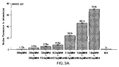
lwtNef-GFP and 7.8-500 ng of SMRwt peptide by Chariot. As shown in Figure 3A
the
SMRwt peptide inhibits the vesicle secretion in Jurkat cells. Figure 3B is a
dose-response
curve showing that the SMRwt peptide inhibits the vesicle secretion in Jurkat
cells in a dose-
dependent manner.
EXAMPLE 3: INHIBITION OF VESICLE SECRETION AND VIRION PARTICLE
RELEASE FROM HIV INFECTED CELLS
3.1 Experiment I
]079] I. Jurkat cells were co-transfected as shown below: (transfection
efficiency
30-40% by Chariot Kit):
#1. pNL4-3 + Nef SMR wt (antagonist) 4 plates
#2. pNL4-3 + Nef SMR mt (nonfunctional antagonist) 4 plates
#3. pNL4-3 + sM1 (negative control peptide) 4 plates
CA 02761121 2011-11-04
WO 2010/144231 PCT/US2010/035698
19
[080J pNL4-3 is a clone containing the viral genome. Transfection into cells
allows
expression of the viral genome and ultimately virion formation and release.
The amount of
virion production in the extracellular supernatant is measured through p24
protein (viral
protein). Samples were collected at 48 hours and 96 hours post-infection.
[081J At 48hr postinfection two plates in each group were removed and analyzed
by:
a. p24 assay
b. Nef assay
c. Infectivity assay
At 96hr postinfection the other two plates in each group were removed and
analyzed by:
a. p24 assay
b. Nef assay
c. Infectivity assay
[082] As shown in Table I, the amount of virus production is reduced
drastically at
96 hours in the presence of the peptide antagonist (NefSMRwt) with no effect
seen for the
negative control peptide (NefSMRmut). The Data suggest that the antagonist
blocks
production and/or release of virus particles from infected cells.
Table is Results of Experiment I
Cells Time Sample P24 Pg/ml Effect (a/b)
Jurkat 48hr pNL4-3+NefSMRwt (a) 0 1
pNL4-3+NefSMRmut (a) 0 1
pNL4-3+sM l (b) 0 1
Jurkat 96hr pNL4-3+NefSMRwt (a) 0 <0.033
pNL4-3+NefSMRmut (a) 15 0.5
pNL4-3+sMl (b) 30 1
293 48hr pNL4-3+NefSMRwt (a) 90 2
pNL4-3+NefSMRmut (a) 30 0.67
pNL4-3+sMl (b) 45 1.0
293 96hr pNL4-3+NefSMRwt (a) 345 0.359
pNL4-3+NefSMRmut (a) 1125 1.17
pNL4-3+sMl (b) 960 1
CA 02761121 2011-11-04
WO 2010/144231 PCT/US2010/035698
3.2 Experiment II
[083] Jurkat cells, HEK293 cells, THP-1 monocytes and U937 monocytes were co-
transfected with either R7 or Nef SMR wt (antagonist) or with R7 + Nef SMR mt
(nonfunctional antagonist) by ChariotTM Kit. The transfection efficiency was
30-40%.
[084] R7 is a clone containing the viral genome. Transfection into cells
allows
expression of the viral genome and ultimately virion formation and release.
The amount of
virion production in the extracellular supernatant is measured through p24
protein (viral
protein).
[085] At 2 hours, 3 days, 6 days, 9 days, 13 days, 15 days, 17 days, 20 days,
23 days,
27 days and 36 days post-transfection, 0.5m1 supernatant were collected from
each plate,
mixed with 0.5m1 fresh media and analyzed by p24 ELISA assay. 1.5 ml of
supernatant were
collected from each plate and spun in a TLA 100 rotor at 400,000xg for 1 hour
to create the
pellets. The pellets were used for Western blot analysis with p24 mAb and Nef
mAb.
[086] As shown in Figure 4A, the p24 concentrations increased in Jurkat cells
3 days
post transfection in the negative control (R7/SMRmt) but did not increase in
SMRwt
(antagonist) cultures until 13 days post transfection. Similar results were
also observed in
HEK293 cells and in THP-1 monocytes. There is little p24 in SMRwt transfected
U937
monocytes, suggesting that the cells could not eliminate the SMRwt antagonist.
These data
suggest that SMRwt antagonizes some aspect of viral growth or viral release
from infected
cells. The effect of SMRwt, however, seems to be temporary. It appears that
Jurkat and HEK
cells can degrade the peptide over time, while U937 monocytes cannot degrade
the peptide.
The temporary effect of the peptide may be overcome by using nondegradable
peptide (e.g.,
peptides with sulfur bond, or d-enatomer peptides). Figure 4B is a composite
of confocal
microscopic pictures at day 3, 6, 10, 14, 17 after transfection showing the
blockage of p24
release by R7/SMRwt (panel A) but not by R7/SMRmt (panel B) in Jurkat cells.
The result
matched with data obtained from ELISAIWestern/ and MAGI analysis. Blue stain
is a nuclear
stain, Red stain is cytoplasmic stain, and green FITC stain is for HIV p24
protein. The p24 can
be seen heavily accumulating in the cytoplasm of antagonist treated cells in
day 3, 6, 10
images as compared to negative control treated cells in same images. In day 14
and 17, the
p24 begins to look like that in the negative control treated images. This
matches the fact that
CA 02761121 2011-11-04
WO 2010/144231 PCT/US2010/035698
21
the p24 appears to be released in the MAGUWestern/ELISA data as we think the
intracellular
levels of the peptide are depleted allowing the virus to begin to be released.
[087] Figures 4C-4E are electron microscopic pictures showing Jurkat cells
transfected with R7/SMRwt or R7ISMRmt at day 6 (Figure 4C) and day 14 (Figure
4D) post
transfection. On day 6, viral particles or nucleocapsid can be observed
accumulating in to
cytoplasm and within the MVBs inside the cells treated with R7/SMRwt. No viral
particles
can be observed accumulating on the extracellular surface of these
cells(Figure 4C, panel A-1
and). In contrast, very few MVBs can be observed inside the cell treated with
R7/SMRmt,
with most of the viral particles observed accumulating on the extracellular
surface of the cell
and polarized on the southern pole of the cell (Figure 4C, panels B-1). On day
14, viral
particles can be observed accumulating on the extracellular surface of the
cell treated with
R7/SMRwt (Figure 4D, panel A-2) nonpolarized across the entire membrane
surface, very
much as observed in cells treated with R7/SMRmt, the negative control (Figure
4D, panel B-
2). Higher magnification images are shown in the following pictures of both
six and 14 day
antagonist and negative control peptide images to show the electron dense
`viral particles'
accumulating as described above (Figure 4E). The evidence shows that the SMRwt
antagonist
delays release of virus from infected cells as measured by EM.
[088J The results of the Western blot analysis are shown in Figures 5-8. The
results
are summarized in Figure 9. As shown in Figures 3-9, the amount of virus
production in the
presence of the peptide antagonist (NefSMRwt) is drastically reduced to zero
or close to zero.
No effect is observed for the negative control peptide (NefSMRmut). Assays of
the cell lysates
show that the production of p24 is the same in all assay conditions suggesting
no effect on
viral protein expression. The data suggest that the antagonist blocks release
of virus particles
from infected cells. This is possibly due to antagonism of trafficking of
viral component(s) to
the cytoplasmic membrane. Ultimately this would (i) shutdown the HW infection
and (ii)
block progression to AIDS.
3.3 Experiment III
10891 Magi/CXCR4 cells were exposed to 48 hour conditioned supernatants from
Jurkat cells, HEK293 cells, THP-1 monocytes, or U937 monocytes transfected
with either R7
viral DNAISMRwt peptide or R7 viral DNA/SMRmt peptide. These cells were then
fixed and
stained with X-Gal. Figure 1OA shows Magi/CXCR4 cells exposed to a I ng/ml
dilution of
CA 02761121 2011-11-04
WO 2010/144231 PCT/US2010/035698
22
p24 supernatant from Jurkat cells transfected with R7/SMRwt. Figure IOB shows
Magi/CXCR4 cells exposed to a l nglml dilution of p24 supernatant from Jurkat
cells
transfected with R7/SMRmt. Cells productively infected with R7 are easily
visualized under
light microscopy by their blue nuclear staining. Magnification x20. Note the
cells treated with
R7 and the peptide antagonist (SMRwt) display drastically reduced numbers of
blue staining
cells, while the cells treated with R7 and the negative control peptide
(SMRmt) display many
blue staining cells. This is indicative of virus in the conditioned
supernatant from the
R7/negative control peptide treated cells and no virus in conditioned
supernatant from the
R7/antagonist treated cells.
[090] These Magi cultures were quantitated for blue staining cells. The data
was
plotted as a function of time post-transfection. As shown in Figure 11, the
numbers of
infected cells in the presence of the supernatant from NefSMRwt transfected
Jurkat cells or
Ne#SMRwt transfected THP-1 monocytes are significantly reduced. The data
suggest that the
antagonist blocks release of virus particles from infected cells. This is
possibly due to
antagonism of trafficking of viral component(s) to the cytoplasmic membrane.
Ultimately this
would shutdown the HIV infection and block progression to AIDS.
[091] In summary, these experiments demonstrate that this technology could be
used
to force cells to make and extracellularly secrete any protein or epitope, so
that the protein or
epitope can be easily purified from the cells. The vesicles could also be used
for chemotherapy
if loaded with a targeting epitope (e.g., antibody epitope to a tumor marker)
and an antitumor
protein or epitope. Further, with this technology the vector could be
transfected into the
specific patient cells so as to be using self vesicles.
[092] Because the protein is also located on the outer membrane of the
vesicles, they
could also be used to induce an immune response. Thus, for example, flu
epitopes may be
loaded into the vector and expressed on the outside of the vesicles to induce
immune response
to flu virus.
EXAMPLE 4: EFFECT OF THE SECRETION ANTAGONIST (HIV NEF SMRWT
PEPTIDE) ON HIV-1 GAGWT-GFP-INDUCED SECRETION
[093] Cells were co-transfected with 0.5 g of pQBI-HIV Gag-GFP (expresses wild
type Gag protein) and either 0.5 g of HIV-1 Nef SMRwt, SMRmut peptide, sM1
peptide or
untransfected controls with Chariot for 48 hours. The conditioned supernatants
from these
CA 02761121 2011-11-04
WO 2010/144231 PCT/US2010/035698
23
cultures were assayed for GFP fluorescence by plate reader. The results are
shown in Table 11.
Particle secretion levels are displayed relative to the untransfected control
which is arbitrarily
set as lx (negative control; 100%).
Table II: Increase in Secretion (relative to untransfected cells) Fluorescent
Plate
Reader Assay
Cell Lines HIV-1 Gag- HIV-1 Gag- GFP/sMl Untransfected
GFPISMRwt GFP/SMRmt
Jurkat 47.46x 58.22x 1.09x lx
ly 1.22y
HEK293 24.22x 25.96x 2.05x lx
ly 1.08y
THP-1 41.3x 45.06x 0.85x lx
ly 1.08y
U937 43.95x 43.82x 1.05x lx
ly ly
CellLines HIV 1 Nef HIV-1 Nef- GFP/sMl Untransfected
GFP/SMRwt GFPISMRmt
H 2.3x 57.04x 1.05x lx
1.Oy 24.8y
x -- exp conditionlUT;
y - SMRmt/SMRwt
[094] Gag has been shown to be secreted from Gag-transfected cells in what are
called `virus-like particles'. These virus-like particles are very much like
vesicles. It has been
suggested that the virus (which has been described as a Gag type vesicle) is
released from cells
via the exosome pathway. The secretion antagonist SMRwt had no effect on Gag
virus-like
particle release. This suggests that the Gag trafficking pathway and the Nef
trafficking
pathway differ at least one point. This point is that factor(s) in the pathway
that the antagonist
manipulates.
CA 02761121 2011-11-04
WO 2010/144231 PCT/US2010/035698
24
EXAMPLE 5: EFFECT OF THE SECRETION ANTAGONIST HIV NEF SMRWT
PEPTIDE) ON HIV-1 GAGWT-GFP-INDUCED SECRETION IN PRESENCE OF WTNEF
PROTEIN.
(095] Cells were transfected with the pQBI-HIV Gag-GFP construct, wtNef-RFP,
and either the antagonist (SMRwt peptide), the negative control SMRmt peptide,
or a random
peptide sM1 with Chariot for 48 hours. The conditioned supernatants from these
cultures were
assayed for GFP fluorescence by plate reader. The results are shown in Table
III. Particle
secretion levels are displayed relative to the untransfected control which is
arbitrarily set as 1 x
(negative control; 100%).
Table HI: Inhibition of Secretion (relative to untransfected cells)
Fluorescent
Plate Reader Assay
Cell Lines Gag-GFP Gag-GFP Gag-GFP Untransfected
+HN-lwtNef +HIV-wwtNef- +HIV-lwtNef-
RFP +SMRwt RFP +SMRmt RFP +sMl
Jurkat 1.3x 55.73x 61.99x lx
1.16x 43.51x 40,36x lx
THP-1 0.95x 49.27x 43.1 lx lx
Monocyte 0.91x 22.24x 20.51x lx
(096] As shown in Table III, in the presence of wtNef-RFP the SMRwt antagonist
peptide blocks release of Gag virus-like particles. These results show that
the SMRwt does
not antagonize Gag VLP formation and release when Gag is in the cell alone,
but SMRwt does
antagonize Gag VLP formation and release when Gag and Nef are both in a cell.
It suggests
that Nef is directing Gag release into a pathway different from the pathway
Gag takes when it
is in a cell by itself. It also explains why the SMRwt peptide can block HIV
virus release but
not Gag virus-like particle release (when only Gag is present).
(097] As shown in Table III, In the presence of wtNef-RFP the SMRwt antagonist
peptide does block the release of Gag virus-like particles in the presence of
wtNef-RFP.
CA 02761121 2011-11-04
WO 2010/144231 PCT/US2010/035698
EXAMPLE 6: CELLULAR TOXICITY ASSAY FOR SMRWT PEPTIDE
[098] SMRwt or SMRmut (negative control) peptide alone were transfected into
Jurkat cells using Chariot. The transfected cells were allowed to grow for 48
hours. The cells
were assayed by Fluorescein diacetate (FD; taken up by live cells and
converted to FITC
making cells fluoresce green) and propidium iodide (PI; diffuse across porous
membranes of
dying cells fluoresing red inside those cells) for cytotoxicity.
[0991 As shown in Figure 12, only a very small number of dying cells (<2%)
were
detected in SMRwt transfected cells (panel A-i). Further, the number of dying
cells in
SMRwt transfected cells is similar to that seen in SMRmut transfected cells
(Panel B-1).
These results suggest that the SMRwt antagonist has very little or no
cytotoxicity in Jurkat
cells.
EXAMPLE 7: IDENTIFYING CELLULAR FACTORS THAT INTERACT WITH THE
ANTAGONIST
A. Identification of cellular factors that bind the SMRwt peptide and regulate
secretion.
[0100] SMRwt vs. SMRmt peptides were used in conjunction with FLAG immuno-
precipitation on Jurkat cell lysates to pulldown cellular factors that
interacted with the SMRwt
antagonist, but not with the SMRznt negative control. The cellular factor(s)
that interact with
the antagonist are analyzed by the FLAG IP assay. Briefly, cell lysates are
combined with
AminoLink Plus resin coupled to FLAG-tagged SMR peptides. SMR-specific
cellular proteins
(ROY) bind to the SMR peptides on the resin. Non-specific contaminants (G BIV)
are washed
off of the resin and removed by centrifugation. The SMR-specific cell proteins
(ROY) are
eluted and collected. Some strongly bound contaminants (V) are also eluted and
collected.
101011 The pulidown products were separated on SDS PAGE ((Figure 13, panel A).
Bands that appeared in the SMRwt lane but not in the SMRmt lane were cut out
and purified.
Five bands were identified and purified in this manner (Figure 13, panel A).
MALDI TOF
MS/MS and LC/MS/MS were used to identify these protein products and were found
to be
Mortalin/GRP75; Myosin 10; Vimentin; GRP78; HSC70. Among these proteins,
mortalinlGRP75 is a member of the Hsp70 family of chaperones, It is located in
both
mitochondria and cytoplasm, and has been implicated in multiple functions
ranging from stress
response, intracellular trafficking, antigen processing, and control of cell
proliferation,
differentiation, and tumorigenesis. Mortalin interacts with p53, and is shown
to be involved in
CA 02761121 2011-11-04
WO 2010/144231 PCT/US2010/035698
26
apoptosis and vesicle transport (MAC complex). It is also found in
microvesicles released by
tumor cells.
[0102) The gel was also Western probed with a-Mortalin antibody. A protein
with a
molecular weight of -75 kDa was detected in the lanes containing the cell
lysate, the
antagonist eluate, and the antagonist affinity resin (Figure 13, panel B,
Lanes 1, 3, and 5), but
not in the negative control or negative control peptide eluate's lanes (Figure
13, panel B, Lanes
2 and 4).
Mortalin Antibody inhibition of vesicle secretion.
[0103[ Chariot transfection of a Mortalin/GRP75 antibody into Jurkat cells
with the
wtNefGFP control was used to knockdown the endogenous MOrtalinIGRP75 protein
to
observe the effect on Nef-induced secretion (Figure 14). A-tubulin antibody
was chariot
transfected into matched cells as a negative control. We observed that the
MortalinlGFP75
antibody blocked NEf-indcued secretion while the a-tubulin antibody had no
effect on Nef-
induced secretion. This showed that Mortalin is important in Nef-induced
exosome secretion.
Mortalin antibody hybridizes to eluate band.
[0104) Mortalin is also known as glucose-regulated protein 75 (GRP75), or
peptide-
binding protein 74 (PBP74). Mortalin is a 679 amino acid long, uninducible
member of the
heat shock protein 70 families. It has a high degree of identity with other
family members
including Escherichia coli DnaK. Although the crystal structure of Mortalin
has not been
deduced, based on the evolutionary conservation within the Hsp70 family, it is
expected to
have two principal domains, the N-terminal ATPase nucleotide binding domain
(NBD) and C-
terminal substrate binding domain (SBD), joined by a protease-sensitive site.
The NBD is
highly conserved across the family, while the SBD displays significant
diversity possibly
explaining the variation among Hsp70 family members in substrate specificity.
Its chaperone
activities are intimately linked with the ATP-hydrolysis function.
[0105) Mortalin has been found to be localized to the mitochondria as well as
to
various cytoplasmic vesicles, including early endocytic vesicles (See, e.g.,
Kanai et al., Genes
Cells, 2007, 12:797-810; Kaul et al., Exp. Gerontol., 2002, 37:1157-1164;
Singh et al., Exp
Cell Res, 1997, 234:205-216 and Van Buskirk et al., J Immunol. 1991, 146:500-
506). Mortalin
binds directly to several proteins (e.g., p53 and FGF-1) and regulates their
intracellular
trafficking (see, e.g., Kaul et al., J Biol Chem, 2005, 280:39373-39379;
Mizukoshi et al.,
CA 02761121 2011-11-04
WO 2010/144231 PCT/US2010/035698
27
Biochem Biophys Res Commun, 2001, 280:1203-1209; Mizukoshi et al., Biochem J,
1999,
343:461-466; and Prudovsky et al., J Cell Biochem, 2008, 103:1327-1343)
through the non-
classical pathway (i.e., exosomal pathway). Cells under attack by the host
immune system
release membrane vesicles through Mortalin expression, and Mortalin is found
in those
vesicles (Pilzer et al., Int Immunol, 2005, 17:1239-1248). Mortalin is also
found in the
exosomes released by various tumor cells (Choi et al., J Proteome Res, 2007,
6:4646-4655;
Staubach et al., Proteomics, 2009).
[01061 Mortalin has been found to play multiple major functions in the cell
(reviewed
in Kaul et al., Exp Ger ontol, 2007, 42:263-274). It serves a major
housekeeping function in
the cellular translocation system of import and export of proteins. Although
not induced by
heat, mild stress responses induce Mortalin allowing it to serve as a guardian
against stress and
apoptosis. Decreased expression of Mortalin, or expression of mutant forms of
Mortalin, lead
to senescence, while increased expression of Mortalin leads to immortality,
with the aberrant
form being cancer.
[0107] Evidence clearly implicates Mortalin in transformation of normal cells
to
cancer cells, as well as in the chemotherapy resistance of those cells.
Mortalin was found to be
over-expressed in tumor cells of various origins (Wadhwa et al., Int J Cancer,
2006, 118:2973-
2980). The murine Mortalin was found to change its subcellular location from
mitochondria,
in normal cells, to the cytosol in cancerous cells (Wadhwa et al., J Biol Chen
1998,
273:29586-29591). Mortalin was found to interact with p53. Further, this
interaction promotes
sequestration of p53 in the cytoplasm, thereby inhibiting its nuclear activity
(Kaul et al., Supra
2007, 42:263-274; Yi et al., Mol Cell Proteomics, 2008, 7:315-325; Czamecka et
al., Cancer
Biol Ther, 2006, 5:714-720), inducing the resistance of some tumors to
radiotherapy and
chemotherapy. Finally, as discussed above, Mortalin has been linked with
intracellular
trafficking leading to exosome release and has been shown to be in exosomal
vesicles (Pilzer
et al. pringer Semin Immunopathol, 2005, 27:375-387; Choi et al., J Proteome
Res, 2007,
6:4646-4655; Staubach et al., Proteomics, 2009). Tumor cells (e.g., breast
tumors) have been
found to secrete, in a regulated manner, exosomes that carry tumor antigens,
and are capable of
presenting these antigens or transmitting them to antigen presenting cells (Yu
et al., J
Immunol, 2007, 178:6867-6875). These tumor exosomes cause immune suppression
through
immune cell killing or dysregulation, thereby promoting a state of immune
privilege that
CA 02761121 2011-11-04
WO 2010/144231 PCT/US2010/035698
28
allows for tumor growth. Thus, through a variety of mechanisms, the tumor
manipulates
Mortalin enhancing its own fitness.
]0108] Heat shock 70 family proteins have been found to be linked with breast
cancer.
They have clear associations with poor differentiation, lymph node metastasis,
increased cell
proliferation, block of apoptosis, and higher clinical stage in breast cancer.
All these
morphologies are markers of poor clinical outcome (Calder-wood et al., Int J
Hyperthermia,
2008, 24:31-39; Calderwood et al., Trends Biochem Sci, 2006, 31:164-172;
Ciocca et a1., Cell
Stress Chaperones, 2005, 10:86-103). Additionally, it has been clearly shown
that over-
expression of Mortalin contributes to carcinogenesis in many cell types,
specifically having
been observed in breast cancer cells (Wadhwa et al., Int J Cancer, 2006,
118:2973-2980).
[01091 It is clear from the literature that Mortalin is a potential target for
cancer
immunotherapy, and there are a number of studies looking to develop
therapeutics (Wadhwa et
al., Cancer Therapy, 2010, 1:173-178; Walker et al., Am J Pathol, 2006,
168:1526-1530;
Deocaris et al., Cancer Lett, 2007, 252:259-269; Pilzer et al., Int J Cancer,
2009; Parolini et
al., J Biol Chem, 2009). For example, MKT-077 is a mitochondrion-seeking
delocalized
cationic dye that causes selective death of cancer cells (Deocaris et al.,
Cancer Lett, 2007,
252:259-269). Its cellular targets include oncogenic Ras, F-actin, telomerase,
and Mortalin
(hmot-2)/mthsp7O (Parolini et al., J Biol Chem, 2009). MKT-077 binds to the
nucleotide-
binding domain (NBD) of Mortalin and causes tertiary structural changes in the
protein,
inactivating its chaperone function, and inducing senescence in human tumor
cell lines. In
clinical trials, this molecule was found to cause renal toxicity, although
there is some evidence
now suggesting lower doses could be less toxic.
EXAMPLE 8: OTHER DRUGS THAT INHIBIT VESICLE/VIRUS RELEASE
[0110] The HIV Nef SMRwt peptide may be used in conjunction with drugs that
have
been approved by the FDA for use in other conditions and have been identified
as having
efficacy in blocking virus release as well as vesicle release. Examples of
such drags are:
dimethyl amiloride and omeprazole.
[0111] A cotransfection assay has been developed that can be used to screen
for
agents that block secretion. In procedure one (Figure 15), NefGFP, NefRFP, or
Nef linked to
any fluorescent tag, is transfected into a cell line and the cell is treated
with an agent or
chemical during a 48 hr incubation period. Then, at 48hr post transfection,
the conditioned
CA 02761121 2011-11-04
WO 2010/144231 PCT/US2010/035698
29
supernatant is assayed for the fluorescent molecule (by various techniques).
In procedure two
(Figure 16), the cell is treated with a fluorescent label like N-Rh-PE that
will label
endogenously made exosomes. The cell is allowed to incubate for at least 24
hours in the
prescence or absence of a chemical or small peptide antagonist. The
conditioned supernatant is
then assayed for N-Rh-PE labeled microvesicles/exosomes (by various
techniques). The lack
of the fluorescent tag in the conditioned supernatant is a sign that the
chemical agent has
blocked the exosome secretion pathway blocking Nef induction of that pathway.
This
procedure should be able to be modified to develop a high throughput assay for
screening of
agents that block secretion.
[01121 The above description is for the purpose of teaching the person of
ordinary
skill in the art how to practice the present invention, and it is not intended
to detail all those
obvious modifications and variations of it which will become apparent to the
skilled worker
upon reading the description. It is intended, however, that all such obvious
modifications and
variations be included within the scope of the present embodiment, which is
defined by the
following claims. The claims are intended to cover the claimed components and
steps in any
sequence which is effective to meet the objectives there intended, unless the
context
specifically indicates the contrary.