Note: Descriptions are shown in the official language in which they were submitted.
CA 02761637 2011-11-10
WO 2010/130538 PCT/EP2010/055252
1
METHOD AND APPARATUS FOR ELECTRICAL CORTEX STIMULATION.
FIELD OF THE INVENTION
The invention relates to methods and apparatuses for
electrical stimulation of the cortex.
BACKGROUND OF THE INVENTION
Electrical brain stimulation is a known method for
treating a number of neurologic diseases, among which
Parkinson's disease.
Electrical brain stimulation includes Deep Brain
Stimulation (DBS) and Epidural Motor Cortex Stimulation
(EMCS).
In DBS, electrodes are deeply implanted in the
patient's brain, in the subthalamic nucleus, which requires
a long and heavy surgical operation. An example of a DBS
method is disclosed for instance in US-A-2008/046025 (Tass
et al .) .
In EMCS, on the contrary, electrodes are implanted
superficially on the dura mater, which requires quicker and
less invasive surgery, with less risk for the patient. An
example of EMCS method is disclosed for instance by
Franzini et al. (Neurol. Res. 2003; 25: 123-26).
The present invention relates more particularly to
cortical stimulation (CS).
OBJECTS AND SUMMARY OF THE INVENTION
One objective of the present invention is to improve
the efficiency of known CS methods.
To this end, according to the invention, a method for
CS is provided, in which an electrode array, having at
least one electrode implanted in the cortex of a patient,
eventually through the dura mater, is controlled by a
control system,
said method including at least the following steps:
(a) a measuring step wherein a number n of electric
signals is collected by said control system from the
CA 02761637 2011-11-10
WO 2010/130538 PCT/EP2010/055252
2
cortex, each at a respective electrode of said electrode
array, n being at least 1;
(b) a processing step wherein said control system
determines n stimulation signals, by a virtual neural field
having a virtual array of n points corresponding to each
electrode of the electrode array having collected an
electric signal at the measuring step (a), said virtual
array receiving the collected signal as an input on each of
the n points and said virtual neural field being adapted to
control a frequency spectrum of neural activity in the
cortex, each stimulation signal being determined by a value
of said virtual potential at each virtual point of the
virtual array;
(c) a stimulation step wherein said stimulation
signals are emitted in the cortex by said control system,
respectively at the electrodes of said electrode array
corresponding respectively to the points of the virtual
array.
Thanks to these dispositions:
- implantation of the control system and electrode
array does not require heavy surgery and is safer for the
patient, due to the relatively superficial implantation of
the electrode array;
- the control system operates in a similar way as
the cortex itself, the activity of which can also be
modeled as a continuous neural field (see in particular
Wilson and Cowan, Kybernetik, 1973, 13(2):55-80; and Amari,
Biol. Cybern., 1977, 27(2) :77-87) : this contributes to an
operation of the control system which is closer to
biological operation and therefore more efficient;
- the control system operates in closed loop with
the cortex, thus stimulating the cortex only when
necessary, for instance only when tremor is present in the
case of the treatment of Parkinson's disease: this results
in minimal disturbance of the normal motor cortex activity,
CA 02761637 2011-11-10
WO 2010/130538 PCT/EP2010/055252
3
and in less power consumption by the control system which
is of special importance when such control system is
implanted and works on battery;
- depending on the number of electrodes of the
electrode array, the spatial resolution of the stimulation
method may be high (and the stimulation is selective since
all the electrodes are controlled individually), thus
enabling to measure and stimulate the cortex activity at a
mesoscopic scale corresponding to the scale of the electric
waves in the cortex, which also results in better
efficiency of the invention in such case.
In various embodiments of the method of the
invention, one may possibly have recourse in addition to
one and/or other of the following steps:
- n is at least 50;
each collected signal and the corresponding
stimulation signal are respectively collected and emitted
in turn through the same electrode of the electrode array;
said electrode array has an electrode density of
at least 4 electrodes / mm2;
said electrode array covers a surface area
comprised between 16 and 1000 mm2 on the cortex;
said electrode array is implanted in the primary
motor cortex;
- said measuring step (a), processing step (b) and
stimulation step (c) are cyclically reiterated;
said stimulating step (c) is carried out only if
a triggering condition is satisfied by said collected
signal at a triggering step (a') which takes place between
said measuring step (a) and said stimulation step (c);
said triggering step (a') takes place between
said measuring step (a) and said processing step (b), and
said processing step (b) is carried out only if said
triggering condition is satisfied;
- at said triggering step (a'), the control system
CA 02761637 2011-11-10
WO 2010/130538 PCT/EP2010/055252
4
determines an amplitude of the collected signal for at
least one predetermined frequency, and said triggering
condition includes having said amplitude being larger than
a predetermined threshold (such amplitude may be for
instance the amplitude of the frequency spectrum of the
collected signal in a certain bandwidth corresponding to
said predetermined frequency);
the virtual neural field attenuates or augments
neural activity in the cortex in a predetermined bandwidth;
- the virtual neural field attenuates neural
activity of the cortex in said predetermined bandwidth,
which includes a target frequency of 10 Hz .
Another object of the present invention is an
apparatus for cortical stimulation, comprising:
- an electrode array including a number n of
electrodes adapted to be implanted in the cortex of a
patient, eventually through the dura mater, n being at
least 1;
a control system controlling said electrode
array, said control system being adapted to:
(a) collecting a number n of electric signals, each
at a respective electrode of said electrode array;
(b) determining n stimulation signals, by a virtual
neural field having a virtual array of n points
corresponding to each electrode of the electrode array
collecting said electric signals, said virtual array being
configured to receive the collected signal as an input of
each of the n points and said virtual neural field being
adapted to control the frequency spectrum of neural
activity in said cortex, each stimulation signal being
determined by a value of said virtual potential at each
point of the virtual array;
(c) emitting said stimulation signals in the cortex,
respectively through the electrodes of said electrode array
corresponding to the points of the virtual array.
CA 02761637 2011-11-10
WO 2010/130538 PCT/EP2010/055252
In various embodiments of the apparatus of the
invention, one may possibly have recourse in addition to
one and/or other of the following arrangements:
n is at least 50;
5 - the control system is adapted to respectively
collect and emit in turn each collected signal and the
corresponding stimulation signal through the same electrode
of the electrode array;
said electrode array has an electrode density of
at least 4 electrodes / mm2;
said electrode array has a surface area comprised
between 16 and 1000 mm2;
the control system is adapted to cyclically
reiterate measuring of the collected signals, determining
the stimulation signals and emitting said stimulation
signals;
the control system is adapted to emit said
stimulating signals only if a triggering condition is
satisfied by said collected signal;
- the control system is adapted to determine an
amplitude of the collected signal for at least one
predetermined frequency, and said triggering condition
includes having said amplitude being larger than a
predetermined threshold (such amplitude may be for instance
the amplitude of the frequency spectrum of the collected
signal in a certain bandwidth corresponding to said
predetermined frequency);
the virtual neural field is adapted to attenuate
or augment neural activity of the cortex in a predetermined
bandwidth;
the virtual neural field is adapted to attenuate
neural activity of the cortex in said predetermined
bandwidth, which includes a target frequency of 10 Hz
BRIEF DESCRIPTION OF THE DRAWINGS
Other features and advantages of the invention
CA 02761637 2011-11-10
WO 2010/130538 PCT/EP2010/055252
6
appear from the following detailed description of one
embodiment thereof, given by way of non-limiting examples,
and with reference to the accompanying drawings.
In the drawings:
- Figure 1 is a diagrammatic view showing a
possible implantation of an electrical stimulation
apparatus in a patient's head, according to one embodiment
of the invention;
Figure 2 is a detailed cutout view of the patient
head, showing the electrical stimulation apparatus of
Figure 1;
Figure 3 shows an example of electrode array
useable in the apparatus of Figure 2;
Figure 4 is a block diagram of the electrical
stimulation apparatus of Figure 2;
and Figure 5 is a diagram showing a simulation of
the activity of a neural mass of the cortex as a function
of time, with and without control by the electrical
stimulation apparatus according to the invention.
MORE DETAILED DESCRIPTION
As shown in Figure 1, the present invention provides
for a new electrical stimulation apparatus 1 which may be
implanted in the head 2 of a human patient P, for carrying
out Cortex Stimulation, i.e. for applying electrical
stimuli in the cortex of the patient P. The electrical
stimulation apparatus 1 may be used for instance for
treating Parkinson's disease or other movement disorders
such as essential tremor, dystonia or other neurological or
neuropsychological disorders. In a variant, the electrical
stimulation apparatus 1 might also be used for increasing
the cortical activity in predetermined frequency bandwidths
for cerebral augmentation purposes.
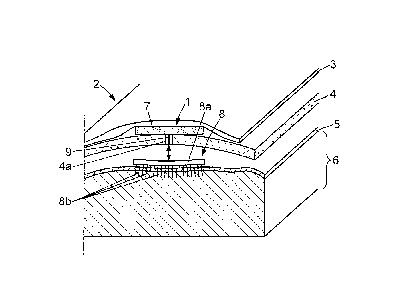
As shown in Figure 2, the head 2 of the patient
includes skin 3 covering the skull 4. The skull 4 covers a
thick membrane called the dura mater 5, which in turn
CA 02761637 2011-11-10
WO 2010/130538 PCT/EP2010/055252
7
covers the cortex 6, i.e. the superficial part of the
brain. The electrical stimulation apparatus 1 may be
implanted on any cortical target, for instance on a
particular area of the brain called the primary motor
cortex (generally referenced as the Ml zone), situated in
the posterior portion of the frontal lobe of the brain.
As shown in Figure 2, the electrical stimulation
apparatus 1 may include for instance:
a control system 7 which may be implanted for
instance between the skin 3 and the skull 4 of the
patient's head 2;
an electrode array 8 which may be implanted
between the skull 4 and the dura mater 5, or between the
dura mater 5 and the cortex 6 in correspondence with any
suitable part of the cortex 6, e.g. the primary motor
cortex;
a connection 9 - for instance a wire connection -
between the central processing unit 7 and the electrode
array 8.
The control system 7 is an electronic microcircuit
fitted with a battery.
The electrode array 8 may include for instance:
a base plate 8a which is disposed between the
skull 4 and the dura mater 5 or between the dura mater 5
and the cortex 6; the base plate 8a can be either rigid, or
preferably in the form of a flexible mat;
and at least one electrode 8b, preferably a
plurality of metal electrodes 8b which protrude downwardly
from the base plate 8a toward the cortex 6 and may
penetrate superficially in the cortex, eventually through
the dura mater 5, so as to be in direct contact with the
cortex.
As a variant, the electrical stimulation apparatus
might be a unitary device including both the control system
7 and the electrode array in a single block which would
CA 02761637 2011-11-10
WO 2010/130538 PCT/EP2010/055252
8
then be located between the skull 4 and the dura mater 5,
or between the dura mater 5 and the cortex 6.
The base plate 8a of the electrode array includes a
micro circuit which connects the wire connection 9 to the
electrodes 8b individually, so that said electrodes 8b be
connected individually to the control system 7.
The electrode array 8 is implanted after trepanation
of the patient, and a hole 4a is left in the skull 4 after
surgery for the wire connection 9.
As shown in Figures 3 and 4, the electrodes 8b may
extend on a height h of a few millimeters from the base
plate 8a, e.g. 1 to 3 mm. The electrode array 8 may include
at least 50 electrodes 8b, preferably more than 100
electrodes, disposed on the base plate 8a at a density of
at least 4 electrodes / mm2 (e.g. 4 to 100 electrodes /
mm2, preferably 5 to 50 electrodes / mm2). The base plate
8a may extend on a width 1 of a few millimeters (e.g. about
4 mm to about 1 cm) and a length L of a few millimeters
(e.g. about 4 mm to a few cm), thus covering a surface area
comprised between 16 mm2 and a few cm2 (e.g. 16 to 1000 mm2,
preferably 16 to 100 mm2) on the cortical tissue.
With the above electrode density of the electrode
array 8, each electrode 8b corresponds to a neural mass of
about 100 to 1000 neurons and is able to map part of the
primary motor cortex at a mesoscopic scale. Therefore, the
electrode array is well adapted to collect electric signals
(voltages) from the cortex and send electric stimulation
signals to the cortex with a good spatial resolution, and
more especially with a spatial resolution which is of
similar scale as the electric phenomena taking place in the
cortex.
The electrode array may be similar to already existing
electrode arrays used as brain implants, for instance
"Utah" type electrode arrays marketed by Cybernetics
Neurotechnology Systems Inc., USA.
CA 02761637 2011-11-10
WO 2010/130538 PCT/EP2010/055252
9
The control system 7 (CPU) is shown on the block
diagram of Figure 4. The control system 7 can be an
electronic autonomous microcircuit including a battery (not
shown) and communicating with each electrode 8b of the
electrode array 8 through the wire connection 9 (INT.) or
through any other communication interface. The control
system 7 includes the following modules, part of which can
be either software or hardware modules:
an amplifier 10 (AMPL.) for receiving and
amplifying analogic electric signals collected by each
electrode 8a, the amplifier also including analogic-numeric
converters for sampling and converting all amplified
collected signals in numeric form (the sampling rate may be
for instance of about 1kHz and the amplification may be
such that the amplified collected signals have a maximum
amplitude of e.g. 1V, the maximum amplitude of the
collected signals being for instance of about 100 pV
(microvolts) before amplification);
a digital processor 11 (NFE) for receiving the
amplified signals from the amplifier 10 and for solving a
neural field equation which will be explained in details
hereafter and for emitting electrical stimulation signals
for each electrode 8b; the processor 11 receives the
collected signals after amplification by amplifier 10; in a
variant, the processor might be an analogic circuit (see in
particular Zou et al., Network: computation in Neural
Sytems, Informa, Sept. 2006; 1 7 (3) : 211-233) , in which case
the collected signals may be sent in analogic form to the
processor 11;
- a buffer 12 (BUFF.) connected to the processor 11
for storing past values of the collected signals;
a triggering module 13 (TRIG.) which is connected
to the output of amplifier 10 for performing frequency
analysis of the collected signals; the triggering module 13
is connected to the processor 11 for inhibiting calculation
CA 02761637 2011-11-10
WO 2010/130538 PCT/EP2010/055252
of stimulation signals (or at least emission of such
stimulation signals) by the processor 11 when the amplitude
of the frequency spectrum of the collected signals does not
exceed a predetermined threshold in a certain frequency
5 range - for instance around 10 Hz (e.g. 8 - 12 Hz) in the
case of the treatment of Parkinson's disease;
a voltage - frequency converter 14 (CONN.)
connected to the output of the processor 11 for converting
the virtual potential signals transmitted by the processor
10 into frequency signals values;
a stimulation module 15 (STIM.) connected to the
output of the converter 14 for transforming each frequency
signal from the converter 14 into a series of voltage
pulses having an instantaneous frequency corresponding to
the frequency computed by the converter 14; the stimulation
signals are transmitted by the stimulation parameter module
15 to the corresponding electrodes 8b of the electrode
array; said stimulation module 15 may also be adapted for
managing the parameters of the electric stimulation (e.g.,
maximum frequency, amplitude, pulse width); this
stimulation module may be activated by an outside apparatus
(not shown) to adjust such parameters through a contactless
link (e.g. by radio communication or by a communication
through induction);
- a synchronizing module 16 (SYNC.), e.g. a clock,
to guarantee that the reception of the collected signals
from the electrodes 8b occurs at a different time from the
transmission of the stimulation signals to the same
electrodes (each electrode is used alternately to collect
the signals from the cortex and to send the stimulation
signals to the cortex).
The operation of the electric stimulation apparatus 1
will now be described.
This operation includes a cycle of 4 steps which are
cyclically and continuously reiterated by the control
CA 02761637 2011-11-10
WO 2010/130538 PCT/EP2010/055252
11
system 7:
(a) a measuring step wherein a number n of electric
signals is collected by said control system 7 from the
cortex 6, each through a respective electrode 8b of said
electrode array 8; in practice, n may be the number of
electrodes 8b of the electrode array 8;
(a') a triggering step wherein the control system 7
(and more particularly the triggering module 13) determines
the amplitude of the collected signal for at least one
predetermined frequency (e.g. 10 Hz) and checks whether
said amplitude is larger than a predetermined threshold
(such amplitude may be for instance the amplitude of the
frequency spectrum of the collected signal in a certain
bandwidth corresponding to said predetermined frequency,
e.g. 8-12 Hz) : if it is larger, then the stimulation step
(c) and possibly the processing step (b) may be inhibited
and the process starts again at step (a), otherwise the
process continues to step (b);
(b) a processing step wherein said control system
determines n stimulation signals, by a virtual neural field
having a virtual array of n points corresponding to each
electrode of the electrode array having collected an
electric signal at the measuring step (a), said virtual
array receiving the collected signal as an input on each of
the n points and said virtual neural field being adapted to
control the frequency spectrum of neural activity in said
cortical target , each stimulation signal being determined
by a value of said virtual potential at each point of the
virtual array;
(c) a stimulation step wherein said stimulation
signals are emitted in the cortex by said control system,
respectively through the electrodes of said electrode array
corresponding respectively to points of the virtual array
(each collected signal and the corresponding stimulation
signal are respectively collected and emitted in turn
CA 02761637 2011-11-10
WO 2010/130538 PCT/EP2010/055252
12
through the same electrode of the electrode array).
When the invention is used for treating Parkinson's
disease, the virtual neural field will be adapted to
attenuate neural activity in the cortical target in a
predetermined bandwidth in the low frequencies. This target
bandwidth may include for instance a target frequency of 10
Hz and may range for instance from 8 to 12 Hz.
The equation of the continuous virtual neural field,
which is solved by processor 11 in the control system 7,
can be written as follows:
LVa(x,t)=I(x,t)+fla fWa(d(x,Y))=S[Va(Y,t-d(x,Y))-9].dy (1)
V
wherein:
z
L is an operator equal to 2. ate +Y-at +l (i . e .
LV(x,t) =2.a2V(x,t) +7 a ; in the example considered
at 2 y at
here, A may be for instance 0 and y may be for instance 1,
so that LV(x,t) = aVa(X ,t)+V(x,t)) ;
Va is a potential in the virtual neural field
(the index "a" stands for the virtual neural field
hereafter), corresponding to a voltage,
- x is a spatial position in the virtual neural
field (NB: in a 2D virtual neural field as considered here,
x is a 2D vector);
t is time;
Q is the spatial domain of the neural field (i.e.
the surface area of the virtual array, corresponding to the
surface area of the electrode array);
d(x,y) is the distance between two spatial
positions x, y in the spatial domain Q;
v is the propagating speed of the signal in the
virtual neural field;
Ra is the synaptic strength in the virtual neural
CA 02761637 2011-11-10
WO 2010/130538 PCT/EP2010/055252
13
field;
- Wa(d(x,y)) is the connectivity kernel of the
virtual field, i.e. the probability that the neural masses
at positions x and y be synaptically connected;
- S(V) is a sigmoid function which provides a
correspondence between the potential V and the
corresponding firing rate of the neurons (i.e. a potential
value V is transformed into an electric discharge
frequency);
- e is the firing threshold;
I(x,t) is an external input: here, I(x,t) is a
function of the electric signals Vr(x,t) collected through
the electrodes 8b (the index "r" stands for the real neural
field hereafter, i.e. the cortical neural field) and
applied to the points of the virtual array in the virtual
neural field.
In the example considered here, the following formulas
may be used for I, W and S:
I(x,t)_flay=fW,,(d(x,Y))=S[V,(Y,t-2a,)-8].dy (la)
W(d(x,y)= [a,exp(-d(x,y)2-airexp(-r2d(x,y)2)] (lb)
(see Atay and Hutt, SIAM J. Appl. Math., 2005, 65(2):644-
666; formula (lb) may be used for all connectivity kernels
mentioned here, to wit Wr, Wa, War, Wra) ;
S(V)= f. (lc)
l+exp[-2(V -9)]
where:
Rar is the synaptic strength between the real and
the virtual neural fields;
- War is the connectivity kernel between the real
and the virtual neural fields, i.e. the probability that
neural masses respectively at positions x and y
respectively in the real and virtual fields be synaptically
connected;
CA 02761637 2011-11-10
WO 2010/130538 PCT/EP2010/055252
14
ear = d(x'y) is a delay;
V
ae/ai are the excitatory/inhibitory synaptic
weights respectively; and
r is the ratio of spatial ranges between
excitatory and inhibitory fibers;
fmax is the maximum discharge rate of the neurons
in the neural field;
A is a non-dimensional parameter.
In the example considered here, typical values of the
above parameters may be as follows:
- fmax: about 100 Hz;
- v: about 1 m/s;
- e: about 3 mV;
- Rar Rrr Rrar Rar: about 2;
- ae: about 50;
- ai : about 30;
- r: about 0.5.
Based on the above equations (1) to (lc) and on the
measured electric signals Vr(x,t), the processor 11
computes values of the virtual potential Va(x,t) for each
point of the virtual area corresponding to the electrode 8b
of the electrode array 8 at which Vr (x,t) was measured.
Each virtual potential Va(x,t) corresponding to each
point of the virtual array is then converted by the
converter 14 into a stimulation frequency fs(x) which is
given by the sigmoid function S(V(x,t)) mentioned above.
Then, each stimulation frequency fs(x) is converted
into a series of pulses by the stimulation parameter module
15 which shapes the stimulation signals E(x,t) in
amplitude, pulse width and maximum pulse frequency (fo -
1/To, where To is the total time between the beginning of
two consecutive pulses of the same stimulation signal
E(x,t)). The stimulation parameter module then transmits
the stimulation signal E(x,t) to the corresponding
CA 02761637 2011-11-10
WO 2010/130538 PCT/EP2010/055252
electrodes 8b of the electrode array 8 to enable emission
thereof in the cortex 6. In the example considered here,
all stimulation signals E(x,t) may have the same maximum
frequency f0, amplitude and pulse width, and the
5 stimulation signals E(x,t) sent simultaneously to the
various electrodes 8b differ from each other by the number
of pulses emitted during the time frame allotted to
transmission of the stimulation signals. For instance, the
amplitude of the pulses may be of about 1V and the pulse
10 width may be of about 50 to 150 ps (microseconds),
preferably about 60 to 90 ps. The pulse maximum frequency
f0 may be for instance in the range 300 - 500 Hz.
The stimulation module 15 may transmit the stimulation
signals with a predetermined delay T, which is at least 50
15 is after the recording period because of the processing by
the control system 7. Such delay is compatible with a real-
time functioning of the invention, appropriate for the
control of a biological system.
Between consecutive recording periods, stimulation
signals are transmitted to the electrode array 8 if the
triggering condition of module 13 is fulfilled.
The efficiency of the invention for creating a closed
loop between the cortex and the virtual neural field can be
explained when considering that the cortex itself functions
as a continuous neural field. Indeed neural field models
have successfully explained and predicted cortical
phenomena such as travelling waves and visual patterns
during hallucinations (see in particular Ermentrout and
Cowan, Biol. Cybern., 1979, 3:137-150). Then, the equation
of this real neural field may be expressed as follows:
LV,(x,t)=E(x,t)+,8,f W,(d(x,y))=S[V (y,t-d(x,Y))-9].dy (2)
V
wherein:
L is the operator already defined for equation
(1) ;
CA 02761637 2011-11-10
WO 2010/130538 PCT/EP2010/055252
16
- Vr is the mean potential in the real neural
field, corresponding to a voltage in the real neural field
of the cortex (i.e., the cortical target),
x is a spatial position in the real neural field
(NB: in a 2D virtual neural field as considered here, x is
a 2D vector);
t is time;
Q is the spatial domain of the neural field (i.e.
the surface area of the virtual array, corresponding to the
surface area of the electrode array);
d(x,y) is the distance between two spatial
positions x, y in the spatial domain Q;
v is the propagating speed of the signal in the
real neural field;
- Rr is the synaptic strength in the real neural
field;
- Wr(d(x,y)) is the connectivity kernel of the real
neural field, i.e. the probability that the neural masses
at positions x and y be synaptically connected;
- S(V) is a sigmoid function which provides a
correspondence between the potential V and the
corresponding firing rate of the neurons (i.e. a potential
value is transformed into an electric discharge frequency);
S may be expressed for instance by the above formula (lc);
- e is the firing threshold;
E(x,t) is the electric stimulation coming from
the stimulation apparatus 1.
The electric stimulation E(x,t) depends upon the
potential V throughout the neural field
E(x,t)= J [V,(y,t-r)].dy (3)
where jV1(y,t-r)] is an unknown function and where r-d(x,y)
V
is a delay. Although this function is unknown, it may be
rewritten in the following form, supposing that the virtual
neural field behaves like the real cortical network, i.e.,
CA 02761637 2011-11-10
WO 2010/130538 PCT/EP2010/055252
17
the virtual neural field is described by the same equation
as the cortical target:
;[V, (Y, t -,r)] =,8,,.W,, (d (x, Y)) = S [Va (Y, t - rra) - 9] ( 4 )
where Rra is the synaptic strength between the virtual and
the real neural field and Wra is the connectivity kernel
between the virtual and the real neural fields, i.e. the
probability that neural masses respectively at positions x
and y respectively in the virtual and real fields be
synaptically connected.
When bringing equation (4) in equation (2), one
obtains equation (5):
LVr(x,t)=/-ra f Wra(d(x,Y)).S[V (y,t-zra)-9].dy+/-r f 4V (d(x,Y)).S[V(y,t-
d(x,Y))-9].dy
v
(5)
As already explained above, the equation of the
virtual neural field can be written in a similar form (6):
,7y
LVa (x, t) = f ar f War (d(x,Y))." [V r (Y, t - Zar) - 9].dy + fla f Wa (d (x,
Y)). [Va (Y, t - d (x, y) ) - e].LG
v
(6)
Therefore, the coupling between the virtual neural
field and the real neural field can be modelled by the two
coupled integro-differential equations (5) and (6).
The parameters of the virtual neural field are adapted
to obtain the desired control of the frequency spectrum of
the cortical target, and therefore the frequency spectrum
of the real potential (voltage) signals in the cortex, in
order to attenuate the cortical activity at certain
frequencies (to alleviate a disorder) or to increase such
cortical activity at certain frequencies (e.g., for sensory
augmentation purposes). For instance, in the treatment of
Parkinson's disease, it will be suitable to attenuate the
potential signals in a predetermined bandwidth in the low
frequencies (e.g. around 10 Hz, for instance in the target
bandwidth 8 - 12 Hz as mentioned above).
Two possible approaches may be used to set these
parameters.
CA 02761637 2011-11-10
WO 2010/130538 PCT/EP2010/055252
18
A. The first approach consists in an analytical
study of the system formed by the two neural field
equations (5) and (6) (one for the virtual neural field of
the stimulation apparatus 1, one for real neural field of
the cortical target). The different steps in this first
approach may be summarized as:
1) computing the equilibrium state of the system;
2) writing down the linearized equations around the
equilibrium to obtain the expression of the mean potential
in response to small external inputs;
3) computing the Green function of the cortical
target, i.e., the response function to external inputs;
4) using the Green function to compute the auto-
correlation function of the potential;
5) using the Wiener-Khinchin theorem stating that
the power spectrum is the Fourier transform of the auto-
correlation function; and
6) obtaining an analytical expression of the power
spectrum of neuronal activity in the neuronal target
depending on the parameters of the virtual array.
Consequently, depending on the frequency band to be
attenuated (to alleviate a disorder) or increased (e.g.,
for sensory augmentation purposes), parameter values may be
chosen from the analytical expression of the power
spectrum.
B. The second approach consists in a numerical study
of the system formed by two neural field equations (5) and
(6). To do so, both neural field equations are solved using
a numerical method such as for instance Euler's method or
fourth-order Runge-Kutta's method. As a result, potential
values are computed at each point of space and time for
both the cortical target and the virtual array. A
spectrogram (i.e., a time-frequency analysis) of the
computed potential in the cortical target gives the power
spectrum of neuronal activity depending on the parameters
CA 02761637 2011-11-10
WO 2010/130538 PCT/EP2010/055252
19
of the virtual array. Thus, it is possible to investigate,
depending on parameters of the virtual array, which
frequency bands are decreased (for therapeutic purpose) or
increased (for augmentation purpose) . Several theoretical
results exist to guide such numerical studies. For
instance, the more general distance-dependent connectivity
kernel used in neural field models is (see in particular
Atay and Hutt, SIAM J. Appl. Math., 2005, 65(2):644-666):
W(z) _ -Laeexp(-z2)-ai xrxexp(-r2z2)]
where:
z is the distance between two points in the
neural field;
ae/ai are the excitatory/inhibitory synaptic
weights respectively, and
- r is the ratio of spatial ranges between
excitatory and inhibitory fibers.
Analytical studies, (see for example Atay and Hutt,
SIAM J. Appl. Math., 2005, 65 (2) :644-666showed that,
depending on the form of W(z), different neuronal activity
patterns could be observed. For instance, it is known that
a locally excitatory/lateral inhibitory (ae>ai>0 and r>1)
connectivity kernel allows both stationary and propagating
waves of neuronal activity throughout the cortex.
Consequently, with guidelines from analytical study of
neural fields equations, it is possible to determine
appropriate parameter values for efficient cortical target
control using numerical simulations.
Simplified example:
One simplified example of the invention, in which
space is not taken into account (i.e. equivalent to using
an electrode array with a single electrode 8b), has been
carried out by numerical simulation. In this example, the
real neural field is considered to include both an
CA 02761637 2011-11-10
WO 2010/130538 PCT/EP2010/055252
excitatory population (index E) and an inhibitory
population (index I). Without control, the excitatory
population creates a strong pathological 5 Hz activity
(typically occurring during Parkinson's disease in the
5 subthalamic nucleus). The stimulation of the virtual
population (index A) of the virtual neural field is
provided to the excitatory population E and the evolution
of both real and virtual neural fields is described by the
two coupled equations (7) and (8) (only the potential of
10 the excitatory population E is mentioned hereafter as far
as the real neural field is concerned):
ICE udE +VE =aSE(VE)-bSE(VE) -2SI(VA) (7)
ZA uVA +VA = f`)E(VE) (8)
In this example, TE = TA = 6 ms (milliseconds) are the
15 membrane time constants of the two neural fields; a = 0.05;
b = 0.1; e = f = 0.05 (a, b, e, f are non-dimensional
synaptic weights); and SE, SI, SA, are the sigmoid functions
of the three populations.
Figure 5 shows a diagram of simulated electrical
20 signals in the cortex in this example, showing the
amplitudes of the electric signals as a function of time.
The amplitude of the signals is represented by the firing
rate of the neurons in Hz/cell. Figure 5 show that the
stimulation signals emitted according to the invention are
efficient to control the 5 Hz, high amplitude signals.