Note: Descriptions are shown in the official language in which they were submitted.
CA 02761856 2011-11-14
WO 2010/130782 PCT/EP2010/056542
1
Use of the GTPase Rab27B to diagnose and treat
poor prognosis estrogen-receptor-positive breast cancer
Technical field of the invention
The present invention relates to evaluating the prognosis of patients with
estrogen receptor-
positive breast cancer on the basis of Rab27B expression. The invention
further relates to a kit
comprising an assay for measuring Rab27B levels in said patients and to the
usage of Rab27B
as a target to screen for drugs capable of inhibiting or diminishing
metastasis of said cancer.
Furthermore, the invention discloses compounds which can be used to treat
Rab27B-positive
poor prognosis estrogen receptor-positive breast cancer.
Background art
Cancers achieve invasive growth by delivering critical factors into the tumor
microenvironment (1), but the molecular mechanisms for the secretion of these
pro-invasive
factors remain largely unknown. One likely process involves vesicle
exocytosis, whose role in
tumor progression was first reported by Palmer and co-workers (2). They showed
that ectopic
expression of BAIAP3, a Munc 13-like effector of regulated exocytosis,
enhanced the
malignancy of cancer cells.
Key players in exocytic and endocytic membrane trafficking include the Rab
GTPases, which
serve as molecular switches oscillating between their GTP-bound active and GDP-
bound
inactive conformations. Rabs recruit specific protein complexes to elicit
their biological
functions (3-6); they are post-translationally modified by
geranylgeranylation, which binds
them to lipophilic membranes (7).
The secretory pathway can be divided into constitutive and regulated portions
(8). In the
constitutive pathway, release of vesicle content occurs at a constant rate,
and vesicles do not
accumulate to an appreciable extent (9). In contrast, regulated secretion
involves two distinct
steps. Newly synthesized proteins are first stored within vesicular structures
and are then
released upon stimulation (10). Certain Rab GTPases, referred to as secretory
Rabs, control
this secretory process; they include Rab26, Rab37, Rab3A/B/C/D, and Rab27A/B
(11). Rab26
and Rab37 are thought to modulate secretion in specialized cell types, whereas
the Rab3 and
Rab27 subfamilies function as more generic regulators of secretion (12-16).
The Rab27
CA 02761856 2011-11-14
WO 2010/130782 PCT/EP2010/056542
2
subfamily has the highest homology (41-44%) to members of the Rab3 subfamily;
Rab27A
and Rab27B exhibit 71% identity at the amino acid level (17).
Rab proteins of the endocytic (e.g., Rab25, Rab23 and Rab5) (18-21) and
constitutive
secretory pathways (e.g. Rab8) (22) play significant roles in malignancy and
Rab GTPases
active in exocytosis/secretion could also be critical for cancer progression.
WO 2006/091776 discloses a method for predicting prostate cancer progression
via
determining the expression level of a set of genes such as the gene encoding
for Rab27. WO
03/004989 further discloses that Rab27B is over-expressed in breast cancer
cells and that
Rab27B can be used to screen for the presence of breast cancer. Hendrix et al.
(40) further
indicates that Rab27B is a potential biomarker in breast cancer progression.
US 2007/0218512
indicates that human matrix metalloproteinase 26 (MMP 26) can be used as a
biomarker,
possibly in combination with an additional biomarker such as Rab27B, for
evaluating the
prognosis of cancers, among them ER-positive breast cancers. Recently, Wang
and co-workers
showed that up-regulation of Rab27A further enhances the already established
invasive and
metastatic phenotypes of the human breast cancer cell lines MDA-MB-231 and MDA-
MB-
435 (23, 36). In these models, Rab27A had a peri-nuclear and non-cytoskeleton
associated
localization pattern, suggesting a non-secretory function of Rab27A in MDA-MB
cell lines.
Human Rab27A and B are further structurally very similar and are functional
homologues
with respect to melanosome transport (35).
ER positive breast cancers, which comprise the majority of breast
malignancies, carry a better
prognosis for disease-free survival and overall survival than ER-negative
breast cancers (37).
Nevertheless, some ER-positive breast cancers are more invasive and tend to
metastasize more
frequently than other ER-positive tumors. A low degree of differentiation and
the presence of
metastasis in the axillary lymph nodes are typical characteristics. The
underlying reasons for
the more aggressive character are poorly understood. In this regard, Wright et
al. (41) recently
demonstrate in Figure 3 of their publication that a lower level of Rab27B
expression was
found in ER-negative breast cancer tissue samples compared to the Rab27B
expression in ER-
positive samples which suggests that relatively increased Rab27B expression
correlates with a
positive outcome of disease.
However, it is currently still unknown which biomarker can be used to evaluate
the prognosis
of patients with estrogen receptor-positive breast cancer and especially the
subset of patients
CA 02761856 2011-11-14
WO 2010/130782 PCT/EP2010/056542
3
with ER-positive breast cancers which are more invasive and tend to
metastasize more
frequently, or, can be used as target for drugs to treat the latter subset of
patients.
Thus, needed in the art are reliable methods for stratifying, prognosing and
treating the ER-
positive breast cancers which are more invasive and tend to metastasize more
frequently than
other ER-positive tumors, as well as predicting treatment outcomes.
Brief description of fitures
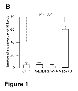
Figure 1. Effect of ectopic expression of Rab3D, Rab27A, or Rab27B on the
formation of
cellular extensions and invasiveness. A) Morphology of MCF-7 cells transiently
transfected
with GFP, GFP-Rab3D, GFP-Rab27A or GFP-Rab27B expressing plasmids. 24 hours
after
transfection, cells were fixed and nuclei were stained with DAPI. Laser
scanning confocal
images show punctuate GFP signal that is indicative of localization of GFP-
fusion protein to
vesicles. Scale bar, 20 gm. B) Matrigel invasion assay with GFP-Rab
transfected MCF-7 cells.
24 hours after transient transfection with GFP-Rab expressing plasmids, 105
MCF-7 cells were
seeded on top of a Matrigel-coated filter and their migration towards medium
containing
serum was quantified by microscopic evaluation (total magnification 400x). The
mean total
number of invading cells counted after 72 hours from 10 different fields is
shown with the
upper 95% confidence intervals from the means of three independent experiments
performed
in triplicate. P-values were calculated using two-sided Student's t-tests.
Statistically
significant P-values are indicated. C and D) Morphology and invasiveness of
GFP-Rab
transfected MCF-7 and T47D breast cancer cells. In (C) phase contrast images
are shown of
cells seeded on type I collagen matrix 24 hours after transient transfection.
In (D) the invasion
index was calculated by counting the number of invading and non-invading cells
into type I
collagen matrix in ten fields. Invasion indices are means and upper 95%
confidence intervals
derived from the means of three independent experiments performed in
triplicate. P-values
were calculated using x2-tests; statistically significant P-values are
indicated. Scale bar, 50
gm. In (A) and (C), arrows indicate cellular extensions and local spreading.
CA 02761856 2011-11-14
WO 2010/130782 PCT/EP2010/056542
4
Figure 2. Rab27B GTP- and geranylgeranyl-dependent cancer cell invasion and
cell cycle
progression in vitro. A) Phase contrast images showing morphology of MCF-7
cells stably
transfected to express GFP, GFP-Rab27A, GFP-Rab27B (wild type, WT), or GFP-
Rab27B
mutants. GFP-Rab27B Q78L (constitutive active), N1331 (dominant negative) and
GER
(impaired geranylgeranylation and vesicle targeting) were the mutants used.
Arrows indicate
cellular extensions and local spreading. Scale bar, 50 gm. B) Quantification
of type I collagen
invasion by the cells shown in (A). Invasion assays were performed as in
Figure 1,D. Invasion
indices are means and upper 95% confidence intervals derived from the means of
three
independent experiments performed in triplicate. P-values were calculated
using x2-tests.
Statistically significant P-values are indicated. C) Laser scanning confocal
images of the F-
actin cytoskeleton (phalloidin-TRITC) and GFP localization in MCF-7 GFP and
GFP-Rab27B
cells cultured for 24 hours on a collagen type I matrix. Arrow indicates
cortical F-actin and
arrowhead indicates membrane blebs. Scale bar, 20 gm. D) Invasion by Rab27B-
expressing
MCF-7 cells in which Rab27B was depleted. MCF-7 cells that expressed GFP-
Rab27B, with
or without transfection of control siRNA (siCON) or Rab27B siRNAs (siRab27B 1
and/or 2),
were seeded on a Matrigel-coated filter. The inset panel shows the impact of
the Rab27B
siRNAs on Rab27B expression in these cells by immunoblotting. The numbers of
invasive
cells were counted after 72 hours in 10 different fields and are expressed as
the mean with
upper 95% confidence intervals of three independent experiments performed in
triplicate. P-
values shown are for comparisons with the siCON transfection using two-sided
Student's t-
tests. E) Effect of Rab27B on cell cycle progression. MCF-7 GFP and GFP-Rab27B
cells were
grown to 50% confluence, followed by 24 hours serum starvation, and 24 hours
serum-
induced (0.5%) cell cycle progression. Percentages of MCF-7 GFP and GFP-Rab27B
cells in
G1, S and G2 stage of the cell cycle, as measured by flow cytometry, are
represented as the
means with upper 95% confidence intervals of two independent experiments. F)
Western blot
analysis in mutant Rab27B-transfected MCF-7 cells of the positive (cyclin A
and E) and
negative (p27) G1 to S phase cell cycle regulators. Protein levels were
quantified as
immunostaining intensity relative to tubulin. G) Measurement of cell
proliferation rates of
MCF-7 cells stably expressing GFP, GFP-Rab27B, or GFP-Rab27B mutants as in
(A). 10 000
cells were plated into each well of a total of 15 wells on day 1 in order to
establish one growth
curve under each condition in triplicate. The total number of cells per well
was manually
CA 02761856 2011-11-14
WO 2010/130782 PCT/EP2010/056542
counted every 2 days until day 8. Mean number of cells is plotted with upper
95% confidence
intervals. P-values were calculated using the two-way repeated measures ANOVA
test.
Statistically significant P-values are indicated; data were compared with the
GFP control. H)
Measurement of cell proliferation rates of MCF-7 GFP-Rab27B cells transiently
transfected
5 with control (siCON) or pooled Rab27B siRNAs (siRab27B1 and 2). The
experiment was
performed as in (G). An inset panel shows the effect of this siRNA on cyclin A
expression in
MCF-7 GFP-Rab27B cells. Tubulin was used as loading control.
Figure 3. Effect of Rab27B on invasive tumor growth in vivo. Nude mice were
injected in the
mammary fat pad with MCF-7 cells expressing GFP, GFP-Rab27A, GFP-Rab27B (wild
type,
WT), or mutant GFP-Rab27B proteins (Q78L, T23N, N133I, and GER). A)
Tumorigenesis in
nude mice with MCF-7 GFP-Rab27B xenografts vs controls. Mice with MCF-7 GFP-
Rab27B
xenografts (lower panel) developed hemorrhagic ascites (blue and swollen
appearance of the
ventral side) and tumor aggregates (arrow) in the peritoneal cavity and
attached to organs
such as the ovary. MCF-7 GFP xenografts (upper panel) developed no hemorrhagic
ascites.
Inset: Pelleted tumor aggregates from the peritoneal fluid of one mouse. Scale
bar, 13 mm. B)
Effect of Rab27B expression on survival of mice with xenografts. Kaplan-Meier
curves and
log-rank testing (95% confidence intervals, P = .031) are shown for nude mice
injected with
MCF-7 GFP cells (n=10) versus MCF-7 GFP-Rab27B cells (n=40; four different
clones with
10 mice per group). C) Expression of GFP-Rab27B in tumor aggregates. A western
blot
loaded with 60 gg peritoneal tumor aggregate and immunostained with primary
Rab27B and
tubulin antibodies is shown. D) Hematoxylin and eosin (H&E) staining of a
peritoneal tumor
aggregate. Scale bar, 100 gm. E) H&E staining of MCF-7 GFP (upper panel) and
GFP-
Rab27B (lower panel) xenografts. Arrowheads indicate striated muscle tissue;
arrows
indicate areas of muscular invasion by cancer cells to the peritoneal side
(P). Scale bar, 100
gm. F) Relative invasiveness of xenografts expressing WT and mutant Rab27B
proteins.
Percentage of invasive tumors was determined by the total number of mice with
an invasive
xenograft in the peritoneal wall as assessed by macroscopic observation and
immunohistochemistry (n=10 mice per group). Precise percentages for a single
experiment are
shown. G) Cellular localization of Rab27B in MCF-7 GFP-Rab27B xenografts.
Arrow
indicates peripheral Rab27B distribution; arrowheads indicate Rab27B vesicle
clustering
CA 02761856 2011-11-14
WO 2010/130782 PCT/EP2010/056542
6
appearing in the cytoplasm and at cell-cell contact. Scale bar, 25 gm. H) Mean
tumor volume
in nude mice bearing xenografts that expressed WT or mutant Rab27B proteins (n
= 10 mice
per group). Tumor size was assessed weekly by measurement of the external
diameter of the
xenografts for 10 weeks. GFP expression was maintained in the xenografts
throughout this
time period (data not shown). Error bars represent 95% confidence intervals.
I) Mean tumor
weight after surgical resection of xenografts expressing WT or mutant Rab27B
proteins. Mice
were killed at variable time points (ie, the ethical endpoint which limits
hemorrhagic ascites
formation, or the experimental end point at 10 weeks) after injection of
stably transfected
MCF-7 cells (n= 10 mice per group). Error bars represent upper 95% confidence
intervals. P-
values were calculated using two-sided Student's t-tests; statistically
significant P-values are
indicated. J) Immunohistochemical staining of MCF-7 GFP and GFP-Rab27B
xenografts to
detect Ki67, a proliferation marker. The mean number of proliferating MCF-7
GFP-Rab27B
cells, calculated from 18 images of three primary tumors per cell line, was
85.50 4.04 vs
32.56 2.68 proliferating control cells (two-sided Student's t-test, P < .001).
Scale bar, 50 gm.
Figure 4. Selective stimulation of HSP90a secretion by Rab27B through GTP- and
geranylgeranyl-dependent mechanisms. A) Secretome profiling of invasive MCF-7
GFP-
Rab27B cancer cells identified HSP90a and HSP90[3. The number of matched
peptides and
the percentage of sequence coverage are indicated for both proteins. The MS/MS
spectrum
recorded on a [M+2H]2+ ion at m/z 618.69, corresponds to a unique peptide
[DQVANSAFVER], derived from HSP90a. Peptides fragment along the amide backbone
to
produce sequence-specific fragment ions; ions containing the C-terminal
fragment are known
as `y' ions, whereas ions containing the N-terminal fragment are known as `b'
ions. The
search engine Mascot uses this information to report probability-based scores
for each peptide.
See Methods for more details. B) Quantification of HSP90a levels in
conditioned media (CM)
of GFP- vs GFP-Rab27B- expressing MCF-7 cells using enzyme-linked
immunosorbent
assay ELISA. Results are means with upper 95% confidence intervals of two
independent
experiments with three replicates. C) Western blot analysis of HSP90a and (3
in CM (upper
panel) and in total protein lysates (lower panel) of transfected MCF-7 cells.
Relative intensity
was quantified with HSP90[3 or tubulin as a loading control. D) Impact of
Rab27B silencing
(siRab27B 1 and 2) versus control silencing (siCON) on the expression of GFP-
Rab27B
CA 02761856 2011-11-14
WO 2010/130782 PCT/EP2010/056542
7
protein (lower panel) and secretion of HSP90a and R (upper panel) in the CM of
MCF-7
GFP-Rab27B cells. Protein levels were quantified as immunostaining intensity
relative to
tubulin and HSP90(3 respectively.
Figure 5. The role of HSP90a and MMP-2 in Rab27B-dependent invasion. A) Phase
contrast
images showing morphology (upper panels) and quantification of collagen type I
invasion by
MCF-7 GFP-Rab27B cells (lower panel) treated with the HSP90a inhibitors 17-AAG
and GA
(1 M) for 24 hours or left untreated (Control, Con). B) Morphology (upper
panels) and
quantification (lower panel) of the invasive phenotype induced by GFP-Rab27B
in MCF-7
cells cultured on collagen type I matrix treated for 6 hours with HSP90a-
neutralizing antibody
(1 gg/mL) or the control IgG isotype. C) Morphology (upper panel) and
quantification
(lower panel) of the invasive phenotype induced by GFP-Rab27B in MCF-7 cells
cultured on
collagen type I matrix and treated for 24 hours in the presence or absence
(Control, Con) of
recombinant (rec) HSP90a protein (1, 5 and 10 gg/mL) or recombinant HSP90(3
protein (10
gg/mL). In A, B and C, arrows indicate cellular extensions and local
spreading. Scale bar,
100 gm.. Invasion indices are means and upper 95% confidence intervals derived
from the
means of three independent experiments performed in triplicate. P-values are
calculated using
the x2-test; statistically significant P-values are indicated. D) Measurement
of cell proliferation
rates of MCF-7 GFP cells treated with recombinant HSP90a (10 g/mL) or left
untreated
(Con) and of MCF-7 GFP-Rab27B cells challenged with a HSP90a-neutralizing
antibody (5
gg/mL) or control immunoglobulin (Con IgG). Proliferation assay was performed
as in Figure
2,G. Mean number of cells is plotted with upper 95% confidence intervals. P-
values are
calculated using the two-way repeated measures ANOVA test. E) Cyclin A
expression was
evaluated in MCF-7 GFP cells treated with recombinant HSP90a (10 g/mL) or left
untreated
(Con) and in MCF-7 GFP-Rab27B cells challenged with HSP90(x-neutralizing or
control
antibody. Intensity was quantified relative to tubulin. F) Analysis of MMP-2
activity in
conditioned media (CM) from cultured MCF-7 cells expressing GFP, GFP-Rab27B
(wild
type, WT), or the GFP-Rab27B mutants (Q78L, N1331, and GER) by gelatin
zymography. G)
Gelatin zymography of MMP-2 activity in CM from MCF-7 GFP-Rab27B cells that
were pre-
incubated with exogenously added proMMP-2 (100 ng/mL) in serum-free medium for
24
CA 02761856 2011-11-14
WO 2010/130782 PCT/EP2010/056542
8
hours. In F and G, arrowhead indicates 72 kDa proMMP-2 and arrow indicates 68
kDa
active protease.
Figure 6. Rab27B expression in clinical breast cancer specimens. A)
Representative Rab27B
stained primary breast cancer samples that illustrate immunohistochemical
scores of 0, 1, and
2. Scale bar, 100 gm. B) Associations of Rab27B immunohistochemical scores
with estrogen
receptor (ER) status and other clinicopathological data for 59 primary breast
tumors. The x2-
test was used to test for differences between categorical variables. C)
Relative levels of
Rab3D, Rab27A, and Rab27B mRNA expression in normal tissue (N, n=5) versus
primary
breast carcinoma (T, n=20). D) Expression of Rab27B mRNA in 5 normal tissues
versus 20
primary breast carcinomas. Tumor samples were divided into three groups
according to ER
status and lymph node (LN) involvement. In C) and D) mRNA expression was
measured by
quantitative RT-PCR in triplicate. Horizontal bars represent median for each
group (two-sided
Mann-Whitney test).
Description of the invention
The present invention relates to the surprising finding that one particular
secretory rat brain
(Rab) protein, Rab27B, promotes cancer cell invasion, tumor growth and
metastasis. Rab27A,
which is structurally very similar to Rab27B, does not have such an effect.
Further surprising
is the fact that, in clinical samples, upregulation of endogenous levels of
Rab27B mRNA and
protein correlates with lymph node metastasis and differentiation grade in ER-
positive breast
tumors. In contrast, the recent data by Wright et al. (41) suggested that
increased Rab27B
expression correlates with a positive outcome of disease.
Hence, the present invention relates to the use of the guanosine triphosphate
hydrolaze
(GTPase) Rat brain (Rab) 27B as a biomarker to evaluate the prognosis of a
patient with
estrogen receptor-positive breast cancer in vitro. With the term `biomarker'
is meant a
characteristic that is objectively measured and evaluated as an indicator of
normal biologic
processes, pathogenic processes, or pharmacologic responses to a therapeutic
intervention.
Hence, the biomarker Rab27B can be used, among other uses, to: 1) diagnose
estrogen
receptor-positive breast cancer with the potential to metastasize and/or to
develop in a grade 3
tumor (see further); 2) evaluate the prognosis of said breast cancer which
encompasses
CA 02761856 2011-11-14
WO 2010/130782 PCT/EP2010/056542
9
predictions about the likely course of disease or disease progression,
particularly with respect
to the likelihood of metastasis, disease remission, disease relapse, tumor
recurrence and death;
3) therapeutically stratify patients with estrogen receptor-positive breast
cancer (i.e. scoring
said patients, see further) in order to decide which therapy, such as
(adjuvant) chemotherapy,
should be given to said patient; and 4) monitor disease progression once a
particular therapy
has been administered to said patients.
In particular, the present invention relates to the latter usage, wherein an
increased level of
Rab27B in a patient sample, compared to a control sample, indicates a poor
prognosis. The
term ' a patient sample' includes, but is not limited to, a primary tumor
sample, circulating
breast cancer cells or a biofluid such as blood, serum, plasma lymph, urine,
saliva, nipple
aspirates, gynecological fluids or any other bodily secretion or derivative
thereof. In this
regard, it should be noted that Rab27B protein can be detected intracellularly
(often as part of
a membrane), or, extracellularly as a secreted form or as part of a secreted
vesicle (i.e. as part
of the so-called exosome). Methods for collecting various samples are well
known in the art.
In some embodiments, a breast tissue sample is obtained by, for example, fine
needle
aspiration biopsy, core needle biopsy or excisional biopsy. The term `poor
prognosis'
corresponds with positive lymph node metastasis and/or a poor differentiation
grade. The term
`a poor differentiation grade' refers to the so-called `Bloom-Richardson
grade' (BR grade,
(38)) which is a histological grade assigned by pathologists to invasive
breast cancers and is
the most common type of cancer grade system currently used. It is a semi-
quantitative grading
method based on three morphologic features of invasive breast cancers. The
morphologic
features that are used are:
1. The degree of tumor tubule formation (percentage cancer composed of tubular
structures)
2. The mitotic activity of the tumor (rate of cell division)
3. The nuclear pleomorphism of tumor cells (nuclear grade, change in cell size
and
uniformity)
CA 02761856 2011-11-14
WO 2010/130782 PCT/EP2010/056542
Each of these features is assigned a score ranging from 1 to 3. The scores are
then added
together for a final sum that will range between 3 and 9. This value is then
used to grade the
tumor as follows:
Grade 1 (I) tumor (well-differentiated)
5 Grade 2 (II) tumor (moderately-differentiated)
Grade 3 (III) tumor (poorly-differentiated)
The terms `an increased level of Rab27B in a patient sample, compared to a
control sample'
depends on which level of Rab27B is measured and how this level is measured.
With a
`control sample' is meant a similar sample as indicated above taken from a
healthy patient not
10 having estrogen receptor-positive breast cancer and/or a patient having
estrogen receptor-
positive breast cancer but without lymph node metastasis. In a particular
embodiment, the
present invention relates to the latter usages wherein the level of Rab27B is
determined by
measuring the expression of Rab27B protein or nucleic acids such as mRNA
expression of
Rab27B. Measuring proteins and nucleic acid levels (such as mRNA levels) are
well known in
the art and can be undertaken by any method known in the art including but not
limited to
Western blots, Northern blots, Southern blots, ELISA, immunoprecipitation,
immunofluorescense, flow cytometry, Rab27B activation test (i.e. GTP vs GDP-
bound
Rab27B), immunohistochemistry, nucleic acid hybridization techniques, nucleic
acid reverse
transcription methods, and nucleic acid amplification methods such as qPCR.
The latter
techniques are, for example, described in detail in US 2007/0218512. In
particular
embodiments, expression of a biomarker is detected on a protein level using
antibodies that
are directed against specific biomarker proteins. These antibodies can be used
in various
methods such as Western blot, ELISA, immunoprecipitation or
immunohistochemistry.
Likewise, immunostaining of breast tumor tissue can be combined with
assessment of clinical
information, conventional prognostic methods, and expression of other
molecular markers
known in the art.
With regard to `increased levels of Rab27B protein compared to a control', the
present
invention further relates in particular to any of the latter usages, wherein
more than 30% of
CA 02761856 2011-11-14
WO 2010/130782 PCT/EP2010/056542
11
cancer cells of a sample taken from a patient show Rab27B protein membrane
localization
and/or vesicle clustering. The Rab27B protein signal was scored on the
following scale:
-score 0: no or weak cytoplasmic staining and less than 5% (5% not included)
of cancer cells
with membrane localization or vesicle clustering,
-score 1: cytoplasmic staining and between 5% and 30% of the cancer cells with
prominent
membrane localization and vesicle clustering,
-score 2: cytoplasmic staining and more than (>) 30% (30% not included) of the
cancer cells
with prominent membrane localization and vesicle clustering.
In this regard, the present invention discloses a statistically significant,
positive correlation
between Rab27B protein score 2, positive lymph node metastasis and a higher
tumor grade,
such as grades II and III (i.e. grades 2 and 3).
The present invention further relates to the latter usages, wherein the level
of mRNA
expression of Rab27B is higher in a patient with lymph node metastasis
compared with a
patient without lymph node metastasis. For example, the present invention
discloses that the
median expression of Rab27B was two-fold higher in the estrogen-positive
patients with
lymph node metastasis compared with those without lymph node metastasis and
was 11-fold
higher compared to normal tissue. The term `higher' in relation to nucleic
acid levels such as
mRNA levels thus refers to at least 1.1, 1.2, 1.3...2, 2.1, 2.2, 2.2, 3,
4,...10, 11, 12, 122.1,
12.2, 12.3...-fold `higher' levels compared to the levels determined in a
control sample.
The present invention also relates to a kit comprising reagents to perform an
assay for
measuring Rab27B levels in a patient having estrogen receptor-positive breast
cancer in vitro
in order to determine if said patient is at risk to develop lymph node
metastasis. The term `kit'
refers to any manufacture (e.g. a package or a container) comprising at least
one reagent (e.g.
an antibody, a nucleic acid probe, etc.) for performing and assay which
specifically detects the
expression of Rab27B. Positive and/or negative controls can be included in the
kits to validate
the activity and correct usage of reagents employed in accordance with the
present invention.
The design and use of controls is standard and well within the routine
capabilities of those of
ordinary skill in the art. The kit can be promoted, distributed, or sold as a
unit for performing
the methods or usages of the present invention. Additionally, the kits can
contain a package
insert describing the kit and methods/usages for its use.
CA 02761856 2011-11-14
WO 2010/130782 PCT/EP2010/056542
12
Preferred assays to perform via the kit are a Rab27B immunohistochemistry
assay or
Quantitative RT-PCR assay on tissues or cells such as biopsies, primary breast
cancer samples
or circulating breast cancer cells of the patients, or, a sandwich-type ELISA
on bio-fluids of
primary breast cancer samples of the patients.
The present invention also relates to the use of Rab27B as a target to screen
for drugs capable
of inhibiting or diminishing metastasis of estrogen receptor-positive breast
cancer in a patient.
Screening assays are well-known in the art and are, for example, described in
detail in WO
03/004989 and WO 2006/091776. The latter assays aim to identify modulators
(antibodies,
peptides, peptidomimetics, small molecules, nucleic acids or other drugs)
which: a) bind to
Rab27B, b) have a modulatory (i.e. stimulatory or inhibitory) effect on the
activity of Rab27B,
c) have a modulatory effect on the interactions of said biomarkers with one or
more of their
substrates or binding partners, or d) have a modulatory effect on the
expression of said
biomarkers. Such assays typically comprise a reaction between Rab27B or
nucleic acids
encoding said protein, and, the modulators or test compounds. Said test
compounds (or
modulators or drugs) may be obtained from any available source, including
libraries of natural
and/or synthetic compounds. The screening methods of the invention will
provide `hits' or
`leads' that possess a desired but not optimized biological activity. Lead
optimization
performed on these compounds to fulfill all physicochemical, pharmacokinetic
and
toxicological factors required for clinical usefulness may provide improved
drug candidates. It
should be noted that in the latter screening assays also fragments or variants
of Rab27B or the
corresponding encoding nucleic acids can be used as long as these fragments or
variants will
provide hits or leads that possess the desired biological activity. A fragment
is a shorter
portion of Rab27B or of their encoding nucleic acids. A variant encodes for-
or has an amino
acid sequence that has at least 70% or 75% sequence identity, preferably at
least 80 % or 85 %
sequence identity and more preferably at least 90%, 91 %, 92 %, 93%, 94%,
95%,96%, 97%,
98% or 99% sequence identity with Rab27B.
The present invention further relates to compounds capable of interfering with
the mRNA
expression of Rab27B or the biological activity of Rab27B protein for use to
treat progression
of estrogen receptor-positive breast cancer in a patient. Said compounds
include antibodies
such as camelantibodies or nanobodies (Van Impe et al (51); Delanote et al.
(45)), peptides
CA 02761856 2011-11-14
WO 2010/130782 PCT/EP2010/056542
13
such as the so-called Trojan peptides (Gratton et al (47)) or Alpha bodies
(www.complix.be),
peptidomimetics, small molecules, nucleic acids or any other drug as indicated
above.
The present invention particularly relates to a compound capable of
interfering with the
mRNA expression of Rab27B or the biological activity of Rab27B protein for use
to treat
progression of estrogen receptor-positive breast cancer in a patient wherein
said compound is
chosen from the list consisting of. 1) a Rab27B-specific small interfering RNA
molecule(siRNA) as the present invention demonstrates that targeting of Rab27B
by single or
pooled siRNA's depletes Rab27B protein and is accompanied by loss of the
invasive
phenotype of human breast cancer cells, 2) a peptide targeting a functional
domain of Rab27B
or a peptide targeting a Rab27B-specific domain, or, 3) a small molecule
inhibiting the
enzymatic activity of geranylgeranyltransferases as described by Lackner et
al. (39).
More particularly, the present invention relates to a) Rab27B-specific small
interfering RNA
molecules which target or bind to the Rab27B nucleic acid sequences
5'AAACGTGTGGTTTATAATGCA3' or 5'TAGGAATAGACTTTCGGGAAA3', b)
peptides targeting or binding to the Rab27B functional amino acid domains
VGIDFREKRVVYNAQ (which corresponds to the amino acid positions 42-56 of Rab27B
protein), AQGPNGSSGKAFKVH (amino acid region 55-69) or ERFRSLTTAFFRDAM
(amino acid region 79-93), or, c) peptides targeting or binding to the Rab27B-
specific 15
amino acid C-terminal tail consisting of the amino acids GNSGNLDGEKPPEKK.
By the term `treatment' is meant the medical management of a patient with the
intent to cure,
ameliorate, stabilize, or prevent a disease. It is further understood that
appropriate doses of
said compounds (which can also be denominated as drugs or pharmaceutical
compositions)
depends upon a number of factors within the knowledge of the ordinary skilled
physician. The
dose of these compounds will vary, for example, depending upon the identity,
size, and
condition of the patient being treated, upon the route of administration of
said compounds (i.e.
parenteral (intravenous, intradermal, subcutaneous), oral, transdermal,
transmucosal or rectal)
and upon the effect which the skilled physician desires the compound to have.
A
pharmaceutical composition is formulated to be compatible with its intended
route of
administration. Suitable diluents, solvents, antioxidants, chelating agents,
buffers, carriers,
isotonic agents, binding agents, adjuvants, flavoring agents, propellants,
detergents and the
like are described in detail in, for example, WO 03/004989.
CA 02761856 2011-11-14
WO 2010/130782 PCT/EP2010/056542
14
The following non-limitative examples are given in order to further illustrate
the present
invention.
Examples
1. Rab27B as a biomarker to monitor disease progression
Materials and Methods
Cell Lines, Expression Vectors and Transfections
Three ER-positive, non-invasive, and non-metastatic human breast cancer cell
lines, MCF-7,
T47D, and ZR75.1 (23) (ATCC, Manassas, VA), were maintained in Dulbecco's
Minimal
Essential Medium supplemented with 10% fetal bovine serum, 100 U/mL
penicillin, and 100
g/mL streptomycin (Invitrogen, Carlsbad, CA). To prepare serum-free
conditioned medium
(CM), 2 x 10⁷ cells per flask of each cell type were washed three times and
incubated for 24
hours at 37 C with 15 mL serum-free culture medium. The medium was harvested,
centrifuged at 1,250 g for 5 minutes at 4 C, and passed through a 0.22 m
filter. CM was 30x
concentrated at 4 C in centriprep tubes YM-10 (Millipore, Billerica, MA).
To generate cells that expressed green fluorescent protein (GFP)-Rab fusion
proteins,
Rab3D, Rab27A, and Rab27B cDNAs were fused in-frame to GFP into the peGFP-C1
vector
(Clontech, Mountain View, CA) and confirmed by sequencing. The source of
Rab27B and
Rab27A cDNA and GFP fusion constructs were described previously (34, 35); the
Rab3D
cDNA was purchased from Origene Inc. (Rockville, MD). Mutant forms of Rab27B
that
encoded the T23N, N1331, and Q78L proteins and a geranylgeranyl-binding mutant
(GER)
were generated by PCR site-directed mutagenesis (Retrogen Inc., San Diego,
CA). Breast
cancer cell lines MCF-7, T47D, and ZR75.1 that stably or transiently
overexpressed GFP-Rab
fusion proteins were then generated by electroporation using the Cell Line
Nucleofector Kit V
according to the manufacturer's protocol (Amaxa, Gaithersburg, MD). To
establish stable cell
lines, transfected cells were selected in G418 (1 mg/mL) (Invitrogen) for 4
weeks. At least
four clones of each cell line were used for in vitro experiments to exclude
clonal variation.
CA 02761856 2011-11-14
WO 2010/130782 PCT/EP2010/056542
Animal experiments were performed with one representative clone except for
wild type (WT)
GFP-Rab27B cells, of which four clones were tested.
Rab27B-specific HiPerformance guaranteed siRNAs (siRab27B-1 target = 5' AAA
CGT GTG GTT TAT AAT GCA 3' and siRab27B-2 target = 5' TAG GAA TAG ACT TTC
5 GGG AAA 3') and a scrambled RNAi negative control were purchased from Qiagen
(Venlo,
Netherlands). RNAi transfections were performed by electroporation using the
Cell Line
Nucleofector Kit V according to the manufacturer's protocol (Amaxa).
Antibodies and Reagents
10 The following primary antibodies were used for Western blot analysis or
immunohistochemistry: mouse monoclonal anti-GFP (1:1000) (MAB3580; Millipore),
mouse
monoclonal anti-tubulin (1:1000) (T5168; Sigma-Aldrich, St Louis, MO), rabbit
polyclonal
anti-Rab27B (1:1000) (24), mouse monoclonal anti-cyclin E (1:500) (AHF0312;
Invitrogen),
mouse monoclonal anti-cyclin A (1:250) (33-4900; Zymed Laboratories, San
Francisco, CA),
15 rabbit monoclonal anti-Ki67 (1:25) (RM-9106-R7; NeoMarker, Fremont, CA),
rabbit
polyclonal anti-p27 (1:1000) (sc-527; Santa Cruz Biotechnology, Santa Cruz,
CA), rabbit
polyclonal anti-HSP90a and R (1:1000) (PA3-012, PA3-013; Affinity Bioreagents,
Golden,
CO). Secondary antibodies coupled to horseradish peroxidase, Alexa-444, Alexa-
555, or
biotin were obtained from Amersham Pharmacia Biotech (Diegem, Belgium) or
Invitrogen.
The nuclear stain, 4',6-diamidino-2-phenylindole (DAPI), and a filamentous
actin stain,
phalloidin-tetramethyl rhodamine isothiocyanate (TRITC), were purchased from
Sigma-
Aldrich.
The HSP90 inhibitors, geldanamycin (GA) and 17-(allylamino)-17-
demethoxygeldanamycin (17-AAG) were purchased from Biomol (Exeter, UK). A
rabbit
polyclonal anti-HSP90a neutralizing antibody (SPS-771) and the HSP90a and
HSP90(3 recombinant proteins were obtained from Stressgen (Ann Arbor, MI).
Recombinant
proMMP-2 protein and the human Proteome Profiler apoptosis antibody array were
obtained
from R&D systems (Minneapolis, MN). The apoptosis array allows the
simultaneous detection
of 35 apoptosis and proliferation-related proteins in a single sample and was
used according to
the manufacturer's protocol.
CA 02761856 2011-11-14
WO 2010/130782 PCT/EP2010/056542
16
Invasion Assays
For the type I collagen invasion assay, the following precooled components
were gently
combined and defined as type I collagen solution: four volumes of type I
collagen (stock is
3.49 mg/mL), five volumes of calcium-and magnesium-free Hank's balanced salt
solution, one
volume of MEM (10x), one volume of 0.25 M NaHCO3, 2.65 volumes of culture
medium and
0.3 volumes of 1 M NaOH. For each test-condition, 1.25 mL of type I collagen
solution was
added to one well of 6-well plate, homogeneously spread and gelified on a flat
surface in a
humidified atmosphere of 10% CO2 in air at 37 C for at least one hour. GFP or
Rab
transfected MCF-7, T47D, or ZR75.1 single-cells (2 x 10⁵) suspended in 1 mL
culture
medium were seeded on top of the type I collagen gel and incubated on a flat
surface in a
humidified atmosphere of 10% CO2 in air at 37 C. Test products such as GA, 17-
AAG, anti-
HSP90a neutralizing antibody and HSP90 recombinant proteins were added to the
culture
medium in the desired concentrations.
Cell morphology was studied and invasion was scored after 24 hours (De Wever
et al.,
(44)). The factor shape refers to a value that is affected by an object's
shape but is independent
of its dimensions. It was calculated as (perimeter)' - (4ir area), which
describes the deviation
of an object from a geometric circle. It gives a minimal value of 1 for a
perfect circle and
larger values for shapes having a higher ratio of perimeter to area. The
number of invasive and
non-invasive cells was counted in ten randomly selected microscopic fields
with a 20x
objective and 1Ox eye piece by two blinded observers using an inverted phase
contrast
microscope (DMI 3000B, Leica, Wetzlar, Germany). The invasion index was
calculated as the
ratio of the number of cells that invaded the gel divided by the total number
of cells counted in
each field. Collagen matrices were fixed in 3% paraformaldehyde for 10 minutes
and
phalloidin-TRITC stained as previously described (28). Cells were imaged with
a Zeiss 510
META confocal laser-scanning microscope (Carl Zeiss, Micro-imaging Inc.,
Heidelberg,
Germany) using a 488 argon and a 543 helium-neon laser. Images were acquired
using a Plan
Apochromat 63x Phase 1.4 oil differential interference contrast (DIC)
objective or a Plan
Apochromat 100x Phase 1.4 oil DIC objective. All of the images shown are
collapsed z-
stacks.
CA 02761856 2011-11-14
WO 2010/130782 PCT/EP2010/056542
17
For the Matrigel invasion assays, 105 cells in serum-free culture medium were
plated in the top
transwell chamber with Matrigel-coated membrane (24-well insert; pore size 8
m; Becton
Dickinson), culture medium was used as a chemoattractant in the lower chamber
(27). After
48 hours, a cotton swab removed the cells that did not invade through the
pores. Cells on the
lower surface of the membrane were stained with DAPI. Invasive cells were
counted in 10
microscopic fields per filter using a fluorescence microscope (Axiovert 200M,
Carl Zeiss)
with a 40x objective (29).
Protein Analysis
For Western blot analysis MCF-7 cells (1-10 x 106) were harvested in Laemmli
lysis buffer
(0.125 M Tris-HCl [pH=6.8], 10% glycerol, 2.3% SDS). Cell lysates (25 g) and
CM (20 L)
were suspended in 10 L reducing sample buffer (1M Tris-HCl [pH=6.8], 30%
glycerol, 6%
SDS, 3% (3-mercaptoethanol, 0.005% bromophenol blue) and boiled for 5 minutes
at 95 C.
Samples were run on NuPage 4-20% Bis-Tris gradient gels (Invitrogen),
transferred to PVDF
membranes, blocked in 5% non-fat milk in PBS with 0.5% Tween-20, and
immunostained.
Scanning densitometry was carried out with the Quantity One Program (Bio-Rad).
Quantitative determination of HSP90a in medium that was conditioned by MCF-7
breast cancer cells stably expressing GFP and GFP-Rab27B was performed with a
HSP90a
ELISA kit (Stressgen) according to the manufacturer's instructions.
For gelatin zymography, CM (20 L) was resuspended in 10 L non-reducing
sample
buffer (0.5 M Tris-HCl [pH=6.8], 20% glycerol, 4% SDS, 0.005% bromophenol
blue) without
boiling. Samples were loaded on Novex 10% zymogram gelatin substrate gels
(Invitrogen).
After electrophoresis, gels were washed twice for 30 minutes in a 2% Triton X-
100 (Bio-Rad)
water solution at room temperature and incubated overnight at 37 C in MMP
substrate buffer
(50 mM Tris-HCl [pH 7.5], 10 mM CaC12). Gels were rinsed again in distilled
water and
stained with Coomassie Brilliant Blue as described above. Proteolytic
activities appeared as
clear bands of lysis against a dark background of stained gelatin.
CA 02761856 2011-11-14
WO 2010/130782 PCT/EP2010/056542
18
Flow Cytometric Cell Cycle Analysis and Cell Proliferation Assay
For analysis of cell cycle distribution, the Coulter DNA Prep Reagents Kit
(Beckman Coulter)
was used. Serum-induced cell cycle progression was analyzed by growing MCF-7
GFP and
GFP-Rab27B stably transfected cells to 50% confluence, followed by serum
starvation for 24
hours, and incubation in Dulbecco's Minimal Essential Medium supplemented with
0.5% fetal
bovine serum, 100 U/mL penicillin, and 100 g/mL streptomycin (Invitrogen) for
24 hours.
Cells were harvested by trypsinization, washed with PBS and exposed to DNA
Prep Lyse for 1
minute, followed by incubation with DNA Prep Stain for 15 minutes at room
temperature in
the dark. Cellular DNA content was monitored on a Beckman Coulter Cytomics
FC500 flow
cytometer (Beckman Coulter). Cell cycle fractions were quantified using
WinCycle software
(Phoenix Flow Systems).
To examine whether Rab27B affects cell proliferation in a GTP-, geranylgeranyl-
, and
HSP90(x-dependent manner, three sets of experiments were conducted: 1)
proliferation rates
of MCF-7 cells stably expressing GFP, GFP-Rab27B, GFP-Rab27B Q78L, GFP-Rab27B
T23N, and GFP-Rab27B GER were compared; 2) proliferation rates of MCF-7 GFP-
Rab27B
cells transiently targeted with control or Rab27B siRNAs were studied; and 3)
proliferation
rates of MCF-7 GFP cells, treated with recombinant HSP90a, and MCF-7 GFP-
Rab27B cells,
challenged with control IgG or anti-HSP90a neutralizing antibody, were
evaluated. To obtain
a growth curve under each condition, triplicate wells of seeded cells were
each counted five
times. Two investigators independently counted the total number of cells in
each well every 2
days for a total of 8 days with the use of a manual hemocytometer.
Animal studies
Animal studies were in accordance with a protocol approved by the Local Ethics
Committee
of Ghent University Hospital. At the age of 4 weeks (1 week before cell
inoculation), female
Swiss nu/nu mice (10 mice per group) (Charles River Laboratories, Brussels,
Belgium) were
primed with a 1 mg estradiol pellet (Organon Laboratories, Cambridge, U.K.)
implanted
subcutaneously in the neck through surgical incision. Viable cells were
injected into the
mammary fat pad as a 50 gL suspension of 106 cells in Matrigel (Becton
Dickinson). Tumor
volume was estimated by using the equation, V = 0.4 x a x b, where V is
volume, a is the
2
CA 02761856 2011-11-14
WO 2010/130782 PCT/EP2010/056542
19
length of the major axis of the tumor, and b is the length of its minor axis.
Intraperitoneal
metastasis formation was assessed weekly via palpation and visual analysis of
the blue and
swollen appearance of the abdomen. Mouse survival time was defined as the time
from
injection until the animals died or were euthanized by cervical dislocation
per the protocol
approved by the ethics committee, which specifically limited hemorrhagic
ascites formation.
Development of ascites was monitored by the measurement of abdominal
circumference and body weight. Ascites formation was scored positive when the
abdominal
circumference increased at least 15%. For the assessment of survival, per
Local Ethics
Committee of Ghent University Hospital guidelines, mice were euthanized when
the
abdominal circumference increased 60% above normal controls. Ascites fluid was
collected
and hematological parameters (number of erythrocytes, hemoglobin and
hematocrit) were
evaluated by flow cytometry using an ADVIA 120 Hematology System (Bayer
Corporation,
Tarrytown, NY).
Primary tumors and peritoneal metastasis were extracted, weighed, and fixed in
4%
buffered formol for 12 hours, followed by a wash with PBS and transfer to 70%
ethanol, and
then embedded in paraffin, sectioned, and stained with hematoxylin and eosin
(H&E). Lung,
liver, and spleen were analyzed for macroscopic metastasis.
Immunohistochemistry (IHC)
using anti-Rab27B and anti-Ki67 antibodies was performed on paraffin sections,
using a
NexES automated slide staining system (Ventana Medical Systems, Tucson, AZ).
Primary
tumors were scored as invasive if they were firmly attached to the abdominal
wall and if H&E
staining revealed massive infiltration of the muscular tissue of the abdominal
wall by cancer
cells. Proliferation was quantified as the percentage of Ki67-positive cancer
cells per high
power field (objective 40x and eye piece 1Ox) averaged across 18 images from a
total of three
primary tumors per cell line.
GFP-Rab27B Vesicle Isolation
Parental or GFP-Rab27B MCF-7 cells (2 x 10⁸ cells) were trypsinized and
resuspended in
culture medium. The cell suspension was centrifuged for 10 minutes at 500 x g,
followed by
three washes with 5 mL Dulbecco's phosphate buffered saline (PBSD+). The cell
pellet was
resuspended in 1 mL homogenization solution (250 mM sucrose in PBSD+
supplemented with
CA 02761856 2011-11-14
WO 2010/130782 PCT/EP2010/056542
protease inhibitor cocktail (Roche Applied Science, Indianapolis, IN). Cells
were
homogenized on ice via sonication on a Vibracell VCX130 (4 pulses of 5 seconds
with
amplitude of 30% each separated by 15 second intervals) (Sonics and Materials
Inc., Newton,
CT). Different centrifugations were performed using a 70.1 Ti rotor Beckman
Coulter
5 centrifuge (Beckman Coulter, Fullerton, CA): low speed centrifugation at
3,000 x g for 10
minutes at 4 C, followed by high speed centrifugation at 30,000 x g for 60
minutes at 4 C. A
sample of the supernatant and the pellet was collected after each
centrifugation step to confirm
the presence of vesicle membrane-bound GFP-Rab27B in the supernatant via
Western blot
analysis. Next, the supernatant was incubated at a 1:1 ratio (v/v) with anti-
GFP-labeled
10 magnetic microbeads suspended in homogenization solution (50 gL microbeads
/ 10 x 106
cells) (MACS MicroBeads, Miltenyi Biotec, Auburn, CA) for 30 minutes on ice.
Total
samples (2 mL) were loaded on the automated MACS separator (Miltenyi Biotec).
Vesicles
were eluted in elution buffer (Miltenyi Biotec). After elution, homogenization
buffer was
added in a 1:1 (v/v) ratio. The purity of the vesicle fraction was checked
before and after
15 magnetic separation via flow cytometry (Calibur, Becton Dickinson, Franklin
Lakes, NJ).
Vesicles were pelletedby centrifugation at 140,000 x g for 1 hour.
Liquid Chromatography-Mass Spectrometry/Mass Spectrometry (LC-MS/MS)
Vesicle pellets and CM (20 L) were suspended in 60 gL and 10 gL reducing
sample buffer
20 respectively (1M Tris-HC1 [pH=6.8], 30% glycerol, 6% SDS, 3% (3-
mercaptoethanol, 0.005%
bromophenol blue) and boiled for 5 minutes at 95 C. Samples were run on
NuPAGE 4-20%
Bis-Tris gradient gels (Invitrogen) in denaturating sodium dodecyl sulphate
buffer, stained
with 0.5% Coomassie Brilliant Blue (Bio-Rad, Hercules, CA) in 40% methanol and
10%
acetic acid for 20 minutes, and destained in a solution composed of 40%
methanol and 10%
acetic acid. Gel bands were processed and analyzed by LC-MS/MS as previously
described
(25). Raw MS/MS files were submitted to the NIH MASCOT Cluster (26) using
MASCOT
DAEMON. Data were searched against the UNIPROT-SPROT + UNIPROT-TREMBL
database as described (25). For each peptide identification, MASCOT reports a
probability-
based ion score, which is defined as - 10*log10(P), where P is the absolute
probability that
the observed match between the experimental data and the database sequence is
a random
CA 02761856 2011-11-14
WO 2010/130782 PCT/EP2010/056542
21
event. The significance threshold for inclusion of each peptide in the output
file is the
individual ion score meeting or exceeding its MASCOT identity score threshold
(P < .05).
MASS SIEVE was used to parse the MS/MS data from MASCOT and generate protein
parsimony reports (http://www.proteomecommons.org/dev/masssieve). Each protein
was
assigned to the functional classification based on the Gene Ontology
annotation system using
the DAVID database bioinformatics resources (http://david.abcc.ncifcrf.gov).
Only peptides
that were detected in two separate experiments were retained.
Patient Samples, Quantitative RT-PCR, Immunohistochemistry and FISH
Clinical data and primary breast carcinoma samples were collected for every
consecutive
patient with stage Ito IV breast cancer at Ghent University Hospital between
January 11, 2008
and December 31, 2008. Written informed consent was obtained from each patient
according
to the recommendations of the local ethics committee. Adjacent histologically
normal breast
tissue was collected in the same tissue sample from each patient. One part of
the tumor, with
adjacent normal tissue, was snap-frozen immediately and stored at -80 C for
blinded
quantitative RT-PCR and Western blot analysis and one part containing tumor
and normal
cells was formalin-fixed for Rab27B IHC.
Western blotting was performed on lysates prepared from microdissected tumor
tissue.
Briefly, one H&E stained section was mounted with a cover slip, and the
remaining adjacent
serial sections were left without a cover slip for tissue removal. Using the
covered H&E-
stained slide as the template, areas that were not of interest (containing
stroma and
accumulated collagen) were removed. The remaining epithelial tissue, obtained
from a
minimum of 10 sections, was lysed and analyzed by Western blotting.
The Rab27B protein IHC signal was scored on the following scale taking into
account
both the proportion of cells stained and the intensity staining in those
cells: score 0, weak or
absent cytoplasmic staining and fewer than 5% of cancer cells containing
Rab27B localized to
the plasma membrane or vesicle clusters; score 1, cytoplasmic staining and
between 5 and
30% of the cancer cells containing Rab27B localized prominently to the plasma
membrane or
clustered vesicles; score 2, cytoplasmic staining and more than 30% of the
cancer cells
containing Rab27B localized prominently to the membrane and vesicles; two
observers
quantified independently.
CA 02761856 2011-11-14
WO 2010/130782 PCT/EP2010/056542
22
Total RNA was isolated using the Trizol reagent (Invitrogen) according to the
manufacturer's protocol. RNA was treated with a DNase kit (DNA-free) to remove
all
remaining DNA according to the manufacturer's protocol (Applied Biosystems,
Austin, TX).
RNA concentration and purity were measured on the Nanodrop ND-1000 (Nanodrop
Technologies, Wilmington, DE). First strand cDNA was synthesized using a high
capacity
RNA-to-cDNA kit (Applied Biosystems) according to the manufacturer's
guidelines. Q-RT-
PCR was performed utilizing 100 ng cDNA, Taqman gene expression master mix
reagent and
Assays-On-Demand (Applied Biosystems) for Rab27B (Assay ID Hs00188156_ml),
Rab27A
(Assay ID Hs00608302ml), Rab3D (Assay ID Hs00269915), and a control gene, PIAA
(37),
(Assay ID Hs99999904ml) on an ABI PRISM 7900 HT Sequence Detection System
(Applied Biosystems) using the comparative CT method (AACT)~_an approach to
measure
relative gene expression. The cycling conditions were as follows: 2 minutes at
50 C, 10
minutes at 95 C, and 40 cycles at 95 C for 15 seconds and 60 C for 60
seconds (30).
Fluorescence in situ hybridization (FISH) was performed with a dedicated
Rab27B
probe set (RP11-99A1 and RP11-839G9; Chori, BACPAC Resources, Oakland, CA).
Deparaffinized and heat pretreated tissue sections were digested with pepsin
(8.5 mM NaCl
[pH = 2]; Sigma) and dehydrated in graded ethanol (75%, 80%, and 100%). The
tissues on the
slides were denatured at 82 C for 5 minutes and hybridized at 45 C for 18
hours with the
Rab27B probe set in a S2450 Hybridizer Instrument for In Situ Hybridization
(DAKO,
Stockholm, Sweden). In each case, 20 non-overlapping, intact, interphase tumor
nuclei
identified by DAPI staining were evaluated, and Rab27B copy numbers in each
nucleus were
assessed. The patient samples were considered to contain amplified, or
polysomic Rab27B
gene expression if more than two signals were seen in at least 10% of the
tumor cells.
Statistical analysis
All statistical calculations were performed using MedCalc (Version 11.0,
Mariakerke,
Belgium). Comparisons were performed using a two-sided unpaired Student's t-
test following
D'Agostino-Pearson testing for normal distribution (Matrigel invasion assays,
factor shape
calculation, Ki67 proliferation index and tumor weight) or x2-test (collagen
type I invasion
assays). For the cell proliferation assays data were compared by two-way
repeated measures
analysis of variance (ANOVA) test. Kaplan-Meier curves and log-rank testing
were used for
CA 02761856 2011-11-14
WO 2010/130782 PCT/EP2010/056542
23
survival analyses. Rab27B, Rab27A and Rab3D mRNA levels in clinical samples
were
compared with the Mann-Whitney rank sum test. Frequency tables of the Rab27B
immunohistochemistry data were analyzed by the x2-test. All data presented are
representative
of at least three independent experiments. All statistical tests were two-
sided. P-values less
than .05 were considered to be statistically significant, and where
appropriate the difference of
means and the 95% confidence interval (95% Cl) are indicated.
CA 02761856 2011-11-14
WO 2010/130782 PCT/EP2010/056542
24
Results
Effect of Rab27B overexpression on morphology and invasion
After transient transfection of human MCF-7 breast cancer cells, GFP-tagged
Rab3D,
Rab27A, and Rab27B each displayed a vesicular distribution (Figure 1,A). MCF-7
cells
transfected with a GFP control plasmid exhibited no morphological changes,
whereas those
transfected with GFP-Rab3D or GFP-Rab27A exhibited limited ruffling at the
cell surface
(Figure 1,A). By contrast, cells in which GFP-Rab27B was overexpressed formed
cellular
extensions and a spread morphology, and had a statistically significantly
increased ability to
invade Matrigel compared with the other three transfected cell types (number
of invading
cells, Rab27B-expressing vs control, mean = 60.1 vs 5.0 cells, difference =
55.1 cells, 95% Cl
= 49.6 to 60.6 cells; P < .001) (Figure 1,B). When MCF-7, T47D, or ZR75.1
breast cancer
cells were transfected with GFP-Rab27B, the cells assumed a similarly changed
morphology
and were more invasive than control cells on a type I collagen substrate
(number of invading
cells of the total number of cells, Rab27B-expressing vs control: MCF-7 cells,
24 of 234
[10%] vs 2 of 212 [0.9%], P < .001); T47D cells, 16 of 229 [7%] vs 5 of 215
[2%], (P = .02).
GFP-Rab27A and GFP-Rab3D had no such effect (Figure 1, C and D).
Involvement of Rab27B in matrix invasion and GI to S phase cell cycle
progression
Next, we established MCF-7 cells that stably expressed GFP, GFP-Rab27B, GFP-
Rab27A,
and each of four mutants of GFP-Rab27B; GFP-Rab27B Q78L is a constitutively
active
mutant defective in GTP hydrolysis, GFP-Rab27B-T23N and GFP-Rab27B-N1331 are
dominant negative mutants defective in GTP binding, and the GFP-Rab27B-GER
mutant is
impaired in geranylgeranyl modification and vesicle membrane targeting.
Laser scanning confocal microscopy revealed a vesicular distribution for the
GFP-
Rab27A, GFP-Rab27B and GFP-Rab27B-Q78L proteins in these cells, but a complete
loss of
vesicular localization for the GFP-Rab27B-T23N and GFP-Rab27B-GER proteins.
Local
spreading and invasion in type I collagen, apparent in GFP-Rab27B transfected
breast cancer
cells, were also characteristic of GFP-Rab27B-Q78L-transfected cells (number
of invading
cells of the total number of cells, wild type GFP-Rab27B-expressing cells vs
GFP-Rab27B-
Q78L-expressing cells vs control: 27 of 224 [12%] vs 27 of210 [13%] vs 3 of
211 [1%]; (P <
.001, for both GFP-Rab27B WT and Q78L vs control) (Figure 2, A and B). By
contrast, GFP-
CA 02761856 2011-11-14
WO 2010/130782 PCT/EP2010/056542
Rab27A, GFP-Rab27B-T23N, GFP-Rab27B-N133I, and GFP-Rab27B-GER-expressing
MCF-7 cells did not change morphology nor invade the collagen matrix. F-actin
staining with
phalloidin-TRITC revealed a rounded appearance for MCF-7 GFP control cells,
with
membrane blebs and prominent cortical F-actin (Figure 2,C). MCF-7 GFP-Rab27B
cells
5 showed elongated cell morphology, with multiple protrusions. GFP-Rab27B
vesicles
accumulated at the cell periphery (Figure 2,C). We quantified cell spreading
by calculating the
factor shape of the cells, (perimeter)' - (4ir area), which describes the
deviation of the shape
from a geometric circle. For control cells, this value was 1.65 0.23,
indicating poor spreading;
for GFP-Rab27B cells, the value was 5.59 0.35, indicating statistically
significant spreading
10 (difference = 3.94, 95% Cl = 3.74 to 4.13; P < .001). Transient targeting
of Rab27B by single
or pooled siRNAs depleted Rab27B protein by 70-80%, as assessed by western
blotting, and
was accompanied by loss of the elongated cell morphology (factor shape value,
after
transfection with pooled siRNAs, was 2.1 0.3) and loss of invasion into
Matrigel and collagen
type I matrices (Figure 2,D).
15 Next, we investigated the impact of Rab27B expression on cell cycle
progression and
proliferation. The results of a screen using a commercial "proteome profiler"
antibody array
indicated that ectopic expression of GFP-Rab27B was associated with a
mitogenic signature in
MCF-7 cells (data not shown). Cell cycle progression was studied by flow
cytometric cell
cycle analysis after serum starvation followed by readdition of 0.5% serum. We
found that
20 GFP-Rab27B initiates G1 to S phase transitions in MCF-7 cells (Figure 2,
E). In addition,
expression of the positive cell cycle regulators cyclin A and cyclin E
increased, whereas
expression of the negative cell cycle regulator p27 decreased, in MCF-7 cells
transfected with
GFP-Rab27B or GFP-Rab27B-Q78L (Figure 2, F). By contrast, transfection of GFP-
Rab27B-
T23N, -N1331, or -GER was associated with increased expression of p27 but
reduced
25 expression of cyclin A and cyclin E. MCF-7 cells that expressed GFP-Rab27B
consistently
demonstrated much higher levels of cell proliferation than control cells
transfected with only
GFP at limiting (0.5%) serum concentrations (P < .001) (Figure 2, G).
Furthermore, GFP-
Rab27B enhanced MCF-7 proliferation under limiting serum concentrations in a
GTP- and
geranylgeranyl-dependent manner. A similar enhancement of growth under low
serum
concentrations was observed following transfection of GFP-Rab27B into T47D and
ZR75.1
breast cancer cells (data not shown). In supporting experiments, transient
targeting of Rab27B
CA 02761856 2011-11-14
WO 2010/130782 PCT/EP2010/056542
26
by a combination of both siRNAs precluded Rab27B-stimulated proliferation (P <
.001) and
Rab27B-induced cyclin A expression (Figure 2, H).
Effect of Rab27B overexpression on invasive tumor growth in nude mice
To further investigate whether Rab27B enhances invasive tumor growth in vivo,
we implanted
106 MCF-7 cells stably transfected with GFP-Rab27A, or GFP-Rab27B and its
mutants, or a
similar number of control GFP-transfected MCF-7 cells into the mammary fat
pads of Swiss
nu/nu mice, and monitored tumor and metastasis formation for 10 weeks. All
mice displayed
visible mammary tumors 2 weeks after injection. No apparent toxicity was
observed in mice
bearing control MCF-7 GFP xenografts (n=10), but 37.5% of the mice bearing MCF-
7 GFP-
Rab27B xenografts (n=40) developed hemorrhagic ascites in the peritoneal
cavity that resulted
in death (at 10 weeks, MCF-7 GFP vs GFP-Rab27B injected mice, survival was
100% vs
62.5%, hazard ratio of death = 0.26, 95% Cl = 0.08 to 0.88; P = .03) (Figure
3, A and B).
Ascites fluid was collected from six of these mice; the mean volume was 1.6
0.2 mL and the
number of red blood cells present was approximately 20% of that in the
peripheral blood (2.2
0.35 x 10⁶/mm3). The tumor aggregates present in the ascites yielded a 57 kDa
GFP-Rab27B
immunoreactive protein (Figure 3, C), indicating they were derived from the
xenograft, and
consisted of a rim of five to ten cell layers surrounding a necrotic center
(Figure 3, D).
The primary MCF-7 GFP-Rab27B xenografts showed massive muscular invasion
compared with the MCF-7 GFP xenografts (Figure 3, E). At 10 weeks,
approximately 80%
and 60% of nude mice injected with GFP-Rab27B and GFP-Rab27B-Q78L MCF-7 cells,
respectively, developed invasive xenografts (Figure 3, F). By contrast, MCF-7
xenografts that
expressed either GFP alone, GFP-Rab27B-T23N, GFP-Rab27B-N133I, GFP-Rab27B-GER
or
GFP-Rab27A were nearly all noninvasive, ie, confined within fibrotic capsules.
Immunohistochemistry of the primary GFP-Rab27B xenograft with a specific
Rab27B
antibody revealed Rab27B localization in the cytoplasm and at cell-cell
contacts (Figure 3, G).
Also at 10 weeks, the MCF-7 GFP-Rab27B and GFP-Rab27B-Q78L xenografts had an
approximately eightfold larger volume and fourfold increased resected tumor
weight than the
MCF-7 GFP, GFP-Rab27B-T23N, GFP-Rab27B-N133I, GFP-Rab27B-GER and GFP-
Rab27A xenografts (means: weight of control GFP xenograft = 0.11 g, WT
xenograft = 0.39
CA 02761856 2011-11-14
WO 2010/130782 PCT/EP2010/056542
27
g, Q78L xenograft = 0.35 g; difference: control vs WT= 0.28 g, 95% Cl = 0.26
to 0.30, P <
.001; difference, control vs Q78L = 0.24 g, 95% Cl = 0.21 to 0.26, P < .001)
(Figure 3, H and
I). Furthermore, Ki67 staining showed that 86% of MCF-7 GFP-Rab27B cancer
cells were in
a proliferative state compared with 33% of MCF-7 GFP cells (difference = 53%,
95% Cl
=
48% to 58%, P <.001) (Figure 3,J).
Functional implication of HSP90a secretion in Rab27B-overexpressing cells
GFP-Rab27B secretory vesicles were isolated from MCF-7 GFP-Rab27B cells by a
combination of differential centrifugation and enrichment using anti-GFP
antibody-coated
magnetic beads and a benchtop automated magnetic cell sorter. Proteomic
analysis was
performed on 97% pure GFP-Rab27B vesicles, as measured by flow cytometry.
HSP90a is
known to play an essential extracellular role in cancer cell invasion (31) and
was identified
with high confidence (ie, in two separate experiments). Polyacrylamide gel
analysis of the
conditioned media from MCF-7 GFP-Rab27B cells revealed 90 kDa proteins that
were
identified by mass spectrometry as HSP90a and HSP90(3 (Figure 4, A). ELISA
assays
confirmed that HSP90a secretion was sevenfold higher in the media prepared
from MCF-7
GFP-Rab27B cells compared with MCF-7 GFP cells (Figure 4, B). Western blotting
measured
HSP90a levels that were 4.4- and 4.9-fold higher in the conditioned media of
MCF-7 cells that
expressed GFP-Rab27B and constitutively active GFP-Rab27B-Q78L, respectively,
compared
with media from control MCF-7 GFP cells (Figure 4, C, upper panel); cells that
expressed
GFP-Rab27A, GFP-Rab27B-GER, or the GTP-binding mutant showed much less HSP90a
secretion, ie, 0.4-fold, 0.4-fold, or 1.8-fold, respectively, that of MCF-7
GFP cells. However,
western blot analysis revealed no difference in intracellular levels of HSP90a
or (3 among
MCF-7 cells expressing GFP, GFP-Rab27A, and GFP-Rab27B or its mutants (Figure
4, C,
lower panel). In spite of this finding, 60% depletion of Rab27B protein
expression by RNA
interference (Figure 4, D, lower panel), was associated with a 50% reduction
in HSP90a
secretion into the media of GFP-Rab27B MCF-7 cells (Figure 4, D, upper panel),
whereas
HSP90(3 secretion remained unchanged.
In additional experiments, we examined the ability of HSP90 or its inhibitors
to affect
invasive growth. We first explored the ability of 1 gM concentrations of an
HSP90 inhibitor,
geldanamycin (GA), or its derivative, 17-AAG (32), to reverse the invasive
potential of MCF-
CA 02761856 2011-11-14
WO 2010/130782 PCT/EP2010/056542
28
7 GFP-Rab27B cells in the type I collagen invasion assay described previously.
Each of these
drugs was able to inhibit invasion by 85% - 100% (P < .001) (Figure 5, A) at
this
concentration, and less than 1% toxicity was observed in Trypan blue exclusion
assays. GA
and 17-AAG are able to inhibit both secreted and intracellular HSP90a and
HSP90(3.
Therefore, to determine whether invasion could be inhibited by reducing only
extracellular
HSP90 activity, we also tested the effect of an anti-HSP90a-specific
neutralizing antibody,
which reversed the invasive phenotype of MCF-7 GFP-Rab27B cells by 4.3-fold (P
< .001)
(Figure 5, B). Finally, we examined whether addition of HSP90 to the cell
culture medium
could promote invasion. We observed a dose-dependent increase in type I
collagen invasion
by MCF-7 cells treated with 1-10 g/mL recombinant HSP90a (P = .04 at 5 gg/mL,
and P =
.003 at 10 gg/mL, x2-test) (Figure 5, C); however, addition of 10 gg/mL
recombinant HSP90(3
had no effect.
Next, we examined the role of HSP90a in Rab27B-induced proliferation and
Rab27B-
increased cyclin A expression. The anti-HSP90a-specific neutralizing antibody
(5 gg/mL)
reversed the increased proliferation of MCF-7 GFP-Rab27B cells by fivefold (P
< .001)
(Figure 5, D) and inhibited cyclin A expression by twofold (Figure 5, E). In
accordance, we
observed increased proliferation of MCF-7 cells upon addition of 10 gg/mL
recombinant
HSP90a to the culture medium (P < .001) (Figure 5, D) and increased cyclin A
expression
(Figure 5, E) at a 10 gg/mL concentration that was similar to that found in
the secretome of
Rab27B overexpressing cells.
What is the molecular mechanism of HSP90a in promoting invasive growth? It is
known that HSP90a serves as an extracellular chaperone for MMP-2, a protease
that degrades
extracellular matrix (31); the active form is 68 kDa, produced by cleavage of
a peptide from
the 72 kDa pro-protein. Extracellular 68 kDa MMP-2 activity increased 2.1-fold
in MCF-7
cells transfected with GFP-Rab27B and 5.3-fold in MCF-7 cells expressing
constitutively
active GFP-Rab27B Q78L, but was decreased in MCF-7 variants transfected with
the
dominant negative or geranylgeranyl mutants of Rab27B (Figure 5, F). In
agreement,
recombinant proMMP-2 that was exogenously added to MCF-7 GFP-Rab27B cells was
activated in an HSP90a dependent manner as demonstrated by the inhibitory
effects of the
specific anti-HSP90a-neutralizing antibody (Figure 5, G).
CA 02761856 2011-11-14
WO 2010/130782 PCT/EP2010/056542
29
Expression of Rab27B in primary human breast tumors
We next analyzed the expression of the Rab27B protein in 59 primary breast
tumors by
immunohistochemistry using our Rab27B-specific antibody (Figure 6, A and B and
Table 1).
Breast tumors with no or weak cytoplasmic Rab27B expression and with less than
5% of
cancer cells showing membrane localization and/or vesicle clustering, ie score
= 0, were ER-
negative (10 of 10, 100%), whereas tumors with cytoplasmic Rab27B distribution
and
prominent membrane localization and/or vesicle clustering, ie a score 1 or 2,
were ER-
positive (49 of 49, 100%; P < .001). Conversely, ER status was perfectly
associated with
Rab27B status. Furthermore, there was a statistically significant association
between Rab27B
score 2 (>30% of cancer cells showing prominent Rab27B localization at the
plasma
membrane or vesicle clusters) and positive lymph node metastases (P < .00 1)
as well as tumor
grade (P = .001) (Figure 6, B). Lysates from MCF-7 GFP-Rab27B cells or from
epithelial
tissues microdissected from fresh frozen primary human breast cancer tissue
with
immunohistochemical scores of 0, 1 or 2 were subjected to western blotting
with our Rab27B-
specific polyclonal antibody. Similar Rab27B expression levels were observed
in MCF-7 cells
that stably expressed ectopic GFP-Rab27B and in microdissected breast tissue
with an
immunohistochemistry score of 2 (that tended to metastasize more frequently to
the lymph
nodes). Endogenous Rab27B levels in non-invasive MCF-7 cells had expression
levels similar
to those in microdissected breast tissue with an immunohistochemistry score of
1 (that had a
less aggressive character); ER-negative breast tumors did not express Rab27B.
We next investigated the relative expression of Rab3D, Rab27A, and Rab27B
mRNAs
in 20 tumor samples by quantitative RT-PCR. Median expression of Rab3D and
Rab27A did
not statistically significantly differ between normal and tumor tissue (P =
1.0 and P = .369
respectively) (Figure 6, Q. By contrast, median expression of Rab27B was
tenfold higher in
tumor tissue compared with normal tissue (P = .004, Mann-Whitney test). To
investigate the
relationship between Rab27B mRNA expression and clinical parameters, the 20
tumor
samples were divided into two groups according to ER status (Figure 6, D). As
might be
predicted from our previous immunohistochemistry results, Rab27B mRNA
expression levels
statistically significantly differed between ER-negative vs ER-positive tumor
samples (P =
.019 and P < .001) whereas no statistically significant difference was
observed between
CA 02761856 2011-11-14
WO 2010/130782 PCT/EP2010/056542
normal samples and ER-negative tumors (P = .22). In addition, the median
accumulation of
Rab27B mRNA was twofold higher in the ER positive group of patients with lymph
node
metastases compared with those without lymph node metastases (P = .049)
(Figure 6, D). On
17 of the 20 tumor samples we performed both quantitative RT-PCR and
5 immunohistochemistry, and demonstrated that in 14 of 17 (82%) samples
analyzed, Rab27B
mRNA expression strictly followed protein expression. We performed FISH
analysis on 10
tumor samples randomly selected among the 17 tumor samples that had an
immunohistochemical score of 2, but this test revealed no amplification of the
RAB27B gene
(Table 1).
Table 1. Tumor composition and Rab27B association with clinico-pathological
parameters.
Type Grading Max Positive ER PR Her2/Neu Rab27B
diameter LN Protein mRNA levels FISH
(mm) (#) (Score) Normal Tumor
1 IDCA 3 25 2 + + - 2
2 IDCA 2 7 0 - + - 0.55
3 IDCA 2 18 3 + + - 1 3.82 8.78
4 Lob 2 21 0 + - - 1 9.59
5 IDCA 2 11 0 + + - 1 0.85 9.42
6 IDCA 3 21 0 + + - 2 13.59 No amplification
7 IDCA/DCIS 1 2 - - +/- 0
8 IDCA 2 12 0 + + - 1 0.89 3.44
9 IDCA 3 27 0 + + - 1 9.53
10 DCIS 1 + + - 2 13.17 No amplification
11 IDCA 3 17 1 + + - 2 34.5 No amplification
12 IDCA 3 12 0 + - - 15.17
13 IDCA 3 20 1 + + - 2
14 IDCA 2 10 0 + + - 2 No amplification
IDCA 3 19 0 + + - 1
16 IDCA 3 54 1 + - - 2
17 IDCA 2 9 0 + + - 1
18 IDCA 3 18 1 + - - 1
CA 02761856 2011-11-14
WO 2010/130782 PCT/EP2010/056542
31
19 IDCA 3 35 0 - - - 0 2.05
20 IDCA 1 6 0 + + - 1 17.54
21 IDCA 3 8 0 + + - 1
22 Lob 18 0 + + - 1
23 IDCA 3 22 2 - - - 5.79
24 IDCA 3 24 1 + + - 2 0.82 58.17 No amplification
25 IDCA 3 9 4 - - + 0
26 IDCA 1 10 1 + - - 2.53 19.15
27 IDCA 3 11 1 - - - 0
28 IDCA 3 11 0 + + - 1
29 IDCA 1 12 0 + + - 1 10.26
30 IDCA 3 8 0 + + - 1
31 IDCA 2 5 0 + + - 1
32 IDCA 3 8 1 - - + 0
33 IDCA 3 35 4 - - + 0
34 IDCA 2 13 0 + + - 1
35 IDCA 3 10 0 + + - 2 No amplification
36 IDCA/Lob 2 29 0 + + - 1
37 Lob 2 75 0 + + - 1 3.87
38 Lob 45 0 + + - 2
39 Lob 17 2 + + - 2
40 Lob 30 1 + - - 1 17.8
41 Lob 32 2 + - - 2
42 Lob 40 12 + + - 1
43 IDCA 3 35 2 + + - 2
44 IDCA 2 13 1 + + - 2 No amplification
45 IDCA 3 22 0 - - - 0
46 IDCA 3 10 3 - - - 0 5.33
47 IDCA 2 9 0 + + - 1
48 IDCA 2 25 0 + + - 1
49 IDCA 2 25 2 + + - 1
50 IDCA 2 8 0 + + - 1
51 IDCA 3 50 2 - - - 0 5.73
52 IDCA 2 8 0 + + - 1
53 IDCA 3 17 1 - - - 0
54 IDCA 3 9 2 + + - 1
CA 02761856 2011-11-14
WO 2010/130782 PCT/EP2010/056542
32
55 IDCA 2 30 6 + + - 2 No amplification
56 IDCA 2 11 0 + + - 1
57 IDCA 11 + - - 1
58 Muc 2 10 0 + + - 1
59 IDCA 2 43 + - - 1
60 DCIS/IDCA 1 + + - 2 No amplification
61 Lob 33 0 + + - 1
62 IDCA/DCIS 23 1 + - - 1
63 IDCA 3 7 2 + - - 2 No amplification
Abbreviations: DCIS: ductal breast carcinoma in situ; ER: estrogen receptor;
IDCA: invasive
ductal breast carcinoma; LN: lymph node; Lob: lobular breast carcinoma; Muc:
mucous breast
carcinoma; PR: progesteron receptor; +: positive; -: negative.
Taken together, the present invention discloses a new key mechanism linking
the secretory
small GTPase Rab27B with HSP90a secretion and leading to MMP-2 stabilization,
activation
and cancer cell invasion. It is shown that human breast cancer cells can
recruit the Rab27B
regulated secretory pathway to deliver pro-invasive signals involved in the
degradation of
extracellular matrix components. In addition to stimulating the reorganization
of the actin
cytoskeleton, the secretory Rab27B small GTPase can also induce GUS cell cycle
progression
(Figure 2). As a consequence, the present invention indicates that Rab27B
promotes the
invasive growth of primary tumors and the multiplication of peritoneal
metastases established
from MCF-7 human breast cancer xenografts (Figure 3). The functional impact of
the Rab27B
small GTPase in vitro and in vivo depends exclusively upon lipid targeting
(i.e.,
geranylgeranylation) and GTP binding (Figure 2 and 3). Moreover, the Rab27A
isoform,
which is structurally very similar to Rab27B and is a functional homologue
with respect to
melanosome transport (35) and which is critically involved in granule
exocytosis in human
neutrophils (34), does not mimick Rab27B!
Proteomic analysis of purified GFP-Rab27B vesicles and of the secretome of
breast cancer
cells expressing Rab27B identified HSP90a as a potential pro-invasive factor.
The present
invention shows that intracellular HSP90a, but not P, is secreted into the
extracellular
environment in a Rab27B-specific, GTP-dependent and geranylgeranyl-dependent
manner
CA 02761856 2011-11-14
WO 2010/130782 PCT/EP2010/056542
33
(Figure 4, A-C). Consistent with this finding, Rab27B siRNA targeting, as well
as HSP90a
neutralizing antibodies, pharmacological inhibitors and recombinant proteins
demonstrated the
critical role for Rab27B and HSP90a in enhancing breast cancer cell invasion
(Figure 4, D and
Figure 5, A-C). MMP-2 activation depends upon HSP90a secretion, and correlates
with
Rab27B activity (Figure 5, D and E). Hence, the present invention identified
Rab27B
expression as a key factor for the increased invasiveness, tumor size and
metastasis of various
ER-positive breast cancer cell lines, both in vitro and in vivo. Critically,
in human breast
cancer specimens the presence of Rab27B protein proved to be associated with a
low degree
of differentiation, lymph node metastasis and a positive ER-status (Figure 6,
A and B). In
agreement, levels of Rab27B mRNA were highest in ER-positive breast cancers
with lymph
node metastasis and lowest in ER-negative tumors (Figure 6, Q. Based on this
body of
evidence, Rab27B serves as a major effector of invasiveness and metastasis,
and provides an
important marker in the signature of ER-positive breast cancers with poor
prognosis.
2. R 27B4arget n compounds to treat est 02en-Positive breast cancer.
Three types of compounds have capabilities to treat Rab27B positive poor
prognosis estrogen-
positive breast cancer, namely:
1) Genetic compounds such as Rab27B-specific small interfering RNA molecules
(siRNAs)
having as targets -within the nucleic acids encoding for Rab27B- for example
the nucleic acid
sequences 5' AAA CGT GTG GTT TAT AAT GCA 3' (siRab27B target 1) and 5' TAG
GAA TAG ACT TTC GGG AAA 3' (siRab27B target 2). These siRNA compounds (50nM)
are electroporated in MCF-7 GFP-Rab27B breast cancer cells as described above.
2) Protein-peptide compounds such as the so-called `Trojan peptides'
containing a target
sequence (see below) fused with the antennapedia peptide or other peptides
(Gratton et al.
(47)) or `Alpha bodies' (www.complix.be) or `nano bodies' (Van Impe et al.
(51); Delanote et
al. (45)) targeting functional domains of Rab27B such as the amino acid
regions
corresponding to the amino acids 42-56 (= VGIDFREKRVVYNAQ), 55-69 AQGPNG
CA 02761856 2011-11-14
WO 2010/130782 PCT/EP2010/056542
34
SSGKAFKVH, or, 79-93 ERFRSLTTAFFRDAM, or targeting the Rab27B-specific 15AA C-
terminal tail consisting of the amino acids GNSGNLDGEKPPEKK. Trojan peptides
are added
to the culture medium of MCF-7 GFP-Rab27B breast cancer cells in
concentrations ranging
from 0.05mM to 10mM. A cDNA, encoding the VHH sequences of the nanobodies
targeting
the Rab27 functional domains, is subcloned in pcDNA3.1/V5-His-TOPO vector
(Invitrogen)
and overexpressed in MCF-7 GFP-Rab27B breast cancer cells.
To avoid Trojan peptide uptake by any cell in vivo, the Trojan peptide
activity is temporally
masked in the blood stream and later released near the targeted Rab27B
positive breast cancer
tissue. Such a strategy has been previously described with a poly-Arg peptide
masked by a
polyanionic peptide made with Asp and Glu residues (Jiang et al. (48)). It was
shown in vitro
and in vivo that under this form, the peptide was not able anymore to enter
the cells, that the
cleavage was specific of the MMP2 protease (MMP2 is also activated in Rab27B
positive
tumors; as described herein) and that this occurred mainly in the very close
environment of the
tumor. Therefore, a Trojan peptide linked to an inhibitory moiety through a
linker sensitive to
the secreted protease is designed. Since it is generally well-documented that
the protease half-
life is very short once released in the extracellular milieu, this instability
leads in fact to its
almost exclusive concentration in the close vicinity of the targeted cell
type. Therefore, these
strategies are used to selectively deliver Trojan peptides with Rab27B
interfering activity into
a targeted cell type
3) Small molecule compounds from the tetrahydrobenzodiazepine class, targeting
Rab-
geranylgeranylation (BMS1, BMS2 or BMS3) as described in detail in ref. 39.
Geranylgeranyl
transferase prenylates exclusively the GTPases of Rab family, and inhibition
of this enzyme
induces apoptosis in cancer cells and inhibits poor prognosis ER-positive
tumors. The doses
for in vitro use range between 0.1 and 10 M. Doses for in vivo use range
between 10-
75mg/kg.
A cellular analysis is performed to assess a functional role of Rab27B
targeted therapy. It is
established that untreated cultured cells show a peripheral distribution of
GFP-Rab27B
CA 02761856 2011-11-14
WO 2010/130782 PCT/EP2010/056542
positive vesicles (as described herein). Perturbation of peripheral vesicle
localization inhibits
secretion of vesicle content and therefore invasion. Quantification of
peripheral and
perinuclear vesicular distribution of GFP-Rab27B positive vesicles in treated
versus non-
treated cells is quantified by laser scanning confocal microscopy, combined
with an actin-stain
5 and DAPI stain to visualize the cell boundaries and nucleus, respectively.
To analyse GFP-
Rab27B distribution, the total area containing GFP-Rab27B vesicles is outlined
with a solid
line, and 75% of the area surrounded by the solid line is indicated by a
broken red line. We
define the cell periphery as the outermost 25% of the cell area (exemplified
in Kuroda and
Fukuda (49)). The total GFP signals in a single cell and the GFP signals in
the peripheral part
10 of the cell are quantified, and the percentage of peripheral GFP-Rab27B
vesicles, that is, GFP-
Rab27B vesicles present in the outermost 25% of the cell area are calculated.
More than 60
randomly selected cells (more than 20 cells per dish, three independent dishes
for each
condition) are examined. Data are expressed by box-and whisker plots as means
95%
confidence intervals of three independent experiments and are analysed by
Student's t-test. It
15 is known that Rab27B interacts directly with several of its effector
proteins and indirectly with
actin-associated motor proteins (Fukuda (46)). Re-distribution of secretory
vesicles towards
the perinuclear area often indicate a perturbation of the binding between the
rab and its
effector and/or motor protein. GFP-Rab27B-effector association is analysed in
treated versus
non-treated cells by biochemical co-immunoprecipitation assays followed by
western blotting
20 (Kuroda and Fukuda (49)). It has been established that secretion of HSP90
alpha is Rab27B-
dependent and that HSP90 alpha is a key pro-invasive factor in the Rab27B-
dependent
invasion process (as described herein), measurement of secreted HSP90 alpha
with ELISA in
conditioned medium of treated versus non-treated cells is an indicator of
Rab27B-dependent
secretion.
The functional role of the Rab27B-targeting compounds in invasion and
metastasis is studied
using well known techniques such as Matrigel-and native collagen type I
invasion assays,
morphometry, and growth curve -and cell cycle analysis as described in detail
in De Wever et
al. (44); Albini and Benelli (43); Ahmed et al. (42)). Local invasive growth
and peritoneal
metastasis formation is analysed in a Swiss nu/nu orthotopic mouse model.
Female mice are
CA 02761856 2011-11-14
WO 2010/130782 PCT/EP2010/056542
36
primed with an estradiol pellet and one week later the mammary fat pad is
prepared by
injecting 106 MCF-7 GFP-Rab27B breast cancer cells engineered to express the
Rab27B-
targeted compounds as described above. Alternatively, mice with orthotopically
injected
MCF-7 GFP-Rab27B breast cancer cells receive bi-weekly intraperitoneal
injection of
geranylgeranyl transferase inhibitor BMS 1, BMS2 or BMS3 (Lackner et al.,
2005) in doses as
indicated above. Tumor volume is estimated by using the equation, V = 0.4 x a
x b2, where
`V' is volume, `a' is the length of the major axis of the tumor, and `b' is
the length of its minor
axis. Intraperitoneal metastasis formation is assessed weekly via palpation
and visual analysis
of the blue and swollen appearance of the abdomen. Mouse survival time is
defined as the
time from injection until the animals died or were euthanized by cervical
dislocation when the
abdominal circumference increased 60% above normal controls.
Kaplan-Meier curves and log-rank testing are used for survival analyses. For
tumor weights,
comparisons are performed using a two-sided unpaired Student's t-test
following D'Agostino-
Pearson testing for normal distribution .
References
1. Comoglio PM, Trusolino L. Invasive growth: from development to metastasis.
J Clin
Invest. 2002;109(7):857-862.
2. Palmer RE, Lee SB, Wong JC, et al. Induction of BAIAP3 by the EWS-WT1
chimeric
fusion implicates regulated exocytosis in tumorigenesis. Cancer Cell.
2002;2(6):497-
505.
3. Pereira-Leal JB, Seabra MC. Evolution of the Rab family of small GTP-
binding
proteins. JMolBiol. 2001;313(4):889-901.
4. Zerial M, McBride H. Rab proteins as membrane organizers. Nat Rev Mol Cell
Biol.
2001;2(3):107-117.
5. Vetter IR, Wittinghofer A. The guanine nucleotide-binding switch in three
dimensions.
Science. 2001;294(5545):1299-1304.
6. Pfeffer SR. Structural clues to Rab GTPase functional diversity. J Biol
Chem.
2005;280(16):15485-15488.
CA 02761856 2011-11-14
WO 2010/130782 PCT/EP2010/056542
37
7. Leung KF, Baron R, Seabra MC. Thematic review series: lipid
posttranslational
modifications. Geranylgeranylation of Rab GTPases. JLipid Res. 2006;47(3):467-
475.
8. Burgess TL, Kelly RB. Constitutive and regulated secretion of proteins.
Annu Rev Cell
Biol. 1987;3:243-293.
9. Chavez RA, Miller SG, Moore H-PH. A biosynthetic regulated secretory
pathway in
constitutive secretory cells. JCell Biol. 1996;133(6):1177-1191.
10. Burgoyne RD, Morgan A. Secretory granule exocytosis. Physiol Rev.
2003;83(2):581-
632.
11. Fukuda M. Regulation of secretory vesicle traffic by Rab small GTPases.
Cell Mol Life
Sci. 2008;65(18):2801-2813.
12. Nashida T, Imai A, Shimomura H. Relation of Rab26 to the amylase release
from rat
parotid acinar cells. Arch Oral Biol. 2006;51(2):89-95.
13. Masuda ES, Luo Y, Young C, et al. Rab37 is a novel mast cell specific
GTPase
localized to secretory granules. FEES Lett. 2000;470(1):61-64.
14. Takai Y, Sasaki T, Shirataki H, Nakanishi H. Rab3A small GTP-binding
protein in
Ca(2+)-dependent exocytosis. Genes Cells. 1996;1(7):615-632.
15. Gomi H, Mori K, Itohara S, Izumi T. Rab27b is expressed in a wide range of
exocytic
cells and involved in the delivery of secretory granules near the plasma
membrane. Mol
Biol Cell. 2007;18(11):4377-4386.
16. Tolmachova T, Anders R, Stinchcombe J, et al. A general role for Rab27a in
secretory
cells. Mol Biol Cell. 2004;15(1):332-344.
17. Chen D, Guo J, Miki T, Tachibana M, Gahl WA. Molecular cloning and
characterization of rab27a and rab27b, novel human rab proteins shared by
melanocytes and platelets. Biochem Mol Med. 1997;60(1):27-37.
18. Cheng KW, Lahad JP, Kuo W-1, et al. The RAB25 small GTPase determines
aggressiveness of ovarian and breast cancers. Nat Med. 2004; 10(11): 1251-
1256.
19. Liu Y-J, Wang Q, Li W, et al. Rab23 is a potential biological target for
treating
hepatocellular carcinoma. World J Gastroenterol. 2007;13(7):1010-1017.
20. Hou Q, Wu YH, Grabsch H, et al. Integrative genomics identifies RAB23 as
an
invasion mediator gene in diffuse-type gastric cancer. Cancer Res.
2008;68(12):4623-
4630.
CA 02761856 2011-11-14
WO 2010/130782 PCT/EP2010/056542
38
21. Fukui K, Tamura S, Wada A, et al. Expression of Rab5a in hepatocellular
carcinoma:
possible involvement in epidermal growth factor signaling. Hepatol Res.
2007;37(11):957-965.
22. Bravo-Cordero JJ, Marrero-Diaz R, Megias D, et al. MT1-MMP proinvasive
activity is
regulated by a novel Rab8-dependent exocytic pathway. EMBO J. 2007;26(6):1499-
1510.
23. Neve RM, Chin K, Fridlyand J, et al. A collection of breast cancer cell
lines for the
study of functionally distinct cancer subtypes. Cancer Cell. 2006;10(6):515-
527.
24. Barral DC, Ramalho JS, Anders R, et al. Functional redundancy of Rab27
proteins and
the pathogenesis of Griscelli syndrome. JClin Invest. 2002; 110(2):247-257.
25. Maynard DM, Heijnen HFG, Home MK, White JG, Gahl WA. Proteomic analysis of
platelet a-granules using mass spectrometry. J Thromb Haemost. 2007;5(9):1945-
1955.
26. Perkins DN, Pappin DJ, Creasy DM, Cottrell JS. Probability-based protein
identification by searching sequence databases using mass spectrometry data.
Electrophoresis. 1999;20(18):3551-3567.
27. De Wever 0, Nguyen Q-D, Van Hoorde L, et al. Tenascin-C and SF/HGF
produced by
myofibroblasts in vitro provide convergent pro-invasive signals to human colon
cancer
cells through RhoA and Rac. FASEB J. 2004;18(9):1016-1018.
28. Pinner S, Sahai E. PDK1 regulates cancer cell motility by antagonising
inhibition of
ROCK1 by RhoE. Nat Cell Biol. 2008;10(2):127-137.
29. Denys H, Derycke L, Hendrix A, et al. Differential impact of TGF-13 and
EGF on
fibroblast differentiation and invasion reciprocally promotes colon cancer
cell invasion.
Cancer Lett. 2008;266(2):263-274.
30. McNeill RE, Miller N, Kerin MJ. Evaluation and validation of candidate
endogenous
control genes for real-time quantitative PCR studies of breast cancer. BMC Mol
Biol.
2007;8:107.
31. Eustace BK, Sakurai T, Stewart JK, et al. Functional proteomic screens
reveal an
essential extracellular role for hsp90a in cancer cell invasiveness. Nat Cell
Biol.
2004;6(6):507-514.
CA 02761856 2011-11-14
WO 2010/130782 PCT/EP2010/056542
39
32. Sharp S, Workman P. Inhibitors of the HSP90 molecular chaperone: current
status. Adv
Cancer Res. 2006;95:323-348.
33. Mosesson Y, Mills GB, Yarden Y. Derailed endocytosis: an emerging feature
of
cancer. Nat Rev Cancer. 2008;8(11):835-850.
34. Herrero-Turrion MJ, Calafat J, Janssen H, Fukuda M, Mollinedo F. Rab27a
regulates
exocytosis of tertiary and specific granules in human neutrophils. J Immunol.
2008;181(6):3793-3803.
35. Westbroek W, Lambert J, De Schepper S, et al. Rab27b is up-regulated in
human
Griscelli syndrome type II melanocytes and linked to the actin cytoskeleton
via exon F-
Myosin Va transcripts. Pigment Cell Res. 2004;17(5):498-505.
36. Wang J-S, Wang F-B, Zhang Q-G, Shen Z-Z, Shao Z-M. Enhanced expression of
Rab27A gene by breast cancer cells promoting invasiveness and the metastasis
potential by secretion of insulin-like growth factor-II. Mol Cancer Res.
2008;6(3):372-
382.
37. Pagani 0, Price KN, Gelber RD, et al. Patterns of recurrence of early
breast cancer
according to estrogen receptor status: a therapeutic target for a quarter of a
century.
Breast Cancer Res Treat. 2009; doi: 10.1007/s 10549-008-0282-0.
38. Bloom H, Richardson W (1957). "Histological grading and prognosis in
breast cancer;
a study of 1409 cases of which 359 have been followed for 15 years" Br J
Cancer 11
(3): 359-77.
39. Lackner MR, Kindt RM, Carroll PM, Brown K, Cancilla MR, Chen C, de Silva
H,
Franke Y, Guan B, Heuer T, Hung T, Keegan K, Lee JM, Manne V, O'Brien C, Parry
D, Perez-Villar JJ, Reddy RK, Xiao H, Zhan H, Cockett M, Plowman G, Fitzgerald
K,
Costa M, Ross-Macdonald P. Chemical genetics identifies Rab geranylgeranyl
transferase as an apoptotic target of farnesyl transferase inhibitors. Cancer
Cell. 7:325-
36, 2005
40. Hendrix A. et al. New insights in the link between Rab27b GTPase and
breast cancer.
Mol. Biol. Cell Vol 17 (supp) 2006, Abstract N 169.
41. Wright et al. Estrogen regulates vesicle trafficking gene expression in
EFF-3, EFM-19
and MCF-7 breast cancer cells. Int J Clin Exp Pathol 2009: 463-475.
CA 02761856 2011-11-14
WO 2010/130782 PCT/EP2010/056542
42. Ahmed AU, Schmidt RL, Park CH, Reed NR, Hesse SE, Thomas CF, Molina JR,
Deschamps C, Yang P, Aubry MC, Tang AH. Effect of disrupting seven-in-absentia
homolog 2 function on lung cancer cell growth.J Natl Cancer Inst. 2008 Nov
19;100(22):1606-29.
5 43. Albini A, Benelli R. The chemoinvasion assay: a method to assess tumor
and
endothelial cell invasion and its modulation.Nat Protoc. 2007;2(3):504-11.
44. De Wever 0, Hendrix A, De Boeck A, Westbroek W, Braems G, Emami S, Sabbah
M,
Gespach C, Bracke M. Modeling and quantification of cancer cell invasion
through
collagen type I matrices.Int J Dev Biol. 2010;54(5):887-96
10 45. Delanote V, Vanloo B, Catillon M, Friederich E, Vandekerckhove J,
Gettemans J. An
alpaca single-domain antibody blocks filopodia formation by obstructing L-
plastin-
mediated F-actin bundling.FASEB J. 2010 Jan;24(1):105-18.
46. Fukuda M. Versatile role of Rab27 in membrane trafficking: focus on the
Rab27
effector families.J Biochem. 2005 Jan; 137(1):9-16.
15 47. Gratton JP, Yu J, Griffith JW, Babbitt RW, Scotland RS, Hickey R,
Giordano FJ, Sessa
WC. Cell-permeable peptides improve cellular uptake and therapeutic gene
delivery of
replication-deficient viruses in cells and in vivo.Nat Med. 2003 Mar;9(3):357-
62.
48. Jiang T, Olson ES, Nguyen QT, Roy M, Jennings PA, Tsien RY. Tumor imaging
by
means of proteolytic activation of cell-penetrating peptides.Proc Natl Acad
Sci U S A.
20 2004 Dec 21;101(51):17867-72.
49. Kuroda TS, Fukuda M. Rab27A-binding protein Slp2-a is required for
peripheral
melanosome distribution and elongated cell shape in melanocytes.Nat Cell Biol.
2004
Dec;6(12):1195-203.
50. Nguyen QT, Olson ES, Aguilera TA, Jiang T, Scadeng M, Ellies LG, Tsien RY.
25 Surgery with molecular fluorescence imaging using activatable cell-
penetrating
peptides decreases residual cancer and improves survival.Proc Natl Acad Sci U
S A.
2010 Mar 2;107(9):4317-22.
CA 02761856 2011-11-14
WO 2010/130782 PCT/EP2010/056542
41
51. Van Impe K, Hubert T, De Corte V, Vanloo B, Boucherie C, Vandekerckhove J,
Gettemans J. A new role for nuclear transport factor 2 and Ran: nuclear import
of
CapG.Traffic. 2008 May;9(5):695-707.