Note: Descriptions are shown in the official language in which they were submitted.
CA 02762365 2011-11-17
WO 2011/000100 PCT/CA2010/001019
BIOCOMPATIBLE POLYMER FIBRES FOR NEUROIMPLANTS
FIELD OF THE INVENTION
The present invention relates to biocompatible polymer fibres for
neuroimplants. More
specifically, the present inventions relates to biocompatible parallel polymer
fibres for
neuroimplants.
BACKGROUND OF THE INVENTION
Brain injury and stroke are leading causes of death and disability worldwide
(Green and
Shuaib 2006; International Brain Injury Association, 2008). In Canada and the
US, brain injury
and stroke affect approximately 2 million people every year, of which more
than 300,000
individuals die and at least another 300,000 end up with disabilities. The
survivors join the
current 10 million individuals who suffer from the chronic consequences of
brain injury and
stroke (Stroke Recovery Canada, 2004; Brain Injury Association of Nipissing:
BIAN, 2005;
International Brain Injury Association, 2007; Stroke Facts from Genetech,
2007). Disabilities
include problems with sensory processing, motor function, communication,
cognition, and
mental health. In addition, a significant percentage of people who survive
stroke are at the risk
of another stroke. In many cases, strokes increase the risk for Alzheimer's
disease,
Parkinson's disease and other brain disorders that become more prevalent with
age (Centre
for Chronic Disease Prevention and Control Canada, 2008; National Institute of
Neurological
Disorders and Stroke, NINDS, 2006; Wen et al., 2008).
The treatments available for brain injury patients are very limited and
include stabilization,
monitoring, surgery and rehabilitation, depending on the case. In particular,
surgical treatments
are used to prevent secondary injury by helping to maintain blood flow and
oxygen to the brain
and minimize inflammation and pressure. While the bleeding inside the skull
cavity is removed
or drained, an intracranial pressure monitoring device may be placed
surgically to supervise
and control pressure. In cases of extensive injuries caused unintentionally or
through surgical
procedures to remove tumours, the damaged or diseased tissue is removed to
make space for
the living brain tissue. As a result, the neurons located in the damaged
region lose their
connections with the rest of the brain and need to functionally reconnect to
prevent
neurophysiological and cognitive problems. In many cases, the cavity left by
the excised
tumour is filled with absorbable hemostat (an oxidized regenerated cellulose
product
manufactured by Johnson & Johnson) to reduce inflammation. However, the
commercially
available hemostats do not facilitate neuroregeneration.
1
CA 02762365 2011-11-17
WO 2011/000100 PCT/CA2010/001019
An extensive list of growth factors, neurotrophic factors, cytokines and drugs
has also been
explored as potential therapies. However, only a limited number of them may
actually have the
potential to effectively offset the brain injury or stroke-related problems.
Common approaches
to treatment of stroke include blood thinner medications, blood clot-
dissolving drugs (such as
recombinant tissue plasminogen activator, rt-PA), endarterectomy, and other
surgeries.
However, rt-PA must be administered within three hours of stroke, which
excludes more than
95% of patients; furthermore, rt-PA does not provide reperfusion, and it
increases the risk of
symptomatic intracranial haemorrhage (Green and Shuaib 2006). Other
neuroprotective drugs
that reduce damage following brain injury or stroke have also been tested;
however, none has
been able to demonstrate efficacy in clinical trials (Marklund et al., 2006).
The efficient delivery of the right factor in a clinically-relevant time
window may improve
functional recovery after brain injury or stroke. Among commercially-available
products, bone
morphogenetic proteins (BMPs) are considered as one of the most promising
candidates due
to their role in modulating tissue repair and their long history of safe
application in other
diseases. To date, a few studies have suggested that stroke and other brain
diseases may
also benefit from BMP7. For instance, the intracisternal or
intracerebroventricular
administration of BMP7 improves motor function for at least two weeks after
ischemia in
rodents (Kawabata et at., 1998; Ren et at., 2000; Chou et at., 2006). However,
multiple
injections are required, possibly due to the short half life of BMP7 (10-30
minutes).
Cell implantation, in general, has been explored in the animal models of brain
injury and
stroke, and in a limited number of clinical trials (Borlongan et at. 1998;
Kondziolka et al. 2000;
Kelly et al. 2004; Lindvall et at. 2004; Muller et al. 2006; Wieloch and
Nikolich 2006; Lindvall
and Kokaia 2006). Clinical trials have shown the safety and feasibility of
exogenous
teratocarcinoma-derived neurons NT2N, mesenchymal stromal cells (MSC) and
endothelial
progenitors in stroke patients (Kondziolka et at., 2000; Kondziolka et al.,
2005; Bang et at.,
2005; Yip et at., 2008). Furthermore, they have shown functional synaptic
communication
between host brain and NT2N graft. These trials have been complemented by
genetic
modification of NT2N and MSC to deliver specific growth factors in the stroke
animal models
(Watson et at., 2003; Longhi et al., 2004; Horita et at., 2006; Zhao et at.,
2006; Hara et al.,
2008). There is also evidence that human fetal neural stem cells can enhance
functional
recovery by secreting glial cell line-derived neurotrophic factor (GDNF) in
rats suffering form
traumatic brain injury (Gao et at., 2006).
Several studies have shown that the adhesion, survival and proliferation of
neural cells require
an appropriate microenvironment (Park et at., 2002; Teng et at., 2002; Bani-
Yaghoub et al.
2005). To achieve regeneration and functional reconnectivity, implants must
fill the gaps in the
2
CA 02762365 2011-11-17
WO 2011/000100 PCT/CA2010/001019
brain tissue formed during phagocytosis of dying cells and scar tissue
formation. While the
injection of cells into the damaged region may partially reduce the gap size,
many cells must
be injected to fill the gap after injury; of these cells, many die or fail to
functionally connect to
the host tissue.
Cells seeded on synthetic biocompatible polymers seem to have the advantage of
a more
permissive environment for connectivity. So far, a number of polymers have
been successfully
used to generate reciprocal interactions between graft and host in the post-
stroke cortex, the
Parkinson's disease striatum, injured visual cortex and injured spinal cord
(Sautter et al. 1998;
Park et al., 2002; Teng et al., 2002; Ahn et at, 2005; Tatard et al. 2007).
Among these
polymers, polylactic acid (PLA), polyglycolic acid (PGA) and polylactic-co-
glycolic acid (PLGA)
have been approved by the Food and Drug Administration (FDA) and demonstrate
optimal
mechanical strength, biocompatibility and biodegradability (Bueno et al.
2007). PLA, PGA, and
PLGA have successfully been used in reconstructive surgery to repair damaged
peripheral
nerves (such as facial, digital and plantar nerves) in patients, and have
shown promise as
synthetic nerve guides (Schlosshauer et al., 2006). In addition to nerve
guides, commercially-
available polymer mesh (PGA mesh, Japan) have been used to repair incidental
dural tears in
patients (Shimada et al. 2006). However, neither the design nor the dimensions
of nerve
guides is suitable for regeneration of damaged brain tissue.
These problems continue to encourage new research to further understand the
mechanisms
by which neurons are formed, and to develop novel strategies that promote
brain repair.
SUMMARY OF THE INVENTION
The present invention relates to biocompatible polymer fibres for
neuroimplants. More
specifically, the present inventions relates to flexible biocompatible
parallel polymer fibres for
neuroimplants.
In one aspect, the present invention provides a neuroimplant comprising
biocompatible
polymer fibres, wherein the polymer fibres are grouped in a parallel
arrangement, and wherein
the group of fibres are flexible. The fibres of the neuroimplant just
described may be formed
from thermoplastic material. For example, the fibres may be poly(glycolic
acid) fibres, polylactic
acid fibres, or a combination thereof. The polymer fibres within the
meuroimplant may also be
in substantial contact with one another.
The neuroimplants may further comprise cells that facilitate the regeneration
of brain tissue.
Such cells may be embryonic stem cells, neural stem cells, neural progenitors,
NT2 cells,
3
CA 02762365 2011-11-17
WO 2011/000100 PCT/CA2010/001019
amniotic fluid cells, amniotic fluid stem cells, blood cord cells, or a
combination thereof. The
cells may be engineered to deliver neurotrophic, neuroprotective, or
neuroregenerative factors
to the brain. The factors may include glial cell line-derived neurotrophic
factor (GDNF) and/or
bone morphogenetic protein 7 (BMP7), or a combination thereof.
The present invention further encompasses a method of facilitating the repair
of damaged brain
tissue, comprising placing a neuroimplant as described herein in the damaged
area, and allowing
the regeneration of neurons to occur. The neuroimplant may additionally
comprise cells that
facilitate the regeneration of brain tissue, which may or may not be
engineered to deliver
neurotrophic factors, neuroprotective factors, or neuroregenerative factors,
or a combination
thereof to the brain (as described above). The method as described may further
comprise a
step of inducing the expression of the neurotrophic factors, neuroprotective
factors, and/or
neuroregenerative factors.
The neuroimplant as described above may provide a template for cell
attachment, survival,
proliferation and differentiation, neurite growth, tissue
reconstitution/regeneration and functional
connectivity and recovery. The topological features of the implant may
facilitate the reconstruction
of damaged brain after injury, stroke or tumour excision, by serving as a
template to reconnect the
injured brain tracts.
Neuroimplants in accordance with the present invention support cell adhesion
and survival.
Seeding of mouse embryonic stem (ES) cells, neural stem (NS) cells, neural
progenitors (NP) and
neuroblasts, and human NT2 cells on neuroimplants of the present invention
shows that these
cells can differentiate into neurons on the neuroimplants. Neurites from these
cell types followed
the pattern of PGA fibres by extending along the fibres. Furthermore, the
production of specific
factors by these cells as well as human amniotic fluid (AF) cells carried by
the neuroimplants of
the present invention was confirmed by ELISA and other methods. Also, the
neuroimplants
presently described were shown to have a beneficial effect in the regeneration
of mouse motor
cortex following injury.
Additional aspects and advantages of the present invention will be apparent in
view of the
following description. The detailed description and examples, while indicating
preferred
embodiments of the invention, are given by way of illustration only, as
various changes and
modifications within the scope of the invention will become apparent to those
skilled in the art
in light of the teachings of this invention.
4
CA 02762365 2011-11-17
WO 2011/000100 PCT/CA2010/001019
BRIEF DESCRIPTION OF THE DRAWINGS
These and other features of the invention will now be described by way of
example, with
reference to the appended drawings, wherein:
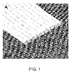
FIGURE 1A is a perspective view of a portion of a neuroimplant in accordance
with the present
invention. The neuroimplant is flat and is comprised of parallel polymer
fibres. FIGURE 1 B is a
perspective view of a neuroimplant of the present invention where the polymer
fibres are
formed to a C-shape. Figure 1 C shows another embodiment of the neuroimplant
of the present
invention, having multiple layers. Cells may be grown on and between fibres of
the present
neuroimplant. FIGURE 1D shows a Hoffman modulation contrast image of a
neuroimplant
prepared in accordance with the present invention.
FIGURE 2A shows a schematic of the BMP7 lentiviral vector. FIGURE 2B shows
confirmation
of BMP7 transgene expression by fluorescence microscopy 18 hours after
transfecting the
packaging HEK 293SF-PacLv cells. Scale bar: 50 pm.FIGURE 3 shows the BMP7-
Lentivirus
titration and protein production for non-infected 293GPG cells (FIGURE 3A);
1:100 BMP7-Lv
infected 293GPG cells (FIGURE 3B); 1:10 BMP7-Lv infected 293GPG cells (FIGURE
3C); and
1:1 BMP7-Lv infected 293GPG cells (FIGURE 3D). FIGURE 3E is a bar graph
showing that at
least 75% of the cells were infected with BMP7 lentivirus at 1:1 dilution.
Figure 3F is a western
blot of the infected HEK 293GPG cultures showing production of BMP7 protein .
BMP7 protein
was present in the cultures as early as 48 hours following infection. Samples
included: mouse
cerebrospinal fluid (lane 1), cells infected with GFP-Lv (lane 2), medium from
BMP7 lentivirus
infected cultures (lane 3), medium (10x concentrated) from GFP-Lv infected
cultures (lane 4),
medium (1 Ox concentrated) from BMP7 lentivirus infected cultures (lane 5).
FIGURE 4 shows that BMP7 is consistently produced and released into the medium
from
approximately 1x10⁶ BMP7-Lv infected 293 GPG cells. FIGURES 4A and B show
ELISA
results for cells 3 and 28 days after infection, respectively. FIGURE 4C is a
bar graph showing
the amount of BMP7 secreted over a 24-hour period, in nanograms; approximately
350 ng of
BMP7 is secreted into the media every 24 hours. FIGURE 4D shows western blot
analysis of
the biological activity of BMP7 protein produced by lentiviral system (Lv-
BMP7) compared to
that of commercially available recombinant human BMP7 (rBMP7). Lane 2: primary
embryonic
day 13 (El 3) cortical progenitor cells treated with GFP-Control media; Lanes
3: 1 ng/mL of
rBMP7, Lanes 4-5: Lv-BMP7. FIGURE 4E is a bar graph showing that, similar to
recombinant
human BMP7 (rhBMP7), there was a significant increase in the number of MAP2
positive
neurons in the embryonic day 13 (E13) cortical progenitor cultures treated
with the lentivirally-
made BMP7 (Lv-BMP7) for 5 days (*, ** p < 0.001).
CA 02762365 2011-11-17
WO 2011/000100 PCT/CA2010/001019
FIGURE 5A shows seeding of, mouse N2a cells on neuroimplants. Both N2a (FIGURE
5B)
and mouse embryonic stem (ES) cells (FIGURES 5C-D) can differentiate into
neurons on
neuroimplants. Both N2a and ES cells have been stained with the cell survival
dye, 5CFDA.
FIGURES 6A and B show GFP-tagged human amniotic fluid cells grown on
neuroimplants.
FIGURE 6C shows human amniotic fluid cells tagged with GDNF-GFP, while FIGURE
6D
shows human amniotic fluid cells tagged with BMP7-GFP.
FIGURE 7 shows high resolution digital photographs of the healthy (FIGURE 7A)
and injured
(FIGURE 7B, circled) brains. Corresponding immunohistochemical images show
intact
neurons (arrowheads) in the healthy motor cortex (FIGURE 7C) and neurons
affected by injury
(FIGURE 7D), showing MAP2 immunoreactivity. Cb: cerebellum, Ncx: neocortex,
OB: olfactory
bulb. *: lost tissue, Scale bar: A and B 1.6 mm, C and D 70 pm.
FIGURE 8 shows tissue reconstitution in the motor cortex after receiving a
neuroimplant.
FIGURE 8A shows an adult mouse left motor cortex (arrow) two months after
injury, having
received no cell or polymer implantation); the right motor cortex has been
used as control.
FIGURE 8B shows the left motor cortex (arrow) one month after injury and
implantation with
the neuroimplant (PGA polymer + cells) of the present invention; the right
motor cortex
(asterisk) is 15 minutes post-injury was used as an internal control.
DETAILED DESCRIPTION OF THE INVENTION
The present invention relates to biocompatible polymer fibres for
neuroimplants. More
specifically, the present invention relates to flexible biocompatible parallel
polymer fibres for
neuroimplants.
In one aspect, the present invention provides a neuroimplant comprising
biocompatible
polymer fibres, wherein the polymer fibres are grouped in a parallel
arrangement, and wherein
the group of fibres are flexible.
The neuroimplant of the present invention, also referred to herein as "neural
implant" or
"implant", is intended for implantation into brain tissue. The present
neuroimplant has
topological features that facilitate the reconstruction of damaged brain after
injury, stroke or
tumour excision, by serving as a template to reconnect the injured brain
tracts.
The neuroimplant of the present invention is comprised of biocompatible
polymer fibres. By the
term "biocompatible", it is meant that the fibres are compatible for placement
in a living system or
6
CA 02762365 2011-11-17
WO 2011/000100 PCT/CA2010/001019
tissue; "biocompatible" also indicates that the polymer fibres can integrate
with the tissue without
eliciting an immune response in the organism.
By the term "polymer fibres", it is meant a synthetic material that is a
continuous filament. The
polymer fibres are synthesized from chemical moieties using physical processes
well-known in
the art. The polymer fibres used in the present invention may be a single
polymer, a co-polymer,
or blend of polymers. The neuroimplant may comprise a number of fibres,
wherein individual
fibres may be made of the same or different materials.
The polymer fibres may be biodegradable or non-degradable. A biodegradable
polymer fibre may
be degraded within a time interval that is compatible for neuroregeneration of
the brain; this time
interval may depend on the size and severity of the damage. For example, and
without wishing to
be limiting in any manner, the polymer fibres may be substantially degraded in
5 to 15 weeks; for
example, the polymer fibres may be substantially degraded in 5, 6, 7, 8, 9,
10, 11, 12, 13, 14, or
15 weeks, or any time there between, or within a range of times defined by any
two values just
recited.
The polymer fibres may be made of any suitable material, including but not
limited to: polyester;
polyethylene; polymethacrylic; polyacrylic; polysulfone; polyurethane; nylon
(polyamide); aliphatic
polyesters; poly(amino acids); copoly(ether-esters); polyalkylene oxalates;
polyamides;
poly(iminocarbonates); polyorthoesters; polyoxaesters; polyamidoesters;
poly(anhydrides);
polyphosphazenes; polyphosphoester; and biopolymers. In a non-limiting
example, the polymer
fibres may be polylactic acid (PLA) fibres, for example poly(L-lactic acid) or
poly(DL-lactic acid);
poly(glycolic acid) (PGA) fibres; polylactic-co-glycolic acid (PLGA) fibres;
polycaprolactone
polyanhydride fibres; chitosan fibres; sulfonated chitosan fibres;
polyglycolide fibers; poly-4-
hydroxybutyrate fibres; or polyphosphoester fibres. In a specific, non-
limiting example, polymer
fibres may be formed from thermoplastic material; the polymer fibres may be
PGA and/or PLA
fibres.
The size of the polymer fibres in the neuroimplant of the present invention
may be any size
suitable for regeneration of brain tissue. The polymer fibres may have a
diameter of about 5 to
about 120 microns; for example, the diameter of the fibres may be 5, 10, 15,
20, 25, 30, 35, 40,
45, 50, 55, 60, 65, 70, 75, 80, 85, 90, 95, 100, 105, 110, 115 or 120 microns,
or any size
therebetween, or any range of sizes defined by any two values just recited.
The neuroimplant of
the present invention may comprise polymer fibres of the same diameter, or of
varying diameters.
As would be recognized by a person of skill in the art, the length of the
polymer fibres would vary
based on the physical requirements of the neuroimplant.
7
CA 02762365 2011-11-17
WO 2011/000100 PCT/CA2010/001019
The neuroimplant of the present invention may comprise a suitable number of
polymer fibres.
Without wishing to be limiting in any manner, the neuroimplant may comprise 5-
500 polymer
fibres; for example, the neuroimplant may comprise 5, 10, 15, 20, 25, 30, 35,
40, 45, 50, 75, 100,
125, 150, 175, 200, 225, 250, 275, 300, 325, 350, 375, 400, 425, 450, 475, or
500 polymer fibres,
or any amount therebetween. The amount of fibres within the implant may vary
based on the type
of polymer used, as well as the size of the fibres; the amount of fibres in
the neuroimplant may be
determined by a skilled person based on these variables.
The size of the implant, the diameter of the fibres, the number of fibres, the
type of polymer(s) and
the rate of degradation of the neuroimplant of the present invention may be
adjusted in
accordance with the physical requirements of the particular application. As
would be understood
by a person of skill in the art, polymer type, molecular weight, and blend may
be adjusted in order
to address the needs of the application at hand.
Importantly, the polymer fibres of the neuroimplant are in a parallel
arrangement. By the term
"parallel arrangement", it is meant that the long axes (also referred to
herein as "length") of the
fibres are placed parallel to each other (see Figure IA). This feature differs
from the currently
used polymer mesh (Shimada et al., 2006), which has randomly-oriented fibres
that lack the
architecture or topology required to reconnect damaged brain tracts. Without
wishing to be
limiting, the parallel arrangement and proper orientation of the polymer
fibres in the neuroimplant
of the present invention presents regular features that may allow neurons to
attach, grow and
expand linearly; this may allow the neurons to communicate and link with each
other and may
provide improved conditions for neurite growth.
Furthermore, the fibres in parallel arrangement must be in substantial contact
with one another.
By "substantial contact", it is meant that the fibres contact each other along
at least part of their
length on at least one side. While some areas of non-contact are permissible,
these must not
interfere with the overall design or integrity of the neuroimplant. Areas of
non-contact may be
located at regular intervals, or at varying intervals along the length of the
neuroimplant. The
polymer fibres may be bonded or consolidated together to maintain contact
between each other;
the bonding may be permanent. The fibres may be bonded together using any
suitable method
known in the art. For example, and without wishing to be limiting in any
manner, gradually
heating thermoplastic fibres above their glass transition temperature, but
before complete flow,
followed by cooling would allow them to be bonded together. Bonding of the
fibres should not
alter the arrangement, configuration or shape of the fibres or the
neuroimplant.
The polymer fibres may be grouped (also referred to herein as "bundled")
together in various
configurations, provided they remain in a parallel arrangement. For example,
and without wishing
8
CA 02762365 2011-11-17
WO 2011/000100 PCT/CA2010/001019
to be limiting in any manner, the polymer fibres may be grouped in a monolayer
of bonded fibres
(see for example, Figure 1A), in multiple layers bonded fibres (see for
example, Figure 1C), in
a cylinder (hollow or filled), or any other suitable configuration. These
configurations, together
with the parallel arrangement of the fibres, create channels between the
fibres that may
encourage regeneration of neurons.
The group of fibres in the neuroimplant of the present invention may be
flexible. By the term
"flexible", it is meant that the group(s) of fibres may be formed into a
desired geometry or shape.
The desired shape may vary based on the area of the brain tissue receiving the
implant and/or
the type of implant required. Generally, the implant may be required to be
flat, to be curved, or to
include curved sections along its length. For example, and without wishing to
be limiting in any
manner, the group of fibres may be formed into a flat implant, or one that is
C-shaped (see Figure
1 B), U-shaped, S-shaped, J-shaped, semi-cylindrical, or any other suitable
shape. The group of
fibres may be shaped using any suitable method known in the art. For example,
and without
wishing to be limiting in any manner, the group of fibres may be formed into
the desired shape
along its length during the bonding process described above; in this non-
limiting example,
thermoplastic fibres are heated while in contact with a mandrel to form the
fibres into the desired
shape (mandrel shape). For example, flat or curved shapes may be obtained
using a flat plate or
cylinder, respectively, on which the fibres are rolled, then consolidated or
bonded under heat.
Once formed into the desired shape, the group of fibres retains the shape
after removal from the
mandrel. Non-limiting examples of shapes of neuroimplants of the present
invention are shown in
Figure 1.
The neuroimplants of the present invention may further comprise cells that
facilitate the
regeneration of brain tissue. As would be recognized by one of skill in the
art, the type of cells
to be used in conjunction with the neuroimplant will vary based on the
organism receiving the
implant. For example, and without wishing to be limiting in any manner, the
cells may be
mouse embryonic stem cells, mouse neural stem cells, mouse neural progenitors,
mouse N2a
cells, human embryonic stem cells, human neural stem cells, human neural
progenitors, NT2
cells (including NT2 differentiated cells such as NT2 neurons and astrocytes),
human amniotic
fluid cells, human amniotic fluid stem cells, human blood cord cells, or any
other suitable type
of cell. In a specific, non-limiting example, the cells may be embryonic stem
cells, neural stem
cells, neural progenitors, NT2 cells, amniotic fluid cells, amniotic fluid
stem cells, blood cord
cells, or a combination thereof.
The cells may be engineered to deliver neurotrophic factors, neuroprotective
factors, or
neuroregenerative factors, or a combination thereof to the brain. For example,
and without
wishing to be limiting in any manner, the cells may be genetically engineered
to produce one
9
CA 02762365 2011-11-17
WO 2011/000100 PCT/CA2010/001019
or more than one factor known to be involved in tissue repair following the
implantation; for
example, the factors may be glial cell line-derived neurotrophic factor (GDNF)
and/or bone
morphogenetic protein 7 (BMP7). The production and the amount of factor(s)
secreted by the
engineered cells may be regulated. This regulation may be achieved by any
suitable method
known in the art. For example and without wishing to be limiting in any
manner, an inducible
lentiviral delivery system may be used to regulate factor expression in these
cells under a
tetracycline (Tet)-responsive bi-directional promoter; this allows for tight
regulation of factor
expression, thus enabling controlled delivery.
The present invention also encompasses a method of facilitating the repair of
damaged brain
tissue, comprising placing a neuroimplant as described above in the damaged
area, and allowing
the regeneration of neurons to occur. The neuroimplant may additionally
comprise cells that
facilitate the regeneration of brain tissue, which may or may not be
engineered to deliver
neurotrophic factors, neuroprotective factors, or neuroregenerative factors,
or a combination
thereof to the brain (as described above). The method as described may further
comprise a
step of inducing the expression of the neurotrophic factors, neuroprotective
factors, and/or
neuroregenerative factors.
The neuroimplant as described above may provide a template for cell
attachment, survival,
proliferation and differentiation, neurite growth, tissue
reconstitution/regeneration and functional
connectivity and recovery. The topological features of the implant may
facilitate the reconstruction
of damaged brain after injury, stroke or tumour excision, by serving as a
template to reconnect the
injured brain tracts.
Neuroimplants in accordance with the present invention support cell adhesion
and survival.
Seeding of various neural cell types (see above) on neuroimplants of the
present invention shows
that cells can differentiate into neurons on the neuroimplants. Neurites from
both cell types
followed the pattern of PGA fibres by extending along the fibres. The
production of specific
factors by cells carried by the neuroimplants of the present invention was
confirmed by ELISA and
other methods. Also, the neuroimplants presently described were shown to have
a beneficial
effect in the regeneration of mouse motor cortex following injury.
The present invention will be further illustrated in the following examples.
However, it is to be
understood that these examples are for illustrative purposes only and should
not be used to
limit the scope of the present invention in any manner.
CA 02762365 2011-11-17
WO 2011/000100 PCT/CA2010/001019
Example 1: Preparation of the polymer fibre neuroimplant
A neuroimplant in accordance with the present invention was prepared as
described below.
Purasorb PG (PURAC), a polyglycolic acid (PGA), was used for the preparation
of the neuro-
implant, due to its degradation time characteristics (within a few weeks).
First, fibres of various
diameters (5 to 120 microns) were produced from PGA using a capillary
rheometer in
combination with a rotating wheel winder. The barrel temperature was set at
280 C and the
fibre was formed at room temperature to allow for very fast cooling and to
avoid crystallization.
Differential scanning calorimetric analysis showed that the fibres were
completely amorphous
(data not shown). The fibres were stored at -18 C after production.
The neuroimplant was produced by rolling a long PGA fibre around either a
metallic plate or
cylinder ("mandrel"). The implants produced had dimensions of about 3 mm in
length. Once
the fibres were closely rolled around the mandrel, they were subjected to high
temperature
(about 210 C) either in an air convection oven or using a hot air stream on
the surface of the
fibres such that only the fibre surface was melted. The exposure time to high
temperature (with
continuous rotation of the mandrel) was about 5 minutes and depended on the
desired degree
of bonding. A Hoffman modulation contrast image of a prepared neuroimplant is
shown in
Figure 1 D.
Example 2: Construction of lentiviral vectors
An inducible lentiviral delivery system was prepared for BMP7 expression in
cells under a
tetracycline (Tet)-responsive bi-directional promoter.
A safe and efficient lentiviral vector, pTetO7CSII-CMV-GFPq (kindly provided
by Dr. Bernard
Massie, NRC-BRI, (Broussau et al., 2008)) was utilized for cloning. The
plasmid pDWC01 was
constructed through standard cloning procedures and isolated with Qiagen
MaxiPrep kit.
Briefly, the sequence encoding BMP7 was cut from pCMV-SPORT6-BMP7 (Open
Biosystems)
with the restriction endonucleases Agel and Xhol. The vector pTetO7CSII-CMV-
GFPq was
linearized with Age[ and Xhol to form compatible ends for ligation. To
construct the lentiviral
BMP7 vector (pDWC01), the cut BMP7 DNA fragment was ligated (T4 DNA ligase,
NEB) into
pTetO7CSII-CMV-GFPq, upstream of an Internal Ribosomal Entry Site and Green
Fluorescent
Protein (IRES-GFP). The resulting plasmid encoded for a third generation
transfer lentivector
with the transgenes BMP7 and GFP under the control of a CMV promoter (Figure
2A). Similar
techniques were used to make GDNF-GFP lentiviral vector (Sandhu et al., 2009).
Both BMP7
and GDNF inserts were sequenced to ensure their accuracy.
11
CA 02762365 2011-11-17
WO 2011/000100 PCT/CA2010/001019
Example 3: Isolation of neural stem and neural progenitor cells
Neural stem and neural progenitor cells were isolated from mice, in
preparation for transfection
and implantation.
Timed-pregnant mice were sacrificed by CO2 inhalation at embryonic day 13
(E13), according
to a protocol approved by the NRC-IBS Animal Care Committee (ACC), as
previously
described (Bani-Yaghoub et at., 2006). The uteruses were aseptically removed
and transferred
sequentially to two Petri dishes containing calcium- and magnesium-free Hank's
balanced salt
solution (HBSS, Invitrogen Corporation, Burlington, ON) to rinse away blood.
Embryos were
dissected out of the amniotic sacs and examined for morphological hallmarks to
ensure the
accuracy of the gestational timing. The heads and the telencephalons were
sequentially
isolated under a dissection microscope and transferred into the new plates
containing HBSS.
The dorsal and ventral telencephalic regions were dissected out and freed of
meninges and
dissected further to isolate the ventricular zone (VZ).
Tissues were mechanically dissociated in Dulbecco's Modified Eagle Medium,
high glucose, L-
glutamine (DMEM; Invitrogen) and filtered through a 40 m nylon cell strainer
(Falcon, VWR,
Mississauga, ON). The dissociated cells were quickly assessed for viability by
the trypan blue
exclusion assay. Neural stem cells were examined for the self-renewal and
multipotential
properties, using neurosphere assays (Bani-Yaghoub et al., 2006). In brief,
cells were
deposited into the uncoated 96-well plates (Nunc) in DMEM (Invitrogen) + N2
supplement
(Invitrogen) + fibroblast growth factor 2 (FGF2, 20 ng/ml, Invitrogen) at a
density of 1 cell/well
(plating efficiency: - 40%). Single cells were repeatedly monitored under a
light microscope for
the neurosphere formation, using the same culture condition. Neurospheres were
dissociated
with trypsin and transferred onto the PLL-coated neuroimplants in DMEM + 5%
fetal bovine
serum (FBS) + N2 supplement and examined 1-10 days later for the expression of
neuronal
markers. Neural progenitors were obtained from the E13.5 VZ and seeded
directly onto the
PLL-coated neuroimplants and treated with DMEM + 5% fetal bovine serum (FBS) +
N2
supplement.
Example 4: Transduction of cells with the GDNF- or BMP7-IRES-GFP lentivirus
The lentiviral delivery system of Example 2 was introduced to cells, yielding
cells that express
GDNF and/or BMP7.
The 293SF-PacLV packaging cells were seeded in 10 cm dishes and transfected
with the
plasmid pDWC01 (3`d generation lentivirus encoding BMP7 or GDNF and control
green
fluorescent protein (GFP)), using Lipofectamine 2000 (Invitrogen) (Broussau et
at., 2008). Six
12
CA 02762365 2011-11-17
WO 2011/000100 PCT/CA2010/001019
hours after transfection, medium was replaced with fresh medium supplemented
with 1 fag/ml
doxycycline and 10 fag/ml cumate (4-Isopropylbenzoic acid). The medium
containing lentivirus
was harvested at 72 h after transfection, filtered with 0.45 pm filters and
concentrated with
Amicon Ultra-15 spin columns (100,000 mol. wt. cut off, Millipore). Then, the
virus was applied
to neural progenitors, including amniotic fluid cells, after which the
transduced cells were
selected (Bani-Yaghoub et al., 2006; Sandhu et al., 2009).
The sample results of Figure 2B confirm BMP7 transgene expression by
fluorescence
microscopy 18 hours after transfecting the packaging HEK 293SF-PacLv cells.
Example 5: FACS-based Titration and Lentiviral Infection
The fluorescent-activated cell sorting (FACS)-analysis was used to determine
the transducing
units (TU)/ mL of BMP7-Lv or GDNF produced by transfected 293SF cells (Example
4) 48 hrs
post-transfection.
Briefly, HEK 293GPG cells were seeded in six-well plates at a density of 1.0E6
cells/well and
incubated at 37 C in 5% CO2 for 24 hrs or until cells were approximately 85-
90% confluent
(-2.0E6 cells/well). To remove potential cell debris prior to infection, the
medium was replaced
with 1.7 mL/well of fresh DMEM with 1 % FBS. Serial dilutions were prepared
with DMEM in the
ratios 1:1, 1:10 and 1:100 from 30x concentrated lentiviral-containing medium.
Each 293GPG-
containing well was transduced with 300 pL of the desired lentiviral serial
preparation.
Polybrene was added to a final concentration of 8 pg/mL for each the control
and the infection
wells and the plates were subsequently incubated at 37 C in 5% CO2. Following
a 48 hr
incubation period, the infection efficiency was verified with fluorescent
microscopy via the
examination of GFP expression. The cells were prepared for FACS analysis,
first by removing
the control and infection medium from each well and washing with 1x phosphate-
buffered
saline (PBS). Next, 200 pL of 0.25% Trypsin was added to each well and
following a short 1
min incubation period at RT, the cells were resuspended in 1 mL/well of PBS
containing 10%
FBS, briefly vortexed to dissociate the cells and stored on ice. An aliquot of
the sample was
counted using a hemocytometer to determine the approximate cell density per
well. The
samples were immediately analyzed on a MoFlo flow cytometer (DakoCytomation,
Copenhagen, Denmark) using Summit software. For each sample at least 40,000
events were
collected. The titer of the virus was determined using the following formula:
transducing
units/mI = [(% Infected Cells) x (Total Cell Number in Well) x (Dilution
Factor)]/ (Volume of
lnoculum Added to Cells).
Figures 3A-D show the BMP7-lentivirus titration via FACS analysis of non-
infected 293GPG
cells, 1:100 BMP7-Lv infected 293GPG cells, 1:10 BMP7-Lv infected 293GPG
cells, and 1:1
13
CA 02762365 2011-11-17
WO 2011/000100 PCT/CA2010/001019
BMP7-Lv infected 293GPG cells, respectively. These results show that at least
75% of the
cells were infected with BMP7 lentivirus at 1:1 dilution (Figure 3E). A
western blot of the
infected HEK 293GPG cultures (Figure 3F) indicates that BMP7 was present in
the cultures as
early as 48 hours following infection.
Example 6: BMP7 and GDNF ELISA
The level of BMP7 and GDNF proteins expressed by the cells of Example 4 was
quantified
using a human BMP7 or GDNF ELISA development kit, according to the
manufacturer's
protocol (R&D Systems, Minneapolis, MN, USA).
BMP7: Briefly, 96-well flat-bottomed Maxisorp plates (Nunc International) were
coated with the
capture antibody (mouse anti-human BMP7 capture antibody) diluted 1:180 with
1x PBS, pH
7.2 and incubated overnight at room temperature (RT). Following overnight
incubation, the
wells were blocked for 1 hr at room temperature with 200 pL of Reagent Diluent
(PBS + 1%
BSA, pH 7.2) per well. Standards for BMP7, ranging from a low of 125 pg/mL to
a high of 8000
pg/mL were prepared using recombinant human BMP7 (R&D Systems) diluted in
Reagent
Diluent and the samples were prepared in serial dilutions (1:1, 1:10, 1:100)
with PBS.
Approximately 100 pU well of each standard and sample dilution were applied to
the plate in
duplicate and incubated at RT for 2 hrs. The wells were washed 5x with 200 pL
/ well of Wash
Buffer (PBS, 0.05% (vlv) Tween 20, pH 7.2) followed by the addition of 100 pU
well of BMP7
detection antibody (biotinylated mouse anti-human BMP7 antibody) diluted 1:180
in Reagent
Diluent + 2% heat-inactivated goat serum. Following a 2 hr incubation period
at RT and
another wash step, 100 pL of streptavidin-conjugated horseradish peroxidase
(streptavidin-
HRP, R&D Systems) diluted in Reagent Diluent (1:200) was applied to each well
and
incubated at RT for 20 min. The wells were again washed (5x) with Wash Buffer
and color
development was achieved by adding 100 pL of a 1:1 mixture of
tetramethylbenzidine (TMB;
Sigma-Aldrich, Oakville, Ontario): H202 per well. The plates were incubated
for 20 min at room
temperature in the dark and the reaction was stopped by the addition of 50 pL
2 N HCI per
well. The absorbance was measured using a SpectraMax 340 microplate reader
(Molecular
Devices, Sunnyvale, Ca, USA) at 450 nm and the amount of BMP7 was calculated
from the
standard curves in the detection limit range.
GDNF: The amount of GDNF released in HAF cultures transduced with Lenti-GDNF
or Lenti-
GFP was measured using a GDNF ER,ax Immunoassay system according to the
manufacturer's instructions (Promega, Madison, WI). In brief, Maxisorp 96-
well, flat-bottomed
ELISA plates (Nalgene Nunc International) were coated with anti-GDNF
monoclonal antibody
14
CA 02762365 2011-11-17
WO 2011/000100 PCT/CA2010/001019
diluted in carbonate coating buffer, pH 8.2 and incubated overnight at 4 C.
Wells were
blocked for 1 hour at room temperature with 1x blocking buffer (200 pL/well).
GDNF standards
ranging from 0-1000 pg/100 pL were prepared using recombinant human GDNF and
sample
dilutions (100 pL, dilutions ranging from 5-fold to 20-fold) were applied to
the wells. All
samples were incubated with shaking for 6 hours at room temperature and then
washed with
TBS-T (20 mM Tris-HCI, pH 7.6, 150 mM NaCl, 0.05% (v/v) Tween 20). The
captured GDNF
was bound by a specific polyclonal antibody on incubating overnight at 4 C.
After washing, the
amount of bound polyclonal antibody specific to GDNF was then detected by a
species specific
(chicken) antibody conjugated to horse radish peroxidase incubated overnight
at 4 C.
Following washes with TBS-T, horseradish peroxidase-conjugated anti-chicken
IgY antibody
was added to the plates and incubated with shaking at room temperature for 2
hours. The
plates were again washed with TBS-T, and 100 pL of the enzyme substrate
(Tetramethylbenzidine One solution) was added. The plates were incubated for
15 min at
room temperature in the dark and the reaction was stopped by the addition of
100 pL 1 N HCI
per well. The absorbance was measured at 450 nm and the amount of GDNF was
calculated
from the standard curve in the linear range.
ELISA results are shown in Figures 4A-4C. The level of BMP7 secretion was
markedly high in
BMP7-Lv infected 293GPG cultures. After 3 days, the level of BMP7 secreted by
1x10⁶ cells
was up to 330 ng over a 24-hr period. To determine the long-term BMP7
producing capacity of
the infected cultures, the level of BMP7 was determined 4 weeks following
infection. The level
of BMP7 was consistent 4 weeks later with a maximum yield of 390 ng of BMP7
secreted over
a 24-hr period. The biological activity of the BMP7 protein produced by
lentiviral system (Lv-
BMP7) was verified by comparing with that of the commercially available
recombinant human
BMP7 (Figure 4D). In brief, primary embryonic day 13 (E13) cortical progenitor
cells were
treated with GFP-Control media (lane 2), 1 ng/mL of rBMP7 or Lv-BMP7 (lanes 3
and 4) and 30
ng/mL Lv-BMP7 (lane 5) for 1.5 hrs to examine SMAD 1/5/8 activation and
translocation to the
nucleus.
Using similar ELISA methods, approximately, 10 ng of GDNF was secreted from
1x10⁶ human
amniotic fluid (AF) cells within 24 hours. Both BMP7 and GDNF were
consistently produced
and released into the media. Additionally, results (figure 4E) show that there
was a significant
increase in the number of MAP2 positive neurons in the embryonic day 13 (E13)
cortical
progenitor cultures treated with the lentivirally-made BMP7 (Lv-BMP7) for 5
days.
CA 02762365 2011-11-17
WO 2011/000100 PCT/CA2010/001019
Example 7: Neuroimplant seeding and evaluation
To construct neuroimplants, cells (mouse or human ES, NS, NP, NT2 or AF) were
seeded on
the scaffolds.
Initially, seeding was done in the presence of Dulbecco's Modified Eagle
Medium (DMEM) +
10% fetal bovine serum (FBS), and then in DMEM + 0.5% FBS + N2 supplement
(i.e., prior to
implantation). While the size of the neuroimplant and cell density are easily
adjustable, cells
were seeded at a density of 2.5x10³-1x10⁵ cells on neuroscaffolds that
approximate the size of
2.5 week old male C57BL/6 mouse primary motor cortex (I: 3 mm x w: 2 mm X 1
mm).
Figure 5 shows results of the seeding of N2a and mouse embryonic stem cells on
the
neuroimplant of the present invention. Both N2a (Figure 5B) and mouse
embryonic stem (ES)
cells (Figures 5C-D) can differentiate into neurons on neuroimplants, and
neurites from both
cell types follow the pattern of PGA scaffold by extending along the scaffold
fibres. Thus, it is
presently shown that the neuroimplant design allows the formation of organized
neurite growth.
Figures 6 show that cells can grow on neuroimplants and secrete
neurotrophic/neuroprotective
/neuroregenerative factors; specifically, the GFP (Figures 6A-B), GFP-GDNF
(Figure 6C), and
BMP7-GFP human amniotic fluid cells (Example 4) were grown on neuroimplants.
The
production of GDNF factors by cells was confirmed by ELISA and other methods
(see Example
6).
The in vivo performance of the neuroimplant of the present invention was also
evaluated.
Injury was mechanically introduced to the left motor cortex of adult mouse
brains (Figures 7B,
circled, and Figure 8A). In brief, 56-77 day old C57BI/6 or CD1 mice (Charles
River Labs, St
Constant, QC) were anesthetized using isoflurane gas (Aerrane, Baxter,
Montreal, QC). The
animals were placed in a stereotaxic frame and the skull was exposed. The
injury site was
marked on the bone, using specific coordinates (from Lat +0.7 mm, AP - 0.25 mm
to - 1.0 mm
to Lat + 2.4 mm AP +1.25 mm to + 3.0 mm) and the bone was removed with a
dental drill. The
motor cortex was injured, using a sterile graduated needle/knife to the depth
of 1 mm (DV 1
mm). Figure 7 shows images of healthy (Figure 7A) and injured (Figure 7B)
adult mouse
brains. In addition to the control non-injured mice (Figure 7A), the right
motor cortex was used
as internal control (non-injured hemisphere in Figures 7B and 8A).
Corresponding
immunohistochemical images show intact neurons (arrowheads) in the healthy
motor cortex
(Figure 7C). In contrast, neurons are significantly affected by injury, as
evidenced by
morphological features and MAP2 immunoreactivity (Figure 7D). A representative
image of the
left motor cortex that had not received cell or polymer implantation (Figure
8A, arrow) has been
shown two months after injury. In another case, the left motor cortex received
the PGA
16
CA 02762365 2011-11-17
WO 2011/000100 PCT/CA2010/001019
polymer neuroimplant seeded with cells (see above) and was evaluated one month
after injury
(Figure 8B, arrow). To better compare the significance of the repair in the
left motor cortex
after implantation (Figure 8B, arrow), an acute injury was introduced to the
right motor cortex
of the same mouse 15 minutes before the brain was taken out (Figure 8B,
denoted by
asterisk).
Together, Figure 8 shows tissue reconstitution in the motor cortex after
receiving a
neuroimplant of the present invention. In the absence of any implantation, the
injured adult
mouse left motor cortex shows little improvement 2 months post-injury. In
contrast,
implantation of the neuroimplant (PGA polymer + cells) of the present
invention in the left
motor cortex shows significant regeneration of the brain tissue one month post-
injury.
The embodiments and examples described herein are illustrative and are not
meant to limit the
scope of the invention as claimed. Variations of the foregoing embodiments,
including
alternatives, modifications and equivalents, are intended by the inventors to
be encompassed
by the claims. Furthermore, the discussed combination of features might not be
necessary for
the inventive solution.
REFERENCES
All patents, patent applications and publications referred to herein are
hereby incorporated by
reference.
Ahn, Y.H., Bensadoun, J.C., Aebischer, P., Zurn, A.D., Seiger, A., Bjorklund,
A., Lindvall, 0.,
Wahlberg, L., Brundin, P., Kaminski, Schierle, G.S. 2005. Increased fiber
outgrowth from xeno-
transplanted human embryonic dopaminergic neurons with co-implants of polymer-
encapsulated genetically modified cells releasing glial cell line-derived
neurotrophic factor.
Brain Res Bull 66:135-142.
Bang SM, Kim YK, Park YH, Sohn SK, Lee JJ, Cho EK, Ryoo BY, Chung IJ, Yoon SS,
Kim HJ,
Lee JH, Yoon HJ, Park S. 2005. High-dose therapy and autologous stem cell
transplantation in
Korean patients with aggressive T/NK-cell lymphoma. Leuk Lymphoma. 11:1599-
1604.
Bani-Yaghoub M, Tremblay R, Voicu R, Mealing G, Monette R, Py C, Faid K,
Sikorska M.
2005. Neurogenesis and neuronal communication on micropatterned neurochips.
Biotechnol
Bioeng 92:336-345.
17
CA 02762365 2011-11-17
WO 2011/000100 PCT/CA2010/001019
Bani-Yaghoub, M., Tremblay, R.G., Lei, J.X., Zhang, D., Zurakowski, B.,
Sandhu, J.K., Smith,
B., Ribecco-Lutkiewicz, M., Kennedy, J., Walker, P.R. and Sikorska, M. (2006)
Role of Sox2 in
the development of the neocortex. Dev Biol 295:52-66.
Bian C, Song X, Liu Z, Zhang H. 2005. Design proposal of imaging activities of
cultured neural
network on a silicon substrate with neural-electronic-optical integrated
microsystem. Conf Proc
IEEE Eng Med Biol Soc. 7:7600-3.
Borlongan CV, Saporta S, Sanberg PR. 1998. Intrastriatal transplantation of
rat adrenal
chromaffin cells seeded on microcarrier beads promote long-term functional
recovery in
hemiparkinsonian rats. Exp Neurol. 2:203-14.
Brain Injury Association of Nipissing: BIAN (2005) http://dawn.thot.net/brain/
Broussau S, Jabbour N, Lachapelle G, Durocher Y, Tom R, Transfiguracion J,
Gilbert R,
Massie B. 2008. Inducible packaging cells for large-scale production of
lentiviral vectors in
serum-free suspension culture. Mol Ther. 3:500-7.
Bueno, EM., Laevsky, G., Barabino, G.A.. 2007. Enhancing cell seeding of
scaffolds in tissue
engineering through manipulation of hydrodynamic parameters. J Biotechnol
129:516-531.
Centre for Chronic Disease Prevention and Control Canada (2008)
http://www.phac-
aspc.gc.ca/ccdpc-cpcmc/
Chang, C.F., Lin, S.Z., Chiang, Y.H., Morales, M., Chou, J., Lein, P., Chen,
H.L., Hoffer, B.J.,
Wang, Y. 2003. Intravenous administration of bone morphogenetic protein-7
after ischemia
improves motor function in stroke rats. Stroke 34:558-564.
Chou, J., Harvey, B.K., Chang, C.F., Shen, H., Morales, M., Wang, Y. 2006.
Neuroregenerative effects of BMP7 after stroke in rats. J Neurol Sci 240:21-
29.
De Coppi P, Callegari A, Chiavegato A, Gasparotto L, Piccoli M, Taiani J,
Pozzobon M, Boldrin
L, Okabe M, Cozzi E, Atala A, Gamba P, Sartore S. 2007. Amniotic fluid and
bone marrow
derived mesenchymal stem cells can be converted to smooth muscle cells in the
cryo-injured
rat bladder and prevent compensatory hypertrophy of surviving smooth muscle
cells. J Urol.
1:369-76.
Gao J, Prough DS, McAdoo DJ, Grady JJ, Parsley MO, Ma L, Tarensenko YI, Wu P.
2006.
Transplantation of primed human fetal neural stem cells improves cognitive
function in rats
after traumatic brain injury. Exp Neurol. 2:281-92.
18
CA 02762365 2011-11-17
WO 2011/000100 PCT/CA2010/001019
Green AR, Shuaib A. 2006. Therapeutic strategies for the treatment of stroke.
Drug Discov
Today. 15-16:681-93.
Hara K, Yasuhara T, Maki M, Matsukawa N, Masuda T, Yu SJ, Ali M, Yu G, Xu L,
Kim SU,
Hess DC, Bortongan CV. 2008. Neural progenitor NT2N cell lines from
teratocarcinoma for
transplantation therapy in stroke. Prog Neurobiol. 3:318-34.
Horita Y, Honmou 0, Harada K, Houkin K, Hamada H, Kocsis JD. 2006. Intravenous
administration of glial cell line-derived neurotrophic factor gene-modified
human mesenchymal
stem cells protects against injury in a cerebral ischemia model in the adult
rat. J Neurosci Res.
2006 Nov 15;84(7):1495-504
International Brain Injury Association, 2007.
http://www.internationalbrain.org/
International Brain Injury Association, 2008.
http://www.internationalbrain.orgf
Kawabata, M., Imamura, T., Miyazono, K. 1998. Signal transduction by bone
morphogenetic
proteins. Cytokine Growth Factor Rev 9:49-61.
Kelly, S., Bliss, T.M., Shah, A.K., Sun, G.H., Ma, M., Foo, W.C., Masel, J.,
Yenari, M.A.,
Weissman, I.L., Uchida, N., Palmer, T., Steinberg, GK. 2004. Transplanted
human fetal neural
stem cells survive, migrate, and differentiate in ischemic rat cerebral
cortex. Proc NO Acad Sci
USA 101:11839-11844.
Kondziolka, D., Wechsler, L., Goldstein, S., Meltzer, C., Thulborn, K.R.,
Gebel, J., Jannetta,
P., DeCesare, S., Elder, E.M., McGrogan, M., Reitman, M.A., Bynum, L. 2000.
Transplantation
of cultured human neuronal cells for patients with stroke. Neurology 55:565-
569.
Kondziolka D, Steinberg GK, Wechsler L, Meltzer CC, Elder E, Gebel J, Decesare
S, Jovin T,
Zafonte R, Lebowitz J, Flickinger JC, Tong D, Marks MP, Jamieson C, Luu D,
Bell-Stephens T,
Teraoka J. 2005. Neurotransplantation for patients with subcortical motor
stroke: a phase 2
randomized trial. J Neurosurg. 1:38-45.
Longhi L, Watson DJ, Saatman KE, Thompson HJ, Zhang C, Fujimoto S, Royo N,
Castelbuono D, Raghupathi R, Trojanowski JQ, Lee VM, Wolfe JH, Stocchetti N,
McIntosh TK.
2004. Ex vivo gene therapy using targeted engraftment of NGF-expressing human
NT2N
neurons attenuates cognitive deficits following traumatic brain injury in
mice.J Neurotrauma.
12:1723-36.
19
CA 02762365 2011-11-17
WO 2011/000100 PCT/CA2010/001019
Lindvall, 0., Kokaia, Z., Martinez-Serrano, A. 2004. Stem cell therapy for
human
neurodegenerative disorders-how to make it work. Nat Med 10 Suppl:S42-50.:S42-
S50.
Lindvall, 0., Kokaia, Z. 2006. Stem cells for the treatment of neurological
disorders. Nature
441:1094-1096.
Marklund N, Bakshi A, Castelbuono DJ, Conte V, McIntosh TK. 2006. Evaluation
of
pharmacological treatment strategies in traumatic brain injury. Curr Pharm
Des. 13:1645-80.
Muller, F.J., Snyder, E.Y., Loring, J.F. 2006. Gene therapy: can neural stem
cells deliver? Nat
Rev Neurosci 7:75-84.
National Institute of Neurological Disorders and Stroke, NINDS, 2006
www.ninds.nih.gov
Park, K.I., Teng, Y.D., Snyder, E.Y. 2002. The injured brain interacts
reciprocally with neural
stem cells supported by scaffolds to reconstitute lost tissue. Nat Biotechnol
20:1111-1117.
Ren, J., Kaplan, P.L., Charette, M.F., Speller, H., Finklestein, S.P. 2000.
Time window of
intracisternal osteogenic protein-1 in enhancing functional recovery after
stroke.
Neuropharmacology. 39:860-5.
Sandhu JK, Gardaneh M, Iwasiow R, Lanthier P, Gangaraju S, Ribecco-Lutkiewicz
M,
Tremblay R, Kiuchi K, Sikorska M. 2008. Astrocyte-secreted GDNF and
glutathione antioxidant
system protect neurons against 6OHDA cytotoxicity. Neurobiol Dis. 3:405-14.
Sautter, J., Sabel, M., Sommer, C., Strecker, S., Weidner, N., Oertel, W.H.,
Kiessling, M. 1998.
BDNF and TrkB expression in intrastriatal ventral mesencephalic grafts in a
rat model of
Parkinson's disease. J Neural Transm 105:253-263.
Schlosshauer, B., Dreesmann, L., Schaller, H.E., Sinis, N. 2006. Synthetic
nerve guide
implants in humans: a comprehensive survey. Neurosurgery 59:740-747.
Shimada Y, Hongo M, Miyakoshi N, Sugawara T, Kasukawa Y, Ando S, Ishikawa Y,
Itoi E.
2006. Dural substitute with polyglycolic acid mesh and fibrin glue for dural
repair: technical
note and preliminary results. J Orthop Sci. 5:454-8
Simic, P., Vukicevic, S. 2007. Bone morphogenetic proteins: from developmental
signals to
tissue regeneration. Conference on bone morphogenetic proteins. EMBO Rep 8:327-
331.
Stroke Facts from Genetech, 2002.
CA 02762365 2011-11-17
WO 2011/000100 PCT/CA2010/001019
www.gene.com/gene/products/education/vascular/stroke-factsheet.html
Stroke Recovery Canada (2004) http://www.strokerecoverycanada.coml
Tatard, V.M., Sindji, L., Branton, J.G., Ubert-Pouessel, A., Colleau, J.,
Benoit, J.P., Montero-
Menei, C.N. 2007. Pharmacologically active microcarriers releasing glial cell
line - derived
neurotrophic factor: Survival and differentiation of embryonic dopaminergic
neurons after
grafting in hemiparkinsonian rats. Biomaterials 28:1978-1988.
Teng, Y.D., Lavik, E.B., Qu, X., Park, K.I., Ourednik, J., Zurakowski, D.,
Langer, R., Snyder,
E.Y. 2002. Functional recovery following traumatic spinal cord injury mediated
by a unique
polymer scaffold seeded with neural stem cells. Proc Natl Acad Sci U S A
99:3024-3029.
Watson DJ, Longhi L, Lee EB, Fulp CT, Fujimoto S, Royo NC, Passini MA,
Trojanowski JQ,
Lee VM, McIntosh TK, Wolfe JH. 2003. Genetically modified NT2N human neuronal
cells
mediate long-term gene expression as CNS grafts in vivo and improve functional
cognitive
outcome following experimental traumatic brain injury.
J Neuropathol Exp Neurol. 4:368-80.
Wen, H., Dou, Z., Finni, T., Havu, M., Kang, Z., Cheng, S., Sipila, S., Sinha,
S., Usenius, J.P.,
Cheng, S. 2008. Thigh muscle function in stroke patients revealed by velocity-
encoded cine
phase-contrast magnetic resonance imaging. Muscle Nerve, March 11.
Wieloch, T., Nikolich, K. 2006. Mechanisms of neural plasticity following
brain injury. Curr Opin
Neurobiol 16:258-264.
Yip S, Shah K. 2008. Stem-cell based therapies for brain tumors. Curr Opin Mol
Ther. 4:334-
42.
Zhao B, Cooper LJ, Brahma A, MacNeil S, Rimmer S, Fullwood NJ. 2006.
Development of a
three-dimensional organ culture model for corneal wound healing and corneal
transplantation. Invest Ophthalmol Vis Sci. 7:2840-6.
21