Note: Descriptions are shown in the official language in which they were submitted.
CA 02762669 2011-11-18
WO 2010/135212
PCT/US2010/035054
X-RAY MICROSCOPY FOR CHARACTERIZING HOLE
SHAPE AND DIMENSIONS IN SURGICAL NEEDLES
TECHNICAL FIELD
The field of art to which this invention relates is x-ray microscopy, in
particular, x-ray microscopy for use with surgical needles and surgical needle
manufacturing processes.
BACKGROUND OF THE INVENTION
Surgical needle and suture combinations are well known in the surgical
arts. Surgical needles and sutures are a fundamental mainstay of surgical
procedures and trauma repair. Surgical sutures are conventionally woven or
braided from natural or synthetic polymeric materials including silk,
polyesters, polydioxanone, polylactide, and the like. The sutures may also be
constructed from a monofilament. The sutures may be bioabsorbable or
nonabsorbable.
Surgical sutures are typically mounted to conventional surgical needles
to create a needle and suture combination for use by the surgeon to
approximate tissue, etc. A conventional surgical needle is typically an
elongated, curved structure having a distal piercing tip and a proximal suture
mounting section. The needles may optionally have cutting edges to assist in
tissue penetration. The proximal suture mounting sections may have
CA 02762669 2011-11-18
WO 2010/135212
PCT/US2010/035054
2
conventional blind boreholes or channels for receiving the end of a suture.
One or both ends of a surgical suture may be mounted in the channel or
borehole and secured therein in a conventional manner, including conventional
mechanical swaging in which the suture mounting end of the surgical needle is
partially compressed, as well as adhesives, cements, etc. Surgical needles are
conventionally made from biocompatible materials, especially metals and
metal alloys such as surgical grade stainless steels.
Early in the development of surgical needles, channels were used to
attach suture to the needle. This was an improvement over needles having
eyelets wherein a suture was threaded through the eyelet in the field.
However, channels, when closed (i.e., swaged), create a bump (to a lesser or
greater degree) in the distal portion of the channel. Such bumps may be
undesirable to surgeons and other medical professionals since a bump may
disrupt the smooth passage of the needle through tissue. This characteristic
of
channeled needles was eliminated with the introduction of mechanically
drilled boreholes for suture mounting, however mechanical drilling can only
be utilized for low strength alloys and large diameter holes. The relatively
recent utilization of laser drilling was an important advancement in this art
and
addresses this issue as it allows small diameter boreholes to be drilled in
small
diameter wires, especially wires made from high strength alloys, which are
currently off-limits for the most part to mechanical drilling due to
technological limitations.
CA 02762669 2011-11-18
WO 2010/135212
PCT/US2010/035054
3
Drilled boreholes in surgical needles are particularly desirable since
the profile of the needle body is not altered in the same manner as when a
channel is punched into the proximal suture mounting end of the needle. A
smooth profile is desirable to the surgeon since it is believed to reduce
tissue
trauma and to reduce the force required to pull the needle through tissue with
a
commensurate reduction in drag. Drilled boreholes in surgical needles may be
produced in a number of conventional manners. Two conventional methods
used to drill boreholes, as previously mentioned, include mechanical drilling
and laser drilling.
11)
There are distinct differences between mechanically drilled and laser
drilled boreholes. Mechanically drilled boreholes are typically uniform and
precise in shape and profile as they take on the shape of the drill.
Mechanically drilled surgical needles are easily inspected using conventional
plug gages (i.e., machined cylindrical members having a constant diameter or,
optionally, tapering from proximal to distal). Although mechanical drilling
will typically produce a borehole having relatively precise dimensions and a
precise configuration, there are several disadvantages that may be associated
with mechanical drilling. These include slow drilling speeds in an automated
high speed manufacturing system, drill wear and life, the difficulty in
manufacturing production grade drills for needles having fine wire sizes,
increased costs, and the inability to drill small diameter holes in high
strength
alloys in small wire sizes
CA 02762669 2014-03-10
4
Although laser drilling overcomes these problems, laser drilled holes, on
the other hand, pose several other unique problems, although certainly
manageable,
that have yet to be addressed. Laser drilled needles tend to have several
issues
associated with the use of a laser to drill a borehole. For example, in cases
where
the laser melts the material to form the hole, there is the potential for
recast to form
on the interior of the hole, and such recast may affect suture attachment.
Other
issues may include the consistency of the borehole profile and the smoothness
of
the borehole, as well as the possibility of blow-outs.
Laser drilling processes have been developed for drilling boreholes in
surgical needles. Examples of such processes are included in the following
U.S.
Patents and Patent Application: US6018860, US 5776268, US 5701656, US
5661893, US 5644834, US 5630268, US 5539973, US6252195, and
US20050109741. Such laser drilling processes have many advantages, including
adaptability for high speed manufacturing processes, efficiencies and cost,
the
ability to drill small holes in small wire diameters in substantially any
material, and
reduced maintenance.
Although laser drilling processes have all of these advantages, as
previously mentioned the boreholes drilled by lasers typically do not have the
same
precise dimensional configuration as mechanically drilled boreholes. Laser
drilling utilizes a conventional laser that emits a laser beam, which is
typically
tapered or Gaussian, in shape. This means that the bore hole drilled
CA 02762669 2011-11-18
WO 2010/135212
PCT/US2010/035054
by the laser beam is typically tapered as it gets deeper. The laser beam used
for drilling is engineered with respect to parameters such as energy level,
pulse, waveform, etc., to produce a borehole having a desired configuration
and characteristics including borehole depth, length, cross-section, and
5 orientation about the longitudinal axis of the needle and about the
center of the
needle wire body, such that the laser drilled borehole is capable of
sufficiently
and effectively accepting an end of a surgical suture for mounting and
affixation.
This is the result of the very nature of laser drilling wherein a high
energy, pulsed laser beam essentially liquefies or vaporizes the target metal
in
the proximal, suture-mounting end of the needle upon which the beam is
directed. In some laser drilling, the molten material will reform
inconsistently
within the hole; this reformed material is commonly called recast, as
mentioned previously. The recast can create a non-uniform hole condition
which may affect suture insertion and attachment.
In order to effectively affix or mount the end of a surgical suture in a
laser-drilled borehole in a surgical needle, the borehole should have a
substantially uniform diameter, similar to a bore hole produced in a
mechanically drilled needle, albeit tapered as mentioned above. Similarly, the
length of the borehole must have maximum and minimum dimensions. A
length that is too long may weaken the needle, while too short may result in
needle/suture separation. And, the borehole must be relatively centered about
the longitudinal axis of the proximal end of the suture needle.
CA 02762669 2011-11-18
WO 2010/135212
PCT/US2010/035054
6
The present state of the art with respect to the measurement of the
dimensions of laser-drilled boreholes is to use conventional mechanical pin
gages, as is the conventional standard for mechanically drilled boreholes. The
use of pin gages is typically a manual procedure wherein statistically
significant quantities of needles are selected from lots of drilled needles,
and
the pin gages are manually inserted by an inspector into the drilled
boreholes.
The resulting data is recorded. There are several disadvantages associated
with the use of mechanical pin gages. While pin gages are ideally suited for
mechanically-drilled needles, they are not especially suitable for laser
drilled
needle manufacturing for several reasons. First of all, pin gages are not
adapted for use in high speed manufacturing processes. Also, the pin gages
used to measure very small diameter boreholes are expensive and difficult to
manufacture, and for the finer diameters are easily damaged. In addition, the
use of pin gages will not provide information with respect to the presence of
re-cast. Pin gages can easily measure a mechanically-drilled borehole as it is
cylindrical in nature and has a regular profile, but a laser drilled borehole
in a
surgical needle is not typically cylindrical in profile and may contain re-
cast
and varying diameters along the length of the borehole. Thus, a pin gage can
only approximate the minor diameter measurement of a laser drilled borehole,
and provides no other information with respect to other important parameters
such as taper, length, degree of centeredness, irregularities, degree of
skewing,
etc. The presence of re-cast may cause a misrepresentation of the true minor
diameter of the laser drilled hole. Further, as mentioned above, the pin gage
measurements fail to address potential variants in the borehole profile. The
CA 02762669 2011-11-18
WO 2010/135212
PCT/US2010/035054
7
use of a pin gage does not indicate the major diameter or provide a
representation of the variation in the borehole profile. Therefore the only
measurement a pin gage can provide is an indication of the smallest potential
diameter of the borehole, without a value or determination of variations in
diameter, profile, degree of skewedness, and other critical parameters.
Another disadvantage associated with the use of pin gages is that pin
gages do not provide real time data that can be used to immediately adjust
production-processing parameters. Statistical sampling of a batch of drilled
needles may indicate that the boreholes are out of specification, requiring
the
destruction of an entire out-of-specification batch of needles. Other
disadvantages include: pin gage wear, whether the gage is a minus or plus in
tolerance with respect to the required borehole measurement, and acceptance
of boreholes that meet the pin gage criteria, but have undetected internal
geometries that inhibit, or preclude subsequent suture attachment. Pin gage
measuring is a manual process and, consequently, is not a procedure that can
keep pace with a high speed surgical needle manufacturing processes required
in modern needle manufacturing processes and typically associated with laser
drilling. Statistical sampling of laser drilled needles, although possible, if
one
were willing to accept any attendant disadvantages, is potentially prohibitive
and it would not be possible to inspect a statistically relevant sample in
real-
time. Therefore it is typically necessary to use a reduced sample size, which
may lead to false positives, possibly resulting in the destruction of laser
drilled
needles that, if inspected at acceptable levels, would not result in such a
loss
and the commensurate expense associated with the loss of a production batch
CA 02762669 2011-11-18
WO 2010/135212
PCT/US2010/035054
8
of needles. Another disadvantage of pin gage inspection methods includes the
possible acceptance of boreholes that meet the pin gage criteria, but have
undetected defects, internal geometries or configurations that inhibit, or
preclude subsequent effective suture insertion and attachment, possibly
resulting in failures in the field.
As discussed above, the conventional means of measurement for
drilled boreholes, i.e., plug gaging, does not work well with laser drilled
holes
because of the numerous attendant disadvantages. Given the inconsistent
lo profile of a laser hole, plug gaging only can provide the user with an
indication of the minor diameter of the inconsistent profile, but fails to
provide
a measure of the major diameter and/or the hole profile. This is a serious
drawback, as variation on the hole profile and the differences between the
minor and major hole diameters directly affects the ability to secure the
suture
to the needle. In mechanically drilled holes this is not a factor as the hole
is a
reflection of the drill geometry. Another drawback is that pin gaging is
extremely time-consuming and only as accurate as the pin gage is
manufactured and maintained. Unfortunately, there are no options available
other than physical destruction, specifically, mechanically cross-sectioning a
needle and examining the shape of the borehole, which is difficult, laborious,
time consuming, and not cost-effective to do with a statistically significant
sample size, and does not provide real time information which can be used to
control production processes.
CA 02762669 2011-11-18
WO 2010/135212
PCT/US2010/035054
9
Therefore, there is a need in this art for novel methods of
characterizing drilled boreholes in a high speed manufacturing environment
and using such characterizations to adjust and control laser drilling and
subsequent manufacturing processes. The significant benefit of which is to
improve yields, product performance, and improve product consistency.
SUMMARY OF THE INVENTION
Accordingly, a novel method of characterizing drilled boreholes in
surgical needles is disclosed. In this method, an x-ray beam is directed from
an x-ray generator at a surgical needle, preferably the proximal end of a
surgical needle containing a drilled borehole. An image of the proximal end
of the needle is digitally generated from a sensor, which the x-ray beam
impinges upon. At least the proximal end of the needle is located between the
x-ray generator and the sensor. The image includes the laser-drilled borehole.
The digital image is processed to determine a deviation from a standard
dimensional specification for the borehole. It is particularly preferred that
the
borehole be laser drilled.
Another aspect of the present invention is a method of controlling a
laser drilling apparatus during a borehole drilling process. In this method, a
laser is provided that emits a laser beam at the proximal end of a surgical
needle to drill a borehole therein. An x-ray beam is directed from an x-ray
generator at a surgical needle, preferably the proximal end of a surgical
needle
containing the laser-drilled borehole. An image of the proximal end of the
needle is digitally generated from a sensor, which the x-ray beam impinges
CA 02762669 2011-11-18
WO 2010/135212
PCT/US2010/035054
upon. At least the proximal end of the needle is located between the x-ray
generator and the sensor. The image includes the laser-drilled borehole. The
digital image is processed to determine a deviation from a standard
dimensional specification for the borehole. Then an algorithm is provided to
5 determine the appropriate corrections to the parameters for the laser
beam to
provide for a drilled bore hole in the surgical needles that is within the
specification.
Yet another aspect of the present invention is a method of
lo characterizing laser-drilled boreholes in surgical needles. In this
method, an
x-ray beam is directed from an x-ray generator at the surgical needle,
preferably the proximal end of a surgical needle containing a laser-drilled
borehole. An image of the proximal end of the needle is digitally generated
from a sensor, which the x-ray beam impinges upon. At least the proximal
end of the needle is located between the x-ray generator and the sensor. The
image includes the laser-drilled borehole. The digital image is processed to
determine a deviation from a standard dimensional specification for the
borehole and to determine the measurements of the borehole. The needle or a
carrier strip carrying the needle is marked with a code containing the
measurements of the image, and each needle is provided with a digital
identity.
Optionally, downstream process steps can utilize this information to
control the attachment of the suture in the drilled boreholes by varying
compression variables such as pressure, time, and dwell to compensate for
CA 02762669 2011-11-18
WO 2010/135212
PCT/US2010/035054
11
subtle changes in the borehole profile as determined by the x-ray analysis.
This greatly contributes to the consistency and efficacy of the suture/needle
interface or attachment and directly contributes to the performance of the
component,
These and other aspects and advantages of the present invention will
become more apparent from the following description and accompanying
drawings.
BRIEF DESCRIPTION OF THE DRAWINGS
FIG. 1 is a flow diagram of a drilled laser borehole process of the
present invention.
FIG. 2 is a schematic illustrating the geometric characteristics and
parameters of a mechanical drill
FIG. 3 is a schematic of a pin gage inserted into a borehole of a laser
drilled surgical needle; the needle is illustrated in cross-section.
FIG. 4 is a schematic showing the distal end of a surgical suture
mounted and swaged in a laser drilled borehole in the proximal end of a
surgical needle.
CA 02762669 2011-11-18
WO 2010/135212
PCT/US2010/035054
12
FIG. 5 is a schematic illustrating the distal end of a surgical suture
swaged into a mechanically drilled borehole in the proximal end of a surgical
needle.
FIG. 6 is a photograph of a mounted and cross-sectioned laser drilled
surgical needle showing the borehole.
FIG. 7 is a photograph of a mounted and cross-sectioned surgical
needle showing that the laser drilled borehole has recast present.
FIG. 8 is a photograph of a mounted cross-section of the proximal end
of a laser drilled surgical needle. The drilled bore hole is seen to have a
blowout.
FIG. 9 is a photograph of a mounted cross-section of a proximal end of
a laser drilled needle, wherein inconsistencies or variations in borehole
diameter are readily visible along the length of the borehole
FIG. 10 is a perspective x-ray image of a mechanically drilled surgical
needle.
FIG.11 is a perspective x-ray image of a laser drilled surgical needle.
FIG. 12 is an x-ray image of a stake-swaged needle laser drilled
needle.
CA 02762669 2011-11-18
WO 2010/135212
PCT/US2010/035054
13
FIG. 13 is an x-ray image of the proximal end of a laser drilled surgical
needle in which a potential blowout defect is visible.
FIG. 14 is a perspective x-ray image of the distal end of a laser drilled
surgical needle that has been swaged in which internal cracks resulting from
the swaging process are visible.
DETAILED DESCRIPTION OF THE INVENTION
The terms "surgical needle" and "needle" are used interchangeably
herein. There is a general recognition in the art of surgical needle
manufacturing that a laser drilled borehole diameter is not as consistent as a
mechanically drilled borehole. In mechanically drilled boreholes, the drill
defines the borehole diameter whereas in laser drilling, the focus and energy
and other known characteristics of the laser beam control the hole diameter.
For mechanical drills, the geometry of the drill is very important with
respect
to borehole accuracy, especially the flute length and flute symmetry of the
mechanical drill. FIG. 2 identifies and illustrates key drill geometrical
characteristics of a mechanical drill 300, including web width 302, flute
length
304, included angle 306, and symmetry. Since a borehole diameter is
physically defined by the drill, pin gaging a resulting mechanically drilled
borehole in a surgical needle is appropriate as a testing means, since the
drill
will, both theoretically and practically, drill consistently throughout the
depth
of the borehole. Pin gaging will indicate nonconformities such as off
CA 02762669 2011-11-18
WO 2010/135212
PCT/US2010/035054
14
specification diameter or even out of round boreholes caused for example by a
flexing drill, worn or broken drill, or flexing needle.
As mentioned above, in laser drilling the focus and energy of the beam,
along with other parameters, are critical to borehole diameter consistency, as
well as depth and other parameters of the shape of the hole. Variations in
diameter are detrimental to accurate borehole gaging and attachment. If the
diameter varies, pin gaging will only allow the inspector to ascertain the
diameter of the smallest diameter; this precludes measurement of the greater
diameter. Consequently, an inaccurate evaluation of hole diameter is obtained
over the length of the swage area (see FIG. 3) when pin gaging is employed
for diameter measurement. As illustrated in FIG. 3, the proximal end 115 of a
surgical needle 110 is seen to have a laser drilled bore hole 120 having
proximal opening 122, end 126 and elongated cavity 130. The cavity 130 is
seen to have several diameters along its length. The pin gage 140 inserted
into
bore hole 120 is only capable of determining the minimum diameter 142 of the
bore hole cavity 140. Also illustrated is one section of a swage die 150.
The attachment concern with borehole diameter variation associated
with laser drilling (i.e., varying diameter along the longitudinal length of
the
borehole) is related to the nature of the swaging process. Swaging is
conventionally based upon fixed displacement, and this means that the
swaging dies will close to the same point each time. Any variation in suture
diameter, suture density, needle barrel diameter, or borehole diameter will
affect attachment strength values. This is illustrated in FIG. 4. As seen in
CA 02762669 2011-11-18
WO 2010/135212
PCT/US2010/035054
FIG. 4, the surgical needle 110 has proximal end 115. The needle has laser
drilled longitudinal borehole 120 having proximal opening 122, end 126 and
elongated cavity 130. A distal end 162 of a suture 160 is seen to be inserted
in
cavity 130 through opening 122. Swage die members 150 are seen to be
5 located on either side of the proximal end 115 of needle 110. Due to the
irregular shape of bore hole cavity 130, it is not possible to completely
insert
the end 162 of suture 160 into cavity 130. This is due to the irregularly
formed shape of the laser drilled borehole 120 having various minor and major
dimensions along the length of the cavity 130. When mechanically swaged by
10 swage members 150, the sides 124 of the bore hole 120 will not uniformly
engage and compress suture end 162 along its length, potentially
compromising retention in the borehole 120. A needle 200 having a
mechanically drilled bore hole 220 is illustrated in FIG. 5. The borehole 220
is seen to have cavity 230 having a uniform or substantially constant diameter
15 229. The distal end 262 of suture 260 is seen to be completely emplaced
within borehole 220 and uniformly engaged by sides 224 when the end 215 of
needle 200 is mechanically swaged by swage die members 250.
If the borehole diameter of a laser drilled borehole can be controlled in
a consistent regular manner, similar to a mechanically drilled needle, the
consistency of pull values of attached sutures from boreholes and
improvement in yields will be significant. The presence of recast will affect
borehole measurements in laser-drilled needles. Recast is a phenomenon
wherein the melted material reforms in the hole and alters the intended shape
of the hole as a consequence. It is sometimes difficult to isolate recast and
can
CA 02762669 2011-11-18
WO 2010/135212
PCT/US2010/035054
16
result in a smaller measurement and the impression that the hole diameter is
smaller than it actually is.
FIG. 6 is a photograph of a mounted, and cross-sectioned, laser-drilled
needle. It clearly shows why pin gaging is disadvantageous and impractical
with laser-drilled needles in that the accuracy of the testing varies with the
degree of trueness of the laser-drilled borehole. It is apparent and can be
seen
that the borehole cavity meanders and the inconsistency of the diameter is
readily seen throughout the length of the hole, thereby effectively rendering
lo useless pin gaging as an effective method of in-process laser drilled
borehole
measurements.
Referring to FIG. 7, a photograph of a cross-sectioned needle shows
that the laser-drilled borehole has recast present, as mentioned above, that
can
influence the pin gaging inspection of the hole diameter of a borehole and may
lead to an incorrect conclusion as to the maximum dimensions of the borehole
diameter. The bore hole of the needle o :FIG. 7 also shows a tapered section
just past recast bumps that are seen protruding inwardly from the sides of the
borehole. These recast bumps may lead to an incorrect conclusion as to
maximum hole diameter and the shape of the bore hole along its length. Note
that it is possible to see these anomalies only by cutting the needle in cross-
section which also results in the destruction of the needle. Additionally, one
must be fortunate to cross-section the needle in the correct plane to reveal
such
anomalies - often missed due to cross-sectioning. Other than pin gaging,
which may be inaccurate and potentially misleading, there are no means
CA 02762669 2011-11-18
WO 2010/135212
PCT/US2010/035054
17
available to determine laser drilled borehole diameters, borehole
concentricity,
and borehole uniformity with a measuring method that is not destructive,
however the novel methods of the present invention provide for such
determinations.
Given that such hole inconsistencies attendant with laser drilled
needles may result in inconsistent needle pulloff (i.e., suture pullout)
performance, a non-destructive testing system would provide the ability to
'see' the borehole without destroying the needle, and this would provide the
capability to determine beforehand whether or not a needle is suitable for
suture attachment or if the attachment method needs to be modified to
compensate for the borehole variability.
The novel methods of the present invention provide for the use of X-
ray imaging and analysis to evaluate borehole diameters and borehole profiles.
FIG. 8 is a photo of a mounted cross-section of the proximal end of a
laser drilled surgical needle. The drilled bore hole can be seen to have a
blowout wherein the laser beam caused the side of the needle surrounding the
bore hole to open to the exterior creating a lateral hole or opening in the
needle into the borehole cavity. This is undesirable because it creates a
cosmetic blemish, can weaken the wall resulting in a potential for breakage,
and could create a sharp surface that could cut the user or cut tissue where
it is
undesired. Referring now to FIG. 9, a photograph of a cross-section of a
proximal end of a laser drilled needle is seen. The inconsistencies in
borehole
CA 02762669 2011-11-18
WO 2010/135212
PCT/US2010/035054
18
diameters are readily visible along the length of the borehole; also seen are
the
major and minor diameters.
FIGS. 10, 11 and 12 are images illustrating examples of needles that
were x-rayed. FIG. 10 shows the distal end of a mechanically drilled needle.
The uniformity of the bore hole is readily observed and is seen to be regular
in
shape as opposed to a laser drilled borehole. FIG. 11 is an x-ray image of the
proximal end of a laser drilled needle. The taper of the borehole can be
clearly
seen. It is apparent that the entire length of the borehole is not available
to use
due to this taper, whereas in a mechanically drilled needle the entire length
of
the hole can be utilized (i.e., used for receiving the distal end of a
suture).
FIG. 12 is an x-ray image of a stake-swaged needle laser drilled
needle. This image illustrates the capability of an x-ray image to present the
results of the attachment process, something that cannot be done by any other
means that is not destructive.
X-ray imaging is also well suited to manufacturing. Multiple images
may be examined and real-time information and evaluation is possible since
only a few milliseconds is needed to grab a picture and perform dimensional
and profile evaluations; this being dependent upon the x-ray aperture and the
computer speed.
One distinct possibility is to couple the picture evaluations to the laser
controls and utilize the inspection results to fine tune or control the laser
to
CA 02762669 2011-11-18
WO 2010/135212
PCT/US2010/035054
19
optimize consistency and minimize variability. One especially significant
perspective of x-ray inspection is that it is capable of inspecting wire/hole
diameters down to a very small diameter. This is something not achievable
pin gaging especially at a high-speed rate. An x-ray system can inspect
multiple needles whereas pin gaging can only inspect one needle at a time.
Pin gaging is also susceptible to the tolerance of the pins, how much they
have
worn, their concentricity (not bent from use), and the expertise of the
inspector. These traits are all eliminated with x-ray inspection.
It is also impossible to pin gage needles at any significant rate. An x-
ray system can inspect at also any rate needed by scanning mu1tip13 needles at
a time. An x-ray system will also provide instant electronic archival of the
results, eliminating paperwork errors and time to transfer data from the
measurement to the recording sheet.
Figure 13 is an x-ray image that depicts a potential blowout condition.
This picture shows a needle wherein the laser borehole was drilled off center
and the resulting profile has in a thin wall condition where the borehole is
almost through the sidewall. This condition can lead to a premature failure of
the needle and potential breakage due to the site being a weakened area.
Figure 14 is an x-ray image that depicts a needle with a borehole where
the needle material has cracked and separated partially. This is a concern in
case the crack propagates to the surface resulting in a weakened area that can
CA 02762669 2011-11-18
WO 2010/135212
PCT/US2010/035054
potentially lead to breakage or the crack may affect a mounted suture such
that
the suture is cut and fails thus separating from the needle prematurely.
The novel x-ray characterization methods of the present invention
5 provide for a method of characterizing drilled boreholes in automated
needle
manufacturing processes. They may be utilized with processes that utilize
mechanical drilling methods or with processes that utilize laser drilling. The
characterization processes of the present invention are particularly preferred
for use in laser drilling processes. The x-ray devices or machines that may be
10 utilized in the processes of the present invention will have the
following
characteristics. The x-ray devices will have the ability to transport and
appropriately position individual, or multiple, drilled needles within the x-
ray
unit between an x-ray emitting source and a sensor. The units will further
have the capability to expose the needle(s) to x-rays emitted by the source,
and
15 to obtain and digitize resulting x-ray image(s). The units will also be
capable
of comparing the digitized images to a digital template or series of specific
borehole dimensional requirements and provide and generate an instructive
disposition signal regarding borehole acceptability. In addition, the units
will
have processing capability to effectively process the instructive signal to
sort
20 or otherwise identify individual needles as to their acceptability, or
specific
borehole dimensions, and/or to adjust laser parameters to produce boreholes
within specified requirements. The x-ray units will be conventional,
commercially available units that may be modified for the processes of the
present invention, for example, an x-ray unit manufactured by Envision
Product Design located in Anchorage, Alaska.
CA 02762669 2011-11-18
WO 2010/135212
PCT/US2010/035054
21
The x-ray devices useful in the practice of the processes of the present
invention provide a digitized output of the image of a drilled borehole that
is
compared with the dimensions of a standard. This comparison may be done in
several manners including the following manner. The image captured and
generated by the x-ray unit is pixilated. These pixels are evaluated for light
density. This density is compared to templates that have been likewise
pixilated. Since the dimension of a pixel is a known measurement, the system
counts the number of pixels within the light density determined by the
template and converts this count into a linear measure. If the resulting value
is
within tolerance or outside of tolerance the appropriate indication is
conveyed.
Additionally, these measurements may be tracked and statistical conclusions
made on an on-going basis for track-and-trend or for statistical control.
Further, the information can be optionally placed by inking, laser etching, or
other known means upon the needle and/or its carrier for downstream
intelligence. In this manner, each characterization for each needle is stored
digitally by providing a digital identity for each needle and then storing the
image corresponding to the digital identity in a computer database.
A preferred x-ray system that can be used in the practice of the present
invention is an integrated inspection system including a shielded cabinet, a
130kV X-ray source located in the top of the cabinet, a 4"x4" CMOS imaging
panel located on an adjustable height platform under the source, a four axis
manipulator for positioning a sample under the source, and a computer
workstation with software. To image a needle, the process begins with
CA 02762669 2011-11-18
WO 2010/135212
PCT/US2010/035054
22
positioning the imaging panel a required distance from the X-ray source,
attaching the sample needle to be inspected to the inspection plate on the
manipulator, then moving the sample into position based upon the orientation
and geometric magnification required. If a previous imaging technique has
not been developed, the next step will involve calibration and various test
shots to determine the optimum photon energy (voltage or kV) and photon
flux (generator current or mA). The optimum kV and mA parameters will be
unique to a needle material, thickness and shot geometry and selected to
provide the widest possible range of image grayscale values in order to
provide the highest possible image contrast. If a previous imaging technique
has been developed, then imaging can begin with small adjustments required
to orient the needle for viewing an Area of Interest (A0I). In general, shots
will involve geometric magnification that results from the needle being
located
off the imaging panel plane and moved toward the source. As the needle
moves closer to the source and away from the imaging panel, the resulting
image on the panel will become larger creating a magnified view of the
needle. Magnification up to 15X will be possible depending upon the size of
the AOI. To acquire an X-ray image the x-ray source is activated,
illuminating the needle with an X-ray photon beam that is projected onto the
X-ray panel. The needle in the middle of the beam creates a shadow on the
panel corresponding to the density of the needle which varies based upon
material and geometry or thickness. The panel converts X-ray beam intensity
as attenuated by the part into electrical signals corresponding to a range
between saturation and no measurable X-ray energy. This signal is digitized
into a 12 bit range represented visually by a grayscale value range from 0 to
CA 02762669 2011-11-18
WO 2010/135212
PCT/US2010/035054
23
4096 and presented on an LCD display. Window and leveling tools are then
applied to the image to select a narrower range of grayscale values that
contain
the relevant data, adjusted to maximize the contrast of the image within the
range of values of interest. After an image has been acquired and adjusted for
best viewing, analysis and interpretation can be completed and the image
evaluated based upon inspection requirements. Typical evaluation may
include dimensional analysis of features using tools that have been calibrated
to the X-ray
The novel process of the present invention for characterizing the
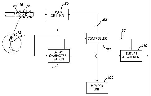
drilled boreholes in surgical needles is illustrated schematically in FIG. 1.
As
seen in FIG. 1, surgical needles 10 having laser drilled bore holes 30 in
their
proximal ends 12 are mounted to strips 40 for moving the needles between
production stations. The mounted needles 10 are first moved to laser drilling
station 50 where a conventional laser is used having a laser beam with desired
waveform and parameters sufficiently effective to drill the boreholes 30 in
the
proximal ends 20 of the surgical needles 10. Such parameters include
conventional parameters, e.g., focal point, pulses and power. The needles 10
and strip 40 are then moved to x-ray characterization station 70. At station
70,
each individual needle 10 is x-rayed and a digital characterization of the
needle including the borehole 30 in the distal end 12 is obtained. Each needle
is given a digital identification number at station 70 and the x-ray
characterization is transmitted to controller/processor 90.
Controller/processor
90 is a conventional computer or data processor. The characterizations for
each needle 10 are stored by controller/processor 90 in memory unit 100, and
CA 02762669 2011-11-18
WO 2010/135212
PCT/US2010/035054
24
are analyzed to determine the dimensional characteristics of the borehole
including longitudinal orientation, center, maximum and minimum diameter,
maximum and minimum length, and maximum and minimum wall thickness
about the borehole. The dimensional characteristics are compared to a
standard template, and deviations are noted. The characteristics for each
needle and deviations from standard are optionally used to generate a signal
92
that is sent back to the laser drilling station 50 to a computer that controls
the
laser drilling station in order to adjust the characteristics of the laser
drilling
beam including parameters such as the waveform, pulse, energy, power, focal
point, pulses and time to provide for a drilled borehole having a minimal
deviation from the specified dimensions. Also optionally, the information
related to the borehole dimensions can be used to generate a signal 95 that is
sent to a controller/processor for suture attachment system 110, such as a
mechanical swaging system, for computing and controlling the attachment
pressure and dwell to optimize yields and performance of the suture/needle
interface. In this manner, each drilled needle will have a customized set of
attachment parameters depending upon the characteristics of the borehole in
that needle. Optionally, each needle is marked with a unique identifier; this
can be done in a conventional manner including for example, laser etching or
ink jet printing. In addition to the identifier (e.g., bar code), the data
from the
x-ray characterization step for each needle including characteristics and
deviations from a standard may optionally be marked on each needle,
The surgical needles that can be processed using the novel methods of
the present invention include conventional surgical needles having suture
CA 02762669 2011-11-18
WO 2010/135212
PCT/US2010/035054
mounting ends, preferably with proximal drilled boreholes. The surgical
needles can be made from conventional biocompatible materials and
equivalents thereof including but not limited to martensitic stainless steel
(e.g.,
UNS 42000), austenitic stainless steel (e.g., UNS 30200), maraging stainless
5 steel (e.g., UNS S45500, UNS 46910, and ETHALLOY brand stainless steel),
and refractory alloy systems (e.g., Tungsten-Rhenium) as well as polymeric
materials and ceramic materials and composites. The needles may have wire
sizes ranging from 1.0 mil to 70 mil, preferably from about 6.0 mil to about
12 mil and will have a variety of conventional lengths. The novel x-ray and
10 laser drilling processes of the present invention have numerous
advantages and
implications that include the following. x-ray imaging can be performed at the
speed of laser drilling in a high speed manufacturing process. The imaging is
non-destructive, so that tested needles can be used for finished product. The
processes eliminate costly and potentially inaccurate plug gaging. X-ray
15 images can be digitized, magnified, and interrogated by a computer
against a
profile and/or pre-defined measurements, which can create a realistic
measurement and permit informed disposition of every needle manufactured.
The x-ray imaging system and process may be linked back to a laser drilling
station, whereby the measurements of the borehole are fed back to the laser to
20 make adjustments to fine tune and/or adjust the parameters of the laser
beam
and thereby control the borehole shape and aspect ratio (depth-to-diameter
measurement) by adjusting parameters such as focus, time, energy, pulses, or
position. The process of the present invention may be further enhanced to
improve quality disposition by marking each needle or the needle carrier
25 adjacent to the needle with a pass/fail or actual measurement in a code
or
CA 02762669 2014-03-10
26 =
actual measurement in a code or actual numbers for disposition later in the
manufacturing process along with a unique digital identity. The data can
further be
employed to control the process for attaching the suture to the needle (e.g.,
by
conventional swaging processes) through adjustments to pressure, dwell, and
closing forces when attaching the needle to suture. This will result in
optimized
yields and further improved quality of the finished product by ensuring
needle/suture attachment integrity.
Although this invention has been shown and described with respect to
detailed embodiments thereof, it will be understood by those skilled in the
art that
various changes in form and detail thereof may be made.
DOCSTOR 2954670\1