Note: Descriptions are shown in the official language in which they were submitted.
CA 02763347 2016-02-08
METHODS FOR TREATMENT OF DISEASE IJSENG
AN EPIMETABOLIC SHIFTER (COENZYME 010)
Background of the Invention:
Cancer is presently one of the leading causes of death in developed nations
and is
25 a serious threat to modern society. Cancer can develop in any tissue of
any organ at any
age. Worldwide, more than 10 million people are diagnosed with cancer every
year and
it is estimated that this number will grow to 15 million new cases every year
by 2020. It
is believed that cancer causes six million deaths every year or 12% of the
deaths
worldwide.
30 The etiology of cancer is not clearly understood. Cancer has been
linked to or
associated with many factors over the many years of ongoing research including
genetic
- 1
CA 02763347 2016-02-08
susceptibility, chromosome breakage disorders, viruses, environmental factors
and
immunologic disorders. Cancer encompasses a large category of medical
conditions.
Cancer cells can arise in almost any organ and/or tissue of the body. Cancer
develops
when cells in a part of the body begin to grow or differentiate out of
control.
Although recent research has vastly increased our understanding of many of the
molecular mechanisms of tumorigenesis and has provided numerous new avenues
for
the treatment of cancer, standard treatments for most malignancies remain
gross
resection, chemotherapy, and radiotherapy. While increasingly successful, each
of these
treatments may cause numerous undesired side effects. For example, surgery may
result
in pain, traumatic injury to healthy tissue, and scarring. Radiation therapy
has the
advantage of killing cancer cells but it also damages non-cancerous tissue at
the same
time. Chemotherapy involves the administration of various anti-cancer drugs to
a
patient. These standard treatments often are accompanied by adverse side
effects, e.g.,
nausea, immune suppression, gastric ulceration and secondary tumorigenesis.
Over the years, many individuals and companies have conducted extensive
research searching for improvements in the treatments for the wide array of
cancers.
Companies are developing bioactive agents including chemical entities, e.g.,
small
molecules, and biologics, e.g., antibodies, with the desire of providing more
beneficial
therapies for cancer. Some of the bioactive agents tested have worked and
provided
beneficial therapeutic effects in some individuals or cancer types and others
have failed
or had minimal therapeutic effects in their testing protocols. Other bioactive
agents
studied to date have mechanisms of action that are not entirely understood.
Coenzyme Q10, also referred to herein as CoQ10, Q10, ubiquinone, or
ubidecarenone, is a popular nutritional supplement and can be found in capsule
form in
nutritional stores, health food stores, pharmacies, and the like, as a vitamin-
like
supplement to help protect the immune system through the antioxidant
properties of
ubiquinol, the reduced form of CoQ10. CoQ10 is art-recognized and further
described
in International Publication No. WO 2005/069916.
CoQ10 is found throughout most tissues of the human body and the tissues of
other mammals. The tissue distribution and redox state of CoQ10 in humans has
been
reviewed in a review article by Bhagavan HN, et al., Coenzyme Q10: Absorption,
tissue
uptake, metabolism and pharmaeokinetic, Free Radical Research 40(5), 445-453
(2006)
-2
CA 02763347 2011-11-10
WO 2010/13250720 PCT/US2010/034453
(hereinafter, Bhagavan, et al.). The authors report that "as a general rule,
tissues with
high-energy requirements or metabolic activity such as the heart, kidney,
liver and
muscle contain relatively high concentrations of CoQ10." The authors further
report
that "[a] major portion of CoQ10 in tissues is in the reduced form as the
hydroquinone
or ubiquinol, with the exception of brain and lungs," which "appears to be a
reflection of
increased oxidative stress in these two tissues." In particular, Bhagavan et
al. reports
that in heart, kidney, liver, muscle, intestine and blood (plasma), about 61%,
75%, 95%,
65%, 95% and 96%, respectively, of CoQ10 is in the reduced form. Similarly,
Ruiz-
Jiminez, etal., Determination of the ubiquinol-10 and ubiquinone-10 (coenzyme
Q10) in
human serum by liquid chromatography tandem mass spectrometry to evaluate the
oxidative stress, J. Chroma A 1175(2). 242-248 (2007) (hereinafter Ruiz-
Jiminez, et al.)
reports that when human plasma was evaluated for Q10 and the reduced form of
Q10
(Q10H2), the majority (90%) of the molecule was found in the reduced form.
CoQ10 is very lipophilic and, for the most part, insoluble in water. Due to
its
insolubility in water, limited solubility in lipids, and relatively large
molecular weight,
the efficiency of absorption of orally administered CoQ10 is poor. Bhagavan,
et al.
reports that "in one study with rats it was reported that only about 2-3% of
orally-
administered CoQ10 was absorbed." Bhagavan, et al. further reports that
"[d]ata from
rat studies indicate that CoQ10 is reduced to ubiquinol either during or
following
absorption in the intestine."
CoQ10 has been associated with cancer in the literature for many years.
Described below are some representative but not all inclusive examples of the
reported
associations in the literature. Karl Folkers, et al., Survival of Cancer
Patients on
Therapy with Coenzyme Q10, Biochemical and Biophysical Research Communication
192, 241-245 (1993) (herein after "Folkers, et al.") describes eight case
histories of
cancer patients "on therapy with CoQ10" and their stories of survival.. ."for
periods of
5-15 years." CoQ10 was orally administered to eight patients having different
types of
cancer, including pancreatic carcinoma, adenocarcinoma, laryngeal carcinoma,
breast,
colon, lung and prostate cancer. Folkers, et al. sets forth that "these
results now justify
systemic protocols." Lockwood, et al., Progress on Therapy of Breast Cancer
with
Vitamin Q10 and the Regression of Metastases, Biochemical and Biophysical
Research
Communication 212, 172-177 (1995) (hereinafter "Lockwood, etal.") is another
review
article that reports on the "[p]rogress on therapy of breast cancer with
Vitamin
-3
MEl 9938730v.1
CA 02763347 2016-02-08
Q10".Lockwood, et al. refers to Folkers, et at., which "covers 35 years of
international
research on animals and humans which revealed variable levels of vitamin Q10
in non-
tumor and tumor tissues and includes data on vitamin Q10 which are intrinsic
to the host
defense system as based on increased survivors of treated mice with tumors".
Lockwood, et al. further sets forth that "[Ole potential of vitamin Q10
therapy of human
cancer became evident in 1961" relying on a study that determined the blood
levels of
CoQ10 in 199 Swedish and American cancer patients that revealed variable
levels of
deficiencies in cases of breast cancer. U.S. Patent No.6,417,233, issued July
9,2002
(hereinafter Sears, et al.) describes compositions containing lipid-soluble
benzoquinones, e.g., coenzyme Q10, for the prevention and/or treatment of
mitochondriopathies. Sears, et al. sets forth that "CoQ10 treatment has been
reported to
provide some benefits in cancer patients (see column 2, lines 30-31)."
As of the date of filing of this application, the National Cancer Institute
reports
that no well-designed clinical trials involving large numbers of patients of
CoQ10 in
cancer treatment have been conducted since "the way the studies were done and
the
amount of information reported made it unclear if the benefits were caused by
the
coenzyme Q10 or by something else.- See The National Cancer Institute (NCI),
(September 29, 2008). In particular, the NCI cites three small studies on the
use of
CoQ10 as an adjuvant therapy after standard treatment in breast cancer
patients, in
which some patients appeared to be helped by the treatment, and reiterates
that
"weaknesses in study design and reporting, however, made it unclear if
benefits were
caused by the coenzyme Q10 or by something else." The NCI specifies that
"these
studies had the following weaknesses: the studies were not randomized or
controlled; the
.. patients used other supplements in addition to coenzyme Q10; the patients
received
standard treatments before or during the coenzyme Q10 therapy; and details
were not
reported for all patients in the studies." The NCI further reports on
"anecdotal reports
that coenzyme Q10 has helped some cancer patients live longer, including
patients with
cancers of the pancreas, lung, colon, rectum and prostate," but states that
'the patients
described in these reports, however, also received treatments other than
coenzyme Q10
including chemotherapy, radiation therapy and surgery."
US Patent Application Publication 2006/0035981, published February 16, 2006
(hereinafter "Mazzio 2006") describes methods and formulations for treating or
-4
CA 02763347 2011-11-10
WO 2010/13250720 PCT/US2010/034453
preventing human and animal cancers using compositions that exploit the
vulnerability
of cancers with regards to its anaerobic requirement for non-oxidative
phosphorylation
of glucose to derive energy, which is opposite to the host. The formulations
of Mazzio
2006 contain one or more compounds that synergistically promote oxidative
metabolism
and/or impede lactic acid dehydrogenase or anaerobic glucose metabolism and
more
particularly are described as containing "2.3-dimethoxy-5-methy1-1,4-
benzoquinone
(herein also termed "DMBQ") (quinoid base) and options for the entire
ubiquinone
series including corresponding hydroquinones, ubichromenols, ubichromanols or
synthesized/natural derivatives and analogues. See Mazzio 2006 at page 3,
paragraph
0010. Mazzio 2006 establishes "the short chain ubiquinones (CoQ<3) as anti-
cancer
agents and even further establishes that `2,3-dimethoxy-5-methyl-1,4-
benzoquinone
(DMBQ) is in excess of 1000 times more potent than CoQ10 as an anti-cancer
agent."
See Mazzio 2006 at page 3, paragraph 0011. Mazzio 2006 further set forth that
the
study "did not find CoQ10 to be as lethal as expected" and like "previous
studies that
have employed CoQ10 against cancer have been somewhat contradictory". See
Mazzio
2006 at pages 3-4 for an extensive list of citations supporting this
statement.
US Patent Application Publication 2007/0248693, published October 25, 2007
(herein after "Mazzio 2007") also describes nutraceutical compositions and
their use for
treating or preventing cancer. Again, this published patent application
focuses on the
short chain ubiquinones and specifically sets forth that CoQ10 is not a
critical
component of this invention. According to Mazzio 2007 "while CoQ10 can
increase the
Vmax of mitochondrial complex II activity in cancer cells (Mazzio and Soliman,
Biochem Pharmacol. 67:1167-84, 2004), this did not control the rate of
mitochondrial
respiration or 02 utilization through complex W. And, CoQ10 was not as lethal
as
expected. Likewise, results of CoQ10 against cancer have been contradictory."
See
Mazzio 2007 at page 5. paragraph 0019.
Summary of the Invention:
Applicants have previously described topical formulations of CoQ10 and
methods for reducing the rate of tumor growth in animal subjects (Hsia et al.,
WO
2005/069916 published August 4, 2005). In the experiments described in Hsia et
al.,
CoQ10 was shown to increase the rate of apoptosis in a culture of skin cancer
cells but
-5
MEl 9938730v.1
CA 02763347 2011-11-10
WO 2010/13250720 PCT/US2010/034453
not normal cells. Moreover, treatment of tumor-bearing animals with a topical
formulation of CoQ10 was shown to dramatically reduce the rate of tumor growth
in the
animals. The present invention is based, at least in part, upon a more
complete
understanding of the role of CoQ10 within a human and/or cell. In particular,
the
methods and formulations of the present invention are based, at least in part,
upon the
knowledge gained about the therapeutic activity of CoQ10 for oncological
disorders
learned by designing and implementing human clinical trials and/or by
administering
CoQ10 to human subjects and observing the surprising and unexpected results
that occur
during these trials and/or treatment regimens. The methods and formulations of
the
present invention are further based, at least in part, upon insight gained
into the
therapeutic mechanism of CoQ10 from extensive studies of CoQ10 treatment of
cells in
vitro.
Specifically, in at least one embodiment, the methods and formulations of the
present invention are based, at least in part, on the surprising discovery
that application
of Coenzyme Q10 (also referred to as CoQ10 or Q10 herein) to cells results in
selective
induction of an apoptotic response in cancer cells, with no effect or, in some
cases, a
positive effect on growth of normal cells. Moreover, in at least one
additional
embodiment, it was unexpectedly found that cell lines derived from aggressive
cancers
were more sensitive to CoQ10 (e.g., required lower concentrations and/or
treatment time
of CoQ10 for cytotoxicity and/or induction of apoptosis) as compared to cell
lines
derived from less aggressive or non-aggressive cancers. A time and dose
response of
mitochondrial Q10 levels was observed, wherein after 48 hours, the level of
Q10 in cell
mitochondria was increased by six fold. In at least one additional embodiment.
the
invention is further based on the surprising and unexpected discovery that the
Q10 is
maintained in the supplied oxidized form (pro-oxidant) and not converted to
the reduced
(anti-oxidant) form of Q10H2 in any significant amounts. In another
embodiment, the
invention is still further based on the discovery that the expression of a
significant
number of genes are modulated in cells treated with the oxidized from of Q10.
These
modulated proteins were found to be clustered into several cellular pathways,
including
apoptosis, cancer biology and cell growth, glycolysis and metabolism,
molecular
transport, and cellular signaling.
Taken together, the results described herein have provided insight into the
therapeutic mechanism of Q10. For example, while not wishing to be bound by
theory,
-6
MEl 9938730v.1
CA 02763347 2011-11-10
WO 2010/13250720 PCT/US2010/034453
Applicants' discoveries indicate that Q10 and, in particular, the oxidized
form of Q10,
induces a metabolic shift to the cell microenvironment. Differential
metabolism is
known to occur in cancer cells (the Warburg effect), whereby most cancer cells
predominantly produce energy by glycolysis followed by lactic acid
fermentation in the
cytosol, rather than by oxidative phosphorylation (oxidation of pyruvate) in
the
mitochondria. Applicants' discoveries indicate that Q10 is capable of shifting
the
metabolic state of cancer cells from anaerobic use of glucose to mitochondrial
oxidative
phosphorylation.
Accordingly, the present invention provides, in one aspect, methods for
treating
or preventing oncological disorders in humans by topically administering
Coenzyme
Q10 to the human such that treatment or prevention occurs. In some
embodiments, the
CoQ10 induces apoptosis or cell death mechanism in a cancerous cell of the
oncological
disorder. In other embodiments, the CoQ10 inhibits angiogenesis in a cancerous
cell of
the oncological disorder. In certain other embodiments, the CoQ10 induces a
modulation of the immune-related elements within the microenvironment in a
cancerous
cell of the oncological disorder while in other embodiments, the CoQ10 induces
a
change in cell cycle control in a cancerous cell of the oncological disorder.
In an
embodiment, the topical administration is via a dose selected for providing
efficacy in
humans for the particular disorder being treated. In certain embodiments,
treatment or
prevention of the disorder occurs by the administration of the oxidized form
of
Coenzyme Q10.
In one embodiment, a population of humans are treated and at least 25% of the
population had a dimishment of symptoms as measured by art-recognized
endpoints
including tissue pathology, clinical observations, photographic analyses, CT-
scan, MRI
imaging, blood, serum or plasma markers of cancer. In one embodiment, a
population
of humans are treated and at least 50% of the population had a dimishment of
symptoms
as measured by art-recognized endpoints including tissue pathology, clinical
observations, photographic analyses, CT-scan, MRI imaging, blood, serum or
plasma
markers of cancer, and physical measurement of the treated site before and
after
treatment. In other embodiments, a population of humans are treated and at
least 5%,
10%, 15%, 20%, 25%, 30%, 35%, 40%, 45%, 50%, 55%, 60%, 65%, 70%, 75%, 80%,
85%, 90%, 95%, 98% or more of the population had a dimishment of symptoms as
measured by art-recognized endpoints including tissue pathology, clinical
observations,
-7
MEl 9938730v.1
CA 02763347 2011-11-10
WO 2010/13250720 PCT/US2010/034453
photographic analyses, CT-scan, MRI imaging, blood, serum or plasma markers of
cancer. It should be understood that ranges having any one of these values as
the upper
or lower limits are also intended to be part of this invention, e.g., 10% to
25%, 15% to
35%, 25% to 50%, 35% to 60%, 40% to 70%, 50% to 75%, 60% to 85% or 70% to 90%.
In various embodiments, the population of humans treated may be about 3
patients, about 5 patients, about 10 patients, about 15 patients, about 20
patients, about
25 patients, about 30 patients, about 35 patients, about 40 patients, about 50
patients,
about 60 patients, about 70 patients, about 80 patients, about 90 patients,
about 100
patients, about 125 patients, about 150 patients, about 160 patients, about
175 patients,
about 200 patients, about 250 patients, about 300 patients, about 400 patients
or more.
In one embodiment, the population of humans treated is It should be understood
that
ranges having any one of these values as the upper or lower limits are also
intended to be
part of this invention, e.g., about 10 to about 25, about 15 to about 35,
about 25 to about
50, or about 20 to about 160 patients.
It will be understood that a skilled artisan would be able, upon examination
of
one or more art-recognized endpoints, to recognize a patient that had a
diminishment of
symptoms based upon common knowledge in the art. For example, a skilled
artisan
would be able to examine and compare photographs of a skin cancer lesion, such
as in
situ cutaneous squamous cell carcinoma, before and after treatment (e.g., such
as the
photographs provided herein in the Examples) and be able to recognize a
diminishment
of symptoms based upon, for example, a diminishment in size of the lesion,
color of the
lesion, or any other visual characteristic of the lesion typically indicative
of the cancer.
In another example, a skilled artisan would be able to examine and compare the
tissue
pathology of, e.g., a skin cancer, before and after treatment and be able to
recognize a
diminishment of symptoms based upon a change in tissue pathology indicating,
e.g., a
diminishment in oncogenicity or in severity of the cancer. In another example,
a skilled
artisan would be able to examine and compare a CT-scan or MRI image of a tumor
or
sites of metastatic lesions before and after treatment, and be able to
recognize a
dimishment of symptoms based upon, for example, a diminishment in size of a
primary
tumor or a diminishment in size or number of metastatic lesions.
In one embodiment, a population of human patients (e.g., about 160 patients)
with superficial basal cell carcinoma are treated with placebo cream (0%
CoQ10),
placebo plus 1.5% by weight CoQ10 in a topical cream base, 1.5% CoQ10 cream
plus
-8
MEl 9938730v.1
CA 02763347 2011-11-10
WO 2010/13250720 PCT/US2010/034453
3% by weight CoQ10 cream, or 3% by weight CoQ10 cream alone and at least 25%
of
the total patient population had a diminishment of symptoms as measured by art-
recognized endpoints including tissue pathology, clinical observations by
trained
experts, photographic analyses, CT-scan, MRI imaging, blood, serum or plasma
markers
of cancer, physical measurement of the treated site before and after
treatment,
pathological examination for sBCC before and after treatment, and digital high-
resolution clinical photography.
In one embodiment, a population of human patients (e.g., about 25 patients)
with
squamous cell carcinoma in situ (SCCIS) are treated for a relatively short
treatment
.. course (six weeks vs. standard treatment of 16-20 weeks) with a cream
containing 3% by
weight Coenzyme Q10 and at least 50% of the population had a diminishment of
symptoms as measured by art-recognized endpoints including tissue pathology,
clinical
observations by trained experts , photographic analyses, CT-scan, MRI imaging,
blood,
serum or plasma markers of cancer, physical measurement of the treated site
before and
after treatment, pathological examination for SCCIS before and after
treatment, and
digital high-resolution clinical photography.
In one embodiment, a population of humans are treated and at least 25% of the
population had a systemic Coenzyme Q10 level that was therapeutic for the
disorder
being treated. In other embodiments, a population of humans are treated and at
least
5%, 10%, 15%, 20%, 25%, 30%, 35%, 40%, 45%, 50%, 55%, 60%, 65%, 70%, 75%,
80%, 85%, 90%, 95% or more of the population had a systemic Coenzyme Q10 level
that was therapeutic for the disorder being treated. It should be understood
that ranges
having any one of these values as the upper or lower limits are also intended
to be part
of this invention, e.g., 10% to 25%, 15% to 35%, 25% to 50%, 35% to 60%, 40%
to
70%, 50% to 75%, 60% to 85% or 70% to 90%.
In certain embodiments, the oncological disorder being treated or prevented is
not a disorder that is typically treated or prevented by topical
administration with the
expectation of systemic delivery of an active agent in therapeutically
effective levels.
In some embodiments, the concentration of Coenzyme Q10 in the tissues of the
.. humans being treated is different that that of a control standard of human
tissue
representative of a healthy or normal state.
-9
MEl 9938730v.1
CA 02763347 2011-11-10
WO 2010/13250720 PCT/US2010/034453
In certain other embodiments of the invention, the form of Coenzyme Q10 that
is
administered to the human is different than the predominant form found in
systemic
circulation within the human.
In certain embodiments of the invention, methods are provided for treating or
.. preventing an oncological disorder in a human by topically administering
Coenzyme
Q10 to the human such that treatment or prevention occurs, wherein the human
is
administered a topical dose of Coenzyme Q10 in a topical vehicle where
Coenzyme Ql 0
is applied to the target tissue in the range of about 0.01 to about 0.5
milligrams of
coenzyme Q10 per square centimeter of skin. In one embodiment, Coenzyme Q10 is
.. applied to the target tissue in the range of about 0.09 to about 0.15 mg
CoQ10 per square
centimeter of skin. In various embodiments, Coenzyme Q10 is applied to the
target
tissue in the range of about 0.001 to about 5.0, about 0.005 to about 1.0,
about 0.005 to
about 0.5, about 0.01 to about 0.5, about 0.025 to about 0.5, about 0.05 to
about 0.4,
about 0.05 to about 0.30 , about 0.10 to about 0.25, or about 0.10 to 0.20 mg
CoQ10 per
square centimeter of skin. In other embodiments, Coenzyme Q10 is applied to
the target
tissue at a dose of about 0.01, 0.02, 0.03, 0.04, 0.05, 0.06, 0.07, 0.08,
0.09, 0.10, 0.11,
0.12, 0.13, 0.14, 0.15, 0.16, 0.17, 0.18, 0.19, 0.20, 0.21, 0.22, 0.23, 0.24,
0.25, 0.26,
0.27, 0.28, 0.29, 0.30, 0.31, 0.32, 0.33, 0.34, 0.35, 0.36, 0.37, 0.38, 0.39,
0.40, 0.41,
0.42, 0.43, 0.44, 0.45, 0.46, 0.47, 0.48, 0.49 or 0.5 mg CoQ10 per square
centimeter of
.. skin. In one embodiment, Coenzyme Q10 is applied to the target tissue at a
dose of
about 0.12 mg CoQ10 per square centimeter of skin It should be understood that
ranges
having any one of these values as the upper or lower limits are also intended
to be part
of this invention, e.g., about 0.03 to about 0.12, about 0.05 to about 0.15,
about 0.1 to
about 0.20, or about 0.32 to about 0.49 mg CoQ10 per square centimeter of
skin.
In another embodiment of the invention, the Coenzyme Q10 is administered in
the form of a CoQ10 cream at a dosage of between 0.5 and 10 milligrams of the
CoQ10
cream per square centimeter of skin, wherein the CoQ10 cream comprises between
1
and 5% of Coenzyme Q10. In one embodiment, the CoQ10 cream comprises about 3%
of Coenzyme Q10. In other embodiments, the CoQ10 cream comprises about 1%,
1.5%,
2%, 2.5%, 3%, 3.5%, 4%, 4.5% or 5% of Coenzyme Q10. In various embodiments,
the
CoQ10 cream is administered at a dosage of about 0.5, 1.0, 1.5, 2.0, 2.5, 3.0,
3.5, 4.0,
4.5, 5.0, 5.5, 6.0, 6.5, 7.0, 7.5, 8.0, 8.5, 9.0, 9.5 or 10 milligrams of
CoQ10 cream per
square centimeter of skin. It should be understood that ranges having any one
of these
- 10
MEl 9938730v.1
CA 02763347 2011-11-10
WO 2010/13250720 PCT/US2010/034453
values as the upper or lower limits are also intended to be part of this
invention, e.g.,
between about 0.5 and about 5.0, about 1.5 and 2.5, or about 2.5 and 5.5 mg
CoQ10
cream per square centimeter of skin.
In another embodiment, the Coenzyme Q10 is administered in the form of a
CoQ10 cream at a dosage of between 3 and 5 milligrams of the CoQ10 cream per
square
centimeter of skin, wherein the CoQ10 cream comprises between 1 and 5% of
Coenzyme Q10. In one embodiment, the CoQ10 cream comprises about 3% of
Coenzyme Q10. In other embodiments, the CoQ10 cream comprises about 1%, 1.5%,
2%, 2.5%, 3%, 3.5%, 4%, 4.5% or 5% of Coenzyme Q10. In various embodiments,
the
CoQ10 cream is administered at a dosage of about 3Ø 3.1, 3.2, 3.3, 3.4, 3.5,
3.6, 3.7.
3.8, 3.9, 4.0, 4.1, 4.2, 4.3, 4.4, 4.5. 4.6, 4.7, 4.8, 4.9 or 5.0 milligrams
of CoQ10 cream
per square centimeter of skin. It should be understood that ranges having any
one of
these values as the upper or lower limits are also intended to be part of this
invention,
e.g., between about 3.0 and about 4.0, about 3.3 and 5.3, or about 4.5 and 4.9
mg CoQ10
cream per square centimeter of skin.
In certain embodiments of the invention, the oncological disorder being
treated
or prevented is Squamous Cell Carcinoma. In certain other embodiments, the
oncological disorder being treated or prevented is Basal Cell Carcinoma. Other
embodiments of the invention, the oncological disorder being prevented is SCC,
and the
method prevents the pre-cancerous lesion actinic keratosis from progressing
into SCC.
In other embodiments, the oncological disorder being treated or prevented is
melanoma.
Certain aspects of the invention provide methods for treating or preventing an
oncological disorder in a human by topically administering Coenzyme Q10 to the
human
such that treatment or prevention occurs, wherein the Coenzyme Q10 is
topically applied
one or more times per 24 hours for six weeks or more.
The invention also provides, in another aspect, methods for treating or
preventing
aggressive oncological disorders in humans. These methods include
administering
Coenzyme Q10 to the human at a selected lower dosage than a dosage regimen
used or
selected for less aggressive or non-aggressive oncological disorder, so that
treatment or
prevention of the aggressive oncological disorder occurs. In certain
embodiments the
aggressive oncological disorder includes pancreatic carcinoma, hepatocellular
carcinoma, Ewing's sarcoma, metastatic breast cancer, metastatic melanoma,
brain
cancer (astrocytoma, glioblastoma), neuroendocrine cancer, colon cancer, lung
cancer,
- 11
MEl 9938730v.1
CA 02763347 2011-11-10
WO 2010/13250720 PCT/US2010/034453
osteosarcoma, androgen-independent prostate cancer, ovarian cancer and non-
Hodgkin's
Lymphoma. In a related aspect, the invention provides a method for treating or
preventing a non-aggressive oncological disorder in a human which includes
administering Coenzyme Q10 to the human at a selected higher dosage over a
dosage
regimen used or selected for aggressive oncological disorders so that
treatment or
prevention of the non-aggressive oncological disorder occurs. In certain
embodiments,
the non-aggressive oncological disorder includes non-metastatic breast cancer,
androgen-dependent prostate cancer, small cell lung cancer and acute
lymphocytic
leukemia. In certain embodiments, the intermediate comprises: (a) benzoquinone
or at
least one molecule that facilitates the biosynthesis of the benzoquinone ring,
and (b) at
least one molecule that facilitates the synthesis of and/or attachment of
isoprenoid units
to the benzoquinone ring. In other embodiments, said at least one molecule
which
facilitates the biosynthesis of the benzoquinone ring comprises: L-
Phenylalanine, DL-
Phenylalanine, D-Phenylalanine, L-Tyrosine, DL-Tyrosine, D-Tyrosine, 4-hydroxy-
phenylpyruvate, 3-methoxy-4-hydroxymandelate (vanillylmandelate or VMA),
vanillic
acid, pyridoxine, or panthenol. In other embodiments, said at least one
molecule which
facilitates the synthesis of and/or attachment of isoprenoid units to the
benzoquinone
ring comprises: phenylacetate, 4-hydroxy-benzoate, mevalonic acid,
acetylglycine,
acetyl-CoA, or farnesyl. In other embodiments, the intermediate comprises: (a)
one or
more of L-Phenylalanine, L-Tyrosine, and 4-hydroxyphenylpyruvate; and, (b) one
or
more of 4-hydroxy benzoate, phenylacetate, and benzoquinone. In other
embodiments,
the intermediate: (a) inhibits Bc1-2 expression and/or promotes Caspase-3
expression;
and/or, (b) inhibits cell proliferation. It was unexpected that these lower
dosages were
therapeutic for the aggressive oncological disorders and the higher dosages
were
therapeutic for the non-aggressive oncological disorders.
A selected lower dosage of CoQ10 for the treatment of aggressive oncological
disorders is intended to include a dosage that is lower than a dosage regimen
that is
typically used or selected for less aggressive or non-aggressive oncological
disorders. In
various embodiments, the selected lower dosage of CoQ10 is about 1.5-fold
lower, about
2 fold lower, about 3-fold lower, about 4-fold lower, about 5-fold lower or
about 10-fold
lower than a dosage regimen that is typically used or selected for less
aggressive or non-
aggressive oncological disorders. It will be understood that a selected lower
dosage of
CoQ10 also includes a shorter treatment time (e.g., 1.5 fold, 2 fold, 3 fold,
4 fold, 5 fold
- 12
MEl 9938730v.1
CA 02763347 2011-11-10
WO 2010/13250720 PCT/US2010/034453
or 10 fold shorter treatment time) of CoQ10 or less frequent administration
(e.g., half as
frequent, 3 fold, 4 fold, 5 fold, 10 fold, 20 fold or 24 fold less frequent)
of CoQ10 as
compared to the treatment time or administration protocol typically used or
selected for
less aggressive or on-aggressive oncological disorders. In various
embodiments, the
selected lower dosage of coenzyme Q10 for the treatment of aggressive
oncological
disorders includes about 0.0001 to about 5.0, about 0.001 to about 1.0, about
0.001 to
about 0.5, about 0.001 to about 0.4, about 0.001 to about 0.30 , about 0.001
to about
0.25, about 0.001 to 0.20, about 0.001 to about 0.12, or about 0.001 to about
0.09 mg
CoQ10 per square centimeter of skin. In other embodiments, Coenzyme Q10 is
applied
to the target tissue at a dose of about 0.0001, 0.001, 0.01, 0.02, 0.03, 0.04,
0.05, 0.06,
0.07, 0.08, 0.09, 0.10, 0.11, 0.12, 0.13, 0.14, 0.15, 0.16. 0.17, 0.18, 0.19,
0.20, 0.21,
0.22, 0.23. 0.24, 0.25, 0.26, 0.27, 0.28, 0.29, 0.30, 0.31, 0.32, 0.33, 0.34,
0.35, 0.36,
0.37, 0.38, 0.39, 0.40, 0.41, 0.42, 0.43, 0.44, 0.45, 0.46, 0.47, 0.48, 0.49
or 0.5 mg
CoQ10 per square centimeter of skin. It should be understood that ranges
having any
one of these values as the upper or lower limits are also intended to be part
of this
invention, e.g., about 0.005 to about 0.09 mg CoQ10 per square centimeter of
skin.
A selected higher dosage of CoQ10 for the treatment of non-aggressive
oncological disorders is intended to include a dosage that is higher than a
dosage
regimen that is typically used or selected for aggressive oncological
disorders. In
various embodiments, the selected higher dosage of CoQ10 is about 1.5-fold,
about 2
fold, about 3-fold, about 4-fold, about 5-fold or about 10-fold higher than a
dosage
regimen that is typically used or selected for aggressive oncological
disorders. It will be
understood that a selected lower dosage of CoQ10 also includes a longer
treatment time
(e.g., 1.5 fold, 2 fold, 3 fold, 4 fold, 5 fold or 10 fold longer treatment
time) of CoQ10 or
more frequent administration (e.g., 1.5 fold, 2 fold, 3 fold. 4 fold, 5 fold,
10 fold, 20 fold
or 24 fold more frequent) of CoQ10 as compared to the treatment time or
administration
protocol typically used or selected for aggressive oncological disorders. In
various
embodiments, the selected higher dosage of coenzyme Q10 for the treatment of
aggressive oncological disorders includes about 0.001 to about 10Ø about
0.005 to
about 10.0, about 0.01 to about 10.0, about 0.05 to about 5.0, about 0.05 to
about 2.0,
about 0.05 to about 1.0, about 0.05 to about 0.7, about 0.10 to about 0.50, or
about 0.12
to 0.5 mg CoQ10 per square centimeter of skin In other embodiments, Coenzyme
Q10
is applied to the target tissue at a dose of about 0.001, 0.01, 0.02, 0.03,
0.04, 0.05, 0.06,
- 13
MEl 9938730v.1
CA 02763347 2011-11-10
WO 2010/13250720 PCT/US2010/034453
0.07, 0.08. 0.09, 0.10, 0.11, 0.12, 0.13, 0.14, 0.15, 0.16, 0.17, 0.18, 0.19,
0.20, 0.21,
0.22, 0.23, 0.24, 0.25, 0.26, 0.27, 0.28, 0.29, 0.30, 0.31, 0.32, 0.33, 0.34,
0.35, 0.36,
0.37, 0.38, 0.39, 0.40, 0.41, 0.42, 0.43, 0.44, 0.45, 0.46, 0.47, 0.48, 0.49,
0.5 mg, 0.6 mg,
0.7 mg., 0.8 mg., 0.9 mg or 1.0 mg CoQ10 per square centimeter of skin. It
should be
understood that ranges having any one of these values as the upper or lower
limits are
also intended to be part of this invention, e.g., about 0.15 to about 0.5 mg
CoQ10 per
square centimeter of skin.
In another aspect, the invention provides a method for treating or preventing
an
oncological disorder in a human, comprising administering Coenzyme Q10 to the
human
such that it is maintained in its oxidized form during treatment of the
oncological
disorder. In one embodiment, the oncological disorder being treated is not a
disorder
typically treated via topical administration, e.g., breast or prostate cancer,
with the
expectation of systemic delivery of an active agent at therapeutically
effective levels.
The present invention provides, in yet another aspect, methods for blocking
anaerobic use of glucose and augmenting mitochondrial oxidative
phosphorylation in a
human. These methods include selecting or treating a human subject suffering
from an
oncological disorder and administering to said human a therapeutically
effective amount
of coenzyme Q10 or an intermediate in the coenzyme Q10 biosynthesis pathway
thereby
blocking anaerobic use of glucose and augmenting mitochondrial oxidative
phosphorylation. hl some embodiments, the method further includes upregulating
the
expression of one or more genes selected from the group consisting of HNF4-
alpha, Bcl-
xl, Bc1-xS, BNIP-2, Bc1-2, Birc6, Bc1-2-L11 (Bim), XIAP, BRAF, Bax. c-Jun,
Bmf,
PUMA, cMyc, transaldolase 1, COQ1, COQ3, COQ6, prenyltransferase, 4-
hydrobenzoate, neutrophil cytosolic factor 2, nitric oxide synthase 2A,
superoxide
dismutase 2, VDAC, Bax channel, ANT, Cytochrome c, complex 1, complex II,
complex III. complex IV, Foxo 3a, DJ-1, IDH-1, Cpt1C and Cam Kinase II and any
one
or more of genes listed in Tables 2-4 & 6-28 and/or downregulating the
expression of
one or more genes selected from the group consisting of HNF4-alpha, Bcl-xl,
Bc1-xS,
BNIP-2, Bc1-2, Birc6, Bc1-2-L11 (Bim), XIAP, BRAF, Bax, c-Jun, Bmf, PUMA,
cMyc,
transaldolase 1, COQ1, COQ3, COQ6, prenyltransferase. 4-hydrobenzoate,
neutrophil
cytosolic factor 2, nitric oxide synthase 2A, superoxide dismutase 2, VDAC,
Bax
channel, ANT, Cytochrome c. complex 1, complex II, complex III, complex IV,
Foxo
- 14
MEl 9938730v.1
CA 02763347 2011-11-10
WO 2010/13250720 PCT/US2010/034453
3a. DJ-1, IDH-1, Cpt1C and Cam Kinase II, thereby blocking anaerobic use of
glucose
and augmenting mitochondrial oxidative phosphorylation.
The present invention provides, in a related aspect, methods for blocking
anaerobic use of glucose and augmenting mitochondrial oxidative
phosphorylation in a
human, involving selecting a human subject suffering from an aggressive
oncological
disorder and administering to said human a therapeutically effective amount of
Coenzyme Q10 or an intermediate in the Coenzyme Q10 biosynthesis pathway
thereby
blocking anaerobic use of glucose and augmenting mitochondria' oxidative
phosphorylation. In some embodiments, the oncological disorder is selected
from the
group consisting of pancreatic carcinoma, hepatocellular carcinoma, Ewing's
sarcoma,
metastatic breast cancer, metastatic melanoma, brain cancer (astrocytoma,
glioblastoma), neuroendocrine cancer, colon cancer, lung cancer, osteosarcoma,
androgen-independent prostate cancer, ovarian cancer and non-Hodgkin's
Lymphoma..
The present invention provides, in a related aspect, methods for blocking
anaerobic use of glucose and augmenting mitochondrial oxidative
phosphorylation in a
human. These methods include selecting a human subject suffering from a non-
aggressive oncological disorder and administering to said human a
therapeutically
effective amount of Coenzyme Q10 or an intermediate in the Coenzyme Q10
biosynthesis pathway thereby blocking anaerobic use of glucose and augmenting
mitochondrial oxidative phosphorylation. In some embodiments, the oncological
disorder is selected from the group consisting of non-metastatic breast
cancer, androgen-
dependent prostate cancer, small cell lung cancer and acute lymphocytic
leukemia.
In another aspect, the invention provides a method for treating an oncological
disorder in a human. This process includes administering Coenzyme Ql 0 to a
human in
need thereof in a dosing regimen such that the permeability of the cell
membranes of the
human is modulated and treatment occurs.
In some embodiments of the invention, the treatment or prevention of the
oncological disorder occurs via an interaction of CoQ10 with a protein
selected from the
group consisting of HNF4-alpha, Bcl-xl, Bc1-xS, BNIP-2, Bc1-2, Birc6, Bc1-2-
L11
(Bim), XIAP, BRAF, Bax. c-Jun, Bmf, PUMA, cMyc, transaldolase 1, COQ1, COQ3,
COQ6, prenyltransferase, 4-hydrobenzoate, neutrophil cytosolic factor 2,
nitric oxide
synthase 2A, superoxide dismutase 2, VDAC, Bax channel, ANT, Cytochrome c,
complex 1, complex II, complex III, complex IV, Foxo 3a, DJ-1, IDH-1, Cpt1C
and
- 15
MEl 9938730v.1
CA 02763347 2011-11-10
WO 2010/13250720 PCT/US2010/034453
Cam Kinase II and any one or more of genes listed in Tables 2-4 & 6-28. In
some
embodiments the oncological disorder is selected from the group consisting of
leukemia,
a lymphoma, a melanoma, a carcinoma or a sarcoma.
In certain embodiments of the invention, the oncological disorder is selected
from the group consisting of a leukemia, a lymphoma, a melanoma, a carcinoma
and a
sarcoma.
In certain embodiments of the invention, the methods further include a
treatment
regimen which includes any one of or a combination of surgery, radiation,
hormone
therapy, antibody therapy, therapy with growth factors, cytokines, and
chemotherapy.
Certain aspects of the invention provide methods for the preparation of a
Coenzyme Q10 cream 3% which includes the steps of preparing a Phase A, B, C, D
and
E and combining all the phases such that an oil-in-water emulsion of 3% CoQ10
cream
is formed.
In some embodiments. the Phase A ingredients include Alkyl C12_15 benzoate NF
at 4.00 %w/w, cetyl alcohol NF at 2.00 %w/w, glyceryl stearate/PEG-100 at 4.5
%w/w
and stearyl alcohol NF at 1.50 %w/w while the Phase B ingredients include
diethylene
glycol monoethyl ether NF at 5.00 %w/w, glycerin USP at 2.00 %w/w, propylene
glycol
USP at 1.50 %w/w, phenoxyethanol NF at 0.475 %w/w, purified water USP at
16.725
%w/w and Carbomer Dispersion 2% at 40.00 %w/w and the Phase C ingredients
include
lactic acid USP at 0.50 %w/w, sodium lactate solution USP at 2.00 %w/w,
trolarnine NF
at 1.30 %w/w, and purified water USP at 2.50 %w/w. Furthermore in these
embodiments the Phase D ingredients include titanium dioxide USP at 1.00 %w/w
while
the Phase E ingredients include CoQ10 21% concentrate at 15 %w/w.
In certain other embodiments, the Phase A ingredients include capric/caprylic
triglyceride at 4.00 %w/w, cetyl alcohol NF at 2.00 %w/w, glyceril
stearate/PEG-100 at
4.5% and stearyl alcohol NF at 1.5 %w/w while the Phase B ingredients include
diethylene glycol monoethyl ether NF at 5.00 %w/w. glycerin USP at 2.00 %w/w,
propylene glycol USP at 1.50 %w/w, phenoxyethanol NF at 0.475 %w/w, purified
water
USP at 16.725 %w/w and Carbomer Dispersion 2% at 40.00 %w/w and the Phase C
ingredients include lactic acid USP at 0.50 %w/w, sodium lactate solution USP
at 2.00
%w/w, trolamine NF at 1.30 %w/w. and purified water USP at 2.50 %w/w.
Furthermore
in these embodiments the Phase D ingredients include titanium dioxide USP at
1.00
%w/w while the Phase E ingredients include CoQ10 21% concentrate at 15 %w/w.
- 16
MEl 9938730v.1
CA 02763347 2011-11-10
WO 2010/13250720 PCT/US2010/034453
In certain embodiments of the invention, methods are provided for the
preparation of a Coenzyme Q10 cream 3% which include the steps of (1) adding
the
Phase A ingredients to a suitable container and heating to 70-80 degrees C in
a water
bath; (2) adding the Phase B ingredients, excluding the Carbomer Dispersion,
to a
suitable container and mixing to form a mixed Phase B; (3) placing the Phase E
ingredients into a suitable container and melting them at 50-60 degrees C
using a water
bath to form a melted Phase E; (4) adding the Carbomer Dispersion to a Mix
Tank and
heating to 70-80 degrees C while mixing; (5) adding the mixed Phase B to the
Mix Tank
while maintaining the temperature at 70-80 degrees C; (6) adding the Phase C
ingredients to the Mix Tank while maintaining the temperature at 70-80 degrees
C; (7)
adding the Phase D ingredients to the Mix Tank and then continue mixing and
homogenizing the contents of the Mix Tank; then (8) stopping the
homogenization and
cooling the contents of the Mix Tank to 50-60 degrees C; then (9)
discontinuing the
mixing and adding the melted Phase E to the Mix Tank to form a dispersion;
(10)
mixing is then resumed until the dispersion is smooth and uniform; then (11)
cooling the
contents of the Mix Tank to 45-50 degrees C.
In some other embodiments of the invention, a pharmaceutical composition
comprising CoQ10 cream 3% is provided. The cream includes a phase A having C12-
15
alkyl benzoate at 4.00 %w/w of the composition, cetyl alcohol at 2.00 %w/w of
the
composition, stearyl alcohol at 1.5 %w/w, glyceryl stearate and PEG-100 at 4.5
%w/w; a
phase B having glycerin at 2.00 %w/w, propylene glycol at 1.5 %w/w,
ethoxydiglycol at
5.0 %w/w, phenoxyethanol at 0.475 %w/w, a carbomer dispersion at 40.00 %w/w,
purified water at 16.725 %w/w; a phase C having triethanolamine at 1.300 %w/w,
lactic
acid at 0.500 %w/w, sodium lactate solution at 2.000 %w/w, water at 2.5 %w/w;
a phase
D having titanium dioxide at 1.000 %w/w; and a phase E having CoQ10 21%
concentrate at 15.000 %w/w. In some embodiments the Carbomer Dispersion
includes
water, phenoxyethanol, propylene glycol and Carbomer 940.
In some other embodiments of the invention, a pharmaceutical composition
comprising CoQ10 cream 3% is provided. The cream includes a phase A having
Capric/Caprylic triglyceride at 4.00 %w/w of the composition, cetyl alcohol at
2.00
%w/w of the composition, stearyl alcohol at 1.5 %w/w, glyceryl stearate and
PEG-100
at 4.5 %w/w; a phase B having glycerin at 2.00 %w/w, propylene glycol at 1.5
%w/w,
ethoxydiglycol at 5.0 %w/w, phenoxyethanol at 0.475 %w/w, a carbomer
dispersion at
- 17
MEl 9938730v.1
CA 02763347 2011-11-10
WO 2010/13250720 PCT/US2010/034453
40.00 %w/w, purified water at 16.725 %w/w; a phase C having triethanolamine at
1.300
%w/w, lactic acid at 0.500 %w/w, sodium lactate solution at 2.000 %w/w, water
at 2.5
%w/w; a phase D having titanium dioxide at 1.000 %w/w; and a phase E having
CoQ10
21% concentrate at 15.000 %w/w. In some embodiments the Carbomer Dispersion
includes water, phenoxyethanol, propylene glycol and Carbomer 940.
In some other embodiments of the invention, a pharmaceutical composition
comprising CoQl 0 cream 1.5% is provided. The cream includes a phase A having
C12_15
alkyl benzoate at 5.000 %w/w, cetyl alcohol at 2.000 %w/w, stearyl alcohol at
1.5
%w/w, glyceryl stearate and PEG-100 stearate at 4.500 %w/w; a phase B having
glycerin at 2.000 %w/w, propylene at 1.750 %w/w, ethoxydiglycol at 5.000 %w/w,
phenoxyethanol at 0.463 %w/w, a carbomer dispersion at 50 %w/vv, and purified
water
at 11.377 %w/w; a phase C having triethanolamine at 1.3 %w/w, lactic acid at
0.400
%w/w, sodium lactate solution at 2.000 %w/w. and water at 4.210 %w/w; a phase
D
having titanium dioxide at 1.000 %w/w; and a phase E having CoQ10 21%
concentrate
at 1.500 %w/w.
In some other embodiments of the invention, a pharmaceutical composition
comprising CoQ10 cream 1.5% is provided. The cream includes a phase A having
Capric/Caprylic triglyceride at 5.000 %w/w, cetyl alcohol at 2.000 %w/vv,
stearyl
alcohol at 1.5 %w/w, glyceryl stearate and PEG-100 stearate at 4.500 %w/w; a
phase B
.. having glycerin at 2.000 %w/w, propylene at 1.750 %w/w, ethoxydiglycol at
5.000
%w/w, phenoxyethanol at 0.463 %w/w, a carbomer dispersion at 50 %w/w, and
purified
water at 11.377 %w/w; a phase C having triethanolamine at 1.3 %w/w, lactic
acid at
0.400 %w/w, sodium lactate solution at 2.000 %w/w, and water at 4.210 %w/w; a
phase
D having titanium dioxide at 1.000 %w/w; and a phase E having CoQ10 21%
concentrate at 1.500 %w/w. In some embodiments the Carbomer Dispersion
includes
water, phenoxyethanol and propylene glycol.
In certain embodiments, methods are provided for treating or preventing CoQ10
responsive disorder in a human, comprising: topically administering Coenzyme
Q10
(CoQ10) to the human such that treatment or prevention occurs. In certain
other
embodiments, the CoQ10 responsive disorder is an oncological disorder.
Brief Description of the Drawings:
- 18
MEl 9938730v.1
Various embodiments of the present disclosure will be described herein below
with reference to the figures wherein:
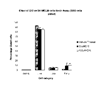
Figure 1: Sensitivity of SK-MEL-28 to 24 hours of Q10 treatment measured by
the amount of early and late apoptotic cells.
Figure 2: Sensitivity of SKBR3 to 24 hours of Q10 treatment measured by the
amount of early and late apoptotic cells.
Figure 3: Sensitivity of PaCa2 to 24 hours of Q10 treatment measured by the
amount of early and late apoptotic cells.
Figure 4: Sensitivity of PC-3 to 24 hours of Q10 treatment measured by the
amount of early and late apoptotic cells.
Figure 5: Sensitivity of HepG2 to 24 hours of Q10 treatment measured by the
amount of early and late apoptotic cells.
Figure 6: Sensitivity of MCF-7 to 24 hours of Q10 treatment measured by the
amount of early and late apoptotic cells.
Figure 7: Measurement of apoptotic cells upon 24 hour treatment with Q10, as
measured by Apostrand ELISA method.
Figure 8: Example gel analysis of 2-D gel electrophoresis. Spots excised for
identification are marked.
Figure 9: Network of interaction between proteins identified by 2-D gel
electrophoresis as being modulated by Q10 in SK-MEL-28 cells.
Figure 10: The pentose phosphate pathway adapted from Verhoeven et al. (Am.
J. Hum. Genet. 2001 68(5):1086-1092).
Figure 11: 2-D gel of the mitochondrial enriched material of SK-MEL-28 cells.
Spots excised and identified by mass spectrometry characterization are marked.
Figure 12: Comparative plot of the relative amounts of Q10 present in SK-MEL-
28 mitochondria following the exogenous addition of 100 M Q10 into the
culture
medium.
Figures 13A and 13B: Apoptosis pathway mapping known processes.
Figure 14: Western blot analysis of Bcl-xl.
Figure 15: Western blot analysis of SK-MEL-28 sample set proved with a
Vimentin antibody.
19
Date Recue/Date Received 2020-08-17
CA 02763347 2011-11-10
WO 2010/13250720 PCT/US2010/034453
Figure 16: Western blot analysis of cell lysis from a number of cell lines,
evaluated with five antibodies targeting oxidative phosphorylation complexes
(MitoSciences #MS601).
Figure 17: Western blot comparison of Fl-alpha levels.
Figure 18: Western blot comparison of Q10 response with C-III-Core 2.
Figure 19: Western blot comparison of Q10 response with C-II-30.
Figure 20: Western blot comparison of Q10 response with C-IV-COX II.
Figure 21: Western blot comparison of Q10 response with C-I-20 (ND6).
Figure 22: Western blot analysis of a variety of cell types against five
mitochondrial protein.
Figure 23: Western blot comparison of Q10 response with Complex V protein
Figure 24: Western blot comparison of Q10 response with C-III-Core 1.
Figure 25: Western blot comparison of Q10 response with Porin (VDAC1).
Figure 26: Western blot comparison of Q10 response with Cyclophilin D.
Figure 27: Western blot comparison of Q10 response with Cytochrome C.
Figure 28: Theoretical model of Q10 (spheres) inserted into the lipid binding
channel of HNF4a1pha (1M7W.pdb) in the Helix 10 open conformation.
Figure 29: Graph depicting the epidermal CoQ10 concentration in a male pig
after treatment with a composition of the present disclosure having a
permeation
enhancer.
Figure 30: Graph depicting the epidermal CoQ10 concentration in a female pig
after treatment with a control composition.
Figure 31: Photographic depiction of a pre-treated target legion 1.
Figure 32: Photographic depiction of a post-treated target legion 1.
Figure 33: Photographic depiction of a pre-treated target legion 2.
Figure 34: Photographic depiction of a post-treated target legion 2.
Figure 35: Photographic depiction of a pre-treated target legion 3.
Figure 36: Photographic depiction of a post-treated target legion 3.
- 20
MEl 9938730v.1
CA 02763347 2011-11-10
WO 2010/13250720 PCT/US2010/034453
Figure 37: OCR in HDFa cells in various glucose conditions in normoxic and
hypoxic conditions.
Figure 38: OCR in HASMC cells in various glucose conditions in normoxic and
hypoxic conditions.
Figure 39: OCR values in MCF-7 breast cancer cells in the absence and presence
of CoQ10 and stressors.
Figure 40: OCR values in PaCa-2 pancreatic cancer cells in the absence and
presence of CoQ10 and stressors.
Detailed Description of the Invention:
Definitions
As used herein, each of the following terms has the meaning associated with it
in
this section.
The articles "a" and "an" are used herein to refer to one or to more than one
(i.e.
to at least one) of the grammatical object of the article. By way of example,
"an
element" means one element or more than one element.
The term "including" is used herein to mean, and is used interchangeably with,
the phrase "including but not limited to".
The term "or" is used herein to mean, and is used interchangeably with, the
term
"and/or," unless context clearly indicates otherwise.
The term "such as" is used herein to mean, and is used interchangeably, with
the
phrase "such as but not limited to".
A "patient" or "subject" to be treated by the method of the invention can mean
either a human or non-human animal, preferably a mammal. It should be noted
that
clinical observations described herein were made with human subjects and, in
at least
some embodiments, the subjects are human.
"Therapeutically effective amount" means the amount of a compound that, when
administered to a patient for treating a disease, is sufficient to effect such
treatment for
the disease. When administered for preventing a disease, the amount is
sufficient to
avoid or delay onset of the disease. The "therapeutically effective amount"
will vary
- 21
MEl 9938730v.1
CA 02763347 2011-11-10
WO 2010/13250720 PCT/US2010/034453
depending on the compound, the disease and its severity and the age, weight,
etc., of the
patient to be treated.
"Preventing" or "prevention" refers to a reduction in risk of acquiring a
disease
or disorder (i.e., causing at least one of the clinical symptoms of the
disease not to
develop in a patient that may be exposed to or predisposed to the disease but
does not
yet experience or display symptoms of the disease).
The term "prophylactic" or "therapeutic" treatment refers to administration to
the
subject of one or more of the subject compositions. If it is administered
prior to clinical
manifestation of the unwanted condition (e.g., disease or other unwanted state
of the
host animal) then the treatment is prophylactic, i.e., it protects the host
against
developing the unwanted condition, whereas if administered after manifestation
of the
unwanted condition, the treatment is therapeutic (i.e., it is intended to
diminish.
ameliorate or maintain the existing unwanted condition or side effects
therefrom).
The ten-n "therapeutic effect" refers to a local or systemic effect in
animals,
particularly mammals, and more particularly humans caused by a
pharmacologically
active substance. The term thus means any substance intended for use in the
diagnosis,
cure, mitigation, treatment or prevention of disease or in the enhancement of
desirable
physical or mental development and conditions in an animal or human. The
phrase
"therapeutically-effective amount" means that amount of such a substance that
produces
some desired local or systemic effect at a reasonable benefit/risk ratio
applicable to any
treatment. In certain embodiments, a therapeutically-effective amount of a
compound
will depend on its therapeutic index, solubility, and the like. For example,
certain
compounds discovered by the methods of the present invention may be
administered in a
sufficient amount to produce a reasonable benefit/risk ratio applicable to
such treatment.
By "patient" is meant any animal (e.g., a human), including horses, dogs,
cats,
pigs, goats, rabbits, hamsters, monkeys, guinea pigs, rats, mice, lizards,
snakes, sheep,
cattle, fish, and birds
"Metabolic pathway" refers to a sequence of enzyme-mediated reactions that
transform one compound to another and provide intermediates and energy for
cellular
functions. The metabolic pathway can be linear or cyclic.
- 22
MEl 9938730v.1
CA 02763347 2011-11-10
WO 2010/13250720 PCT/US2010/034453
"Metabolic state" refers to the molecular content of a particular cellular,
multicellular or tissue environment at a even point in time as measured by
various
chemical and biological indicators as they relate to a state of health or
disease.
The term "microarray" refers to an array of distinct polynucleotides,
oligonucleotides, polypeptides (e.g., antibodies) or peptides synthesized on a
substrate,
such as paper, nylon or other type of membrane, filter, chip, glass slide, or
any other
suitable solid support.
The terms "disorders" and "diseases" are used inclusively and refer to any
deviation from the normal structure or function of any part, organ or system
of the body
(or any combination thereof). A specific disease is manifested by
characteristic
symptoms and signs, including biological, chemical and physical changes, and
is often
associated with a variety of other factors including, but not limited to,
demographic,
environmental, employment, genetic and medically historical factors. Certain
characteristic signs, symptoms, and related factors can be quantitated through
a variety
of methods to yield important diagnostic information.
The term "expression" is used herein to mean the process by which a
polypeptide
is produced from DNA. The process involves the transcription of the gene into
mRNA
and the translation of this mRNA into a polypeptide. Depending on the context
in which
used, "expression" may refer to the production of RNA, protein or both.
The terms "level of expression of a gene" or "gene expression level" refer to
the
level of mRNA, as well as pre-mRNA nascent transcript(s), transcript
processing
intermediates, mature mRNA(s) and degradation products, or the level of
protein,
encoded by the gene in the cell.
The term "modulation" refers to upregulation (i.e., activation or
stimulation),
downregulation (i.e., inhibition or suppression) of a response, or the two in
combination
or apart. A "modulator" is a compound or molecule that modulates, and may be,
e.g., an
agonist, antagonist, activator, stimulator, suppressor, or inhibitor.
The term "Trolamine," as used herein, refers to Trolamine NF, Triethanolamine,
TEAlan , TEAlan 99%, Triethanolamine, 99%, Triethanolamine, NF or
Triethanolamine, 99%, NF. These terms may be used interchangeably herein.
The term "intermediate of the coenzyme biosynthesis pathway" as used herein,
characterizes those compounds that are formed between the chemical/biological
- 23
MEl 9938730v.1
CA 02763347 2016-02-08
conversion of tyrosine and Acetyl-CoA to ubiquinone. Intermediates of the
coenzyme
biosynthesis pathway include 3-hexapreny1-4-hydroxybenzoate, 3-hexapreny1-4,5-
dihydroxybenzoate, 3-hexapreny1-4-hydroxy-5-methoxybenzoate, 2-hexapreny1-6-
methoxy-1,4-benzoquinone, 2-hexapreny1-3-methyl-6-methoxy-1,4-benzoquinone, 2-
hexapreny1-3-methy1-5-hydroxy-6-methoxy-1,4-benzoquinone, 3-Octapreny1-4-
hydroxybenzoate, 2-octaprenylphenol, 2-octapreny1-6-metholxyphenol, 2-
octapreny1-3-
methy1-6-methoxy-1,4-benzoquinone, 2-octapreny1-3-methy1-5-hydroxy-6-methoxy-
1,4-
benzoquinone, 2-decapreny1-3-methyl-5-hydroxy-6-methoxy-1,4-benzoquinone, 2-
decapreny1-3-methy1-6-methoxy-1.4-benzoquinone, 2-decapreny1-6-methoxy-1,4-
benzoquinone, 2-decapreny1-6-methoxyphenol, 3-decapreny1-4-hydroxy-5-
methoxybenzoate, 3-decapreny1-4,5-dihydroxybenzoate, 3-decapreny1-4-
hydroxybenzoate, 4-hydroxy phenylpyruvate, 4-hydroxyphenyllactate, 4-hydroxy-
benzoate, 4-hydroxycinnamate and hexaprenydiphosphate.
Reference will now be made in detail to preferred embodiments of the
invention.
While the invention will be described in conjunction with the preferred
embodiments, it
will be understood that it is not intended to limit the invention to those
preferred
embodiments. To the contrary, it is intended to cover alternatives,
modifications, and
equivalents.
IL Environmental influencers
The present invention provides methods of treating oncological disorders by
administration of an Environmental influencer. "Environmental influencers"
(Env-
influencers) are molecules that influence or modulate the disease environment
of a
human in a beneficial manner allowing the human's disease environment to
shift,
reestablish back to or maintain a normal or healthy environment leading to a
normal
state. Env-influencers include both Multidimensional Intracellular Molecules
(MEVIs)
and Epimetabolic shifters (Epi-shifters) as defined below.
As used herein, "oncological disorder" refers to all types of cancer or
neoplasm
or malignant tumors found in humans, including, but not limited to: leukemias,
lymphomas, melanomas, carcinomas and sarcomas. As used herein, the terms or
language "oncological disorder", "cancer," "neoplasm," and "tumor," are used
-24
CA 02763347 2011-11-10
WO 2010/13250720 PCT/US2010/034453
interchangeably and in either the singular or plural form, refer to cells that
have
undergone a malignant transformation that makes them pathological to the host
organism. In some embodiments the oncological disorder is a Coenzyme Q10
responsive
state.
In some embodiments, the oncological disorder or cancer is characterized by a
lack of apoptosis. In other embodiments, the oncological disorder or cancer is
characterized by increased angiogenesis. In other embodiments, the oncological
disorder or cancer is characterized by extracellular matrix (ECM) degradation.
In yet
other embodiments, the oncological disorder or cancer is characterized by loss
of cell
cycle control. In still other embodiments, the oncological disorder or cancer
is
characterized by a shift in metabolic governance from mitochondrial oxidative
phosphorylation to increased utilization and/or dependency on lactate and
glycolytic
flux. In further embodiments, the oncological disorder or cancer is
characterized by
adapted immunomodulatory mechanisms that have evaded immunosurveilance. In one
embodiment, the oncological disorder or cancer is characterized by at least
two of the
above features, e.g., increased angiogenesis and ECM degradation. In one
embodiment,
the oncological disorder or cancer is characterized by at least three of the
above features.
In one embodiment, the oncological disorder or cancer is characterized by at
least four
of the above features. In one embodiment, the oncological disorder or cancer
is
characterized by at least five of the above features. In one embodiment, the
oncological
disorder or cancer is characterized by all six of the above features.
Accordingly, in some embodiments, the compounds of the present invention
function by restoring the capacity for apoptosis or inducing apoptosis. In
other
embodiments, the compounds of the present invention function by reducing,
decreasing
or inhibiting angiogenesis. In still other embodiments, the compounds of the
present
invention function by restoring re-establishing extracellular matrix. In other
embodiments, the compounds of the present invention function by restoring cell
cycle
control. In still other embodiments, the compounds of the present invention
function by
shifting metabolic governance back from glycolysis to mitochondrial oxidative
phosphorylation. In further embodiments, the compounds of the present
invention
function by restoring immunosurveilance or restoring the body's ability to
recognize the
cancer cell as foreign.
- 25
MEl 9938730v.1
CA 02763347 2011-11-10
WO 2010/13250720 PCT/US2010/034453
Without wishing to be bound by any particular theory, it is believed that
there is
typically a coordinated cascade of events that aggregate to develop into
cancer. That is,
in some embodiments, cancer is not singularly dependent on a 1 gene-1 protein-
root
causality. In some embodiments, cancer is a physiologic disease state that
manifests into
tissue changes and alterations that become tumors, altered tissue states,
e.g., energetics,
compromised extracellular matrix integrity that allows for metastatic
potential, lack of
immunosurveilance and/or altered state of angiogenesis.
Primary cancer cells (that is, cells obtained from near the site of malignant
transformation) can be readily distinguished from non-cancerous cells by well-
established techniques, particularly histological examination. The definition
of a cancer
cell, as used herein, includes not only a primary cancer cell, but also cancer
stem cells,
as well as cancer progenitor cells or any cell derived from a cancer cell
ancestor. This
includes metastasized cancer cells, and in vitro cultures and cell lines
derived from
cancer cells. When referring to a type of cancer that normally manifests as a
solid
tumor, a "clinically detectable" tumor is one that is detectable on the basis
of tumor
mass; e.g., by procedures such as CAT scan, MR imaging, X-ray, ultrasound or
palpation, and/or which is detectable because of the expression of one or more
cancer-
specific antigens in a sample obtainable from a patient.
1. Multidimensional Intracellular Molecule (MIM)
The term "Multidimensional Intracellular Molecule (MIM)", is an isolated
version or synthetically produced version of an endogenous molecule that is
naturally
produced by the body and/or is present in at least one cell of a human. A MEV'
is
characterized by one or more, two or more, three or more, or all of the
following
functions. MIMs are capable of entering a cell, and the entry into the cell
includes
complete or partial entry into the cell, as long as the biologically active
portion of the
molecule wholly enters the cell. MIMs are capable of inducing a signal
transduction
and/or gene expression mechanism within a cell. MIMs are multidimensional in
that the
molecules have both a therapeutic and a carrier, e.g., drug delivery, effect.
MIMs also
are multidimensional in that the molecules act one way in a disease state and
a different
way in a normal state. For example, in the case of CoQ-10, administration of
CoQ-10 to
a melanoma cell in the presence of VEGF leads to a decreased level of Bc12
which, in
turn, leads to a decreased oncogenic potential for the melanoma cell. In
contrast, in a
- 26
MEl 9938730v.1
CA 02763347 2011-11-10
WO 2010/13250720 PCT/US2010/034453
normal fibroblast, co-administration of CoQ-10 and VEFG has no effect on the
levels of
Bc12. Preferably, MIMs selectively act in cells of a disease state, and have
substantially
no effect in (matching) cells of a normal state. Preferably, MIMs selectively
renders
cells of a disease state closer in phenotype, metabolic state, genotype, mRNA
/ protein
expression level, etc. to (matching) cells of a normal state.
In one embodiment, a MIM is also an epi-shifter. In another embodiment, a
MIM is not an epi-shifter. The skilled artisan will appreciate that a MIM of
the
invention is also intended to encompass a mixture of two or more endogenous
molecules, wherein the mixture is characterized by one or more of the
foregoing
functions. The endogenous molecules in the mixture are present at a ratio such
that the
mixture functions as a MIM.
MIMs can be lipid based or non-lipid based molecules. Examples of MIMs
include, but are not limited to, CoQ10, acetyl Co-A, palmityl Co-A, L-
carnitine, amino
acids such as, for example, tyrosine, phenylalanine, and cysteine. In one
embodiment,
the MIM is a small molecule. In one embodiment of the invention, the MIM is
not
CoQ10. MIMs can be routinely identified by one of skill in the art using any
of the
assays described in detail herein.
In some embodiments. MIMs include compounds in the Vitamin B family, or
nucleosides, mononucleotides or dinucleotides that comprise a compound in the
Vitamin
B family. Compounds in the vitamin B family include, for example, thiamine
(vitamin
B1), niacin (also known as nicotinic acid or Vitamin B3), or pyridoxine
(vitamin B6) as
well as provitamins such as panthenol (provitamin B5). In some embodiments.
the MIM
is selected from thiamine, niacin and pyridoxine. Nucleosides, mononucleotides
or
dinucleotides that comprise a compound in the vitamin B family include, for
example.
nucleosides, mononucleotides or dinucleotides which include an adenine or a
niacin
(nicotinic acid) molecule. In some embodiments, the MIM is selected from
adenosine,
adenosine diphosphate (ADP), flavin adenosine dinucleotide (FAD, which
comprises
parts of vitamin B2 and ADP) and nicotinic acid dinucleotide.
In other embodiments, the MIMs include amino acids. Examples of amino acids
include, for example, tyrosine (e.g., L-tyrosine), cysteine, phenylalanine
(e.g., L-
phenylalanine) and alanine. In some embodiments, the amino acid is
phenylalanine or
alanine. In some embodiments, the MIMs include amino acid derivatives such as
4-
hydroxyphenylpyruvate or acetylglycine.
- 27
MEl 9938730v.1
CA 02763347 2011-11-10
WO 2010/13250720 PCT/US2010/034453
In some embodiment, the MINI is a glucose analog, e.g., a glucose molecule
wherein one -OH or -CH2OH substituent has been replaced with a -COOH, a -000-
or
an -NH2 substituent. Examples of glucose analogs include glucosamine,
glucoronic
acid, glucoronide and glucoronate.
In some embodiments. the MINI is selected from compounds of formula (I):
0 R3
XO CH
R1 R2
R4 n
(I)
wherein
n is an integer of 0 or 1;
R1, R2, R3 and R4, when present, are each independently selected from
hydrogen and hydroxyl or RI and R2 are taken together with the carbon on which
they
are attached to form a carbonyl (C=0) group;
W is -COOH or -N(CH3)3+; and
X is hydrogen, a negative charge or a alkali metal cation, such as Na+ or.
It is to be understood that when n is 0, the CHR3 group is bonded to the W
substituent.
In some embodiments. W is -N(CH3)3+. In some embodiments, the MIM is a
carnitine, such as L-carnitine.
In some embodiments. the MINI is a dicarboxylic acid. In some embodiments,
W is -COOH. In some embodiments. R3 is hydrogen. In some embodiments, n is 0.
In
some embodiments. R1 and R2 are each independently hydrogen. In some
embodiments, W is -COOH, R3 is hydrogen, n is 0 and RI and R2 are each
independently hydrogen. In some embodiments, n is 1. In some embodiments R1
and
R2 are taken together with the carbon on which they are attached to form a
carbonyl
(C=0) group. In some embodiments, R4 is hydrogen. In some embodiments, R4 is
hydroxyl. In some embodiments, W is -COOH, R3 is hydrogen, n is 1 and RI and
R2
are taken together with the carbon on which they are attached to form a
carbonyl (C=0)
group.
- 28
MEl 9938730v.1
CA 02763347 2011-11-10
WO 2010/13250720 PCT/US2010/034453
In some embodiments. the MIM is an intermediate of the Krebs Cycle, the excess
of which drives the Krebs Cycle towards productive oxidative phosphorylation.
Exemplary Krebs Cycle intermediates that are MIMs include succinic acid or
succinate,
malic acid or malate, and a-ketoglutaric acid or a-ketoglutarate.
In some embodiments. the MINI is a building block of CoQ10, which has the
following structure:
0
H3C0
H3C0 CH3
0
Thus, building blocks of CoQ10 include, but are not limited to, phenylalanine,
tyrosine, 4-hydroxyphenylpyruvate, phenylacetate, 3-methoxy-4-
hydroxymandelate,
10 vanillic acid, 4-hydroxybenzoate, mevalonic acid, famesyl, 2,3-dimethoxy-5-
methyl-p-
benzoquinone, as well as the corresponding acids or ions thereof. In some
embodiments, the MINI is selected from phenylalanine, tyrosine, 4-
hydroxyphenylpyruvate, phenylacetate and 4-hydroxybenzoate.
2. Epimetabolic Shifters (Epi-shifters)
As used herein, an "epimetabolic shifter" (epi-shifter) is a molecule
(endogenous
or exogenous) that modulates the metabolic shift from a healthy (or normal)
state to a
disease state and vice versa, thereby maintaining or reestablishing cellular,
tissue, organ,
system and/or host health in a human. Epi-shifters are capable of effectuating
normalization in a tissue microenvironment. For example, an epi- shifter
includes any
molecule which is capable, when added to or depleted from a cell, of affecting
the
microenvironment (e.g., the metabolic state) of a cell. The skilled artisan
will appreciate
that an epi-shifter of the invention is also intended to encompass a mixture
of two or
more molecules, wherein the mixture is characterized by one or more of the
foregoing
functions. The molecules in the mixture are present at a ratio such that the
mixture
functions as an epi-shifter. Examples of epi-shifters include, but are not
limited to,
CoQ-10; vitamin D3; ECM components such as fibronectin; immunomodulators, such
as
- 29
MEl 9938730v.1
CA 02763347 2011-11-10
WO 2010/13250720 PCT/US2010/034453
TNFa or any of the interleukins, e.g., IL-5, IL-12, IL-23; angiogenic factors;
and
apoptotic factors.
In some embodiments. the epi-shifter is an enzyme, such as an enzyme that
either
directly participates in catalyzing one or more reactions in the Krebs Cycle,
or produces
a Krebs Cycle intermediate, the excess of which drive the Krebs Cycle. In some
embodiments, the enzyme is an enzyme of the non-oxidative phase of the pentose
phosphate pathway, such as transaldolase, or transketolase. In other
embodiments, the
enzyme is a component enzyme or enzyme complex that facilitates the Krebs
Cycle,
such as a synthase or a ligase. Exemplary enzymes include succinyl CoA
synthase
(Krebs Cycle enzyme) or pyruvate carboxylase (a ligase that catalyzes the
reversible
carboxylation of pyruvate to form oxaloacetate (OAA), a Krebs Cycle
intermediate).
In some embodiments, the epi-shifter is a building block of CoQ10. Building
blocks of CoQ10 include, but are not limited to, phenylalanine, tyrosine, 4-
hydroxyphenylpyruvate, phenylacetate, 3-methoxy-4-hydroxymandelate, vanillic
acid,
4-hydroxybenzoate, mevalonic acid, farnesyl, 2,3-dimethoxy-5-methyl-p-
benzoquinone,
as well as the corresponding acids or ions thereof. In some embodiments, the
epi-shifter
is selected from phenylalanine, tyrosine, 4-hydroxyphenylpyruvate,
phenylacetate and 4-
hydroxybenzoate.
In some embodiments, the epi-shifter is a compound in the Vitamin B family.
Compounds in the vitamin B family include, for example, riboflavin (vitamin
B2), or
analogs thereof. Epi-shifters also include any analogs or pro-drugs that may
be
metabolized in vivo to any of the endogenous MIMs, such as those described
herein.
In one embodiment, the epi-shifter also is a MIM. In one embodiment, the epi-
shifter is not CoQ10. Epi-shifters can be routinely identified by one of skill
in the art
using any of the assays described in detail herein.
In some embodiments, the compounds of the present invention, e.g., the MIMs or
epi-shifters described herein, may be used to treat a Coenzyme Q10 responsive
state in a
subject in need thereof. The language "Coenzyme Q10 responsive state," or
"CoQ10
responsive state," includes diseases, disorders, states and/or conditions
which can be
treated, prevented, or otherwise ameliorated by the administration of Coenzyme
Q10.
Without wishing to be bound by any particular theory, and as described further
herein, it
is believed that CoQ10 functions, at least partially, by inducing a metabolic
shift to the
cell microenvironment, such as a shift towards the type and/or level of
oxidative
- 30
MEl 9938730v.1
CA 02763347 2011-11-10
WO 2010/13250720 PCT/US2010/034453
phosphorylation in normal state cells. Accordingly, in some embodiments, CoQ10
responsive states are states that arise from an altered metabolism of cell
microenvironment. Coenzyme Q10 responsive states include, for example,
oncological
disorders, which, for example, may be biased towards glycolysis and lactate
biosynthesis. In some embodiments, CoQ10 responsive oncological disorders
include
liver cancer, pancreatic cancer, breast cancer, prostate cancer, liver cancer,
or bone
cancer, squamous cell carcinomas, basal cell carcinomas, melanomas, and
actinic
keratosis, among others. Coenzyme Q10 responsive states further include other
oncological disorders as described herein.
Coenzyme Q10 responsive states also include, for example, metabolic disorders
such as obesity, diabetes, pre-diabetes, Metabolic Syndrome, satiety, and
endocrine
abnormalities. Coenzyme Q10 responsive states further include other metabolic
disorders as described herein.
In some embodiments, the compounds of the present invention, e.g., the MIMs or
epi-shifters described herein, share a common activity with Coenzyme Q10. As
used
herein, the phrase "share a common activity with Coenzyme Q10" refers to the
ability of
a compound to exhibit at least a portion of the same or similar activity as
Coenzyme
Q10. In some embodiments, the compounds of the present invention exhibit 25%
or
more of the activity of Coenzyme Q10. In some embodiments, the compounds of
the
present invention exhibit up to and including about 130% of the activity of
Coenzyme
Q10. In some embodiments, the compounds of the present invention exhibit about
30%,
31%, 32%, 33%, 34%, 35%, 36%, 37%, 38%, 39%, 40%, 41%, 42%, 43%, 44%, 45%,
46%, 47%, 48%, 49%, 50%, 51%, 52%, 53%, 54%, 55%, 56%, 57%, 58%, 59%, 60%,
61%, 62%, 63%, 64%, 65%, 66%, 67%, 68%, 69%, 70%, 71%, 72%, 73%, 74%, 75%,
76%, 77%, 78%, 79%, 80%, 81%, 82%, 83%, 84%, 85%, 86%, 87%, 88%, 89%, 90%,
91%, 92%, 93%, 94%, 95%, 96%, 97%, 98%, 99%, 100%, 101%, 102%, 103%, 104%,
105%, 106%, 107%, 108%, 109%, 110%, 111%, 112%, 113%, 114%, 115%, 116%,
117%, 118%, 119%, 120%, 121%, 122%, 123%, 124%, 125%, 126%, 127%, 128%,
129%, or 130% of the activity of Coenzyme Q10. It is to be understood that
each of the
values listed in this paragraph may be modified by the term "about."
Additionally, it is
to be understood that any range which is defined by any two values listed in
this
paragraph is meant to be encompassed by the present invention. For example, in
some
embodiments, the compounds of the present invention exhibit between about 50%
and
- 31
ME1 9938730v.1
CA 02763347 2011-11-10
WO 2010/13250720 PCT/US2010/034453
about 100% of the activity of Coenzyme Q10. In some embodiments, the activity
shared
by Coenzyme Q10 and the compounds of the present invention is the ability to
induce a
shift in cellular metabolism. In certain embodiments, the activity shared by
of CoQ10
and the compounds of the present invention is measured by OCR (Oxigen
Consumption
Rate) and/or ECAR (ExtraCellular Acidification Rate).
HI. Assays useful for identifying MIMs/Epi-shifters
Techniques and methods of the present invention employed to separate and
identify molecules and compounds of interest include but are not limited to:
liquid
chromatography (LC), high-pressure liquid chromatography (HPLC), mass
spectroscopy
(MS), gas chromatography (GC), liquid chromatography/mass spectroscopy (LC-
MS),
gas chromatography/mass spectroscopy (GC-MS). nuclear magnetic resonance
(NMR),
magnetic resonance imaging (MRI), Fourier Transform InfraRed (FT-IR), and
inductively coupled plasma mass spectrometry (ICP-MS). It is further
understood that
mass spectrometry techniques include, but are not limited to, the use of
magnetic-sector
and double focusing instruments, transmission quadrapole instruments,
quadrupole ion-
trap instruments, time-of-flight instruments (TOF), Fourier transform ion
cyclotron
resonance instruments (FT-MS) and matrix-assisted laser desorption/ionization
time-of-
flight mass spectrometry (MALDI-TOF MS).
Quantification of Bioenergetic molecule levels:
Environmental influencers (e.g., MIMs or Epi-shifters) may be identified by
changes in cellular bioenergetic molecule levels (e.g., ATP, pyruvate, ADP,
NADH,
NAD, NADPH, NADP, acetylCoA, FADH2) of cells to which a candidate epi-shifter
has been applied. Exemplary assays of bioenergetic molecule levels use
colorometric,
fluorescence, and/or bioluminescent-based methods. Examples of such assays are
provided below.
Levels of ATP within cells can be measured with a number of assays and
systems known in the art. For example, in one system, cytoplasmic ATP released
from
lysed cells reacts with luciferin and the enzyme luciferase to produce light.
This
bioluminescence is measured by a bioluminometer and the intracellular ATP
concentration of the lysed cells can be calculated (EnzyLightTm ATP Assay Kit
(EATP-
- 32
MEl 9938730v.1
CA 02763347 2011-11-10
WO 2010/13250720 PCT/US2010/034453
100), BioAssay Systems, Hayward, CA). In another system, for example, both ATP
and
its dephosphorylated form, ADP, are calculated via bioluminescence; after ATP
levels
are calculated, ADP is transformed into ATP and then detected and calculated
using the
same luciferase system (ApoSENSORTm ADP/ATP Ratio Assay Kit, Bio Vision Inc.,
Mountain View, CA).
Pyruvate is an important intermediate in cellular metabolic pathways. Pyruvate
may be converted into carbohydrate via gluconeogenesis, converted into fatty
acid or
metabolized via acetyl CoA, or converted into alanine or ethanol, depending
upon the
metabolic state of a cell. Thus detection of pyruvate levels provides a
measure of the
metabolic activity and state of a cell sample. One assay to detect pyruvate,
for example,
uses both a colorimetric and fluorimetric to detect pyruvate concentrations
within
different ranges (EnzyChromTmPyruvate Assay Kit (Cat# EPYR-100), BioAssay
Systems, Hayward, CA).
Environmental influencers (e.g., MIMs or Epi-shifters) may influence the
process of oxidative phosphorylation carried out by mitochondria in cells,
which are
involved in the generation and maintenance of bioenergetic molecules in cells.
In
addition to assays that detect changes in cellular energetics in cell cultures
and samples
directly (described below), assays exist that detect and quantify the effects
of
compounds on discrete enzymes and complexes of mitochondria in cells. For
example,
the MT-OXC MitoTox'm Complete OXPHOS Activity Assay (MitoSciences Inc.,
Eugene. OR) can detect and quantify the effects of compounds applied directly
to
complexes Ito V extracted from mitochondria. Assays for the detection and
quantification of effects on individual mitochondrial complexes such as NADH
dehydrogenase (Complex I), cytochrome c oxidase (Complex W) and ATP synthase
(Complex V) are also available (MitoSciences Inc.. Eugene, OR).
Measurement of Cellular Energetics:
Environmental influencers (e.g., MIMs or Epi-shifters) may also be identified
by
changes in cellular energetics. One example of the measurement of cellular
energetics
are the real-time measures of the consumption of molecular oxygen and/or the
change in
pH of the media of a cell culture. For example, the ability of a potential epi-
shifter to
modulate the metabolic state of a cell may be analyzed using, for example, the
XF24
- 33
MEl 9938730v.1
CA 02763347 2011-11-10
WO 2010/13250720 PCT/US2010/034453
Analyzer (Seahorse, Inc.). This technology allows for real time detection of
oxygen and
pH changes in a monolayer of cells in order to evaluate the bioener2etics of a
cell
microenvironment. The XF24 Analyzer measures and compares the rates of oxygen
consumption (OCR), which is a measure of aerobic metabolism, and extracellular
acidification (ECAR), which is a measure of glycolysis, both key indicators of
cellular
energetics.
Measurement of Oxidative Phosphorylation and Mitochondrial Function
Oxidative Phosphorylation is a process by which ATP is generated via the
oxidation of nutrient compounds, carried out in eukaryotes via protein
complexes
embedded in the membranes of mitochondria. As the primary source of ATP in the
cells
of most organisms, changes in oxidative phosphorylation activity can strongly
alter
metabolism and energy balance within a cell. In some embodiments of the
invention,
environmental influencers (e.g., MIMs or Epi-shifters) may be detected and/or
identified by their effects on oxidative phosphorylation. In some embodiments,
environmental influencers (e.g., MIMs or Epi-shifters) may be detected and/or
identified by their effects on specific aspects of oxidative phosphorylation,
including,
but not limited to, the electron transport chain and ATP synthesis.
The membrane-embedded protein complexes of the mitochondria that carry out
processes involved in oxidative phosphorylation perform specific tasks and are
numbered I, II, III and IV. These complexes, along with the trans-inner
membrane ATP
synthase (also known as Complex V), are the key entities involved in the
oxidative
phosphorylation process. In addition to assays that can examine the effects of
environmental influencers (e.g., MIMs or Epi-shifters) on mitochondiial
function in
general and the oxidative phosphorylation process in particular, assays are
available that
can be used to examine the effects of an epi-shifter on an individual complex
separately
from other complexes.
Complex I, also known as NADH-coenzyme Q oxidoreductase or NADH
dehydrogenase, is the first protein in the electron transport chain. In some
embodiments,
the detection and quantification of the effect of an epi-shifter on the
production of NAD+
by Complex I may be performed. For example, the complex can be immunocaptured
from a sample in a 96-well plate; the oxidation of NADH to NAD+ takes place
concurrently with the reduction of a dye molecule which has an increased
absorbance at
- 34
MEl 9938730v.1
CA 02763347 2011-11-10
WO 2010/13250720 PCT/US2010/034453
450 nM (Complex I Enzyme Activity Microplate Assay Kit, MitoSciences Inc.,
Eugene,
OR).
Complex IV, also known as cytochrome c oxidase (COX), is the last protein in
the electron transport chain. In some embodiments, the detection and
quantification of
the effect of an epi- shifter on the oxidation of cytochrome c and the
reduction of oxygen
to water by Complex IV may be performed. For example, COX can be
immunocaptured
in a microwell plate and the oxidation of COX measured with a colorimetric
assay
(Complex IV Enzyme Activity Microplate Assay Kit, MitoSciences Inc., Eugene,
OR).
The final enzyme in the oxidative phosphorylation process is ATP synthase
(Complex V), which uses the proton gradient created by the other complexes to
power
the synthesis of ATP from ADP. In some embodiments, the detection and
quantification
of the effect of an epi-shifter on the activity of ATP synthase may be
performed. For
example, both the activity of ATP synthase and the amount of ATP synthase in a
sample
may be measured for ATP synthase that has been immunocaptured in a microwell
plate
well. The enzyme can also function as an ATPase under certain conditions, thus
in this
assay for ATP synthase activity, the rate at which ATP is reduced to ADP is
measured
by detecting the simultaneous oxidation of NADH to NAD+. The amount of ATP is
calculated using a labeled antibody to ATPase (ATP synthase Duplexing
(Activity +
Quantity) Microplate Assay Kit, MitoSciences Inc., Eugene, OR).Additional
assays for
oxidative phosphorylation include assays that test for effects on the activity
of
Complexes II and III. For example, the MT-OXC MitoToxTm Complete OXPHOS
System (MitoSciences Inc., Eugene, OR) can be used to evaluate effects of a
compound
on Complex II and III as well as Complex I, IV and V, to provide data on the
effects of a
compound on the entire oxidative phosphorylation system.
As noted above, real-time observation of intact cell samples can be made using
probes for changes in oxygen consumption and pH in cell culture media. These
assays
of cell energetics provide a broad overview of mitochondrial function and the
effects of
potential environmental influencers (e.g., MIMs or Epi-shifters) on the
activity of
mitochondria within the cells of the sample.
Environmental influencers (e.g., MIMs or Epi-shifters) may also affect
mitochondrial permeability transition (MPT), a phenomena in which the
mitochondrial
membranes experience an increase in permeability due to the formation of
mitochondrial
permeability transition pores (MPTP). An increase in mitochondrial
permeability can
- 35
MEl 9938730v.1
CA 02763347 2011-11-10
WO 2010/13250720 PCT/US2010/034453
lead to mitochondria' swelling, an inability to conduct oxidative
phosphorylation and
ATP generation and cell death. MPT may be involved with induction of
apoptosis.
(See, for example, Halestrap, A.P., Biochem. Soc. Trans. 34:232-237 (2006) and
Lena,
A. etal. Journal of Translational Med. 7:13-26 (2009), hereby incorporated by
reference
in their entirety.)
In some embodiments, the detection and quantification of the effect of an
environmental influencer (e.g., MIM or epi-shifter) on the formation,
discontinuation
and/or effects of MPT and MPTPs are measured. For example, assays can detect
MPT
through the use of specialized dye molecules (calcein) that are localized
within the inner
membranes of mitochondria and other cytosolic compartments. The application of
another molecule, CoCb, serves to squelch the fluorescence of the calcein dye
in the
cytosol. CoC17cannot access, however, the interior of the mitochondria, thus
the calcein
fluorescence in the mitochondria is not squelched unless MPT has occurred and
CoC12
can access the interior of the mitochondria via MPTPs. Loss of mitochondrial-
specific
fluorescence signals that MPT has occurred. Flow cytometry can be used to
evaluate
cellular and organelle fluorescence (MitoProbeTm Transition Pore Assay Kit,
Molecular
Probes, Eugene, OR). Additional assays utilize a fluorescence microscope for
evaluating experimental results (1mage-iffm LIVE Mitochondrial Transition Pore
Assay
Kit, Molecular Probes, Eugene, OR).
Measurement of Cellular Proliferation and Inflammation
In some embodiments of the invention, environmental influencers (e.g., MIMs or
Epi-shifters) may be identified and evaluated by their effects on the
production or
activity of molecules associated with cellular proliferation and/or
inflammation. These
molecules include, but are not limited to, cytokines, growth factors,
hormones,
components of the extra-cellular matrix, chemokines, neuropeptides,
neurotransmitters,
neurotrophins and other molecules involved in cellular signaling, as well as
intracellular
molecules, such as those involved in signal transduction.
Vascular endothelial growth factor (VEGF) is a growth factor with potent
angiogenic, vasculogenic and mitogenic properties. VEGF stimulates endothelial
permeability and swelling and VEGF activity is implicated in numerous diseases
and
disorders, including rheumatoid arthritis, metastatic cancer, age-related
macular
degeneration and diabetic retinopathy.
- 36
MEl 9938730v.1
CA 02763347 2011-11-10
WO 2010/13250720
PCT/US2010/034453
In some embodiments of the invention, an environmental influencer (e.g., MINI
or Epi-shifter) may be identified and characterized by its effects on the
production of
VEGF. For example, cells maintained in hypoxic conditions or in conditions
mimicking
acidosis will exhibit increased VEGF production. VEGF secreted into media can
be
assayed using an ELISA or other antibody-based assays, using available anti-
VEGF
antibodies (R&D Systems, Minneapolis, MN). In some embodiments of the
invention,
an Epi-shifter may be identified and/or characterized based on its effect(s)
on the
responsiveness of cells to VEGF and/or based on its effect(s) on the
expression or
activity of the VEGF receptor.
Implicated in both healthy immune system function as well as in autoimmune
diseases, tumor necrosis factor (TNF) is a key mediator of inflammation and
immune
system activation. In some embodiments of the invention, an Epi-shifter may be
identified and characterized by its effects on the production or the activity
of TNF. For
example, TNF produced by cultured cells and secreted into media can be
quantified via
ELISA and other antibody-based assays known in the art. Furthermore, in some
embodiments an environmental influencer may be identified and characterized by
its
effect(s) on the expression of receptors for TNF (Human TNF RI Duoset, R&D
Systems,
Minneapolis, MN).
The components of the extracellular matrix (ECM) play roles in both the
structure of cells and tissues and in signaling processes. For example, latent
transforming growth factor beta binding proteins are ECM components that
create a
reservoir of transforming growth factor beta (TGFf3) within the ECM. Matrix-
bound
TGFI3 can be released later during the process of matrix remodeling and can
exert
growth factor effects on nearby cells (Dallas, S. Methods in Mol. Biol.
139:231-243
(2000)).
In some embodiments, an environmental influencer (e.g., MIM or Epi-shifter)
may be identified or characterized by its effect(s) on the creation of ECM by
cultured
cells. Researchers have developed techniques with which the creation of ECM by
cells,
as well as the composition of the ECM, can be studied and quantified. For
example, the
__ synthesis of ECM by cells can be evaluated by embedding the cells in a
hydrogel before
incubation. Biochemical and other analyses are performed on the ECM generated
by the
cells after cell harvest and digestion of the hydrogel (Strehin, I. and
Elisseeff, J. Methods
in Mol. Bio. 522:349-362 (2009)).
- 37
MEl 9938730v.1
CA 02763347 2011-11-10
WO 2010/13250720 PCT/US2010/034453
In some embodiments, the effect of environmental influencer (e.g., MIM or epi-
shifter) on the production, status of or lack of ECM or one of its components
in an
organism may be identified or characterized. Techniques for creating
conditional
knock-out (KO) mice have been developed that allow for the knockout of
particular
ECM genes only in discrete types of cells or at certain stages of development
(Brancaccio, M. et al. Methods in Mol Bio. 522:15-50 (2009)). The effect of
the
application or administration of an epi-shifter or potential epi-shifter on
the activity or
absence of a particular ECM component in a particular tissue or at a
particular stage of
development may thus be evaluated.
Measurement of Plasma Membrane Integrity and Cell Death
Environmental influencers (e.g., MIMs or Epi-shifters) may be identified by
changes in the plasma membrane integrity of a cell sample and/or by changes in
the
number or percentage of cells that undergo apoptosis, necrosis or cellular
changes that
demonstrate an increased or reduced likelihood of cell death.
An assay for lactate dehydrogenase (LDH) can provide a measurement of
cellular status and damage levels. LDH is a stable and relatively abundant
cytoplasmic
enzyme. When plasma membranes lose physical integrity, LDH escapes to the
extracellular compartment. Higher concentrations of LDH correlate with higher
levels
of plasma membrane damage and cell death. Examples of LDH assays include
assays
that use a colorimetric system to detect and quantify levels of LDH in a
sample, wherein
the reduced form of a tetrazolium salt is produced via the activity of the LDH
enzyme
(QuantiChromm4 Lactate Dehydrogenase Kit (DLDH-100), BioAssay Systems,
Hayward, CA; LDH Cytotoxicity Detection Kit, Clontech, Mountain View, CA).
Apoptosis is a process of programmed cell death that may have a variety of
different initiating events. A number of assays can detect changes in the rate
and/or
number of cells that undergo apoptosis. One type of assay that is used to
detect and
quantify apoptosis is a capase assay. Capases are aspartic acid-specific
cysteine
proteases that are activated via proteolytic cleavage during apoptosis.
Examples of
assays that detect activated capases include PhiPhiLux0 (OncoImmunin, Inc.,
Gaithersburg, MD) and Caspase-Glo 3/7 Assay Systems (Promega Corp., Madison,
WI). Additional assays that can detect apoptosis and changes in the percentage
or
- 38
MEl 9938730v.1
CA 02763347 2011-11-10
WO 2010/13250720 PCT/US2010/034453
number of cells undergoing apoptosis in comparative samples include TUNEL/DNA
fragmentation assays. These assays detect the 180 to 200 base pair DNA
fragments
generated by nucleases during the execution phase of apoptosis. Exemplary
TUNEL/DNA fragmentation assays include the In Situ Cell Death Detection Kit
(Roche
.. Applied Science, Indianapolis, IN) and the DeadEndTM Colorimetric and
Fluorometric
TUNEL Systems (Promega Corp.. Madison, WI).
Some apoptosis assays detect and quantify proteins associated with an
apoptotic
and/or a non-apoptotic state. For example. the MultiTox-Fluor Multiplex
Cytotoxicity
Assay (Promega Corp., Madison, WI) uses a single substrate, fluorimetric
system to
.. detect and quantify proteases specific to live and dead cells, thus
providing a ratio of
living cells to cells that have undergone apoptosis in a cell or tissue
sample.
Additional assays available for detecting and quantifying apoptosis include
assays that detect cell permeability (e.g., APOPercentageTM APOPTOSIS Assay,
Biocolor, UK) and assays for Annexin V (e.g., Annexin V-Biotin Apoptosis
Detection
.. Kit, BioVision Inc., Mountain View, CA).
/V. Treatment of Oncological Disorders
The present invention provides methods of treating or preventing an
oncological
disorder in a human, comprising administering CoQ10 to the human in an amount
sufficient to treat or prevent the oncological disorder, thereby treating or
preventing the
oncological disorder.
The present invention also provides CoQ10 compositions and methods of
preparing the same. Preferably, the compositions comprise at least about 1% to
about
25% CoQ10 w/w. CoQ10 can be obtained from Asahi Kasei N&P (Hokkaido, Japan) as
UBIDECARENONE (USP). CoQ10 can also be obtained from Kaneka Q10 as Kaneka
Q10 (USP UBIDECARENONE) in powdered form (Pasadena, Texas, USA). CoQ10
used in the methods exemplified herein have the following characteristics:
residual
solvents meet USP 467 requirement; water content is less than 0.0%, less than
0.05% or
less than 0.2%; residue on ignition is 0.0%, less than 0.05%, or less than
0.2% less than;
heavy metal content is less than 0.002%, or less than 0.001%; purity of
between 98-
100% or 99.9%, or 99.5%. Methods of preparing the compositions are provided in
the
examples section below.
- 39
MEl 9938730v.1
CA 02763347 2011-11-10
WO 2010/13250720 PCT/US2010/034453
As used herein, "oncolo2ical disorder" refers to all types of cancer or
neoplasm
or malignant tumors found in humans, including, but not limited to: leukemias,
lymphomas, melanomas, carcinomas and sarcomas. As used herein, the terms or
language "oncological disorder", "cancer," "neoplasm," and "tumor," are used
interchangeably and in either the singular or plural form, refer to cells that
have
undergone a malignant transformation that makes them pathological to the host
organism. Primary cancer cells (that is, cells obtained from near the site of
malignant
transformation) can be readily distinguished from non-cancerous cells by well-
established techniques, particularly histological examination. The definition
of a cancer
cell, as used herein, includes not only a primary cancer cell, but also cancer
stem cells,
as well as cancer progenitor cells or any cell derived from a cancer cell
ancestor. This
includes metastasized cancer cells, and in vitro cultures and cell lines
derived from
cancer cells. When referring to a type of cancer that normally manifests as a
solid
tumor, a "clinically detectable" tumor is one that is detectable on the basis
of tumor
mass; e.g., by procedures such as CAT scan, MR imaging, X-ray, ultrasound or
palpation, and/or which is detectable because of the expression of one or more
cancer-
specific antigens in a sample obtainable from a patient.
The term "sarcoma" generally refers to a tumor which is made up of a substance
like the embryonic connective tissue and is generally composed of closely
packed cells
embedded in a fibrillar or homogeneous substance. Examples of sarcomas which
can be
treated with an environmental influencer of the invention include, but are not
limited to,
a chondrosarcoma, fibrosarcoma, lymphosarcoma. melanosarcoma, myxosarcoma,
osteosarcoma, Abemethy's sarcoma, adipose sarcoma, liposarcoma, alveolar soft
part
sarcoma. ameloblastic sarcoma, botryoid sarcoma, chloroma sarcoma, chorio
carcinoma.
embryonal sarcoma, Wilms' tumor sarcoma, endometrial sarcoma, stromal sarcoma,
Ewing's sarcoma, fascial sarcoma, fibroblastic sarcoma, giant cell sarcoma,
granulocytic
sarcoma. Hodgkin's sarcoma, idiopathic multiple pigmented hemorrhagic sarcoma,
immunoblastic sarcoma of B cells, lymphoma, immunoblastic sarcoma of T-cells,
Jensen's sarcoma, Kaposi's sarcoma, Kupffer cell sarcoma, angiosarcoma,
leukosarcoma,
malignant mesenchyrnoma sarcoma, parosteal sarcoma, reticulocytic sarcoma,
Rous
sarcoma. serocystic sarcoma, synovial sarcoma, and telangiectaltic sarcoma.
The term "melanoma" is taken to mean a tumor arising from the melanocytic
system of the skin and other organs. Melanomas which can be treated with an
- 40
MEl 9938730v.1
CA 02763347 2011-11-10
WO 2010/13250720 PCT/US2010/034453
environmental influencer of the invention include, but are not limited to, for
example,
acral-lentiginous melanoma, amelanotic melanoma, benign juvenile melanoma,
Cloudman's melanoma, S91 melanoma, Harding-Passey melanoma, juvenile melanoma,
lentigo maligna melanoma, malignant melanoma, nodular melanoma, subungal
melanoma, and superficial spreading melanoma.
The term "carcinoma" refers to a malignant new growth made up of epithelial
cells tending to infiltrate the surrounding tissues and give rise to
metastases. Carcinomas
which can be treated with an environmental influencer of the invention
include, but are
not limited to, for example, acinar carcinoma, acinous carcinoma, adenocystic
carcinoma, adenoid cystic carcinoma, carcinoma adenomatosum, carcinoma of
adrenal
cortex, alveolar carcinoma, alveolar cell carcinoma, basal cell carcinoma,
carcinoma
basocellulare, basaloid carcinoma, basosquamous cell carcinoma,
bronchioalveolar
carcinoma, bronchiolar carcinoma, bronchogenic carcinoma, cerebriform
carcinoma,
cholangiocellular carcinoma, chorionic carcinoma, colloid carcinoma, comedo
carcinoma, corpus carcinoma, cribriform carcinoma, carcinoma en cuirasse,
carcinoma
cutaneum, cylindrical carcinoma, cylindrical cell carcinoma, duct carcinoma,
carcinoma
durum, embryonal carcinoma, encephaloid carcinoma, epiermoid carcinoma,
carcinoma
epitheliale adenoides, exophytic carcinoma, carcinoma ex ulcere, carcinoma
fibrosum,
gelatinifon-n carcinoma, gelatinous carcinoma, giant cell carcinoma, carcinoma
gigantocellulare, glandular carcinoma, granulosa cell carcinoma, hair-matrix
carcinoma,
hematoid carcinoma, hepatocellular carcinoma, Hurthle cell carcinoma, hyaline
carcinoma, hypemephroid carcinoma, infantile embryonal carcinoma, carcinoma in
situ,
intraepidermal carcinoma, intraepithelial carcinoma, Krompecher's carcinoma.
Kulchitzky-cell carcinoma, large-cell carcinoma, lenticular carcinoma,
carcinoma
lenticulare, lipomatous carcinoma, lymphoepithelial carcinoma, carcinoma
medullare,
medullary carcinoma, melanotic carcinoma, carcinoma molle, mucinous carcinoma,
carcinoma muciparum, carcinoma mucocellulare, mucoepidermoid carcinoma,
carcinoma mucosum, mucous carcinoma, carcinoma myxomatodes, nasopharyngeal
carcinoma, oat cell carcinoma, carcinoma ossificans, osteoid carcinoma,
papillary
carcinoma, periportal carcinoma, preinvasive carcinoma, prickle cell
carcinoma,
pultaceous carcinoma, renal cell carcinoma of kidney, reserve cell carcinoma,
carcinoma
sarcomatodes, schneiderian carcinoma, scirrhous carcinoma, carcinoma scroti,
signet-
ring cell carcinoma, carcinoma simplex, small-cell carcinoma, solanoid
carcinoma,
- 41
MEl 9938730v.1
CA 02763347 2011-11-10
WO 2010/13250720 PCT/US2010/034453
spheroidal cell carcinoma, spindle cell carcinoma, carcinoma spongiosum,
squamous
carcinoma, squamous cell carcinoma, string carcinoma, carcinoma
telangiectaticum,
carcinoma telangiectodes, transitional cell carcinoma, carcinoma tuberosum,
tuberous
carcinoma, verrucous carcinoma, and carcinoma villosum.
In general, CoQ10 may be used to prophylactically or therapeutically treat any
neoplasm. In one embodiment, CoQ10 is used to treat solid tumors. In various
embodiments of the invention, CoQ10 is used for treatment, of various types of
skin
cancer (e.g., Squamous cell Carcinoma or Basal Cell Carcinoma), pancreatic
cancer,
breast cancer, prostate cancer, liver cancer, or bone cancer. In one
embodiment, CoQ10
is used for treatment of a skin oncological disorder including, but not
limited to,
squamous cell carcinomas (including SCCIS (in situ) and more aggressive
squamous
cell carcinomas), basal cell carcinomas (including superficial, nodular and
infiltrating
basal cell carcinomas), melanomas, and actinic keratosis. However, treatment
using
CoQ10 is not limited to the foregoing types of cancers. Examples of cancers
amenable
to treatment with CoQ10 include, but are not limited to, cancer of the brain,
head and
neck, prostate, breast, testicular, pancreas, liver, colon, bladder, kidney,
lung, non-small
cell lung, melanoma, mesothelioma, uterus, cervix, ovary, sarcoma, bone,
stomach and
Medulloblastoma.
Additional cancers which can be treated with CoQ10 include, for example,
Hodgkin's Disease, Non-Hodgkin's Lymphoma, multiple myeloma, neuroblastoma,
breast cancer, ovarian cancer, lung cancer, rhabdomyosarcoma, primary
thrombocytosis,
primary macroglobulinemia, small-cell lung tumors, primary brain tumors,
stomach
cancer, colon cancer, malignant pancreatic insulanoma, malignant carcinoid,
urinary
bladder cancer, premalignant skin lesions, testicular cancer, lymphomas,
thyroid cancer,
neuroblastoma, esophageal cancer, genitourinary tract cancer, malignant
hypercalcemia,
cervical cancer, endometrial cancer, adrenal cortical cancer, and prostate
cancer. In one
embodiment, the oncological disorder or cancer which can be treated with CoQ10
is not
melanoma.
The definition of a cancer cell, as used herein, is intended to include a
cancer cell
that produces energy by anaerobic glycolysis (e.g., glycolysis followed by
lactic acid
fermantion in the cytosol ) , aerobic glycolysis (e.g., glycolysis followed by
oxidation of
pyruvate in the mitochondria), or a combination of anaerobic glycolysis and
aerobic
glycolysis. In one embodiment, a cancer cell produces energy predominantly by
- 42
MEl 9938730v.1
CA 02763347 2011-11-10
WO 2010/13250720 PCT/US2010/034453
anaerobic glycolysis (e.g., at least 50%, 60%, 70%, 80%, 90%, 95% or more of
the cell's
energy is produced by anaerobic glycolysis). In one embodiment, a cancer cell
produces
energy predominantly by aerobic glycolysis (e.g., at least 50%, 60%, 70%, 80%,
90%,
95% or more of the cell's energy is produced by anaerobic glycolysis). The
definition of
cancer cells, as used herein, is also intended to include a cancer cell
population or
mixture of cancer cells comprising cells that produce energy by anaerobic
glycolysis and
cells that produce energy by aerobic glycolysis. In one embodiment, a cancer
cell
population comprises predominantly cells that produce energy by anaerobic
glycolysis
(e.g., at least 50%, 60%, 70%, 80%, 90%, 95% or more of the cells in the
population
produce energy by anaerobic glycolysis). In one embodiment, a cancer cell
population
comprises predominantly cells that produce energy by aerobic glycolysis (e.g.,
at least
50%, 60%, 70%, 80%, 90%, 95% or more of the cells in the population).
As used herein, the phrase "anaerobic use of glucose" or "anaerobic
glycolysis"
refers to cellular production of energy by glycolysis followed by lactic acid
fermentation
in the cytosol. For example, many cancer cells produce energy by anaerobic
glycolysis.
As used herein, the phrase "aerobic glycolysis" or "mitochondria' oxidative
phosphorylation" refers to cellular production of energy by glycolysis
followed by
oxidation of pyruvate in mitochondria.
As used herein, the phrase "capable of blocking anaerobic use of glucose and
augmenting mitochondrial oxidative phosphorylation" refers to the ability of
an
environmental influencer (e.g., an epitmetabolic shifter) to induce a shift or
change in
the metabolic state of a cell from anaerobic glycolysis to aerobic glycolysis
or
mitochondrial oxidative phosphorylation.
In some embodiments of the invention, the oncological disorder being treated
is
not a disorder typically treated via topical administration with the
expectation of
systemic delivery of an active agent at therapeutically effective levels. As
used herein,
the phrase "not a disorder typically treated via topical administration"
refers to
oncological disorders that are not typically or routinely treated with a
therapeutic agent
via topical administration but rather are typically treated with a therapeutic
agent via, for
example, intravenous administration. Oncological disorders not typically
treated via
topical administration include, but are not limited to, breast cancer,
prostate cancer, liver
cancer, pancreatic cancer, and bone cancer.
- 43
MEl 9938730v.1
CA 02763347 2011-11-10
WO 2010/13250720 PCT/US2010/034453
The present invention also provides a method for treating or preventing an
aggressive oncological disorder in a human, comprising administering CoQ10 to
the
human at a selected lower dose than the dosage regimen used or selected for
less
aggressive or non-aggressive oncological disorders, thereby treating or
preventing the
aggressive oncological disorder. In a related aspect, the invention provides a
method
for treating or preventing a non-aggressive oncological disorder in a human,
comprising
administering an environmental influencer to the human at a selected higher
dose over
the dosage regimen used or selected for aggressive oncological disorders,
thereby
treating or preventing the non-aggressive oncological disorder.
As used herein, the term "aggressive oncological disorder" refers to an
oncological disorder involving a fast-growing tumor. An aggressive oncological
disorder typically does not respond or responds poorly to therapeutic
treatment.
Examples of an aggressive oncological disorder include, but are not limited
to,
pancreatic carcinoma, hepatocellular carcinoma, Ewing's sarcoma, metastatic
breast
cancer, metastatic melanoma, brain cancer (astrocytoma, glioblastoma),
neuroendocrine
cancer, colon cancer, lung cancer, osteosarcoma, androgen-independent prostate
cancer,
ovarian cancer and non-Hodgkin's Lymphoma.
As used herein, the term "non-aggressive oncological disorder" refers to an
oncological disorder involving a slow-growing tumor. A non-aggressive
oncological
disorder typically responds favorably or moderately to therapeutic treatment.
Examples
of a non-aggressive oncological disorder include, but are not limited to, non-
metastatic
breast cancer, androgen-dependent prostate cancer, small cell lung cancer and
acute
lymphocytic leukemia. In one embodiment, non-aggressive oncological disorders
include any oncological disorder that is not an aggressive oncological
disorder.
In one embodiment, CoQ10 reduces tumor size, inhibits tumor growth and/or
prolongs the survival time of a tumor-bearing subject. Accordingly, this
invention also
relates to a method of treating tumors in a human or other animal by
administering to
such human or animal an effective, non-toxic amount of CoQ10. One skilled in
the art
would be able, by routine experimentation, to determine what an effective, non-
toxic
amount of CoQ10 would be for the purpose of treating malignancies. For
example, a
therapeutically active amount of CoQ10 may vary according to factors such as
the
disease stage (e.g., stage I versus stage IV), age, sex, medical complications
(e.g.,
immunosuppressed conditions or diseases) and weight of the subject, and the
ability of
- 44
MEl 9938730v.1
CA 02763347 2011-11-10
WO 2010/13250720 PCT/US2010/034453
the CoQ10 to elicit a desired response in the subject. The dosage regimen may
be
adjusted to provide the optimum therapeutic response. For example, several
divided
doses may be administered daily, or the dose may be proportionally reduced as
indicated
by the exigencies of the therapeutic situation.
V. Therapeutic Targets for Oncological Disorders
The present invention provides methods for identifying therapeutic targets for
oncological disorders. The invention further provides therapeutic targets
identified by
such methods. The identification of a therapeutic target involves, generally,
the
exogenous application of an Env-influencer or candidate Env-influencer to a
cell or
panel of cell lines, and the subsequent evaluation of changes induced to a
treated cell as
compared to a control, untreated cell. Induced cellular changes which are
monitored
include, but are not limited to. changes to the morphology, physiology or
composition,
e.g., RNA, protein, lipid or metabolite levels, of the cell. Induced cellular
changes as a
result of treatment by a candidate Env-influencer can be monitored by using
any of the
assays described herein. For example, changes in gene expression at the mRNA
level
can be evaluated by real-time PCR arrays, while changes in gene expression at
the
protein level can be monitored by using antibody microarrays and 2-D gel
electrophoresis. Genes identified as being modulated by the candidate Env-
influencer
(e.g., at the mRNA and/or protein level) are then evaluated from a Systems
Biology
perspective using pathway analysis (Ingenuity IPA software) and by a review of
the
known literature. Genes identified as potential therapeutic targets are next
submitted to
confirmatory assays such as Western blot analysis, siRNA knock-down, or
recombinant
protein production and characterization methods. Screening assays can then be
used to
identify modulators of the targets. Modulators of the therapeutic targets are
useful as
novel therapeutic agents for oncological disorders. Modulators of therapeutic
targets can
be routinely identified using screening assays described in detail herein, or
by using
routine methodologies known to the skilled artisan.
Genes identified herein as being modulated (e.g., upmodulated or
downmodulated, at either the mRNA or protein level) by the MIM/Epi-shifter,
CoQ10,
are drug targets of the invention. Drug targets of the invention include, but
are not
limited to, the genes subsequently listed in Tables 1-28 (e.g., 2-4 & 6-28)
herein. Based
- 45
MEl 9938730v.1
CA 02763347 2011-11-10
WO 2010/13250720
PCT/US2010/034453
on the results of experiments described by Applicants herein, the key proteins
modulated
by Q10 are associated with or can be classified into different pathways or
groups of
molecules, including transcription factors, apoptotic response, pentose
phosphate
pathway, biosynthetic pathway, oxidative stress (pro-oxidant), membrane
alterations,
and oxidative phosphorylation metabolism. The key proteins modulated by CoQ10,
based on the results provided herein, are summarized as follows. A key protein
modulated by CoQ10 and which is a transcription factor is HNF4alpha. Key
proteins
that are modulated by CoQ10 and associated with the apoptotic response include
Bcl-xl,
Bcl-xl, Bc1-xS, BNIP-2, Bc1-2, Birc6, Bc1-2-L11 (Bim), XIAP, BRAF, Bax, c-Jun,
Bmf,
PUMA, and cMyc. A key protein that is modulated by CoQ10 and associated with
the
pentose phosphate pathway is transaldolase 1. Key proteins that are modulated
by
CoQ10 and associated with a biosynthetic pathway include COQ1, COQ3, COQ6,
prenyltransferase and 4-hydroxybenzoate. Key proteins that are modulated by
CoQ10
and associated with oxidative stress (pro-oxidant) include Neutrophil
cytosolic factor 2,
nitric oxide synthase 2A and superoxide dismutase 2 (mitochondrial). Key
proteins that
are modulated by CoQl 0 and associated with oxidative phosphorylation
metabolism
include Cytochrome c, complex I, complex II, complex III and complex IV.
Further key
proteins that are directly or indirectly modulated by CoQ10 include Foxo 3a,
DJ-1, IDH-
1, Cpt1C and Cam Kinase II.
Accordingly, in one embodiment of the invention, a drug target may include
HNF4-alpha, Bcl-xl, Bc1-xS, BNIP-2, Bc1-2, Birc6, Bc1-2-L11 (Bim), XIAP, BRAF,
Bax, c-Jun, Bmf, PUMA, cMyc, transaldolase 1, COQ1, COQ3, COQ6,
prenyltransferase, 4-hydrobenzoate, neutrophil cytosolic factor 2, nitric
oxide synthase
2A, superoxide dismutase 2, VDAC, Bax channel, ANT, Cytochrome c, complex 1,
complex II, complex III, complex IV, Foxo 3a, DJ-1. IDH-1, Cpt1C and Cam
Kinase II.
In a preferred embodiment, a drug target may include HNF4A, Transaldolase,
NM23
and BSCv. In one embodiment, the drug target is TNF4A. In one embodiment, the
drug
target is transaldolase. In one embodiment, the drug target is NM23. In one
embodiment, the drug target is BSCv. Screening assays useful for identifying
.. modulators of identified drug targets are described below.
VI. Screening Assays
- 46
MEl 9938730v.1
CA 02763347 2011-11-10
WO 2010/13250720 PCT/US2010/034453
The invention also provides methods (also referred to herein as "screening
assays") for identifying modulators, i.e., candidate or test compounds or
agents (e.g.,
proteins, peptides, peptidomimetics, peptoids, small molecules or other
drugs), which
modulate the expression and/or activity of an identified therapeutic target of
the
invention. Such assays typically comprise a reaction between a therapeutic
target of the
invention and one or more assay components. The other components may be either
the
test compound itself, or a combination of test compounds and a natural binding
partner
of a marker of the invention. Compounds identified via assays such as those
described
herein may be useful, for example, for treating or preventing a oncological
disorder.
The test compounds used in the screening assays of the present invention may
be
obtained from any available source, including systematic libraries of natural
and/or
synthetic compounds. Test compounds may also be obtained by any of the
numerous
approaches in combinatorial library methods known in the art, including:
biological
libraries; peptoid libraries (libraries of molecules having the
functionalities of peptides,
but with a novel, non-peptide backbone which are resistant to enzymatic
degradation but
which nevertheless remain bioactive; see, e.g., Zuckermann et al., 1994, J.
Med. Chem.
37:2678-85); spatially addressable parallel solid phase or solution phase
libraries;
synthetic library methods requiring deconvolution; the 'one-bead one-compound'
library
method; and synthetic library methods using affinity chromatography selection.
The
biological library and peptoid library approaches are limited to peptide
libraries, while
the other four approaches are applicable to peptide, non-peptide oligomer or
small
molecule libraries of compounds (Lam. 1997, Anticancer Drug Des. 12:145).
Examples of methods for the synthesis of molecular libraries can be found in
the
art, for example in: DeWitt et al. (1993) Proc. Natl. Acad. Sci. U.S.A.
90:6909; Erb et
al. (1994) Proc. Natl. Acad. Sci. USA 91:11422; Zuckermann etal. (1994). .I.
Med.
Chem. 37:2678; Cho etal. (1993) Science 261:1303; Carrell etal. (1994) Angew.
Chem.
Int. Ed. Engl. 33:2059; Carell etal. (1994) Angew. Chem. Int. Ed. Engl.
33:2061; and in
Gallop et al. (1994) J. Med. Chem. 37:1233.
Libraries of compounds may be presented in solution (e.g., Houghten, 1992,
Biotechniques 13:412-421), or on beads (Lam, 1991, Nature 354:82-84), chips
(Fodor,
1993, Nature 364:555-556), bacteria and/or spores, (Ladner, USP 5,223,409),
plasmids
(Cull et al, 1992, Proc Nati Acad Sci USA 89:1865-1869) or on phage (Scott and
Smith,
1990, Science 249:386-390; Devlin, 1990, Science 249:404-406; Cwirla et al,
1990,
- 47
MEl 9938730v.1
CA 02763347 2011-11-10
WO 2010/13250720 PCT/US2010/034453
Proc. Nall. Acad. Sci. 87:6378-6382; Felici, 1991, J. Mol. Biol. 222:301-310;
Ladner,
supra.).
The screening methods of the invention comprise contacting a cell with a test
compound and determining the ability of the test compound to modulate the
expression
and/or activity of a therapeutic target of the invention in the cell. The
expression and/or
activity of a therapeutic target of the invention can be determined as
described herein.
The expression and/or activity of a therapeutic target of the invention can
also be
determined by using routine methods known to the skilled artisan. In one
embodiment,
a compound is selected based on its ability to increase expression and/or
activity of a
therapeutic target of the invention. In one embodiment, a compound is selected
based
on its ability increase expression and/or activity of a therapeutic target
selected from the
protein listed in Tables 1-28 (e.2., 2-4 & 6-28), wherein the therapeutic
target is
upmodulated by CoQ10 (e.g., exhibits a positive-fold change). In one
embodiment, a
compound is selected based on its ability to decrese expression and/or
activity of a
therapeutic target of the invention. In one embodiment, a compound is selected
based
on its ability to decrease expression and/or activity of a therapeutic target
selected from
the proteins listed in Tables 1-28 (e.g., 2-4 & 6-28), wherein the therapeutic
targetis is
downmodulated by CoQ10 (e.g., exhibits a negative-fold change)..
In another embodiment, the invention provides assays for screening candidate
or
test compounds which are substrates of a therapeutic target of the invention
or
biologically active portions thereof. In yet another embodiment, the invention
provides
assays for screening candidate or test compounds which bind to a therapeutic
target of
the invention or biologically active portions thereof. Determining the ability
of the test
compound to directly bind to a therapeutic target can be accomplished, for
example, by
coupling the compound with a radioisotope or enzymatic label such that binding
of the
compound to the drug target can be determined by detecting the labeled marker
compound in a complex. For example, compounds (e.g., marker substrates) can be
labeled with 1311, 1251, 35S, 14C, or 3H, either directly or indirectly, and
the radioisotope
detected by direct counting of radioemission or by scintillation counting.
Alternatively.
assay components can be enzymatically labeled with, for example, horseradish
peroxidase, alkaline phosphatase, or luciferase, and the enzymatic label
detected by
determination of conversion of an appropriate substrate to product.
- 48
MEl 9938730v.1
CA 02763347 2011-11-10
WO 2010/13250720 PCT/US2010/034453
This invention further pertains to novel agents identified by the above-
described
screening assays. Accordingly, it is within the scope of this invention to
further use an
agent identified as described herein in an appropriate animal model. For
example, an
agent capable of modulating the expression and/or activity of a marker of the
invention
identified as described herein can be used in an animal model to determine the
efficacy,
toxicity, or side effects of treatment with such an agent. Alternatively, an
agent
identified as described herein can be used in an animal model to determine the
mechanism of action of such an agent. Furthermore, this invention pertains to
uses of
novel agents identified by the above-described screening assays for treatment
as
described above.
VII. Pharmaceutical Compositions and Pharmaceutical Administration
The present invention provides compositions comprising CoQ10. CoQ10 can be
incorporated into pharmaceutical compositions suitable for administration to a
subject.
Typically, the pharmaceutical composition comprises CoQ10 and a
pharmaceutically
acceptable carrier. As used herein, "pharmaceutically acceptable carrier"
includes any
and all solvents, dispersion media, coatings, antibacterial and antifungal
agents, isotonic
and absorption delaying agents, and the like that are physiologically
compatible.
Examples of pharmaceutically acceptable carriers include one or more of water,
saline,
phosphate buffered saline, dextrose, glycerol, ethanol and the like, as well
as
combinations thereof. In many cases, it will be preferable to include isotonic
agents, for
example, sugars, polyalcohols such as mannitol, sorbitol, or sodium chloride
in the
composition. Pharmaceutically acceptable carriers may further comprise minor
amounts
of auxiliary substances such as wetting or emulsifying agents, preservatives
or buffers,
which enhance the shelf life or effectiveness of the environmental influencer.
The compositions of this invention may be in a variety of forms. These
include,
for example, liquid, semi-solid and solid dosage forms, such as liquid
solutions (e.g.,
injectable and infusible solutions), dispersions or suspensions, tablets,
pills, powders,
creams, lotions, liniments, ointments or pastes, drops for administration to
the eye, ear or
nose, liposomes and suppositories. The preferred form depends on the intended
mode of
administration and therapeutic application.
- 49
MEl 9938730v.1
CA 02763347 2011-11-10
WO 2010/13250720 PCT/US2010/034453
CoQ10 can be administered by a variety of methods known in the art. For many
therapeutic applications, the preferred route/mode of administration is
topical,
subcutaneous injection, intravenous injection or infusion. As will be
appreciated by the
skilled artisan, the route and/or mode of administration will vary depending
upon the
.. desired results. In certain embodiments, the active compound may be
prepared with a
carrier that will protect the compound against rapid release, such as a
controlled release
formulation, including implants, transdermal patches, and microencapsulated
delivery
systems. Biodegradable, biocompatible polymers can be used, such as ethylene
vinyl
acetate, polyanhydrides, polyglycolic acid, collagen, polyorthoesters, and
polylactic
acid. Many methods for the preparation of such formulations are patented or
generally
known to those skilled in the art. See, e.g., Sustained and Controlled Release
Drug
Delivery Systems, J.R. Robinson, ed., Marcel Dekker, Inc., New York, 1978. In
one
embodiment, the mode of administration is parenteral (e.g., intravenous,
subcutaneous,
intraperitoneal, intramuscular). In one embodiment, the environmental
influencer is
.. administered by intravenous infusion or injection. In another embodiment,
the
environmental influencer is administered by intramuscular or subcutaneous
injection. In
a preferred embodiment, the environmental influencer is administered
topically.
Therapeutic compositions typically must be sterile and stable under the
conditions of manufacture and storage. The composition can be formulated as a
solution, microemulsion, dispersion, liposome, or other ordered structure
suitable to high
drug concentration. Sterile injectable solutions can be prepared by
incorporating the
active compound (i.e., environmental influencer) in the required amount in an
appropriate solvent with one or a combination of ingredients enumerated above,
as
required. followed by filtered sterilization. Generally, dispersions are
prepared by
incorporating the active compound into a sterile vehicle that contains a basic
dispersion
medium and the required other ingredients from those enumerated above. In the
case of
sterile, lyophilized powders for the preparation of sterile injectable
solutions, the
preferred methods of preparation are vacuum drying and spray-drying that
yields a
powder of the active ingredient plus any additional desired ingredient from a
previously
sterile-filtered solution thereof. The proper fluidity of a solution can be
maintained, for
example, by the use of a coating such as lecithin, by the maintenance of the
required
particle size in the case of dispersion and by the use of surfactants.
Prolonged
absorption of injectable compositions can be brought about by including in the
- 50
MEl 9938730v.1
CA 02763347 2011-11-10
WO 2010/13250720 PCT/US2010/034453
composition an agent that delays absorption, for example, monostearate salts
and
gelatin.
Techniques and formulations generally may be found in Remmington's
Pharmaceutical Sciences, Meade Publishing Co., Easton, Pa. For systemic
administration, injection is preferred, including intramuscular, intravenous,
intraperitoneal, and subcutaneous. For injection, the compounds of the
invention can be
formulated in liquid solutions, preferably in physiologically compatible
buffers such as
Hank's solution or Ringer's solution. In addition, the compounds may be
formulated in
solid form and redissolved or suspended immediately prior to use. Lyophilized
forms are
also included.
For oral administration, the pharmaceutical compositions may take the form of,
for example, tablets or capsules prepared by conventional means with
pharmaceutically
acceptable excipients such as binding agents (e.g., pregelatinised maize
starch,
polyvinylpyrrolidone or hydroxypropyl methylcellulose); fillers (e.g.,
lactose,
microcrystalline cellulose or calcium hydrogen phosphate); lubricants (e.g.,
magnesium
stearate, talc or silica); disintegrants (e.g., potato starch or sodium starch
glycolate); or
wetting agents (e.g., sodium lauryl sulphate). The tablets may be coated by
methods well
known in the art. Liquid preparations for oral administration may take the
form of, for
example, solutions, syrups or suspensions, or they may be presented as a dry
product for
constitution with water or other suitable vehicle before use. Such liquid
preparations
may be prepared by conventional means with pharmaceutically acceptable
additives
such as suspending agents (e.g., sorbitol syrup, cellulose derivatives or
hydrogenated
edible fats); emulsifying agents (e.g., lecithin or acacia); non-aqueous
vehicles (e.g.,
ationd oil, oily esters, ethyl alcohol or fractionated vegetable oils); and
preservatives
(e.g., methyl or propyl-p-hydroxybenzoates or sorbic acid). The preparations
may also
contain buffer salts, flavoring, coloring and sweetening agents as
appropriate.
Preparations for oral administration may be suitably formulated to give
controlled release of the active compound. For buccal administration the
compositions
may take the form of tablets or lozenges formulated in conventional manner.
For
administration by inhalation, the compounds for use according to the present
invention
are conveniently delivered in the form of an aerosol spray presentation from
pressurized
packs or a nebuliser, with the use of a suitable propellant, e.g.,
dichlorodifluoromethane,
trichlorofluoromethane, dichlorotetrafluoroethane, carbon dioxide or other
suitable gas.
- 51
MEl 9938730v.1
CA 02763347 2011-11-10
WO 2010/13250720
PCT/US2010/034453
In the case of a pressurized aerosol the dosage unit may be determined by
providing a
valve to deliver a metered amount. Capsules and cartridges of e.g., gelatin
for use in an
inhaler or insufflator may be formulated containing a powder mix of the
compound and
a suitable powder base such as lactose or starch.
The compounds may be formulated for parenteral administration by injection,
e.g., by bolus injection or continuous infusion. Formulations for injection
may be
presented in unit dosage form, e.g., in ampoules or in multi-dose containers,
with an
added preservative. The compositions may take such forms as suspensions,
solutions or
emulsions in oily or aqueous vehicles, and may contain formulatory agents such
as
suspending, stabilizing and/or dispersing agents. Alternatively, the active
ingredient may
be in powder form for constitution with a suitable vehicle, e.g., sterile
pyrogen-free
water, before use.
The compounds may also be formulated in rectal compositions such as
suppositories or retention enemas, e.g., containing conventional suppository
bases such
.. as cocoa butter or other glycerides.
In addition to the formulations described previously, the compounds may also
be
formulated as a depot preparation. Such long acting formulations may be
administered
by implantation (for example subcutaneously or intramuscularly) or by
intramuscular
injection. Thus, for example, the compounds may be formulated with suitable
polymeric
or hydrophobic materials (for example as an emulsion in an acceptable oil) or
ion
exchange resins, or as sparingly soluble derivatives, for example, as a
sparingly soluble
salt.
Systemic administration can also be by transmucosal or transdermal means. For
transmucosal or transdermal administration, penetrants appropriate to the
barrier to be
permeated are used in the formulation. Such penetrants are generally known in
the art,
and include, for example, for transmucosal administration bile salts and
fusidic acid
derivatives in addition, detergents may be used to facilitate permeation.
Transmucosal
administration may be through nasal sprays or using suppositories. For topical
administration, the compound(s) of the invention are formulated into
ointments, salves,
gels, or creams as generally known in the art. A wash solution can be used
locally to
treat an injury or inflammation to accelerate healing.
The compositions may, if desired, be presented in a pack or dispenser device
which may contain one or more unit dosage forms containing the active
ingredient. The
- 52
MEl 9938730v.1
CA 02763347 2011-11-10
WO 2010/13250720 PCT/US2010/034453
pack may for example comprise metal or plastic foil, such as a blister pack.
The pack or
dispenser device may be accompanied by instructions for administration.
For therapies involving the administration of nucleic acids, the compound(s)
of
the invention can be formulated for a variety of modes of administration,
including
.. systemic and topical or localized administration. Techniques and
formulations generally
may be found in Remmington's Pharmaceutical Sciences, Meade Publishing Co.,
Easton,
Pa. For systemic administration, injection is preferred, including
intramuscular,
intravenous, intraperitoneal, intranodal. and subcutaneous. For injection, the
compound(s) of the invention can be formulated in liquid solutions, preferably
in
physiologically compatible buffers such as Hank's solution or Ringer's
solution. In
addition, the compound(s) may be formulated in solid form and redissolved or
suspended immediately prior to use. Lyophilized forms are also included.
In a preferred embodiment of the invention, the compositions comprising
CoQ10 are administered topically. It is preferable to present the active
ingredient, i.e.
CoQ10, as a pharmaceutical formulation. The active ingredient may comprise,
for
topical administration, from about 0.001% to about 20% w/w, by weight of the
formulation in the final product, although it may comprise as much as 30% w/w,
preferably from about 1% to about 20% w/w of the formulation. The topical
formulations of the present invention, comprise an active ingredient together
with one or
.. more acceptable carrier(s) therefor and optionally any other therapeutic
ingredients(s).
The carrier(s) should be "acceptable" in the sense of being compatible with
the other
ingredients of the formulation and not deleterious to the recipient thereof.
In treating a patient exhibiting a disorder of interest, a therapeutically
effective
amount of an agent or agents such as these is administered. A therapeutically
effective
dose refers to that amount of the compound that results in amelioration of
symptoms or a
prolongation of survival in a patient.
Toxicity and therapeutic efficacy of such compounds can be determined by
standard pharmaceutical procedures in cell cultures or experimental animals,
e.g., for
determining the LD50 (the dose lethal to 50% of the population) and the ED50
(the dose
therapeutically effective in 50% of the population). The dose ratio between
toxic and
therapeutic effects is the therapeutic index and it can be expressed as the
ratio
LD50/ED50. Compounds which exhibit large therapeutic indices are preferred.
The data
obtained from these cell culture assays and animal studies can be used in
formulating a
- 53
MEl 9938730v.1
CA 02763347 2011-11-10
WO 2010/13250720 PCT/US2010/034453
range of dosage for use in human. The dosage of such compounds lies preferably
within
a range of circulating concentrations that include the EDco with little or no
toxicity. The
dosage may vary within this range depending upon the dosage form employed and
the
route of administration utilized.
For any compound used in the method of the invention, the therapeutically
effective dose can be estimated initially from cell culture assays. For
example, a dose
can be formulated in animal models to achieve a circulating plasma
concentration range
that includes the IC50 as determined in cell culture. Such information can be
used to
more accurately determine useful doses in humans. Levels in plasma may be
measured,
for example, by HPLC.
The exact formulation, route of administration and dosage can be chosen by the
individual physician in view of the patient's condition. (See e.g. Fingl et
al., in The
Pharmacological Basis of Therapeutics, 1975, Ch. 1 p. 1). It should be noted
that the
attending physician would know how to and when to terminate, interrupt, or
adjust
.. administration due to toxicity, or to organ dysfunctions. Conversely, the
attending
physician would also know to adjust treatment to higher levels if the clinical
response
were not adequate (precluding toxicity). The magnitude of an administrated
dose in the
management of the oneogenic disorder of interest will vary with the severity
of the
condition to be treated and to the route of administration. The severity of
the condition
may, for example, be evaluated, in part, by standard prognostic evaluation
methods.
Further, the dose and perhaps dose frequency, will also vary according to the
age, body
weight, and response of the individual patient. A program comparable to that
discussed
above may be used in veterinary medicine.
Depending on the specific conditions being treated, such agents may be
formulated and administered systemically or locally. Techniques for
formulation and
administration may be found in Remington's Pharmaceutical Sciences, 18th ed.,
Mack
Publishing Co., Easton, Pa. (1990). Suitable routes may include oral, rectal,
transdermal,
vaginal, transmucosal, or intestinal administration; parenteral delivery,
including
intramuscular, subcutaneous, intramedullary injections, as well as
intrathecal, direct
intraventricular, intravenous, intraperitoneal, intranasal, or intraocular
injections, just to
name a few.
The compositions described above may be administered to a subject in any
suitable formulation. In addition to treatment of a oncological disorder with
topical
- 54
MEl 9938730v.1
CA 02763347 2011-11-10
WO 2010/13250720 PCT/US2010/034453
formulations of CoQ10, in other aspects of the invention CoQ10 might be
delivered by
other methods. For example, CoQ10 might be formulated for parenteral delivery,
e.g.,
for subcutaneous, intravenous, intramuscular, or intratumoral injection. Other
methods
of delivery, for example, liposomal delivery or diffusion from a device
impregnated with
the composition might be used. The compositions may be administered in a
single bolus,
multiple injections, or by continuous infusion (for example, intravenously or
by
peritoneal dialysis). For parenteral administration, the compositions are
preferably
formulated in a sterilized pyrogen-free form. Compositions of the invention
can also be
administered in vitro to a cell (for example, to induce apoptosis in a cancer
cell in an in
vitro culture) by simply adding the composition to the fluid in which the cell
is
contained.
For injection, the agents of the invention may be formulated in aqueous
solutions, preferably in physiologically compatible buffers such as Hanks's
solution,
Ringer's solution, or physiological saline buffer. For such transmucosal
administration,
penetrants appropriate to the barrier to be permeated are used in the
formulation. Such
penetrants are generally known in the art.
Use of pharmaceutically acceptable carriers to formulate the compounds herein
disclosed for the practice of the invention into dosages suitable for systemic
administration is within the scope of the invention. With proper choice of
carrier and
suitable manufacturing practice, the compositions of the present invention, in
particular,
those formulated as solutions, may be administered parenterally, such as by
intravenous
injection. The compounds can be formulated readily using pharmaceutically
acceptable
carriers well known in the art into dosages suitable for oral administration.
Such carriers
enable the compounds of the invention to be formulated as tablets, pills,
capsules,
liquids, gels, syrups, slurries, suspensions. and the like, for oral ingestion
by a patient to
be treated.
Agents intended to be administered intracellularly may be administered using
techniques well known to those of ordinary skill in the art. For example, such
agents
may be encapsulated into liposomes, then administered as described above.
Liposomes
are spherical lipid bilayers with aqueous interiors. All molecules present in
an aqueous
solution at the time of liposome formation are incorporated into the aqueous
interior.
The liposomal contents are both protected from the external microenvironment
and,
because liposomes fuse with cell membranes, are efficiently delivered into the
cell
- 55
MEl 9938730v.1
CA 02763347 2011-11-10
WO 2010/13250720
PCT/US2010/034453
cytoplasm. Additionally, due to their hydrophobicity, small organic molecules
may be
directly administered intracellularly.
Pharmaceutical compositions suitable for use in the present invention include
compositions wherein the active ingredients are contained in an effective
amount to
achieve its intended purpose. Determination of the effective amounts is well
within the
capability of those skilled in the art, especially in light of the detailed
disclosure
provided herein. In addition to the active ingredients, these pharmaceutical
compositions
may contain suitable pharmaceutically acceptable carriers comprising
excipients and
auxiliaries which facilitate processing of the active compounds into
preparations which
can be used pharmaceutically. The preparations formulated for oral
administration may
be in the form of tablets, dragees, capsules, or solutions. The pharmaceutical
compositions of the present invention may be manufactured in a manner that is
itself
known, e.g., by means of conventional mixing, dissolving, granulating, dragee-
making,
levitating, emulsifying, encapsulating, entrapping or lyophilizing processes.
Formulations suitable for topical administration include liquid or semi-liquid
preparations suitable for penetration through the skin to the site of where
treatment is
required. such as liniments, lotions, creams, ointments or pastes, and drops
suitable for
administration to the eye, ear, or nose. Drops according to the present
invention may
comprise sterile aqueous or oily solutions or suspensions and may be prepared
by
dissolving the active ingredient in a suitable aqueous solution of a
bactericidal and/or
fungicidal agent and/or any other suitable preservative, and preferably
including a
surface active agent. The resulting solution may then be clarified and
sterilized by
filtration and transferred to the container by an aseptic technique. Examples
of
bactericidal and fungicidal agents suitable for inclusion in the drops are
phenylmercuric
nitrate or acetate (0.002%), benzalkonium chloride (0.01%) and chlorhexidine
acetate
(0.01%). Suitable solvents for the preparation of an oily solution include
glycerol,
diluted alcohol and propylene glycol.
Lotions according to the present invention include those suitable for
application
to the skin or eye. An eye lotion may comprise a sterile aqueous solution
optionally
containing a bactericide and may be prepared by methods similar to those for
the
preparation of drops. Lotions or liniments for application to the skin may
also include an
agent to hasten drying and to cool the skin, such as an alcohol or acetone,
and/or a
moisturizer such as glycerol or an oil such as castor oil or arachis oil.
- 56
MEl 9938730v.1
CA 02763347 2011-11-10
WO 2010/13250720 PCT/US2010/034453
Creams, ointments or pastes according to the present invention are semi-solid
formulations of the active ingredient for external application. They may be
made by
mixing the active ingredient in finely-divided or powdered form, alone or in
solution or
suspension in an aqueous or non-aqueous fluid, with the aid of suitable
machinery, with
a greasy or non-greasy basis. The basis may comprise hydrocarbons such as
hard, soft or
liquid paraffin, glycerol, beeswax, a metallic soap; a mucilage; an oil of
natural origin
such as almond, corn. arachis, castor or olive oil; wool fat or its
derivatives, or a fatty
acid such as stearic or oleic acid together with an alcohol such as propylene
glycol or
macrogels. The formulation may incorporate any suitable surface active agent
such as an
anionic, cationic or non-ionic surface active such as sorbitan esters or
polyoxyethylene
derivatives thereof. Suspending agents such as natural gums, cellulose
derivatives or
inorganic materials such as silicaceous silicas, and other ingredients such as
lanolin, may
also be included.
Pharmaceutical formulations for parenteral administration include aqueous
solutions of the active compounds in water-soluble form. Additionally,
suspensions of
the active compounds may be prepared as appropriate oily injection
suspensions.
Suitable lipophilic solvents or vehicles include fatty oils such as sesame
oil, or synthetic
fatty acid esters, such as ethyl oleate or triglycerides, or liposomes.
Aqueous injection
suspensions may contain substances which increase the viscosity of the
suspension, such
as sodium carboxymethyl cellulose, sorbitol, or dextran. Optionally, the
suspension may
also contain suitable stabilizers or agents which increase the solubility of
the compounds
to allow for the preparation of highly concentrated solutions.
Pharmaceutical preparations for oral use can be obtained by combining the
active
compounds with solid excipient, optionally grinding a resulting mixture, and
processing
the mixture of granules, after adding suitable auxiliaries, if desired, to
obtain tablets or
dragee cores. Suitable excipients are, in particular, fillers such as sugars,
including
lactose, sucrose, mannitol, or sorbitol; cellulose preparations such as, for
example, maize
starch, wheat starch, rice starch, potato starch, gelatin, gum tragacanth,
methyl cellulose,
hydroxypropylmethyl-cellulose, sodium carboxy-methylcellulose, and/or
polyvinyl
pyrrolidone (PVP). If desired, disintegrating agents may be added, such as the
cross-
linked polyvinyl pyrrolidone, agar, or alginic acid or a salt thereof such as
sodium
alginate.
- 57
MEl 9938730v.1
CA 02763347 2011-11-10
WO 2010/13250720 PCT/US2010/034453
Dragee cores are provided with suitable coating. For this purpose,
concentrated
sugar solutions may be used, which may optionally contain gum arabic, talc,
polyvinyl
pyrrolidone, carbopol gel, polyethylene glycol, and/or titanium dioxide,
lacquer
solutions, and suitable organic solvents or solvent mixtures. Dyestuffs or
pigments may
be added to the tablets or dragee coatings for identification or to
characterize different
combinations of active compound doses.
Pharmaceutical preparations which can be used orally include push-fit capsules
made of gelatin, as well as soft, sealed capsules made of gelatin and a
plasticizer, such as
glycerol or sorbitol. The push-fit capsules can contain the active ingredients
in
admixture with filler such as lactose, binders such as starches, and/or
lubricants such as
talc or magnesium stearate and, optionally, stabilizers. In soft capsules, the
active
compounds may be dissolved or suspended in suitable liquids, such as fatty
oils, liquid
paraffin, or liquid polyethylene glycols. In addition, stabilizers may be
added.
The composition can include a buffer system, if desired. Buffer systems are
chosen to maintain or buffer the pH of compositions within a desired range.
The term
"buffer system" or "buffer" as used herein refers to a solute agent or agents
which, when
in a water solution. stabilize such solution against a major change in pH (or
hydrogen
ion concentration or activity) when acids or bases are added thereto. Solute
agent or
agents which are thus responsible for a resistance or change in pH from a
starting
buffered pH value in the range indicated above are well known. While there are
countless suitable buffers, potassium phosphate monohydrate is a preferred
buffer.
The final pH value of the pharmaceutical composition may vary within the
physiological compatible range. Necessarily, the final pH value is one not
irritating to
human skin and preferably such that transdermal transport of the active
compound, i.e.
CoQl 0 is facilitated. Without violating this constraint, the pH may be
selected to
improve CoQ10 compound stability and to adjust consistency when required. In
one
embodiment, the preferred pH value is about 3.0 to about 7.4, more preferably
about 3.0
to about 6.5, most preferably from about 3.5 to about 6Ø
For preferred topical delivery vehicles the remaining component of the
composition is water, which is necessarily purified, e.g., deionized water.
Such delivery
vehicle compositions contain water in the range of more than about 50 to about
95
percent, based on the total weight of the composition. The specific amount of
water
present is not critical, however, being adjustable to obtain the desired
viscosity (usually
- 58
MEl 9938730v.1
CA 02763347 2011-11-10
WO 2010/13250720 PCT/US2010/034453
about 50 cps to about 10,000 cps) and/or concentration of the other
components. The
topical delivery vehicle preferably has a viscosity of at least about 30
centipoises.
Other known transdermal skin penetration enhancers can also be used to
facilitate delivery of CoQ10. Illustrative are sulfoxides such as
dimethylsulfoxide
(DMSO) and the like; cyclic amides such as 1-dodecylazacycloheptane-2-one
(Azone.TM., a registered trademark of Nelson Research, Inc.) and the like;
amides such
as N,N-dimethyl acetamide (DMA) N,N-diethyl toluamide, N,N-dimethyl formamide,
N,N-dimethyl octamide, N,N-dimethyl decamide, and the like; pyrrolidone
derivatives
such as N-methyl-2-pyrrolidone, 2-pyrrolidone, 2-pyrrolidone-5-carboxylic
acid, N-(2-
hydroxyethyl)-2-pyn-olidone or fatty acid esters thereof, 1-laury1-4-
methoxycarbony1-2-
pyrrolidone, N-tallowalkylpyrrolidones, and the like; polyols such as
propylene glycol,
ethylene glycol, polyethylene glycol, dipropylene glycol, glycerol,
hexanetriol, and the
like; linear and branched fatty acids such as oleic, linoleic, lauric,
valeric. heptanoic,
caproic, myristic, isovaleric, neopentanoic, trimethyl hexanoic, isostearic,
and the like;
alcohols such as ethanol, propanol, butanol, octanol, oleyl, stearyl,
linoleyl, and the like;
anionic surfactants such as sodium laurate, sodium lauryl sulfate, and the
like; cationic
surfactants such as benzalkonium chloride, dodecyltrimethylammonium chloride,
cetyltrimethylammonium bromide, and the like: non-ionic surfactants such as
the
propoxylated polyoxyethylene ethers, e.g., Poloxamer 231, Poloxamer 182,
Poloxamer
184, and the like, the ethoxylated fatty acids, e.g., Tween 20, Myjr 45, and
the like, the
sorbitan derivatives, e.g.. Tween 40, Tween 60, Tween 80, Span 60, and the
like, the
ethoxylated alcohols, e.g., polyoxyethylene (4) lauryl ether (Brij 30),
polyoxyethylene
(2) oleyl ether (Brij 93), and the like, lecithin and lecithin derivatives,
and the like; the
terpenes such as D-limonene, .alpha.-pinene, .beta.-carene, .alpha.-terpineol,
carvol,
carvone, menthone, limonene oxide, .alpha.-pinene oxide, eucalyptus oil, and
the like.
Also suitable as skin penetration enhancers are organic acids and esters such
as
salicyclic acid, methyl salicylate, citric acid, succinic acid, and the like.
In one embodiment, the present invention provides CoQ10 compositions and
methods of preparing the same. Preferably, the compositions comprise at least
about 1%
to about 25% CoQ10 w/w. CoQ10 can be obtained from Asahi Kasei N&P (Hokkaido,
Japan) as UBIDECARENONE (USP). CoQ10 can also be obtained from Kaneka Q10
as Kaneka Q10 (USP UBIDECARENONE) in powdered form (Pasadena, Texas, USA).
CoQ10 used in the methods exemplified herein have the following
characteristics:
- 59
MEl 9938730v.1
CA 02763347 2011-11-10
WO 2010/13250720 PCT/US2010/034453
residual solvents meet USP 467 requirement; water content is less than 0.0%,
less than
0.05% or less than 0.2%; residue on ignition is 0.0%, less than 0.05%, or less
than 0.2%
less than; heavy metal content is less than 0.002%, or less than 0.001%;
purity of
between 98-100% or 99.9%, or 99.5%. Methods of preparing the compositions are
provided in the examples section below.
In certain embodiments of the invention, methods are provided for treating or
preventing an oncological disorder in a human by topically administering
Coenzyme
Q10 to the human such that treatment or prevention occurs, wherein the human
is
administered a topical dose of Coenzyme Q10 in a topical vehicle where
Coenzyme Q10
is applied to the target tissue in the range of about 0.01 to about 0.5
milligrams of
coenzyme Q10 per square centimeter of skin. In one embodiment, Coenzyme Q10 is
applied to the target tissue in the range of about 0.09 to about 0.15 mg CoQ10
per square
centimeter of skin. In various embodiments, Coenzyme Q10 is applied to the
target
tissue in the range of about 0.001 to about 5.0, about 0.005 to about 1.0,
about 0.005 to
about 0.5, about 0.01 to about 0.5, about 0.025 to about 0.5, about 0.05 to
about 0.4,
about 0.05 to about 0.30 , about 0.10 to about 0.25, or about 0.10 to 0.20 mg
CoQ10 per
square centimeter of skin. In other embodiments, Coenzyme Q10 is applied to
the target
tissue at a dose of about 0.01, 0.02, 0.03, 0.04, 0.05, 0.06, 0.07, 0.08,
0.09, 0.10, 0.11,
0.12, 0.13. 0.14, 0.15, 0.16, 0.17, 0.18, 0.19, 0.20, 0.21, 0.22, 0.23, 0.24,
0.25, 0.26,
0.27, 0.28, 0.29, 0.30, 0.31, 0.32, 0.33, 0.34, 0.35, 0.36. 0.37, 0.38, 0.39,
0.40, 0.41,
0.42, 0.43. 0.44, 0.45, 0.46, 0.47, 0.48, 0.49 or 0.5 mg CoQ10 per square
centimeter of
skin. In one embodiment, Coenzyme Q10 is applied to the target tissue at a
dose of
about 0.12 mg CoQ10 per square centimeter of skin It should be understood that
ranges
having any one of these values as the upper or lower limits are also intended
to be part
of this invention, e.g., about 0.03 to about 0.12, about 0.05 to about 0.15,
about 0.1 to
about 0.20, or about 0.32 to about 0.49 mg CoQ10 per square centimeter of
skin.
In another embodiment of the invention, the Coenzyme Q10 is administered in
the form of a CoQ10 cream at a dosage of between 0.5 and 10 milligrams of the
CoQ10
cream per square centimeter of skin, wherein the CoQ10 cream comprises between
1
and 5% of Coenzyme Q10. In one embodiment, the CoQ10 cream comprises about 3%
of Coenzyme Q10. In other embodiments, the CoQ10 cream comprises about 1%,
1.5%,
2%, 2.5%, 3%, 3.5%, 4%, 4.5% or 5% of Coenzyme Q10. In various embodiments,
the
CoQ10 cream is administered at a dosage of about 0.5, 1.0, 1.5, 2.0, 2.5, 3.0,
3.5, 4.0,
- 60
MEl 9938730v.1
CA 02763347 2011-11-10
WO 2010/13250720 PCT/US2010/034453
4.5, 5.0, 5.5, 6.0, 6.5, 7.0, 7.5, 8.0, 8.5, 9.0, 9.5 or 10 milligrams of
CoQ10 cream per
square centimeter of skin. It should be understood that ranges having any one
of these
values as the upper or lower limits are also intended to be part of this
invention, e.g.,
between about 0.5 and about 5.0, about 1.5 and 2.5, or about 2.5 and 5.5 mg
CoQ10
cream per square centimeter of skin.
In another embodiment, the Coenzyme Q10 is administered in the form of a
CoQl 0 cream at a dosage of between 3 and 5 milligrams of the CoQl 0 cream per
square
centimeter of skin, wherein the CoQ10 cream comprises between I and 5% of
Coenzyme Q10. In one embodiment, the CoQ10 cream comprises about 3% of
Coenzyme Q10. In other embodiments, the CoQ10 cream comprises about 1%, 1.5%,
2%, 2.5%, 3%, 3.5%, 4%, 4.5% or 5% of Coenzyme Q10. In various embodiments,
the
CoQ10 cream is administered at a dosage of about 3.0, 3.1, 3.2, 3.3, 3.4, 3.5,
3.6, 3.7,
3.8, 3.9, 4.0, 4.1, 4.2, 4.3, 4.4, 4.5. 4.6, 4.7, 4.8, 4.9 or 5.0 milligrams
of CoQ10 cream
per square centimeter of skin. It should be understood that ranges having any
one of
these values as the upper or lower limits are also intended to be part of this
invention,
e.g., between about 3.0 and about 4.0, about 3.3 and 5.3, or about 4.5 and 4.9
mg CoQ10
cream per square centimeter of skin.
Certain aspects of the invention provide methods for treating or preventing an
oncological disorder in a human by topically administering Coenzyme Q10 to the
human
such that treatment or prevention occurs, wherein the Coenzyme Q10 is
topically applied
one or more times per 24 hours for six weeks or more.
Certain aspects of the invention provide methods for the preparation of a
Coenzyme Q10 cream 3% which includes the steps of preparing a Phase A, B, C, D
and
E and combining all the phases such that an oil-in-water emulsion of 3% CoQ10
cream
is formed.
In certain embodiments, the MIMS and Epi-shifters disclosed herein exclude
those that are conventionally used as a dietary supplement. In certain
embodiments,
these MIMS and/or Epi-shifter that are disclosed herein are of pharmaceutical
grade. In
certain embodiments, the MIMS and/or Epi-shifter of pharmaceutical grade has a
purity
between about 95% and about 100% and include all values between 95% and 100%.
In
certain embodiments, the purity of the MIMS and/or Epi-shifter is 95%, 96%,
97%,
98%, 99%, 99.1%, 99.2%, 99.3%, 99.4%, 99.5%, 99.6%, 99.7%, 99.8%, 99.9%, 99.9
or
100%. In certain embodiments, the MIMS and/or Epi-shifter is free of end
toxins. In
- 61
MEl 9938730v.1
CA 02763347 2011-11-10
WO 2010/13250720 PCT/US2010/034453
other embodiments, the MIMS and/or Epi-shifter is free of foreign protein
materials. In
certain embodiments, the MIMS and/or Epi-shifter is CoQ10.
In some embodiments. the Phase A ingredients include Alkyl C12_15 benzoate NF
at 4.00 %w/w, cetyl alcohol NF at 2.00 %w/w, glyceryl stearate/PEG-100 at 4.5
%w/w
and stearyl alcohol NF at 1.50 %w/w while the Phase B ingredients include
diethylene
glycol monoethyl ether NF at 5.00 %w/w, glycerin USP at 2.00 %w/w, propylene
glycol
USP at 1.50 %w/w, phenoxyethanol NF at 0.475 %w/w, purified water USP at
16.725
%w/w and Carbomer Dispersion 2% at 40.00 %w/w and the Phase C ingredients
include
lactic acid USP at 0.50 %w/w, sodium lactate solution USP at 2.00 %w/w,
trolarnine NF
at 1.30 %w/w, and purified water USP at 2.50 %w/w. Furthermore in these
embodiments the Phase D ingredients include titanium dioxide USP at 1.00 %w/w
while
the Phase E ingredients include CoQ10 21% concentrate at 15 %w/w.
In certain other embodiments, the Phase A ingredients include capric/caprylic
triglyceride at 4.00 %w/w, cetyl alcohol NF at 2.00 %w/w, glyceril
stearate/PEG-100 at
4.5% and stearyl alcohol NF at 1.5 %w/w while the Phase B ingredients include
diethylene glycol monoethyl ether NF at 5.00 %w/w, glycerin USP at 2.00 %w/w,
propylene glycol USP at 1.50 %w/w, phenoxyethanol NF at 0.475 %w/w, purified
water
USP at 16.725 %w/w and Carbomer Dispersion 2% at 40.00 %w/w and the Phase C
ingredients include lactic acid USP at 0.50 %w/w, sodium lactate solution USP
at 2.00
%w/w, trolamine NF at 1.30 %w/w, and purified water USP at 2.50 %w/w.
Furthermore
in these embodiments the Phase D ingredients include titanium dioxide USP at
1.00
%w/w while the Phase E ingredients include CoQ10 21% concentrate at 15 %w/w.
In certain embodiments of the invention, methods are provided for the
preparation of a Coenzyme Ql 0 cream 3% which include the steps of (1) adding
the
Phase A ingredients to a suitable container and heating to 70-80 degrees C in
a water
bath; (2) adding the Phase B ingredients, excluding the Carbomer Dispersion,
to a
suitable container and mixing to form a mixed Phase B; (3) placing the Phase E
ingredients into a suitable container and melting them at 50-60 degrees C
using a water
bath to form a melted Phase E; (4) adding the Carbomer Dispersion to a Mix
Tank and
heating to 70-80 degrees C while mixing; (5) adding the mixed Phase B to the
Mix Tank
while maintaining the temperature at 70-80 degrees C; (6) adding the Phase C
ingredients to the Mix Tank while maintaining the temperature at 70-80 degrees
C; (7)
adding the Phase D ingredients to the Mix Tank and then continue mixing and
- 62
MEl 9938730v.1
CA 02763347 2011-11-10
WO 2010/13250720 PCT/US2010/034453
homogenizing the contents of the Mix Tank; then (8) stopping the
homogenization and
cooling the contents of the Mix Tank to 50-60 degrees C; then (9)
discontinuing the
mixing and adding the melted Phase E to the Mix Tank to form a dispersion;
(10)
mixing is then resumed until the dispersion is smooth and uniform; then (11)
cooling the
contents of the Mix Tank to 45-50 degrees C.
In some other embodiments of the invention, a pharmaceutical composition
comprising CoQl 0 cream 3% is provided. The cream includes a phase A having
C19_15
alkyl benzoate at 4.00 %w/w of the composition, cetyl alcohol at 2.00 %w/w of
the
composition, stearyl alcohol at 1.5 %w/w, glyceryl stearate and PEG-100 at 4.5
%w/w; a
phase B having glycerin at 2.00 %w/vv, propylene glycol at 1.5 %w/w,
ethoxydiglycol at
5.0 %w/w, phenoxyethanol at 0.475 %w/w, a carbomer dispersion at 40.00 %w/vv,
purified water at 16.725 %w/w; a phase C having triethanolamine at 1.300 %w/w,
lactic
acid at 0.500 %w/w, sodium lactate solution at 2.000 %w/w, water at 2.5 %w/w;
a phase
D having titanium dioxide at 1.000 %w/w; and a phase E having CoQ10 21%
concentrate at 15.000 %w/w. In some embodiments the Carbomer Dispersion
includes
water, phenoxyethanol, propylene glycol and Carbomer 940.
In some other embodiments of the invention, a pharmaceutical composition
comprising CoQ10 cream 3% is provided. The cream includes a phase A having
Capric/Caprylic triglyceride at 4.00 %w/w of the composition, cetyl alcohol at
2.00
%w/w of the composition, stearyl alcohol at 1.5 %w/w, glyceryl stearate and
PEG-100
at 4.5 %w/w; a phase B having glycerin at 2.00 %w/w, propylene glycol at 1.5
%w/w,
ethoxydiglycol at 5.0 %w/w, phenoxyethanol at 0.475 %w/w, a carbomer
dispersion at
40.00 %w/w, purified water at 16.725 %w/w; a phase C having triethanolamine at
1.300
%w/w, lactic acid at 0.500 %w/w, sodium lactate solution at 2.000 %w/w, water
at 2.5
%w/w; a phase D having titanium dioxide at 1.000 %w/w; and a phase E having
CoQ10
21% concentrate at 15.000 %w/w. In some embodiments the Carbomer Dispersion
includes water, phenoxyethanol, propylene glycol and Carbomer 940.
In some other embodiments of the invention, a pharmaceutical composition
comprising CoQ10 cream 1.5% is provided. The cream includes a phase A having
C12-15
alkyl benzoate at 5.000 %w/vv, cetyl alcohol at 2.000 %w/w, stearyl alcohol at
1.5
%w/w, glyceryl stearate and PEG-100 stearate at 4.500 %w/w; a phase B having
glycerin at 2.000 %w/w, propylene at 1.750 %w/w, ethoxydiglycol at 5.000 %w/w,
phenoxyethanol at 0.463 %w/w, a carbomer dispersion at 50 %w/w, and purified
water
- 63
MEl 9938730v.1
CA 02763347 2011-11-10
WO 2010/13250720 PCT/US2010/034453
at 11.377 %w/w; a phase C having triethanolamine at 1.3 %w/w, lactic acid at
0.400
%w/w, sodium lactate solution at 2.000 %w/w. and water at 4.210 %w/w; a phase
D
having titanium dioxide at 1.000 %w/w; and a phase E having CoQ10 21%
concentrate
at 1.500 %w/w.
In some other embodiments of the invention, a pharmaceutical composition
comprising CoQ10 cream 1.5% is provided. The cream includes a phase A having
Capric/Caprylic triglyceride at 5.000 %w/w, cetyl alcohol at 2.000 %w/w,
stearyl
alcohol at 1.5 %w/w, glyceryl stearate and PEG-100 stearate at 4.500 %w/w; a
phase B
having glycerin at 2.000 %w/w, propylene at 1.750 %w/w, ethoxydiglycol at
5.000
%w/w, phenoxyethanol at 0.463 %w/w, a carbomer dispersion at 50 %w/w, and
purified
water at 11.377 %w/w; a phase C having triethanolamine at 1.3 %w/w, lactic
acid at
0.400 %w/w, sodium lactate solution at 2.000 %w/w, and water at 4.210 %w/w; a
phase
D having titanium dioxide at 1.000 %w/w; and a phase E having CoQ10 21%
concentrate at 1.500 %w/w. In some embodiments the Carbomer Dispersion
includes
water, phenoxyethanol and propylene glycol.
1. Combination Therapies
In certain embodiments, CoQ10 and/or pharmaceutical compositions thereof can
be used in combination therapy with at least one other therapeutic agent.
CoQ10 and/or
pharmaceutical composition thereof and the other therapeutic agent can act
additively or,
more preferably, synergistically. In one embodiment, CoQ10 and/or a
pharmaceutical
composition thereof is administered concurrently with the administration of
another
therapeutic agent. In another embodiment, a compound and/or pharmaceutical
composition thereof is administered prior or subsequent to administration of
another
therapeutic agent.
In one embodiment, the therapeutic methods of the invention comprise
additional
agents. For example, in one embodiment, an additional agent for use in the
therapeutic
methods of the invention of the invention is a chemotherapeutic agent.
Chemotherapeutic agents generally belong to various classes including, for
example: 1. Topoisomerase II inhibitors (cytotoxic antibiotics), such as the
antracyclines/anthracenediones, e.g., doxorubicin, epirubicin, idarubicin and
nemorubicin, the anthraquinones, e.g., mitoxantrone and losoxantrone, and the
- 64
MEl 9938730v.1
CA 02763347 2011-11-10
WO 2010/13250720 PCT/US2010/034453
podophillotoxines, e.g., etoposide and teniposide; 2. Agents that affect
microtubule
formation (mitotic inhibitors), such as plant alkaloids (e.g., a compound
belonging to a
family of alkaline, nitrogen-containing molecules derived from plants that are
biologically active and cytotoxic), e.g., taxanes, e.g., paclitaxel and
docetaxel, and the
vinka alkaloids, e.g., vinblastine, vincristine, and vinorelbine, and
derivatives of
podophyllotoxin; 3. Alkylating agents, such as nitrogen mustards,
ethyleneimine
compounds, alkyl sulphonates and other compounds with an alkylating action
such as
nitrosoureas, dacarbazine, cyclophosphamide, ifosfamide and melphalan; 4.
Antimetabolites (nucleoside inhibitors), for example, folates, e.g., folic
acid,
fiuropyrimidines, purine or pyrimidine analogues such as 5-fluorouracil,
capecitabine,
gemcitabine, methotrexate and edatrexate; 5. Topoisomerase I inhibitors, such
as
topotecan, irinotecan, and 9- nitrocamptothecin, and camptothecin derivatives;
and 6.
Platinum compounds/complexes, such as cisplatin, oxaliplatin, and carboplatin;
Exemplary chemotherapeutic agents for use in the methods of the invention
include, but
are not limited to, amifostine (ethyol), cisplatin, dacarbazine (DTIC),
dactinomycin,
mechlorethamine (nitrogen mustard), streptozocin, cyclophosphamide,
carrnustine
(BCNU), lomustine (CCNU), doxorubicin (adriamycin), doxorubicin lipo (doxil),
gemcitabine (gemzar), daunorubicin, daunorubicin lipo (daunoxome),
procarbazine,
mitomycin, cytarabine, etoposide, methotrexate, 5- fluorouracil (5-FU),
vinblastine,
vincristine, bleomycin, paclitaxel (taxol), docetaxel (taxotere), aldesleukin,
asparaginase,
busulfan, carboplatin, cladribine, camptothecin, CPT-I1 ,10-hydroxy-7-ethyl-
camptothecin (SN38), dacarbazine, S-I capecitabine, ftorafur,
5'deoxyflurouridine, UFT,
eniluracil, deoxycytidine, 5-azacytosine, 5- azadeoxycytosine, allopurinol, 2-
chloro
adenosine, trimetrexate, aminopterin, methylene-10-deazaaminopterin (MDAM),
oxaplatin, picoplatin, tetraplatin, satraplatin, platinum-DACH, ormaplatin, C1-
973, JM-
216, and analogs thereof, epirubicin, etoposide phosphate, 9-
aminocamptothecin, 10,
11-methylenedioxycamptothecin. karenitecin, 9-nitrocamptothecin, TAS 103,
vindesine,
L-phenylalanine mustard, ifosphamidemefosphamide, perfosfamide, trophosphamide
carmustine, semustine, epothilones A-E, tomudex, 6-mercaptopurine, 6-
thioguanine,
amsacrine, etoposide phosphate, karenitecin, acyclovir, valacyclovir,
ganciclovir,
amantadine, rimantadine, lamivudine, zidovudine, bevacizumab, trastuzumab,
rituximab,
5-Fluorouracil, Capecitabine, Pentostatin, Trimetrexate, Cladribine,
floxuridine,
fludarabine, hydroxyurea, ifosfamide, idarubicin, mesna, irinotecan,
mitoxantrone,
- 65
MEl 9938730v.1
CA 02763347 2011-11-10
WO 2010/13250720 PCT/US2010/034453
topotecan, leuprolide, megestrol, melphalan, mercaptopurine, plicamycin,
mitotane,
pegaspargase, pentostatin, pipobroman, plicamycin, streptozocin, tamoxifen,
teniposide,
testolactone, thioguanine, thiotepa, uracil mustard, vinorelbine,
chlorambucil, cisplatin,
doxorubicin, paclitaxel (taxol) and bleomycin, and combinations thereof which
are
readily apparent to one of skill in the art based on the appropriate standard
of care for a
particular tumor or cancer.
In another embodiment, an additional agent for use in the combination
therapies
of the invention is a biologic agent.
Biological agents (also called biologies) are the products of a biological
system,
e.g., an organism, cell, or recombinant system. Examples of such biologic
agents include
nucleic acid molecules (e.g., anti sense nucleic acid molecules), interferons,
interleukins,
colony-stimulating factors, antibodies, e.g., monoclonal antibodies, anti-
angiogenesis
agents, and cytokines. Exemplary biologic agents are discussed in more detail
below and
generally belong to various classes including, for example: 1. Hormones,
hormonal
analogues, and hormonal complexes, e.g., estrogens and estrogen analogs,
progesterone,
progesterone analogs and progestins, androgens, adrenocorticosteroids,
antiestrogens,
antiandrogens, antitestosterones, adrenal steroid inhibitors, and anti-
leuteinizing
hormones; and 2. Enzymes, proteins, peptides, polyclonal and/or monoclonal
antibodies,
such as interleukins, interferons, colony stimulating factor, etc.
In one embodiment, the biologic is an interfereon. Interferons (IFN) are a
type
biologic agent that naturally occurs in the body. Interferons are also
produced in the
laboratory and given to cancer patients in biological therapy. They have been
shown to
improve the way a cancer patient's immune system acts against cancer cells.
Interferons may work directly on cancer cells to slow their growth, or they
may
cause cancer cells to change into cells with more normal behavior. Some
interferons
may also stimulate natural killer cells (NK) cells, T cells, and macrophages
which are
types of white blood cells in the bloodstream that help to fight cancer cells.
In one embodiment, the biologic is an interleukin. Interleukins (IL) stimulate
the
growth and activity of many immune cells. They are proteins (cytokines and
chemokines) that occur naturally in the body, but can also be made in the
laboratory.
Some interleukins stimulate the growth and activity of immune cells, such as
lymphocytes, which work to destroy cancer cells.
- 66
MEl 9938730v.1
CA 02763347 2011-11-10
WO 2010/13250720 PCT/US2010/034453
In another embodiment, the biologic is a colony-stimulating factor.
Colony-stimulating factors (CSFs) are proteins given to patients to encourage
stem cells within the bone marrow to produce more blood cells. The body
constantly
needs new white blood cells, red blood cells, and platelets, especially when
cancer is
present. CSFs are given, along with chemotherapy, to help boost the immune
system.
When cancer patients receive chemotherapy, the bone marrow's ability to
produce new
blood cells is suppressed, making patients more prone to developing
infections. Parts of
the immune system cannot function without blood cells, thus colony-stimulating
factors
encourage the bone marrow stem cells to produce white blood cells, platelets,
and red
blood cells.
With proper cell production, other cancer treatments can continue enabling
patients to safely receive higher doses of chemotherapy.
In another embodiment, the biologic is an antibody. Antibodies, e.g.,
monoclonal
antibodies, are agents, produced in the laboratory, that bind to cancer cells.
When cancer-destroying agents are introduced into the body, they seek out the
antibodies and kill the cancer cells. Monoclonal antibody agents do not
destroy healthy
cells. Monoclonal antibodies achieve their therapeutic effect through various
mechanisms. They can have direct effects in producing apoptosis or programmed
cell
death. They can block growth factor receptors, effectively arresting
proliferation of
tumor cells. In cells that express monoclonal antibodies, they can bring about
anti
idiotype antibody formation.
Examples of antibodies which may be used in the combination treatment of the
invention include anti-CD20 antibodies, such as, but not limited to,
cetuximab,
Tositumomab, rituximab, and Ibritumomab. Anti-HER2 antibodies may also be used
in
combination with an environmental influencer for the treatment of cancer. In
one
embodiment, the anti-HER2 antibody is Trastuzumab (Herceptin). Other examples
of
antibodies which may be used in combination with an environmental influencer
for the
treatment of cancer include anti-CD52 antibodies (e.g., Alemtuzumab), anti-CD-
22
antibodies (e.g., Epratuzumab), and anti-CD33 antibodies (e.g., Gemtuzumab
ozogamicin). Anti-VEGF antibodies may also be used in combination with an
environmental influencer for the treatment of cancer. In one embodiment, the
anti-
VEGF antibody is bevacizumab. In other embodiments, the biologic agent is an
antibody
- 67
MEl 9938730v.1
CA 02763347 2011-11-10
WO 2010/13250720 PCT/US2010/034453
which is an anti-EGFR antibody e.g., cetuximab. Another example is the anti-
glycoprotein 17-1A antibody edrecolomab.
In another embodiment, the biologic is a cytokine. Cytokine therapy uses
proteins (cytokines) to help a subject's immune system recognize and destroy
those cells
that are cancerous. Cytokines are produced naturally in the body by the immune
system,
but can also be produced in the laboratory. This therapy is used with advanced
melanoma and with adjuvant therapy (therapy given after or in addition to the
primary
cancer treatment). Cytokine therapy reaches all parts of the body to kill
cancer cells and
prevent tumors from growing.
In another embodiment, the biologic is a fusion protein. For example,
recombinant human Apo2L/TRAIL (Genentech) may be used in a combination
therapy.
Apo2/TRAlL is the first dual pro-apoptotic receptor agonist designed to
activate both
pro-apoptotic receptors DR4 and DR5, which are involved in the regulation of
apoptosis
(programmed cell death).
In one embodiment, the biologic is an antisense nucleic acid molecule.
As used herein, an "antisense" nucleic acid comprises a nucleotide sequence
which is complementary to a "sense" nucleic acid encoding a protein, e.g.,
complementary to the coding strand of a double-stranded cDNA molecule,
complementary to an mRNA sequence or complementary to the coding strand of a
gene.
Accordingly, an antisense nucleic acid can hydrogen bond to a sense nucleic
acid.
In one embodiment, a biologic agent is an siRNA molecule, e.g., of a molecule
that enhances angiogenesis, e.g., bFGF, VEGF and EGFR. hi one embodiment, a
biologic agent that inhibits angiogenesis mediates RNAi. RNA interference
(RNAi) is a
post-transcriptional, targeted gene-silencing technique that uses double-
stranded RNA
(dsRNA) to degrade messenger RNA (mRNA) containing the same sequence as the
dsRNA (Sharp, P.A. and Zamore, P.D. 287, 2431-2432 (2000); Zamore, P.D., et
al. Cell
101, 25-33 (2000). Tuschl, T. et al. Genes Dev. 13, 3191-3197 (1999); Cottrell
TR, and
Doering TL. 2003. Trends Microbiol. 11:37-43; Bushman F.2003. MoI Therapy. 7:9-
10;
McManus MT and Sharp PA. 2002. Nat Rev Genet. 3.737-47). The process occurs
when
an endogenous ribonuclease cleaves the longer dsRNA into shorter, e.g., 21- or
22-
nucleotide-long RNAs, termed small interfering RNAs or siRNAs. The smaller RNA
segments then mediate the degradation of the target mRNA. Kits for synthesis
of RNAi
are commercially available from, e.g. New England Biolabs or Ambion. In one
- 68
MEl 9938730v.1
CA 02763347 2011-11-10
WO 2010/13250720
PCT/US2010/034453
embodiment one or more chemistries for use in antisense RNA can be employed in
molecules that mediate RNAi.
The use of antisense nucleic acids to downregulate the expression of a
particular
protein in a cell is well known in the art (see e.g., Weintraub, H. et al.,
Antisense RNA
as a molecular tool for genetic analysis, Reviews - Trends in Genetics, Vol.
1(1) 1986;
Askari, F.K. and McDonnell, W.M. (1996) N. Eng. J. Med. 334:316- 318; Bennett,
M.R.
and Schwartz, S.M. (1995) Circulation 92:1981-1993; Mercola, D. and Cohen,
J.S.
(1995) Cancer Gene Ther. 2:47-59; Rossi, JJ. (1995) Br. Med. Bull. 51.217-225;
Wagner, R.W. (1994) Nature 372:333-335). An antisense nucleic acid molecule
comprises a nucleotide sequence that is complementary to the coding strand of
another
nucleic acid molecule (e.g., an mRNA sequence) and accordingly is capable of
hydrogen
bonding to the coding strand of the other nucleic acid molecule. Antisense
sequences
complementary to a sequence of an mRNA can be complementary to a sequence
found
in the coding region of the mRNA, the 5' or 3' untranslated region of the mRNA
or a
region bridging the coding region and an untranslated region (e.g., at the
junction of the
5' untranslated region and the coding region). Furthermore, an antisense
nucleic acid can
be complementary in sequence to a regulatory region of the gene encoding the
mRNA,
for instance a transcription initiation sequence or regulatory element.
Preferably, an
antisense nucleic acid is designed so as to be complementary to a region
preceding or
spanning the initiation codon on the coding strand or in the 3' untranslated
region of an
mRNA.
Given the coding strand sequences of a molecule that enhances angiogenesis,
antisense nucleic acids of the invention can be designed according to the
rules of Watson
and Crick base pairing. The antisense nucleic acid molecule can be
complementary to
the entire coding region of the mRNA, but more preferably is an
oligonucleotide which
is antisense to only a portion of the coding or noncoding region of the mRNA.
For
example, the antisense oligonucleotide can be complementary to the region
surrounding
the translation start site of the mRNA. An antisense oligonucleotide can be,
for example.
about 5. 10, 15, 20, 25, 30, 35, 40, 45 or 50 nucleotides in length.
An antisense nucleic acid of the invention can be constructed using chemical
synthesis and enzymatic ligation reactions using procedures known in the art.
For
example, an antisense nucleic acid (e.g., an antisense oligonucleotide) can be
chemically
synthesized using naturally occurring nucleotides or variously modified
nucleotides
- 69
MEl 9938730v.1
CA 02763347 2011-11-10
WO 2010/13250720 PCT/US2010/034453
designed to increase the biological stability of the molecules or to increase
the physical
stability of the duplex formed between the antisense and sense nucleic acids,
e.g.,
phosphorothioate derivatives and acridine substituted nucleotides can be used.
Examples
of modified nucleotides which can be used to generate the antisense nucleic
acid include
5- fluorouracil, 5-bromouracil, 5-chlorouracil, 5-iodouracil, hypoxanthine,
xantine, 4-
acetylcytosine, 5-(carboxyhydroxylmethyl) uracil, 5-carboxymethylaminomethy1-2-
thiouridine, 5-carboxymethylaminomethyl uracil, dihydrouracil, beta-D-
galactosylqueosine, inosine, N6-isopentenyladenine, 1-methylguanine, 1-
methylinosine,
2,2-dimethylguanine, 2-methyladenine, 2-methylguanine, 3-methylcytosine, 5-
methylcytosine, N6-adenine, 7-methylguanine, 5-methylaminomethyluracil, 5-
methoxyaminomethy1-2-thiouracil, beta-D-mannosylqueosine, 5'-
methoxycarboxymethyluracil, 5-methoxyuracil, 2-methylthio-N6-
isopentenyladenine,
uracil-5-oxyacetic acid (v), wybutoxosine, pseudouracil, queosine, 2-
thiocytosine, 5-
methy1-2-thiouracil, 2-thiouracil, 4-thiouracil, 5-methyluracil, uracil-5-
oxyacetic acid
methylester, uracil-5-oxyacetic acid (v), 5-methyl-2-thiouracil, 3-(3-amino-3-
N-2-
carboxypropyl) uracil, (acp3)w, and 2,6-diaminopurine. To inhibit expression
in cells,
one or more antisense oligonucleotides can be used. Alternatively, the
antisense nucleic
acid can be produced biologically using an expression vector into which a
nucleic acid
has been subcloned in an antisense orientation (i.e., RNA transcribed from the
inserted
nucleic acid will be of an antisense orientation to a target nucleic acid of
interest,
described further in the following subsection).
In yet another embodiment, the antisense nucleic acid molecule of the
invention
is an a-anomeric nucleic acid molecule. An a-anomeric nucleic acid molecule
forms
specific double-stranded hybrids with complementary RNA in which, contrary to
the
usual a-units, the strands run parallel to each other (Gaultier et al. (1987)
Nucleic Acids.
Res. 15:6625-6641). The antisense nucleic acid molecule can also comprise a 2'-
o-
methylribonucleotide (Inoue et al. (1987) Nucleic Acids Res. 15:6131- 6148) or
a
chimeric RNA-DNA analogue (Inoue et al. (1987) FEBS Lett. 215:327-330).
In another embodiment, an antisense nucleic acid of the invention is a
compound
that mediates RNAi. RNA interfering agents include, but are not limited to,
nucleic acid
molecules including RNA molecules which are homologous to the target gene or
genomic sequence, "short interfering RNA" (siRNA), "short hairpin" or "small
hairpin
RNA" (shRNA), and small molecules which interfere with or inhibit expression
of a
- 70
MEl 9938730v.1
CA 02763347 2011-11-10
WO 2010/13250720 PCT/US2010/034453
target gene by RNA interference (RNAi). RNA interference is a post-
transcriptional,
targeted gene-silencing technique that uses double-stranded RNA (dsRNA) to
degrade
messenger RNA (mRNA) containing the same sequence as the dsRNA (Sharp, P.A.
and
Zamore, P.D. 287, 2431-2432 (2000); Zamore, P.D., et al. Cell 101, 25-33
(2000).
Tuschl, T. et al. Genes Dev. 13, 3191-3197 (1999)). The process occurs when an
endogenous ribonuclease cleaves the longer dsRNA into shorter, 21- or 22-
nucleotide-
long RNAs, termed small interfering RNAs or siRNAs. The smaller RNA segments
then
mediate the degradation of the target mRNA. Kits for synthesis of RNAi are
commercially available from, e.g. New England Biolabs and Ambion. In one
embodiment one or more of the chemistries described above for use in antisense
RNA
can be employed.
Nucleic acid molecules encoding molecules that, e.g., inhibit angiogenesis,
may
be introduced into the subject in a form suitable for expression of the
encoded protein in
the cells of the subject may also be used in the methods of the invention.
Exemplary
molecules that inhibit angiogenesis include, but are not limited to, TSP-I,
TSP-2, IFN-g,
IFN-a, angiostatin, endostatin, tumastatin. canstatin, VEGI, PEDF, vasohibin,
and the 16
kDa fragment of prolactin 2-Methoxyestradiol (see, Kerbel (2004) J. Clin
Invest
114:884, for review).
For example, a full length or partial cDNA sequence is cloned into a
recombinant
expression vector and the vector is transfected into a cell using standard
molecular
biology techniques. The cDNA can be obtained, for example, by amplification
using the
polyrnerase chain reaction (PCR) or by screening an appropriate cDNA library.
The
nucleotide sequences of the cDNA can be used for the design of PCR primers
that allow
for amplification of a cDNA by standard PCR methods or for the design of a
hybridization probe that can be used to screen a cDNA library using standard
hybridization methods. Following isolation or amplification of the cDNA, the
DNA
fragment is introduced into a suitable expression vector.
Exemplary biologic agents for use in the methods of the invention include, but
are not limited to, gefitinib (Iressa), anastrazole, diethylstilbesterol,
estradiol, premarin,
raloxifene, progesterone, norethynodrel, esthisterone, dimesthisterone,
megestrol
acetate, medroxyprogesterone acetate, hydroxyprogesterone caproate,
norethisterone,
methyltestosterone, testosterone, dexamthasone, prednisone, Cortisol,
solumedrol,
tamoxifen, fulvestrant, toremifene, aminoglutethimide, testolactone,
droloxifene,
-71
MEl 9938730v.1
CA 02763347 2016-02-08
anastrozole, bicalutamide, flutamide, nilutamide, goserelin, flutamide,
leuprolide,
triptorelin, aminoglutethimide, mitotane, 2oserelin, cetuximab, erlotinib,
imatinib,
Tositumomab, Alemtuzumab. Trastuzumab, Gemtuzumab, Rituximab, Ibritumomab
tiuxetan. Bevacizumab, Denileukin diftitox, Daclizumab, interferon alpha,
interferon
beta, anti-4-1BB, anti-4-1BBL, anti-CD40, anti-CD 154, anti- 0X40, anti-OX4OL,
anti-
CD28, anti-CD80, anti-CD86, anti-CD70, anti-CD27, anti- HVEM, anti-LIGHT, anti-
GITR, anti-GITRL, anti-CTLA-4, soluble OX4OL, soluble 4-IBBL, soluble CD154,
soluble GITRL, soluble LIGHT, soluble CD70, soluble CD80, soluble CD86,
soluble
CTLA4-Ig, GVAX , and combinations thereof which are readily apparent to one of
skill in the art based on the appropriate standard of care for a particular
tumor or cancer.
The soluble forms of agents may be made as, for example fusion proteins, by
operatively
linking the agent with, for example, lg-Fc region.
It should be noted that more than one additional agent, e.g., 1. 2, 3, 4, 5,
may be
administered in combination with CoQ10. For example, in one embodiment two
chemotherapeutic agents may be administered in combination with CoQ10. In
another
embodiment, a chemotherapeutic agent, a biologic agent, and CoQ10 may be
administered.
Various forms of the biologic agents may be used. These include, without
limitation, such forms as proform molecules, uncharged molecules, molecular
complexes, salts, ethers, esters, amides, and the like, which are biologically
activated
when implanted, injected or otherwise inserted into the tumor.
This invention is further illustrated by the following examples which should
not
be construed as limiting.
Exemplification of the Invention:
Example 1: Identification of CoQ10 as a MIM
In order to evaluate CoQ10 as a potential MIM, CoQ10 in oxidized form was
exogenously added to a panel of cell lines, including both cancer cell lines
and normal
control cell lines, and the changes induced to the cellular microenvironment
profile for
each cell line in the panel were assessed. Changes to cell
morphology/physiology, and
- 72
CA 02763347 2011-11-10
WO 2010/13250720 PCT/US2010/034453
to cell composition, including both mRNA and protein levels, were evaluated
and
compared for the diseased cells as compared to normal cells. The results of
these
experiments identified CoQ10 and, in particular, the oxidized form of CoQ10,
as a MIM.
In a first set of experiments, changes to cell morphology/physiology were
evaluated by examining the sensitivy and apoptotic response of cells to CoQ10.
A panel
of skin cell lines including a control cell lines (primary culture of
keratinocytes and
melanocytes) and several skin cancers cell lines (SK-MEL-28, a non-metastatic
skin
melanoma; SK-MEL-2, a metastatic skin melanoma; or SCC, a squamous cell
carcinoma; PaCa2, a pancreatic cancer cell line: or HEP-G2, a liver cancer
cell line)
were treated with various levels of Coenzyme Q10. The results of these
experiments
demonstrated that the cancer cell lines exhibited an altered dose dependent
response as
compared to the control cell lines, with an induction of apoptosis and cell
death in the
cancer cells only. Exemplary experiments are described in detail in Example 3
below.
Assays were next employed to assess changes in the composition of the cell
following treatment with CoQ10. Changes in gene expression at the mRNA level
were
analyzed using Real-Time PCR array methodology. Exemplary experiments are
described in detail in Examples 6 and 9-13 below. In complementary
experiments,
changes in gene expression at the protein level were analyzed by using
antibody
microarray methodology, 2-dimensional gel electrophoresis followed by protein
identificuation using mass spectrometry characterization, and by western blot
analysis.
Exemplary experiments are described in detail below in Examples 4, 7 and 8,
respectively. The results from these assays demonstrated that significant
changes in
gene expression, both at the mRNA and protein levels, were induced in the cell
lines
examined due to the addition of the oxidized form of CoQ10. Genes modulated by
CoQ10 treatment were found to be clustered into several cellular pathways,
including
apoptosis, cancer biology and cell growth, glycolysis and metabolism,
molecular
transport, and cellular signaling.
Experiments were carried out to confirm the entry of CoQ10 into cells and to
determine the level and form of CoQ10 present in the cells. In particular. the
level of
Coenzyme Q10, as well as the form of CoQ10 (i.e., oxidized or reduced),
present in the
mitochondria was determined by analyzing mitochondrial enriched preparations
from
cells treated with CoQ10. The level of Coenzyme Q10 present in the
mitochondria was
confirmed to increase in a time and dose dependent manner with the addition of
- 73
MEl 9938730v.1
CA 02763347 2011-11-10
WO 2010/13250720 PCT/US2010/034453
exogenous Q10. In a surprising and unexpected result, CoQ10 was determined to
be
present in the mitochondria primarily in oxidized form. In addition, changes
in levels of
proteins from mitochondria enriched samples were analyzed by using 2-D gel
electrophoresis and protein identification by mass spectrometry
characterization. The
results from these experiments demonstrated that the levels of the oxidized
form of
CoQ10 in the mitochondria over the time course examined correlated with a wide
variety of cellular changes, as evidenced by the modulation of mRNA and
protein levels
for specific proteins related to metabolic and apoptotic pathways. Exemplary
experiments are described in detail in Example 5 below.
The results described by Applicants herein identified the endogenous molecule
CoQ10 and, in particular, the oxidized form of CoQ10, as a MIM. For example,
the
results identified CoQ10 as a MIM, since CoQ10 was observed to induce changes
in
gene expression at both the mRNA and protein level. The results identified
CoQ10 as
having multidimentional character, since CoQ10 induced differential changes in
cell
morphology/physiology and cell composition (e.g., differential changes in gene
expression at both the mRNA and protein level), in a disease state (e.g.,
cancer) as
compared to a normal (e.g., non-cancerous) state. Moreover, the results
identified
CoQ10 as having multidimensional character in that CoQ10 was capable of
entering a
cell, and thus exhibited both therapeutic and carrier effects.
Example 2: Methods for Identifying Disease Relevant Processes and
Biomarkers for Oncological Disorders
From the cell based assays in which cell lines were treated with a
molecule of interest, the differences in treated vs non-treated cells is
evaluated by
mRNA arrays, protein antibody arrays, and 2D gel electrophoresis. The proteins
identified from comparative sample analysis to be modulated by the MIM or Epi-
shifter,
are evaluated from a Systems Biology perspective with pathway analysis
(Ingenuity IPA
software) and a review of the known literature. Proteins identified as
potential
therapeutic or biomarker targets are submitted to confirmatory assays such as
Western
blot analysis, siRNA knock-down, or recombinant protein production and
characterization methods.
- 74
MEl 9938730v.1
CA 02763347 2011-11-10
WO 2010/13250720 PCT/US2010/034453
Materials and Methods for Examples 3-8
Coenzyme 010 stock
A 500 pM Coenzyme Q10 (5% isopropanol in cell growth media) was prepared
as follows. A 10 mL 500 IJ M Coenzyme Q10 stock was made fresh every time.
Molecular Weight: 863.34
(0.0005 mol/L)(0.010 L)(863.34 g/mol) = 0.004317 g
To make 10 mL of 500 [.I.M stock, 4.32, mg Coenzyme Q10 was weighted out in a
mL falcon tube, and 500 pL isopropanol was added. The solution was warmed in a
10 50-60 C water bath while swirling to dissolve completely. To this
solution, 9.5 mL of
media (the same media in which the cells are grown) was added.
Cell Culture
Cells were obtained from the American Type Culture Collection or Gibco. Cells
15 were grown in DMEM/F-12 media supplemented with 5% fetal bovine serum,
0.25
ug/mL Amphotericin, 100 ug/mL Streptomycin, and 100 U mL-1 penicillin. Cells
were
maintained in an atmosphere of 95% air and 5% CO2 at 37 degrees C.
Coenzyme 010 Treatment and Total Protein Isolation
Cells were grown to 85% confluency prior to exposure with Q10. Supplemented
media was conditioned with Q10 to 50 and 100 micro molar concentrations.
Flasks
were treated with control, 50 M Q10. and 100 p M Q10 in triplicate. Protein
was
isolated from the treated and control flask after 4, 8, 12, and 24 hours. For
isolation of
proteins, cells were washed three times with 5 mL of ice cold PBS at a pH of
7.4. The
cells were then scraped in 3 mL of PBS, pelleted by centrifuge, and re-
suspended in a
lysis buffer at pH 7.4 (80 mM TRIS-HC1, 1% SDS, with protease and phosphotase
inhibitors). Protein concentrations were quantified using the BCA method.
Cell Lines
The cell lines listed below were propagated and a cell bank established for
each.
Large scale production of cells for various assays were performed and the
material
- 75
MEl 9938730v.1
CA 02763347 2016-02-08
harvested for analysis. In general, when a cell specific media was not
required for
maintenance of cell lines, the media used for cell growth was DMEMF-12 with 5%
serum. Cells were typically grown to 75-80% confluence (clear spacing) prior
to
splitting and use in cell assays and standard practice methods followed. The
following
cell lines were established for experiments:
SK-MEL-28 (non-metastatic skin melanoma)
SK-MEL-2 (metastatic skin melanoma)
HEKa (kerantinocytes, skin control)
HEMa (melanocyte, skin control)
nFIB (neonatal fibroblasts)
HEP-G2 (liver cancer) [SBH cell line]
SkBr-3 (breast cancer, fler2 overexpressed)
MCF-7 (breast cancer, p53 mutation)
PC-3 (prostate cancer) [SBH cell line]
SkBr-3 (human breast adenocareinoma)
NCI-ES-0808
SCC (squamous cell carcinoma)
PaCa-2
NIH-3T3
Cell culture:
Cells were obtained for the American Type Culture Collection or Gibco. Cells
were grown in DMEM/F-12 media supplemented with 5% fetal bovine serum, 0.25
ue/mL Amphotericin, 100 ug/mL Streptomycin, and 100 U mL-1 penicillin. Cells
were
maintained in an atmosphere of 95% air and 5% CO2 at 37 degrees C.
Skin malignant melanoma SK-MEL28 cells were grown and maintained in
TM
DMEM/F12 with Glutamax (Invitrogen, Carlsbad CA) supplemented with 5% FBS,
amphotericin and penicillin/streptomycin. Cells were grown at 37 C with 5%
CO2.
Details of additional cell line and growth conditions are outlined in the
table below.
- 76
CA 02763347 2011-11-10
WO 2010/13250720
PCT/US2010/034453
Table 1. Cell lines analyzed for sensitivity to Q10.
Cell Line Description Growth Conditions
PaCa2 Pancreatic Carcinoma DMEM/F12 with Glutamax + 10%FBS,
2.5%Horse Serum, amphotericin,
penicillin/streptomycin.
HepG2 Hepatocellular MEM with Earles Salts supplemented with 10%
Carcinoma FBS, amphotericin, penicillin/streptomycin,
sodium pyruvate and non-essential amino acids.
PC3 Prostate DMEM/F12 with Glutamax, supplemented with
Adenocarcinoma 5%FBS, amphotericin and
penicillin/streptomycin.
SKBr3 Breast Cancer DMEM/F12 with Glutamax supplemented with
5% FBS and amphotericin,
penicillin/streptomycin.
MCF-7 Breast Cancer DMEM/F12 with Glutamax supplemented with
5% FBS and amphotericin,
penicillin/streptomycin.
010 treatment of SKMEL28 cells:
SK-MEL28 cells were treated with 100 uM Q10 or the control vehicle. The
formulation of the Q10 was as follows. In a 15 mL capped tube, 4.32 mg of Q10
(supplied by Cytotech) was transferred and then dissolved by the addition of
5001_1 L of
isopropanol. The resulting solution was warmed in a 65 C water bath and
vortexed at
high speed. The Q10/isopropanol solution was made to a volume of 10 mL with
the
addition of equilibrated cell culture media. The stock solution was then
vortexed to
ensure maximum solubility of Q10. The stock solution was diluted (2 mL of
stock with
8 mL of media) to obtain a final concentration of 100 uM Q10. For the control
vehicle,
9.5 mL of media was added to 500 tit of isopropanol. The control stock was
further
diluted (2 mL of stock) with 8 mL of media. Cells were harvested 6, 16, 24, 48
or 72
.. hours after the start of the treatment.
010 treatment of SCC cells:
SCC cells were treated with 100 uM Q10 (prepared as described above) either
for 6 hours or 24 hours. The control cells were untreated cells. Cells were
harvested and
pelleted at the different times after treatment and the pellets were flash
frozen and stored
at -80 C until the RNA was isolated at XTAL as described below.
- 77
MEl 9938730v.1
CA 02763347 2016-02-08
RNA isolation:
Cells were lysed for RNA isolation at different treatment times using the
RNeasy
Mini kit (Qiagen, Inc., Valencia CA) kit following the manufacturer's
instructions. RNA
was quantified by measuring Optical Density at 260 nm.
First Strand Synthesis:
First strand cDNA was synthesized from 1 1.tg of total RNA using the RT2 First
Strand Synthesis kit (SABiosciences., Frederick MD) as per manufacturer's
recommendations.
Real-time PCR:
Products from the first strand synthesis were diluted with water, mixed with
the
SYBR green master mix (SABiosciences., Frederick MD) and loaded onto PCR
arrays.
Real time PCR was run on the PCR Arrays (Apoptosis Arrays, Diabetes Arrays,
Oxidative stress and Antioxidant defense Arrays and Heat Shock Protein
Arrays.)
(SABiosciences, Frederick MD) on a Biorad CFX96.
Determining cell line sensitivity to Coenzyme Q10 by Nexin Assay for
Apoptosis:
The percentage of cells in early and late apoptosis was quantified following
24
hours of Coenzyme Q10 treatment. Early and late apoptosis was used as a marker
to
understand the differences in sensitivity of various cancer cell lines to
Coenzyme Q10.
The different cell lines tested were PaCa2. HepG2, PC-3, SKBr3, MCF-7 and SK-
MEL28. Cells were allowed to adhere overnight in 96-well plates. These cells
were
treated with either control vehicle, 504M Q10 or 100 gIVI Coenzyme Q10. After
24
hours, the presence of apoptotic cells was estimated on a PCA96 flow cytometer
(Guava
Technologies, Hayward, CA). In addition, some cells were treated with 4 1.1M
Staurosporine for 2 hours as a positive control for apoptosis. Cells were
first washed
TM
with PBS and detached with 50 I of Accumax (Innovative Cell Technologies, San
Diego, CA) at room temperature. The dissociation was stopped by addition of
culture
medium containing 1% Pluronic F-68 (Sigma-Aldrich, St.Louis, MO). Then 100
RI., of
TM
Nexin reagent (Guava Technologies, Hayward, CA) was added to each of the
wells.
- 78
CA 02763347 2016-02-08
After 20 minutes of incubation in the dark, the assay was performed in low
binding
plates to minimize reattachment of cells to the substrate. The Nexin Reagent
contains
two dyes. Annexin-V-PE which detects phosphotidyl serine on the outside of a
cell; a
characteristic of early apoptotic cells. The second dye, 7-AAD permeates only
late
apoptotic cells while being excluded from live (healthy) and early apoptotic
cells. The
percentage of four populations of cells; live, early apoptotic, late apoptotic
and debris
TM
was determined using the Cytosoft 2.5.7 software (Guava Technologies, Hayward,
CA).
Immunoblotting
Approximately 50 ng of protein were assayed per sample by immunoblotting.
All treatments were run in triplicate with controls. Proteins were separated
on 12%
TRIS-HCl gels, transferred via electrophoresis to nitro-cellulose membranes
and
blocked using a 5% milk and TBST solution prior to incubation with primary
antibodies.
The primary antibodies were incubated overnight at 4 degrees C in a 5% BSA and
TBST
solution. Secondary antibodies were incubated for one hour at 4 degrees. All
antibodies
were purchased from Cell Signaling Technology. Antibodies were used at a ratio
of
1:1000, with the exception of pActin at a ratio of 1:5000. Blots were
developed and
results were quantified using the NIH Java based densitometer analysis
software Image
J. All blots were also probed for and normalized to their respective 13Actin
expression.
Two-Dimensional Electrophoresis
Before isoelectric focusing (IEF), samples were solubilized in 40 mM Tris, 7 M
urea. 2 M thiourea, and 1% C7 zwitterionic detergent, reduced with
tributylphosphine,
and alkylated with 10 mM acrylamide for 90 min at room temperature. After the
sample
was run through a 10-kDa cutoff Amicon Ultra device with at least 3 volumes of
the
resuspension buffer, consisting of 7 M urea, 2 M thiourea, and 2% CHAPS to
reduce the
conductivity of the sample. One hundred micrograms of protein were subjected
to IEF
on 11-cm pH 3 to 10, pH 4 to 7 or pH 6 to 11 immobilized pH gradient strips
(GE,
Amersham, USA) to 100,000 volts hour. After IEF, immobilized pH gradient
strips
were equilibrated in 6 M urea, 2% SDS, 50 mM Tris-acetate buffer, pH 7.0, and
0.01%
bromphenol blue and subjected to SDS-polyacrylamide gel electrophoresis on 8
to 16%
Tris-HC1 Precast Gel, 1 mm (Bio-Rad, USA). The gels were run in duplicate.
They
- 79
CA 02763347 2016-02-08
TM
were either fixed, stained in SYPRO Ruby, 80 mL/gel (Invitrogen, USA) and
imaged on
Fuji FLA-5100 laser scanner or transferred onto PVDF membrane.
Additional information was obtained for a control sample to test the utility
of
protein identification through the use of methods that utilize dPC (Protein
Forest Inc.)
selective pl fractionation, followed by trypsin digestion of the dPC plug with
mass spec
identification and semi-quantization (Nanomate or LC/LTQ/MS). The dPC analysis
performed with a control sample demonstrated its utility in identifying a
large subset of
proteins. The materials produced during the studies were archived so that they
may be
utilized as a resource should the future need arise
2D Gel Image Analysis:
Analysis of all gel images was performed using Progenesis Discovery and Pro
(Nonlinear Dynamics Inc., Newcastle upon Tyne, UK). After spot detection,
matching,
background subtraction, normalization, and filtering, data for SYPRO Ruby gel
images
was exported. Pairwise comparisons between groups were performed using the
Student's t test in Progenesis Discovery to identify spots whose expression
was
significantly altered (p> 0.05).
Antibody Array:
An antibody microarray (Panorama XP725 Antibody Array, Sigma) was utilized
to screen over 700 protein antibodies to assess changes at the protein
concentration level
in Q10 treated cells (SK-MEL-28. SCC). The expression of a protein in a cell
extract is
detected when it is bound by a corresponding antibody spotted on the slide.
Prior to
binding, the proteins are directly labeled with a fluorescent dye which is
used for
fluorescent visualization and quantitative analysis. The array is used for
comparing
protein expression profiles of two samples (test versus reference samples),
each labeled
with a different CyDye (Cy3 or Cy5) and the two samples are applied
simultaneously at
equal protein concentrations on the array. Fluorescent signal intensity for
each sample is
then recorded individually at the wavelength corresponding to the dye label of
the
sample and compared.
- 80
CA 02763347 2011-11-10
WO 2010/13250720 PCT/US2010/034453
High doses of Coenzyme Q10 regulates expression of genes involved in the
apoptotic, diabetic and oxidative stress pathways in cultured SKMEL-28 cells.
Experimental details: SKMEL-28 cells (ATCC Catalog # HTB-72) are non
metastatic, skin melanoma cells that were cultured in DMEM-F12 containing
Glutamax
(Invitro2en Cat# 10565-042) supplemented with 5% FBS, Penicillin, Streptomycin
and
Amphotericin, were treated with the vehicle or 100 uM Coenzyme Q10 for varying
amounts of time. Any changes in gene expression consequent to Coenzyme Ql 0
treatment were quantified using Real time PCR Arrays (Apoptosis Cat #PAHS-1 2,
Diabetes Cat #PAHS-023 and Oxidative Stress Cat #PAHS-065). (SABiosciences,
Frederick, MD).
A stock concentration of 500 uM Coenzyme Q10 was prepared by dissolving
4.32 mg in 500u1 of isopropanol which was further diluted to 10m1 by addition
of media.
Alternate vortexing and heating to 65 oC dissolved the Coenzyme Q10. 2 ml of
the
stock solution was diluted to 10 ml with media to get a 100 uM Q10 containing
media
that was used to treat cells. A vehicle was prepared in parallel with a
similar protocol
except that the Coenzyme Q10 was not added.
SKMEL-28 cells were plated at a density of 1x10⁵ cells/well in a 6-well plate.
After 24 hours, when cells had attached and were at 50% confluence, either the
vehicle
or 100 uM Q10 was added. Cells were harvested by at 6, 16, 24, 48 or 72 hours
after
Q10 treatment while the vehicle treated cells were harvested after 24 hours.
Cells were
lysed for RNA isolation at different treatment times using the RNeasy Mini kit
(Qiagen,
Inc., Valencia CA Cat #74104) kit following the manufacturer's instructions
using a spin
column and on-column DNase treatment. RNA was quantified by measuring
absorbance
at 260 nm.
Real time PCR was preceded by first strand cDNA synthesis using 0.4-lug of
total RNA as the template using the RT2 First Strand Synthesis kit
(SABiosciences.,
Frederick MD Cat# C-03) with a genomic DNA elimination step as per
manufacturer's
recommendations. Products from the first strand synthesis were diluted with
water,
mixed with the SYBR green master mix (SABiosciences., Frederick MD Cat#PA-010-
12) and loaded onto PCR arrays that contain primer assays for 84 different
genes linked
- 81
MEl 9938730v.1
CA 02763347 2016-02-08
within a common pathway, 5 housekeeping genes used for normalization, reverse
transcription and PCR controls. Real time PCR was run on a Biorad Cfx96. The
amplification was initiated with a hot start to activate the enzyme, followed
by 40 cycles
each of (95 C-15 second denaturation step and 60 C-1 minute annealing and
extension
step) followed by a melting curve program. Ct values, the output from the PCR
thermocycler for all treatment groups were organized on an excel spreadsheet
and
loaded onto the comparative analysis software.
Purification of Mitochondria Enriched Samples:
Experimental details: SKMEL-28, NCI-ES0808 and NIH-313 cells that were
treated with 100 M Q10 for 24 or 48 hours along with cells that were
harvested at tr---0
were harvested by washing and scraping from T160 flasks. Cells were
centrifuged,
pelleted, flash frozen and stored at -80 oC until the mitochondria were
isolated. Cell
pellets were thawed, resuspended and ruptured in Dounce homogenizer. The
homogenate was centrifuged and mitochondria were isolated using reagents and
the
protocol recommended by the Mitochondria Isolation kit for Cultured cells
(MitoSciences, Eugene OR, Cat # MS852). The mitochondrial fraction was
aliquoted
and stored at -80 oC.
Coenzyme 010 and Ubiquinol-10 Quantification Method:
A method for the simultaneous determination of Coenzyme Q10 (Q10) and the
reduced form ubiquinol-10 (Q10H2) was implemented based upon a recently
published
method (Ruiz-Jimenez, 2007, J. Chromatogr. A, 1175, 242-248) through the use
of LC-
MS/MS with electrospray ionization (ESI) in the positive ion mode. The highly
selective
identification and sensitive quantitation of both Q10 and Q101-12 is possible,
along with
the identification of other selected lipids. An aliquot of the mitochondrial
enriched
samples from SK-MEL-28 treated with 100 M Q10 was subjected to a conventional
pre-treatment based on protein precipitation (100 I of packed cells sonicated
in 300 gl
of 1-propanol), liquid¨liquid extraction (add 100 1 of water to supernatant
and extract
X3 with 200 p.1 of n-hexane), evaporation of combined hexane extracts to
dryness and
reconstitution in 50 ul of 95:5 methanol/hexane (v/v). Analysis was by LC-
MS/MS on a
- 82
CA 02763347 2011-11-10
WO 2010/13250720 PCT/US2010/034453
Waters Quattro II triple quadrupole mass spectrometer with a Prism RP 1 X 100
mm, 5
p.m particle size column (Keystone Scientific). Isocratic elution with 4 mM
ammonium
formate in 20% isopropyl alcohol 80% methanol at a flow rate of 50 pl/min. Ten
I of
each sample was injected. MRM analysis was performed using m/z 882.7>197.00
(Q10H2) and m/z 880.80>197.00 (Q10) transitions with cone voltage of 40 and
collision
energy of 30.
EXAMPLE 3: Sensitivity of Cell Lines to CoQ10
A number of cell lines were tested for their sensitivity to Q10 after 24 hours
of
application by using a reagent (Nexin reagent) that contains a combination of
two dyes,
7AAD and Annexin-V-PE. The 7AAD dye will enter into cells with permeabilized
cell
membranes; primarily those cells that are in late apoptosis. Annexin-V-PE is a
dye that
binds to Phosphotidyl serine, which is exposed on the outer surface of the
plasma
membrane in early apoptotic cells. The Nexin reagent thus can be used to
differentiate
between different populations of apoptotic cells in a flow cytometer.
PaCa2 cells showed an increase in both early and late apoptotic cells (between
5-
10% of gated cells) with 50 pM Q10 and 100 iuM Q10 after 24 hours of Q10
application. PC-3 cells also showed an increase in both early and late
apoptotic
population with 50 p.M and 100 iu M Q10, although the increase was less when
compared
to PaCa2 cells. MCF-7 and SK-MEL28 cells showed an increase only in early
apoptotic
population with 50 p.M and 100 iuM Q10. HepG2 cells were also sensitive to 50
ILIM
Q10 treatment, where there was an increase of about 20% of the gated populated
in the
late apoptotic and early apoptotic stages. SKBr3 was the only cell line tested
that did not
show any significant increases of early and late apoptosis with either 50 iuM
or 100 iuM
Q10 treatment. The results are depicted in Figures 1-6.
To provide additional confirmation that Q10 treatment causes an apoptotic
response in HepG2 liver cancer cells, a second apoptosis assay was evaluated
using the
ApoStrandTM ELISA based method that measures single-stranded DNA. The
ApoStrand" ELISA is based on the sensitivity of DNA in apoptotic cells to
formamide
denaturation and the detection of the denatured DNA with a monoclonal antibody
to
single-stranded DNA (ssDNA). Treatment of the liver cancer cell line HepG2
with 50
and 100 p.M Q10 resulted in detectable apoptosis, with a dose-response of 17%
and
32%, respectively (Figure 7). These results are consistent with the
observation of Q10
- 83
MEl 9938730v.1
CA 02763347 2011-11-10
WO 2010/13250720 PCT/US2010/034453
inducing apoptosis in other cancer cell lines from other tissues (e.g., SCC,
SKMEL-28,
MCF-7, and PC-3).
EXAMPLE 4: Proteomic Analysis of Cells treated with Q10
Cell pellets of samples treated with Q10 were analyzed using proteomic
methods. The cell pellets were lysed and treated for use in 2-D gel and
Western blot
analysis. Three cell types (SKMEL-28, SCC, and nFib) were treated with Q10 and
submitted to proteomic characterization by 2-D gel electrophoresis.
Proteomic Analysis of SKMEL-28 Cells Treated with 010
The first experimental set processed and evaluated by Western blot and 2-D gel
electrophoresis was the skin cancer cell line SKMEL-28. This experimental set
involved
SK-MEL-28 cells treated at 3, 6, 12, and 24 hours with 0, 50 or 100 RM Q10.
The set of Q10 treated SK-MEL-28 samples were subjected to 2-D gel
electrophoreses (Figure 8) and were analyzed to identify protein-level changes
relative
to the control samples. A comparative analysis of 943 spots across all twenty-
four gels
was performed, comparing the control sample against all of the treated
samples. The
analysis included the identification of spot changes over the time course due
to increase,
decrease, or post-translational modification.
The analysis found thirty-two statistically significant differential spot
changes.
From this, twenty non-redundant spots were excised and submitted for protein
identification by trypsin digestion and mass spectrometry characterization.
The
characterized peptides were searched against protein databases with Mascot and
MSRAT software analysis to identify the protein (Table 2).
Table 2: Proteins identified to have a differential responseto Q10 treatment
in
SKMEL-28 cell.
- 84
MEl 9938730v.1
CA 02763347 2011-11-10
WO 2010/13250720
PCT/US2010/034453
( 'vile. It - -. -----
i.,...,..,... 9.0/11 :1: ::.,:::::.:T:.:
:.:.:::::,...:...:...4 :.:,:m.:.:ili:
:::::.:::::.:.:::x:.:.,:m:.:.:::x:.:.,:m:.:.:::x:.:.:x::1:
:.,:m:.:.:::x:.:j1 '....,..:::................:::...i!
3 50 1528 down 1.234 ea thepsin D Cr l'SD
peptidase
3 50 702 down 1.575 chaperonin containing TCP1, subunit CCT3
.. other
3
3 50 74 down 1.383 eukaryotic translation initiation Elf
33 translation regulator
factor 3
3 50 829 down 1.074 Ribosomal protein P2 RPLP2 other
3 50 368 down 1.121 transaldolase 1 TALD01 enzyme
6 50 452 up -1.464 eukaryotic translation initiation MFG
translation regulator
factor 6 _
50 6 175 up -1.32 Stomatin; HSPC322 STOM other
6 50 827 up -1.457 Tyrosine 3/Tryptophan 5- YWHAZ
enzyme
monooxygenase activation protein _
6 50 139 up -1.628 Vimentin VIM other
6 50 218 up -1.416 Vimentin VIM other
6 50 218 up -1.212 Vimentin VIM other
6 50 139 up -1.036 Vimentin VIM other
6 50 507 down 1.379 Lamin B1 LMNB1 other
6 50 571 down 1.832 mitochandrial import receptor TOMM22
transporter
Tom22
12 50 166 up -1.171 ALG-2 interacting protein 1 PDCD6IP
other
12 50 550 up -1.747 peptidylprolyl isomerase A PPIA
enzyme
12 50 613 down 1.802 galectin-1 LGALS1 other
12 50 242 down 1.373 Phosphoglycerate mutase; PGAM2
phosphatase
Posphomannomutase 2
24 50 326 down 1.385 glycyl-tRNA synthase GARS enzyme
24 50 419 down 1.451 Mago-nashi homolog MAGOH other
3 100 528 down -1.036 cathepsin D CTSD
peptidase
3 100 702 down 1.151 chaperonin
containing TCP1, subunit CCT3 other
3
3 100 74 down 1.122 eukaryotic translation initiation
ElF3G translation regulator
factor 3
3 100 829 _down 1.145 Ribosomal protein P2
RPLP2 other
3 100 368 down _1.209 transaldolase 1
_TALD01 _enzyme
6 100 139 up -1.829 Vimentin VIM other
6 100 218 up -1.761 Vimentin VIM other
6 100 452 down 1.134 eukaryotic translation
initiation ElF6 translation regulator
factor 6
6 100 252 down 1.4 Sec 13 protein, Keratin II ?
6 100 827 down 1.12 Tyrosine 3/Tryptophan 5- YWHAZ
enzyme
monooxygenase activation protein
12 100 76 up -1.679 galectin-1; keratin II LGALS1 other
A key finding in this experiment was the decrease of Transaldolase 1, which
supports the premise that Q10 acts by altering the metabolic state within the
cancer cell.
Transaldolase 1 is an enzyme in the pentose phosphate pathway (also known as
the
hexose monophosphate shunt). Transaldolase (EC:2.2.1.2) catalyses the
reversible
transfer of a three-carbon ketol unit from sedoheptulose 7-phosphate to
glyceraldehyde
3-phosphate to form erythrose 4-phosphate and fructose 6-phosphate. This
enzyme,
together with transketolase, provides a link between the glycolytic and
pentose-
phosphate pathways. This is relevant to nucleotide and NADPH synthesis, to
facilitate
- 85
MEI 9938730v.1
CA 02763347 2011-11-10
WO 2010/13250720 PCT/US2010/034453
production of reducing equivalents for biosynthetic reactions and maintenance
of a
reducing environment.
A recent publication (Basta, P., et.al. August 2008, Cancer Detect Prevention,
32,
200-208) provided evidence of genetic polymorphism in Transaldolase and was
linked
to squamous cell carcinoma of the head and neck. Another recent publication
(Qian, Y.,
et.al. May 2008, Biochem J, 415, 123-134) identified transaldolase deficiency
as a
modulator of mitochondria] homoeostasis, Ca2+ fluxing and apoptosis.
From these initial results, the other proteins identified by 2-D gel
electrophoresis
as being modulated by Q10 in SK-MEL-28 were analyzed for known relationships
(Figure 9). A functional evaluation of these proteins revealed that there was
a group
involved in 14-3-3-mediated signaling (PDCP6IP, YWHAZ, and VIM), along with
individual proteins linked to a variety of processes [cell cycle; pentose
phosphate
pathway (TALD01); ceramide signaling (CTSD); aminoacyl-tRNA biosynthesis
(GARS). and mitochondrial protein import (T0M22)].
Proteomic Analysis of SCC Cells Treated with 010
Another skin cancer cell line, Squamous Cell Carcinoma (SCC), was also
prepared and analyzed by 2-D gel electrophoreses as a follow-up experiment the
previous SK-MEL-28 analysis The SCC cells were treated with 100 IJ M Q10 for 6
hour
or 24 hours before harvesting. A control of untreated cells was also
harvested. The cell
pellets were lysed and the samples were subjected to 2-D electrophoresis (in
duplicate).
Analysis of over six hundred protein spots in the comparative study was
performed,
comparing the control sample against the six hour and twenty-four hour
treatments.
The top twenty-five statistically significant differential spot changes were
evaluated from the comparative analysis of the 2-D electrophoresis gels. From
this,
twelve spots were excised and submitted for identification by trypsin
digestion and mass
spectrometry characterization (results summarized in Table 3 below).
Table 3: Proteins identified to have a differential response to 100 11/1 Q10
treatment in SCC cells at 6 and 24 hours.
Response
Cellular (fold
Spot # Protein Name localization Function change)
331 Transaldolase TAI D01 Cytoplasm Enzyme Decrease
- 86
MEl 9938730v.1
CA 02763347 2011-11-10
WO 2010/13250720 PCT/US2010/034453
1 (1.5) at 6 and
14 hr
Human BSCv Decrease
(chromosome (2.1) at 6 and
20 reading Plasma strictosidine 24 hr
23 frame 3) C200RF3 membrane synthase
Increase (-
1.2) at 6 hr,
Nucleus, decrease at
54 NM23 protein NME1 (mitochondria?) Kinase 24 hr
two Human Decrease
ESTs from (2.6) at 6 hr,
MCF7 breast further
cancer cell decrease at
116 line (HSP 70) HSP70 24 hr
Heat shock Response to Increase (-
27kDa protein environmental 1.9) at 6 and
176 1 HSPB 1 Cytoplasm stresses 24 hr
Decrease
intermediate (2.3) at 6 and
135 Keratin I KRT I Cytoplasm filaments 24 hr
Increase (-
intermediate 1.6) at 6 and
50 Keratin 14 KRT14 Cytoplasm filaments 24 hr
Increase (-
intermediate 1.5) at 6 and
68 Keratin 13 KRT13 Cytoplasm filaments 24 hr
Decrease
Proteasome Proteasome (1.6) at 24 hr
49 Beta 7 PSMB7 Cytoplasm subunit only
Proteasome Decrease
activator (1.3) at 24 hr
93 subunit 3 PSME3 Cytoplasm peptidase only
Rho GDP Decrease
dissociation (1.5) at 6 hr
inhibitor only
66 (GDI) alpha ARHGDIA Cytoplasm Inhibitor
Decrease
1 Unknown? (9.5)
Transaldolase 1: As previously observed in the SKMEL-28 cells treated with
Q10, the enzyme Transaldolase 1 was modulated with a decrease in levels. This
provides
an independent confirmation of the previously observation of a linkage between
Q10
and alterations in transaldolase (and thus the metabolic state of the cell).
- 87
MEl 9938730v.1
CA 02763347 2011-11-10
WO 2010/13250720 PCT/US2010/034453
Transaldolase is an enzyme in the non-oxidative phase of the pentose phosphate
pathway (Figure 10). The pentose phosphate pathway is critical in the
metabolic state of
cells for the generation of nicotinamide adenine dinucleotide phosphate
(reduced
NADH), for reductive biosynthesis, and in the formation of ribose which is an
essential
component of ATP, DNA, and RNA. Transaldolase also links the pentose phosphate
pathway to glycolysis. Glycolysis is the metabolic pathway by which cancer
cells obtain
the energy needed for cell survival, as the mitochondrial process of oxidative
phosphorylation is not utilized. Q10 is an essential coenzyme factor required
for
oxidatative phosphorylation and mitochondria' ATP production.
BSCv: Spot 23 was a novel human protein from Chromosome 20 named BSCv.
BSCv protein is also known as Adipocyte plasma membrane-associated protein
(Gene
names: APMAP or C20orf3) and is predicted to be a single-pass type II membrane
protein with sequence similarity to the strictosidine synthase family of
proteins. Q10
treatment caused a reduction in the levels of this protein. This protein is
not well
characterized, nor has its homology with strictosidine synthases been
confirmed.
Interestingly, this protein has been associated with a role in adipocyte
differentiation
(Albrektsen et al., 2001). Recent proteomic studies of human omental adipose
tissue
identified BSCv as one of nine proteins with differential expression for
polcystic ovary
syndrome (PCOS) from morbidly obese women (Corton, 2008 Hum. Reprod. 23: 651-
661). As a cell surface protein that responds to Q10, an antibody against BSCv
would
be useful as a biomarker. Based on the current results and the literature
available, BSCv
may a have a potential role in cancer and diabetes.
NM23A: Non-metastatic cells 1, protein (NM23A, also known as NME1) is
thought to be a metastasis suppressor. This gene (NME1) was identified because
of its
reduced mRNA transcript levels in highly metastatic cells. The protein has
activity as a
nucleoside diphosphate kinase (NDK) and exists as a hexamer composed of 'A'
(encoded
by this gene) and 'B' (encoded by NME2) isoforms. Mutations in this gene have
been
identified in aggressive neuroblastomas. NDK activities maintain an
equilibrium
between the concentrations of different nucleoside triphosphates such as, for
example,
when GTP produced in the citric acid (Krebs) cycle is converted to ATP. The
NDK
complex is associated with p53 through interaction with STRAP. It is
noteworthy that
STRAP is linked to HNF4A. Thus, NM23A is a potential protein involved in
pathways
important for cell control and disease treatment.
- 88
MEl 9938730v.1
CA 02763347 2011-11-10
WO 2010/13250720 PCT/US2010/034453
Rho GDP dissociation inhibitor (GDI) alpha: GDI Regulates the GDP/GTP
exchange reaction of the Rho proteins by inhibiting the dissociation of GDP
from them,
and the subsequent binding of GTP to them. The protein is upregulated in
cancer cells.
EXAMPLE 5: Mitochondrial Enrichment Analysis
Several lines of evidence suggested that a closer evaluation of the role of
mitochondrial proteins and cancer biology and Q10 response was warranted.
First, there
is the essential role of Q10 in the mitochondrial oxidative phosphorylation
process for
energy production in normal cells. However, the metabolic shift that occurs in
cancer
cells is to energy production through the alternative pathway of glycolysis,
which does
not require Q10. Second, the apoptotic response of cells requires
mitochondrial proteins
to occur. Q10 has been established as stimulating apoptosis in cancer cells
(Bc1-2 family
proteins, cytochrome c). Finally, new mitochondria] proteins were identified
as being
modulated by Q10 treatment, as exemplified by the modulation in protein levels
of the
mitochondrial import receptor protein T0M22 (see experiments described
herein).
Production of Mitochondrial Enriched Samples
The skin cancer SKMEL-28 cells were treated with 100 iuM Q10 or a mock
vehicle for 6, 19, or 48 hours. The cells were harvested by washing and
scraping the
cells from T-160 flasks (4 for each time point). The cells were collected by
centrifugation and the pellets flash frozen and stored at -80 C. The cell
pellets were
resuspended and ruptured using a 2 mL Dounce homogenizer. The reagents and
method
were obtained from a Mitochondria Isolation Kit for Cultured Cells
(MitoSciences, Cat#
M5852). The resultant mitochondria samples were divided into 75 tL aliquots (4-
5
aliquots per sample) and stored at -80 C.
Proteomic Analysis of Mitochondria Enriched Samples Isolated from SK-MEL-28
Cells
Treated with Q10
2-D gel electrophoresis was performed on proteins solubilized from two
aliquots
of the SK-MEL-28 mitochondria enriched samples treated with 100 [1.M Q10 for
6, 19,
and 48 hours (along with the corresponding mock vehicle controls). The samples
were
- 89
MEl 9938730v.1
CA 02763347 2011-11-10
WO 2010/13250720 PCT/US2010/034453
subjected to 2-D electrophoresis (in duplicate). Analysis of 525 protein spots
in the
comparative study was performed, comparing the control samples against the
other time
point samples (Figure 11).
The nine statistically significant differential spot changes were selected
from the
comparative analysis of the 2-D electrophoresis gels. From these, 9 spots were
excised
and submitted for identification by trypsin digestion and mass spectrometry
characterization
Table 4: Proteins identified to have a differential response to Q10 treatment
in
SKMEL-28 mitochondria.
Spot Response (fold
Protein Name Function change)
Up (1.3) at 6 hr,
drop to low levels
11 Unknown protein after this
Down (1.3) at 6 hr,
Unknown, same as drops more for 19
131 spot #11, modified and 48 hr
acyl-CoA Cleaves fatty acyl- Down (1.3) at 6 hr,
thioesterase 7 CoA's into free fatty back to normal at
279 isoform hB ACHb ACOT7 acids and CoA 48 hr
catalyzes the Up (1.5) at 6 hr,
production of back to normal at
phosphoenolpyriry ate 48 hr
from pyruvate and
372 Pyruvate kinase PKM2 ATP
Protein disulfide Up at 19 and 48 hr
110 ER60 protein PDIA3 isomerase
185 Keratin 10 KRT10 intermediate filament (Jr) only at 19
hr
202 Beta-Actin Structural protein Up only at 19 hr
carbohydrate-binding Up only at 19 hr
protein of the
endoplasmic reticulum
and a candidate player
in the early steps of
protein N-
246 Malectin MLEC glycosylation
Conserved Up at 48 hr
Coiled-coil domain hypothetical protein ¨
75 containing 58 CCDC58 nuclear pore forming
Acyl-CoA thioesterase 7: Acyl-CoA thioesterase 7 (ACOT7) is a member of
the enzyme family that catalyzes the hydrolysis of fatty acyl-CoA to free
fatty acid and
CoA. This enzyme thus has a role in the regulation of lipid metabolism and
cellular
signaling. ACOT7 has a preference for long-chain acyl-CoA substrates with
fatty acid
- 90
MEl 9938730v.1
CA 02763347 2011-11-10
WO 2010/13250720 PCT/US2010/034453
chains of 8-16 carbon atoms (C8-C16). The exact cellular function is ACOT7 is
not
fully understood. The transcription of this gene is activated by sterol
regulatory
element-binding protein 2, thus suggesting a function in cholesterol
metabolism.
The results in this Example indicate that ACOT7 is potentially involved in the
metabolism of Q10, either directly or indirectly. Thus, targeting ACOT7 could
facilitate
modulation of intercellular levels of Q10 and thus impact cellular Q10
effects.
Pyruvate kinase: Pyruvate kinase is an enzyme involved in the last step of
glycolysis. It catalyzes the transfer of a phosphate group from
phosphoenolpyruvate
(PEP) to ADP, yielding one molecule of pyruvate and one molecule of ATP.
pyruvate
kinase
phosphoenolpyruvate (PEP) (PK) pyruvate (Pyr)
a
llY111.0! rase
0
ADP ATP
0 0h
-7- 0
GO_ P
The protein is presumably that of PKM2, the type 2 isoform, as this was
identified from the mitochondria enriched SK-MEL-28 sample. This isoform is
well
known to be involved in tumor cell formation and regulation.
Quantification of 010 Levels in Mitochondria
A method for the simultaneous determination of Coenzyme Q10, (Q10) and the
reduced form ubiquinol-10 (Q10H2) was implemented based upon a recently
published
method (Ruiz-Jimenez, 2007, J. Chroma A, 1175, 242-248) through the use of
LC¨MS-
MS with electrospray ionization (ESI) in the positive mode. The highly
selective
identification and sensitive quantitation of both Q10 and Q10H2 is possible,
along with
- 91
MEl 9938730v.1
CA 02763347 2011-11-10
WO 2010/13250720 PCT/US2010/034453
the identification of other selected lipids. An aliquot of the mitochondrial
enriched
samples from SK-MEL-28 treated with 100 u.M Q10 were subject to a conventional
pre-
treatment based on protein precipitation, liquid¨liquid extraction,
evaporation to dryness
and reconstitution with 95:5 methanol/hexane (v/v).
In this analysis, Q10, Q10H2, and Q9 were quantitated (Table 5). The levels of
the related molecule Q9 were low, and near the level of detection. The level
of the
untreated samples were relatively consistent, with the 6 hour Q10 treated
sample having
this same level. To control for sample variance in total material, the levels
of cholesterol
was also measured to confirm that the differences were not due to sample size
eiTors.
When the Q10 levels were corrected against total protein values obtained by
protein
extraction other aliquots of the same mitochondria' preps, the relative ratios
were
comparative. Thus, a significant increase in Q10 levels was obtained at 19
hours (-3-
fold) with an even larger increase by the 48 hour time point (¨ 6-fold)
(Figure 12).
Table 5: HPLC-MS Quantification results for the levels of Q10 present in
mitochondrial enriched samples from SK-MEL-28 cells treated with 100 M Q10
in the media.
Peak Area ng/Sample I kg/sample
File Sample Injection Q9 Q10 Q9 Q10 Q10H2
Cholesterol
081204-05 100 ng Std 245,342 352792
081204-06 6 hr mock#1 10% 2560 32649
081204-07 Solvent Blank#1 5 ul 3781 3174
081204-08 Solvent Blank#2 5 ul 2396 4399[
081204-09 6 hr mock#2 20% 1572 36328
081204-10 Solvent Blank#3 10 ul 1722 25041
081204-11 48 hr Q10 treated 20% 4879 164496
081204-12 48 hr mock 20% 2412 25552 = ' s :ffatiam
?=
081204-13 6 hr Q10 treated 20% 692 2542]
081204-14 19 hr Q10 treated 20% 1161
081204-15 19 hr mock 20% 901 19999 anath ';
A surprising result from this study was the finding that the Ql 0 was supplied
to
the cells as the oxidized form. For the 48 hour samples, the reduced form
Q10H2 was
also measured and found to be present in significantly lower amounts (0.28
ng/sample of
CoQ10H2 as compared to 46.63 ng/sample of CoQ10). There was a general increase
(3-
fold) in the levels of Q10H2 in the Q10 treated 48 hour sample, although the
levels were
near the presumed detection limit of the assay. Interestingly, the oxidized
form (Q10)
can act as a pro-oxidant in biological systems. According to the literature,
when human
- 92
MEI 9938730v.1
CA 02763347 2011-11-10
WO 2010/13250720 PCT/US2010/034453
plasma was evaluated for Q10 and Q10H2, the majority (90%) of the molecule was
found in the reduced form of Ql0H2 (Ruiz-Jimenez, 2007, J. Chroma A, 1175, 242-
248)
which can act as an anti-oxidant.
Thus, these results confirm and quantitate that the levels of Q10 increase in
the
mitochondria upon the exogenous addition of Q10 to the media. A surprising and
unexpected discovery was that Q10 was maintained in the supplied oxidized form
(pro-
oxidant) and not converted to the reduced (anti-oxidant) form of Ql 0H2 in any
significant amounts.
EXAMPLE 6: Real-Time PCR Arrays
Experiment 1: Apoptosis Array
As discussed above in Example 3, exposure of cancer cells to Q10 induces a
portion of these cells to die due to apoptotic processes. To identify proteins
that were
involved in the Q10 response, real-time polymerase chain reaction (RT-PCR)
methods
.. were employed to identify changes in the level of mRNA for genes/proteins
involved in
targeted pathway arrays for apoptosis.
Using PCR arrays as a screening tool, a spectrum of molecular targets that
would
potentially offer an insight to the mode of biological action of Q10 within
the cells were
thus evaluated. Changes in mRNA levels were evaluated using real-time PCR
quantification to assess mRNA levels in pre-selected subsets containing 80
pathway
specific targets.
For the interpretation of mRNA results, the genes that were altered in their
mRNA transcription by a two-fold level were identified and evaluated. The
level of
gene transcription to produce mRNA only provides a rough estimate of potential
changes in the level of the expressed protein. The skilled artisan will
appreciate that
each mRNA may have different rates at which it is degraded or its translation
inefficiently, thus resulting in differing amounts of protein.
SkBr-3 cells treated with 50um 010 for 24 hours
The assay method of RT-PCR was utilized to provide a measure of mRNA level
changes to a total of 84 apoptotic pathway related proteins. The experiments
with the
real-time PCR apoptosis analysis on SkBr3 with Q10 (24 hr) identified the
following
- 93
MEl 9938730v.1
CA 02763347 2011-11-10
WO 2010/13250720 PCT/US2010/034453
mRNA's being affected: Bc12, Bc12L1, Bc12L11, Birc6, Bax, Xiap, Hprtl, Apafl,
Abll,
Braf. These results again provided supporting evidence for the apoptotic
response of
cancer cells to Q10 treatment.
Table 6A.
]Apotol rfillomt Uninn 1feq g)00.0%*
iiucvilL11i0w
BCL2L1 13.1957 Hs.516966 NM_138578 BCL2-
like 1 BCL-XL/S
BN1P2 6.3291 lls.646490 NA4_004330
BCL2/adenovirus E 1B BNIP-2/NIP2
19kDa interacting
protein 2
BCL2 5.4717 Hs.150749 NM_000633 B-cell CEL/lymphoma 2 Bc1-2
BIRC6 4.7966 Hs.150107 NM_016252
Baculoviral TAP APOLLON/B
repeat-containing 6 RUCE
(apollon)
BCL2L11 4.6012 Hs.469658 NM_006538 BCI,2-
like 11 (apoptosis B AM/BIM
facilitator)
XIAP 4.3832 11s.356076 NA4_001167 X-linked inhibitor of
API3/BIRC4
apoptosis
BRAE 4.3832 Hs.550061 NM_004333 V-raf
murine sarcoma B-raf
viral oncogene homolog 1/BRAE1
B1
BAX 3.896 Hs.631546 NM_004324 BCL2-associated X Bax zeta
protein
APAF1 2.6244 Hs.708112 NM_001160 Apoptotic
peptidase CED4/DKEZp
activating factor 1 781B1145
I IPRT1 -160.6748 11s.412707 NM
000194 Hypoxanthine I IGPRT/I IPRT
phosphoribosyltransferase
1 (Lesch-Nyhan
syndrome)
Results that are consistent from three independent experiments from SK-MEL-28
cells are summarized below in Table 6B. Many genes are regulated in SCC cells
as well
with 1001u M Q10 treatment. The genes in the Apoptosis array that appear to be
regulated in SCC cells are described in Table 7. We find that many genes are
regulated
at 6 hours, both in SK-MEL-28 cells and in SCC cells. By 24 hours, the
regulation is
decreased. Genes that appear to be regulated in both SK-MEL-28 cells and in
SCC cells
are described in Table 8.
Table 6B: Genes in SK-MEL-28 cells regulated by 100 pM Q10 treatment when
analyzed by the Apoptosis Array.
Possible
Symbol Description Regulation Location Functions
- 94
ME1 9938730v.1
CA 02763347 2011-11-10
WO 2010/13250720
PCT/US2010/034453
C-abl oncogene 1,
receptor tyrosine Down Regulated at
ABL1 kinase 72 hours Nucleus Tyrosine Kinase
Anti-apoptotic,
BCL2-associated Up Regulated at 48 glucocorticoid
BAG1 athanogene hours Cytoplasm receptor pathway
B-cell CLL/lymphoma Down Regulated at
BCL2 2 48 hours Cytoplasm Cell death
Regulates
Caspases,
BCL2-related protein Down Regulated at phosphorylates
BCL2A1 Al 48 hours Cytoplasm TP73
Down Regulated at
BCL2L1 BCL2-like 1 72 hours Cytoplasm Caspase Inhibitor
BCL2-like 10 Down Regulated at
BCL2L10 (apoptosis facilitator) 48 hours Cytoplasm Caspase
Activator
Pro-Apoptotic,
BCL2-like 11 Down Regulated at Caspase3
BCL2L11 (apoptosis facilitator) 48 hours Cytoplasm
Activator
Baculoviral 1AP Down Regulated at
BIRC3 repeat-containing 3 6 hours Cytoplasm Anti-
apoptotic
Baculoviral IAP Down Regulated at
BIRC8 repeat-containing 8 48 hours Cytoplasm
Activates Caspase
Caspase recruitment
domain family, Down Regulated at
CARD8 member 8 48 hours Nucleus Caspase Activator
Caspase 14, apoptosis-
related cysteine Down Regulated at Apoptosis related
CASP14 peptidase 48 hours Cytoplasm cysteine peptidase
Caspase 5, apoptosis-
related cysteine Down Regulated at Apoptosis related
CASP5 peptidase 48 hours Cytoplasm cysteine peptidase
CD40 ligand (TNF
superfamily, member
5, hyper-IgM Down Regulated at Extracellula CD40 receptor
CD4OLG syndrome) 48 hours r Space binding
Cell death-inducing Up Regulated at 48
CIDEA Dfl-A-like effector a hours Cytoplasm Pro-
Apoptotic
Fas (TNFRSF6)-
associated via death Down Regulated at
FADD domain 6 hours Cytoplasm Pro-Apoptotic
Fas (TNF receptor
superfamily, member Up Regulated at 48 Plasma
FAS 6) hours Membrane Pro-Apoptotic
Fas ligand (TNF
superfamily, member Down Regulated at Extracellula
FASLG 6) 48 hours r Space Pro-Apoptotic
Growth arrest and
DNA-damage- Up Regulated at 48
GADD45A inducible, alpha hours Nucleus Growth
Arrest
Harakiri, BCL2
interacting protein
(contains only BH3 Down Regulated at
HRK domain) 48 hours Cytoplasm Pro-Apoptotic
- 95
MEl 9938730v.1
CA 02763347 2011-11-10
WO 2010/13250720
PCT/US2010/034453
PYD and CARD Down Regulated at Apoptotic Protease
PYCARD domain containing 6 hours Cytoplasm Activator
Tumor necrosis factor Up Regulated at 48
(TNF superfamily, hours then down Extracellula TNF receptor
TNF member 2) regulated r Space binding
Tumor necrosis factor Up Regulated at 48
receptor superfamily, hours then clown Plasma
TNFRSF10A member 10a regulated Membrane Caspase Activator
Tumor necrosis factor
receptor superfamily, Down Regulated at Plasma p53
signaling.
TNFRSF1OB member 10b 72 hours Membrane caspase activation.
Tumor necrosis factor
receptor superfamily, Down Regulated at Plasma
TNFRSF1A member lA 72 hours Membrane Pro-apoptotic
Tumor necrosis factor
receptor superfamily, Down Regulated at Plasma
TNFRSF21 member 21 48 hours Membrane Activates Caspase
Down Regulated at Plasma
CD27 CD27 molecule 48 hours Membrane Caspase Inhibitor
Tumor necrosis factor
receptor superfamily, Down Regulated at Plasma
TNFRSF9 member 9 48 hours Membrane Pro-apoptotic
Tumor necrosis factor
(ligand) superfamily, Upregulated at 48 Extracellula
TNFSF10 member 10 hours r Space Pro-apoptotic
Down Regulated at Transcription
TP73 Tumor protein p73 48 hours Nucleus factor
TNF receptor- Down Regulated at Zinc-finger
TRA13 associated factor 3 48 hours Cytoplasm domain
TNF receptor- Down Regulated at Zinc-finger
TRAF4 associated factor 4 48 hours Cytoplasm domain
Table 7: Genes in SCC cells that are regulated by 100 pM Q10 treatment when
analyzed by the Apoptosis Array.
Symbol Description Regulation.
Down regulated at 6 hours
V-akt murine thymoma viral oncogene and then up regulated at 24
AKT1 homolog 1 hours.
BAG4 BCL2-associated athanogene 4 Up regulated at 24 hours.
BAX BCL2-associated X protein Up regulated at 24 hours.
BCL2 B-cell CLL/Iymphoma 2 Up regulated at 24 hours.
Down regulated at 6 hours
and then up regulated at 24
BCL2L1 BCL2-like 1 hours.
BIRC3 Baculoviral IAP repeat-containing 3 Down regulated at 6 hours.
BCL2/adenovirus El B 19kDa interacting
BN IP3 protein 3 Down regulated at 24 hours.
Caspase recruitment domain family, member
CARD6 6 Down regulated at 6 hours.
Caspase 6, apoptosis-related cysteine
CASP6 peptidase Up regulated at 24 hours.
Caspase 7, apoptosis-related cysteine
CASP7 peptidase Up regulated at 24 hours.
- 96
MEI 9938730v.1
CA 02763347 2011-11-10
WO 2010/132507'20
PCT/US2010/034453
CD40 molecule, TNF receptor superfamily
CD40 member 5 Down regulated at 6 hours.
FADD Fas (TNFRSF6)-associated via death domain Up regulated at 24
hours.
Growth arrest and DNA-damage-inducible,
GADD45A alpha Up regulated at 24 hours.
Harakiri, BCL2 interacting protein (contains
HRK only BH3 domain) Up regulated at 24 hours.
Tumor necrosis factor receptor superfamily,
TNFRSF21 member 21 Down regulated at 6 hours.
Down regulated at 6 hours
Tumor necrosis factor receptor superfamily, and then up regulated at 24
TNFRSF25 member 25 hours.
0D27 0D27 molecule Down regulated at 6 hours.
Tumor necrosis factor receptor superfamily,
TNFRSF9 member 9 Down regulated at 6 hours.
Tumor necrosis factor (ligand) superfamily,
TNFSF10 member 10 Up regulated at 24 hours.
CD70 CD70 molecule Down regulated at 6 hours.
TP53 Tumor protein p53 Up regulated at 24 hours.
Down regulated at 6 hours
and then up regulated at 24
TP73 Tumor protein p73 hours.
TRAF2 TNF receptor-associated factor 2 Up regulated at 24 hours.
- 97
ME1 9938730v.1
CA 02763347 2011-11-10
WO 2010/13250720 PCT/US2010/034453
Table 8: Genes from the apoptosis array regulated with 100 M Q10 treatment in
both SK-MEL-28 and SCC cells.
Symbol Description
BCL2 B-cell CLL/Iymphoma 2
BCL2L1 BCL2-like 1 (Bc1-xl)
BIRC3 Baculoviral IAP repeat-containing 3
FADD Fas (TNFRSF6)-associated via death domain
GADD45A Growth arrest and DNA-damage-inducible, alpha
TNFRSF21 Tumor necrosis factor receptor superfamily, member 21
CD27 CD27 molecule
TNFRSF9 Tumor necrosis factor receptor superfamily, member 9
TNFSF10 Tumor necrosis factor (ligand) superfamily, member 10
TP73 Tumor protein p73
TRAF2 TNF receptor-associated factor 2
Interestingly, the altered mRNA levels showed a significant up-regulation in a
series of apoptitic proteins, with Bc1-xl one of the highest. This was also
observed in the
protein array experiments on SK-MEL-28 cells.
Bc1-xl is a transmembrane molecule in the mitochondria (Bc1-xl stands for
"Basal cell lymphoma-extra large"). It is involved in the signal transduction
pathway of
the FAS-L and is one of several anti-apoptotic proteins which are members of
the Bc1-2
family of proteins. It has been implicated in the survival of cancer cells.
However, it is
known that alternative splicing of human Bcl-x mRNA may result in at least two
distinct
Bcl-x mRNA species, Bc1-xL and Bc1-xS. The predominant protein product (233
amino
acids) is the larger Bcl-x mRNA, Bc1-xL, which inhibits cell death upon growth
factor
withdrawal (Boise et al., 1993 . Cell 74, 597-608). Bc1-xS, on the other hand,
inhibits
the ability of Bc1-2 to inhibit cell death and renders cells more susceptible
to apoptotic
cell death. The employed assays utilized do not distinguish which isoform of
Bcl-x is
being upregulated. The Bcl-x isoform being upregulated by CoQ10 in these
studies may
be determined by routine methods known in the art, e.g., by using RT-PCR
methods to
evaluate the ratio of the two mRNA splicing isoforms (Bc1-xL vs Bc1-sL).
From the survey of apoptotic related proteins it was observed multiple pro-
and
anti-apoptotic factors were in the BCL-2 family or that interact with these
factors have
modulated expression levels (BCL2L11, BNIP2, BAG1, HRK, BAK1, BCL2,
BCL2L1). These proteins govern mitochondrial outer membrane permeabilization.
An early marker for apoptotic response is observed with the upregulation of
Caspase-9 (16 hour) which is consistent with previous observations of
apoptosis with
caspase 3/7 proteins. Induction of stress signaling pathways causes release of
- 98
MEl 9938730v.1
CA 02763347 2011-11-10
WO 2010/13250720 PCT/US2010/034453
cytochrome c from mitochondria and activation of apaf-1 (apoptosome), which in
turn
cleaves the pro-enzyme of caspase-9 into the active form. Once intiated
caspase-9 goes
on to cleave procaspase-3 & procaspase-7 to trigger additional apoptotic
pathways.
There is also a consistent linkage to the tumor necrosis factor receptor
family of
proteins being modulated.
A strong down regulation of tumor protein p73 is also noted. Analyses of many
tumors typically found in humans including breast and ovarian cancer show a
high
expression of p73 when compared to normal tissues in corresponding areas.
Recent
finding are suggesting that deregulated over expression of transcription
factors within
the body involved in cell cycle regulation and synthesis of DNA in mammalian
cells
(i.e.: E2F-1), induces the expression of p73. The suggestion is that p73 may
be an
oncoprotein, but may involve different mechanism that the related p53 protein.
A
schematic showing mapping of the apoptosis pathway is provided in Figure 13.
SKMEL-28 Cells
From the survey of apoptotic related proteins it was observed multiple pro-
and
anti-apoptotic factors were in the BCL-2 family or that interact with these
factors have
modulated expression levels (BCL2L11, BNIP2, BAG1, HRK, BAK1, BCL2,
BCL2L1). These proteins govern mitochondria] outer membrane permeabilizati on.
.. An early marker for apoptotic response is observed with the upregulation of
Caspase-9
(16 hour) which is consistent with previous observations of apoptosis with
caspase 3/7
proteins. Induction of stress signaling pathways causes release of cytochrome
c from
mitochondria and activation of apaf-1 (apoptosome), which in turn cleaves the
pro-
enzyme of caspase-9 into the active form. Once inflated caspase-9 goes on to
cleave
procaspase-3 & procaspase-7 to trigger additional apoptotic pathways.
Table 9: Changes in mRNA levels for SKMEL-28 cells treated with 100 laM A10,
evaluated by RT-PCR arrays focused around apoptotic pathways.
Refseq Description Symbol 6 hr Q10
16 hr Q10 24 hr Q10 72 hr Q10
NM 006538 BCL2-like 11 BCL2L11 2.13 2.41 1.92 2.51
(apoptosis
facilitator)
NM 000875 Insulin-like growth IGF1R 1.77 1.09 1.33
1.25
factor 1 receptor
NM 004048 Beta-2- B2M 1.74 1.76 1.58 3.11
microglobulin
- 99
MEl 9938730v.1
CA 02763347 2011-11-10
WO 2010/13250720 PCT/US2010/034453
NM 003921 B-cell BCL10 1.55 1.87 1.48 -3.11
CLL/Iymphoma 10
NM 004330 BCL2/adenovirus BNIP2 1.46 1.51 1.57 -
1.61
E1B 19kDa
interacting protein 2
NM_005157 C-abl oncogene 1, ABL1 1.42 2.77 -1.22 -
2.03
receptor tyrosine
kin ase
NM 004323 BCL2-associated BAG1 1.41 1.44 -1.61 -
2.45
athanogene
NM 001229 Caspase 9, CASP9 1.32 3.96 1.83 1.14
apoptosis-related
cysteine peptidase
NM 003806 Harakiri, BCL2 HRK 1.18 4.52 2.73 -1.14
interacting protein
(contains only BH3
domain) -
NM 001924 Growth arrest and GADD45A 1.07 3.34 1.13 -
2.36
DNA-damage-
inducible, alpha
NM_001188 BCL2- BAK1 1.06 2.73 -1.00 -4.54
antagonist/killer 1
NM_004295 TNF receptor- TRAF4 -1.91 2.63 -1.58 -740.66
associated factor 4
NM 003842 Tumor necrosis TNFRSF1 -2.07 1.53 -1.81 -710.49
factor receptor OB
superfamily,
member 10b
NM_000633 B-cell BCL2 -2.98 -1.63 - -2.82 -11.36
CLL/Iymphoma 2
NM 001242 CD27 molecule 0D27 -3.40 -2.38 -1.35 -12.72
NM 014430 Cell death-inducing CIDEB -3.48 1.56 -3.69 -
2.59
DFFA-like effector b
NM_001065 Tumor necrosis TNFRSF1 -4.53 2.28 -3.30 1.22
factor receptor A
superfamily,
member 1A
NM_005427 Tumor protein p73 1P73 -4.66 -9.80 -8.71 -
26.96
NM 003844 Tumor necrosis TNFRSF1 -4.84 -5.26 -4.33 -11.84
factor receptor OA
superfamily,
member 10a
NM_138578 BCL2-like 1 BCL2L1 -4.94 -1.80 -6.17 -7.04
NM 001165 Baculoviral IAP BIRC3 -13.68 -1.98 -2.42
-3.42
repeat-containing 3
There is a consistent linkage to the tumor necrosis factor receptor family of
proteins being modulated.
A strong down regulation of tumor protein p73 is also noted. Analyses of many
tumors typically found in humans including breast and ovarian cancer show a
high
expression of p73 when compared to normal tissues in corresponding areas.
Recent
finding are suggesting that deregulated over expression of transcription
factors within
the body involved in cell cycle regulation and synthesis of DNA in mammalian
cells
- 100
ME1 9938730v.1
CA 02763347 2011-11-10
WO 2010/13250720
PCT/US2010/034453
(i.e.: E2F-1), induces the expression of p73. The suggestion is that p73 may
be an
oncoprotein, but may involve different mechanism that the related p53 protein.
Experiment 2: Real-time PCR Arrays using Oxidative Stress and Antioxidant
defense Array
To identify proteins that were involved in the Q10 response, real-time
polymerase chain reaction (RT-PCR) methods were employed to identify changes
in the
level of mRNA's for genes/proteins involved in targeted pathway arrays for
oxidative
stress and antioxidant defense.
Table 10 below lists the genes that are regulated in SK-MEL28 cells with 100
p.M Q10 treatment. Results are given only for those genes that are regulated
in two
independent experiments. Although there is a significant amount of gene
regulation
seen at 6 hours, most significant changes in RNA levels are seen at 48 hours.
Table 10: Genes in SK-MEL-28 cells that are regulated by 100 M 010
treatement
as seen in the Oxidative Stress and Antioxidant Defense Arrays.
Symbol Description Regulation Location Possible Functions.
Down Regulation at Extracellu Carrier protein,
anti-
ALB Albumin 48 hours lar space apoptotic
Up regulation from 16 Produces free radicals.
AOX1 Aldehyde oxidase 1 hours Cytoplasm drug metabolic
process.
Down Regulation at Extracellu
MOE Apolipoprotein E 48 hours lar space Lipid metabolism
ATOX ATX1 antioxidant protein 1 Down Regulation at
1 homolog (yeast) 48 hours Cytoplasm Copper metabolism
BCL2/adenovirus E1B 19kDa Down Regulation at
BNIP3 interacting protein 3 48 hours Cytoplasm Anti-
apoptotic
Cold shock domain Down Regulation at Transcriptional
CSDE1 containing El, RNA-binding 48 hours .. Cytoplasm
regulation.
Cytochrome b-245, alpha Down Regulation at
CYBA polypeptide 48 hours Cytoplasm Apoptotic,
Down Regulation at
CYGB Cytoglobin 48 hours Cytoplasm Peroxidase,
Transporter.
Electron carrier, binds
DIICR 24-dehydrocholesterol Down Regulation at 6
to TP53, involved in
24 reductase hours Cytoplasm apoptosis.
DUOX Up Regulation at 48 Plasma Calcium ion
binding,
1 Dual oxidase 1 hours Membrane electron carrier.
DUOX Down Regulation at
2 Dual oxidase 2 48 hours Unknown Calcium ion binding.
Epoxide hydrolase 2, Down Regulation at Arachidonic acide
EPHX2 cytoplasmic 48 hours Cytoplasm metabolism.
Down Regulation at Phenyl alanine
EPX Eosinophil peroxidase 48 hours Cytoplasm
metabolism, apoptosis.
Electron carrier, binds
Glutathione peroxidase 2 Down Regulation at to TP53, involved in
GPX2 (gastrointestinal) 48 hours Cytoplasm apoptosis.
- 101
MEI 9938730v.1
CA 02763347 2011-11-10
WO 2010/13250720 PCT/US2010/034453
Arachidonic acid
Glutathione peroxidase 3 Up Regulation at 48 Extracellu metabolims, up
GPX3 (plasma) hours lar space regulated in
carcinomas.
Glutathione peroxidase 5
(epididymal androgen-related Up Regulation at 48 Extracellu
Arachidonic acid
GPX5 protein) hours lar space metabolism.
Glutathione peroxidase 6 Down Regulation at Extracellu Arachidonic
acid
GPX6 (olfactory) 48 hours lar space metabolism.
Glutamate and
Down Regulation at glutathione metabolism,
GSR Glutathione reductase 48 hours Cytoplasm apoptosis.
Transcriptional
General transcription factor TI, Down Regulation at 6 activator,
transcription
GTF2I i hours Nucleus of fos.
Keratin 1 (epidermolytic Up Regulation at 48
KRT1 hyperkeratosis) hours Cytoplasm Sugar Binding.
Down Regulation at Extracellu Phenyl alanine
LPO Lactoperoxidase 48 hours lar space metabolism.
Mannose-binding lectin Complement signaling,
(protein C) 2, soluble Down Regulation at Extracellu pattern
recognition in
MBL2 (opsonic defect) 48 hours lar space receptors.
MGST Microsomal glutathione S- Upregulation at 16
3 transferase 3 hours Cytoplasm Xenobiotic metabolism.
Down Regulation at Anti-apoptotic, phenyl
MPO Myeloperoxidase 48 hours Cytoplasm alanine metabolism.
MpV17 mitochondrial inner Down Regulation at 6 Maintenance of
MPV17 membrane protein hours Cytoplasm mitochondrial DNA.
Down Regulation at
MT3 Metallothionein 3 48 hours Cytoplasm Copper ion binding.
Neutrophil cytosolic factor 1,
(chronic granulomatous Down Regulation
NCF1 disease, autosomal 1) from 6 hours Cyoplasm Produces
free radicals.
Neutrophil cytosolic factor 2
(65kDa, chronic
granulomatous disease, Up Regulation at 48
NCF2 autosomal 2) hours Cytoplasm Electron carrier.
Non-metastatic cells 5,
protein expressed in
(nucleoside-diphosphate Down Regulation at Kinase, Purine and
NME5 kinase) 48 hours Jnknown pyrimidine
metabolism.
Nitric oxide synthase 2A Down Regulation at Glucocorticoid receptor
NOS2A (inducible, hepatocytes) 48 hours Cytoplasm signaling, apoptosis.
Down Regulation at Responds to oxidative
OXR1 Oxidation resistance 1 48 hours Cytoplasm stress.
PDLIM PDZ and LIM domain 1 Up Regulation at 48 Transcriptional
1 (elfin) hours Cytoplasm activator.
Phosphoinositide-binding Down Regulation at
PIP3-E protein PIP3-E 48 hours Cytoplasm Peroxidase.
Role in phenyl alanine
Down Regulation at 6 metabolism. Role in
PRDX2 Peroxiredoxin 2 hours Cytoplasm cell death.
Down Regulation
PRDX4 Peroxiredoxin 4 from 24 hours Cytoplasm Thioredoxin
peroxidase.
Phosphatidylinositol 3,4,5-
trisphosphate-dependent RAC Down Regulation at Forms oxygen free
PREX1 exchanger 1 48 hours Cytoplasm radicals.
Down Regulation at Extracellu
PRG3 Proteoglycan 3 48 hours lar space Role in cell death.
Prostaglandin-endoperoxide arachidonic acid
synthase 1 (prostaglandin 0/H Down Regulation at metabolism,
PTGS1 synthase and cyclooxygenase) 48 hours Cytoplasm prostaglandin
synthesis.
Prostaglandin-endoperoxide arachidonic acid
synthase 2 (prostaglandin G/H Up Regulation at 48 metabolism,
PTGS2 synthase and cyclooxygenase) hours Cytoplasm prostaglandin
synthesis.
- 102
MEI 9938730v.1
CA 02763347 2011-11-10
WO 2010/13250720 PCT/US2010/034453
binds to TRAF4,
Peroxidasin homolog Up Regulation at 48 calcium ion binding,
PXDN (Drosophila) hours tinknown iron ion binding.
peroxidase, calcium ion
PXDN Peroxidasin homolog Down Regulation at
binding, iron ion
(Drosophila)-like 48 hours Unknown binding.
apoptotic, copper ion
lip Regulation at 16 binding, ubiquitin
RNF7 Ring finger protein 7 hours Nucleus pathway.
Senim/glucocorticoid Down Regulation at Kinase, potasium
SGK2 regulated kinase 2 48 hours Cytoplasm channel regulator.
Sirtuin (silent mating type
information regulation 2 Jp regulation at 16
SIRT2 homolog) 2 (S. cerevisiae) hours Nucleus
Transcription factor.
Superoxide dismutase 1,
soluble (amyotrophic lateral Up Regulation at 16 Apoptotic, Caspase
SOD1 sclerosis 1 (adult)) hours Cytoplasm Activator.
Superoxide dismutase 2, Jp regulation at 16 Apoptotic, Regulated by
SOD2 mitochondrial hours Cytoplasm TNF.
Superoxide dismutase 3, Down Regulation at Extracellu
SOD3 extracellular 48 hours lar space Pro-apoptotic
Sulfiredoxin 1 homolog (S. Down Regulation at DNA binding,
SRXN1 cerevisiae) 48 hours Cytoplasm oxidoreductase
iodination of
thyroglobulin, tyrosine
metabolism,
Down Regulation at Plasma phenylalanine
TPO Thyroid peroxidase 48 hours Membrane metabolism.
Actin cytoskeleton
Down Regulation at signaling, integrin
TTN Titin 48 hours Cytoplasm signaling
TXND Thioredoxin domain- Down Regulation at
C2 containing 2 (spermatozoa) 48 hours Cytoplasm Pyrimidine
metabolism
The Neutrophil cytosolic factor 2 (NCF2, 65kDa, chronic granulomatous disease,
autosomal 2) was one of the initial top induced mRNA's (observed at 6 hours).
Subsequently at the 16 hour time point and onward, Neutrophil cytosolic factor
1
(NCF1) (chronic granulomatous disease, autosomal 1) was induced at very high
levels
after an initial lag phase.
Neutrophil cytosolic factor 2 is the cytosolic subunit of the multi-protein
complex known as NADPH oxidase commonly found in neutrophils. This oxidase
produces a burst of superoxide which is delivered to the lumen of the
neutrophil
phagosome.
The NADPH oxidase (nicotinamide adenine dinucleotide phosphate-oxidase) is a
membrane-bound enzyme complex. It can be found in the plasma membrane as well
as
in the membrane of phagosome. It is made up of six subunits. These subunits
are:
a Rho guanosine triphosphatase (GTPase), usually Racl or Rac2 (Rac stands for
Rho-
related C3 botulinum toxin substrate)
= Five "phox" units. (Phox stands for phagocytic oxidase.)
- 103
ME1 9938730v.1
CA 02763347 2011-11-10
WO 2010/13250720 PCT/US2010/034453
o P91-PHOX (contains heme)
o p22phox
o p4Ophox
o p47ph0x (NCF1)
o p67phox (NCF2)
It is noted that another NADPH oxidase levels do not change. The enzyme is
NOX5, which is a novel NADPH oxidase that generates superoxide and functions
as a
H+ channel in a Ca(2+)-dependent manner
In addtition Phosphatidylinositol 3,4,5-trisphosphate-dependent RAC exchanger
l(PREX1) was also upregulated. This protein acts as a guanine nucleotide
exchange
factor for the RHO family of small GTP-binding proteins (RACs). It has been
shown to
bind to and activate RAC1 by exchanging bound GDP for free GTP. The encoded
protein, which is found mainly in the cytoplasm, is activated by
phosphatidylinositol-
3,4,5-trisphosphate and the beta-gamma subunits of heterotrimeric G proteins.
The second major early induced protein was Nitric oxide synthase 2A
(inducible.
hepatocytes) (NOS2A). Nitric oxide is a reactive free radical which acts as a
biologic
mediator in several processes, including neurotransmission and antimicrobial
and
antitumoral activities. This gene encodes a nitric oxide synthase which is
expressed in
liver and is inducible by a combination of lipopolysaccharide and certain
cytokines.
Superoxide dismutase 2, mitochondrial (SOD2) is a member of the
iron/manganese superoxide dismutase family. It encodes a mitochondrial protein
that
forms a homotetramer and binds one manganese ion per subunit. This protein
binds to
the superoxide byproducts of oxidative phosphorylation and converts them to
hydrogen
peroxide and diatomic oxygen. Mutations in this gene have been associated with
idiopathic cardiomyopathy (IDC), premature aging, sporadic motor neuron
disease, and
cancer.
An example of a down regulated protein is Forkhead box MI (FOXM1), which
is known to play a key role in cell cycle progression where endogenous FOXM1
expression peaks at S and G2/M phases. Recent studies have shown that FOXMl,
regulates expression of a large array of G2/M-specific genes, such as Plkl,
cyclin B2,
Nek2 and CENPF, and plays an important role in maintenance of chromosomal
segregation and genomic stability. The FOXM1 gene is now known as a human
proto-
- 104
MEl 9938730v.1
CA 02763347 2011-11-10
WO 2010/13250720 PCT/US2010/034453
oncogene. Abnormal upregulation of FOXM1 is involved in the oncogenesis of
basal
cell carcinoma (BCC). FOXMI upregulation was subsequently found in the
majority of
solid human cancers including liver, breast, lung, prostate, cervix of uterus,
colon,
pancreas, and brain. Further studies with BCC and Q10 should evaluate FOXM1
levels.
SKMEL-28 Cells
Further experiments were carried out using SKMEL-28 cells. The level of
mRNA present in SKMEL-28 cells treated with 100 [tM Q10 were compared to the
levels in untreated cells at various time points using real-time PCR methods
(RT-PCR).
The PCR array (SABiosciences) is a set of optimized real-time PCR primer
assays on
96-well plates for pathway or disease focused genes as well as appropriate RNA
quality
controls. The PCR array performs gene expression analysis with real-time PCR
sensitivity and the multi-gene profiling capability of a microarray.
Table 11: Listing and classification of mRNA levels evaluated in the Oxidative
Stress and Antioxidant Defense PCR Array. After six hours of treatment with
100
1.01 Q10 on SKMEL-28 cells, the largest changes to the mRNA levels are
indicated
by highlighting the protein code ( increased - bold; decreased - underlined;
or no
change - grey).
Antioxiclantn:
Giutathione Peroxidases (GPx): GPM., tat GPX3, GPX4, Gsrzl.
Peroxiredoxins (TPx): PRDX1, PRDX2, PRDX3, PRDX4, PRDX5, PRDX6.
Qther Peroxidases. CAT, CYGB, EPX:, GpRL56, rien,
mist
PIGS2, PXDN, PXDNL, TPO, TIN,
Other Antioxidants: APOE, GSR, Mn3, SELS, SOD], NON, SRXN.L, TXNDC2, IX RD
Genes Involved in Reactive Ox !gen Species (OS) Matabolismg
Superoxide Dismutases (SOD): SODi,
Other Genes in voived in Superoxide Metabolism; ALOX:i2,
CCS, CYBA, DUOXI, DIJOX2, wiT3, NCFE,
RG
NA,O, R1.
NCF2, NDS2A, = = -
Other Genes Involved n ROS Metabolism: A0X1, BN1P3, MPV17,
Oxidative Stress Responsive Genes ANOPT17, APOE, ATOM, CATCC1.5,
CYGB, DGKX, D.HCR24,
Ducxxi,..pyox2, DUSPI, EPX, FOAM-I&GLkXL GPR156 , GPX1, bgt, GPX3, GPX1,
fi525Nalani, GSS,
KU!, MB I.2, MsRA, N M 65, NUCIF1; OXRI, OX5Pt
1,01.JM s PNKP, P1DX2, PRDX5,
PRDX6, PRNP, RNF7, WM% SEES, i-"IA/sai = C = 2, SP,XN1. STK25,
TIN, irionj.
Srr.-K2, SIRT SODI., ' ' ' =
___________________________________________________________________
Table 12: Time course evaluation of 100 p,M treatment of SKMEL-28. The mRNA
level changes were monitored by RT-PCR methods and oxidative stress and
antioxidant defense proteins array was evaluated.
Rase(' Symbol Descri pt ion 6 hr Q10 16 hr 24 hr 48 72
hr
QI0 Q11)
- 105
MEI 9938730v.1
CA 02763347 2011-11-10
WO 2010/13250720 PCT/US2010/034453
NM_000265 NCF1 Neutrophil cytosolic factor 0 high 3.3829
15.7838 31.5369
1, (chronic granulomatous
disease, autosomal 1)
NM_012423 RPL13A Ribosomal protein L13a -0.9025 3.1857 2.5492
4.9253 7.82
NM_020820 PREX1 Phosphatidylinositol -3.2971
2.867 0.3222 6.3719 7.476
3,4,5-trisphosphate-depend
ent RAC exchanger 1
NM_012237 SIRT2 Sirtuin (silent mating type -0.9025 4.0829
4.4766 5.7166 6.6257
information regulation 2
homolog) 2 (S.
cerevisiae)
NM_005125 CCS Copper chaperone for -0.6206 3.0077 3.452
2.9801 6.1539
superoxide di smutase
NM_181652 PRDX5 Peroxiredoxin 5 -2.995 3.0454 3.5381
4.7955 6.0169
NM 016276 SGK2 Serum/glucocorticoid 0 0 0 0.5995 5.937
regulated kinase 2
NM_003551 NME5 Non-metastatic cells 5, -0.6652 3.1138
3.3694 3.1549 5.782
protein expressed in
(nucleoside-diphosphate
kinase)
NM_004417 DUSP1 Dual specificity -0.6998 0.5902 2.7713
3.321 5.5375
phosphatase 1
NM_001752 CAT Catalase -0.8589
2.8424 0.1046 3.8557 5.3988
NM_000041 APOE Apolipoprotein E -0.8212 3.2069 -0.9543
3.7694 5.3315
NM_000101 CYBA Cytochrome b-245, alpha -0.3945 4.3475
3.9208 6.2452 5.0762
polypeptide
NM_000433 NCE2 Neutrophil cytosolic factor 1.2266 3.0077
0.0954 5.476 0
2 (65kDa, chronic
granulomatous disease,
autosomal 2)
NM_000963 PTGS 2 Pro staglandin-endoperoxide -0.6912
2.7046 2.6552 4.0553 -3.3022
synthase 2 (prostaglandin
G/H synthase and
cyclooxygenase)
NM_183079 PRNP Prion protein (p27-30) -0.2144 3.5236 2.9086
5.0837 -3.9396
(Creutzfeldt-Jakob
disease,
Gerstmann-Strausler-Schei
nker syndrome, fatal
familial insomnia)
NM_004052 BNIP3 BCL2/adenovirus El B -2.9376 3.3288 4.312 -
18.2069 -4.8424
19kDa interacting protein
3
NM_000242 MBE2 Manno se-binding lectin -0.3622 -1.9072 -3.0142
-1.1854 -6.4544
(protein C) 2. soluble
(opsonic defect)
NM_021953 FOXM1 Forkhead box M1 -0.8135 0.068 -0.9216
3.3655 -10.0953
The Neutrophil cytosolic factor 2 (NCF2, 65kDa, chronic granulomatous disease,
autosomal 2) was one of the initial top induced mRNA's (observed at 6 hours).
Subsequently at the 16 hour time point and onward, Neutrophil cytosolic factor
1
(NCF1) (chronic granulomatous disease, autosomal 1) was induced at very high
levels
after an initial lag phase.
- 106
ME1 9938730v.1
CA 02763347 2011-11-10
WO 2010/13250720
PCT/US2010/034453
Neutrophil cytosolic factor 2 is the cytosolic subunit of the multi-protein
complex known as NADPH oxidase commonly found in neutrophils. This oxidase
produces a burst of superoxide which is delivered to the lumen of the
neutrophil
phagosome. The NADPH oxidase (nicotinamide adenine dinucleotide phosphate-
oxidase) is a membrane-bound enzyme complex. It can be found in the plasma
membrane as well as in the membrane of phagosome. It is made up of six
subunits.
These subunits are: = a Rho guanosine triphosphatase (GTPase), usually Racl or
Rac2
(Rac stands for Rho-related C3 botulinum toxin substrate)
= Five "phox" (phagocytic oxidase) units.
P91-PHOX (contains heme)
p22phox
p4Ophox
p47ph0x (NCF1)
p67ph0x (NCF2)
It is noted that another NADPH oxidase levels do not change. The enzyme is
NOX5, which is a novel NADPH oxidase that generates superoxide and functions
as a
H+ channel in a Ca(2+)-dependent manner
In addtition Phosphatidylinositol 3,4,5-trisphosphate-dependent RAC exchanger
l(PREX1) was also upregulated. This protein acts as a guanine nucleotide
exchange
factor for the RHO family of small GTP-binding proteins (RACs). It has been
shown to
bind to and activate RAC1 by exchanging bound GDP for free GTP. The encoded
protein, which is found mainly in the cytoplasm, is activated by
phosphatidylinosito1-
3,4,5-trisphosphate and the beta-gamma subunits of heterotrimeric G proteins.
The second major early induced protein was Nitric oxide synthase 2A
(inducible,
hepatocytes) (NOS2A). Nitric oxide is a reactive free radical which acts as a
biologic
mediator in several processes, including neurotransmission and antimicrobial
and
antitumoral activities. This gene encodes a nitric oxide synthase which is
expressed in
liver and is inducible by a combination of lipopolysaccharide and certain
cytokines.
An example of a down regulated protein is FOXMl, which is known to play a
key role in cell cycle progression where endogenous FOXM1 expression peaks at
S and
G2/M phases. Recent studies have shown that FOXMl, regulates expression of a
large
array of G2/M-specific genes, such as Plkl, cyclin B2, Nek2 and CENPF, and
plays an
important role in maintenance of chromosomal segregation and genomic
stability. The
- 107
MEl 9938730v.1
CA 02763347 2011-11-10
WO 2010/13250720
PCT/US2010/034453
FOXM1 gene is now known as a human proto-oncogene. Abnormal upregulation of
FOXM1 is involved in the oncogenesis of basal cell carcinoma (BCC). FOXM1
upregulation was subsequently found in the majority of solid human cancers
including
liver, breast, lung, prostate, cervix, uterus, colon, pancreas, and brain.
Experiment 3: Real-Time PCR Arrays using Heat Shock Array
Heat Shock Arrays were run for SCC cells and the data of regulated genes is
summarized below in Table 13.
Table 13: Genes from the Heat Shock Protein array regulated with 100 NI Q10
treatment in SCC cells.
Symbol Description Regulation. Location. Possible functions.
Chaperonin Protein folding and
containing TCP1, Down regulated protein complex
CCT6B subunit 6B (zeta 2) at 24 hours Cytoplasm assembly.
Dual (Hsp40) Responds to DNA
homolog, subfamily Up regulated at 6 damage and changes in
DNAJA1 A, member 1 hours. Nucleus protein folding.
DnaJ (IIsp40)
related, subfamily B, Down regulated Protein folding and
DNAJB13 member 13 at 6 hours. Unknown apoptosis.
Binds to HSP, involved in
DnaJ (Hsp40) protein folding and in
homolog, subfamily Down regulated protein complex
DNAJB5 B, member 5 at 6 hours. Unknown assembly.
Binds to IISP, involved in
DnaJ (Hsp40) protein folding and in
homolog, subfamily Down regulated protein complex
DNAJC12 C, member 12 at 6 hours. Unknown assembly.
Binds to HSP, involved in
DnaJ (Hsp40) protein folding and in
homolog, subfamily Down regulated protein complex
DNAJC4 C, member 4 at 6 hours. Cytoplasm assembly.
Involved in protein
DnaJ (Hsp40) folding responds to
homolog, subfamily Down regulated changes in protein
DNAJC5B C, member 5 beta at 6 hours. Unknown folding.
Regulates TNF, binds
Heat shock 70kDa Up regulated at 6 BAG1, STUB1, TP53,
IISPA8 protein 8 hours. Cytoplasm involved in apoptosis.
Binds to IISPA8,
important for protein
Heat shock folding, responds to
105kDa/110kDa Up regulated at 6 protein unfolding and
HSPH1 protein 1 hours. Cytoplasm stress.
Experiment 4: Real-Time PCR Arrays using Diabetes Array
- 108
MEl 9938730v.1
CA 02763347 2011-11-10
WO 2010/13250720 PCT/US2010/034453
The experiments described in this example were performed to test the overall
hypothesis that Q10 would have an impact on multiple genes and alter the
metabolic
state of a cell. The mRNA from SKMEL-28 cells treated with 100 [tM Q10 was
evaluated by RT-PCR against a panel of target proteins involved in diabetes
and related
pathways. Results from this experiment demonstrate that several proteins
involved in
glycolyic pathways and insulin processing are altered in their mRNA expression
levels
(summarized in Table 14).
Table 14: Major mRNA level changes to SKMEL-28 cellstreated with 100iuM Q10
for 16 hours.
Refseq Description Symbol Fold Change after
16 hours (100 p.M
Q10)
NM_000162 Glucoki nase GCK 8.5386
(hexokinase 4)
NM_178849 IIepatocyte nuclear IINF4A 8.421
factor 4, alpha
NM_005249 Forkhead box Cl FOXG1 4.6396
N1V1_000599 Insulin-like growth IGEBP5 2.2721
factor binding protein 5
NM 001101 Actin, beta ACTB -2.0936
NM_002863 Phosphorylase, PYGL -2.65
glycogen; liver (Hers
disease, glycogen
storage disease type VI)
NM 001065 'Tumor necrosis factor INFRSE1A -2.8011
receptor superfamily,
member lA
NM_021158 Tribbles homoloe 3 1RIB3 -2.8011
(Drosophila)
NM 003749 Insulin receptor 1RS2 -2.9404
substrate 2
NM 004578 RAB4A, member RAS RAB4A -3.1296
oncogene family
NM_004176 Sterol regulatory SREBF1 -3.5455
element binding
transcription factor 1
NM_004969 Insulin-degrading IDE -4.4878
enzyme
- 109
MEl 9938730v.1
CA 02763347 2011-11-10
WO 2010/13250720 PCT/US2010/034453
NM_005026 Phosphoinositide-3- PIK3CD -6.8971
kinase, catalytic, delta
polypeptide
NM_000208 Insulin receptor INSR -8.6099
NM_003376 Vascular endothelial VEGFA -15.5194
growth factor A
NM_001315 Mitogen-activated MAPK14 -74.3366
protein kinase 14
The results of this initial experiment show that the mRNA levels for a variety
of
insulin related proteins were modulated in both directions. The results
indicate that Q10
would have an impact on diabetic disease treatment and/or evaluation.
Further experiments werenext conducted to confirm the results above obtained
from SK-MEL-28 cells treated with Q10. Many of the genes in SK-MEL-28 cells
are
regulated as early as 6 hours after Q10 treatment. However, the initial
regulation
becomes less evident by 16 and 24 hours. Around 48 hours, we find that many of
the
genes in the Diabetes array are again strongly regulated. Results that are
consistent from
two or more or independent experiments are summarized below in Table 15. SCC
cells
also appeared to exhibit regulation in some genes, both at 6 and 24 hours
after Q10
treatment. These results from SCC cells are summarized in Table 16 while genes
that
are regulated both in SK-MEL-28 cells and in SCC cells are summarized in Table
17.
Table 15: Genes in SK-MEL-28 cells regulated by 100 M Q10 treatment when
analyzed by the Diabetes Array.
Possible
Symbol Description Regulation. Location Function
cAMP signaling,
Adrenergic, beta-3-, Down Regulated at Plasma 3-protein
ADRB3 receptor 48 hours membrane signaling
Carcinoembryonic Anti-apoptotic,
antigen-related cell positive
CEACA adhesion molecule 1 Down Regulated at Extracellular regulation of
M1 (biliary glycoprotein) 48 hours space
angiogenesis.
Glucocorticoid
receptor
CCAAT/enhancer signaling,
binding protein Up regulated at 48 VDR/RXR
CEBPA (C/EBP), alpha hours Nucleus activation.
Cytotoxic T- T cell receptor
lymphocyte-associated Down Regulated at Plasma signaling,
CTLA4 protein 4 48 hours Membrane activates CASP8.
Dual specificity Down Regulated at
DUSP4 phosphatase 4 48 hours Nucleus Phosphatase
- 110
MEI 9938730v.1
CA 02763347 2011-11-10
WO 2010/132507'20
PCT/US2010/034453
Negative
Ectonucleotide regulator of the
pyrophosphatase/phosp Down Regulated at Plasma insulin receptor
ENPP1 hodiesterase 1 48 hours membrane pathway
Forkhead box C2 Anti-apoptotic,
(MFH-1, mesenchyme Down Regulated at transcription
FOXC2 forkhead 1) 48 hours Nucleus factor
Pentose
Phosphate
Up regulated at 48 Pathway,
Glucose-6-phosphate hours, then down Glutathione
G6PD dehydrogenase regulated Cytoplasm metabolism.
Heme oxygenase Down Regulated at Heme oxygenase
HMOX1 (decycling) 1 48 hours Cytoplasm decycling
Intercellular adhesion Regulated by
molecule 1 (CD54), atorvastatin,
human rhinovirus Down Regulated at Plasma processes some
ICAM1 receptor 48 hours membrane caspases.
Up regulation by
Down Regulated at Plasma TP73, binds to
IL4R Interleukin 4 receptor 48 hours membrane IRS1 and
IRS2
Up regulated at 48
Insulin receptor hours then down Plasma Binds Insulin
IRS1 substrate 1 regulated membrane receptor
Insulin receptor Down Regulated at Plasma
IRS2 substrate 2 48 hours membrane IGF-1 signaling
N-ethylmaleimide- Down Regulated at
NSF sensitive factor 48 hours Cytoplasm GABA signaling
Phosphoinositide-3-
kinase, catalytic, delta Down Regulated at
PIK3CD polypeptide 48 hours Cytoplasm Kinase
Peroxisome proliferator-
activated receptor Down Regulated at Transcriptional
PPARG gamma 48 hours Nucleus factor
PRKCB Down Regulated at
1 Protein kinase C, beta 1 48 hours Cytoplasm PKC family
Selectin L (lymphocyte Down Regulated at Plasma Activates RAS,
SELL adhesion molecule 1) 48 hours membrane MAPK
Sterol regulatory Up regulated at 48
SREBF element binding hours then down Transcriptional
1 transcription factor 1 regulated Nucleus factor
STXBP Syntaxin binding protein Down Regulated at Present in myelin
1 1 48 hours Cytoplasm enriched fraction.
Up regulated at 48
Transforming growth hours then down Extracellular
TGFB1 factor, beta 1 regulated space Pro-apoptotic
Down Regulated at Transcriptional
NKX2-1 NK2 homeobox 1 48 hours Nucleus activator
Tumor necrosis factor
(TNF superfamily, Up regulated at 48 Extracellular
TNF member 2) hours space Pro-apoptotic
Tumor necrosis factor
TNFRS receptor superfamily, Down Regulated at Plasma
F1A member 1A 72 hours membrane Pro-apoptotic
Up regulated at 58
Vascular endothelial hours then down
VEGFA growth factor A regulated Cytoplasm Kinase
-111
ME1 9938730v.1
CA 02763347 2011-11-10
WO 2010/13250720 PCT/US2010/034453
Table 16: Genes in SCC cells regulated by 100 M Q10 treatment when analyzed
by the Diabetes Array.
Symbol Description Regulation.
G6PD Glucose-6-phosphate dehydrogenase Down regulated at 6 hours.
Intercellular adhesion molecule 1 (CD54),
ICAM1 Down regulated at 6 hours.
human rhinovirus receptor
INPPL1 Inositol polyphosphate phosphatase-like 1 Down regulated at
6 hours.
NOS3 Nitric oxide synthase 3 (endothelial cell) Down regulated at
6 hours.
Phosphoinositide-3-kinase, catalytic, delta
PIK3CD Down regulated at 6 hours.
polypeptide
Peroxisome proliferative activated receptor,
PPARA Down regulated at 6 hours.
alpha
Phosphorylase, glycogen; liver (Hers
PYGL Down regulated at 6 hours.
disease, glycogen storage disease type VI)
Sterol regulatory element binding
SREBF1 Down regulated at 6 hours.
transcription factor 1
STXBP2 Syntaxin binding protein 2 Down regulated at 6 hours.
Tumor necrosis factor (TNF superfamily,
TNF Down regulated at 6 hours.
member 2)
Tumor necrosis factor receptor superfamily, Down regulated at 6 and 24
TNFRSF1A
member 1A hours.
VEGFA Vascular endothelial growth factor A Down regulated at 6
hours.
Table 17:Genes from the diabetes array regulated with 100 M Q10 treatment for
both SK-MEL-28 and SCC cells.
Symbol Description.
G6PD Glucose-6-phosphate dehydrogenase
ICAM1 Intercellular adhesion molecule 1 (0D54), human rhinovirus
receptor
PIK3CD Phosphoinositide-3-kinase, catalytic, delta polypeptide
SREBF1 Sterol regulatory element binding transcription factor 1
TNF Tumor necrosis factor (TNF superfamily, member 2)
TNFRSF1A Tumor necrosis factor receptor superfamily, member 1A
VEGFA Vascular endothelial growth factor A
The mRNA levels for a variety of insulin related proteins were modulated in
both
directions. Q10 has an impact on regulation of cellular metabolism, and thus
influences
metabolic disregluation diseases such as diabetes. Two proteins that were
significantly
modulated are further discussed below.
Mitogen-activated protein kinase 14 (MAPK14): Mitogen-activated protein
kinase 14 (MAPK14) is a member of the MAP kinase family. MAP kinases act as an
integration point for multiple biochemical signals, and are involved in a wide
variety of
cellular processes such as proliferation, differentiation, transcription
regulation and
development. Results from this experiment show that the MAPK14 was
significantly
down-regulated.
-112
ME1 9938730v.1
CA 02763347 2011-11-10
WO 2010/13250720 PCT/US2010/034453
Hepatocyte nuclear factor 4, alpha (HNF4A): HNF4 (Hepatocyte Nuclear
Factor 4) is a nuclear receptor protein mostly expressed in the liver, gut,
kidney, and
pancreatic beta cells that is critical for liver development. In humans, there
are two
isoforms of NHF4, alpha and gamma encoded by two separate genes HNF4A and
HNF4G respectively. (See, e.g., Chartier FL, Bossu JP, Laudet V, Fruchart JC,
Laine B
(1994). "Cloning and sequencing of cDNAs encoding the human hepatocyte nuclear
factor 4 indicate the presence of two isoforms in human liver". Gene 147 (2):
269-72.)
HNF4 was originally classified as an orphan receptor. However HNF4 was found
later to be constitutively active by virtue of being continuously bound to a
variety of
fatty acids. (See, e.g., Sladek F (2002). "Desperately seeking...something".
Mol Cell 10
(2): 219-221 and Jump DB, Botolin D, Wang Y. Xu J. Christian B, Demeure 0
(2005).
"Fatty acid regulation of hepatic gene transcription". J Nutr 135 (11)). The
ligand
binding domain of HNF4, as with other nuclear receptors, adopts a canonical
alpha
helical sandwich fold (see, e.g., Wisely GB, Miller AB, Davis RG, Thornquest
AD Jr,
Johnson R, Spitzer T, Sefler A, Shearer B, Moore JT, Miller AB, Willson TM,
Williams
SP (2002). "Hepatocyte nuclear factor 4 is a transcription factor that
constitutively binds
fatty acids". Structure 10 (9): 1225-34 and Dhe-Paganon S, Duda K, Iwamoto M,
Chi
YI, Shoelson SE (2002). "Crystal structure of the HNF4 alpha ligand binding
domain in
complex with endogenous fatty acid ligand". J Biol Chem 277 (41): 37973-6) and
interacts with co-activator proteins. (See, e.g., Duda K, Chi YI, Shoelson SE
(2004).
"Structural basis for HNF-4a1pha activation by ligand and coactivator
binding". J Biol
Chem 279 (22): 23311-6).
Mutations in the HNF4-a gene have been linked to maturity onset diabetes of
the
young (MODY). (See, e.g., Fajans SS, Bell GI, Polonsky KS (2001). "Molecular
mechanisms and clinical pathophysiology of maturity-onset diabetes of the
young". N
Engl J Med 345 (13): 971-80.)
Hepatocyte nuclear factor 4 (HNF4) is a tissue-specific transcription factor
known to regulate a large number of genes in hepatocytes and pancreatic cells.
Although
HNF4 is highly expressed in some sections of the kidney, little is known about
its role in
this organ and about HNF4-regulated genes in the kidney cells. The abundance
and
activity of HNF4 are frequently reduced in renal cell carcinoma (RCC)
indicating some
tumor suppressing function of HNF4 in renal cells. Interestingly, many of the
genes
regulated by HNF4 have been shown to be deregulated in RCC microarray studies.
- 113
MEl 9938730v.1
CA 02763347 2011-11-10
WO 2010/13250720 PCT/US2010/034453
These genes (ACYI, WTI, SELENBP1, COBL, EFHD I, AGXT2L1, ALDH5A1,
THEM2, ABCB1, FLJ14146, CSPG2, TRIIVI9 and HEY I) are good candidates for
genes
whose activity is changed upon the decrease of HNF4 in RCC.
In the structure of the ligand binding domain of HNF4alpha (1M7W.pdb; Dhe-
.. Pa2anon (2002) JBC, 277, 37973); a small lipid was observed and which co-
purified
from E. coli production. The crystal contains two conformations of the
protein, where
the elongated helix 10 and short helix 12 have alternate conformations. Upon
examination of the lipid binding region, it was interesting to observe that
there are two
exits regions. One exit region holds the small lipids head group, and it is
noted that
several pocket regions are co-localized with this exit port. A hypothesis
would be that
Q10 binds specifically to this transcription factor. When Q10 in modeled into
this lipid
binding tunnel, the Q10 ring would fit into the surface pocket (Figure 28). A
known
loss-of-function mutation (E276Q) would have the potential to order the
residues lining
this surface pocket, and thus have a negative impact on the putative Q10
binding.
In addition, with this Q10 binding model, the hydrophobic tail would extend
out
of the internal cavity and would then interact with the elongated helix 10.
Thus, this
interaction could potential alter the conformation of the helix 10/12 group.
This may
then alter the activation/inactivation equilibrium of the transcription factor
activity.
EXAMPLE 7: Antibody MicroArray Analysis
The evaluation of protein concentration due to the presence of Q10 was
evaluated through the utilization of antibody microarray methods. The
microarray
contained antibodies for over 700 proteins, sampling a broad range of protein
types and
potential pathway markers.
An initial experiment to assess changes at the protein concentration level in
cells
treated with Q10 was conducted with an antibody microarray (Panorama XP725
Antibody Array, Sigma) and SK-MEL-28 cells treated for 6 or 24 hour. The cells
were
harvested and extracted to obtain a soluble protein supernatant. Two portions
of protein
(-1 mg total) from each sample (at 1 mg/mL) were each label with fluorescent
dye (Cy3
.. and Cy5, respectively). The excess dye was removed from the protein and the
material
utilized for the microarray incubations. To compare two time point samples,
equal
amounts of protein were mixed, with each sample being of the different label
type (e.g.,
-114
MEl 9938730v.1
CA 02763347 2011-11-10
WO 2010/13250720
PCT/US2010/034453
3 hour extract labeled with Cy3 was mixed with the 24 hour extract labeled
with Cy5).
After incubation with the microarray chip (according to manufactures
recommended
protocols), the chips were washed and dried. The microarrays were scanned with
a
fluorescent laser scanner to measure the relative fluorescence intensity of
the Cy3 and
Cy5 dyes.
Table 18: Proteins with increased levels in SK-MEL-28 cells after 24 hour
treatment with 50 M Q10
Name Ratio Name Ratio
Cdkl 0.1 Heat Shock Protein 110 0.4
Serine Threonine Protein
DcR1 0.1 Phosphatase lgl 0.4
Protein Kinase Cb2 0.1 COX II 0.5
Tumor Necrosis Factor Soluble
Receptor II 0.1 HSP70 0.5
BAD 0.1 BI,K 0.5
Caspase13 0.2 Cytokeratin 8 12 0.5
E1311 PAKFMON 0.2 131513R1 0.5
Zyxin 0.2 FOXC2 0.5
Serine Threonine Protein
Cdc25A 0.3 Phosphatase 2 A Bg 0.5
PIASx 0.3 M51I6 0.5
Nerve Growth Factor b 0.3 DR6 0.5
Protein Tyrosine Phosphatase
PEST 0.3 Rad17 0.5
hBRM hSNF2a 0.4 BAF57 0.5
Transforming Growth Factorb
GRP94 0.4 pan 0.5
Calmodulin 0.4 BTK 0.5
Serine Threonine Protein SerineThreonine Protein
Phosphatase 2C a b 0.4 Phosphatase 2 A/B pan2 0.5
ARC 0.4 CNPase 0.5
NeurabinII 0.4 SynCAM 0.5
Proliferating Cell Nuclear
Nitric Oxide Synthase bNOS 0.4 Antigen 0.5
Serine Threonine Protein
Phosphatase lb 0.4
Table 19: Proteins with increased levels in SK-MEL-28 cells after 24 hour
treatment with 50 M Q10
Name Ratio Name Ratio
Bc1xL 4.2 Claspin 2.1
BID 3.7 GRP75 2.1
Bmf 3.7 Caspasc 6 2.1
PUMA bbc3 3.0 TT ,P2 2.1
Zip Kinase 2.8 aActinin 2.1
Bmf 2.8 Vitronectin 2.1
- 115
ME1 9938730v.1
CA 02763347 2011-11-10
WO 2010/13250720 PCT/US2010/034453
DcR2 2.7 DRAK1 2A
E2F1 2.7 PTEN 2.1
FAK pTyr577 2.5 Grb2 2.1
FKHRL1 1FOX03a 2.5 HDAC4 2.0
MTBP 2.5 HDAC7 2.0
Connexin 32 2.5 Nitric Oxide Synthase bNOS 2.0
Annexin VII 2.4 HDAC2 2.0
p63 2.4 p38 MAPK 2.0
SUM01 2.4 Reelin 2.0
IAfadin 2.3 Protein Kinase Cd 2.0
MDMX 2.3 cerbB3 2.0
Pyk2 2.3 hSNF5 INI1 2.0
RIP Receptor Interacting Protein 2.3 Protein Kinase
Ca 2.0
RICK 2.3 Glutamate receptor NMDAR 2a 2.0
IKKa 2.3 Leptin 2.0
Bclx 2.3 Dimethyl Histone H3 diMeLys4 2.0
Afadin 2.2 BID 2.0
Proliferating Cell Protein Ki67 2.2 MeCP2 2.0
Histone H3 pSer28 2.2 Nerve growth factor receptor p75 2.0
CASK LIN2 2.2 Myosin Light Chain Kinase 2.0
Centrin 2.2 cRaf pSer621 2.0
TOM22 2.1 GRP78 RIP 2.0
Nitric Oxide Synthase Endothelial
eNOS 2.1 cMyc 2.0
Protein Kinase Ba 2.1 Rafl 2.0
Laminin 2.1 MTA2 MTAlL 2.0
Myosin lb Nuclear 2.1 Sir2 2.0
Caspase 7 2.1 ATF2 pThr69 71 2.0
MAP Kinase 2 ERK2 2.1 Protein Kinase C 2.0
KIF17 2.1 Protein Kinase Cb2 2.0
In order to confirm the previously observed apoptosis proteins, and to expand
the
evaluation into a larger number of pro-apoptosis and anti-apoptosis proteins,
two assay
methods were chosen which were capable of screening the broad family of
proteins
potentially involved.
First, an antibody micro array (Panorama XP725 Antibody Array, Sigma) was
utilized to screen over 700 protein antibodies to assess changes at the
protein
concentration level in SK-MEL-28 cells treated for 24 hours with 50 itt.M Q10.
From the Antibody array experiments, on SKMEL-28 with Q10 (24 hr), the
following are some of the identified proteins with altered levels: Bcl-xl,
Bmf, BTK,
BLK, cJun (pSer63), Connexin 32, PUMA bbc3, BID, Par4, cCbl. The key
conclusion
from this initial study was that the expected pro-apoptosis proteins are
altered.
- 116
MEI 9938730v.1
CA 02763347 2011-11-10
WO 2010/13250720 PCT/US2010/034453
Antibody Microarray for SK-MEL-28
An antibody micro array (Panorama XP725 Antibody Array, Sigma) was utilized
to screen over 700 protein antibodies to assess changes at the protein
concentration level
in SK-MEL-28 cells treated for 24 hours with 50 [tIVI Q10.
Table 20: Changes in protein levels in SKMEL-28 treated with 50 ttM Q10
Name Antibody
SKMI411,28 SKMEL28/ IliKa
111 Number Q1.0/ HE Ka ,:=$ Q10/
].] (Sigma) '::" SKN1EL28 control
Mi] HE Ka
0i] control control
Bc1xL B9429 2.46 1.04 1.83
PUMA bbc3 P4743 2.31 1.14 2.14
Bmf B1559 2.23 1.12 2.11
Bmf B1684 2.09 1.13 1.74
cJun pSer63 J2128 1.99 1.14 1.85
BLK B8928 1.94 1.05 1.51
From the Antibody array experiments, on SKMEL-28 with Q10 (24 hr), the
following are some of the identified proteins with altered levels: Bcl-xl,
Bmf, BTK,
BLK, cJun (pSer63), Connexin 32, PUMA bbc3, BID, Par4, cCbl. These data
confirm
that the levels of pro-apoptosis proteins are altered upon incubation with
elevated levels
of exogenously added Q10.
Bc1-xl ("Basal cell lymphoma-extra large") is a transmembrane molecule in the
mitochondria. It is involved in the signal transduction pathway of the FAS-L
and is one
of several anti-apoptotic proteins which are members of the Bc1-2 family of
proteins. It
has been implicated in the survival of cancer cells. However, it is known that
alternative
splicing of human Bcl-x mRNA may result in at least two distinct Bcl-x mRNA
species,
Bc1-xL and Bc1-xS. The predominant protein product (233 amino acids) is the
larger
Bcl-x mRNA, Bc1-xL, which inhibits cell death upon growth factor withdrawal
(Boise et
al., 1993. Cell 74, 597-608). Bc1-xS, on the other hand, inhibits the ability
of Bc1-2 to
inhibit cell death and renders cells more susceptible to apoptotic cell death.
Table 21: Proteins with increased levels in SCC cells after 24 hour treatment
with
100 MM Q10.
Name Ratio Name Ratio
-117
MEI 9938730v.1
CA 02763347 2011-11-10
WO 2010/13250720 PCT/US2010/034453
PUMA bbc3 3.81 Sir2 2.25
HDAC7 3.21 DcR3 2.24
BID 3.12 RbAp48 RbAp46 2.21
MTBP 3.00 OGIcNAc Transferase 2.21
p38 MAP Kinase
NonActivated 2.93 GRP78 BiP 2.20
PKR 2.87 Sin3A 2.20
TRAIL 2.86 p63 2.20
DR5 2.86 Presenilinl 2.19
Cdk3 2.82 PML 2.18
NCadherin 2.71 PAK1pThr212 2.17
Reelin 2.68 HDAC8 2.16
p35 Cdk5 Regulator 2.63 HDAC6 2.15
Nitric Oxide Synthase
HDAC10 2.60 Inducible iNOS 2.15
RAP1 2.59 Neurofibromin 2.15
PSF 2.56 Syntaxin 6 2.13
cMyc i 2.55 i Parkin 2.12
methyl Histone H3
MeLys9 2.54 Rad17 2.11
HDAC1 2.51 Nitric Oxide Synthase bNOS 2.10
Fl A 2.48 TIS7 2.09
0P18 Stathmin (stathmin
ROCK1 2.45 1/oncoprotein 1 8 ) 2.08
Bim 2.45 phospho-b-Catenin pSer45 2.07
FXR2 2.44 Neurabin I I 2.07
DEDAF 2.44 e Tubulin 2.07
DcR1 2.40 PKB pThr308 2.07
APRIL 2.40 Ornithine Decarboxylase 2.07
PRMT1 2.36 P53 BP1 2.06
Pyk2 pTyr580 2.34 Pyk2 2.05
Vitronectin 2.33 HDAC5 2.05
Synaptopodin 2.32 Connexin 43 2.05
Caspasel3 2.30 al Syntrophin 2.04
Syntaxin 8 2.29 MRP1 2.04
DR6 2.29 cerbB4 2.03
BLK 2.28 S Nitrosocysteine 2.03
ROCK2 2.28 SGK 2.02
Rab5 2.01
Ubiquitin Cterminal Hydrolase
Li 2.01
Myosin lb Nuclear 2.00
Par4 Prostate Apoptosis
Response 4 2.00
Table 22: Proteins with reduced levels in SCC cells after 24 hour treatment
with
100 M Q10.
Name Ratio
AP1 0.68
Centrin 0.55
- 118
ME1 9938730v.1
CA 02763347 2011-11-10
WO 2010/13250720 PCT/US2010/034453
CUGBP1 0.67
Cystatin A 0.69
Cytokeratin CK5 0.60
Fibronectin 0.63
gParvin 0.70
Growth Factor Independence1 0.63
Nerve Growth Factor b 0.60
ProCaspase 8 0.72
Rab7 0.62
Rab9 0.73
Serine Threonine Protein Phosphatase 1g1 0.71
Serine Threonine Protein Phosphatase 2 A
Bg 0.73
SKM1 0.70
SLIPR MAGI3 0.67
Spectrin a and b 0.70
Spred2 0.66
TRF1 0.74
EXAMPLE 8: Western Blot Analysis
The first experiment processed and evaluated by Western blot and 2-D gel
electrophoresis was carried out on the skin cancer cell line SKMEL-28. This
experimental set involved SK-MEL-28 cells treated at 3, 6, 12, and 24 hours
with 50 or
1001.1M Q10.
A variety of cell types were evaluated by Western blot analysis against an
antibody for Bc1-xL (Figure 14), an antibody for Vimentin (Figure 15), a
series of
antibodies for mitochondria] oxidative phosphorylation function (Figures 16-
21) and
__ against a series of antibodies related to mitochondrial membrane integrity
(Figures 22-
27). The results from these experiments demonstrated that several of the
examined
proteins were upregulated or downregulated as a result of cell treatment with
Q10.
EXAMPLE 9: Diabetes related genes identified as being modulated at the
mRNA level by treatment of pancreatic cancer cells (PaCa2) with 100 urn Q10
Diabetes arrays were run for samples treated with 100uM Q10 at various times
after treatment. Experiments were carried out essentially as described above.
The
various genes found to be modulated upon Q10 treatment are summarized in Table
23
below. The results showed that the following genes are modulated by Ql 0
treatment:
ABCC8, ACLY, ADRB3, CCL5, CEACAM1, CEBRA, FOXG I , FOXP3, G6PD,
- 119
MEl 9938730v.1
CA 02763347 2011-11-10
WO 2010/132507.20 PCT/US2010/034453
GLP1R, GPD1, HNF4A, ICAM1, IGFBP5, INPPL1, IRS2, MAPK14, ME1, NFKB1,
PARP1, PIK3C2B, PIK3CD, PPARGC1B, PRICAG2, PTPN1, PYGL, SLC2A4,
SNAP25, HNF1B, TNRFSF1A, TRB33, VAPA, VEGFA, IL4R and IL6.
Table 23: Genes from the diabetes array whose expression is regulated with
100 pM Q10 and their possible functions in a cell.
Up-regulated (grey) and down-regulated (white).
Gene Name Gene Function.
ADRB cAMP signaling, G-protein signaling
CCL5 Natural ligarids for CCR5 and is regulated by TNF.
CEACAMI Anti-apoptotie, positive regulation of atigiogettesis,
Increases Insulin and decreases glucagon secretion from
GLPR 1 the pancreas.
GPDI Carbohydrate metabolism, NADH oxidation.
rAml Regulated by atorvasta= tin processes some caspases.
DNA damage checkpoint, angloosenesis, glucose
MAPKI4 metabolic process. ..
DNA repair, regulates TP53, NOS2A, NFICB,te.lomere
PARP1 maintenance.
Phosphoinositide mediated signaling, regulates A KT and
PIK3C2B AKT1.
PIK3CD Kinase
carbohydrate metabolism, regulates glycogen and
PYGL glycogen synthase.
SLC2A4 regulates glucose and is regulated by INS and insulin.
SNAP25 regulation of insulin secretion, nerotransmitter uptake.
(Th,BPA Glucocorticoid receptor signaling, VDR/RXR activation.
- 120
MEI 9938730v.1
CA 02763347 2011-11-10
WO 2010/13250720
PCT/US2010/034453
FOXP3 Regulates IL4, IL2.
G6PD Pentose Phosphate Pathway, Glutathione metabolism.
IGFBP5 Regulation of cell growth, regulated by IGF1
INPPL1 Regulates Akt and glycogen.
IRS2 IGF-1 signaling
MEI Regulates malic acid and is regulated by T3.
NFKB1 Regulates IL6 and TNF.
PPARGC1B Regulated by MAPK14
PRKAG2 Fatty acid, cholesterol biosynthesis.
PTPN1 dephosphory1ates JAK2 and EGFreceptor kinase.
VEGFA Kinase, angiogenesis.
IL4R Up regulation by TP73, binds to IRS1 and IRS2
HNF1B HNF4A
TNFRSF1A Pro-apoptotic
TRIB3 Regulates AKT1 and negative regulator of NFkB.
VAPA Regulates NFkB, vesicle trafficking.
EXAMPLE 10: Angiogenesis related genes identified as being modulated at
the mRNA level by treatment of pancreatic cancer cells (PaCa2) with 100 M Q10
Angiogenesis arrays were run for samples treated with 100uM Q10 at various
times after treatment. Experiments were carried out essentially as described
above. The
various genes found to be modulated upon Q10 treatment are summarized in Table
24
below. The results showed that the following genes are modulated by Q10
treatment:
AKT1, ANGPTL4, ANGPEP, CCL2, CDH4, CXCL1, EDG1, EFNA3, EFNB2, EGF,
FGF1, ID3, IL1B, IL8, KDR, NRP1, PECAM1, PROK2, SERPINF1, SPHK1, STAB1,
TGFB1, VEGFA and VEGFB.
- 121
MEl 9938730v.1
CA 02763347 2011-11-10
WO 2010/13250720
PCT/US2010/034453
Table 24: A list of genes from the angiogenesis array whose expression is
regulated with 100 pM Q10 and their possible functions in a cell.
Up-regulated (grey) and down-regulated (white).
Gene Gene Function.
antiangiogcnesis negative regulator of apoptosis, lipid====i!
metabolism
CDH5 blood vessel maturation, cell adhesion negative regulator of
cell
FGFI Cell adhesion cell proliferationAKT1 carbohydrate metabolic
process, glycogen biosynthetic process,
glucose metabolic process, insulin receptor signaling pathway,
activation of pro-apoptotic gene products, apoptotic mitochondrial
changes
ANPEP proteolysis, multicellular organismal development, cell
differentiation
CCL2 chemotaxis,anti-apoptosis,JAK-STAT cascade, organ
morphogenesis, viral genome replication
CXCL1 chemotaxis, inflammatory response, immune response,negative
regulation of cell proliferation,actin cytoskeleton organization and
biogenesis.
EDG1 positive regulation of cell proliferation, transmission of
nerve
impulse, regulation of cell adhesion, neuron differentiation,
positive regulation of cell migration, positive regulation of Ras
EFNB2 cell-cell signaling, regulated by VEGFA.
EGF activation of MAPKK activity, positive regulation of mitosis,
DNA replication
ILIB response to glucocorticoid stimulus, apoptosis, signal
transduction,
cell-cell signaling, negative regulation of cell proliferation
IL8 cell cycle arrest
- 122
ME1 9938730v.1
CA 02763347 2011-11-10
WO 2010/13250720 PCT/US2010/034453
KDR VEGF pathway, regulated by AKT.
NRP1 cell adhesion, signal transduction, cell-cell signaling, cell
proliferation, regulated by VEGFA
PECAMI cell adhesion, regulated by TNF.
PROK2 activation of MAPK, anti-apoptosis, cell proliferation,
regulates
AKT,
SPHK1 anti-apoptosis, cell proliferation, regulates mitosis, cell
migration.
STAB 1 inflammatory response, cell adhesion, receptor-mediated
endocytosis, cell-cell signaling, negative regulation of
angiogenesis, defense response to bacterium
VEGFA anti-apoptosis, regulates TNF, regulated by HIFI .
EXAMPLE 11: Apoptosis related genes identified as being modulated at the
mRNA level by treatment of pancreatic cancer cells (PaCa2) with 100 M Q10
Apoptosis arrays were run for samples treated with 100uM Q10 at various times
after treatment. Experiments were carried out essentially as described above.
The
various genes found to be modulated upon Q10 treatment are summarized in Table
25
below. The results showed that the following genes are modulated by Q10
treatment:
ABL1, AKT1, Bc12L1, Bc1AF1, CASP1, CASP2, CASP6, C1DEA, FADD, LTA, TNF,
TNFSF10A and TNFSF10.
Table 25: A list of genes from the apoptosis array whose expression is
regulated with 100 M Q10 and their possible functions in a cell.
Up-regulated (Grey) and down-regulated (white).
$flelW ; Gene Function,,q*
..............................
cASP I . Pro-Apoptotic, Regulates :IL I a. regulated by TNF.
! M] M]
(7.A,S1)(-) Pro-A poptotic. regulates PA RP. MC L I . APP
cell proliferation, differentiation, apoptosis. lipid metabolism,
TNF""" """ and coaizulatioitg
- 123
MEI 9938730v.1
CA 02763347 2011-11-10
WO 2010/13250720 PCT/US2010/034453
FsF
:.:.:.:.:.:I Pio Apoptotic= 1 N
Regulates Bc12L1, TP53, Pro-apoptotic, actin cytoskeleon
ABL1 organization and biogenesis.
Prop-apoptotic, apoptotic mitochondrial changes, carbohydrate
transport, response to heat, glucose metabolism, IGF signaling
AKT1 pathway.
Bc1AF1 Pro-Apoptotic.
Anti-Apoptotic, release of cytochrome c from mitochondria,
Bc12L1 regulates Caspases, binds to BAD, BAX, BC12L11
CASP2 Anti-Apoptotic.
CIDEA Pro-Apoptotic
FADD Pro-Apoptotic
LTA Pro-Apoptotic
TNFSF10A Caspase Activator
EXAMPLE 12: PCR Diabetes Arrays on Liver Cancer (HepG2) Cells
HepG2 (liver cancer) cells were treated with either the vehicle for 24 hours
or
100 uM Q10 for different times. The treatment was initiated on lx105 cells per
well,
following the procedure utilized in the PaCa2 cells (above, Examples 9-11).
However,
the total amount of RNA that was extracted from these samples was lower than
expected. Reverse transcription is normally done using 1 jig of total RNA
(determined
by measurement at 260 nm). The maximum volume that can be used per reverse
transcription is 8 pl. Since the RNA concentration was low, the RT-PCR array
analysis
using the vehicle, and Q10 treated samples from 16 hours and 48 hours was
performed
using 0.44 jig of RNA. The arrays provided an initial analysis of trends and
patterns in
HepG2 gene regulation with 100 p M Q10 treatment, as summarized in Table 26
below.
The results showed that each of the genes PPARGC1A, PRKAA1 and SNAP25 were
downregulated at 16 hours following treatment (by approximately 20 fold, 6
fold and 5
fold, respectively). At 48 hours following treatment, PPARGC1A and PRKAA1 had
- 124
ME1 9938730v.1
CA 02763347 2011-11-10
WO 2010/13250720 PCT/US2010/034453
normalized or were slightly upregulated, while SNAP25 was downregulated by
approximately 2 fold.
Table 26: List of genes regulated in the Diabetes Arrays when HepG2 cells were
treated with 100 p1V1 Q10.
Gene Gene name Gene Function.
peroxisome proliferator-
activated receptor Involved in cell death,
gamma, coactivator 1 proliferation, cellular respiration
PPARGC1A alpha and transmembrane potential.
Regulates TP53 and is involved in
protein kinase, AMP- apoptosis, regulates glycolysis.
activated, alpha 1 regulates metabolic enzyme
PRKAA1 catalytic subunit activities.
Plays in transport, fusion,
synaptosomal-associated exocytosis and release of
SNAP25 protein, 25kDa molecules.
EXAMPLE 13: PCR Angiogenesis Array on Liver Cancer (HEPG2) Cells
HepG2 (liver cancer) cells were treated with either the vehicle for 24 hours
or
100 uM Q10 for different times. The treatment was initiated on lx105 cells per
well,
following the procedure utilized in the PaCa2 cells (above Examples 9-11).
However,
the total amount of RNA that was extracted from these samples was lower than
expected. Reverse transcription is normally done using 1 jug of total RNA
(determined
by measurement at 260 nm). The maximum volume that can be used per reverse
transcription is 8 jil. Since the RNA concentration was low, the RT-PCR array
analysis
using the vehicle, and Q10 treated samples from 16 hours and 48 hours was
performed
using 0.44 jig of RNA. The arrays provided an initial analysis of trends and
patterns in
HepG2 gene regulation with 100 p M Q10 treatment, as summarized in Table 27
below.
The various genes found to be modulated upon Q10 treatment are summarized in
Table
27 below. The results showed that each of the genes ANGPTL3, ANGPTL4, CXCL1,
CXCL3, CXCL5, ENG, MMP2 and TIMP3 were upregulated at 16 hours following
treatment (by approximately 5.5, 3, 3. 3.2, 3, 3, 1 and 6.5 fold, 6 fold and 5
fold,
respectively, over that of control). ID3 was downregulated at 16 hours
following Q10
treatment, by approximately 5 fold over control. At 48 hours following
treatment,
ANGPTL3, CXCL1, CXCL3, ENG and TIMP3 were still upregulated (by
approximately 3.5, 1.5, 3.175, 2 and 3 fold, respectively, over control),
while
- 125
MEl 9938730v.1
CA 02763347 2011-11-10
WO 2010/13250720
PCT/US2010/034453
ANGPTL4, CXCL5, ID3 and MMP2 were downregulated by approximately 1, 1, 2 and
18 fold, respectively, over control.
Table 27. List of genes regulated in the Angiogenesis Arrays when HepG2 cells
were treated with 100 M Q10.
Gene Gene Name. Gene Function.
Predominantly expressed in live, role in cell
migration and adhesion, regulates fatty acid
ANGPTL3 angiopoietin-like 3 and glycerol metabolism.
Regulated by PPARG, apoptosis inhibitor
for vascular endothelial cells, role lipid and
ANGPTL4 andopoietin-like 4 glucose metabolism and insulin
sensitivity.
chemokine (C-X-C motif)
ligand 1 (melanoma growth
CXCL1 stimulating activity, alpha) Role in cell proliferation and
migration
chemokine (C-X-C motif) Chemokine activation, hepatic stellar
cell
CXCL3 ligand 3 activation, migration, proliferation.
Produced along with IL8 when stimulated
chemokine (C-X-C motif) with ILI or TNFA. Role in chemotaxis,
CXCL5 ligand 5 migration, proliferation.
Binds to TGFBR and is involved in
migration, proliferation, attachment and
ENG endoglin invasion.
Regulates MMP2, Regulated by TGFB1,
inhibitor of DNA binding 3, Vitamin D3, Retinoic acid, VEGFA,
dominant negative helix- involved in apoptosis, proliferation,
1D3 loop-helix protein differentiation, migration.
matrix metallopeptidase 2 IIepatic stellate cell activation, IIIF
(gelatinase A, 72kDa signaling, binds to TIMP3, involved in
gelatinase, 72kDa type IV tumorigenesis, apoptosis, proliferation,
MMP2 collagenase) invasiveness, migration and chemotaxis.
Regulates MMP2, ICAM1. Regulated by
'1 GFB, EGF, TNF, EGF and TP53.
TIMP metallopeptidase Involved in apoptosis, cell-cell adhesion
and
TEV1P3 inhibitor 3 malignancy.
Proteins known to be involved in the process of angiogenesis were components
in the RT-PCR array. Angiogenesis is a critical process by which cancer cells
become
malignant. Some of these proteins are also implicated in diabetes.
ANGPTL3 and ANGPTL4: The literature related to ANGPTL3 connects this
protein to the regulation of lipid metabolism. In particular, the literature
(Li, C. Curr
Opin Lipidol. 2006 Apr;17(2):152-6) teaches that both angiopoietins and
angiopoietin-
like proteins share similar domain structures. ANGPTL3 and 4 are the only two
members of this superfamily that inhibit lipoprotein lipase activity. However,
ANGPTL3 and 4 are differentially regulated at multiple levels, suggesting non-
- 126
ME1 9938730v.1
CA 02763347 2011-11-10
WO 2010/13250720 PCT/US2010/034453
redundant functions in vivo. ANGPTL3 and 4 are proteolytically processed into
two
halves and are differentially regulated by nuclear receptors. Transgenic
overexpression
of ANGPTL4 as well as knockout of ANGPTL3 or 4 demonstrate that these two
proteins play essential roles in lipoprotein metabolism: liver-derived ANGPTL3
inhibits
lipoprotein lipase activity primarily in the fed state, while ANGPTL4 plays
important
roles in both fed and fasted states. In addition, ANGPTL4 regulates the tissue-
specific
delivery of lipoprotein-derived fatty acids. ANGPTL4 is thus an endocrine or
autocrine/paracarine inhibitor of lipoprotein lipase depending on its sites of
expression.
Lipoprotein lipase is an enzyme that hydrolyzes lipids in lipoproteins, such
as
those found in chylomicrons and very low-density lipoproteins (VLDL), into
three free
fatty acids and one glycerol molecule. Lipoprotein lipase activity in a given
tissue is the
rate limiting step for the uptake of triglyceride-derived fatty acids.
Imbalances in the
partitioning of fatty acids have major metabolic consequences. High-fat diets
have been
shown to cause tissue-specific overexpression of LPL, which has been
implicated in
tissue-specific insulin resistance and consequent development of type 2
diabetes
mellitus.
The results in this Example indicate that Q10 is modulating proteins involved
in
lipid metabolism and thus warrants further investigation of ANGPTL3/ANGPTL4
and
their related pathways. For example, ANGPTL3/ANGPTL4 have been implicated to
play a role in the following pathways: Akt, cholesterol, fatty acid, HDL-
cholesterol,
HNFl A, ITGA5, ITGA5, ITGAV, ITG83, L-trilodothynonine, LIPG, LPL, Mapk, Nrth,
NR1H3, PPARD, PTK2, RXRA, triacylglerol and 9-cis-retinoic acid.
EXAMPLE 14: PCR Apoptosis Array on Liver Cancer (HEPG2) Cells
Apoptosis arrays were run for samples treated with 100uM Q10 for 16 and 48
hours as described above. However, the array for 48 hours was run choosing FAM
as
the fluorophore instead of SYBR. Both FAM and SYBR fluoresce at the same
wavelength.
The various genes found to be modulated upon Q10 treatment are summarized in
Table 28 below. The results showed that CASP9 was upregulated at 16 hours
following
Q10 treatment, by approximately 61 fold over control, while BAG1 and TNFRSF1A
- 127
MEl 9938730v.1
CA 02763347 2011-11-10
WO 2010/13250720 PCT/US2010/034453
were downregulated at 16 hours following treatment by approximately 6 and 4
fold,
respectively, over that of control. At 48 hours following treatment, CASP9.
BAG1 and
TNFRSF IA were upregulated by approximately 55, 1 and I fold, respectively,
over
control.
Table 28: List of genes regulated in the Apoptosis Arrays when HepG2 cells
were
treated with 100 M Q10.
Gene Gene Name Gene Function.
BAG1 BCL2-associated athanogene Involved with Apoptosis
caspase 9. apoptosis-related Apoptosis through release of
CASP9 cysteine peptidase cytochrome c.
tumor necrosis factor receptor anti-apoptosis, binds many cell
TNFRSF1A superfamily, member lA death factors. regulates ICAM1
EXAMPLE 15: Assessing Ability of MIM or Epi-Shifter to Treat
Oncological Disorder
The ability of a selected MIM or Epi-shifter, e.g., CoQ10, to treat an
oncological
disorder, e.g., melanoma, is evaluated in a murine model. Melanoma tumors are
induced
in mice by SK-MEL28 injection into the subcutaneous layer. The animal study
consists
of both a control and treatment group each containing four mice. The mice are
inoculated with two tumors. A topical formulation of the MIM or Epi-shifter is
applied
to the tumors in the treatment group daily for a period of 30 days, after
which, the
tumors are excised and the mass is determined. A MINI or Epi- shifter is
identified as
effective in treating the tumor when the difference in the overall mean mass
of the
treatment group is significant compared to the control.
Example 16: Identification of a MIM associated with an Oncological
Disorder
In order to evaluate a candidate molecule (e.g., environmental influencer) as
a
potential MIM, the selected candidate MIM is exogenously added to a panel of
cell
lines, including both diseased (cancer) cell lines and normal control cell
lines, and the
changes induced to the cellular microenvironment profile for each cell line in
the panel
are assessed. Changes to cell morphology, physiology, and/or to cell
composition,
- 128
MEl 9938730v.1
CA 02763347 2011-11-10
WO 2010/13250720 PCT/US2010/034453
including for example, mRNA and protein levels, are evaluated and compared for
the
diseased cells as compared to normal cells.
Changes to cell morphology/physiology are evaluated by examining the sensitivy
and apoptotic response of cells to the candidate MIM. These experiments are
carried out
as described in detail in Example 3. Briefly, a panel of cell lines consisting
of at least
one control cell line and at least one cancer cell line are treated with
various
concentrations of the candidate MIM. The sensitivity of the cell lines to the
potential
MIM are evaluated by monitoring cell survival at various times, and over the
range of
applied concentrations. The apoptoic response of the cell lines to the
potential MIM are
evaluated by using, for example, Nexin reagent in combination with flow
cytometry
methodologies. Nexin reagent contains a combination of two dyes, 7AAD and
Annexin-
V-PE, and allows quantification of the population of cells in early and late
apoptosis.
An additional apoptosis assay that measures single-stranded DNA may be used,
using
for example ApostrandTM ELISA methodologies. The sensitivity and apoptotic
response of the disease and control cell lines are evaluated and compared. A
molecule
that displays differential cytotoxicity and/or that differentially induces the
apoptotic
response in the diseased cells as compared to the normal cells is identified
as a MIM.
Changes in the composition of cells following treatment with the candidate MIM
are evaluated. Changes in gene expression at the mRNA level are analyzed using
Real-
Time PCR array methodology. These experiments are carried out as described in
detail
in Examples 6 and 9-13. Briefly, the candidate MIM is exogenously added to one
or
more cell lines including, for example a diseased cell and a normal control
cell line, and
mRNA is extracted from the cells at various times following treatment. The
level of
mRNAs for genes involved in specific pathways are evaluated by using targeted
pathway arrays, including, for example, arrays specific for apoptosis,
oxidative stress
and antioxidate defense, angiogenesis, heat shock or diabetes. The genes that
are altered
in their mRNA transcription by a two-fold level or greater are identified and
evaluated.
A molecule that induces changes in mRNA levels in cells and/or that induces
differential
changes in the level of one or more mRNAs in the diseased cells as compared to
the
normal cells is identified as a MIM.
In complementary experiments, changes in gene expression at the protein level
are analyzed by using antibody microarray methodology, 2-dimensional gel
electrophoresis followed by protein identificuation using mass spectrometry
- 129
MEl 9938730v.1
CA 02763347 2011-11-10
WO 2010/13250720 PCT/US2010/034453
characterization, and by western blot analysis. These experiments are carried
out as
described in detail in Examples 7, 4 and 8, respectively. Briefly, the
candidate MIM is
exogenously added to one or more cell lines, including, for example a diseased
cell and a
normal control cell line, and soluble protein is extracted from the cells at
various times,
e.g., 6 hours or 24 hours, following treatment. Changes induced to protein
levels by the
candidate MIM are evaluated by using an antibody microarray containing
antibodies for
over 700 proteins, sampling a broad range of protein types and potential
pathway
markers. Further complementary proteomic analysis can be carried by employing
2-
dimensional (2-D) gel electrophoresis coupled with mass spectrometry
methodologies.
The candidate MIM is exogenously added to one or more cell lines, including,
for
example a diseased cell and a normal control cell line, and cell pellets are
lysed and
subjected to 2-D gel electrophoresis. The gels are analyzed to identify
changes in
protein levels in treated samples relative to control, untreated samples. The
gels are
analyzed for the identification of spot changes over the time course of
treatment due to
increased levels, decreased levels or post-translational modification. Spots
exhibiting
statistically significant changes are excised and submitted for protein
identification by
tryp sin digestiona dn mass spectrometry characterization. The characterized
peptides
are searched against protein databases with, for example, Mascot and MSRAT
software
analysis to identify the proteins. In addition to the foregoing 2-D gel
analysis and
antibody microarray experiments, potential changes to levels of specific
proteins
induced by the candidate MIM may be evaluated by Western blot analysis. In all
of the
proteomic experiments, proteins with increased or decreased levels in the
various cell
lines are identified and evaluated. A molecule that induces changes in protein
levels in
cells and/or that induces differential changes in the level of one or more
proteins in the
diseased cells as compared to the normal cells is identified as a MIM.
Genes found to be modulated by treatment with a candidate MIM from the
foregoing experiments are subjected to cellular and biochemical pathway
analysis and
can thereby be categorized into various cellular pathways, including, for
example
apoptosis, cancer biology and cell growth, glycolysis and metabolism,
molecular
transport, and cellular signaling.
Experiments are carried out to confirm the entry of a candidate MIM into
cells,
to determine if the candidate MIM becomes localized within the cell, and to
determine
the level and form of the candidate MIM present in the cells. These
experiments are
- 130
MEl 9938730v.1
CA 02763347 2011-11-10
WO 2010/13250720 PCT/US2010/034453
carried out, for example, as described in detail in Example 5. For example, to
determine
the level and the form of the candidate MINI present in the mitochondria,
mitochondrial
enriched preparations from cells treated with the candidate MIM are prepared
and
analyzed. The level of the candidate MIM present in the mitochondria can
thereby be
confirmed to increase in a time and dose dependent manner with the addition of
exogenous candidate MIM. In addition, changes in levels of proteins from
mitochondria
enriched samples are analyzed by using 2-D gel electrophoresis and protein
identification by mass spectrometry characterization, as described above for
total cell
protein samples. Candidate MIMs that are found to enter the cell and to be
present at
increased levels, e.g., in the mitochondria, are identified as a MIM. The
levels of the
candidate MIM in the cell, or, for example, specifically in the mitochondria,
over the
time course examined can be correlated with other observed cellular changes,
as
evidenced by, for example, the modulation of mRNA and protein levels for
specific
proteins.
Candidate MIMs observed to induce changes in cell composition, e.g., to induce
changes in gene expression at the mRNA or protein level, are identified as a
MIM.
Candidate MIMs observed to induce differential changes in cell morphology,
physiology
or cell composition (e.g., differential changes in gene expression at the mRNA
or protein
level), in a disease state (e.g.. cancer) as compared to a non-nal (e.g., non-
cancerous)
state are identified as a MINI and, in particular, as having multidimensional
character.
Candidate MIMs found to be capable of entering a cell are identified as a MIM
and, in
particular, as having multidimensional character since the candidate MIM
thereby
exhibits a carrier effect in addition to a therapeutic effect.
Example 17: Identification of CoQ10 As An Epi-shifter Associated With A
Oncological Disorder
A panel of skin cell lines consisting of a control cell lines (primary culture
of
keratinocytes and melanocytes) and several skin cancers cell lines (SK-MEL-28,
a non-
metastatic skin melanoma; SK-MEL-2, a metastatic skin melanoma; or SCC, a
squamous cell carcinoma; PaCa2, a pancreatic cancer cell line; or HEP-G2, a
liver
cancer cell line) were treated with various levels of Coenzyme Q10. The cancer
cell
lines exhibited an altered dose dependent response when compared to the
control cell
- 131
MEl 9938730v.1
CA 02763347 2011-11-10
WO 2010/13250720 PCT/US2010/034453
lines, with an induction of apoptosis and cell death in the cancer cells only.
Detailed
exemplary experiments are presented in, e.g., Example 3 herein.
Assays were employed to assess changes in the mRNA and protein levels
composition of the above-identified cells following treatment with CoQ10.
Changes in
mRNA expression were analyzed using real-time PCR microarrays specific for
each of
apoptosis, oxidative stress and antioxidants, angio2enesis and diabetes.
Changes in
protein expression were analyzed using antibody microan-ay analysis and
western blot
analysis. The results from these assays demonstrated that significant changes
in gene
expression, both at the mRNA and protein levels, were occurring in the cell
lines due to
the addition of the Coenzyme Q10. Numerous genes known to be associated with
or
involved in cellular metabolic processes were observed to be modulated as a
result of
treatment with CoQ10. For example, expression of the nuclear receptor protein
HNF4A
was found to be upmodulated in cells following Q10 treatment. Expression of
transaldolase 1 (TAL) was also modulated in cells treated with Q10. TAL
balances the
levels of NADPH and reactive oxygen intermediate, thereby regulating the
mitochondrial trans-membrande potentional, which is a critical checkpoint of
ATP
synthesis and cell survival. Of particular relevance to oncological disorders,
numerous
genes known to be associated with, e.g., apoptosis, cancer biology and cell
growth, were
identified as being regulated by Q10. Detailed exemplary experiments are
presented in.
e.g., Examples 4, 6, 7, 8 and 9 herein.
Ql 0 is an essential cofactor for exidative phosphorylation processes in the
mitochondria for energy production. The level of Coenzyme Q10, as well as the
form of
CoQ10, present in the mitochondria was determined by analyzing mitochondrial
enriched preparations from cells treated with CoQ10. The level of Coenzyme Q10
.. present in the mitochondria was confirmed to increase in a time and dose
dependent
manner with the addition of exogenous Q10. The time course correlated with a
wide
variety of cellular changes as observed in modulation of mRNA and protein
levels for
specific proteins related to metabolic and apoptotic pathways. Detailed
exemplary
experiments are presented in. e.g., Example 5 herein.
The results described herein identified the endogenous molecule CoQ10 as an
epi-shifter. In particular, the results identified CoQ10 as inducing a shift
in the
metabolic state, and partially restoration of mitochondrial function, in
cells. These
- 132
MEl 9938730v.1
CA 02763347 2011-11-10
WO 2010/13250720 PCT/US2010/034453
conclusions are based on the following interpretation of the data described
herein and
the current knowledge in the relevant art.
Q10 is known to be synthesized, actively transported to, enriched in, and
utilized
in the mitochondrial inner membrane. Q10 is also known to be an essential
cofactor for
oxidative phosphorylation processes in the mitochondrial for energy
production.
However, most cancer cells predominantly produce energy by glycolysis followed
by
lactic acid fermentation in the cytosol, rather than by oxidation of pyruvate
in
mitochondria like most normal cells. The oxidative phosphorylation involves
the
electron transport complexes and cytochrome c. Apoptosis involves the
disruption of
the mitochondria, with permiabilization of the inter mitochondrial membrane by
pro-
apoptitic factors. By utilizing a different metabolic energy synthesis
pathway, cancer
cells are able to mitigate the normal apoptosis response to abnormalities in
the cell.
While not wishing to be bound by theory, Applicants propose that Q10 is
functioning by
upregulating the oxidative phosphorylation pathway proteins, thus switching
the
mitochondrial function back to a state that would recognize the oncogenic
defects and
trigger apoptosis. Thus, Q10 is acting as an Epi-shifter by shifting the
metabolic state of
a cell.
Example 18: Identification of An Epi-shifter Associated With an Oncological
Disorder
A panel of skin cell lines consisting of control cell lines (e.g., primary
culture of
keratinocytes and melanocytes) and cancer cell lines (e.g.. SK-MEL-28, a non-
metastatic skin melanoma; SK-MEL-2, a metastatic skin melanoma; or SCC, a
squamous cell carcinoma; PaCa2, a pancreatic cancer cell line; or HEP-G2, a
liver
cancer cell line) are treated with various levels of a candidate Epi-shifter.
Changes to
cell morphology/physiology are evaluated by examining the sensitivy and
apoptotic
response of cells to the candidate Epi-shifter. These experiments are carried
out as
described in detail in Example 3. Briefly, the sensitivity of the cell lines
to the candidate
Epi-shifter are evaluated by monitoring cell survival at various times, and
over a range
of applied concentrations. The apoptoic response of the cell lines to the
candidate Epi-
shifter are evaluated by using, for example, Nexin reagent in combination with
flow
cytometry methodologies. Nexin reagent contains a combination of two dyes,
7AAD
and Annexin-V-PE, and allows quantification of the population of cells in
early and late
- 133
MEl 9938730v.1
CA 02763347 2011-11-10
WO 2010/13250720 PCT/US2010/034453
apoptosis. An additional apoptosis assay that measures single-stranded DNA may
be
used, using for example ApostrandTM ELISA methodologies. The sensitivity and
apoptotic response of the disease and control cell lines are evaluated and
compared.
Candidate Epi-shifters are evaluated based on their ability to inhibit cell
growth
preferentially or selectively in cancer cells as compared to normal or control
cells.
Candidate Epi-shifters are further evaluated based on their ability to
preferentially or
selectively induce apoptosis in cancer cells as compared to normal or control
cells.
Assays are employed to assess changes in the mRNA and protein level
composition of the above-identified cells following treatment with the
candidate Epi-
shifter. Changes in mRNA levels are analyzed using real-time PCR microarrays.
These
experiments are carried out as described in detail in Examples 6 and 9-13.
Briefly,
mRNA is extracted from the cells at various times following treatment. The
level of
mRNAs for genes involved in specific pathways are evaluated by using targeted
pathway arrays, including, arrays specific for apoptosis, oxidative stress and
antioxidate
defense, angiogenesis, heat shock or diabetes. The genes that are altered in
their mRNA
transcription by a two-fold level or greater are identified and evaluated.
Changes in protein expression are analyzed using antibody microarray analysis,
2-D gel electrophoresis analysis coupled with mass spectrometry
characterization, and
western blot analysis. These experiments are carried out as described in
detail in
Examples 7. 4 and 8, respectively. Briefly, soluble protein is extracted from
the cells at
various times. e.g., 6 hours or 24 hours, following treatment with the
candidate Epi-
shifter. Changes induced to protein levels by the candidate Epi-shifter are
evaluated by
using an antibody microarray containing antibodies for over 700 proteins,
sampling a
broad range of protein types and potential pathway markers. Further
complementary
proteomic analysis can be carried out by employing 2-dimensional (2-D) gel
electrophoresis coupled with mass spectrometry methodologies. The candidate
Epi-
shifter is exogenously added to the cell lines and cell pellets are lysed and
subjected to
2-D gel electrophoresis. The gels are analyzed to identify changes in protein
levels in
treated samples relative to control, untreated samples. The gels are analyzed
for the
identification of spot changes over the time course of treatment due to
increased levels,
decreased levels or post-translational modification. Spots exhibiting
statistically
significant changes are excised and submitted for protein identification by
trypsin
digestion and mass spectrometry characterization. The characterized peptides
are
- 134
MEl 9938730v.1
CA 02763347 2011-11-10
WO 2010/13250720 PCT/US2010/034453
searched against protein databases with, for example, Mascot and MSRAT
software
analysis to identify the proteins. In addition to the foregoing 2-D gel
analysis and
antibody microarray experiments, potential changes to levels of specific
proteins
induced by the candidate MIM may be evaluated by Western blot analysis. In all
of the
proteomic experiments, proteins with increased or decreased levels in the
various cell
lines are identified and evaluated.
Candidate Epi-shifters are evaluated based on changes induced to gene
expression, at the mRNA and/or protein levels, in the cell lines due to the
addition of
the candidate Epi-shifter. In particular, candidate Epi-shifters are evaluated
based on
their ability to modualate genes known to be associated with or involved in
cellular
metabolic processes. Of particular relevance to oncological disorders,
candidate Epi-
shifters are evaluated based on their ability to modulate genes known to be
associated
with, for example, apoptosis, cancer biology and cell growth.
The level of the candidate Epi-shifter, as well as the form of the candidate
Epi-
.. shifter, present in the cell or a particular cell location is determined
using routine
methods known to the skilled artisan. For example, the level of the candidate
Epi-shifter
in mitochondria over time and over a range of doses is determined by analyzing
mitochondrial enriched preparations from cells treated with the candidate Epi-
shifter.
The levels of the candidate Epi- shifter in the mitochondria over the time
course can be
compared and correlated with other cellular changes observed, such as
modulation of
mRNA and protein levels for specific proteins related to metabolic and
apoptotic
pathways.
Candidate Epi-shifters observed to induce a shift in the metabolic state of a
cell
based on the results obtained from the foregoing experiments are identified as
Epi-
shifters. For example, a candidate Epi-shifter that displays cytotoxicity
and/or that
induces apoptosis in a cell is identified as an Epi-shifter. Preferably, a
candidate Epi-
shifter that displays differential cytotoxicity and/or that differentially
induces the
apoptotic response in diseased (cancer) cells as compared to normal cells
(e.g., Epi-
shifters that differentially modulate expression of proteins involved in
apoptosis in
cancer cells as compared to normal cells) is identified as an Epi-shifter.
Example 19: Identification of Vitamin D3 as an Epi-shifter
- 135
MEl 9938730v.1
CA 02763347 2011-11-10
WO 2010/13250720 PCT/US2010/034453
Vitamin D3. or la, 25-dihydroxyvitamin D3 (also known as calcitriol), is a
vitamine D metabolite that is synthesized from vitamin D by a two-step
enzymatic
process. Vitamin D3 interacts with its ubiquitous nuclear vitamin D receptor
(VDR) to
regulate the transcription of a wide spectrum of genes involved in calcium and
phosphate homeostasis as well as in cell division and differentiation. Vitamin
D3 has
been reported to have anticancer effects in numerous model systems, including
squamous cell carcinoma, prostate adenocarcinoma, cancers of the ovary, breast
and
lung (reviewed in Deeb et al. 2007 Nature Reviews Cancer 7:684-700).
The anticancer effects of vitamin D3 are reported to involve multiple
mechanisms, including growth arrest at the G1 phase of the cell cycle,
apoptosis, tumor
cell differentiation, disruption of growth factor-mediated cell survival
signals, and
inhibition of angiogenesis and cell adhesion (reviewed in Deeb et at. 2007
Nature
Reviews Cancer 7:684-700). For example, with particular respect to apoptosis,
Vitamin
D3 has been reported to induce apoptosis by regulating key mediators of
apoptosis, such
as repressing the expression of the anti-apoptotic, pro-survival proteins BCL2
and BCL-
XL, or inducing the expression of pro-apoptotic proteins (e.g., BAX, BAK and
BAD)
(Deeb et at. 2007).. In a further example, with particular respect to
angiogenesis,
Vitamin D3 has been reported to inhibit the proliferation of some tumor-
derived
endothelial cells and to inhibit the expression of vascular endothelial growth
factor
(VEGF) that induces angiogenesis in tumors (reviewed in Masuda and Jones, 2006
Mol.
Cancer Ther. 5(4): 797-8070). In another example, with particular respect to
cell cycle
arrest, Vitamin D3 has been reported to induce gene transcription of the
cyclin-
dependent kinase inhibitor p21WAFI/CIPI and to induce the synthesis and/or
stabilization of the cyclin-dependent kinase inhibiotor p27KIPI protein, both
of which
are critical for induction of G1 arrest. (Deeb et al. 2007)..
Based on the foregoing observations, Vitamin D3 is identified as an Epi-
shifter,
i.e., owing to its ability to shift the metabolic state of a cell. Vitamin D3
is an Epi-
shifter owing to its ability to induce apoptosis in a cell and, in particular,
based on its
ability to differentially inhibit cell growth and induce the apoptotic
response in diseased
(cancer) cells as compared to normal cells (e.g., differentially modulate
expression of
proteins, such as BCL-2, BCL-XL, and BAX, involved in apoptosis in cancer
cells as
compared to normal cells).
- 136
MEl 9938730v.1
CA 02763347 2011-11-10
WO 2010/13250720 PCT/US2010/034453
Example 20: Relative sensitivities of oncogenic and normal cells to
Coenzyme Q10
The effects of Coenzyme Q10 treatment on a variety of oncogenic and normal
cell lines were examined and compared. The sensitivity of cells to Coenzyme
Q10 was
assessed by monitoring induction of apoptosis. CoQ10 treatment of cells was
carried
out as described in detail below in the Materials and Methods. Induction of
apoptosis
was assessed in the treated cells by monitoring indicators of early apoptosis
(e.g., Bc1-2
expression, caspase activation and by using annexin V assays) as described
below.
From these studies, the minimal CoQ10 dosage, e.g., concentration of CoQ10 and
time
of treatment, required to induce apoptosis in the panel of cell lines was
determined.
In an unexpected and surprising result, the data demonstrated that efficacy of
Coenzyme Q10 treatment was greater in cell types that exhibited increased
oncogenicity
and/or greater metastatic potential, i.e., cell types that were derived from
more
aggressive cancers or tumors. The results of these studies are summarized
below in
Table 29. The data demonstrates that CoQ10 is more effective in both a time
and
concentration dependent manner on cells in a more aggressive cancer state.
Moreover, a
surprising divergent effect was observed on normal cells as compared to
oncogenic cells.
Specifically, Coenzyme Q10 was unexpectedly found to exhibit a slightly
supportive
role in a normal tissue environment, wherein increased proliferation and
migration was
observed in normal cells, including keratinocytes and dermal fibroblasts.
The effect of Coenzyme Q10 on gene regulatory and protein mechanisms in
cancer is different in a normal cell. Key cellular machinery and components,
such as
membrance fluidity, transport mechanisms, irnmunomodulation, angio genesis,
cell cycle
control, genomic stability, oxidative control, glycolytic flux, metabolic
control and
integrity of extracellular matrix proteins, are dysregulated and thus the
genetic and
molecular fingerprint of the cell is altered. The disease environment favors
governance
of cellular control processes. The data provided herein suggests that CoQ10
exerts a
greater level of efficacy (e.g., in cancer cells vs. normal cells, and in
cells of a more
aggressive cancer state as compared to cells1 of a less aggressive or non-
aggressive
cancer state) by normalizing some of the key aforementioned processes in a
manner that
allows for restored apoptotic potential.
- 137
MEl 9938730v.1
CA 02763347 2011-11-10
WO 2010/13250720
PCT/US2010/034453
Table 29: Minimal CoQ10 concentration and treatment time required for
induction of early apoptosis in various cell types.
Tissue Origin Indication of Concent Time Level of
(Cell type) Early apoptosis ration (hr) aggressiveness:
(BcI-2, annexin ( M)
V, or caspase 1 = normal
activation) tissue
2 = malignant
3 = metastatic
SKIN:
Keratinocytes (Heka, None N/A N/A 1
Hekn)
Fibroblasts (nFib) None N/A N/A 1
Melanocytes (Hema, None N/A N/A 1
LP)
Melanoma Strong 20 24 2
(Skmel 28)
Melanoma (Skmel 2) Very Strong 25 24 3
SCC, Squamous cell Very Strong 25 24 3
carcinoma
BREAST:
MCF-7 Strong 50 48 2
SkBr-3 Very Strong 50 24 3
BT-20 Strong 100 48 2
ZR-75 Slight 200 72 2
MDA MB 468 Strong 100 48 2
Mammary fiboblasts: None N/A 1
184A1 and 184B5)
(Lawrence Berkeley)
PROSTATE:
PC3 Very Strong 25 24 3
LIVER:
HepG2 Very Strong 50 24 3
Hep3B Very Strong 50 24 3
BONE:
Osteosarcoma (143b) Very Strong 50 48 2
Ewing's sarcoma Extremely strong 5 1 3
(NCI)
PANCREAS: 3
PaCa2 Very Strong 25 24
Heart:
- 138
MEl 9938730v.1
CA 02763347 2011-11-10
WO 2010/13250720 PCT/US2010/034453
Aortic smooth muscle None N/A N/A 1
(HASMC)
Materials and Methods
Cell Preparation and Treatment
Cells prepared in dishes or flasks
Cells were cultured in T-75 flasks with relevant medium supplemented with 10%
Fetal Bovine Serum (FBS), 1% PSA (penicillin, streptomycin, amphotericin B)
(Invitrogen and Cellgro) in a 37 C incubator with 5% CO2 levels until 70-80%
confluence was reached. To harvest cells for treatment, flasks were primed
with 1 mL
Trypsin, aspirated, trypsinized with an additional 3mL, and incubated at 37 C
for 3-5
minutes. Cells were then neutralized with an equal volpme of media and the
subsequent
solution was centrifuged at 10,000 rpm for 8 minutes. The supernatant was
aspirated and
the cells were resuspended with 8.5m1 of media. A mixture of 500u1 of the
resuspension
and 9.5m1 of isopropanol was read twice by a coulter counter and the
appropriate
number of cells to be seeded into each dish was determined. Control and
concentration
ranging from 0-200 M groups were examined in triplicate. From a 500 p M CoQ-10
stock solution, serial dilutions were performed to achieve desired
experimental
concentration in appropriate dishes. Dishes were incubated in a 37 C
incubator with 5%
CO2 levels for 0 ¨ 72 hours depending on cell type and experimental protocol.
Protein Isolation and Quantification
Cells prepared in dishes
Following cell treatment incubation period was complete, protein isolation was
performed. Dishes of all treatment groups were washed twice with 2m1, and once
with
lml of ice cold lx Phosphate Buffered Saline (PBS). The PBS was aspirated from
the
dishes after the initial 2 washes only. Cells were gently scraped and
collected into
microcentrifuge tubes using the final volume from the third wash and
centrifuged at
10,000 rpm for 10 minutes. After centrifugation, the supernatant was aspirated
and the
pellet was lysed with 50 uL of lysis buffer (luL of protease and phosphotase
inhibitor
for every 100 uL of lysis buffer). Samples were then frozen overnight at -20
C.
Cells prepared in flasks
- 139
MEl 9938730v.1
CA 02763347 2016-02-08
After the cell treatment incubation period was complete, protein isolation was
performed. Flasks of all treatment groups were washed twice with 5mL, and once
with
3mL of ice cold lx PBS. The PBS was aspirated from the flasks after the first
2 washes
only. Cells were gently scraped and collected into 15mL centrifuge tubes using
the final
volume from the third wash and centrifuged for at 10,000 rpm for 10 minutes.
After
centrifugation, the supernatant was aspirated and the pellet was lysed with an
appropriate amount of lysis buffer (luL of protease and phosphotase inhibitor
for every
100 uL of lysis buffer). Lysis buffer volgme was dependent on pellet size.
Samples were
transferred in microcentrifuge tubes and frozen overnight at -20 C.
Protein Quantification
Samples were thawed at -4 C and sonicated to ensure homogenization the day
following protein isolation. Protein quantification was performed using the
micro BCA
protein assay kit (Pierce). To prepare samples for Immuno-blotting, a 1:19
solution of
betamercaptoethanol (Sigma) to sample buffer (Bio-Rad) was prepared. Samples
were
diluted 1:1 with the betamercaptoethanol-sample buffer solution, boiled at 95
C for 5
minutes, and frozen overnight at -20 C.
Immuno-blotting
Bc1-2, caspase, 9, cyotochrome c
The volume of sample to load per well was determined using the raw mean
concentration of protein obtained from the BCA protein assay. Approximately 30-
60 pig
of protein were loaded for each treatment time point. Proteins were run in
triplicate on
12% Tris-HC1 ready gels (Bio-Rad) or hand cast gels in lx running buffer at 85
and 100
volts. Proteins were then transferred onto nitrocellulose paper for an hour at
100 volts,
and blocked for another hour in a 5% milk solution. Membranes were placed in
primary
antibody (luL Ab:1000 uL TBST) (Cell Signaling) overnight at -4 C. The
following
day, membranes were washed three times for ten minutes each with Tris-Buffered
Saline
TM
Tween-20 (TBST), and secondary antibody (anti-rabbit; luL Ab: 1000 uL TBST)
was
applied for an hour at -4 C. Membranes were washed again three times for ten
minutes
with TBST and chemoluminescence using Pico or Femto substrate was completed
(Pierce). Membranes were then developed at time intervals that produced the
best visual
- 140
CA 02763347 2011-11-10
WO 2010/13250720
PCT/US2010/034453
results. After developing, membranes were kept in TBST at -4 C until Actin
levels
could be measured.
Actin
Membranes were placed in primary Actin antibody (luL Ab:5000 uL TBST)
(cell signaling) for 1 hour at -4 C, washed three times for ten minutes each
with TBST,
and secondary antibody (anti-mouse; luL Ab: 1000 uL TBST) was applied for an
hour
at -4 C. Membranes were washed again three times for ten minutes each with
TBST
and chemoluminescence using Pico substrate was completed (Pierce). Membranes
were
then developed at time intervals that produced the best visual results.
Annexin V assay
Cells were washed twice in PBS1OX and resuspended in Binding Buffer (0.1 M
HEPES, pH 7.4; 1.4 M NaCl; 25 mM CaCl2). Samples of 100 IA were added to a
culture
tube with 5 pl of annexin-PE dye or 7-ADD. The cells were mixed and incubated
without light at room temperature for 15 minutes. After which, 400 p.1 of 1X
Binding
Buffer was added to each sample and they were subjected to analysis by flow
cytometry.
Examples 21-25 are herein taken from International Application WO
2008/116135. The contents of which are herein incorporated in its entirety.
Example 21: Method of Preparing a CoQ10 22% Concentrate Which
Includes Pentylene Glycol
A concentrate was produced with CoQ10 as the lipophilic bioactive agent.
About 10 kilograms (kg) of polysorbate 80 was placed in a vacuum kettle and
heated to
a temperature of from about 50 C to about 65 C. About 8.8 kg of CoQ10 was
added to
the polysorbate 80 and vacuum was applied with the temperature maintained at
from
about 50 C to about 65 C, and the contents mixed for about 15 minutes. The
resulting
material may be referred to herein as the CoQ10 phase or the first phase. The
CoQ10
was dissolved in the polysorbate 80 with the vacuum kettle sealed, vacuum on,
and
temperature of the mix of polysorbate/CoQ10 from about 50 C to about 55 C.
In a separate kettle, about 15.8 kg of water was heated to a temperature of
from
about 50 C to about 55 C and about 0.2 kg of phenoxyethanol and about 2 kg
of
- 141
MEl 9938730v.1
CA 02763347 2011-11-10
WO 2010/13250720 PCT/US2010/034453
HYDROLITE 5 Pentylene Glycol, USP were added to the water and mixed until
clear
and uniform. About 8 kg of PHOSPHOLIPON 85G was then added until dispersed.
The resulting material may be referred to herein as the water phase or the
second phase.
The water phase achieved a uniform dispersion and hydration of the
Phospholipon-type
lecithin and was added to the CoQ10/Polysorbate liquid as described below at a
temperature from about 50 C to about 55 C.
A SiIverson in-line production scale homogenizer, similar to the SiIverson
L4RT
model used for laboratory scale batches, was utilized to combine the two
phases
described above, (i.e., the CoQ10 phase and the water phase). Homogenization
occurred
using the SiIverson standard emulsion head screen by mixing at full capacity
(from
about 7000 rpm to about 10,000 rpm) for a total of about 5 minutes through a
closed
recirculating loop and under vacuum (from about 18 mm to about 20 mm Hg) at
temperatures of from about 50 C to about 55 C with sweep agitation until the
solubilized CoQ10 was completely encapsulated and uniformly dispersed thereby
creating a thick, uniform liposomal dispersion. The resulting CoQ10
concentrate
possessed CoQ10 at a concentration of about 22% by weight. The PHOSPHOLIPON
85G concentration was about 8% by weight of the total composition, that is, of
the
combination of the two phases described above.
In separate experiments, a one kg laboratory batch of the 22% CoQ10
concentrate described above was produced and samples were taken at 5 minute
intervals
during homogenization. The particle size of the liposomes at the various
sampling times
was determined utilizing laser diffraction equipment (Malvern 2000) following
the
manufacturer's directions. Details of the homogenization process and the
particle sizes
obtained during homogenization are set forth below in Table 30.
Table30.
Process time SiIverson Avg. particle Particle Approx. peak
(minutes) L4RT Head diameter(nm) Intensity; % < temp.
Speed 300nm exposure ( C)
5 7000 108 84.9 55
10 7000 162 57.8 65
15 7000 112 85.4 55
20 7000 149 67.0 62
7000 120 83.0 55
45 7000 107 85.0 55
- 142
MEl 9938730v.1
CA 02763347 2016-02-08
As can be seen from Table 30, the CoQ10 concentrate formula and process
described above was capable of producing liposomes with an average diameter of
107
nm and a particle distribution that included 85% of all liposomes produced
within a size
of from about 59 nm to about 279 nm. A short process time (about 5 minutes)
produced
a liposome dispersion of CoQ10 just as efficiently as a long process time
(about 45
minutes). As can also be seen from the above, optimal liposome particles were
obtained
where the CoQ10 was not exposed to temperatures above about 55 C.
Example 22: Method of Preparing A 2% Carbomer Dispersion
A cross linked acrylic acid polymer dispersion was prepared for use as a
TM
viscosity agent in a cream composition. The acrylic acid utilized, CARBOMER
940,
was prepared in a 2% dispersion with the following components set forth below
in Table
31:
Table 31
Phase Trade Name CTFA Name Percent Amount
(Kg)
1 phenoxyethanol phenoxyethanol 0.500 0.0750
1 hydrolite-5 pentylene glycol 5.000 I 0.7500
2 purified water, USP water 92.500 13.8750
3 ACRITAMER 940 CARBOMER 940 2.000 0.3000
Totals 100.000 15.0000
The manufacturing process was conducted as follows. The equipment was first
cleaned and sanitized. On a benchtop, the phase 1 ingredients were mixed until
clear
and uniform. The required amount of water (phase 2) was weighed and added to a
phase
vessel kettle of the homgenizer described above in Example 1. The water was
heated
with a hot water/steam jacket to a temperature of from about 60 C to about 65
C.
Phase 1 was then added to the phase 2 water with moderate agitation until
clear and
uniform. The phase 1 container was rinsed with process water and the
temperature was
maintained at from about 60 C to about 65 C. The agitator was then turned on
high
and CARBOMER 940 powder (phase 3) was added.
The temperature was maintained at from about 60 C to about 65 C and mixing
continued at medium-high speed of from about 500 rpm to about 800 rpm until
all the
CARBOMER 940 powder was added. The CARBOMER powder was added slowly to
- 143
CA 02763347 2011-11-10
WO 2010/13250720 PCT/US2010/034453
the vortex of the mixture of phases 1 and 2. The powder was hand sifted slowly
so that
the total amount of CARBOMER was added in no less than about 10 minutes.
Mixing continued at medium-high agitation until all powder was thoroughly
dispersed and no "fish-eyes" were present. The manufacturing process was
conducted
so that all of the unneutralized CARBOMER 940 powder was completely dispersed
to
create a smooth translucent dispersion of fully hydrated CARBOMER polymer.
Agitation of the batch was high enough to create a visible vortex, but not so
high to
cause splashing of the batch. Adequate mixing of the batch occurred at a high
speed of
from about 800 rpm to about 1300 rpm over a period of time from about 60
minutes to
about 90 minutes. The batch temperature was maintained at from about 60 C to
about
65 C at the start of mixing and from about 55 C to about 65 C during
mixing. The
elevated temperature assisted in dispersion of the CARBOMER polymer and helped
prevent agglomeration.
The batch was cooled to from about 25 C to about 30 C with chilled water
through a jacket and mixing continued with medium-high agitation. Samples were
taken
to determine microquality, pH, specific gravity and viscosity.
Example 23: Method of Preparing CoQ10 Creams (1.5%, 3.0% and 5.0%)
Using a CoQ10 22% Concentrate
A cream emulsion base was formed utilizing several phases for combination with
the CoQ10 concentrate possessing liposomes of Example 1. Phases A, B, C and D
were
combined to form the base cream. Phase E was the CoQ10 22% concentrate of
Example
1 (22% w/w CoQ10). Details of the preparation of the cream emulsion base and
the
subsequent addition of the CoQ10 22% concentrate of Example 1 are set forth
below.
For preparation of the cream possessing CoQ10 22% concentrate at 1.5% by
weight ("CoQ10 cream 1.5%"), the procedure for combining the various phases
was as
follows with the ingredients set forth below in Tables 32-37:
- 144
MEl 9938730v.1
CA 02763347 2011-11-10
WO 2010/13250720
PCT/US2010/034453
Table 32
_
CoQ10 Cream 1.5%
Phase Trade Name CTFA Name Percent Amount (g)
A RITAMOLLIENT C12-15 ALKYL 5.000 1.0000
TN BENZOATE
A RITA CA CETYL ALCOHOL 2.500 0.5000
A RITA SA STEARYL ALCOHOL 2.000 0.4000
A RITAPRO 165 GLYCERYL STEARATE 4.500 0.9000
AND PEG-100
STEARATE
Phase A (the "Oil Phase") included C12-15 alkyl benzoates, which are light
esters
added for emolliency and spreadability. The cetyl alcohol and stearyl alcohol
were
waxes added to impart body or texture to the cream and the glyceryl stearate
and PEG-
100 stearate mixture was a primary emulsifier included to form an oil-in-water
(o/w)
emulsion. On a benchtop, the Phase A ingredients were weighed in a vacuum
kettle and
heated to from about 70 C to about 75 C in water bath.
Table 33
Phase Trade Name CTFA Name Percent Amount (g)
RITA GLYCERIN glycerin 2.000 0.4000
HYDROLITE-5 pentylene glycol 2.125 0.4250
TRANSCUTOL P ethoxydiglycol 5.000 1.0000
phenoxyethanol phenoxyethanol 0.463 0.0926
ACRITAMER 940, water, CARBOMER 50.000 10.0000
2% dispersion 940
purified water USP Water 11.000 2.2000
Phase B (the "Water Phase"), contained glycerine for skin moisturization and
humectancy; propylene glycol for humectancy, to aid in skin penetration and to
improve
the microbiological preservation profile; ethoxydiglycol to enhance CoQ10 skin
penetration of the liposomes; phenoxyethanol for microbiological preservation;
purified
water as the phase solvent, and CARBOMER 940 dispersion of Example 2 above to
control the rheological properties of the cream formulas and to add stability
to the
primary emulsion.
- 145
MEl 9938730v.1
CA 02763347 2011-11-10
WO 2010/13250720
PCT/US2010/034453
Phase B ingredients were placed in a separate mixing kettle. The ingredients
were mixed with moderate sweep mixing while heating to from about 70 C to
about 75
C (no vacuum). When the Phase B ingredients reached from about 70 C to about
75
C, Phase A ingredients were added at from about 70 C to about 75 C with
moderate
sweep mixing. The mixture of Phases A and B was recirculated through a
SiIverson
homogenizer as described above in Example 1 (standard head) and continued to
the next
part of the process.
Table 34
Phase Trade Name CTFA Name Percent Amount (g)
TEALAN 99% triethanolamine 1.300 0.2600
RITALAC LA USP lactic acid 0.300 0.0600
RITALAC NAL Sodium lactate, water 2.000 0.4000
distilled water Water 3.312 0.6624
In Phase C (the "Neutralization and Buffer Phase"), purified water acted as a
solvent and a diluent for the other ingredients in this phase. Triethanolamine
was the
primary neutralizer of the CARBOMER acrylic acid copolymer in the water phase
(Phase B); sodium lactate solution (60% w/w in water) and lactic acid were
added as a
buffer system to adjust and maintain the final pH of the cream from about 5 to
about 5.5,
which is within the natural pH range of the skin.
On a benchtop, Phase C ingredients were weighed and mixed until uniform and
heated to from about 60 C to about 65 C. The Phase C mixture was then added
to the
vacuum mixing kettle containing Phases A and B with sweep mixer on medium-
high.
Mixing continued while moving to the next part of the process.
Table 35
Phase Trade Name CTFA Name Percent Amount (g)
TITANIUM titanium dioxide 1.000 0.2000
DIOXIDE, #3328
- 146
MEl 9938730v.1
CA 02763347 2011-11-10
WO 2010/13250720
PCT/US2010/034453
Phase D (the "Pigment Phase"). A water-dispersible grade of Titanium Dioxide
powder was used in the formula solely for the purpose of lightening the color
of the final
cream color. The yellow-orange color of the cream, imparted by CoQ10, was
substantially reduced and cosmetically improved by the addition of about 1%
w/w
Titanium Dioxide.
For Phase D of the process, weighed TiO2 was added to the batch (Phases A, B
and C) and mixed and recirculated through the SiIverson homogenizer (high
shear head)
for about 10 minutes or until completely uniform and fully extended (color was
checked
to confirm).
It was important to ensure there was no agglomeration or clumping of the
titanium dioxide on the sweep mixing blades; this was confirmed by visual
inspection.
A SiIverson in line homogenizer as described above in Example 1 was used with
a high
shear screen to insure maximum deagglomeration and grinding of the titanium
dioxide.
The final dispersion of the titanium dioxide was checked with a Hegman PH-175
fineness of grind gauge.
Table 36
Phase Trade Name CTFA Name Percent Amount (g)
CoQ10 WATER, 7.500 1.5000
CONCENTRATE POLYSORBATE 80,
22% UBIQUINONE,
(From Example 1 LECITHIN,
above) PENTYLENE
GLYCOL,
PHENOXYETHANOL
Totals 100.000 20.000
Recirculation was stopped and the batch was cooled to from about 50 C.
to about 55 C with the sweep mixer on medium, at a speed of about 30 rpm. The
previously weighed CoQ10 22% concentrate (Phase E) from Example 1 was warmed
to
from about 45 C to about 50 C and added to the batch (Phases A, B, C and D).
All phases were mixed with sweep agitation at about 60 rpm with a vacuum
- 147
MEl 9938730v.1
CA 02763347 2011-11-10
WO 2010/13250720
PCT/US2010/034453
applied until uniform. Temperature was maintained at about 50 C.
The batch was cooled to from about 35 C to about 45 C with mixing at about
60 rpm and the application of a vacuum.
The resulting material was placed into holding containers.
For preparation of a cream possessing CoQ10 22% concentrate at 3% ("CoQ10
cream 3% ") by weight, the exact same procedure described above for forming
the
cream possessing CoQ10 22% concentrate at 1.5% ("CoQ10 cream 1.5%") by weight
was followed. The materials for each phase, and the amounts utilized, are set
forth
below in Tables 37-41:
Table 37
CoQ10 Cream 3%
Phase Trade Name CTFA Name Percent
Amount (g)
A RITAMOLLIENT C12-15 alkyl benzoate 4.000 0.8000
TN
A RITA CA cetyl alcohol 2.500 0.5000
A RITA SA stearyl alcohol 2.000 0.4000
A RITAPRO 165 glyceryl stearate and 4.500 0.9000
PEG-100 stearate
Table 38
Phase Trade Name CTFA Name Percent
Amount (g)
RITA GLYCERIN glycerin 2.000 0.4000
HYDROLITE-5 pentylene glycol 2.250 0.4500
TRANSCUTOL P ethoxydiglycol 5.000 1.0000
phenoxyethanol phenoxyethanol 0.463 0.0926
ACRITAMER 940, water, CARBOMER 40.000 8.0000
2% dispersion 940
purified water, water 15.000 3.0000
USP
Table 39
- 148
MEl 9938730v.1
CA 02763347 2011-11-10
WO 2010/13250720 PCT/US2010/034453
Phase Trade Name CTFA Name Percent Amount (g)
TEALAN 99% triethanolamine 1.300 0.2600
RITALAC LA Lactic acid 0.500 0.1000
RITALAC NAL sodium lactate, water 2.000 0.4000
purified water, water 2.487 0.4974
USP
Table 40
Phase Trade Name CTFA Name Percent Amount (g)
TITANIUM titanium dioxide 1.000 0.2000
DIOXIDE, #3328
Table 41
Phase Trade Name CTFA Name Percent Amount (g)
CoQ10 water, 15.000 3.0000
CONCENTRATE POLYSORBATE 80,
22% ubiquinone, LECITHIN,
(From Example 1 pentylene glycol,
above) phenoxyethanol
Totals 100.000 20.000
A similar cream was prepared by using the CoQl 0 22% concentrate from
Example 1 in an amount of about 25% by weight to create a cream having CoQ10
22%
concentrate at a concentration of about 5% by weight.
A summary of the contents of CoQ10 creams having 1.5% CoQ10 by weight, 3%
CoQ10 by weight, and 5% CoQ10 by weight are set forth below in Tables 42, 43
and 44
respectively. Note that in all the formulation examples given above and below
for
CoQ10 creams, the amount of CoQ10 22% concentrate used would actually yield a
final
theoretical concentration of CoQ10 22% concentrate of about 5% above the
target
concentration. So, for "CoQ10 Cream 1.5%", the actual batch amount used was
7.5%
by weight of a CoQ10 22% concentrate that yielded 1.58% w/w CoQ10. The "CoQ10
Cream 3%" was made with 15% by weight of the CoQ10 22% concentrate that
yielded a
theoretical content of 3.15% CoQ10 by weight. The 5% excess drug was added to
- 149
MEl 9938730v.1
CA 02763347 2011-11-10
WO 2010/13250720 PCT/US2010/034453
extend the overall shelf life of the product and maintain the drug content
from about
90% to about 110% of the label or expected drug content.
Table 42
CoQ10 CREAK 1,5%
F '''''''''''''''''''''' Pera6M 1 Buivliar
t A I RITAKMILIENT C12-15 aky1 T 5.000¨R I TA
..
= . ITN beramtos
A I RITA eA .. 7 ceIvIalcohd 2.000 RITA
A RITA SA 'zftarktlAipp119). ., ''
A RIIAPRO 16.5 Myce.,Ty sickarata ind 41.500 RITA
. PEG-100 stearate
...=
=
..
= =
8 i RITA Grarine 2.066¨IRITA
: GI YCE RiNE = . ., - . ....:::::::::::::::::......---
............- ....,-
8 HYDROLIT S. pentyleno gN...r..01 2.125 SYNIRISt -
, õ,õõ=õõ..-õ ¨ ..
8 1TRANSCUTO1 EthaxydOycal 5.000 GA II EFOSSE =
z
I P z
. ......
B i Ivenox thariol "Plionox .thag1131 0403 RITA
i 8 I PURIFIED deionizeoi waiter ,
I 11S11)0
II WATER
4-
18 i ACRITAMER 1 woteir, perrVerte 50,000
.940 ditgiersiort, , gtycol, CARBOMER
L.
. 4 1 2tipt.s.:1=.;pfied water water
C abeaoxyethanOt
41.212
6 . - __ .. ' 1:41etImVIarn4v 1 triethanoIamine 1.:3(X) RITA
õ_õõ,..õ......õõ
......6- RITALAC NAL $odiorrt Iactate and .-I000 RITA ''' waier
-4-
c RITALACTX I k,'-',0Ii:J 1 0.400 j RITA
. _______ 1 USP
______________________________________________ 1
0 I TrANIUM titanium dioxIde 1,00 MPSI
i DIOX /DE 713328 I ___
7.500
õ
1 Nposorm POLYSORBATE 80, 1
1
1 toncentrate, a*IquItione, Iecithin,
il
1
2231ANNV (From pantykme 9.tycoi,
,.
:.. *sExnr0.1k1)._ plienonttlanaL_
- 150
ME1 9938730v.1
CA 02763347 2011-11-10
WO 2010/13250720 PCT/US2010/034453
Table 43
Co010 Cream 3%
1 Phase 1 inpreqient ¨T-
i , SAAJ
1
A t14-015 AW 1 BenzcatT 1 . 4.000
A Catyl Ncoh:DI 1 --r----------1----
"riTai
A I Stearyl'Alcohot 1 ¨ ____________ i 1,504fi
A Glyceryl Strearate i-70EG 100 Stearate 1 :11 5.g
r_____ _ ...... ,
B __ 1 F>entVene -Glycol __ L... f --2-72.-5- 0 I
,.
B Ethox di ivcol
_______________________________________ _ ____ . --
g Carbomer ! 40 000
--Et Puffied Water 1a, 566
C Sodium Lactate 2 000
...., .. C._ Puffted Water 2.474
-6- - tiie-iiiar;tolarnine ¨ 1,300
..
C Lactt ---T 0,500
_Azad
__________________ 1 ______________ _ ____ ,
...,..____ _____________________________________________
0 Titanium Dioxide t000
1 ____________________________________ . ____
E Co010 Concentrate 22% 15 MO
_________ From Exampta ill ___
-
i Total _______________________________ , 100.000 _
B Ethoxydiglycol 5.000
B Phenoxyethanol 0.476
B _. Carbomer 40.000
B Purified Water 1 16.000
C Sodium Lactate 2.000
C Purified Water 2.474
C _ Triethanolamine 1.300
C Lactic 0.500
_ Acid . .
D _ Titanium Dioxide 1.000
,
E CoQ10 Concentrate 22% 15.000
(From Example 1)
Total: 100.000
- 151
ME1 9938730v.1
CA 02763347 2011-11-10
WO 2010/13250720 PCT/US2010/034453
Table 44
C00 1 Cream 5% ________________________
etiALI 1 in.jfIt
A 4, C12-C15 Alkyl Benzoate 3 0001
A Cetyl Alcohol _________________________ 2 000
A Stearyi Alcohol 1.b00
A Cilywtyi trearete , µg 'Fifa' 100 &ea:tate 4 500
GiYm": -m õ _____________________________________ 2.000
Pentyiene Gy.001 2 000
6_ I Ethoxytligiycd 5.000
.......... I Phenoxyetheinot _______________ ¨
___________________________________________________ 0,450
B Cerberner 1 _______
000
B PuaGaWaier
= 35.
14.000 1
1
..... C SOdium Lactate 2.000
t C Purified Water
01:37:00
C --------- Triettlianctlamine=
C Lactic f
0.500
Acid=
- - ______________________ ;
D Titanium Dioxide 1,000
E Cc4/10 Concentfate 22% 25.000
(from Example 1,1
IT0411mL1 __________________________________________ 00.000 1
Note: 5% manufacturing overage of CoQ10 22% concentrate was added to the
CoQ10 cream 1.5%, CoQ10 cream 3.0% and the CoQ10 cream 5% batches (1.5% plus
0.075%, 3% plus 0.15%, and 5% plus 2.5%).
Example 24: Topical Application of a CoQ10 Cream (1.5%, 3.0% or 5.0%)
Creams possessing Cog] 0 produced in Example 3 (i.e.. CoQ10 cream 1.5%,
CoQ10 cream 3%, and CoQ10 cream 5%) above were applied to porcine skin. The
topical dose study was conducted on two pigs each, one male and one female.
Each
animal had 6 test areas; three test areas on each side. For each pig, one side
(3 sites) was
dosed once per day for 7 days, while the opposite test side (3 test areas) for
each pig was
dosed only one time on day 1. The creams from Example 3, prepared with
ethoxydiglycol, were used on the male animals. The female animals received 3
test
formulas that contained the same ingredients as the samples produced in
Example 3
above, except they contained 5% 1,3-butylene glycol instead of 5%
ethoxydiglycol.
- 152
ME1 9938730v.1
CA 02763347 2011-11-10
WO 2010/13250720 PCT/US2010/034453
Details of these formulations made with 1,3-butylene glycol, which possessed
1.5%
CoQ10 22% concentrate by weight, 3% CoQ10 22% concentrate by weight and 5%
CoQ10 22% concentrate by weight, are set forth below in Tables 45, 46, and 47
respectively.
Table 45
Co010 Cream 1.5% lion*lai Adive
Butylene Glymi Base
phase jizTekcIttio,L.t L
- ______________________________________________________
I- % wivit -
i
1
A C12-C15 Nkyi Bemzoate
i
,.______
, 5 COO
2.ffo-
_õ.õõõõ..õõõ.
I 45 SeealµAlciakriol 1.500 .
- ______________________________________________
A Glycery Strearate & PEG 100 Star ate 41.500
: 1 ___ ,
B _ Glycerin r 2000
lk Panityiene GlyimA _____________________ 2_125
B li Butyterta Glycol __________________ . 5.0001
- ____ B 1 Phermyethanol ________________________ 0.4 i54
,
_______________________________ i = I, ¨I 50, 000 i
B 1 Carbomer :
1
B Purified Water 1-1 001 i
- õ,,....i
- 1
C ii Sodium Lactate 2.000 i
C ii Purified Water ----1 4,211 i
....4,-
C ii Tr*thanolan'ire 1.300 1
C Lactic Acid r----- .............. .A. 0.4ao i
1 ________________________
D Titanium Dioxide : 1.0C:61
1 ..1 ___________ õ... ________ .
ca.)10 c.n...itrat. 22% i
;
E i (From Exam* :1).
, ............................................ miµ to-ii: 7f00ix 01
i .................................................... ,,...õ.
- 153
ME1 9938730v.1
CA 02763347 2011-11-10
WO 2010/13250720 PCT/US2010/034453
Table 46
CoC110 Cream 3% Nominal Active
Batylens Glycol Base
PIIM. i inaganst
A C12-C15 Akyt Benzastet 40(10
i
A t Cetyl Alconol 2.000
A ,ISitearAtAlcotiot I
1.500
A i C.3.1yceryi Strearate & PEG 100 Statuette , 4.500_,
.
i
, B Glycerin 2..0O,
,.,
I:1_1 fETmp.9,9.1 0.tycoi i4.250.
B 1 qgtittere pils9 ________________________________
B Phenoxyothatiol 0.476
B 4
0000
14 16.000"--- Purified Vlater r ): --
. -1
$ .
¨1
C _______ : Sodim Lactate z 2,000 t
----= -4
C l Purified Water i 2.474 t
------ - - - -,t
C Trietnanotarnine :1______ j _________
----C-7, 1-aotif,', Acid
1
,
D Titanium Dioxide 1 .......... _ 1 1.000
,
i Coa1.0 Concentrate 22%
õs_st,õ J, (From Exetppte,11_,_, _õ_õõ ''' õ___,_,õõ , 1 15.000
....... 1 :i
1 -r -.:
,
Total: 1 100.000
,
- 154
ME1 9938730v.1
CA 02763347 2011-11-10
WO 2010/13250720 PCT/US2010/034453
Table 47
C0010 Cream 5% rsiorninai Active
Butylene Glycol Base
atlaW_ _____________ tnaredient : ____________
,__ ............... ., .................................. 1
= sA-. C12-C 15 Aovi Benzoate
i 3,000 j
A Cetyl Aloohol I ,
4-- 2000. =
A Stearyi NQa.hol -------------- i 1,500 '
A
_______ 8
Et
6 SP`MrYlõ ?'Str.LT-*q.:?,..Pcci 100 Stearate 4.500
Gfrerin ________________________________________________ 000
Pentyl;ane avot t
Butley.ne 0 000
1 1
Phenoxyethand . i
, !
2.
2,000
5.
0.450
1 B Colborne 1 ; 35,000
rirl'tirified Water ________________ ,,
,
I 2.000 C Purified Water
0.750
; ¨ _______
C - . Triethanolamine 1,300
C Lactic 0,500
Add
1 ; ,
D Titanium Diaxtde i 1.000
E I Co0i0. concentrate 22% 1 25.000
(Flom Exam le 11 1
t
' To:kW: 100.000 :
,
All animals received the same dose of each formulation, which was 200 mg, to a
.. 121 cm2 application area applied once or daily for 7 days.
After application, skin samples were obtained and analyzed as follows. The
skin
test area was gently washed with a mild soap and water mixture (e.g., 1% Ivory
Soap in
water or equivalent) to remove any residual topical test formulation. If the
area to be
excised was larger than the dosed area, the dosed area was demarked with
indelible ink
to delineate the skin area that was dosed. A full thickness skin section was
removed by
scalpel with a size approximating 10 cm x 10 cm, to the depth and including
the adipose
layer. Following excision, the skin section was laid flat and wrapped in two
layers of
plastic wrap (SARAN WRAP.TM. or equivalent), and frozen to about -70 C or
colder
in a timely manner. Each skin section was identified as appropriate (e.g.
animal
identification, study number, date, etc.). Samples were maintained at about -
70 C. or
- 155
MEI 9938730v.1
CA 02763347 2011-11-10
WO 2010/13250720 PCT/US2010/034453
lower until examined.
Each skin section was placed in a watertight plastic bag and thawed in from
about 30 C to about 35 C water baths. Once thawed, each skin section was
gently
rinsed with distilled deionized water to remove any residual surface dose and
blood. All
subcutaneous tissue (e.g. adipose) was removed by scalpel to the level of the
papular
dermis.
Each skin section was then tape stripped (TRANSPORE.TM., from 3M) from
about 10 to about 20 times until approximately 10-25% surface glistening was
observed.
This process removed the stratum comeum and any residual surface dose.
On each full skin sheet. 6 areas were demarcated with ink. The demarcated
areas
were 1 cm2 in area.
Each skin section was placed in a watertight plastic bag and immersed in a
.about.65 C (.+/-.3 C) water bath to initiate the separation process of the
epidermis
from the dermis. The test sites were then excised from the skin sheet by
punch, and the
epidermis removed from the dermis by forceps. The individual skin sections
were
weighed and the weight recorded. The individual skin sections were minced with
a
scalpel, placed into pre-labeled tubes, and saved for subsequent analysis.
The skin samples were extracted in isopropanol (IPA) on a shaker for about 47
hours, then stored at about -20 C until further processed. The samples were
then
centrifuged at about 13,500 rpm for about 10 minutes and the supernatant was
collected
into 2 mL amber vials.
Quantification of CoQ10 was performed by High Performance Liquid
Chromatography (HPLC-UV). Briefly, HPLC was conducted on a Hewlett-Packard
1100 Series HPLC system with an Agilent 1100 Series LC/MSD. A solvent system
including about 65% Ethanol and about 35% Methanol was run through an Aquasil
C18
column (about 3 mm×about 100 mm, 5µ) at a flow rate of about 1
mL/min. Ten
microliters of sample were injected. Peak areas were quantified to
concentration using
- 156
MEl 9938730v.1
CA 02763347 2011-11-10
WO 2010/13250720 PCT/US2010/034453
an external standard curve prepared from the neat standard. The curve was
spiked into
IPA due to solubility issues of CoQ10 in water.
The results for the content of CoQ10 in mini-pig skin are summarized in FIGS.
1
and 2, and Tables 48 and 49 below. The 6 replicates per skin section were
corrected to
tissue weight and averaged to obtain a mean for each dosed site.
Table 48: Mean +/- SD Tissue Weight (n=42)
DWIDf # E.pkierri*, (gram& ___ Da rimis (arrll -
5061873 fl.11ale) 0.037 t 0 012 % .. .....
0.882 t 6.122
[5061521 (F"wi4e)
Table 49: Mean: +/- SD Measured Concentration of CoQ10 in Porcine Sin
(n=6/section)
¨Demur 4:1. i Sex Side ,
Dose : Erriderm4 Derrilt
i..
OrI) (i-V9m) 1 OA l'iLj..
6061873 k Mats Left 1.5 137,7 58.2 i 0,72 1' 12
--I]
t
R61873 Male i' Let.
il 3,0 166.7 Ir. 40,3. i < LO
50616,73 i.. mae. Lart 60 ] 163,4 39 1
,z 0.16 L3
'---e--
5061873 1 kia. 1 Right 1.5 µ 519,3 0,93 osail
101.2
5061873 Mtge I. kight 3,0 3'15.3 LICt
1 227J) :
i
5061873 Maki t. Riet 6,0 ' '311:2 < LL,Q --
______________________ 1 128,7
5061673 1 Matt 1_, C*nter 0 .... 24.5 : 11.5 4 11.0
51:1616 1 I Famate , Lett 1,5 1,135.6 Lt
39.2 =¨= ..;. LL.Q. ¨
'5051521 1 Farris Leif 3.0 1 211:6 60.5 <-10-1
5061521 Fm a6 Left
5061521 is, Fertta _BMA__ 5,0 1 211,q
678 LLQ '
1.5
5061521 FerrWe ROI, , ....... .. .0 84.74.µ24.6
.< LW i..
66.81521 riiiiiii . ki2H: 56 ii6:1
:2: `Yd .r¨ ', Li2M:.
5061621 Female Center 0 28,7
<LLQ = below lower level of quality validation range (i.e., not detected)
The data indicated that measurable amounts of CoQ10 were observed in
all epidermal samples and in selected dermal samples.
All dosed sites for the epidermis were found to contain CoQ10 at levels that
were
- 157
ME1 9938730v.1
CA 02763347 2011-11-10
WO 2010/13250720 PCT/US2010/034453
significantly greater than the non-dosed sites (p<0.001).
There were no significant differences between the epidermal contents for CoQ10
across the three dosing concentrations in either the male or female pig skin
sections
(p>0.02)
Between the male and female pig, for the sites from the animal's right side (1-
day
dosing), the epidermal content for the 1.5% CoQ10 and 5% CoQ10 applied doses
from
the male's skin was significantly greater than that seen in the female's skin
(p<0.003).
but not for the 3% CoQ10 dose (p=0.0329). Thus, as can be seen from the data,
the
penetration of the CoQ10 on a single dose basis was significantly greater for
the
ethoxydiglycol formula vs. the butylene glycol formula (p<0.003 for the 1.5%
and 5%
doses and p=0.0329 for the 3% dose).
The epidermal levels for both male and female skin sections, for all three
dose
applications, for the 7-day dosing period (left side), were statistically
identical.
Dermal content was only observed in the male skin sections for the 1.5% CoQ10
and 5% CoQ10 dose applications from the 7-day dosing period (left side), and
the 1.5%
CoQ10 dose application from the 1-day dosing period (right side).
A summary of the data is provided as follows in Table 50:
- 158
MEl 9938730v.1
CA 02763347 2011-11-10
WO 2010/13250720
PCT/US2010/034453
Table 50
111111111111111111111111111111111111111
% Concentration 11111111111111111011111111116111111111M
druoitTig formult3tIon 15 30 50
Amount a**lied rn 200 200 i 200
11.=11.110.1
Total tdrui applied (,:paL 3000 6000 10000
Area applied
cm2 121
''''''''''' 24.79 49.59 8.264
: Male Left side
ffp kiemi s (pg'i
2
CM ) 3.470 6..888
14.0 13.5 8,8
Dermis (tigionrj.. t ' 0,106
% ftZ 2.3 __ 0.0 0.1
Male Right side
00 di
Epidermis 44/
cm21 18.309 8215 10.986
-10-7;74-2". ¨T378' 676- 3.3
Dermis ((cm) 0.582 , 0
% Dose/cm? 2,3 0.0 0,0
If one were to extrapolate the data from Table 50 to the total area of skin,
the
penetration of the CoQ10 would be as set forth below in Table 51.
- 159
MD 9938730v.1
CA 02763347 2011-11-10
WO 2010/13250720 PCT/US2010/034453
Table 51
¨
If expanded out to toiai area:
-------------------------------------------------------- 5
E idermis 419.87 809,248 884,631
= (p9/121 rne)
% Dose 14.0 13.5 8.8
.................. if expanded out to total area:
_______________________________________________ 1,5 3
Epidermis
21 =2) 2215389 ______________ 994.015 1329.306
t'..41: Dose 73.8 I 1.6..6 13,3
A single application of the CoQ10 cream formulation delivered an average of
12%, 17%, or 70% of the applied dose for the respective 5%, 3%, and 1.5% CoQ10
cream formulations. In general, the penetration of the CoQ10 on a single dose
basis was
significantly greater for the ethoxydiglycol formula vs. the butylene glycol
formula
(p<0.003 for the 1.5% and 5% doses and p=0.0329 for the 3% dose). The data
indicated
that there was a rise in epidermal content with applied concentration to 3%
CoQ10 with
the 5% CoQ10 dose being essential equal to the 3% CoQ10 dose. This suggests
that the
skin became saturated with CoQ10 at the 3% CoQ10 dose, or that the vehicle was
unable to deliver more CoQl 0 above the 3% CoQ10 concentration. It can be seen
that
the levels achieved in the skin following 7 days of topical application were
identical
between the 2 animals.
For the ethoxydiglycol formulations, and for the single application data,
average
penetration of 73.8%, 16.6%, and 13.3% for the respective 1.5%, 3% and 5%
ethoxydiglycol containing creams was obtained.
An interesting and unexpected finding was the disproportional amount of CoQ10
found in the epidermis for the 1.5% cream, the lowest dose of CoQ10 tested.
Without
wishing to be bound by any theory, this enhanced penetration of CoQ10 may be a
function of the ratio of CoQl 0 to ethoxydiglycol in the cream formulations,
or may
possibly be related to the ratio of ethoxydiglycol to CoQ10 and the
phospholipid
- 160
ME1 9938730v.1
CA 02763347 2011-11-10
WO 2010/13250720 PCT/US2010/034453
liposome. The relatively higher ratio of ethoxydiglycol to CoQ10 used in the
cream
containing a lower concentration of CoQ10 may be responsible for the higher
amounts
of CoQ10 found in the epidermis.
The 1.5% cream and 3% cream also successfully completed 9 weeks accelerated
testing (storage at about 35 C and about 50 C); passed 5 freeze-thaw cycles
packaged
in both plastic jar and metal tube packaging; and passed USP microbiological
challenge
testing. Results were confirmed for the same system with multiple development
batches
and at 1.5%, 3% and 5% by weight concentrations of CoQ10 in the cream
prototype
formulation base.
Example 25: Method of Forming CoQ10 Creams (1.5%, 3.0% and 5.0%) Using a
CoQ10 21% Concentrate
Creams were produced as described in Example 3 above, except propylene glycol
was
utilized instead of pentylene glycol (1,2-pentane diol; Hydrolite-5, Symrise).
A
concentrate was first produced as described in Example 1 above, with the
components
listed below in Table 52:
Table 52: Batch Formula - CoQ10 Concentrate
Rosa flow Me1 Name OmMity
........
A PoWtbat & NF 2O 5.Q00
A Libidetwepone USP 066t 4 2W
'S Propylene Gtvcc4 LPSP 10,000 2.000
Phenexyethafwl NF t),500. a. IGO
PuriNd Wata LISPTi
3S.50'M 7,100
nal Lecithin NF 176136
The resulting CoQl 0 concentrate (CoQ10 21% concentrate) possessed CoQ10 at
a concentration of about 21% by weight.
A CARBOMER dispersion was prepared as described in Example 2 above for
- 161
MEI 9938730v.1
CA 02763347 2011-11-10
WO 2010/13250720
PCT/US2010/034453
use in forming the cream with the components listed below in Table 53:
Table 53: Batch Formula -Carbomer Dispersion
1 Phase Raw Material Name. Theoretical Quantity
% wivi Kg
. ,
! A Pheroxyethano3 /V 0Z001 0.0900
i A Propylene Glycol LISP ,k , 5.0001 0,9000
õ I ____
B Purified Water USP i 82.500 16,6500
C Carborrieaiii5F-----t¨ 2.000i 0.3600
....................................... 1 ,õ_õõõ =
i
Totals 100.0001 18.000
A cream having 1.5% by weight CoQ10 21% concentrate and another cream
having 3% by weight CoQ10 21% concentrate were prepared as described above in
Example 3, with the components listed below in Tables 54 and 55:
Table 54: Batch Formula - CoQ10 Cream 1.5%
Phase s Ravi Materiai Name
1-Theeratical Quantity :
;.
1%wAtv kg __
1 _________________________________
otk lAikylC12-15BentoateNF 5,000 -13,000
i:--- ,
A Cety.1 Alcohol NE 2.000 i0,400
A IStearyl Alcohd Nf ......................... f:66ii--lo. 300
i!A [Slytetyl Steer-ate/PEG-100
Stee'raTa- ' 4,500 0.g.00
iB . (.3lycerin USP 2,000 10.400
,
B 1Propylerie Glycol USP 1,750 ...... 10.350 = .--
i
!B iDiethylene estYml MonoettlyrEher
NI 5,0.00 i1.000
,
1
B iPhenoxyetkanoi NI 0,463 i0.093 i
i =
B Carbomer Dispersion, 2% ¨ 150.000 10.000 ,
11E-4 Putified Water USP 18,377 1 575
,
B Purified Water LISP (for rinsing) 13, coo 0.600
...................................... ,
G Trotamine NI 31,300 :0,260
C lactic Acid USP 0F10.0----- i0.080
,..1
:,...
r- Sodium: Laotate Sokition USP, 60% 12.000 0,400
C 4Putified Water LISP it 210 0,842
0 Ritanium Dioxide USP :Coo- tiTiai
....._
E, CoQ.1 0 Concentrate, 2 Tll 01% 7.500 1.500
ota[a0,00 20.00
, ,
- 162
MEI 9938730v.1
CA 02763347 2011-11-10
WO 2010/13250720 PCT/US2010/034453
Table 55: Batch Formula -CoQ10 Cream 3%
1Phase Raw Material Name ITheoretical Quantity
1% w1W 19 ..
IA Alkyl.0 _________ 12-15Ben .. zoate N F. .. 4.000 0.800
A 1Cetyl Alcohol NF 2.000 0.400
A Stearyt Alcohol NF 1.500 0 300
A 1Gryceril StearateiPEG-100 Stearate 4.500 10,000
1 ........................................................
__________________________________________________________
B Giyoerin USP 2.000 '0.400 ..
B Propylene Gtywi USP 1 .600 lb.300
tr- __________________________________________________ ---Tbiethyie-ne-
'elico-IMenoethyl Ether 5.000¨ i .000
il----15FiiGiiii;i;o1V------lEZir- IdTa4E-
16- Caner Dispersion, 2% 40.000 8;000
,.
,B Pured Water USP ' 13.725 2.745
--
B l 4Purified Water USP (for rinsing) 1000 0.600
o Trolamine NF 1-300 0.260
,C liactic Acid USP 0,500 0,100 s
------------- ,
IC Sodium LatAate Solution USP, 60% 2.000 0,400
lc 1PuiTtla Water 0-.115 2.500
0,500
,
D Tar Moxide USP 11,000 10.200
4 , 1.¨______/
1E. CoCtl 0 Concentrate, 21% 115.000 i3 000
Tol,a lei 100,000 ii20 .000
Example 26 -- Method of Forming a CoQ10 21% Concentrate Which Includes
Propylene Glycol
A CoQ10 21% concentrate composition was prepared by combining phases A
and B as described below. Phase A included Ubidecarenone USP (CoQ10) at 21
%w/w
and polysorbate 80 NF at 25 %w/w. Phase B included propylene glycol USP at
10.00
%w/w, phenoxyethanol NF at 0.50 %w/w, lecithin NF (PHOSPHOLIPON 85G) at 8.00
%w/w and purified water USP at 35.50 %w/w. All weight percentages are relative
to
the weight of the entire CoQ10 21% concentrate composition. The percentages
and
further details are listed in the following table.
Table 56
Phase Trade Name INCI Name Percent
A RITABATE 80 POLYSORBATE 80 25.000
A UBIDECARENONE UBIQUINONE
21.000
B PURIFIED WATER WATER 35.500
B PROPYLENE GLYCOL PROPYLENE
10.000
- 163
ME1 9938730v.1
CA 02763347 2011-11-10
WO 2010/13250720 PCT/US2010/034453
GLYCOL
PHENOXYETHANOL PHENOXYETHANOL 0.500
PHOSPHOLIPON 85G LECITHIN 8.000
Totals 100.000
The phenoxyethanol and propylene glycol were placed in a suitable container
and mixed until clear. The required amount of water was added to a second
container
(Mix Tank 1). Mix Tank 1 was heated to between 45 and 55 C while being mixed.
The
phenoxyethanol/propylene glycol solution was added to the water and mixed
until it was
clear and uniform. When the contents of the water phase in Mix Tank 1 were
within the
range of 45 to 55 C, Phospholipon G was added with low to moderate mixing.
While
avoiding any foaming, the contents of Mix Tank 1 was mixed until the
Phospholipon
85G was uniformly dispersed. The polysorbate 89 was added to a suitable
container
(Mix Tank 2) and heated to between 50 and 60 C. The Ubidecarenone was then
added
to Mix Tank 2. While maintaining the temperature at between 50 and 60 C Mix
Tank 2
was mixed until all the Ubidecarenone was dissolved. After all the
Ubidecarenone had
been dissolved, the water phase was slowly transferred to Mix Tank 2. When all
materials have been combined, the contents were homogenized until dispersion
is
smooth and uniform. While being careful not to overheat, the temperature was
maintained at between 50 and 60 C. The homogenization was then stopped and
the
contents of Mix Tank 2 were transferred to a suitable container for storage.
Example 27 -- Method of Forming a 0.5 kg Batch of CoQ10 21%
Concentrate Which Includes Propylene Glycol
A 0.5 kg of CoQ10 21% concentrate composition was prepared by combining
phases A and B as described below. Phase A included Ubidecarenone USP (CoQl 0)
at
21 %w/w and polysorbate 80 NF at 25 %w/w. Phase B included propylene glycol
USP
at 10.00 %w/w, phenoxyethanol NF at 0.50 %w/w, lecithin NF (PHOSPHOLIPON 85G)
at 8.00 %w/w and purified water USP at 35.50 %w/w. All weight percentages are
relative to the weight of the entire CoQ10 cream 21% concentrate composition.
The
percentages, amounts and further details are listed above in the following
table.
Table 57
Phase Trade Name INCI Name Percent Amount
- 164
MEl 9938730v.1
CA 02763347 2011-11-10
WO 2010/13250720 PCT/US2010/034453
(Kg)
A R1TABATE 80 POLYSORBATE 80 25.000 0.1250
A UBIDECARENONE UBIQUINONE 21.000 0.1050
PURIFIED WATER WATER 35.500 0.1775
PROPYLENE GLYCOL PROPYLENE 10.000 0.0500
GLYCOL
PHENOXYETHANOL PHENOXYETHANOL 0.500 0.0025
PHOSPHOLIPON 85G LECITHIN 8.000 0.0400
Totals 100.000 0.5000
All equipment was clean and sanitary. Polysorbate 80 was directly weighed in
PK-2 kettle and heat in vacuum kettle PK-2 to 50-55 C. The Ubidecarenone USP
was
weighed on benchtop and the weight double checked by adding to tared PK-2
vessel
(agitators oft). The PK-2 was closed and sealed. The closed and sealed PK-2
was
mixed with sweep mixers on low while maintaining 50-55 C temperature and
vacuum
on for 15 minutes. The Phase was examined to insure all powder has dissolved
in
polysorbate before moving to the next step. In PK-1, the required amount of
water was
added and heated to 50-55 C. On benchtop, phenoxyethanol and Hydrolite-5 were
weighed and mixed until clear and uniform and water was added with moderate
mixing
until clear and uniform. When above water mixture reached 50-55 C, lecithin
was
added with low-moderate mixing, avoid foaming and mixed until dispersed. Water
phase was transfer from PK-1 to a 5 gallon container. With both water phase
and
CoQ10 phase at 50-55 C, the water phase was added to CoQ10 phase with
moderate
sweep mixing. Once all materials have been transferred to PK-2, the batch was
recirculated through SiIverson with standard shear head at 7000 rpm for 3-5
minutes
with PK-2 vessel closed and vacuum on. Temperature was maintained at 50-55 C
and
was not allowed to overheat. Recirculation was stopped and the batch was
cooled to 30-
35 C with moderate sweep mixing. The concentrate was pumped into temporary
transfer containers.
Example 28 -- Method of Preparing a 20 kg Batch of CoQ10 21% Concentrate
Which Includes Propylene Glycol
A 20 kg batch of CoQ10 21% concentrate was prepared by combining the
ingredients of phases A, B and C. Phase A included polysorbate 80 NF at 25.00
%w/w
and Ubidecarenone USP at 21.00 %w/w. Phase B included propylene glycol USP at
- 165
MEl 9938730v.1
CA 02763347 2011-11-10
WO 2010/13250720
PCT/US2010/034453
10.00 %w/w and phenoxyethanol NF at 0.50 %w/w. Phase C included purified water
USP at 35.50 %w/w and lecithin NF at 8.00 %w/w. The percentages, amounts and
further details are presented in the following table.
Table 58
Theoretical Quantity
Phases RM Number Raw Material Name `)/0 w/w gm
A RM-002 RM-002: Polysorbate 80 NF 25.000 5,000
A RM-010 RM-010: Ubidecarenone USP 21.000 4,200
B RM-021 RM-021: Propylene Glycol USP 10.000 2,000
B RM-013 RM-013: Phenoxyethanol NF 0.500 100.0
C RM-011 RM-011: Purified Water USP 35.500 7,100
C RM-017 RM-017: Lecithin NF 8.000 1,600
Totals 100.000 20,000
For purging PK-2 (Lee Vacuum Tank)
RM-019 or Nitrogen 97% NF
RM-020 Nitrogen NF q.s. q.s.
In preparing the 20 kg batch of CoQ10 21% concentrate, the area was cleaned
and verified clean. All equipment was cleaned and within
expiration/calibration. 100.0
gm phenoxyethanol was weighed and placed into a clean beaker.
To prepare Phase B, two thousand grams (2,000 gm) of propylene glycol was
weighed and placed into a clean 2-L SS beaker. Further, 2000 gm of purified
water was
weighed and placed into a clean container labeled "water for rinsing."
To the 2-L SS beaker containing the pre-weighed 2,000 gm propylene glycol, the
pre-weighed 100 gm phenoxyethanol was added. The beaker that contained the
phenoxyethanol was rinsed into the 2-L beaker with 1/3 of the water for
rinsing. The
contents of the 2-L beaker was mixed with a spatula until clear and uniform
and was
labeled as Phase B.
In the following step, 1,600 gm of lecithin was weighed and 5,100 gm of
purified
water was weighed. Appropriately labeled charts were placed in the temperature
recorders TIC-1 for the PK-1 and TIC-2 for the PK-2. 5,100 gm of purified
water was
added to the PK-1 and the water was manually heated in the PK-1 to 50-55 C.
The
agitator was turned on and a slight vortex was maintained. The Phase B
solution was
then slowly added from the 2-L SS beaker into the PK-1. The SS beaker was then
rinsed
with the approximately 1/3 of the water for rinsing. The rinsate was slowly
added to the
- 166
MEl 9938730v.1
CA 02763347 2011-11-10
WO 2010/13250720 PCT/US2010/034453
PK-1. The temperature was manually maintained at 50-55 C. The lecithin NF was
then
slowly added and mixed until it was dispersed. The temperature was manually
maintained at 50-55 C and the mixing continued until Phase B was ready for
transfer to
PK-2.
To prepare Phase A, 5,000 gm of polysorbate 80 was weighed and placed into a
clean container while 4,200 gm of Ubidecarenone USP was weighed. To compound
the
concentrate, the equipment included a Lee Vacuum Tank (PK-2), a SiIverson
Homogenizer (P-2) and a Waukesha Pump (P-1). First, it was confirmed that the
bottom
valve of the PK-2 was closed. The pre-weighed 5,000 gm of polysorbate 80 was
then
added into the PK-2 through the sight glass portal. The sight glass was
replaced on the
PK-2 after the addition was complete.
The PK-2 agitator was then turned on and the polysorbate 80 was manually
heated in the PK-2 to 50-55 C. When the temperature of the polysorbate 80
reached
that temperature range, the 4,200 gm of pre-weighed Ubidecarenone USP was
added
through the access portal on the PK-2. A spatula was use to remove any
Ubidecarenone
which was caked on the agitator blades during addition. When addition was
completed,
the sight glass was replaced. The temperature was manually maintained at
between 50-
55 C and mixed for 15 minutes. The contents of the PK-2 was inspected through
the
sight glass portal to evaluate if the Ubidecarenone was dissolved in the
polysorbate 80.
The PK-1 agitator (A-1) was then turned off.
Through the access portal of the PK-2, the contents of the PK-1 (Phase B) were
added to the PK-2. The PK-1 was then rinsed with the remaining "water for
rinsing."
The PK-2 was manually heated to 50-55 C. The contents of the PK-2 were then
recirculated through the P-1 and P-2 with the SiIverson high shear screen in P-
2 at
maximum rpm for 5-10 minutes. The vacuum was turned on and was maintained at
maximum to prevent foaming. The temperature was manually maintained at 50-55
C.
Four samples were removed: two 30 gm samples for Micro test and two 400 gm
samples for physico/chemical testing. One set of sample was labeled as retain.
The
product was then transferred into a clean HDPE closed-top container. The batch
size
was 20,000 gm.
Example 29 -- Method of Preparing a CoQ10 Cream 1.5%
- 167
MEl 9938730v.1
CA 02763347 2011-11-10
WO 2010/13250720 PCT/US2010/034453
A CoQ10 cream 1.5% composition was prepared by combining phases A-E as
described below. Phase A included alkyl C12_15 benzoate NF at 5.000 %w/w,
cetyl
alcohol NF at 2.000 %w/w, glyceryl stearate/PEG-100 stearate at 4.5 %w/w and
stearyl
alcohol NF at 1.5 %w/w. The percentages and amounts are reflected in the
following
table.
Table 59
Phase Trade Name CTFA Name Percent
A RITAMOLLIEN C12-15 ALKYL 4.000
T TN BENZOATE
A RITA CA CETYL ALCOHOL 2.000
A RITA SA STEARYL ALCOHOL 1.500
A RITAPRO 165 GLYCERYL STEARATE 4.500
AND PEG-100 STEARATE
Phase B included diethylene glycol monoethyl ether NF at 5.000 %w/w, glycerin
USP at 2.000 %w/w, propylene glycol USP at 1.750 %w/w, phenoxyethanol NF at
0.463
%w/w, purified water USP at 11.377 %w/w and carbomer dispersion 2% at 50 %w/w.
The percentages and amounts are reflected in the following table.
Table 60
Phase Trade Name CTFA Name Percent
= RITA GLYCERIN GLYCERIN
2.000
= PROPYLENE GLYCOL PROPYLENE
1.750
GLYCOL
= TRANSCUTOL P ETHOXYDIGLYCOL
5.000
= PHENOXYETHANOL PHENOXYETHANOL
0.463
= ACRITAMER 940,2% WATER,
50.000
DISPERSION PHENOXYETHANOL,
PROPYLENE
GLYCOL, AND
CARBOMER 940
= PURIFIED WATER USP WATER
11.377
Phase C included lactic acid USP at 0.400 %w/w, sodium lactate solution USP at
2.000 %w/w, trolamine NF at 1.300 %w/w and purified water USP at 4.210 %w/w.
The
percentages and amounts are reflected in the following table.
Table 61
- 168
MEl 9938730v.1
CA 02763347 2011-11-10
WO 2010/13250720 PCT/US2010/034453
Phase Trade Name CTFA Name Percent
= TEAlan 99% TRIETHANOLAMINE
1.300
= RITALAC LA USP LACTIC ACID
0.400
= RITALAC NAL SODIUM LACTATE,
2.000
WATER
= DISTILLED WATER WATER 4.210
Phase D included titatinum dioxide USP at 1.000 %w/w. While Phase E
included CoQ10 21% concentrate. 21% at 7.500 %w/w. All weight percentages are
relative to the weight of the entire 1.5% CoQ10 cream composition. The
percentages,
amounts and further details are reflected in the following tables.
Table 62
Phase Trade Name CTFA Name Percent
TITANIUM DIOXIDE, #3328 TITANIUM 1.000
DIOXIDE
Table 63
Phase Trade Name CTFA Name Percent
= CoQ10 21% CONCENTRATE WATER,
7.500
POT YSORBATE 80,
UBIQUINONE,
LECITI IIN,
PROPYLENE
GLYCOL,
PHENOXYETHANOL
In preparing the CoQ10 cream 1.5% composition, Phase A ingredients were
added to a suitable container and heated to between 70 and 80 C in a water
bath. Phase
B ingredients, excluding the Carbomer Dispersion, were added to a PK-2 Kettle
and
mixed with moderate sweep mixing while heating to between 70 and 80 C. The
Phase
C ingredients were added to a suitable container and heated to between 70 and
80 C in a
water bath. The Phase E CoQ10 concentrate was placed in a suitable container
and
melted between 50 and 60 C using a water bath, while mixing as necessary to
assure
uniformity. The Carbomer Dispersion was added to a suitable container (Mix
Tank) and
heated to between 70 and 80 C while mixing. While continuing to mix, Phase B
ingredients were added to the heated Carbomer Dispersion in the Mix Tank while
- 169
MEl 9938730v.1
CA 02763347 2011-11-10
WO 2010/13250720 PCT/US2010/034453
maintaining the temperature. While continuing to mix, Phase C ingredients were
added
to the contents of the Mix Tank while maintaining the temperature. The Mix
Tank was
continually mixed and homogenized. The mixer was then turned off but
homogenization continued while adding the Phase D ingredient to the Mix Tank.
The
mixer was then turned on and the ingredients was mixed and homogenized until
completely uniform and fully extended (check color). Homogenization was then
stopped and the batch was cooled to between 50 and 60 C. The mixer was then
turned
off and the melted CoQ10 concentrate was added to the Mix Tank. The mixer was
then
turned on and the contents mixed/recirculated until dispersion was smooth and
uniform.
The contents of the Mix Tank was then cooled to between 45 and 50 C. The
cooled
contents were then transferred to a suitable container for storage until
packaging.
Example 30 -- Method of Preparing a 0.5 kg Batch of CoQ10 Cream 1.5%
A 1.5% CoQ10 cream composition was prepared by combining phases A-E as
described below. Phase A included alkyl C12_15 benzoate NF at 5.000 %w/w,
cetyl
alcohol NF at 2.000 %w/w, glyceryl stearate/PEG-100 stearate at 4.5 %w/w and
stearyl
alcohol NF at 1.5 %w/w. The percentages, amounts and further details are
reflected in
the following table.
Table 64
Phase Trade Name CTFA Name Percent Amount (kg)
A RITAMOLLIENT C12-15 ALKYL BENZOATE 4.000 0.0250
TN
A RITA CA CETYL ALCOHOL 2.000 0.0100
A RITA SA STEARYL ALCOHOL 1.500 0.0075
A RITAPRO 165 GLYCERYL STEARATE 4.500 0.0225
AND PEG-100 STEARATE
Phase B included diethylene glycol monoethyl ether NF at 5.000 %w/w, glycerin
USP at 2.000 %w/w, propylene glycol USP at 1.750 %w/w, phenoxyethanol NF at
0.463
%w/w, purified water USP at 11.377 %w/w and carbomer dispersion 2% at 50 %w/w.
The percentages, amounts and further details are reflected in the following
table.
Table 65
Phase Trade Name CTFA Name Percent Amount
(kg)
- 170
MEl 9938730v.1
CA 02763347 2011-11-10
WO 2010/13250720
PCT/US2010/034453
= RITA GLYCERIN GLYCERIN 2.000
0.0100
= PROPYLENE GLYCOL PROPYLENE 1.750
0.0088
GLYCOL
= TRANSCUTOL P ETHOXYDIGLYCOL 5.000
0.0250
= PHENOXYETHANOL PHENOXYETHANOL 0.463
0.0023
= ACRITAMER 940, 2% WATER, 50.000
0.2500
DISPERSION PHENOXYETHANOL,
PROPYLENE
GLYCOL, AND
CARBOMER 940
= PURIFIED WATER USP WATER 11.377
0.0569
Phase C included lactic acid USP at 0.400 %w/w, sodium lactate solution USP at
2.000 %w/w, triethanolamine NF at 1.300 %w/w and purified water USP at 4.210
%w/w. The percentages, amounts and further details are reflected in the
following table.
Table 66
Phase Trade Name CTFA Name Percent Amount
(kg)
= TEAlan 99% TRIETHANOLAMINE
1.300 0.0065
= RITALAC LA USP LACTIC ACID 0.400
0.0020
= RITALAC NAL SODIUM LACTATE,
2.000 0.0100
WATER
= DISTILLED WATER WATER 4.210
0.0211
Phase D included titatinum dioxide USP at 1.000 %w/w. While Phase E
included CoQ10 concentrate, 21% at 7.500 %w/w. All weight percentages are
relative
to the weight of the entire 1.5% CoQ10 cream composition. The percentages,
amounts
and further details are reflected in the following table.
Table 67
Phase Trade Name CTFA Name Percent
Amount
(kg)
TITANIUM DIOXIDE, #3328 TITANIUM 1.000 0.0050
DIOXIDE
CoQ10 21% CONCENTRATE WATER, 7.500 0.0375
POLYSORBATE 80,
UBIQUINONE,
LECITHIN,
PROPYLENE
GLYCOL,
PHENOXYETHAN
OL
- 171
MEl 9938730v.1
CA 02763347 2011-11-10
WO 2010/13250720 PCT/US2010/034453
In preparing the 1.5% CoQ10 cream composition, the Phase A ingredients were
weighed and heated to between 70-75 C in a water bath. Phase B ingredients
were
added to a PK-2 Kettle and mixed with moderate sweep mixing while heating to
between 70-75 C. When Phase B reaches 70-75 C Phase A was added at 70-75 C
with moderate sweep mixing. Phase A and B were then recirculated through
SiIverson.
The Phase C ingredients were weighed, mixed until uniform and heated to 60-65
C.
Phase C ingredients were then added to the PK-2 kettle with sweep mixer on
medium-
high. While continuing to mix, Phase D was added to the batch in the PK-2
kettle. The
batch was continually mixed and recirculated through the SiIverson for 10
minutes or
until completely uniform and fully extended.
The circulation was discontinued and the batch was cooled to between 50-55 C
with sweep mixer on medium. After warming the Phase E ingredients to between
45
and 50 C, they were added to the batch and the batch was mixed with vacuum at
moderate speed until uniform. The temperature was maintained at 50 C. The
batch was
then cooled to 30-35 C with low-moderate mixing and vacuum. The batch was
then
pumped to a holding container.
Example 31 -- Method of Preparing a 20 kg Batch CoQ10 Cream 1.5%
A 20 kg batch of CoQ10 Cream 1.5% composition was prepared by combining
the ingredients of Phases A-E. The weight percentages, amounts and further
details of
the ingredients for each phase are presented in the following table.
Table 68
RM Theoretical Quantity
Phase Number Raw Material Name % w/w Gm
RM-026: Capric/Caprylic
A RM-026 5.000 1000.0
Triglyceride
A RM-003 RM-003: Cetyl Alcohol
NF 2.000 400.0
A RM-005 RM-005: Stearyl
Alcohol NF 1.500 300.0
RM-016: Glyceryl Stearate/PEG-
A RM-016 4.500 900.0
100 Stearate
RM-001 RM-001: Glycerin USP 2.000 400.0
RM-021 RM-021: Propylene Glycol USP 1.750
350.0
RM-007 RM-007: Diethylene Glycol 5.000
1000.0
- 172
MEl 9938730v.1
CA 02763347 2011-11-10
WO 2010/13250720 PCT/US2010/034453
Monoethyl Ether NF
RM-013 RM-013: Phenoxyethanol NF 0.465 93.0
IP-003 IP-003: Carbomer Dispersion, 2% 50.000
10000.0
RM-011 RM-011: Purified Water USP 8.375 1675.0
RM-011: Purified Water USP (for
RM-011 3.000 600.0
rinsing)
C RM-009 RM-009: Trolamine NF 1.300 260.0
C RM-006 RM-006: Lactic Acid USP 0.400 80.0
RM-012: Sodium Lactate Solution
C RM-012 2.000 400.0
USP, 60%
C RM-011 RM-011: Purified Water USP 4.210 842.0
D RM-008 RM-008: Titanium Dioxide USP 1.000 200.0
IP-004 IP-004: CoQ10 Concentrate, 21% 7.500 1500.0
Totals 100.00
20000.0
For Purging PK-2 (Vacuum Tank)
RM-019 or Nitrogen 97% NF or
RM-020 Nitrogen NF q.s. q.s.
The chemicals were weighed with special care taken to avoid spillage onto the
weighing pan. First 200 gm of titanium dioxide (Phase D) was weighed. Then 600
gm
of purified water was weighed and labeled as "water for rinsing - Phase B".
In preparing Phase A, 1000 gm of Capric/Caprylic Triglyceride, 400 gm of cetyl
alcohol NF, 300 gm of stearyl alcohol NF, and 900 gm of glyceryl stearate/PEG-
100
were weighed. These pre-weighed Phase A ingredients were then added to a 4-L
SS
beaker and the container labeled as Phase A. Note that this pre-mix must be
used within
24 hours. The Phase A container was then covered and put aside for later use.
In preparing Phase B, 10,000 2m of Carbomer Dispersion 2%, as in example
11 A, was weighed. Further, 400 gm of glycerin USP, 350 gm of propylene
glycol,
1,000 gm of diethylene glycol monoethyl ether NF and 93 gm phenoxyethanol NF
were
weighed. 1675 gm of purified water was weighed and the container was labeled
as
"purified water for Phase B". These pre-weighed Phase B ingredients were added
to a
10-L SS beaker and labeled as Phase B. Note that this pre-mix must be used
within 24
hours. The contents of the Phase B container were manually mixed with a
spatula until
clear and uniform. The Phase B container was then covered and put aside for
later use.
- 173
MEI 9938730v.1
CA 02763347 2011-11-10
WO 2010/13250720
PCT/US2010/034453
In preparing Phase C, 260 gm of triethanolamine NF, 400 gm of sodium lactate
solution USP, 60%, 842 gm of purified water (labeled as "purified water for
Phase C"),
and 80 gm of lactic acid USP were weighed. These pre-weighed Phase C
ingredients
were then added to a 2-L SS beaker in the following order: (1) 260 gm of
triethanolamine, (2) 400 gm sodium lactate solution, 60%, (3) 842 gm purified
water
USP for Phase C and (4) 80 gm lactic acid USP. Note that this pre-mix must be
used
within 24 hours. The contents of the Phase C container were then manually
mixed with
a spatula until clear and uniform. The Phase C container was then covered and
put aside
for later use.
In preparing Phase E, 1500 gm of CoQ10 21% Concentrate was weighed and
covered in a Phase E container and put aside for later use.
In preparing the 20 kg batch of CoQ10 Cream 1.5% 2 water baths are required.
The Phase A beaker was placed into a water bath set to 70-75 C and the
contents were
mixed manually with a spatula until clear and uniform. The Phase C beaker was
then
placed into a water bath set at 60-65 C and the contents of the Phase C
beaker were
mixed manually with a spatula until clear and uniform. Similarly, the Phase E
beaker
was placed into a water bath and set to 50-55 C. The contents of the Phase E
container
were mixed manually with a spatula until clear and uniform.
Additional equipment used included a water bath (E-1), a Lee vacuum Tank (PK-
2), a Waukesha Pump (P-1) and a square hole high shear screen for SiIverson
Homogenizer.
First, it was confirmed that the bottom of the valve of PK-2 was closed and
that
the PK-2 was properly sealed. The sight glass was then removed from the PK-2.
The
pre-weighed 10,000 gm of Carbomer Dispersion 2.0% was then added to the PK-2
through the sight glass portal. A spatula was used to transfer the Carbomer
Dispersion
2.0% from the walls of its container. The TIC-2 (temperature recorder) for the
PK-2
was then turned on and was checked to ensure proper operation. The agitator
for the
PK-2 (A-2) was then turned on and the Carbomer Dispersion 2.0% in the PK-2 was
heated with the steam jacket to 70-80 C. The vacuum was turned off. The sight
glass
was then removed from the PK-2 and Phase B, from the SS beaker, was slowly
added
into PK-2 through the sight glass portal. The Phase B container was then
rinsed with the
- 174
MEl 9938730v.1
CA 02763347 2011-11-10
WO 2010/13250720 PCT/US2010/034453
"water for rinsing Phase B." The rinsate was added into the PK-2 through the
sight glass
portal. Phase A was then slowly added to the PK-2. A spatula was used to
transfer any
Phase A from the walls of the SS beaker. Note that the Phase A temperature
must be
between 70-80 C when added to the PK-2. The bottom valve from the PK-2 was
then
opened. The P-1 (Waukesha Pump) and the P-2 (SiIverson Homogenizer) were
turned
on and the contents of the PK-2 (Vacuum Tank) were homogenized. Phase C was
slowly added to the PK-2 through the access port. Note that the temperature
must be
between 70-80 C when added to the PK-2. It was then ensured that the
Homogenization was for greater than 5 minutes then the A-2 (agitator) was
turned off.
The pre-weighed 200.0 gm titanium dioxide of Phase D was then slowly sifted
through a
100 mesh screen into the PK-2. A spatula was used to dislodge any titanium
dioxide
which sticks to the blades of the PK-2.
The sight glass was then replaced and A-2 was turned on. The contents were
continued to be mixed while recirculating through P-1 (Waukesha Pump) and P-2.
The
contents were homogenized for 10 minutes or until completely uniform and fully
extended (check color). P-2 was stopped after 10 minutes. The contents of the
PK-2
were cooled to 50-60 C. The sight glass was removed and the melted CoQ10 21%
Concentrate (Phase E, as in Example 7A) was slowly added to the PK-2. The
sight glass
was then replaced.
The contents of the PK-2 were mixed until uniform and recirculated through P-
1.
The temperature was maintained at 50-60 C. The nitrogen NF flow was started
and
then the C-2 (vacuum pump) was turned on. Note that it is best to avoid foam
up of the
product. The batch was then cooled to 45-50 C and both the vacuum and the
nitrogen
NF were turned on. When the product had cooled to temperature, the C-2 was
turned off
and any vacuum with the nitrogen NF was relieved. The nitrogen NF flow
remained on.
The outlet valve was then purged with product before collecting samples or
packaging
the product. The product was transferred into pre-weighed, clean, HDPE closed-
top
containers. The A-2, P-1 and nitrogen NF flow were turned off and the batch
was
completed and indicated on the TIC-2 chart.
Two 30 gm (minimum) samples were removed for micro testing and two 400 gm
(minimum) samples were removed for physico/chemical testing. One set of
samples
- 175
MEl 9938730v.1
CA 02763347 2011-11-10
WO 2010/13250720 PCT/US2010/034453
was labeled as "retain". The filled containers were weighed. The yielded batch
size was
20,000 gm.
Example 32 -- Method of Preparing an 18 kg Batch of Carbomer Dispersion
2%
An 18 kg batch of Carbomer Dispersion 2.0% composition was prepared by
combining phases A-C as described below. Phase A included propylene glycol USP
at
0.50 %w/w and phenoxyethanol NF at 5.00 %w/w. Phase B included purified water
USP at 92.50 %w/w while Phase C included Carbomer 940 NF at 2.00 %w/w. All
weight percentages are relative to the weight of the entire CoQ10 cream 2.0%
composition. The percentages and amounts of the ingredients are listed in the
following
table.
Table 69
Phase Trade Name CTFA Name Percent Amount
(kg)
A PHENOXYETHANOL PHENOXYETH 5.00 0.9
ANOL
A PROPYLENE GLYCOL PROPYLENE 0.500 0.09
GLYCOL
PURIFIED WATER, PURIFIED 92.500 16.65
USP WATER
ACRITAMER 940 CARBOMER 2.000 0.3600
940 NF
Totals 100.000 18.00
In a suitable container, the Phase A ingredients were mixed until clear and
uniform. To a second container (Mix Tank) the purified water of Phase B was
added. A
portion of the purified water ("water for rinse") was retained for rinsing the
Phase A
container. The water in the second container was heated to between 60 and 65
C. The
Phase A ingredients were added to the water of Phase B and the "water for
rinse" was
used to rinse the Phase A container. The contents of the Phase A container
were then
mixed until clear and uniform. The mixer speed was increased while slowly
adding
(sprinkle) the Carbomer 940 of Phase C to the Mix Tank. Mixing was continued
until
all powder had been thoroughly dispersed and no "fish-eyes" were present. The
temperature was maintained between 60 to 65 C. The contents were then
transferred to
a suitable container for storage.
- 176
MEl 9938730v.1
CA 02763347 2011-11-10
WO 2010/13250720 PCT/US2010/034453
Example 33 -- Method of Preparing a 3 kg Batch of Carbomer Dispersion
2%
A 3 kg batch of Carbomer Dispersion 2.0% composition was prepared by
combining phases A-C as described below. Phase A included propylene glycol USP
at
5.00 %w/w and phenoxyethanol NF at 0.50 %w/w. Phase B included purified water
USP at 92.50 %w/w while Phase C included Carbomer 940 NF at 2.00 %w/w. All
weight percentages are relative to the weight of the entire CoQ10 cream 2.0%
composition. The percentages, amounts and further details are listed in the
following
table.
Table 70
Phase Trade Name CTFA Name Percent Amount
(kg)
A PIIENOXYETIIANOL PIIENOXYETII 0.500 0.0150
ANOL
A PROPYLENE GLYCOL PROPYLENE 5.000 0.1500
GLYCOL
PURIFIED WATER, WATER 92.500 2.7750
USP
ACRITAMER 940 CARBOMER 2.000 0.0600
940
Totals 100.000 3.0000
All equipment was cleaned and sanitized. On benchtop, phase A ingredients
were mixed until clear and uniform. The required amount of water was weighed
and
added to Kettle PK-1 (phase vessel). The water in PK-1 was heated with hot
water/steam jacket to 60-65 C. Phase A was added to Phase B water with
moderate
agitation until clear and uniform. Phase A container was rinsed with process
water
phase while maintaining the temperature at 60-65 C. Mixing was continued at
medium-
high agitation until all powder had been thoroughly dispersed and no "fish-
eyes" were
present. The contents were sampled for micro quality, pH, specific gravity and
viscosity. The batch was then pumped into a clean & sanitized 5 gallon closed
top
carboy based on pH, specific gravity and viscosity within specifications.
- 177
MEl 9938730v.1
CA 02763347 2011-11-10
WO 2010/13250720 PCT/US2010/034453
Example 34 -- Method of Preparing an 18 kg Batch of Carbomer Dispersion
2%
An 18 kg batch of Carbomer Dispersion 2% was prepared by combining the
ingredients of Phases A, B and C. Phase A included phenoxyethanol NF at 0.5o
%w/w,
propylene glycol USP at 5.00 %w/w, purified water USP at 92.50 %w/w and
Carbomer
940 NF at 2.00 %w/w.
Table 71
M Theoretical Quantity
Phase Number Raw Material Name % w/w GMS
A RM-013 Phenoxyethanol NF 0.500 90.0
A RM-021 Propylene Glycol USP 5.000 900
RM-011 Purified Water USP 92.500 16,650
RM-004 Carbomer 940 NF 2.000 360
Totals 100.000 18,000
The equipment used in this batch preparation included a Sartorius Balance, a
Mettler Balance, a Chart Recorder, a Lee Phase Tank and a 2-L stainless steel
(SS)
beaker.
Before weighing the ingredients, the production area was cleaned and verified
as
being clean. All equipment were likewise cleaned and verified as clean and
within
expiration/calibration. The balance calibration was checked and recorded. The
weighing containers were tared to avoid spillage onto the weighing pan. First
1,650 gms
of purified water was weighed and placed in a container labeled "water for
rinsing."
Another 15,000 gms of purified water was also weighed. 360 gms of Carbomer 940
NF
was also weighed.
Phase A was prepared by weighing 90 gms of phenoxyethanol into a beaker. 900
gms of propylene glycol USP was then weighed into a 2-L SS beaker. The pre-
weighed
phenoxyethanol NF was then added to the 2-L SS beaker containing the pre-
weighed
propylene glycol. Phenoxyethanol residue remaining in the beaker was rinsed
with
approximately 1/3 of the "water for rinsing." The container was then labeled
as Phase
A. Note that the pre-mix must be used within 24 hours.
- 178
MEl 9938730v.1
CA 02763347 2011-11-10
WO 2010/13250720
PCT/US2010/034453
Phase A was mixed with a spatula until clear and uniform. The spatula was
removed while rinsing with 1/3 of the "water for rinsing."
Following the preparation of Phase A, the dispersion was compounded using a
Lee Phase Tank (PK-1). An appropriate labeled chart was placed in the TIC-1
(temperature recorder). The TIC-I (Honeywell Temperature Recorder) was turned
on
and was ensured to be operating properly. Once the bottom valve on the PK-1
was
confirmed to be closed, the pre-weighed purified water USP was added to the PK-
1.
The SS beaker was rinsed into the PK-1 with the remaining "water for rinsing."
The agitator was turned on to moderate and the contents of the PK-1 were
heated
with the hot water/steam jacket to 60-65 C. Acceptable ranges also includes
55-70 C.
The agitator was set to the highest speed without causing splashing. The pre-
weighed
Carbomer 940 NF was evenly sifted through a 50 mesh screen into PK-1 over a
period
of at least 15 minutes but not more than 20 minutes. The targeted temperature
was 60-
65 C, however the acceptable range was 55 - 70 C. The agitator was then
turned on to
high. Mixing continued for at least 1 hour at high agitation until all powder
had been
thoroughly dispersed and no "fish-eyes" were present. A spatula was used to
disperse
any powder that was caught on the edge into the vortex.
Two 30 gm samples of the dispersion was removed for micro testing and two
400 gm sales for physico/chemical testing. One set was labeled as "retain."
The product
was then transferred to clean HDPE closed-top containers. The resulting batch
size was
18,000 gm.
Example 35 -- Method of Preparing a CoQ10 Cream 3% Which Includes
CoQ10 21% Concentrate and Alkyl Benzoate
A CoQ10 cream 3.0% composition was prepared by combining the following
phases. Phase A included alkyl C12_15 benzoate NF at 4.00 %w/w, cetyl alcohol
NF at
2.00 %w/w, dyceryl stearate/PEG-100 stearate at 4.50 %w/w and stearyl alcohol
NF at
1.5 %w/w. The percentages, amounts and further details are listed in the
following
table.
- 179
MEl 9938730v.1
CA 02763347 2011-11-10
WO 2010/13250720 PCT/US2010/034453
Table 72
Phase Trade Name CTFA Name Percent
A RITAMOLLIENT TN C12-15 ALKYL 4.000
BENZOATE
A RITA CA CETYL ALCOHOL 2000.
A RITA SA STEARYL L500
ALCOHOL
A RITAPRO 165 GLYCERYL 4.500
STEARATE AND
PEG-100 STEAR ATE
Phase B included diethylene glycol monoethyl ether NF at 5.00 %w/w, glycerin
USP at 2.00 %w/w, propylene glycol USP at 1.50 %w/w, phenoxyethanol NF at
0.475
%w/w, purified water USP at 16.725 %w/w and Carbomer Dispersion, 2% at 40
%w/w.
The percentages and amounts of the ingredients are listed in the following
table.
Table 73
Phase Trade Name CTFA Name Percent
RITA GLYCERIN GLYCERIN 2.000
PROPYLENE GLYCOL PROPYLENE 1.500
GLYCOL
TRANSCUTOL P ETI IOXYDIGLYCOL 5.000
PI1ENOXYETIIANOL PI IENOXYETIIANOL 0.475
ACRITAMER 940.2% WATER, 40.000
DISPERSION PHENOXYETHANOL,
PROPYLENE
GLYCOL,
CARBOMER 940
PURIFIED WATER, USP WATER 16.725
Phase C included lactic acid USP at 0.50 %w/w, sodium lactate solution USP at
2.00 %w/w, Triethanolamine NF at 1.30 %w/w and purified water USP at 2.50
%w/w.
The percentages and amounts of the ingredients are listed in the following
table.
Table 74
Phase Trade Name CTFA Name Percent
TEALAN 99% TRIETHANOLAMINE 1.300
RITALAC LA LACTIC ACID 0.500
RITALAC NAL SODIUM LACTATE, 2.000
WATER
- 180
MEl 9938730v.1
CA 02763347 2011-11-10
WO 2010/13250720 PCT/US2010/034453
PURIFIED WATER, WATER 2.500
USP
Table 45
Phase D included titanium dioxide USP at 1.00 %w/w while Phase E included
CoQ10 21 % concentrate, at 15.00 %w/w. The percentages and amounts of the
ingredients are listed in the following table.
Table 75
Phase Trade Name CTFA Name Percent
TITANIUM TITANIUM DIOXIDE 1.000
DIOXIDE, #3328
CoQ10 21% PROPYLENE 15
CONCENTRATE GLYCOL,
POLYSORBATE 80,
UBIQUINONE,
WATER,
PHENOXYETHANOL
All weight percentages are relative to the weight of the entire CoQ10 cream
3.0% composition.
The Phase A inuedients were added to a suitable container and heated to
between 70 and 80 C in a water bath. The Phase B ingredients, not including
the
Carbomer Dispersion, were added to a suitable container and mixed. The Phase C
ingredients were also added to a suitable container and then heated to between
70 and 80
C in a water bath. The CoQ10 concentrate of Phase E was placed in a suitable
container and melted between 50 and 60 C using a water bath. The ingredients
were
mixed as necessary to assure uniformity. The Carbomer Dispersion was then
added to a
suitable container (Mix Tank) and heated to between 70 and 80 C while being
mixed.
While the ingredients were being mixed, the Phase B ingredients were added to
the
contents of the Mix Tank while maintaining the temperature. The contents were
continually mixed and homogenized. The mixer was then turned off, however,
homogenization was sustained. While the homogenization continued, the titanium
dioxide of Phase D was added to the Mix Tank. The mixer was then turned on and
the
contents were mixed and further homogenized until completely uniform and fully
extended (check color). Homogenization was then stopped and the batch was
cooled to
between 50 and 60 C. The mixer was then turned off and the melted CoQ10
- 181
MEl 9938730v.1
CA 02763347 2011-11-10
WO 2010/13250720 PCT/US2010/034453
concentrated was added to the Mix Tank. The mixer was subsequently turned on
and
the contents mixed/recirculated until dispersion was smooth and uniform. The
contents
of the Mix Tank were then cooled to between 45 and 50 C. The contents were
then
transferred to a suitable container for storage until unpacking.
Example 36-- Method of Preparing a 0.5 kg Batch of CoQ10 Cream 3%
Which Includes CoQ10 21% Concentrate and Alkyl Benzoate
A 0.5 kg batch of CoQ10 cream 3.0% composition was prepared by combining
the following phases. Phase A included C12_15 alkyl benzoate at 4.00 %w/w,
cetyl
alcohol NF at 2.00 %w/w, glyceryl stearate/PEG-100 stearate at 4.50 %w/w and
stearyl
alcohol NF at 1.5 %w/w. The percentages and amounts are listed in the
following table.
Table 76
Phase Trade Name CTFA Name Percent Amount (kg)
A CAPRYLIC C12-15 alkyl benzoate 4.000 0.0200
A RITA CA CETYL ALCOHOL 2.000 0.0100
A RITA SA STEARYL ALCOHOL 1.500 0.0075
A RITAPRO 165 GLYCERYL STEARATE 4.500 0.0225
AND PEG-100 STEARATE
Phase B included diethylene glycol monoethyl ether NF at 5.00 %w/w, glycerin
USP at 2.00 %w/w, propylene glycol USP at 1.50 %w/w, phenoxyethanol NF at
0.475
%w/w, purified water USP at 16.725 %w/w and Carbomer Dispersion, 2% at 40
%w/w.
The percentages and amounts are listed in the corresponding phase table below.
Table 77
Phase Trade Name CTFA Name Percent Amount
(kg)
RITA GLYCERIN GLYCERIN 2.000 0.0100
PROPYLENE PROPYLENE GLYCOL 1.500 0.0075
GLYCOL
TRANSCUTOL P ETHOXYDIGLYCOL 5.000 0.0250
PHENOXYETHANO PHENOXYETHANOL 0.475 0.0024
ACRITAMER 940, WATER, 40.000 0.2000
2% DISPERSION PHF,NOXYETHANOIõ
PROPYLENE GLYCOL,
CARBOMER 940
- 182
MEl 9938730v.1
CA 02763347 2011-11-10
WO 2010/13250720 PCT/US2010/034453
PURIFIED WATER, WATER 16.725 0.0836
USP
Phase C included lactic acid USP at 0.50 %w/w, sodium lactate solution USP at
2.00 %w/w, triethanolamine NF at 1.30 %w/w and purified water USP at 2.50
%w/w.
The percentages, amounts and further details are listed in the following
table.
Table 78
Phase Trade Name CTFA Name Percent Amount
(kg)
TEALAN 99% TRIETHANOLAMINE 1.300 0.0065
RITALAC LA LACTIC ACID 0.500 0.0025
RITALAC NAL SODIUM LACTATE, 2.000 0.0100
WATER
PURIFIED WATER, WATER 2.500 0.0125
USP
Phase D included titanium dioxide USP at 1.00 %w/w while Phase E included
CoQ10 21 % concentrate at 15.00 %w/w. The percentages, amounts and further
details
are listed in the following table.
Table 79
Phases Trade Name CTFA Name Percent Amount
(kg)
TITANIUM TITANIUM DIOXIDE 1.000 0.0050
DIOXIDE, #3328
CoQ10 21% PROPYLENE 15.000 0.0750
CONCENTRATE GLYCOL,
POLYSORBATE 80,
WATER,
UBIQUINONE,
LECITHIN,
PI IENOXYETI IANOL
All weight percentages are relative to the weight of the entire CoQ10 cream
3.0% composition.
The Phase A ingredients were added to a suitable container and heated to
between 70 and 80 C in a water bath. The Phase B ingredients, not including
the
Carbomer Dispersion, were added to a suitable container and mixed. The Phase C
ingredients were also added to a suitable container and then heated to between
70 and 80
- 183
MEl 9938730v.1
CA 02763347 2011-11-10
WO 2010/13250720 PCT/US2010/034453
C in a water bath. The CoQ10 21% concentrate of Phase E was placed in a
suitable
container and melted between 50 and 60 C using a water bath. The ingredients
were
mixed as necessary to assure uniformity. The Carbomer Dispersion was then
added to a
suitable container (Mix Tank) and heated to between 70 and 80 C while being
mixed.
While the ingredients were being mixed, the Phase B ingredients were added to
the
contents of the Mix Tank while maintaining the temperature. The contents were
continually mixed and homogenized. The mixer was then turned off, however,
homogenization was sustained. While the homogenization continued, the titanium
dioxide of Phase D was added to the Mix Tank. The mixer was then turned on and
the
contents were mixed and further homogenized until completely uniform and fully
extended (check color). Homogenization was then stopped and the batch was
cooled to
between 50 and 60 C. The mixer was then turned off and the melted CoQ10 21%
concentrated was added to the Mix Tank. The mixer was subsequently turned on
and
the contents mixed/recirculated until dispersion was smooth and uniform. The
contents
of the Mix Tank were then cooled to between 45 and 50 C. The contents were
then
transferred to a suitable container for storage until unpacking.
Example 37-- Method of Preparing a 0.5 kg Batch CoQ10 Cream 3% Which
Includes CoQ10 21% Concentrate and Caprylic/Capric Triglyceride
A 0.5 kg batch of CoQ10 cream 3.0% composition was prepared by combining
the following phases. Phase A included Caprylic/Capric Triglyceride at 4.00
%w/w,
cetyl alcohol NF at 2.00 %w/w, glyceryl stearate/PEG-100 stearate at 4.50 %w/w
and
stearyl alcohol NF at 1.5 %w/w. The percentages and amounts are listed in the
following table.
Table 80
Phase Trade Name CTFA Name Percent Amount (kg)
A CAPRYLIC Capric Triglyceride 4.000 0.0200
A RI TA CA CE'1YL ALCOHOL 2.000 0.0100
A RI TA SA STEARYL ALCOHOL 1.500 0.0075
A RI l'APRO 165 GLYCERYL STEARATE 4.500 0.0225
AND PEG-100 STEARATE
- 184
MEl 9938730v.1
CA 02763347 2011-11-10
WO 2010/13250720 PCT/US2010/034453
Phase B included diethylene glycol monoethyl ether NF at 5.00 %w/w, glycerin
USP at 2.00 %w/w, propylene glycol USP at 1.50 %w/w, phenoxyethanol NF at
0.475
%w/w, purified water USP at 16.725 %w/w and Carbomer Dispersion, 2% at 40
%w/w.
The percentages and amounts are listed in the corresponding phase table below.
Table 81
Phase Trade Name CTFA Name Percent Amount
(kg)
RITA GLYCERIN GLYCERIN 2.000 0.0100
PROPYLENE PROPYLENE GLYCOL 1.500 0.0075
GLYCOL
TRANSCUTOL P ETHOXYDIGLYCOL 5.000 0.0250
PHENOXYETHANO PITENOXYETHANOL 0.475 0.0024
ACRITAMER 940, WATER, 40.000 0.2000
2% DISPERSION PI IENOXYETI IANOL,
PROPYLENE GLYCOL,
CARBOMER 940
PURIFIED WATER, WATER 16.725 0.0836
USP
Phase C included lactic acid USP at 0.50 %w/w, sodium lactate solution USP at
2.00 %w/w, triethanolamine NF at 1.30 %w/w and purified water USP at 2.50
%w/w.
The percentages, amounts and further details are listed in the following
table.
Table 82
Phase Trade Name CTFA Name Percent Amount (kg)
TEALAN 99% TRIETHANOLAMINE 1.300 0.0065
RITALAC LA LACTIC ACID 0.500 0.0025
RITALAC NAL SODIUM LACTATE, 2.000 0.0100
WATER
PURIFIED WATER, WATER 2.500 0.0125
USP
Phase D included titanium dioxide USP at 1.00 %w/w while Phase E included
CoQ10 21 % concentrate at 15.00 %w/w. The percentages, amounts and further
details
are listed in the following tables.
Table 83
Phases Trade Name CTFA Name Percent Amount (kg)
- 185
MEl 9938730v.1
CA 02763347 2011-11-10
WO 2010/13250720 PCT/US2010/034453
TITANIUM TITANIUM DIOXIDE 1.000 0.0050
DIOXIDE, #3328
CoQ10 21% PROPYLENE 15.000 0.0750
CONCENTRATE GLYCOL,
POLYSORBATE 80,
WATER,
UBIQUINONE,
LECITHIN,
PHENOXYETHANOL
All weight percentages are relative to the weight of the entire CoQ10 cream
3.0% composition.
The Phase A ingredients were added to a suitable container and heated to
between 70 and 80 C in a water bath. The Phase B ingredients, not including
the
Carbomer Dispersion, were added to a suitable container and mixed. The Phase C
ingredients were also added to a suitable container and then heated to between
70 and 80
C in a water bath. The CoQ10 21% concentrate of Phase E was placed in a
suitable
container and melted between 50 and 60 C using a water bath. The ingredients
were
mixed as necessary to assure uniformity. The Carbomer Dispersion was then
added to a
suitable container (Mix Tank) and heated to between 70 and 80 C while being
mixed.
While the ingredients were being mixed, the Phase B ingredients were added to
the
contents of the Mix Tank while maintaining the temperature. The contents were
continually mixed and homogenized. The mixer was then turned off, however,
homogenization was sustained. While the homogenization continued, the titanium
dioxide of Phase D was added to the Mix Tank. The mixer was then turned on and
the
contents were mixed and further homogenized until completely uniform and fully
extended (check color). Homogenization was then stopped and the batch was
cooled to
between 50 and 60 C. The mixer was then turned off and the melted CoQ10 21%
concentrated was added to the Mix Tank. The mixer was subsequently turned on
and
the contents mixed/recirculated until dispersion was smooth and uniform. The
contents
of the Mix Tank were then cooled to between 45 and 50 C. The contents were
then
transferred to a suitable container for storage until unpacking.
Example 38 -- Method of Preparing a 20 kg batch CoQ10 Cream 3% Which
Includes CoQ10 21% Concentrate and Caprylic/Capric Triglyceride
- 186
MEl 9938730v.1
CA 02763347 2011-11-10
WO 2010/13250720
PCT/US2010/034453
A 20 kg batch of CoQ10 Cream 3.0% composition was prepared by combining
the ingredients of Phases A-E. The weight percentages, amounts and further
details of
the ingredients for each phase are presented in the following table.
Table 84
RM Theoretical Quantity
Phase Number Raw Material Name % w/w Gm
RM-026: Capric/Caprylic
A RM-026 4.000 800.0
Triglyceride
A RM-003 RM-003: Cetyl Alcohol
NF 2.000 400.0
A RM-005 RM-005: Stearyl
Alcohol NF 1.500 300.0
RM-016: Glyceryl Stearate/PEG-
A RM-016 4.500 900.0
100 Stearate
B RM-001 RM-001: Glycerin USP
2.000 400.0
B RM-021 RM-021: Propylene
Glycol USP 1.500 300.0
RM-007: Diethylene Glycol
B RM-007 5.000 1000.0
Monoethyl Ether NF
B RM-013 RM-013: Phenoxyethanol
NF 0.475 95.0
IF-003: Carbomer Dispersion,
B IP-003 40.000 8000.0
B RM-011 RM-011: Purified Water
USP 13.725 2745.0
RM-011: Purified Water USP (for
B RM-011 3.000 600.0
rinsing)
C RM-009 RM-009: Trolamine NF
1.300 260.0
C RM-006 RM-006: Lactic Acid
USP 0.500 100.0
RM-012: Sodium Lactate
C RM-012 2.000 400.0
Solution USP, 60%
C RM-011 RM-011: Purified Water
USP 2.500 500.0
D RM-008 RM-008: Titanium
Dioxide USP 1.000 200.0
IP-004: CoQ10 Concentrate,
E IP-004 15.000 3000.0
21%
Totals 100.00 20000.0
For Purging PK-2 (Vacuum Tank)
RM-019 or Nitrogen 97% NF or
RM-020 Nitrogen NF q.s. q.s.
In preparing the 20 kg batch of CoQ10 Cream 3%, the following procedures
were followed. Before the chemical ingredients were weighed, special care was
taken to
make sure that the area and all equipment was clean. First the chemical
ingredients were
weighed and special care was taken to avoid any spillage onto the weighing
pan.
- 187
MEl 9938730v.1
CA 02763347 2011-11-10
WO 2010/13250720 PCT/US2010/034453
First 200 gm of titanium dioxide USP (Phase D) was weighed. Then 600 gm of
purified water (labeled "water for rinsing-Phase B") was weighed.
In preparing Phase A, 800 gm of Capric/Caprylic Triglyceride, 400 gm of cetyl
alcohol NF, 300 gm of stearyl alcohol NF, 900 gm of glyceryl stearate/PEG-100
stearate
were weighed. These pre-weighed Phase A ingredients were added to a 4-L SS
beaker
and labeled as Phase A. Note that this premix must be used within 24 hours.
The Phase
A container is then covered and put aside for later use.
In preparing Phase B, 8,000 gm of Carbomer Dispersion 2.0%, 400 gm of
glycerin USP, 300 gm of propylene glycol, 1,000 gm diethylene glycol monoethyl
Ether
NF, 95 am of phenoxyethanol NF, and 2,745 gm purified water USP (labeled
"Purified
Water for Phase B") were weighed. These pre-weighed Phase B ingredients were
then
added to a 10-L SS beaker. Note that this pre-mix must be used within 24
hours. The
contents of the Phase B container were manually mixed using a spatula until
clear and
uniform. The Phase B container was then covered and put aside for later use.
In preparing Phase C, 260 gm of triethanolamine NF, 400 gm of sodium lactate
solution USP, 60%, 500 gm of purified water (labeled -Purified Water for Phase
C"),
and 100 gm of lactic acid USP were weighed. These Phase C ingredients were
then
added to a 2-L SS beaker in the following order: (1) 260 gm of trolamine, (2)
400 gm of
sodium lactate solution, 60%, (3) 500 gm of purified water for Phase C, and
(4) 100 gm
of lactic acid USP. The container was labeled Phase C. Note that this premix
must be
used within 24 hours. The contents of the Phase C container were then manually
mixed
with a spatula until clear and uniform. The Phase C container was then covered
and put
aside for later use.
In preparing Phase E, 3,000 gm of CoQ10 21% Concentrate was weighed and
placed in a container labeled Phase E. The Phase E container was covered and
put aside
for later use.
For compounding the CoQ10 Cream 3%, 2 water baths were used to heat Phases
A, C and E. First, the Phase A beaker was placed into a water bath set to 70-
75 C and
the contents were mixed manually with a spatula until clear and uniform. The
Phase C
beaker was placed into a water bath that was set to 60-65 C. The contents of
the Phase
- 188
MEl 9938730v.1
CA 02763347 2011-11-10
WO 2010/13250720 PCT/US2010/034453
C beaker were manually mixed with a spatula until clear and uniform. The Phase
E
beaker was placed into a water bath and was set to a temperature of 50-55 C.
The
contents of the Phase E beaker were manually mixed with a spatula until clear
and
uniform.
For compounding the cream, a water bath (E-1), a Lee Vacuum Tank (PK-2), a
Waukesha Pump (P-1) and a square hole high shear screen for SiIverson
Homogenizer
were used.
Fist, it was confirmed that the bottom valve of PK-2 was closed and that the
PK-
2 was properly sealed. The sight glass was then removed from the PK-2 and the
pre-
weighed 10,000 gm Carbomer Dispersion 2.0% was added to the PK-2 tank through
the
sight glass portal. A spatula was used to transfer any Carbomer Dispersion
2.0% from
the walls of its container. The TIC-2 (temperature recorder) for the PK-2 was
then
turned on and it was ensured that the recorder was properly operational.
The agitator (A-2) for the PK-2 was turned on and the Carbomer Dispersion
2.0% was heated with the steam jacket to 70-80 C. The sight glass was then
removed
from the PK-2 and Phase B was slowly added, from the SS beaker to the PK-2,
through
the sight glass portal. The Phase B container was then rinsed with the "water
for
rinsing-Phase B." This rinsate was then added to the PK-2 through the sight
glass portal.
Phase A was then slowly added to the PK-2. A spatula was used to transfer any
Phase A remaining on the walls of the SS beaker. Note that the temperature of
the Phase
A must be between 70-80 C when added to the PK-2.
The bottom valve of the PK-2 was then opened and the P-1 pump and P-2
(SiIverson homogenizer) were turned on. The contents of the PK-2 were
homogenized.
Phase C was then slowly added to the PK-2 through the access port. Note that
the temperature must be between 70-80 C when added. It was ensured the
homogenization endured for at least 5 minutes, then the A-2 agitator was
turned off.
The pre-weighed 200 un titanium dioxide USP was then slowly sifted through a
100
mesh screen to the PK-2. A spatula was used to dislodge any titanium dioxide
that had
been stuck to the blades of the PK-2.
- 189
MEl 9938730v.1
CA 02763347 2011-11-10
WO 2010/13250720 PCT/US2010/034453
The sight glass was then replaced and the A-2 agitator turned on. The contents
were continued to be mixed while recirculating through P-1 and P-2.
Homogenization
was allowed to continue for 10 minutes or until completely uniform and fully
extended.
After 10 minutes P-2 was stopped. The contents of PK-2 were then cooled to 50-
60 C.
The sight glass was then removed, and the melted CoQ10 21% concentrate of
Phase E
was slowly added through the access port. The sight glass was then replaced.
The contents of the PK-2 were then mixed with A-2 until uniform. The
temperature was maintained at 50-60 C and the contents were recirculated
through the
P-1. The nitrogen NF flow was then started and the C-2 vacuum pump turned on.
Note
that avoidance of foam up of the product is preferred. The batch was then
cooled to 45-
50 C then both the vacuum and the nitrogen were turned on. When the product
was
cooled to temperature, C-2 (vacuum pump) was turned off and any vacuum was
relieved
with the nitrogen NF. The nitrogen NF flow remained on. The outlet valve was
purged
with product before collecting samples or packaging the product.
The product was then transferred into pre-weighed, clean, HDPE closed-top
containers. The A-2 agitator, P-1 Waukesha pump and the nitrogen NF flow were
turned off. The batch was completed and indicated on the TIC-2 chart.
Two 30 gm (minimum) samples were removed for micro testing and two 400 gm
(minimum) samples for physico/chemical testing. One set of samples was labeled
as
"retain". The filled containers were weighed. A batch size of 20 kg was
obtained.
Example 39¨ Method of Treating SCCIS by Topical Application of CoQ10
Cream 3%
A CoQ10 cream 3.0% composition, as described above (e.g., examples 15 and
16), was topically applied to in situ cutaneous squamous cell carcinomas
(SCCIS).
Thirty five (35) subjects were topically treated with a 3.0% CoQ10 water-in-
oil
emulsion cream base medication. The medication was shipped and stored at room
temperature in light-resistant containers.
- 190
MEl 9938730v.1
CA 02763347 2011-11-10
WO 2010/13250720 PCT/US2010/034453
The analysis populations were defined as (1) Intent-to-Treat (ITT) Population,
(2) Safety Population and (3) Per Protocol (PP) population. The ITT population
included all subjects who were dispensed the investigational drug (CoQ10 3%).
The
Safety population included all subjects who took at least one dose of the
investigational
product. The PP population included all subjects who had SCCIS confirmed via
histological results at baseline, had a Week 6 histological examination and
did not miss
any interim visits.
The subjects were otherwise healthy male or female adults with at least one
histologically confirmed non-facial SCCIS lesion. The SCCIS lesions which were
suitable for excision, had a minimum area of 0.5 cm2 and a maximum diameter of
2.0
cm, and were in a location that could be protected from sunlight by clothing
during the
study. At approximately the same time each morning, the subject washed the
lesion site
then dispensed a small pea sized (50-100 mg) amount of the topical cream
medication
onto a piece of wax paper or applicator. The subject then applied the
appropriate
amount of cream to the lesion and surrounding area using a cotton swab or
applicator
stick. The treated area was not washed for at least 8 hours following
application. At
approximately the same time each evening, the procedure was repeated. The
lesion and
surrounding area was protected from sunlight with clothing.
On the first day of treatment the lesion's diameter was measured and the area
was calculated. The lesions were also photographed. At Weeks 1, 2, 3. 4 and 5
the
subjects were evaluated and a record was taken of their vital signs: blood
pressure, pulse
rate, respiratory rate, oral temperature. The following clinical
signs/symptoms of
cutaneous irritation based on the following tables were also graded as a
measure of the
safety of the CoQ10 3% treatment: erythema, peeling, dryness, itching,
burning/stinging.
Table 85: Erythema
0 No observable erythema
1 Slight pinkness, limited to a small area
2 Mild redness over much of the treated area
3 Marked redness over much of the treated area
4 Severe redness, presence of edema, possible erosion
- 191
MEl 9938730v.1
CA 02763347 2011-11-10
WO 2010/13250720
PCT/US2010/034453
Table 86: Peeling/Scaling
0 No observable scaling or peeling
1 Slight flaking or occasional small lifting scales may be present
in isolated areas
2 Moderate flaking/scaling. Cracks easily evident. Edges of scales
lifting over large portion of the
treated area
3 Marked scaling, slight fissuring, cracking and lifting scales on
most of the treated area
4 Large peeling sheets of epidermis present
Table 87: Dryness
0 Oily shine over much of the treated area
1 Normal, no dryness, no appreciable shine
Slightly dry, dull appearance over a small portion of the treated area
3 Moderately dry, very dull appearance over much of the treated area
4 Severely dry, cracking over entire treated area
Table 88: Itching
0 No itching
1 Mild itching on occasion, no impact on daily activities
Mild itching present most of the time, no impact on daily activities or sleep
3 Moderate itching, occasionally interferes with daily activities or
sleep
4 Intense itching that interferes with daily activities or sleep
Table 89: Burning/Stinging
0 No burning/stinging
1 Mild burning/stinging on occasion, no impact on daily activities
Mild burning/stinging present most of the time, no impact on daily activities
or sleep
3 Moderate burning/stinging, occasionally interferes with daily
activities or sleep
4 Intense burning/stinging that interferes with daily activities or
sleep
Safety evaluation by Erythema: At Baseline, 32 subjects (91.4%) had some
degree of erythema. This sign was considered slight to mild in most subjects
(28
subjects, 80%) and 4 subjects (11.4%) had marked (Grade 3) redness. At Week 6,
erythema was absent in 4 subjects (11.8%) and slight or mild in 30 subjects
(88.2%),
while no Grade 3 erythema was observed. The maximum erythema score observed
during the study improved compared with Baseline in 7 subjects, did not change
in 15
- 192
NIE1 9938730v.1
CA 02763347 2011-11-10
WO 2010/13250720 PCT/US2010/034453
subjects, and worsened in 13 subjects. The final erythema score was improved
relative
to Baseline in 11 subjects, was unchanged in 22 subjects, and worsened in 2
subjects.
Safety evaluation by Peeling/Scaling: At Baseline, 27 subjects (77.1%) had
some
degree of peeling or scaling, and 8 subjects (22.9%) had none. At Week 6,
visible
peeling or scaling was absent for 16 subjects (47.1%), was slight in 17
subjects (50%),
and was moderate in only 1 subject (2.9%). The maximum score for
peeling/scaling
observed during the study improved compared with Baseline in 7 subjects, did
not
change in 20 subjects, and worsened in 8 subjects. The final peeling/scaling
score was
improved relative to Baseline in 16 subjects, was unchanged in 14 subjects,
and
worsened in 5 subjects.
Safety evaluation by Dryness: Eight subjects (22.9%) had slight or moderate
dryness at Baseline, while 77.1% had no dryness (Grade 1) or an oily shine
(Grade 0) at
the treatment area. At Week 6, all but 1 subject had Grade 0 or Grade 1
dryness
(97.1%). The maximum dryness score observed during the study improved compared
with Baseline in 7 subjects, did not change in 23 subjects, and worsened in 5
subjects.
The final dryness score was improved relative to Baseline in 13 subjects, was
unchanged
in 21 subjects, and worsened in 1 subject.
Safety evaluation by itching: A majority of subjects at Baseline reported no
itching (74.3%) or mild, occasional itching (20%), while 2 subjects (5.7%)
reported
itching of Grade 2 or 3. At Week 6, itching had improved so that 94.1% had no
itching,
and only 2 subjects (5.9%) had mild, occasional itching. From Week 1 through
Week 6,
no subject had itching worse than Grade 1 (mild, occasional itching). The
maximum
itching score observed during the study improved compared with Baseline in 5
subjects,
did not change in 24 subjects, and worsened in 6 subjects. The final itching
score was
improved relative to Baseline in 9 subjects, was unchanged in 24 subjects, and
worsened
in 2 subjects.
Safety evaluation by burning/stinging: At Baseline, burning or stinging was
absent in most subjects (94.3%) and mild in 2 subjects (5.7%). These scores
remained
virtually unchanged during the study. No subject had a score above Grade 1
(mild), and
no more than 2 subjects had a Grade 1 score at any visit from Day 1 (Baseline)
through
Week 6. The maximum score for burning/stinging observed during the study
improved
- 193
MEI 9938730v.1
CA 02763347 2011-11-10
WO 2010/13250720 PCT/US2010/034453
compared with Baseline in 2 subjects, did not change from "no
burning/stinging" in 28
subjects, and worsened (from none to mild) in 5 subjects. The final
burning/stinging
score was improved relative to Baseline in 2 subjects, was unchanged in 31
subjects, and
worsened in 2 subjects.
The lesions' diameters were also measured and the areas calculated to evaluate
the efficacy of the CoQ10 3% treatment. At the end of the 6 week treatment
period
there was a discontinuation visit where a record was taken of the vital signs
and the
clinical signs/symptoms were graded. A physical examination was also performed
at the
6 week discontinuation visit. At the discontinuation visit fasting blood
samples were
also collected within 3 hours of the final cream application to determine
CoQ10 plasma
concentrations. The lesions were photographed, the diameters were measured and
the
area was calculated.
Results of the topical treatment of SCCIS with CoQ10 cream 3% showed
efficacy as depicted in the before and after photographs of Figures 4-9. The
primary
efficacy endpoint was the percentage of subjects with a complete response
defined as a
negative histology assessment of the target lesion at Week 6. Secondary
efficacy
endpoints are reflected in the percentage of subjects with a partial response,
defined as at
least a 50% decrease in the area (the product of the two principal diameters)
of the
treated lesion at Week 6. The results showed that 23.5% of the ITT population
had a
complete response at Week 6 while 18.5% of the PP Population had a complete
response
at Week 6.
Secondary efficacy results showed that the ITT Population had a 26.5% partial
50% response and 2.9% had a 75% partial response at Week 6. A partial response
was
observed as early as Week 1 in 2 subjects and Week 2 in 8 subjects. The
highest partial
response rates occurred at Weeks 4 and 5. Interestingly, of the 8 subjects
with a
complete response at Week 6, 4 subjects did not have a partial response based
on visual
inspection, and none had a 75% response. In the PP Population, 22.2% had a 50%
partial response while 0% had a 75% partial response. The mean change and mean
percentage change in lesion area were -0.3 ctn2 and -26.1% respectively, in
the IT1'
Population at Week 6 and-0.3 cm2 and -23.4% respectively in the PP Population.
- 194
MEl 9938730v.1
CA 02763347 2011-11-10
WO 2010/13250720 PCT/US2010/034453
Overall, CoQ10 3% cream was safe and well tolerated. Complete cure was
achieved by approximately 25% of subjects in the ITT Population.
Example 40: Method of Treating BCC By Topical Application of CoQ10
Cream 3%
Basal cell carcinoma (BCC) is the most common form of cutaneous malignancy,
and overall the most common form of cancer in the United States. The American
Cancer Society estimates that over 800,000 new cases of basal cell carcinoma
are
diagnosed each year. Superficial basal cell carcinoma (sBCC) rarely
metastasizes and is
usually curable through surgical excision or topical agents.
A CoQ10 cream 3.0% composition, as described above in examples 15-16, was
topically applied to one hundred and sixty (160) otherwise healthy male or
female adults
with one or more histologically confirmed superficial basal cell carcinoma
(sBCC)
lesions. One target lesion, with a minimum area of 0.5 cm2 and a maximum
diameter of
2.0 cm was designated for treatment. The sBCC lesion was non-facial and was
capable
of being protected from sunlight during the study.
This study was a randomized, double-blind vehicle-controlled, parallel study.
Each subject was randomized to one of four (4) study arms: 1.5% CoQl 0 cream
qd
(once daily) plus vehicle cream qd (once daily), 3.0% CoQl 0 cream qd (once
daily) plus
vehicle cream qd (once daily), 3.0% CoQ10 cream bid (twice daily), or vehicle
cream
bid (twice daily). Each arm had 40 patients.
Table 90
AM PM
1. 3% CoQ10 3% CoQ10
2. Vehicle B 3% CoQ10
3. Vehicle A* 1.5% CoQ10
4. Vehicle A Vehicle A
- 195
MEl 9938730v.1
CA 02763347 2011-11-10
WO 2010/13250720
PCT/US2010/034453
At an initial screening visit, the lesion diameters were measured and the area
calculated. The area is determined by measuring the two largest perpendicular
diameters
and multiplying for a result in cm2. The subjects' vital signs were taken and
recorded
and a physical examination was performed. Fasting blood samples were collected
and a
complete blood count (CBC) test was performed as well as a clinical chemistry
with
lipid panel test. Blood samples were collected for determination of baseline
CoQ10
plasma concentrations, duplicate samples were collected and packaged.
Urinalysis was
also performed and for women of childbearing potential a urine pregnancy test
was
performed. The subjects were then graded for the following clinical
signs/symptoms of
cutaneous irritation: erythema, peeling, dryness, itching and
burning/stinging.
On the first day of the study (Day 1), the subjects' vital signs were again
recorded and the clinical signs/symptoms of cutaneous irritation were graded
as in the
screening visit. The subject was also interviewed for adverse events, use of
concurrent
topical products and use of concurrent medications. The target sBCC lesions
were then
photographed and measured for diameter and area as in the screening visit. The
subjects
were then given a medication kit containing the CoQ10 medication. The CoQ10
cream
was applied by the patient twice daily for six weeks to the sBCC lesion and 1
cm of
surrounding skin.
The dosing regimen consisted of washing the lesion site at approximately the
same time each morning, dispensing a small pea sized (50-100 mg) amount of
topical
cream from the AM tub onto a piece of wax paper or applicator. The subject
then
applied the appropriate amount of cream to the lesion and surrounding area
using a Q-tip
or applicator stick. The treated area was not washed for at least 8 hours. At
approximately the same time each evening, the procedure was repeated using
topical
cream from the PM tube.
There were interim visits by the subject which occurred at weeks 1, 2, 3, 4,
and
5. At each of these visits, the vital signs were recorded as at the initial
treatment visit
and the clinical signs/symptoms were graded. The lesion diameter was measured
and
the area was calculated. The subject was then interviewed for adverse events,
use of
- 196
MEl 9938730v.1
CA 02763347 2011-11-10
WO 2010/13250720 PCT/US2010/034453
concurrent topical products, and use of concurrent medications. Clinical
evaluations
were done weekly.
At the end of the treatment period. 6 weeks, the vital signs were recorded as
at
the initial treatment visit and the clinical signs/symptoms were graded. The
lesion
diameter was measured and the area was calculated. A physical examination was
also
performed. Fasting blood samples were also collected after the final cream
application.
Blood samples were collected no more than 3 hours after the final cream
application for
determination of CoQ10 plasma concentrations. A complete blood count (CBC)
test
was performed and a clinical chemistry with lipid panel test was performed.
At four weeks post-treatment (study week 10), the subject returned for final
evaluation and excision of the sBCC lesion site. Vital signs were taken and
recorded
and clinical signs/symptoms, similar to those in the initial screening visit,
were graded.
Treatment results showed, after reviewing the pathology of 110 subjects, that
at
least 20% of the patients who were topically treated with the CoQ10 cream 3%
demonstrated diminishment of symptoms as measured by an art recognized
endpoint. In
particular, 24 out the 110 subjects had no evidence of sBCC based on biopsy of
the
lesion site at 8 weeks.
Example 41: Pharmacokinetic Results of CoQ10 Topical Treatment
Seventy-two BALB/c mice were randomly divided into nine groups of eight
.. mice each (Groups 1-IX). Group I was untreated. On day 0, groups II-VIII
were
topically treated with 0.1 g of the test article(an oil-in-water cream
emulsion containing
3%w/w CoQ10 cream spiked with C-14 radiolabeled API 31510) at a rate of 5
mg/cm2.
The radioactive API 31510 was added to the 3% cream batch to yield an
experimental
cream formulation with a specific activity of approximately 50 Ci/g of
product or 5
H.Ci/application dose. The test article was topically applied to the skin of
the back of
each mouse in Groups II-IX with a glass rod. Immediately following dosing the
group II
animals were sacrificed and a measured amount of blood, urine, feces and the
target
organs (liver, pancreas and spleen) were collected and weighed. The blood was
processed for serum and each organ was homogenized. Groups III-VIII were
sacrificed
at 2, 4, 8, 12, 18 and 24 hours following dosing, respectively, and the same
samples
- 197
MEl 9938730v.1
CA 02763347 2011-11-10
WO 2010/13250720 PCT/US2010/034453
were collected. Group IX was treated topically with 0.1 g of the test article
for seven
days (Days 0-6). On day 7, 24 hours following the final application of the
test article on
Day 6, groups I and IX were sacrificed and the same samples collected as for
the
previous groups. Each sample was measured for disintegrations per minute (DPM)
and
the mean DPM/sample type was calculated for each group.
Evaluation was based on the measurement of levels of radioactivity of serum,
urine, feces and target organs (liver, pancreas and spleen) at the various
time points with
the objective of determining relative levels of percutaneous penetration of
the test article
over 24 hours and determining where the drug accumulates with the method of
application.
A summary of the average sample weights in grams for each group is presented
in the chart below. Please note that the present application as filed does not
contain a
Table 91.
Table 92
Pancreas ................................................ Liver Spleen Feces
Urine Mood
Group I 0.2907 1,4463 0.0776 0..0654 NA 0.43.18
GrwpD 0.1691 1.1352 0.0935 0.0164 NA 0.4530
Group. III. 0.1 a, 00 1,Q$8 0.1777 0.0324 0,0890 0.442.9
Group IV 0.1377 0,9893 0.0846 0.0292 0.0802. 0.3770
Group V 0,17S0 0.7105 0.0750 0.0299 0.0864 0.3222
Group Vi 0,1156 0)1994 0.0595 0.0328 NA 03273
Group VII 0,2864 1,1312 03353 0k0160 0.067.1 0.2077
Group V.11.1 0,1969 1,1729 0,0905 0.õ0$50 0.0097 03093
Group IX O.3063 12912 0,0839 0.1034 NA 0,Z439
Mtatl 0.2011.2 11184
0.1199 0.041,1 04665 0,3572
Disintegrations per minute (DPM) are presented in Table 63 and were measured
on a scintillation counter for each type of tissue sample from each animal.
The average
DPM for each sample type was calculated. After the results were converted to 1
mL
amounts, the averages were then divided by average organ weights to obtain the
DPM
per tissue gram result. The control (Group I) results were then subtracted
from each of
the other group results to remove background radiation amounts and obtain the
actual
number of average DPM per tissue gram for each sample in the group. By
dividing by a
constant of 2,220,000, the results were converted to microcuries per tissue
gram. The
- 198
MEI 9938730v.1
CA 0 2 7 6 3 3 4 7 2011-11-10
WO 2010/13250720
PCT/US2010/034453
final results represent picocuries (rnicrocuries x 1000) per tissue gram. The
results for
the organs were presented in the chart and graph below.
Table 93
Target Organ Re-suits
Pielacuries per tissue &ram
mtemsk,-,
Pancreas Liyer SIteen
NsT&RMX,NSZNIC312.\ `&2:as.
Hr 0 Group T1 1;09 10.34 0,40
Eir 2 - Group -11:1 0,.8 7 0.14 0,09
fir 4 - Group IV 0,47 1,11 1,07
fir S - Group V 6.05 2.10 0,45
lir 1:2 - Group VI 0.13 O.l 0.46
1frI-GriipV1I 0.03 2.02 .4.15
fir 24 - Group VIII 0.07 2,40 0.30
10
Table 94
- 199
MEl 9938730v.1
CA 02763347 2011-11-10
WO 2010/13250720
PCT/US2010/034453
Target Organs Results Graph
01:N. A
4,
1
4,N k
k k
44.*
* µ4.' %M. TR =
uo - ea,ic : ;tk,: " ==
Itt.2 Hr 4 Hr 12 1$ 14:t
}lour after &sin
p ten_ J
The data reflects that the test article accumulated substantially in the
pancreas at
approximately eight (8) hours after dosing and also, in lesser amounts, in the
liver at
eight hours after dosing. The amount of picocuries per tissue gram in the
spleen and
pancreas decreased to nearly zero by 18 hours after dosing. The Hour 0 liver
results
were abnormal because of a single animal with an exceptionally high amount of
DPM at
zero hours after dosing. One possible explanation for this abnormally high
result is that
the animal managed to ingest the test article directly, either by licking its
own skin or
licking its paws after rubbing the dose site. That possibility would send the
test article to
the liver much quicker than percutaneous absorption. After the 8 hour peak,
the liver
amounts decreased slightly at 12 hours but then increased slightly at 18 hours
and
remained consistent through 24 hours.
Table 95
- 200
ME1 9938730v.1
CA 02763347 2011-11-10
WO 2010/13250720 PCT/US2010/034453
Target Orptrt Results (ccut)
Of the amount of test article that secumulau.td in each of the target organs
for all animals in
Groups II¨ VIII, the percentage of pi cures per tissue gams is presented in
the chart below.
PaTICM1iS Liver teet;
I-ir 0 l;'51% 52_30% 15.21%
Hr 2 9.99% 0.71% 342%
iir$ 5 40% 5.61% 40.613%
Hr 8 69.46% 14.16% 17.49%
HT 12 1.49% 4.36% 1149%
Hr 18 034% 10.22% -5.70%
11r24 010% 12.14% 11.41%
Groups II-V111 were dosed with 4.112 microcuries of test article. The chart
below presents the amount of microcuries in each target organ at each time
period.
Table 96
hacreas Liver Sew
airdtWkWzows,
Group ¨ brs 0_0011 0.0101 0_0004
Group III ¨2 brs 0.0001 0.0001
Group 1V-4 lira 0.0005 0.0011 0.0011
Group V 8 Ira 0.0060 0,0028. 0.0005
Group VI¨ 12 hr s 0.0001 0.0010 0.0005
Group VII ¨Mrs. 0.0000 0,0020 -0,0002
Group VIII¨ 24 ho 0.0001, 0.0024 0.0003
By dividing those numbers by 4.112 (the amount of microcuries in each dose), a
percentage results that represents the amount of test article that was in each
organ for
each time period. The chart below presents those percentages.
Table 97
?-43)...1,rtu Spit=
lir 0 0.W% 0.01
2 0.02% 0.00% 0:00%
}Jr 4 0.01% 0,03% 0,0%
IIr 0,15% 0.07% 0.01%
Ni 12 0.00% 0.02% 0.01%
Hr 18 0.00% 0.05% 0.00%
Ur 24 0.00% 0.06% 0.01%
- 201
ME1 9938730v.1
CA 02763347 2011-11-10
WO 2010/13250720 PCT/US2010/034453
The average percentage of test article in microcuries that reached the target
organs is 0.03%, 0.07% and 0.01% for the pancreas, liver and spleen,
respectively.
For the body waste samples, final results were presented in picocuries per mL.
This amount was obtained by converting the DPM to 1 mL, subtracting the Group
I
results, and then dividing by the constant 2,220,000 to obtain microcuries per
mL. By
multiplying that result by 1000, picocuries per mL was obtained. The average
picocuries per mL. for the feces and the urine are presented in the chart and
graph below.
Table 98
MI, I ral.
Feee Urine
Hr 0 - Group II -11,0411 0.0002
Hr 2 - GropiH 18.2417 0.0000
Hr 4 - Group IV 3.9548 0.0105
Re 3 - 6moupY 117.1009 0,0081
Er 12 GroupW 2.7089 -0.0015
Hr 18 - Grtalp VII 0,7791 0.0067
He 24 -Grolv VIII 0.1303 0.00:16.
Table 99
Waste Sample Results Graph
am=
ngimm ,
mem
.siameal
=
X
A 0.666f
AW.+W
M:W.0 = 31-
z
mi 6 It02 1:Tt. a Mr n 14), 11424
Hour aftor Dosing
The data reflects that the test article accumulated substantially in the feces
at
eight hours after dosing, continued to be present at 12 hours after dosing but
was
- 202
MEd 9938730v.1
CA 02763347 2011-11-10
WO 2010/13250720 PCT/US2010/034453
substantially lower by 18 hours after dosing. There was no indication that the
test article
accumulated in the urine at any point during the study.
Blood results were calculated in the same way as the waste sample results were
calculated. The calculations for the picocuries per mL in the blood resulted
in negative
.. numbers because of the high amount of DPM in Group I ¨ Control's blood
results. This
was a result of two animals having higher than expected DPM readings in the
blood.
The results for the blood are presented in the chart and graph below.
Table 100
I naL
Blood
N
M.- 0 - Group -03706'
Br 2 - Croup111 43896
lir 4 - Group IV -n2877
Br 6 - Group V 4M890
11r n - Group Vii -0.1965
'Kr 1.8 - Group VII 02164
Hr 24 - Group WEL 00545
Table 101
Blood Res-ohs Graph
aNkz= ,==
+
VOX .......................
4'6 a m k-t
45000 L
H131,1r after Dining,
The data indicates that the test article did not accumulate in the blood; with
the
exception that test article may have been present in the blood at 18 hours
after dosing. It
is an oddity that there was no significant amounts of test article found in
the blood,
especially since test article was observed in the liver and pancreas. One
explanation is
- 203
MEd 9938730v.1
CA 02763347 2011-11-10
WO 2010/13250720 PCT/US2010/034453
percutaneous absorption of the test article through the skin directly to the
organs;
however, it is unlikely that the test article would not enter the blood after
penetrating the
skin. Another possibility is that the blood immediately recognized a foreign
substance
and deposited the test material in the liver. The last dosing for Group II was
at 10:23 am
and the first sacrificed for Group II was at 10:45 am. Twenty-two minutes may
have
been enough time for the blood to rid itself of the foreign material.
Group IX data was calculated separately as the animals in Group IX were dosed
repeatedly instead of once and were allowed 7 days to absorb the test article.
Group IX
data is presented in the charts below.
Table 102
Pieoarries per time gram for organ samples:
Pantreas Livtr. S.-.=
2.07 1, 1 9
Average picocuries per tissue gram for Groups II-VIII were 1.24, 2.83 and 0.38
for the pancreas, liver and spleen respectively. Group IX results for the
organs were
lower than average for the pancreas, close to average for the liver and higher
than
average for the spleen. The data indicates that there were increasing amounts
of test
article in the spleen by Day 7 and that the amounts of test article in the
liver at Day 7
were comparable to the same amounts found in the liver at 18 and 24 hours
after dosing.
Table 103
Picoeuries per miL for waste samples
forões Urine
Group IX 25µ000 4001.1
Average picocuries per mL for the feces for Groups II ¨ VIII was 27.55 and for
urine was 0.0064. Group IX feces and urine results were not abnormal and
showed only
minimal signs of test article in the feces.
- 204
ME1 9938730v.1
CA 02763347 2011-11-10
WO 2010/13250720 PCT/US2010/034453
Table 104
pi=ere per Ira, for blood
131.4,
somatimasszsgssmw
Group IX -4038
As with other groups, Group IX blood results indicate that the test article
was not
present in the blood.
The overall results indicate that there were no significant differences within
the
weights of the target organs, feces, urine or blood amounts between groups. No
significant amounts of the test article were detected in the urine. There was
no urine
collected from Groups I (control), II (Hour 0) or VI (Hour 12). Except for
Group VII
(18 hours after dosing), the test article was not detected in significant
amounts in the
blood at any time during the study. The highest amounts of picocuries per
tissue gram
and picocuries per mL were recorded for Group V (8 hours after dosing) and
were found
most concentrated in the feces and the pancreas at that time. Also in Group V,
increased
levels of picocuries per tissue gram were found in the liver. Within the
liver, after a
slight dip at Hour 12, levels remained consistent at Hour 18 and Hour 24 and
comparable levels were found in the liver for Group IX (Day 7 animals). After
the peak
at 8 hours after dosing, the pancreas amounts dropped to near zero levels,
including
Group IX results. The spleen amounts rose at Hour 4 and then decreased to near
zero
levels by Hour 18. However those amounts increased for Group IX, indicating
accumulated test article in the spleen on Day 7. There was percutaneous
absorption of
the test article or a metabolite of the test material because the compound was
found in
the liver and the pancreas. It is strange that no test material was present in
the blood in
significant amounts since the expected route of transport would be the blood
flow. A
possible explanation could be rapid clearance of the test article from the
blood by the
liver and pancreas. Another possibility is that the test article could have
been ingested
directly by the animals, either by licking the dose itself or licking paws
that had rubbed
on the dose site.
Example 42: Western Plot
- 205
MEI 9938730v.1
CA 02763347 2011-11-10
WO 2010/13250720 PCT/US2010/034453
Over the past five decades enormous volume of information has been generated
implicating endogenous/exogenous factors influencing specific processes as the
underlying cause of malignant transformations. Clinical and basic literature
provides
evidence that changes in the DNA structure and function play a significant
role in the
initiation and progression of cancer, defining cancer as a genetic disease
(Wooster,
2010; Haiman, 2010). In the early 1920s, Otto Warburg and other investigators
involved in characterizing fundamental changes in etiology of oncogenesis
described
two major observations (a) the ability of cells to transport and utilize
glucose in the
generation of ATP for energy production in the presence of oxygen ¨ also known
as
Warburg Effect and (b) alterations in the mitochondrial structure and function
¨
including changes in the electron transport leading to a decrease in the
production of
mitochondrial ATP. The past few years has seen a resurgence in the
investigating the
central role of cellular bioenergetics in the etiology of cancer i.e. viewing
cancer as a
metabolic disease.
Historically, although mutations in genes has been thought to be responsible
for
changes in gene expression, there is accumulating literature in support of
epigenetic
processes playing a critical role in influencing gene expression in supporting
carcinogenesis. This is evidenced by the observation that mutation rate for
most genes is
low and cannot account for the numerous (spectrum of) mutations found in the
cancer
cells. Epigenetic alteration is regulated by methylation and modification of
histone tails,
both changes inherently linked to the energy (nutrient) status of the cells
since they
require the availability of co-factors e.g. acetyl CoA requirement for histone
acetylation
(ref). The biosynthesis of acetyl CoA depends on glycolysis and Kreb's Cycle,
directly
linking the intracellular energy status to regulation of gene expression and
activity.
In normal cells, mitochondrial oxidative phosphorylation generates sufficient
ATP to meet the energy demands for maintaining normal physiological activities
and
cell survival. A consequence of mitochondrial energy production is the
generation of
reactive oxygen species (ROS), aberrant production of which leads to damage of
mitochondria (refs). It is well established that chronic ROS generation by the
mitochondria leads to cumulative accumulation of genetic mutations, a
phenomenon that
has been implicated in the etiology of carcinogenesis. It has been suggested
that cancer
cells decrease mitochondrial respiration to minimize ROS generation, and
switch to
glycolysis to sustain energy production. Thus, a progressive shift of energy
generation
- 206
MEl 9938730v.1
CA 02763347 2011-11-10
WO 2010/13250720 PCT/US2010/034453
from oxidative phosphorylation to glycolysis would be essential for a cell to
maintain
energy production to maintain physiological functions and could be associated
with the
progression of a normal cell phenotype to that of a cancer cell. The
progressive shift in
cellular energy (bioenergetic) profile in tandem with accumulated alteration
(mutations)
.. in mitochondria] genetic make-up alters the cellular metabolome. Changes in
the whole
cell metabolomic profile as a consequence of mitochondrial phosphorylation to
glycolysis transition corresponds to an abnormal bioenergetic induced
metabolomic
profile and is the underlying cause supporting carcinogenesis. Targeted
intervention
using an endogenous molecule to elicit a cellular metabolomic shift towards
conditions
.. of a non-cancerous normal mitochondrial oxidative phosphorylation
associated cellular
bioenergetic state represents a therapeutic endpoint in the treatment of
cancer.
Coenzyme Q10 as a MIM causing an Epi-Metabolomic Shift
The data presented herein demonstrates that treatment of normal and cancer
cells
with Coenzyme Q10 is associated with changes in the expression of proteins
that
regulate key biochemical terminals within the glycolysis ¨ mitochondrial
oxidative
stress continuum. The combination of data describing assessment of protein
expression
by western blotting and oxygen consumption rates demonstrates that in normal
cells,
there is no significant alteration in normal glycolytic and mitochondrial
respiration rates
.. following exposure to Coenzyme Q10. Thus, the values for expression of the
proteins
and mitochondrial respiration rates in normal cell lines e.g. HDFa (normal
human adult
fibroblast), HASMC (normal human aortic smooth muscle cell), nFib (normal
fibroblast)
and HeKa (normal human keratinocytes) can be considered as representatives of
baseline physiological state. Any deviation in expression of proteins and
mitochondrial
.. respiration rates in cancer cell lines, e.g. HepG2 (liver cancer), PaCa-2
(pancreatic
cancer), MCF7 (breast cancer), SK-MEL (melanoma) and SCC-25 (squamous cell
carcinoma), is representative of alteration due to initiation/progression of
the disease, in
this case cancer. The experimental evidence provides support to the hypothesis
that
exposure of Coenzyme Q10 to cancer cells is associated with cellular
pathophysiological
reorganization that is reminiscent of normal cells. Specifically, the data
provided herein
demonstrates that Coenzyme Q10 exposure in cancer cells is associated with a
shift in
the glycolytic pathways and mitochondrial oxidative phosphorylation
responsible for
- 207
MEl 9938730v.1
CA 02763347 2011-11-10
WO 2010/13250720 PCT/US2010/034453
induction of global reorganization of cellular architecture to that observed
in normal
cells.
In normal cells, the end-points of glycolytic output are linked to
mitochondrial
oxidative phosphorylation (OXPHOS), i.e. generation of pyruvate from glucose
via the
glycolytic pathway for the entry into the Kreb's Cycle (also known as
Tricarboxylic acid
cycle, TCA, or Citric Acid Cycle) to generate reducing equivalents to support
the
mitochondrial OXPHOS for ATP production. Thus, in normal cells the expression
and
functional orientation of gene products involved in glycolysis is primed
towards
adequate generation of pyruvate and its entry into the Kreb's Cycle.
Dysreaulated
expression and function of key proteins participating in glycolysis and Kreb's
Cycle
pathways in cancer cells results in enhanced glycolysis with a significant
decrease in
mitochondrial function. Exposure of cancer cells to Coenzyme Q10, an
endogenous
molecule that selectively influences the mitochondrial respiratory chain,
alters
(normalizes) expression of proteins of the glycolyis and Kreb's Cycle pathways
to
facilitate a bioenergetic shift such that energy production (i.e. ATP
generation) is
restored to the mitochondria.
EXPERIMENTAL PROCEDURE
Western Blot Experiment 1
The cells that were used for the experiment were HDFa, and MCF-7 cells that
were treated or not with Coenzyme Q10 at two different concentrations, 50 iuM
and 100
!_tM, and harvested after 24 hours of treatment. The whole cell pellets were
resuspended
one at a time in 1 mL of C7 buffer and transferred to labeled 15 mL tubes. The
samples
were then sonicated in the cold room on ice using 6 sonic pulses with the
setting at #14.
The samples were spun for a short time to 2500 g after sonication and the
samples
transferred to 2 ml tubes. The pH was verified of each sample (pH should be
9.0) using
the foam remaining in the 50 mL sample tubes.
Alkylation and reduction of samples was performed for each sample by adding
10 ul of 1M acrylamide, 25 ul of tributylphoshene and incubation for 90 mins
with
intermittent mixing. After incubation, 10 ul of 1M DTT was added and the tubes
were
spun at 20,000 g at 20 deg C for 10 minutes and transferred the supernatant to
labeled
Amicon Ultra centrifugal filter units with a 10 k cut off (Millipore catalog #
UFC
801024). The samples were spun for 15 minutes at 2500 g in 2 intervals. The
- 208
MEl 9938730v.1
CA 02763347 2011-11-10
WO 2010/13250720 PCT/US2010/034453
conductivity was measured for Chaps alone as well as the samples using a
conductivity
meter. If the conductivity of samples is high. then 1 ml of chaps was added
for buffer
exchange and spun again at 2500 g until the volume was down to 250 ul. When
the
conductivity was 200 or less the samples were spun in 5 min intervals at 2500
g until the
volume of the supernatant was between 150-100 ul. The sample supernatants were
transferred to eppendorf tubes and Bradford assay was performed using BSA as
standard.
The samples were processed as per standard protocols as described above and
the
amount of protein in each of the samples was determined by Bradford assay.
Sample
volumes equivalent to 10 ug of protein were prepared as shown below with
Lamelli
Loading dye (LDS) and MilliQ water were run on a 4-12% Bis-Tris Novex NuPAGE
gel
(Invitro2en, cat # NP0323Box)
The gels were run for 50 minutes using IX MOPS buffer using a NO VEX Xcell
Surelock system at 200 V. The gels were then transferred for 1 hour using a
NOVEX
Xcell Surelock wet transfer protocol at 30 V. The blots were stained with
Simply Blue
Safestain from Invitrogen (LC6065).
IDHl and ATP Citrate Lyase levels in HDFa and MCF-7 samples.
After transfer each of the blots was placed in between 2 Whatman Filter papers
and dried for 15-20 minutes. After drying the blots were labeled with the
date, the type
of samples and either blot 1 or blot 2 using a HB pencil. The molecular weight
markers
were outlined with the pencil and with single lines for the blue and a doublet
for the
colored markers. The blots were activated with methanol for 5 seconds, washed
with
water for 5 minutes, and TBST for 15 minutes. The blots were blocked for 1
hour with
5% blocking reagent in TBS-T at room temperature and then washed 3 times with
TBS-
T ( 1X-1.5'; 2X 5' each). Blot 1 was probed with the primary antibody for IDN1
(Cell
Signaling # 3997) in TBST with 5% BSA (at 1:1000 dilutions) and blot 2 with
the rabbit
polyclonal antibody for ATP Citrate Lyase in 5% BSA (Cell Signaling #4332) at
1:1000
dilution by incubation overnight at 4 deg C with shaking. After the overnight
incubation
with primary antibodies, the blots were washed 3 times with TBS-T ( 1X-15'; 2X
5'
each) and probed with the secondary antibody (antirabbit; 1:10,000 dilution)
for 1 h on
the orbital tilting shaker at room temperature. After 1 h of incubation with
secondary
antibodies, the blots were washed 3 times with TBS-T ( 1X-15'; 2X 5' each) and
then
- 209
MEl 9938730v.1
CA 02763347 2011-11-10
WO 2010/13250720 PCT/US2010/034453
incubated with ECF reagent for 5 mins and then each blot scanned with 5100
Fuji Laser
scanner at 25 uM resolution, 16 bit, green laser, at 400V and at 500 V.
Actin levels in HDFa and MCF-7 samples.
The above blots were stripped by incubating for 30 minutes with methanol,
followed by two 10 minute washes with TBS-T, then 30 minutes of incubation
with
Stripping buffer at 50 deg C, and followed by two washes with 100 ml or more
of TBS-
T for 30' each. The 2 blots were scanned in laser scanner to check for
complete
stripping. The blots were then activated with methanol for 5 seconds, washed
with
water for 5 minutes, and TBST for 15 minutes. The blots were blocked for 1
hour with
5% blocking reagent in TBS-T at room temperature and then washed 3 times with
TBS-
T ( 1X-15'; 2X 5' each) and probed with the antibody for Actin in 5% BSA
(Sigma
catalog # A5316, clone AC-74) at 1:5000 dilutions for 1 hour at room
temperature with
shaking. After 1 hour of incubation with primary antibody for Actin, the
membranes
were washed 3 times with TBS-T ( 1X-15'; 2X 5' each) and probed with the
secondary
antibody (antimouse; 1:10,000 dilution) for l h on the orbital tilting shaker
at room
temperature. After 1 h of incubation with secondary antibodies, the blots were
washed 3
times with TBS-T ( 1X-15'; 2X 5' each) and then incubated with ECF reagent for
5
minutes and then each blot scanned with 5100 Fuji Laser scanner at 25 uM
resolution,
16 bit, green laser, at 400V and at 500 V.
Western Blot Experiment 2
The cells used in this experiment were SKMEL28. SCC-25, nFib and Heka that
were treated or not with coenzyme Q10 at two different concentrations, 50 uM
or 100
uM, and harvested after 3, 6 and/or 24 hours of treatment. The samples were
processed
and run on a 4-12% Bis-Tris Novex NuPAGE gel as described above. The gels were
run, transferred and stained essentially as described above.
Levels of IDH1 for the 4 Cell lines
After transfer the blot was dried for 15-20 minutes, activated with methanol
for 5
seconds, washed with water for 5 minutes, and TBST for 15 minutes. The blot
was
blocked for 1 hour with 5% blocking reagent in TBS-T at room temperature and
then
washed 3 times with TBS-T ( 1X-15'; 2X 5 each). This was then probed with the
- 210
MEl 9938730v.1
CA 02763347 2011-11-10
WO 2010/13250720 PCT/US2010/034453
primary antibody for IDH I (Cell Signaling #3997) in TBST with 5% BSA (at
1:1000
dilutions) by incubation overnight at 4 deg C with shaking. After the
overnight
incubation with primary antibody for IDH1, the blot was washed 3 times with
TBS-T (
1X-15'; 2X 5' each) and probed with the secondary antibody (antirabbit;
1:10,000
dilution) for 1 h at room temperature. After 1 h of incubation with secondary
antibodies,
the blot was washed 3 times with TBS-T ( 1X-15'; 2X 5 each) and then incubated
with
ECF reagent for 5 mins and then each blot scanned with 5100 Fuji Laser scanner
at 25
uM resolution, 16 bit, green laser, at 400V and at 500 V.
ATP Citrate Lyase levels in 4 different cell lines.
The Isocitrate dehydrogenase blot was stripped by incubating for 30 minutes
with methanol, followed by two 10 minute washes with TBS-T, then 30 minutes of
incubation with stripping buffer at 50 deg C, and followed by two washes with
100 ml or
more of TBS-T for 30' each. The blot was scanned in laser scanner to check for
complete stripping. The blot was activated with methanol for 5 seconds, washed
with
water for 5 minutes, and TBST for 15 minutes. The blot was blocked for 1 hour
with
5% blocking reagent in TBS-T at room temperature and then washed 3 times with
TBS-
T ( 1X-15'; 2X 5' each). This was then probed with the rabbit polyclonal
antibody for
ATP Citrate Lyase in 5% BSA (Cell Signaling #4332) at 1:1000 dilution
overnight at 4
deg C with shaking. After the overnight incubation with primary antibody for
ATP
Citrate Lyase, the membrane was washed 3 times with TBS-T ( 1X-15'; 2X 5'
each) and
probed with the secondary antibody (antirabbit; 1:10,000 dilution) for 1 h on
the orbital
tilting shaker at room temperature. After 1 h of incubation with secondary
antibodies,
the blot was washed 3 times with TBS-T ( 1X-15'; 2X 5' each) and then
incubated with
ECF reagent for 5 minutes and then each blot scanned with 5100 Fuji Laser
scanner at
25 uM resolution, 16 bit, green laser, at 400V and at 500 V.
Actin levels in 4 different cell lines.
The ATP Citrate Lyase blot was stripped by incubating for 30 minutes with
methanol, followed by two 10 minute washes with TBS-T, then 30 minutes of
incubation with Stripping buffer at 50 deg C, and followed by two washes with
100 ml
or more of TBS-T for 30' each. The blot was scanned in laser scanner to check
for
complete stripping. The blot was activated with methanol for 5 seconds, washed
with
- 211
MEl 9938730v.1
CA 02763347 2011-11-10
WO 2010/13250720 PCT/US2010/034453
water for 5 minutes, and TBST for 15 minutes. The blot was blocked for 1 hour
with
5% blocking reagent in TBS-T at room temperature and then washed 3 times with
TBS-
T ( 1X-15'; 2X 5' each) and probed with the antibody for Actin in 5% BSA
(Sigma
catalog # A5316, clone AC-74) at 1:5000 dilutions for 1 hour at room
temperature with
shaking. After 1 hour of incubation with primary antibody for Actin, the
membranes
were washed 3 times with TBS-T ( 1X-15'; 2X 5' each) and probed with the
secondary
antibody (antimouse; 1:10,000 dilution) for 1 h on the orbital tilting shaker
at room
temperature. After 1 h of incubation with secondary antibodies, the blots were
washed 3
times with TBS-T ( 1X-15'; 2X 5' each) and then incubated with ECF reagent for
5
minutes and then each blot scanned with 5100 Fuji Laser scanner at 25 uM
resolution,
16 bit, green laser, at 400V and at 500 V.
Western Blot Experiment 3
The cells used in this experiment were HepG2, HASMC, and PACA2 cells that
were treated or not with Coenzyme Q10 at two different concentrations (50 p M
and 100
pM) and harvested 48 hours of treatment. In this experiment (western blot
experiment
3), and in all of the experiments described below in this Example (i. e . ,
western blot
experiments 4 through 9) , the cells were additionally treated with either 5
mM glucose
( "5G") or 22 mM glucose ("22G"). The samples derived from the cells were
processed
and run on a 4-12% Bis-Tris Novex NuPAGE gel as described above. The gels were
run, transferred and stained essentially as described above.
IDHI, ATP Citrate Lyase and Actin levels in HASMC vs. PACA2 and HepG2.
The levels of IDH1, ATP citrate lyase and actin levels were determined by
probing the blots with primary antibodies for IDH1, ATP citrate lyase and
actin,
essentially as described above.
Western Blot Experiment 4
The cells used in this experiment were HepG2 cells that were treated or not
with
Coenzyme Q10 at two different concentrations, 50 or 100 p M, and harvested
after 24 or
48 hours of treatment. In this experiment, the cells were additionally treated
with either
5 mM glucose ( "5G") or 22 mM glucose ("22G"). The samples derived from the
cells
- 212
MEl 9938730v.1
CA 02763347 2011-11-10
WO 2010/13250720 PCT/US2010/034453
were processed and run on a 4-12% Bis-Tris Novex NuPAGE gel as described
above.
The gels were run, transferred and stained essentially as described above.
Lactate Dehydrogenase levels in HepG2 cells.
After transfer each blot was dried for 15-20 minutes, activated with methanol
for
5 seconds, washed with water for 5 minutes, and TBST for 15 minutes. The blots
were
blocked for 1 hour with 5% blocking reagent in TBS-T at room temperature and
then
washed 3 times with TBS-T ( 1X-15'; 2X 5 each) and probed with the primary
antibody
for Lactate Dehydrogenase (abcam ab2101; polyclonal) in 5% BSA (at 1:1000
dilutions)
.. by incubation overnight at 4 deg C with shaking. After the overnight
incubation with
primary antibody for Lactate Dehydrogenase, the blots were washed 3 times with
TBS-T
( 1X-15'; 2X 5' each) and probed with the secondary antibody (rabbit antigoat;
1:10,000
dilution) for 1 h at room temperature. After 1 h of incubation with secondary
antibodies,
the blots were washed 3 times with TBS-T ( 1X-15'; 2X 5' each) and then
incubated with
ECF reagent for 5 mins and then each blot scanned with 5100 Fuji Laser scanner
at 25
uM resolution, 16 bit, green laser, at 400V and at 500 V.
Pyruvate Kinase Muscle form (PKM2) levels in HepG2 cells.
The lactate dehydrogenase blots were stripped by incubating for 30 minutes
with
methanol, followed by two 10 minute washes with TBS-T, then 30 minutes of
incubation with Stripping buffer at 50 deg C, and followed by two washes with
100 ml
or more of TBS-T for 30' each. The 2 blots were scanned in laser scanner to
check for
complete stripping. The blots were activated with methanol for 5 seconds,
washed with
water for 5 minutes, and TBST for 15 minutes. The blots were blocked for 1
hour with
5% blocking reagent in TBS-T at room temperature and then washed 3 times with
TBS-
T ( 1X-15'; 2X 5' each) and probed with the rabbit polyclonal antibody for
Pyruvate
Kinase M2 in 5% BSA (NOVUS BIOLOGICALS catalog # H00005315-DO1P) at 1:500
dilution overnight at 4deg C with shaking. After the overnight incubation with
primary
antibody for Pyruvate Kinase M2, the membranes were washed 3 times with TBS-T
(
.. 1X-15'; 2X 5' each) and probed with the secondary antibody (antirabbit;
1:10.000
dilution) for 1 h on the orbital tilting shaker at room temperature. After 1 h
of
incubation with secondary antibodies, the blots were washed 3 times with TBS-T
(lx-
15'; 2X 5' each) and then incubated with ECF reagent for 5 minutes and then
each blot
- 213
MEl 9938730v.1
CA 02763347 2011-11-10
WO 2010/13250720 PCT/US2010/034453
scanned with 5100 Fuji Laser scanner at 25 uM resolution, 16 bit, green laser,
at 400V
and at 500 V.
Pyruvate Dehydrogenase beta levels in HepG2 cells.
The pyruvate kinase blots were stripped by incubating for 30 minutes with
methanol, followed by two 10 minute washes with TBS-T, then 30 minutes of
incubation with Stripping buffer at 50 deg C, and followed by two washes with
100 ml
or more of TBS-T for 30' each. The 2 blots were scanned in laser scanner to
check for
complete stripping. After making sure stripping of the antibody and the ECF
reagent has
worked. the blots were activated with methanol for 5 seconds, washed with
water for 5
minutes, and TBST for 15 minutes. The blots are blocked for 1 hour with 5%
blocking
reagent in TBS-T at room temperature and then washed 3 times with TBS-T ( 1X-
15';
2X 5' each) and probed with the antibody for Pyruvate Dehydrogenase in 5% BSA
(ABNOVA catalog # H00005162-M03) at 1:500 dilutions) overnight at 4deg C with
shaking. After the overnight incubation with primary antibody for Pyruvate
Dehydrogenase, the membranes were washed 3 times with TBS-T ( 1X-15'; 2X 5'
each)
and probed with the secondary antibody (antimouse; 1:10,000 dilution) for 1 h
on the
orbital tilting shaker at room temperature. After 1 h of incubation with
secondary
antibodies, the blots were washed 3 times with TBS-T ( 1X-15'; 2X 5' each) and
then
incubated with ECF reagent for 5 minutes and then each blot scanned with 5100
Fuji
Laser scanner at 25 uM resolution, 16 bit, green laser. at 400V and at 500 V.
Actin levels in HepG2 cells.
The Pyruvate Dehydrogenase blots were stripped and then reprobed for actin,
essentially as described above.
Western Blot Experiment 5
The cells used in this experiment were MIAPACA2 (PACA2) cells that were
treated or not with Coenzyme Q10 at two different concentrations, 50 or 100
H.M, and
harvested after 24 or 48 hours of treatment. In this experiment, the cells
were
additionally treated with either 5 mM glucose ( "5G") or 22 mM glucose
("22G"). The
samples derived from the cells were processed and the gels were run,
transferred, stained
and scanned essentially as described above.
- 214
MEl 9938730v.1
CA 02763347 2011-11-10
WO 2010/13250720
PCT/US2010/034453
Lactate Dehydrogenase (LDH) and Pyruvate Dehydrogenase (PDH) levels in PaCa2
cells
The levels of LDH and PDH were determined by probing the blots successively
with primary antibodies for LDH and PDH, essentially as described above.
Caspase 3 levels in PaCa2 cells.
The blots were stripped by incubating for 30 minutes with methanol, followed
by two 10
minute washes with TBS-T, then 30 minutes of incubation with Stripping buffer
at 50
deg C, and followed by two washes with 100 ml or more of TBS-T for 30' each.
The 2
blots were scanned in laser scanner to check for complete stripping. The blots
were
activated with methanol for 5 seconds, washed with water for 5 minutes, and
TBST for
minutes. The blots were blocked for 1 hour with 5% blocking reagent in TBS-T
at
room temperature and then washed 3 times with TBS-T ( 1X-15'; 2X 5' each) and
15 probed with the antibody for Caspase 3 in 5% BSA (Santacruz
Biotechnology # 5c7272)
at 1:200 dilutions) overnight at 4deg C with shaking. After the overnight
incubation with
primary antibody for Caspase 3, the membranes were washed 3 times with TBS-T
(lx-
15'; 2X 5' each) and probed with the secondary antibody (antimouse; 1:10,000
dilution)
for 1 h on the orbital tilting shaker at room temperature. After 1 h of
incubation with
secondary antibodies, the blots were washed 3 times with TBS-T ( 1X-15'; 2X 5'
each)
and then incubated with ECF reagent for 5 minutes and then each blot scanned
with
5100 Fuji Laser scanner at 25 uM resolution, 16 bit, green laser, at 400V and
at 500 V.
Western Blot Experiment 6
The cells that were used for this Western blot experiment were PC-3, HepG2,
MCF-7, HDFa and PACA2 that were treated or not with a Coenzyme Q10 IV
formulation and harvested after 24 hours of treatment. In this experiment, the
cells were
additionally treated with either 5 rnM glucose ( "5G") or 22 naM glucose
("22G"). The
samples derived from the cells were processed and the gels were run,
transferred, stained
and scanned essentially as described above.
Capase 3 and Actin levels in different cell types.
- 215
MEl 9938730v.1
CA 02763347 2011-11-10
WO 2010/13250720 PCT/US2010/034453
The levels of Caspase 3 and actin were determined by probing the blots
successively with primary antibodies for Caspase 3 and actin, essentially as
described
above.
Western Blot Experiment 7
The cells used in this experiment were Human Aortic Smooth Muscle (HASMC)
cells that were treated or not with Coenzyme Q10 at two different
concentrations, 50 IJ M
or 100iuM, and harvested after 24 or 48 hours of treatment. The HASMC samples
were
processed and the gels were run, transferred, stained and scanned essentially
as
described above.
Experimental Protocol for Actin:
The levels of actin were determined by probing the blots with a primary
antibody
for actin. essentially as described above.
Experimental Protocol for Hif lalpha, Caspase3 and PDHB:
The Actin blots were stripped by incubating for 30 minutes with methanol,
followed by two 10 minute washes with TBS-T, then 30 minutes of incubation
with
Stripping buffer at 50 deg C, and followed by two washes with 100 ml or more
of TBS-
T for 30' each. The blots were scanned in laser scanner to check for complete
stripping.
The blots were activated with methanol for 5 seconds, washed with water for 5
minutes,
and TBST for 15 minutes. The blots were blocked for 1 hour with 5% blocking
reagent
in TBS-T at room temperature and then washed 3 times with TBS-T ( 1X-15'; 2X
5'
each) and probed with the primary antibody for Hif 1 alpha, Caspase 3 or PDHB
in 5%
BSA (at 1:200 by incubation overnight at 4 deg C with gentle shaking. The
primary
antibody for Hif 1 alpha (Abcam ab2185; antirabbit) was at 1:500 dilution in
5% BSA.
The primary antibody for Caspase 3 (Santacruz sc7272; antirabbit) was at 1:200
dilution
in 5% BSA. The primary antibody for Pyruvate Dehydrogenase beta (PDHB) (Novus
Biologicals H00005162-M03; antimouse) was at 1:500 dilution in 5% BSA. After
incubation with primary antibodies, the membranes were washed 3 times with TBS-
T (
1X-15'; 2X 5' each) and probed with the secondary antibody (PDHB antimouse;
Hif la
and Caspase 3 antirabbit; 1:10,000 dilution) for 1 h at room temperature.
After 1 h of
incubation with secondary antibodies, the blots were washed 3 times with TBS-T
(lx-
- 216
MEl 9938730v.1
CA 02763347 2011-11-10
WO 2010/13250720 PCT/US2010/034453
15'; 2X 5' each) and then incubated with ECF reagent for 5 minutes and then
each blot
scanned with 5100 Fuji Laser scanner at 25 uM resolution, 16 bit, green laser,
at 400V
and at 500 V.
Experimental Protocol for PKM2, SDHB and SDHC:
The above blots were stripped by incubating for 30 minutes with methanol,
followed by two 10 minute washes with TBS-T, then 30 minutes of incubation
with
Stripping buffer at 50 deg C, and followed by two washes with 100 ml or more
of TBS-
T for 30' each. The blots were scanned in laser scanner to check for complete
stripping.
The blots were activated with methanol for 5 seconds, washed with water for 5
minutes,
and TBST for 15 minutes. The blots were blocked for 1 hour with 5% blocking
reagent
in TBS-T at room temperature and then washed 3 times with TBS-T ( 1X-15'; 2X
5'
each) and probed with the primary antibody for PKM2, SDHB or SDHC in 5% BSA in
TBS-T by incubation overnight at 4 deg C with gentle shaking. The primary
antibody
for SDHC (ABNOVA H00006391-M02; antimouse) was at 1:500 dilution. The primary
antibody for SDHB was from Abcam ab4714-200; antimouse; at 1:1000 dilution.
The
primary antibody for Pyruvate Kinase M2 (PKM2) was from Novus Biologicals
H00005315-DOIP; antirabbit; at 1:500 dilution. After incubation with primary
antibodies, the membranes were washed 3 times with TBS-T (1X-15'; 2X 5' each)
and
probed with the secondary antibody (SDHB & C antimouse; and PKM2 antirabbit;
1:10,000 dilution) for 1 h on the orbital tilting shaker at room temperature.
After 1 h of
incubation, the blots were washed 3 times with TBS-T ( 1X-15'; 2X 5' each) and
incubated with ECF reagent for 5 minutes and then each blot scanned with 5100
Fuji
Laser scanner at 25 uM resolution, 16 bit, green laser, at 400V and at 500 V.
Experimental Protocol for LDH and Bik:
The above blots were stripped by incubating for 30 minutes with methanol,
followed by two 10 minute washes with TBS-T, then 30 minutes of incubation
with
Stripping buffer at 50 deg C, and followed by two washes with 100 ml or more
of TBS-
T for 30' each. The blots were scanned in laser scanner to check for complete
stripping.
The blots were activated with methanol for 5 seconds, washed with water for 5
minutes,
and TBST for 15 minutes. The blots were blocked for 1 hour with 5% blocking
reagent
in TBS-T at room temperature and then washed 3 times with TBS-T ( 1X-15'; 2X
5'
- 217
MEl 9938730v.1
CA 02763347 2011-11-10
WO 2010/13250720 PCT/US2010/034453
each) and probed with the primary antibody for LDH or Bik in 5% BSA in TBS-T
by
incubation overnight at 4 deg C with gentle shaking. The primary antibody for
LDH
was from Abcam ab2101; antigoat; at 1:1000 dilution. The primary antibody for
Bik
was from Cell Signaling #9942; antirabbit: at 1:1000 dilution. After
incubation with
primary antibodies, the membranes were washed 3 times with TBS-T (1X-15'; 2X
5'
each) and probed with the secondary antibody (LDH antigoat; Jackson
Laboratories) and
Bik antirabbit; 1:10,000 dilution) for 1 h on the orbital tilting shaker at
room
temperature. After 1 h of incubation, the blots were washed 3 times with TBS-T
(lx-
15'; 2X 5' each) and incubated with ECF reagent for 5 minutes and then each
blot
scanned with 5100 Fuji Laser scanner at 25 uM resolution, 16 bit, green laser,
at 400V
and at 500 V.
Western Blot Experiment 9
The cells used were HepG2 cells that were treated or not with Coenzyme Q10 at
.. two different concentrations, 50 tM or 100 uM, and harvested after 24 or 48
hours of
treatment. In this experiment, the cells were additionally treated with either
5 mM
glucose ( "5G") or 22 mM glucose ("22G"). The samples derived from the cells
were
processed and the gels were run, transferred, stained and scanned essentially
as
described above.
Experimental Protocol for Actin:
The levels of actin were determined by probing the blots with a primary
antibody
for actin, essentially as described above.
.. Experimental Protocol for Caspase3 and MMP-6:
The Actin blots were stripped by incubating for 30 minutes with methanol,
followed by two 10 minute washes with TBS-T, then 30 minutes of incubation
with
Stripping buffer at 50 deg C, and followed by two washes with 100 ml or more
of TBS-
T for 30' each. The blots were activated with methanol for 5 seconds, washed
with
water for 5 minutes, and TBST for 15 minutes. The blots were blocked for 1
hour with
5% blocking reagent in TBS-T at room temperature and then washed 3 times with
TBS-
T ( 1X-15'; 2X 5' each) and probed with the primary antibody for Caspase 3 or
MMP-6
in 5% BSA by incubation overnight at 4 deg C with gentle shaking. The primary
- 218
MEl 9938730v.1
CA 02763347 2011-11-10
WO 2010/13250720 PCT/US2010/034453
antibody for Caspase 3 (Abcam ab44976-100; antirabbit) was at 1:500 dilution
in 5%
BSA. The primary antibody for MMP-6 (Santacruz scMM0029-ZB5; antimouse) was at
1:100 dilution in 5% BSA. After incubation with primary antibodies, the
membranes
were washed 3 times with TBS-T ( 1X-15'; 2X 5' each) and probed with the
secondary
antibody (MMP-6 antimouse; Caspase 3 antirabbit; 1:10,000 dilution) for 1 h at
room
temperature. After 1 h of incubation with secondary antibodies, the blots were
washed 3
times with TBS-T ( 1X-15'; 2X 5' each) and then incubated with ECF reagent for
5
minutes and then each blot scanned with 5100 Fuji Laser scanner at 25 uM
resolution,
16 bit, green laser, at 400V and at 500 V.
Experimental Protocol for LDH:
The above blots were stripped by incubating for 30 minutes with methanol,
followed by two 10 minute washes with TBS-T, then 30 minutes of incubation
with
stripping buffer at 50 deg C, and followed by two washes with 100 ml or more
of TBS-T
for 30 each. The blots were activated with methanol for 5 seconds, washed with
water
for 5 minutes, and TBST for 15 minutes. The blots ere blocked for 1 hour with
5%
blocking reagent in TBS-T at room temperature and then washed 3 times with TBS-
T (
1X-15'; 2X 5' each) and probed with the primary antibody for LDH in 5% BSA or
5%
milk by incubation overnight at 4 deg C with gentle shaking. The primary
antibody for
LDH 080309b1 (Abcam ab2101; antigoat) was at 1:1000 dilution in 5% BSA. The
primary antibody for LDH 080309b2 (Abcam ab2101; antigoat) was at 1:1000
dilution
in 5% milk. After incubation with primary antibodies, the membranes were
washed 3
times with TBS-T (1X-15'; 2X 5' each) and probed with the secondary antibody
(Jackson Immuno Research antigoat; 1:10,000 dilution; 305-055-045) for 1 h.
After 1 h
of incubation with secondary antibodies, the blots were washed 3 times with
TBS-T (
1X-15'; 2X 5' each) and then incubated with ECF reagent for 5 minutes and then
each
blot scanned with 5100 Fuji Laser scanner at 25 uM resolution, 16 bit, green
laser, at
400V and at 500 V.
Experimental Protocol for Transaldolase and Hifla:
The above blots were stripped by incubating for 30 minutes with methanol,
followed by two 10 minute washes with TBS-T, then 30 minutes of incubation
with
Stripping buffer at 50 deg C, and followed by two washes with 100 ml or more
of TBS-
- 219
MEl 9938730v.1
CA 02763347 2011-11-10
WO 2010/13250720 PCT/US2010/034453
T for 30' each. The blots were activated with methanol for 5 seconds, washed
with
water for 5 minutes, and TBST for 15 minutes. The blots are blocked for 1 hour
with
5% blocking reagent in TBS-T at room temperature and then washed 3 times with
TBS-
T ( 1X-15'; 2X 5' each) and probed with the primary antibody for Transaldolase
or Hifla
.. in 5% BSA by incubation overnight at 4 deg C with gentle shaking. The
primary
antibody for Transaldolase (Abcam ab67467; antimouse) was at 1:500 dilution.
The
primary antibody for Hifla (Abcam ab2185; antirabbit) was at 1:500 dilution.
After
incubation with primary antibodies, the membranes were washed 3 times with TBS-
T
(1X-15'; 2X 5' each) and probed with the secondary antibody (antimouse or
antirabbit;
.. 1:10,000 dilution) for 1 h on the orbital tilting shaker at room
temperature. After 1 h of
incubation with secondary antibodies, the blots were washed 3 times with TBS-T
(lx-
15'; 2X 5' each) and then incubated with ECF reagent for 5 minutes and then
each blot
scanned with 5100 Fuji Laser scanner at 25 uM resolution, 16 bit, green laser,
at 400 &
500V.
Experimental Protocol for IGFBP3 and TP53:
The above blots were stripped by incubating for 30 minutes with methanol,
followed by two 10 minute washes with TBS-T, then 30 minutes of incubation
with
Stripping buffer at 50 deg C, and followed by two washes with 100 ml or more
of TBS-
T for 30' each. The blots were activated with methanol for 5 seconds, washed
with
water for 5 minutes, and TBST for 15 minutes. The blots are blocked for 1 hour
with
5% blocking reagent in TBS-T at room temperature and then washed 3 times with
TBS-
T ( 1X-15'; 2X 5' each) and probed with the primary antibody for IGFBP3 or
TP53 in
5% BSA by incubation overnight at 4 deg C with gentle shaking. The primary
antibody
.. for IGFBP3 (Abcam ab76001; antirabbit) was at 1:100 dilution. The primary
antibody
for TP53 (Sigma Aldrich AV02055; antirabbit) was at 1:100 dilution. After
incubation
with primary antibodies, the membranes were washed 3 times with TBS-T (IX-15';
2X
5' each) and probed with the secondary antibody (antirabbit; 1:10,000
dilution) for 1 h
on the orbital tilting shaker at room temperature. After 1 h of incubation
with secondary
antibodies, the blots were washed 3 times with TBS-T ( 1X-15'; 2X 5' each) and
then
incubated with ECF reagent for 5 minutes and then each blot scanned with 5100
Fuji
Laser scanner at 25 uM resolution, 16 bit, green laser. at 400 & 500V.
- 220
MEl 9938730v.1
CA 02763347 2011-11-10
WO 2010/13250720 PCT/US2010/034453
Experimental Protocol for Transaldolase and PDHB:
The above blots were stripped by incubating for 30 minutes with methanol,
followed by two 10 minute washes with TBS-T, then 30 minutes of incubation
with
Stripping buffer at 50 deg C, and followed by two washes with 100 ml or more
of TBS-
T for 30' each. The blots were activated with methanol for 5 seconds, washed
with
water for 5 minutes, and TBST for 15 minutes. The blots were blocked for 1
hour with
5% blocking reagent in TBS-T at room temperature and then washed 3 times with
TBS-
T ( 1X-15'; 2X 5' each) and probed with the primary antibody for Transaldolase
or
PDHB in 5% BSA by incubation overnight at 4 deg C with gentle shaking. The
primary
antibody for Transaldolase (Santacruz sc51440; antigoat) was at 1:200
dilution. The
primary antibody for PDHB (Novus Biologicals H00005162-M03; antimouse) was at
1:500 dilution. After incubation with primary antibodies, the membranes were
washed 3
times with TBS-T (IX-15'; 2X 5' each) and probed with the secondary antibody
(antigoat or antimouse; 1:10,000 dilution) for 1 h on the orbital tilting
shaker at room
temperature. After 1 h of incubation with secondary antibodies, the blots were
washed 3
times with TBS-T ( 1X-15'; 2X 5' each) and then incubated with ECF reagent for
5
minutes and then each blot scanned with 5100 Fuji Laser scanner at 25 uM
resolution,
16 bit, green laser, at 400 & 500V.
RESULTS
Isocitrate Dehydrogenase- 1 (IDH-1)
Isocitrate dehydrogenase is one of the enzymes that is part of the TCA cycle
that
usually occurs within the mitochondrial matrix. However, IDH1 is the cytosolic
form of
the enzyme that catalyzes the oxidative decarboxylation of isocitrate to a-
ketoglutarate
and generates carbon dioxide in a two step process. IDH1 is the NADP
dependent form
that is present in the cytosol and peroxisome. IDH1 is inactivated by Serl 13
phosphorylation and is expressed in many species including those without a
citric acid
cycle. IDH1 appears to function normally as a tumor suppressor which upon
inactivation contributes to tumorigenesis partly through activation of the HIF-
1 pathway
(Bayley 2010; Reitman, 2010). Recent studies have implicated an inactivating
mutation
in 1DH1 in the etiology of glioblasotoma (Bleeker, 2009; Bleeker, 2010).
- 221
MEl 9938730v.1
CA 02763347 2011-11-10
WO 2010/13250720 PCT/US2010/034453
Treatment with Coenzyme Q10 increased expression of IDH1 in cancer cell lines
including MCF-7, SKMEL28, HepG2 and PaCa-2 cells. There was a moderate
increase
in expression in the SCC25 cell lines. In contrast cultures of primary human
derived
fibroblasts HDFa, nFIB and the human aortic smooth muscle cells HASMC did not
demonstrate significant changes in the expression pattern of the IDH1 in
resposne to
Coenzyme Q10. a-ketoglutarate (a-KG) is a key intermediate in the TCA cycle,
biochemically synthesized from isocitrate and is eventually converted to
succinyl coA
and is a druggable MIM and EpiShifter. The generation of a-KG serves as a
critical
juncture in the TCA cycle as it can be used by the cell to replenish
intermediates of the
cycle, resulting in generation of reducing equivalents to increase oxidative
phosphorylation. Thus, Coenzyme Q10 mediated increase in IDH1 expression would
result in formation of intermediates that can be used by the mitochondrial TCA
cycle to
augment oxidative phosphorylation in cancer cells. The results are summarized
in the
tables below.
Table 105: IDH1 in HDFa and MCF-7
Composition Average Normalized Intensity
HDF, Media 346
HDF24-50-Coenzyme Q10 519
HDF24-100-Coenzyme Q10 600
MCF, Media 221
MCF24-50-Coenzyme Q10 336
MCF24-100-Coenzyme Q10 649
Table 106: I0I11 in HASMC vs. HepG2 after Treatment
Amount - Composition Normalized Intensity
HAS5g48-media 20
HAS5g48-50-Coenzyme Q10 948
HAS5g48-100-Coenzyme Q10 1864
HAS22G48-Media 1917
HAS22G48-50-Coenzyme Q10 1370
HAS22G48-100-Coenzyme Q10 1023
Hep5g48-Media 14892
- 222
MEl 9938730v.1
CA 02763347 2011-11-10
WO 2010/13250720 PCT/US2010/034453
Hep5g48-50-Coenzyme Q10 14106
Hep5g48-100-Coenzyme Q10 15774
Hep22G48-Media 16558
Hep22G48-50-Coenzyme Q10 15537
Hep22G48-100-Coenzyme Q10 27878
Table 107: IDH1 in HASMC vs. PACA2 after Treatment
Amount - Composition Normalized Intensity
HAS5g48-media 562
HAS5g48-50-Coenzyme Q10 509
HAS5g48-100-Coenzyme Q10 627
HAS22G48-Media 822
HAS22G48-50-Coenzyme Q10 1028
HAS22G48-100-Coenzyme Q10 1015
PACA5g48-Media 1095
PACA5g48-50-Coenzyme Q10 1095
PACA5g48-100-Coenzyme Q10 860
PACA22G48-Media 1103
PACA22G48-50-Coenzyme Q10 1503
PACA22G48-100-Coenzyme Q10 1630
ATP Citrate Lyase (ACL)
ATP citrate Lyase (ACL) is a homotetramer (-126kd) enzyme that catalyzes the
formation of acteyl-CoA and oxaloacetate in the cytosol. This reaction is a
very
important first step for the biosynthesis of fatty acids, cholesterol, and
acetylcholine, as
well as for glucogenesis (Towle et al., 1997). Nutrients and hormones regulate
the
expression level and phosphorylation status of this key enzyme. Ser454
phosphorylation
of ACL by Akt and PKA has been reported (Berwick., DC MW et al., 2002; Pierce
MW
et al., 1982).
The data describes the effect of Coenzyme Q10 on ATP citrate Lyase is that in
normal and cancer cells. It is consistently observed that in cancer cells
there is a dose-
dependent decrease in the expression of ACL enzymes. In contrast there appears
to be a
trend towards increased expression of ACL in normal cells. Cytosolic ACL has
been
- 223
MEl 9938730v.1
CA 02763347 2011-11-10
WO 2010/13250720 PCT/US2010/034453
demonstrated to be essential for histone acetylation in cells during growth
factor
stimulation and during differentiation. The fact that ACL utilizes cytosolic
glucose
derived citrate to generate Acetyl CoA essential for histone acetylation, a
process
important in the neoplastic process demonstrates a role of Coenzyme Q10
induced ACL
expression in influencing cancer cell function. Acetyl CoA generated from
citrate by
cytosolic ACL serves as a source for biosynthesis of new lipids and
cholesterol during
cell division. Thus, Coenzyme Q10 induced changes in ACL expression alters
Acetyl
CoA availability for synthesis of lipids and cholesterol in normal versus
cancer cells.
The results are summarized in the tables below.
Table 108: ATPCL in HDFa and MCF-7
Composition Average Normalized Intensity
HDF-Media ¨ 15000
HDF-50-Coenzyme Q10 ¨ 17500
HDF-100-Coenzyme Q10 ¨ 25000
MCF-Media ¨ 7500
MCF-50-Coenzyme Q10 ¨7500
MCF-100-Coenzyme Q10 ¨ 12500
Table 109: ATP Citrate Lysase -kd band in HASMC vs. HepG2
Amount - Composition Normalized Intensity
HAS5g48-media 24557
HAS5g48-50-Coenzyme Q10 23341
HAS5g48-100-Coenzyme Q10 25544
HAS22G48-Media 27014
_
HAS22G48-50-Coenzyme Q10 21439
HAS22G48-100-Coenzyme Q10 19491
Hep5g48-Media 28377
Hep5g48-50-Coenzyme Q10 24106
Hep5g48-100-Coenzyme Q10 22463
_
Hep22G48-Media 24262
Hep22G48-50-Coenzyme Q10 31235
- 224
MD 9938730v.1
CA 02763347 2011-11-10
WO 2010/13250720
PCT/US2010/034453
Hep22G48-100-Coenzyme Q10 50588
Table 110: ATP Citrate Lysase -kd band in HASMC vs. PACA2
Amount - Composition Normalized Intensity
HAS5g48-media 11036
HAS5g48-50-Coenzyme Q10 12056
HAS5g48-100-Coenzyme Q10 15265
HAS22G48-Media 18270
HAS22G48-50-Coenzyme Q10 15857
HAS22G48-100-Coenzyme Q10 13892
PACA5g48-Media 11727
PACA5g48-50-Coenzyme Q10 8027
PACA5g48-100-Coenzyme Q10 4942
PACA22G48-Media 8541
PACA22G48-50-Coenzyme Q10 9537
PACA22G48-100-Coenzyme Q10 14901
Table 111: ATP Citrate Lysase in HepG2 and PACA2 as % of CTRL
Amount - Composition Normalized Intensity
PACA5g48-Media 1.00
PACA5g48-50-Coenzyme Q10 0.68
PACA5g48-100-Coenzyme Q10 0.42
PACA22G48-Media 1.00
PACA22G48-50-Coenzyme Q10 1.12
PACA22G48-100-Coenzyme Q10 1.74
Hep5g48-Media 1.00
Hep5g48-50-Coenzyme Q10 0.85
Hep5g48-100-Coenzyme Q10 0.79
Hep22G48-Media 1.00
Hep22G48-50-Coenzyme Q10 1.29
Hep22G48-100-Coenzyme Q10 2.09
Pyruvate Kinase M2 (PKM2)
- 225
MEl 9938730v.1
CA 02763347 2011-11-10
WO 2010/13250720 PCT/US2010/034453
Pyruvate Kinase is an enzyme involved in the glycolytic pathway. It is
responsible for the transfer of phosphate from phosphoenolpyruvate (PEP) to
adenosine
diphosphophate (ADP) to generate ATP and pyruvate. PKM2 is an isoenzyme of the
glycolytic pyruvate kinase, expression of which is characterized by the
metabolic
function of the tissue i.e. M2 isoenzyme is expressed in normal rapidly
proliferating
cells with high energy needs such as embryonic cells and also expressed in few
normal
differentiated tissues such as lung and pancreatic islet cells that require
high rate of
nucleic acid synthesis. PKM2 is highly expressed in tumor cells due to their
dependence
on glycolytic pathway for meeting cellular energetic requirements. The PKM2
isoform
normally thought to be embryonically restricted is re-expressed in cancerous
cells. Cells
expressing PKM2 favor a stronger aerobic glycolytic phenotype (show a shift in
metabolic phenotype) with increased lactate production and decreased oxidative
phosphorylation. Thus, decrease in expression of PKM2 in cancer cells would
shift or
down-regulate energy generation via the glycolytic pathway, a strategy that is
useful in
the treatment of cancer. Data demonstrates variable expression pattern of
PK1V12 in
normal and cancer cells, with cancer cells demonstrating higher levels of
expression
compared to normal. Treatment of cells with Coenzyme Q10 altered expression
pattern
of the PKM2 upper and lower band levels in normal and cancer cells (Figures 81-
85). In
cancer cells tested, there was a dose-dependent decrease in the PKM2
expression, and no
major changes in normal cells were observed. The results are summarized in the
tables
below.
Table 112: Pyruvate Kinase Muscle form 2 Upper Band in HepG2
Amount - Composition Normalized Volume Normalized
(24 h) Intensity (48 h)
5g-Media 28386 413
5g-50-Coenzyme Q10 29269 303
5g-100-Coenzyme Q10 18307 354
22G-Media 25903 659
22G-50-Coenzyme Q10 22294 562
22G-100-Coenzyme Q10 19560 601
Table 113: Pyruvate Kinase Muscle form 2 Lower Band (58 KD) in HepG2
- 226
MEl 9938730v.1
CA 02763347 2011-11-10
WO 2010/13250720 PCT/US2010/034453
Amount - Composition Normalized Normalized
Volume (24 h) Volume (48 h)
5g-Media 10483 310
5g-50-Coenzyme Q10 11197 185
5g-100-Coenzyme Q10 7642 122
22G-Media 9150 306
22G-50-Coenzyme Ql 0 6302 344
22G-100-Coenzyme Q10 6904 465
Table 114: Pyruvate Kinase Muscle form 2 Upper Band in HASMC Cells after
Treatment
Amount - Composition Normalized Intensity
5g48-Media 608
5g48-50-Coenzyme Q10 811
5g48-100-Coenzyme Q10 611
22G48-Media 516
22G48-50-Coenzyme Q10 595
22G48-100-Coenzyme Q10 496
22G24-Media 301
22G24-50-Coenzyme Q10 477
22G24-100-Coenzyme Q10 701
Lactate Dehydrogenase (LDH)
LDH is an enzyme that catalyzes the interconversion of pyruvate and lactate
with
the simultaneous interconversion of NADH and NAD . It has the ability to
convert
pyruvate to lactate (lactic acid) under low cell oxygen tension for generation
of reducing
equivalents and ATP generation at the expense of mitochondrial oxidative
phosphorylation. Cancer cells typically demonstrate increased expression of
LDH to
maintain the glycolytic flux to generate ATP and reducing equivalents and
reducing
mitochondrial OXPHOS. Thus, reducing the expression of the LDH in cancer cells
would shift metabolism from generation of lactate to facilitate entry of
pyruvate into the
TCA cycle. Treatment with Coenzyme Q10 reduced Lactate Dehydrogenase (LDH)
expression in cancer with minimal effect on normal cells, supporting a role
for
- 227
MEl 9938730v.1
CA 02763347 2011-11-10
WO 2010/13250720 PCT/US2010/034453
Coenzyme Q10 in eliciting a shift in cancer cell bioenergtics for the
generation of ATP
from glycolytic to mitochondrial OXPHOS sources by minimizing the conversion
of
cytoplasmic pyruvate to lactic acid. The results are summarized in the tables
below.
Table 115: Lactate Dehydrogenase in HepG2
Amount - Composition Normalized Normalized Volume
Volume (24 h) (48 h)
5g-Media 7981 5997
5g-50-Coenzyme Q10 7900 5188
5g-100-Coenzyme Q10 6616 7319
22G-Media 9171 7527
22G-50-Coenzyme Q10 7550 6173
22G-100-Coenzyme Q10 7124 9141
Table 116: Lactate Dehydrogenase in HepG2 as % Control from 2 Experiments
Amount - Composition Average Volume as a
% of Control
5g24-Media 1.00
5g24-50-Coenzyme Q10 0.64
5g24-100-Coenzyme Q10 1.06
5g48-Media 1.00
5g48-50-Coenzyme Q10 1.12
5g48-100-Coenzyme Q10 1.21
22G24-Media 1.00
22G24-50-Coenzyme Q10 1.21
22G24-100-Coenzyme Q10 1.44
22G48-Media 1.00
22G48-50-Coenzyme Q10 0.95
22G48-100-Coenzyme Q10 0.67
Table 117: Lactate Dehydrogenase in PACA2
Amount - Composition Normalized Normalized Volume (48 h)
- 228
MEl 9938730v.1
CA 02763347 2011-11-10
WO 2010/13250720 PCT/US2010/034453
Volume (24 h)
5g-Media 2122 2360
5g-50-Coenzyme Q10 5068 2978
5g-100-Coenzyme Q10 3675 2396
22G-Media 4499 2332
22G-50-Coenzyme Q10 10218 2575
22G-100-Coenzyme Q10 7158 3557
Pyruvate Dehydrogenase ¨ B (PDH-E1)
Pyruvate Dehydrogenase beta (PDH-E1) is the first enzyme component that is
part of the
pyruvate dehydrogenase complex (PDC) that converts pyruvate to acetyl CoA. PDH-
El requires thiamine as cofactor for its activity, performs the first two
biochemical
reactions in the PDC complex essential for the conversion of pyruvate to
acetyl CoA to
enter the TCA cycle in the mitochondria. Thus, concomitant decreases in PKM2
and
LDH expression along with increase in expression of PDH-El in cancer cells
would
enhance the rate of entry of pyruvate towards augmenting the mitochondrial
OXPHOS
for generation of ATP. The data shows that for expression of PDH-El in normal
and
cancer cell lines, the baseline expressions of this enzyme is decreased in
cancer
compared to normal cells. Treatment with Coenzyme Q10 is associated with
progressive increase in the expression of the PDH-El proteins in cancer cells
with
minimal changes in the normal cells. The results are summarized in the tables
below.
Table 118: Pyruvate Dehydrogenase Beta in HepG2
Amount - Composition Normalized Normalized Volume
Volume (24 h) (48 h)
5g-Media 517 100
5g-50-Coenzyme Q10 921 123
5g-100-Coenzyme Q10 433 205
22G-Media 484 181
22G-50-Coenzyme Q10 426 232
22G-100-Coenzyme Q10 340 456
Table 119: Pyruvate Dehydrogenase Beta in PACA2
- 229
MEl 9938730v.1
CA 02763347 2011-11-10
WO 2010/13250720 PCT/US2010/034453
Amount - Composition Normalized Normalized Volume
Volume (24 h) (48 h)
5g-Media 323 375
5g-50-Coenzyme Q10 492 339
5g-100-Coenzyme Q10 467 252
22G-Media 572 276
22G-50-Coenzyme Ql 0 924 279
22G-100-Coenzyme Q10 1201 385
Table 120: Pyruvate Dehydrogenase Beta in HASMC after Treatment
Amount - Composition Normalized Volume
5g48-Media 140
5g48-50-Coenzyme Q10 147
5g48-100-Coenzyme Q10 147
22G48-Media 174
22G48-50-Coenzyme Q10 149
22G48-100-Coenzyme Q10 123
22G24-Media 140
22G24-50-Coenzyme Q10 145
22G24-100-Coenzyme Q10 150
Caspase 3
Control of the onset of apoptosis is often exerted at the level of the
initiator
caspases, caspase-2, -9 and -8/10. In the extrinsic pathway of apoptosis,
caspase-8, once
active, directly cleaves and activates executioner caspases (such as caspase-
3). The
active caspase-3 cleaves and activates other caspases (6, 7, and 9) as well as
relevant
targets in the cells (e.g. PARP and DFF). In these studies, the levels of
effectors caspase-
3 protein were measured in the cancer cell lines and in normal cell lines in
response to
Coenzyme Q10. It should be noted although control of apoptosis is through
initiator
caspases, a number of signaling pathways interrupt instead the transmission of
the
apoptotic signal through direct inhibition of effectors caspases. For e.g. P38
MAPK
phosphorylates caspase-3 and suppresses its activity (Alvarado-Kristensson et
al., 2004).
Interestingly, activation of protein phosphates (PP2A) in the same study or
protein
- 230
MEl 9938730v.1
CA 02763347 2011-11-10
WO 2010/13250720 PCT/US2010/034453
kinase C delta (PKC delta) (Voss et al., 2005) can counteract the effect of
p38 MAPK to
amplify the caspase-3 activation and bolster the transmission of the apoptotic
signal.
Therefore, events at the level of caspase-3 activation or after Caspase 3
activation may
determine the ultimate fate of the cell in some cases.
Caspase-3 is a cysteine-aspartic acid protease that plays a central role in
the execution
phase of cell apoptosis. The levels of caspase 3 in the cancer cells were
increased with
Coenzyme Q10 treatment. In contrast the expression of Caspase-3 in normal
cells was
moderately decreased in normal cells. The results are summarized in the tables
below.
Table 121: Caspase 3 in PACA2
Amount-Composition Normalized Normalized Volume
Volume (24 h) (48 h)
5g-Media 324 300
5g-50-Coenzyme Q10 325 701
5g-100-Coenzyme Q10 374 291
22G-Media 344 135
22G-50-Coenzyme Q10 675 497
22G-100-Coenzyme Q10 842 559
Table 122: Caspase 3 in HepG2 cells as % Control from 2 Experiments
Amount - Composition NormalizedVolume as
a % of Control
5g24-Media 1..00
5g24-50-Coenzyme Q10 1.08
5g24-100-Coenzyme Q10 1.76
5g48-Media 1.00
5g48-50-Coenzyme Q10 1.44
5g48-100-Coenzyme Q10 0.95
22G24-Media 1.00
22G24-50-Coenzyme Q10 1.39
22G24-100-Coenzyme Q10 1.78
22G48-Media 1.00
22G48-50-Coenzyme Ql 0 1.50
- 231
MEl 9938730v.1
CA 02763347 2011-11-10
WO 2010/13250720 PCT/US2010/034453
22G48-100-Coenzyme Q10 L45
Table 123: Caspase 3 in HASMC after Treatment
Amount - Composition Normalized Volume
5g48-Media 658
5g48-50-Coenzyme Q10 766
5g48-100-Coenzyme Q10 669
22G48-Media 846
22G48-50-Coenzyme Q10 639
22G48-100-Coenzyme Q10 624
22G24-Media 982
22G24-50-Coenzyme Q10 835
22G24-100-Coenzyme Q10 865
Succinate Dehydrogenase (SDH)
Succinate dehydrogenase, also known as succinate-coenzyme Q reductase is a
complex of the inner mitochondrial membrane that is involved in both TCA and
electron
transport chain. In the TCA, this complex catalyzes the oxidation of succinate
to
fumarate with the concomitant reduction of ubiquinone to ubiquinol. (Baysal et
al.,
Science 2000; and Tomlinson et al., Nature Genetics 2002). Germline mutations
in SDH
B, C and D subunits were found to be initiating events of familial
paraganglioma or
leiomyoma (Baysal et al., Science 2000).
Western blotting analysis was used to characterize expression of SDH Subunit B
in
mitochondrial preparations of cancer cells treated with Coenzyme Q10. The
results
suggest that Coenzyme Q10 treatment is associated with increase SDH protein
levels in
.. the mitochondrion of the cells. These results suggest one of the mechanisms
of action of
Coenzyme Q10 is to shift the metabolism of the cell towards the TCA cycle and
the
mitochondrion by increasing the levels of mitochondrial enzymes such as SDHB.
The
results are summarized in the table below.
Table 124: Succinate Dehydrogenase B in NCIE0808 Mitopreps
Composition - Time Average Normalized Volume
Media 531
- 232
MEl 9938730v.1
CA 02763347 2011-11-10
WO 2010/13250720 PCT/US2010/034453
50 uM Coezyme Q10, 3h 634
100 uM Coenzyme Q10, 3h 964
50 uM Coenzyme Q10, 6h 1077
100 uM Coenzyme Q10, 6h 934
Hypoxia Induced Factor - 1
Hypoxia inducible factor (Hif) is a transcription factor composed of alpha and
beta
subunits. Under normoxia, the protein levels of Hifl alpha are very low owing
to its
continuous degradation via a sequence of post translational events. The shift
between
glycolytic and oxidative phosphorylation is generally considered to be
controlled by the
relative activities of two enzymes PDH and LDH that determine the catabolic
fate of
pyruvate. Hif controls this crucial bifurgation point by inducing LDH levels
and
inhibiting PDH activity by stimulating PDK. Due to this ability to divert
pyruvate
metabolism from mitochondrion to cytosol, Hif is considered a crucial mediator
of the
bioenergetic switch in cancer cells.
Treatment with Coenzyme Q10 decreased Hifl alpha protein levels after in
mitochondrial preparations of cancer cells. In whole cell lysates of normal
cells, the
lower band of Hifl a was observed and showed a decrease as well. The results
are
summarized in the tables below.
Table 125: Hifl alpha Lower Band in HASMC Cells after Treatment
Amount - Composition Normalized Volume
5g48-Media 22244
5g48-50-Coenzyme Q10 21664
5g48-100-Coenzyme Q10 19540
22G48-Media 14752
22G48-50-Coenzyme Q10 17496
22G48-100-Coenzyme Q10 23111
22G24-Media 21073
22G24-50-Coenzyme Q10 18486
22G24-100-Coenzyme Q10 17919
Table 126: Hifl alpha Upper Band in HepG2 after Treatment
- 233
MEl 9938730v.1
CA 02763347 2011-11-10
WO 2010/13250720 PCT/US2010/034453
Amount - Composition Normalized Volume
5g24-Media 12186
5g24-50-Coenzyme Q10 8998
5g24-100-Coenzyme Q10 9315
5g48-Media 8868
5g48-50-Coenzyme Q10 8601
5g48-100-Coenzyme Q10 10192
22G24-Media 11748
22G24-50-Coenzyme Q10 14089
22G24-100-Coenzyme Q10 8530
22G48-Media 8695
22G48-50-Coenzyme Q10 9416
22G48-100-Coenzyme Q10 5608
Example 43: Analysis of Oxygen Consumption Rates (OCR) and
Extracellular Acidification (ECAR) in Normal and Cancer cCells Treated with
CoQ10
This example demonstrates that exposure of cells to treatment by a
representative
MIM / epi-shifter of the invention - CoQ10 - in the absence and/or presence of
stressors
(e.g., hyperglycemia, hypoxia, lactic acid), is associated with a shift
towards glycolysis /
lactate biosynthesis and mitochondrial oxidative phosphorylation (as measured
by
ECAR and OCR values) representative of values observed in a normal cells under
normal physiological conditions.
Applicants have demonstrated in the previous section that treatment with CoQ10
in cancer cells is associated with changes in expression of specific proteins
that enhance
mitochondrial oxidative phosphorylation, with a concomitant decrease in
glycolysis and
lactate biosynthesis. This example shows that a direct measure of
mitochondrial
oxidative phosphorylation can be obtained by measuring the oxygen consumption
rates
(OCR) in cell lines using the SeaHorse XF analyzer, an instrument that
measures
dissolved oxygen and extracellular pH levels in an in vitro experimental
model.
(SeaHorse Biosciences Inc, North Billerica. MA).
- 234
MEl 9938730v.1
CA 02763347 2011-11-10
WO 2010/13250720 PCT/US2010/034453
The pH of the extracellular microenvironment is relatively acidic in tumors
compared to the intracellular (cytoplasmic) pH and surrounding normal tissues.
This
characteristic of tumors serves multiple purposes, including the ability to
invade the
extracellular matrix (ECM), a hallmark attribute of tumor metastasis that
subsequently
initiates signaling cascades that further modulate:
= tumor angiogenesis
= increased activation of arrest mechanisms that control cell cycle turn-
over
= immuno-modulatory mechanisms that facilitate a cellular evasion system
against immunosurveilance
= metabolic control elements that increase dependency on glycolytic flux
and lactate utilization
= dysregulation of key apopototic gene families such as Bc1-2, IAP, EndoG,
AIF that serve to increase oncogenicity
While not wishing to be bound by any particular theory, the acidic pH of the
external microenvironment in the tumor is a consequence of increase in
hydrogen ion
concentrations extruded from the tumor cells due to the increased lactate
production
from an altered glycolytic phenotype.
In this experiment, the OCR and extracellular acidification rate (ECAR) in
normal cells lines were obtained in the presence and absence of CoQ10 to
determine
baseline values. It was observed that in its native nutrient environment, the
basal OCR
rates in normal cells lines are different, and are usually a function of the
physiological
roles of the cells in the body.
For example, one set of experiments were conducted using the non-cancerous
cell line HDFa, which is a human adult dermal fibroblast cell line.
Fibroblasts are cells
that primarily synthesize and secrete extracellular matrix (ECM) components
and
collagen that form the structural framework (stroma) for tissues. In addition,
fibroblasts
are known to serve as tissue ambassadors of numerous functions such as wound
healing
and localized irnmnomodulation. Under normal physiological conditions, energy
requirements in normal fibroblasts are met using a combination of glycolysis
and
oxidative phosphorylation - the glycolysis providing the necessary nutrients
for
synthesis of ECM.
- 235
MEl 9938730v.1
CA 02763347 2011-11-10
WO 2010/13250720
PCT/US2010/034453
In contrast to HDFa, the HASMC (human aortic smooth muscle cell) is found in
arteries, veins, lymphatic vessels, gastrointestinal tracts, respiratory
tract, urinary
bladder and other tissues with the ability to undergo regulated excitation-
contraction
coupling. The ability of smooth muscles such as HASMC cells to undergo
contraction
requires energy provided by ATP. These tissues transition from low energy
modes
wherein ATP may be supplied from mitochondria to high energy modes (during
exercise/stress) where energy is provided by switching to glycolysis for rapid
generation
of ATP. Thus, normal smooth muscle cells can use a combination of
mitochondrial
OXPHOS and glycolysis to meet their energy requirements under normal
physiological
environment.
The differences in their respective physiological roles (i.e., HDFa and HASMC)
were observed in the resting OCR values measured in these cells lines using
the
SeaHorse XF analyzer. Figures 37 and38 below describes the OCR in HDFa and
HASMC cells grown in physiologically normal glucose (about 4.6mM) and high
glucose
.. (hyperglycemic) conditions.
The baseline OCR values for HDFa in the absence of any treatments under
normal oxygen availability is approximately 40 pmoles/min (Figure 37) in the
presence
of 5.5 mM glucose. This value was slightly elevated when the cells were
maintained at
22 mM glucose. In contrast, in HASMC cells, the OCR values at 5.5 mM glucose
is
approximately 90 pmoles/min, and the OCR value declined to approximately 40
pmoles/min while at 22 mM glucose. Thus, under hyperglycemic conditions, there
is a
differential response between HDFa and HASMC, further demonstrating inherent
differences in their respective physiological make-up and function.
Treatment with CoQ10 in cells is associated with changes in OCR that is
representative of conditions observed at normal (5 mM) glucose conditions. The
complexity of physiological response is compounded in the presence of low
oxygen
tension. Thus, CoQ10 exposure is associated with changes in OCR rates in
normal cells
towards a physiological state that is native to a particular cell.
Table 127 below describes the ECAR values (mpH/min) in HDFa cells in the
presence or absence of CoQ10 under normoxic and hypoxic conditions at 5.5 mM
and
- 236
MEl 9938730v.1
CA 02763347 2011-11-10
WO 2010/13250720 PCT/US2010/034453
22 mM glucose. It can be observed that in normal cells, treatment with CoQ10
had
minimal influence on ECAR values, even though it influenced OCR in these
cells. In
high glucose hypoxic conditions, treatment with CoQ10 was associated with
lowering of
elevated ECAR to a value that was observed in untreated normoxic conditions.
Table 127: ECAR values in HDFa cells in the absence and presence of CoQ10
under normoxic and hypoxic conditions at 5.5 mM and 22 mM glucose
Normoxia Hypoxia Normoxia
(5.5mM) (5.5mM) (22mM) Hypoxia (22mM)
Treatment ECAR SEM ECAR SEM ECAR SEM ECAR SEM
Untreated 5 1.32 5 0.62 5 0.62 9 0.81
50 M
31510 6 1.11 5 0.78 5 0.78 6 0.70
100 M
31510 6 0.76 5 1.19 5 1.19 8 1.07
In Table 128 the measured baseline ECAR values (mpH/min) in HASMC were
higher compared to that of HDFa. Induction of hypoxic conditions caused an
increase in
ECAR most likely associated with intracellular hypoxia induced acidosis
secondary to
increased glycolysis.
Table 128: ECAR values in HASMC cells in the absence and presence of CoQ10
under normoxic and hypoxic conditions at 5.5 mM and 22 mM glucose
Normoxic Hypoxic Normoxic
(5.5mM) (5.5mM) (22mM) Hypoxic (22mM)
Treatment ECAR SEM ECAR SEM ECAR SEM ECAR SEM
Untreated 9 2.22 11 2.18 22 2.08 19 1.45
50 M
31510 9 2.13 11 2.54 21 1.72 17 1.60
100 M
31510 9 1.72 13 2.30 22 1.64 17 1.47
Treatment with CoQ10 was observed to be associated with a downward trend of
ECAR rates in hyperglycemic HASMC cells in hypoxic conditions towards a value
that
would be observed in normoxic normal glucose conditions. These data
demonstrate the
- 237
MEl 9938730v.1
CA 02763347 2011-11-10
WO 2010/13250720 PCT/US2010/034453
presence of physiological variables that is inherent to the physiological role
of a specific
type of cell, alterations observed in abnormal conditions (e.g. hyperglycemia)
is shifted
towards normal when treated with CoQ10.
In contrast, cancer cells (e.g., MCF-7. PaCa-2) are inherently primed to
culture at
higher levels of glucose compared to normal cells due to their glycolytic
phenotype for
maintenance in culture. Treatment with CoQ10 caused a consistent reduction in
OCR
values (Figure 39 and Figure 40).
The effects of CoQ10 on OCR values in MCF-7 and PaCa-2 cells was similar to
that of the normal HDFa and HASMC cells, wherein the variable response was
suggestive of a therapeutic response based on individual metabolic profile of
the cancer
cell line.
Table 129: ECAR values in PaCa-2 cells in the absence and presence of CoQ10
under normoxic and hypoxic conditions at 5.5 mM and 22 mM glucose
Normoxia Normoxia
(17mM) Hypoxia (17mM) (22mM) Hypoxia (22mM)
Treatment ECAR SEM ECAR SEM ECAR SEM ECAR SEM
Untreated 21 5.97 16 3.41 24 4.35 36 5.65
50 M
31510 13 3.08 12 1.66 20 5.15 25 4.58
1001J M
31510 14 2.14 17 2.59 19 3.38 30 5.62
Table 129 describes the ECAR values in PaCa-2 cells. In contrast to normal
cells, cancer cells are phenotypically primed to use high glucose for ATP
generation
(enhanced glycolysis) resulting in higher ECAR (Table 129, ECAR for untreated
normoxia 17mM) at 21 mpH/min. Treatment with CoQ10 produces a significant
decrease in ECAR rates under these conditions, most likely associated with a
decrease in
the glycolysis generated lactic acid. The associated decrease in OCR in these
cells was
likely associated with increased efficiency of the mitochondrial OXPHOS.
A similar comparison of OCR and ECAR values (data not shown) were
determined in numerous other normal and cancer cells lines, including: HAEC
(normal
human aortic endothelial cells), MCF-7 (breast cancer), HepG2 (liver cancer)
and highly
metastatic PC-3 (prostate cancer) cell lines. In all of the cell lines tested,
exposure to
- 238
MEl 9938730v.1
CA 02763347 2011-11-10
WO 2010/13250720 PCT/US2010/034453
CoQ10 in the absence and/or presence of stressors (e.g., hyperglycemia,
hypoxia, lactic
acid) was associated with a shift in OCR and ECAR values representative of
values
observed in a normal cells under normal physiological conditions. Thus, the
overall
effect of CoQ10 in the treatment of cancer, including cell death, is an
downstream effect
of its collective influence on proteomic, genomic, metabolomic outcomes in
concert
with shifting of the cellular bioenergetics from glycolysis to mitochondrial
OXPHOS.
Example 44: Building Block Molecules for the Biosynthesis of CoQ10
This example demonstrates that certain precursors of CoQ10 biosynthesis, such
as those for the biosynthesis of the benzoquinone ring, and those for the
biosynthesis of
the isoprenoid repeats and their attachment to the benzoquinone ring
("building block
components"), can be individually administered or administered in combination
to target
cells, and effect down-regulation of the apoptosis inhibitor Bc1-2, and/or up-
regulation
of the apoptosis promoter Caspase-3. Certain precursors or combinations
thereof may
also inhibit cell proliferation. The data suggests that such CoQ10 precursors
may be
used in place of CoQ10 to achieve substantially the same results as CoQ10
administration.
Certain exemplary experimental conditions used in the experiments are listed
below.
Skme1-28 melanoma cells were cultured in DMEM/F12 supplemented with 5%
Fetal Bovine Serum (FBS) and 1X final concentration of Antibiotics. The cells
were
grown to 85% confluency and treated with building block components for 3, 6,
12 and
24 hours. The cells were then pelleted and a Western blot analysis was
performed.
The test building block components included L-Phenlylalanine, DL-
Phenlyalanine, D-Phenlylalanine, L-Tyrosine, DL-Tyrosine, D-Tyrosine, 4-
Hydroxy-
phenylpyruvate, phenyl acetate, 3-methoxy-4-hydroxymandelate
(vanillylmandelate or
VMA), vanillic acid, 4-hydroxy-benzoate, pyridoxine, panthenol, mevalonic
acid,
Acetylglycine, Acetyl-CoA, Farnesyl, and 2,3-Dimethoxy-5-methyl-p-
benzoquinone.
In the Western Blot Analysis, the cells were pelleted in cold PBS, lysed, and
the
protein levels were quantified using a BCA protein assay. The whole cell
lysate was
loaded in a 4% loading 12% running Tris-HCl gel. The proteins were then
transferred to
- 239
MEl 9938730v.1
CA 02763347 2011-11-10
WO 2010/13250720 PCT/US2010/034453
a nitrocellulose paper then blocked with a 5% milk Tris-buffered solution for
1 hour.
The proteins were then exposed to primary antibodies (Bc1-2 and Caspase-3)
overnight.
The nitrocellulose paper was then exposed to Pico Chemilluminescent for 5 min
and the
protein expression was recorded. After exposure, actin was quantified using
the same
.. method. Using ImageJ the levels of protein expression were quantified. A t-
Test was
used to analyze for statistical significance.
Illustrative results of the experiments are summarized below.
Western Blot Analysis of Building Block component L-Phenylalanine: Before
proceeding to the synthesis pathway for the quinone ring structure, L-
Phenylalanine is
converted to tyrosine. A western blot analysis was performed to quantify any
changes in
the expression of the apoptotic proteins in the melanoma cells. The
concentrations
tested were 5 M, 25 M, and 100 M. Initial studies added L-Phenylalanine to
DMEM/F12 medium which contained a concentration of 0.4 M phenylalanine. For
the
5 tiM, 25 M, and 100 pM the final concentration of the L-Phenylalanine in the
medium
was 0.405 M, 0.425 M, and 0.500 M, respectively. These final concentrations
were
tested on the Skme1-28 cells for incubation periods of 3,6, 12 and 24 hours.
The cells
were grown to 80% confluency before adding the treatment medium and harvested
using
the western blot analysis procedure as described above. A statistically
significant
decrease in Bc1-2 was observed for the 100 M L-Phenylalanine after 3 hours
and 12
hours incubation. Fr the 5 pM L-phenylalanine, a statistically significant
decrease in
Bc1-2 was observed after 6 hours of incubation. For the 25 M L-phenylalanine,
a
statistically significant decrease in Bc1-2 and a statistically significant
increase in
Caspase-3 were observed after 12 hours of incubation. A statistically
significant
decrease in Bc1-2 indicates a change in the apoptotic potential and a
statistically
.. significant increase in Caspase-3 confirms the cells are undergoing
apoptosis. There
was a constant trend for the decrease in Bc1-2 compared to the control even
though, due
to sample size and standard deviation, these time points were not
statistically significant
in this experiment.
Western Blot Analysis of Building Block component D-Phenylalanine: D-
Phenylalanine, a chemically synthetic form of the bioactive L-Phenylalanine,
was tested
for comparison to L-phenylalanine. For all three concentrations (5 M, 25 M,
and 100
M of D-Phenylalanine, there was a significant reduction in Bc1-2 expression
after 6
hours of incubation. In addition, for the 5 M and 25 M, there was a
significant
- 240
MEl 9938730v.1
CA 02763347 2011-11-10
WO 2010/13250720 PCT/US2010/034453
reduction after 3 hours of incubation. For the 51.1M and 100 p.M
concentrations, a
significant increase in Caspase-3 expression was observed after 6 hours of
incubation.
Western Blot Analysis of Building Block component DL-Phenylalanine: DL-
Phenylalanine was also tested for comparison to L-Phenylalanine. Again,
concentrations of 5 M, 25 M, and 100 p.M were tested on Skme1-28 cells. The
incubation periods were 3, 6, 12 and 24 hours. A statistically significant
increase in
Caspase-3 was observed after 3 hours of incubation. A statistically
significant decrease
in Bc1-2 was observed after 24 hours of incubation. Although a decreasing Bc1-
2 and
increasing Caspase-3 trend at all other concentrations and incubation time
points, they
were not statistically significant in this experiment.
Western Blot Analysis of Building Block component L-Tyrosine: L-Tyrosine is a
building block component for the synthesis of quinone ring structure of CoQ10.
Initial
testing of L-Tyrosine did not result in a high enough protein concentration
for western
blot analysis. From this study concentrations under 25 p.M were tested for
Western Blot
Analysis. The DMEM/F12 medium used contained L-Tyrosine disodium salt
concentration of 0.398467 M. The initial concentration was increased by 500
nM, 5
M, and 15 p.M. A statistically significant increase in Caspase-3 was observed
for the
500 nM concentration after 12 hours of incubation. A statistically significant
increase in
Caspase-3 was also observed for the 5A statistically significant decrease in
Bc1-2 was
observed for the 5 M concentration after 24 hours of incubation. A
statistically
significant decrease in Bc1-2 was observed for the 500 pM and 5 p.M
concentrations
after 24 hours of incubation.
Western Blot Analysis of Building Block component D-Tyrosine: D-Tyrosine, a
synthetic form of L-Tyrosine, was tested for comparison against the L-Tyrosine
apoptotic effect on the melanonal cells. Based on initial studies with L-
Tyrosine,
concentrations below 25 pM were chosen for the western blot analysis. The
concentrations tested were 1 iu m, 5 pM, and 15 p M. D-Tyrosine showed a
reduction in
Bc1-2 expression for the 5 p M and 15 p M concentrations for 12 and 24 hour
time
periods. Caspase-3 was significantly increased for the concentration of 5 pM
for 3, 12
and 24 time periods. Also there was an increase in Caspase-3 expression for
the 1 jiM
for 12 and 24 hour time period. In addition there is an increase in Caspase-3
expression
for 5 p M for the 12 hour time period.
- 241
MEl 9938730v.1
CA 02763347 2011-11-10
WO 2010/13250720 PCT/US2010/034453
Western Blot Analysis of Building Block component DL-Tyrosine: DL-Tyrosine,
a synthetic form of L-Tyrosine, was also tested for comparison against L-
Tyrosine's
apoptotic effect on the cells. There is a statistical decrease in Bc1-2
expression seen in
the 1 p.M and 15 p.M concentrations after 12 hours incubation and for the 5 M
after 24
hour of incubation. An increase in Caspase-3 expression was also observed for
the 5
pM and 15 pM after 12 hours of incubation.
Western Blot Analysis of Building Block component 4-Hydroxy-phenylpyruvate:
4-Hydroxy-phenylpyruvate is derived from Tyrosine and Phenylalanine amino
acids and
may play a role in the synthesis of the ring structure. The concentration of 1
pM, 5 pM,
and 15 pM were tested for Bc1-2 and Caspase-3 expression. For the 5 M and 15
i_tM
concentrations there is a significant reduction in Bc1-2 expression after 24
hours of
incubation and a significant increase in Caspase-3 expression after 12 hours
of
incubation.
Western Blot Analysis of Building Block component Phenylacetate:
Phenylacetate has the potential to be converted to 4-Hydroxy-benzoate, which
plays a
role in the attachment of the side chain to the ring structure. The
concentration tested
were 1 M, 5 M, and 15 p.M. For phenylacetate there was a decrease in Bc1-2
expression for the concentration of 5 M and 15 M after 12 hours and 24 hours
of
incubation. An increase in Caspase-3 expression was observed for the
concentration of
5 M and 1511M after 12 hours and 24 hours of incubation.
Western Blot Analysis of Building Block component 3-methoxy-4-
hydroxymandelate (vandlylmandelate or VMA): VMA is an additional component for
the synthesis of the CoQ10 quinone ring structure. The concentrations tested
were 100
nM, 250 nM, 500 nM, 1 M, 25 M, 50 M, and 100 M. Though no statistically
significant apoptotic effect was observed in this experiment, the data
indicated a
downward trend of Bc1-2 expression.
Western Blot Analysis of Building Block component Vanillic acid: Vanillic is a
precursor for the synthesis of the quinone ring and was tested at a
concentration of 500
nm, 5 p M, and 15 p.M. A western blot analysis measured Bc1-2 and Caspase-3
expression. Vanillic Acid was shown to significantly reduce Bc1-2 expression
for the
concentrations of 500 nM and 5 p M at the 24 hour incubation time point. For
the 15 p M
concentration there is a reduction in Bc1-2 expression after 3 hours of
incubation. For
- 242
MEl 9938730v.1
CA 02763347 2011-11-10
WO 2010/13250720 PCT/US2010/034453
the cells incubated with 15 M for 24 hours there was a significant increase
in Caspase-
3 expression.
Western Blot Analysis of Building Block component 4-Hydroxybenzoate: 4-
Hydroxybenzoate acid plays a role in the attachment of the isoprenoid side
chain to the
ring structure. The concentrations tested were 500 nM, 1 p M, and 50 M. There
was a
significant reduction in Bc1-2 expression for the 15 p.M concentration after
24 hours of
incubation.
Western Blot Analysis of Building Block component 4-Pyridoxine: Pyridoxine is
another precursor building block for the synthesis of the quinone ring
structure of
CoQl O. The concentrations tested for this compound are 5 M, 25 M, and 100 M.
The cells were assayed for their levels of Bc1-2 and Caspase-3. Pyridoxine
showed a
significant reduction in Bc1-2 after 24 hours of incubation in melanoma cells.
Western Blot Analysis of Building Block component Panthenol: Panthenol plays
a role in the synthesis of the quinone ring structure of CoQ10. The
concentrations tested
on melanoma cells were 5 p M, 25 M, and 100 M. This compound showed a
significant reduction in Bc1-2 expression for the 25 p M concentration.
Western Blot Analysis of Building Block component Mevalonic: Mevalonic Acid
is one of the main components for the synthesis of CoQ10. This compound was
tested at
the concentrations of 500 nM, 1 M, 25 pm, and 50 M. There was no significant
reduction in Bc1-2 expression or an increase in Caspase-3 expression in this
experiment.
Western Blot Analysis of Building Block component Acetylglycine: Another route
for the synthesis of CoQ10 is the isoprenoid (side chain) synthesis. The
addition of
Acetylglycine converts Coenzyme A to Acetyl-CoA which enters the mevalonic
pathway for the synthesis of the isoprenoid synthesis. The concentrations
tested were 5
M, 25 M. and 100 p M. The testing of Acetylglycine showed significant
decrease in
Bc1-2 expression after 12 hours of incubation for the concentration of 5 M
and 25 p M.
A significant decrease in Bc1-2 was recorded for the 100 p M concentration at
the 24
hour incubation time point.
Western Blot Analysis of Building Block component Acetyl-CoA: Acetyl-CoA is a
precursor for the mevalonic pathway for the synthesis of CoQ10. The
concentrations
tested were 500 nm, 1 M, 25 M, and 50 M. There was no significant observed
- 243
MEl 9938730v.1
CA 02763347 2011-11-10
WO 2010/13250720
PCT/US2010/034453
reduction in Bc1-2 or increase in Caspase-3 expression for the time points and
concentrations tested.
Western Blot Analysis of Building Block component L-Tyrosine in combination
with famesyl: L-Tyrosine is one of the precursors for the synthesis of the
quinone ring
structure for CoQ10. Previous experiment tested the reaction of L-Tyro sine in
medium
with L-Phenylalanine and L-Tyrosine. In this study L-Tyrosine was examined in
medium without the addition of L-Phenylalanine and L-Tyrosine. In this study
the final
concentrations of L-Tyrosine tested were 500 nM, 5 M, and 15 M. Farnesyl was
tested at a concentration of 50 M. There was no observed significant response
for the 3
and 6 hour time points.
Western Blot Analysis of Building Block component L-Phenylalanine in
combination with Farnesyl: L-Phenylalanine, a precursor for the synthesis of
the
quinone ring structure, was examine in combination with farnesyl in medium
free of L-
Tyrosine and L-Phenylalanine. A western blot analysis was performed to assay
the
expression of Bc1-2 and Caspase-3. The final concentrations of L-Phenylalanine
were: 5
M, 25 M. and 100 p M. Farnesyl was added at a concentration of 50 p M. This
study
showed a decrease in Bc1-2 expression for most of the concentrations and
combinations
tested as depicted in the table below.
Table 130
L- 3 hr 6 hr 12 hr 24 hr
Phenylalanine Bc1-2 Cas-3 Bc1-2 Cas-3 Bc1-2 Cas-3 Bc1-2 Cas-3
50/1 X
5 jiM w/ X X
Fames yl
M X X
25 M w/ X X
Farnesyl
100 M X X X
100 M w/ X
Farnesyl
Cell Proliferation Assay of the Combination of 4-Hydroxy-Benzoate with
Benzoquinone: This set of experiments used a cell proliferation assay to
assess the effect
of combining different building block molecules on cell proliferation.
- 244
MEl 9938730v.1
CA 02763347 2011-11-10
WO 2010/13250720 PCT/US2010/034453
The first study examined the effect of combining 4-Hydroxy-Benzoate with
Benzoquinone. Cells were incubated for 48 hours, after which a cell count was
performed for the live cells. Each test group was compared to the control, and
each
combination groups were compared to Benzoquinone control. The compounds were
statistically analyzed for the addition of Benzoquinone. The following table
summarizes
the cell count results wherein the X mark indicates a statistical decrease in
cell number.
Table 131
4-Hydroxy Compared to Ctrl Compared to 4- Compared to
Hydroxy to Benzoquinone
compound w/o Control
Benzoquinone
500 nm X
500 nm w/ Benzo X X
(35p M)
500 nm w/ Benzo X X
(70 M)
1 p.m X
1 ium w/ Benz X X
(35 M)
1 im w/ Benzo X X
(70 M)
50 m X
50 !um w/ Benzo X
(35 M)
50 w/ Benzo X X X
(70 M)
There is a significant decrease in cell number for the cells incubated with 4-
Hydroxybenzoic and benzoquinone and in combination. For the combination of 50
M
4-Hydroxybenzoate in combination with 70 M Benzoquinone there is significant
reduction in cell number compared to the Benzoquinone control. This suggests a
synergistic effect for this molar ratio.
Additional studies were performed testing additional molar ratios. For the
first
test 4-Hydroxybenzoic were tested at concentrations of 500 nM, 1 M, and 50
M.
These concentrations were tested in combination with 2,3-Dimethoxy-5-methyl-p-
benzoquinone (Benzo). The concentration of Benzo tested were 25 M, 50 M, and
100
M. Melanoma cells were grown to 80% confThency and seeded in 6 well plates at
a
- 245
MEl 9938730v.1
CA 02763347 2011-11-10
WO 2010/13250720 PCT/US2010/034453
concentration of 40K cells per well. The cells were treated with CoQ10, 4-
Hydroxybenzoate, Benzo, and a combination of 4-Hydroxybenzoate/Benzo.
A T-test was performed with p<0.05 as statistically significant. An X
signifies a
statistical decrease in cell number.
Table 132
Ctrl vs Benzo 251u M X
Ctrl vs Benzo (B) 50 04
Ctrl vs Benzo (B) 100 uM X
Ctrl vs 4-Hydroxybenzoate (HB) 500 nm X
Ctrl vs HB 1 pM X
Ctrl vs HB 50 p..M X
500 nM HB vs 500 nM HB w/ 25 B X
500 nM HB vs 500 nM HB w/ 50 B X
500 nM HB vs 500 nM HB w/ 100 B X
1 uM HB vs 1 jiM HB w/ 25 B X
1 uM HB vs 1 p.M HB w/ 50 B X
1 uM HB vs 1 pM HB w/ 100 B
50 uM HB vs 50 pM HB w/ 25 B X
50 uM HB vs 50 pM HB w/ 50 B X
50 uM HB vs 50 p.M HB w/ 100 B
500 nM HB w/ 25 B vs 25 B X
500 nM HB w/ 50 B vs 50 B X
500 nM HB w/ 100 B vs 100 B X
1 p.M HB w/ 25 B vs 25 B X
1 pM HB w/ 50 B vs 50 B X
1 HB w/ 100 B vs 100 B
50 p.M HB w/ 25 B vs 25 B X
50 t.t.M HB w/ 50 B vs 50 B X
50 pM HB w/ 100 B vs 100B
- 246
MEl 9938730v.1
CA 02763347 2011-11-10
WO 2010/13250720 PCT/US2010/034453
There is a significant decrease in cell proliferation for the treatment medium
containing HB. Moreover the combination of the HB with benzoquinone showed a
significant reduction in cell number compare to the cells incubated with the
corresponding benzoquinone concentrations.
A cell proliferation assay was also performed on neonatal fibroblast cells.
The
concentrations of HB tested were 500 nM, 51.1M, and 25 p M. HB was also tested
in
combination with benzoquinone at a concentrations of 25 uM, 50 uM, and 100 p
M.
Melanoma cells were seeded at 40k cells per well and were treated for 24
hours. The
cells were trypsinized and quantified using a coulter counter.
Statistical analysis did not show a significant reduction in fibroblast cells.
This
indicates minimal to no toxicity in normal cells.
Cell Proliferation Assay of the Combination of phenylacetate and benzoquinone:
Phenyl acetate is a precursor for the synthesis of 4-Hydroxybenzoic acid
(facilitates the
attachment of the ring structure. A cell proliferation assay was performed to
assay the
effect of incubating phenylacetate in combination with CoQ10 and Benzoquinone.
Table 133
Ctrl and 25/25 p M Ben X
Ctrl and 25/50 p M Ben X
Ctrl and 25/100 p..M Ben X
Ctrl and 25/25 p M Q-10 X
Ctrl and 25/25 p M Q-10 X
Ctrl and 25/50 p M Q-10 X
Ctrl and 25/100 FM Q-10 X
Ctrl and Ben 25 X
Ctrl and Ben 50 X
Ctrl and Ben 100 X
Ctrl and Q-10 25
Ctrl and Q-10 50
Ctrl and Q-10 100 X
Ben 25 u.M and 500 nM/25 uM Ben X
Ben 25 u.M and 5 nM/25 M Ben X
- 247
MEl 9938730v.1
CA 02763347 2011-11-10
WO 2010/13250720 PCT/US2010/034453
Ben 25 p..M and 25 nM/25 p..M Ben X
Ben 50 p.M and 500 nM/50 11M Ben X
Ben 50 iuM and 5 nM/50 p,M Ben X
Ben 50 p..M and 25 nM/50 p.M Ben X
Ben 100 p.M and 500 nM/100 M Ben
Ben 100 p.M and 5 nM/100 p.M Ben
Ben 100 pM and 25 nM/100 p.M Ben
Q-10 251a.M and 500 nM/25iuM Q-10 X
Q-10 25 p..M and 5 n1V1/25 p.M Q-10 X
Q-10 25 p..M and 25 nM/25 p..M Q-10 X
Q-10 50 pM and 500 nM/50 WI Q-10 X
Q-10 50 p.M and 5 nM/50 pM Q-10 X
Q-10 50 p.M and 25 nM/50 pM Q-10 X
Q-10 100 I_EM and 500 nM/100 .1\4 Q-10 X
Q-10 10011M and 5 nM/100 p.M Q-10 X
Q-10 100 M and 25 nM/100 pM Q-10 X
The data indicates the addition of phenylacetate in combination with
benzoquinone significantly decreases the cellular proliferation. The
combination with
CoQ10 and phenylacetate significantly decrease the cell number compared to
incubation
with CoQ10 and benzoquinone alone.
Cell Proliferation Assay of the Combination of 4-Hydroxy-Benzoate with
Farnesyl: 4-Hydroxy-Benzoate was incubated in combination with Farnesyl. The
summary of the results are listed below. 4-Hydroxybenzoate groups were
compared to
the control and Farnesyl control groups. The X signifies a statistical
decrease in cell
number.
Table 134
4-Hydroxy - Compared to Ctrl Compared to 4- Compared to
Benzoate Hydroxy to Farnesyl Control
compound w/o
Farnesyl
500 nm X
- 248
MEl 9938730v.1
CA 02763347 2011-11-10
WO 2010/13250720 PCT/US2010/034453
500 nm w/ Farnesyl X
(35 M)
500 nm w/ Farnesyl X
(70pM)
1 pm Error
1 p.m w/ Farnesyl Error
(35p M)
1 p.m w/ Farnesyl Error
(70p M)
50 p.m X
50 p.m w/ Farnesyl X
(35p M)
50 [im w/ Farnesyl X
(70p M)
Cell Proliferation Assay of the Combination of L-Phenylalanine with
Benzoquinone: A cell proliferation assay was performed to test the combination
of L-
Phenylalanine combined with Benzoquinone. Below is a summary of the results of
L-
Phenylalanine compared to the control and Benzoquinone control. The X
signifies a
statistical decrease.
Table 135
L-Phenylalanine Compared to Ctrl Compared to L- Compared to
Phenylalanine to Benzoquinone
compound w/o Control
Benzoquinone
5 p.M
5 jim w/ Benzo X
(50 M)
5 p.m w/ Benzo X
(100p.M)
25 pm
25 p.m w/ Benzo X
(50 M)
25 jim w/ Benzo X
(100p.M)
100 p.m
100 p.m w/ Benzo X X X
(50 pM)
- 249
MEl 9938730v.1
CA 02763347 2011-11-10
WO 2010/13250720 PCT/US2010/034453
100 jam w/ Benzo X X X
(100 uM)
A similar synergistic role is seen for the L-Phenylalanine combined with
Benzoquinone.
Cell Proliferation Assay of the Combination of L-Phenylalanine with Farnesyl:
Preliminary results for combination cell proliferation study of L-
Phenylalanine
incubated in combination with Farnesyl. The L-Phenylalanine were compared to
the
control and Farnesyl control group. An X signifies a statistical decrease in
cell number.
Table 136
L-Phenylalanine Compared to Ctrl Compared to L- Compared to
Phenylalanine to Farnesyl Control
compound w/o
Farnesyl
5 uNI
5 tm w/ Farnesyl
(50 M)
5 um w/ Farnesyl
(100uM)
25 um X
25 im w/ Farnesyl X X X
(50 M)
25 iu m w/ Farnesyl X X X
(100uM)
100 um X
100 iitm w/ Farnesyl X X
(50 OA)
100 pm w/ Farnesyl X
(100 uM)
Cell Proliferation Assay of the Combination of L-Tyrosine with Benzoquinone:
L-Tyrosine was incubated in combination with Benzoquinone after which a cell
count
was performed. The groups were compared the control groups and Benzoquinone
control group.
Table 137
- 250
MEl 9938730v.1
CA 02763347 2011-11-10
WO 2010/13250720 PCT/US2010/034453
L-Tyrosine Compared to Ctrl Compared to L- Compared to
Tyrosine to Benzoquinone
compound w/o Control
Benzoquinone
500 nm
500 nm w/ Benzo
(50 M)
500 nm w/ Benzo
(100 M)
5m X
pm w/ Benz (50 X
P-M)
5 pm w/ Benzo X
(100 M)
m X
15 im w/ Benzo X
(50 M)
15 im w/ Benzo x
(100 FM)
The addition of Benzoquinone did not amplify the effect of L-Tyro sine on the
cell number.
Cell Proliferation Assay of the Combination of L-Tyrosine with Benzoquinone:
5 This study examined the combination of L-Tyrosine with Farnesyl. The
groups were
compared to control and Farnesyl control groups.
Table 138
L-Tyrosine Compared to Ctrl Compared to L- Compared to
Tyrosine to Farnesyl Control
compound w/o
Farnesyl
500 nm
500 nm w/ Farnesyl
(50 M)
500 nm w/ Farnesyl
(50 M)
5 ium X
5 jim w/ Famesyl X
(50 M)
- 251
MEl 9938730v.1
CA 02763347 2011-11-10
WO 2010/13250720 PCT/US2010/034453
p m w/ Farnesyl X
(100 iuM)
ium X
15 tm w/ Farnesyl X
(50 t.M)
15 tm w/ Farnesyl X
(100 p.M)
Combining L-Tyrosine and Farnesyl does not appear to have a synergistic effect
on reducing the cell number in this experiment.
The synthesis of the CoQ10 is divided into two main parts, which consist of
the
5 synthesis of the ring structure and synthesis of the side chain
structure. Here, oncogenic
cells were supplemented with compounds which are precursors for the synthesis
of the
side chain and the ring structure components. Our results have focused the
study to 3
main components involved in the synthesis of the ring structure and two
compounds that
play a role in the attachment of the ring structure to the side chain
structure. The three
10 compounds that have shown a significant reduction in Bc1-2 and increase
in Caspase-3
expression are: 1) L-Phenylalanine, 2) L-Tyrosine and 3) 4-
Hydroxyphenylpyruvate.
The two compounds involved with the attachment of the side chain to the ring
structure
are: 1) 4-hydroxy benzoate and 2) Phenylacetate.
Our results also showed that exogenous delivery of these compounds in
15 combination with 2,3 Dimethoxy-5-methyl-p-benzoquinone (benzoquinone)
significantly inhibits cell proliferation. This indicates a supplementation of
the ring
structure with compounds for the attachment of the side chain to the
benzoquinone ring
may supplement an impaired CoQ10 synthesis mechanism. This may also assist in
the
stabilization of the molecule to maintain the functional properties required
by cellular
processes. Phenylacetate is a precursor for the synthesis of 4-
Hydroxybenzoate, which
exogenous delivery in combination with benzoquinone has a similar effect in
oncogenic
cells.
Example 45: In vivo Effects of Coenzyme Q10 Administration on Pancreatic
Cancer
- 252
MEl 9938730v.1
CA 02763347 2016-02-08
An intravenously administered formulation of coenzyme Q10 was evaluated for
treating
pancreatic cancer in an animal model. Rats with induced pancreatic cancer were
randomized into groups and received one of the following 9 treatments:
= Group A: control
= Group B: saline solution
= Group C: vehicle
= Group D: 5 mg/kg coenzyme Q10
= Group E: 10 mg/kg coenzyme Q10
= Group F: 25 mg/kg coenzyme Q10
= Group G: 50 mg/kg coenzyme Q10
= Group H: 5 mg/kg Doxorubicin
= Group I: 50 mg/kg coenzyme Q10 and 5 mg/kg Doxorubicin
After 28 days, all animals in Groups A and B and the majority of the animals
in
Group C had died. In contrast, most of the animals in Groups D, E and F
remained
alive, with those animals receiving the higher dose of coenzyme Q10 remaining
alive
longer. Indeed, all of the animals receiving the highest dose of coenzyme Q10
(Group
G) remained alive at 28 days. These data demonstrate an overall dose response
curve in
which those animals receiving higher doses had a higher survival rate.
To evaluate the effectiveness of coenzyme Q10 in treating pancreatic cancer in
combination with Doxorubicin, Group H was administered Doxorubicin alone,
while
Group I was administered the combination of Doxorubicin and coenzyme Q10.
After 28
days, a significant number of the animals in Group H had died due to the
toxicity of
Doxorubicin, while those animals in Group I had an increased survival rate.
These data
suggest that, in addition to increasing the survival rate associated with
pancreatic cancer,
coenzyme Q10 can also mitigate the toxic side effects of a chemotherapeutic
regimen.
Equivalents:
Those skilled in the art will recognize, or be able to ascertain using no more
than
.. routine experimentation, many equivalents to the specific embodiments and
methods
described herein.
- 253