Note: Descriptions are shown in the official language in which they were submitted.
CA 02766064 2011-12-19
WO 2010/148412 PCT/US2010/039391
PATENT
Attorney Docket No.: 16354-68-1PC
VACUUM AND POSITIVE PRESSURE VENTILATION SYSTEMS AND
METHODS FOR INTRATHORACIC PRESSURE REGULATION
CROSS-REFERENCES TO RELATED APPLICATIONS
[0001] This application is a nonprovisional of, and claims the benefit of the
filing date of,
U.S. Provisional Patent Application No. 61/218,763 filed June 19, 2009
(Attorney Docket No.
016354-006800US). This application is also related to U.S. Patent Application
No. 11/034,996
filed January 12, 2005 (Attorney Docket No. 016354-005214US), which is a
continuation in
part of U.S. Patent Application No. 10/796,875 filed March 8, 2004 (Attorney
Docket No.
016354-005213US) and a continuation in part of U.S. Patent Application No.
10/660,462 filed
September 1, 2003 (U.S. Patent No. 7,082,945; Attorney Docket No. 016354-00521
1US),
which is a continuation in part of U.S. Patent Application No. 10/460,558
filed June 11, 2003
(U.S. Patent No. 7,185,649; Attorney Docket No. 016354-005210US), which is a
continuation
in part of U.S. Patent Application No. 10/426,161 filed April 28, 2003 (U.S.
Patent No.
7,195,012; Attorney Docket No. 016354-005200US). This application is also
related to U.S.
Patent Nos. 5,730,122 (Attorney Docket No. 016354-000300US), 6,029,667
(Attorney Docket
No. 016354-000310US), and 7,195,013 (Attorney Docket No. 016354-005212US). The
entire
content of each of the above listed filings is incorporated herein by
reference for all purposes.
BACKGROUND OF THE INVENTION
[0002] Embodiments of the present invention relate generally to the field of
systemic, and
intracranial pressures. More specifically, embodiments relate to devices and
methods for
decreasing intracranial pressures and increasing systemic arterial pressures
and systemic vital
organ perfusion, such as those resulting from a traumatic head injury, blood
loss, and other
injuries and illnesses or interventions (e.g. surgery and anesthesia) that
cause low blood
pressure and poor circulation. Embodiments provides a means to maintain
adequate blood
pressure and ventilation in a patient who has low blood pressure and is unable
to breathe
independently in order to maintain vital organ perfusion and oxygenation.
1
CA 02766064 2011-12-19
WO 2010/148412 PCT/US2010/039391
[0003] Decreased organ perfusion results in cell death. Both low systemic
pressures, or in
the case of the brain, high intracranial pressures reduce vital organ
perfusion. Hence, head
trauma and shock are generally regarded as the leading cause of morbidity and
mortality in the
United States for children and young adults. Head trauma often results in
swelling of the brain.
Because the skull cannot expand, the increased pressures within the brain can
lead to death or
serious brain injury. While a number of therapies have been evaluated in order
to reduce brain
swelling, including use of hyperventilation and steroids, an effective way to
treat intracranial
pressures or improve cerebral perfusion pressures remains an important medical
challenge.
Similarly, low blood pressure and multi-organ injury and disease decrease
vital organ perfusion
and when associated with head trauma there is an increase in pressure within
the brain and a
subsequent decrease in cerebral blood flow. These patients have an extremely
high mortality
rate and similarly remain a major medical challenge.
BRIEF SUMMARY OF THE INVENTION
[0004] Embodiments of the present invention encompass techniques for
regulating
intrathoracic pressure, airway pressure, endotracheal pressure, and the volume
of respiratory
gases within the lungs. Advantageously, certain approaches involve decreasing
intracranial or
intraocular and increasing systemic pressures when the thorax is intact.
Similar embodiments
of the present invention can also be used in a patient with the open chest.
Lung volume and
pressure may change, however the intrathoracic pressure may remain unchanged
as the circuit
is open. In some cases, a positive end expiratory pressure (PEEP) can be
provided prior to
application of a vacuum. In some cases, a PEEP can be provided subsequent to
application of a
vacuum. The addition of PEEP may provide additional oxygenation and protection
for a
diseased or compromised lung, more than just the positive pressure breath
would. In some
cases, the use of intrathoracic pressure regulation (IPR) can modulate the
autonomic nervous
system as well as alter cerebral and systemic circulation. And in some cases,
the combination
of IPR and an intra-aortic balloon pump (IABP) can provide an even bigger
effect on
enhancing circulation than either provides alone. In some cases, when IPR
therapy is applied
when the thorax has been opened, for example during open heart surgery, the
lungs are filled
with respiratory gases during the positive pressure phase (inspiration) and
during the expiratory
phase respiratory gases are actively extracted from the lungs. This results in
the rapid
2
CA 02766064 2011-12-19
WO 2010/148412 PCT/US2010/039391
displacement of blood within the lungs into the left atrium, thereby priming
the left heart with
blood. By alternately filling the lungs with respiratory gases and providing
space concurrently
for blood from the right heart, and then extracting respiratory gases and
propelling the blood
within the lung reservoir forward, the lung serves as a peristaltic sponge to
both suck up blood
from the right heart and venous circulation and deliver it to the left heart.
By `wringing out the
sponge' the expansion and contraction of the lung parenchyma provides a novel
means to
propel blood forward in the setting of low or reduced blood circulation. The
addition of PEEP
either before or after this `wringing out' process provides a means to help
maintain oxygenation
and preserve and protect lung function. During this process the delivered
tidal volume during
the inspiratory phase may vary and the rate of respiratory gases removal by
the method or
device may vary, either directly or indirectly with the tidal volume
delivered, thereby providing
a means to achieve the desired target airway pressures and/or intrathoracic
pressures. This
method and devices that provide IPR therapy can therefore be used to enhance
circulation and
increase blood pressure, even when the thorax is open to atmospheric pressure
such as during
or after open heart surgery. It can be applied to both lungs or just one lung,
as long as the
method and device is allowed to move respiratory gases in and out of the
lung(s).
[0005] The changes in pressures in the lung achieved with IPR therapy are a
direct result of
changes in lung respiratory gas volume. With each positive pressure
ventilation the gas volume
is increased and when it is actively extracted it is reduced. In the process
blood is squeezed out
of the lungs and blood can only move forward due to the intact one-way valves
within the heart
(pulmonic and mitral in this case). Thus blood is pumped out of the lungs,
which served as a
giant reservoir, during the gas extraction phase and when the lungs are
inflated respiratory
gases fill the alveoli of the lungs and indirectly restore the arterial and
venous bed architecture
so that blood from the right heart rushes into the lung blood reservoir as
soon as the lungs are
inflated. The active infusion and removal of respiratory gases by the IPR
therapy provides a
novel means to pump blood into the left heart. It is important to note that
when the chest is
open to atmospheric pressure, then changes in lung volumes typically do not
alter intracranial
pressures as the pressures within the non-lung structures in the thorax no
longer vary with
changes in airway or lung pressures.
3
CA 02766064 2011-12-19
WO 2010/148412 PCT/US2010/039391
[0006] In one embodiment, the invention provides a device for decreasing
intracranial or
intraocular pressures and increasing systemic blood pressures and organ
perfusion when the
thorax is intact. The device comprises a housing having an inlet opening and
an outlet opening
that is adapted to be interfaced with a person's airway. The device further
includes a valve
system that is operable to regulate respiratory gas flows through the housing
and into the
person's lungs during spontaneous or artificial inspiration. For a person who
requires artificial
inspiration, the valve system can be attached to a vacuum source. The valve
system assists in
lowering airway pressures during spontaneous inspiration and in non-breathing
patients when
not actively delivering a breath to in turn lower intracranial pressures or
intraocular pressures
and increase systemic perfusion pressures. The valve system may also be used
to continuously
or intermittently lower pressures in the head by lowering the pressures within
the thorax. In
addition, the invention lowers the pressures within the left and right heart,
when positive
pressure ventilations are not being provided. The reduced pressures in the
thorax, including the
heart, draws more blood back to the heart thereby helping to increase the
efficiency of heart
function and cardiac output. The invention can therefore be used to treat
patients suffering from
a number of disease states including but not limited to those suffering from
elevated
intracranial pressures, intra-ocular pressures, shock, hypotension,
circulatory collapse, cardiac
arrest, heart failure, intra-operative hypotension, and those in dialysis. It
can also lower venous
pressures within the abdomen during surgical procedures such as operations on
the liver or
intestines, and simultaneously provide greater blood flow to these and other
vital organs such
as the kidneys, brain, and heart. By lowering venous pressures it can help to
reduce blood loss
during surgical procedures. By the aforementioned described mechanisms the
novel methods
and devices can also treat hypotension and poor circulation associated with
sepsis, poly-
traumatic organ damage, and acute respiratory disease syndrome (ARDS). The
intention may
also be used to reduce venous pressure in `compartment syndrome' and therefore
help to
circulate more blood and preserve tissue viability and function. The invention
is based upon
the discovery that reductions in intrathoracic pressure result in a decrease
in intracranial
pressures and enhancement of blood flow to the heart. In patients with an open
thorax, the
device lowers pressure in the airway and in the lungs, thereby removing
respiratory gases from
the lungs. This results in a `wringing out' of the lungs much like a wet
sponge with each
application of the vacuum and this forces the blood in the lungs into the left
heart as the
4
CA 02766064 2011-12-19
WO 2010/148412 PCT/US2010/039391
pulmonic valve prevent reverse transpulmonary flow. With the next inspiration,
respiratory
gases fill the lungs and blood rushes into the lungs. It is squeezed out with
the next application
of the low level vacuum. As such, the changes in airway pressure provide a
pulmonary pump
to alternatively squeeze blood out of the lungs and with each positive
pressure breath provide
an empty vascular reservoir within the lungs that is rapidly refilled from
blood within the right
heart.
[0007] Such a device may also be used to facilitate movement of cerebral
spinal fluid when the
thorax is intact. In so doing, intracranial pressures may be further reduced.
Such a device may
therefore be used to treat those suffering from head trauma associated with
elevated intracranial
pressures as well as those suffering from conditions that cause low systemic
blood pressure.
[0008] In one aspect, the valve system is configured to open to permit
respiratory gasses to
freely flow to the person's lungs during spontaneous respirations when the
negative
intrathoracic pressure reaches a pressure in the range from about -2 cm H2O to
about -20 cm
H2O in order to reduce intrathoracic pressure and thus reduce intracranial or
intraocular
pressures. In this way, the negative intrathoracic pressure is lowered until a
threshold pressure
is reached, at which time the valve opens. The cycle may be repeated
continuously or
periodically to repetitively lower intrathoracic pressures. In another aspect,
the valve system is
configured to generate an intrathoracic vacuum in the range from about -2 cm
H2O to about -20
cm H2O in order to both reduce intrathoracic pressure and thus reduce
intracranial or
intraocular pressures and to enhance blood flow to the heart. The device may
include or be
used with a means for repetitively compressing the chest to improve blood
circulation in
patents in or with low blood circulation or cardiac arrest. The compression
could be
accomplished with an automated chest compression, a circumferential vest,
manual chest
compression, and the like. This would improve blood flow to the heart and
brain in patients
with low blood circulation. When the device compresses the chest blood is
forced out of the
heart to increase perfusion of the vital organs. When the compression means is
released, blood
flows back into the heart. In some cases, a decompression device could also be
used to actively
lift or decompress the chest to enhance the blood flow back to the heart.
[0009] The device may also include means for causing the person to
artificially inspire
through the valve system. For example, the device may utilize an electrode, an
iron lung
5
CA 02766064 2011-12-19
WO 2010/148412 PCT/US2010/039391
cuirass device, a chest lifting device, a ventilator or the like. By reducing
the pressure within
the chest, respiratory gases flow into the lungs and provide oxygen. By
sequentially
compressing the chest and then decompressing the chest, the chest is turned
into a bellows and
blood is circulated and respiratory gases are exchanged. This action can be
timed with the
natural contractions of the heart, such as by using an ECG. In one embodiment,
the chest is
compressed and then the chest is allowed to recoil to its resting position to
circulate blood and
respiratory gases. After each chest wall recoil, a device is used to lower
intrathoracic pressures
to create an intrathoracic vacuum to enhance blood flow back to the heart. In
another
embodiment, the chest is compressed and then actively decompressed to
circulate blood and
respiratory gases, and after each chest decompression a device is used to
lower intrathoracic
pressures to create an intrathoracic vacuum to enhance blood flow back to the
heart and also
lower intracranial pressures. Devices that may be used to lower intrathoracic
pressures include
any type of vacuum or vacuum source, including those incorporated into a
ventilator. During at
least some of the decompressions, respiratory gases may be permitted to freely
flow to the
lungs to provide proper ventilation.
[0010] In another embodiment, the device may comprise a means to reduce
intrathoracic
pressure by applying a vacuum within the airway. The vacuum may be adjusted in
terms of
frequency, amplitude, and duration. When the thorax is intact this results in
a decrease in
intracranial pressure in proportion to the degree of vacuum applied. Hence,
intracranial
pressures may be reduced simply by manipulating airway pressures to reduce
intrathoracic
pressures. In addition, the vacuum created within the thorax enhances blood
flow back to the
heart, thereby simultaneously increasing cardiac output and systemic vital
organ perfusion.
Such a vacuum may be generated from an external vacuum source, through the
airway or a
chest tube between the ribs, or it may be generated using a ventilator capable
of applying a
negative pressure.
[0011] The device may further include a mechanism for varying the level of
impedance or
resistance of the valve system. It may include adding positive expiratory
pressure when the
chest is being compressed. This device may be used in combination with at
least one
physiological sensor that is configured to monitor at least one physiological
parameter of the
person. In this way, the mechanism for varying the level of intrathoracic
pressure may be
6
CA 02766064 2011-12-19
WO 2010/148412 PCT/US2010/039391
configured to receive signals from the sensor and to vary the level of
impedance of the valve
system based on the signals. Examples of sensors that may be used include
those that measure
respiratory rate, intrathoracic pressure, intratracheal pressure, blood
pressure, right heart
pressure, heart rate, end tidal C02, oxygen level, intracranial perfusion, and
intracranial
pressure. When the thorax is not intact the device may also include a
mechanism for varying
the level of resistance of the valve system. It may include adding positive
expiratory pressure.
This device may be used in combination with at least one physiological sensor
that is
configured to monitor at least one physiological parameter of the person. In
this way, the
mechanism for varying the pressures and/or volume of respiratory gases within
the lungs may
be configured to receive signals from the sensor and to vary the level of
impedance of the valve
system based on the signals. This in turn regulates the amount of respiratory
gas volume
and/or pressure and the speed at which the gases are actively infused into and
extracted from
the lungs. Examples of sensors that may be used include those that measure,
airway pressure,
intratracheal pressure, blood pressure, right heart pressure, heart rate, end
tidal C02, oxygen
level, and left heart pressures.
[0012] In one aspect, a coupling mechanism may be used to couple the valve
system to the
person's airway. Examples of coupling mechanisms include a mouthpiece, an
endotracheal
tube, a supraglottic airway, and a face mask.
[0013] A wide variety of valve systems may be used to repetitively decrease
the person's
intrathoracic pressure or volume of respiratory gases infused into and then
extracted from the
lungs. For example, valve systems that may be used include those having spring-
biased
devices, those having automated, electronic or mechanical systems to occlude
and open a valve
lumen, duck bill valves, ball valves, other pressure sensitive valve systems
capable of opening
a closing when subjected to low pressure differentials triggered either by
spontaneous breathing
and/or external systems to manipulate intrathoracic pressures (such as
ventilators, phrenic nerve
stimulators, iron lungs, and the like).
[0014] In another embodiment, the invention provides a method for decreasing
intracranial or
intraocular pressures when the thorax is intact. Systems and methods are well
suited for use in
patients having an open chest. Lung volume and pressure may change, however
the
intrathoracic pressure may remain unchanged as the circuit is open. When the
chest is open
7
CA 02766064 2011-12-19
WO 2010/148412 PCT/US2010/039391
this approach in general does not lower intracranial pressure. According to
the method, a valve
system is coupled to a person's airway and is configured to at least
periodically reduce or
prevent respiratory gases from flowing to the person's lungs. With the valve
system coupled to
the airway, the person's negative intrathoracic pressure is repetitively
decreased to in turn
repetitively lower pressures in the venous blood vessels that transport blood
out of the head. In
so doing, intracranial and intraocular pressures are reduced. Such a method
also facilitates
movement of cerebral spinal fluid. In so doing, intracranial pressures are
further reduced. As
such, this method may also be used to treat a person suffering from head
trauma that is
associated with elevated intracranial pressures, those suffering from heart
conditions that
increase intracranial pressures, such as atrial fibrillation and heart
failure, and those suffering
from low blood pressure that is caused in part or whole by a decrease in
cardiac output or
function.
[0015] The person's negative intrathoracic pressure may be repetitively
decreased as the
person repeatedly inspires through the valve system. This may be done by the
person's own
efforts (referred to as spontaneous breathing), or by artificially causing the
person to repeatedly
inspire through the valve system. For example, the person's intrathoracic
pressure can be
lowered when the thorax is intact by repeatedly stimulating the phrenic nerve,
by manipulating
the chest with an iron lung cuirass device, by generating negative pressures
within the thorax
using a ventilator, by applying a vacuum within the thorax that can be
regulated by the valve
system, by applying a high frequency ventilator that supplies oscillations at
a rate of about 200
to about 2000 per minute, or the like. Lowering the intrathoracic pressure can
be used to draw
respiratory gases into the lungs, draw more blood back to the heart, or both.
Lowering the
intrathoracic pressure can also be used to lower intracranial and intraocular
pressures.
[0016] In another aspect, the level of impedance of the valve system may be
fixed or
variable. If variable, at least one physiological parameters of the person may
be measured, and
the impedance level may be varied based on the measured parameters.
[0017] To couple the valve system to the airway, a variety of techniques maybe
used, such
as by using a mouthpiece, an endotracheal tube, a face mask or the like.
Further, the
respiratory gases may be prevented from entering the lungs through the valve
system until a
8
CA 02766064 2011-12-19
WO 2010/148412 PCT/US2010/039391
negative intrathoracic pressure in the range from about 0 cm H2O to about -25
cm H2O is
achieved, at which time the valve system permits respiratory gases to flow to
the lungs.
[0018] In another embodiment, the invention provides a method for treating a
person
suffering from head trauma associated with elevated intracranial pressures.
According to the
method, a positive pressure breath is delivered to the person with an intact
thorax. Respiratory
gases are extracted from the person's airway by a vacuum source attached to a
device situated
between the ventilator and the person's airway to create an intrathoracic
vacuum. In turn, this
reduces intracranial pressures and may also lower pressures in the venous
blood vessels that
transport blood out of the head. In some options positive pressure breaths are
delivered to the
lungs to provide respiratory gases. The steps of delivering positive pressure
breaths and
extracting respiratory gases are repeated to continue the treatment. Further,
a positive pressure
breath need not be provided every time before extracting gases, but only when
needed to
provide proper ventilation. In some cases PEEP can be applied either before or
after the
extraction of the gases. With this approach, the method and device provide a 3-
phase means to
modulate airway pressures and when the thorax is intact intrathoracic
pressure: the lungs are
inflated, the gases are removed from the lungs, and the lungs are partially
inflated by PEEP to
reduce atelectasis and help preserve lung integrity. In some cases, blood
volume may be
reduced by the use of diuretics or other means including but not limited to
intentional blood
loss or volume depletion to enhance the effects of lowering intracranial
pressures by lowering
intrathoracic pressures.
[0019] In some options, the patient may also have his or her intrathoracic
pressures externally
manipulated with an external thoracic positive pressure source while being
provided with the
positive pressure breaths and the extraction of gases from the airway.
Examples of external
thoracic positive pressure sources include a mechanical extrathoracic vest, a
body cuirass, a
compression piston, a compression cup and the like. These devices may be
supplied with
energy from a variety of sources, such as pneumatic, electric, combustion and
the like. Further,
the external compressions may be timed with cardiac activity, e.g., with ECG
activity. Further,
the external compressions and/or application of the positive pressure breath
and the vacuum
may be used in combination with invasive means to maintain blood pressure,
such as by
removing blood from the patient. Also, in some cases, the patient's chest may
also need to at
9
CA 02766064 2011-12-19
WO 2010/148412 PCT/US2010/039391
least periodically be decompressed. In such cases, a valve may be placed in
the patient's airway
to prevent air from rushing into the patient's lungs for at least some time in
order to increase the
magnitude of the negative intrathoracic pressure that is created.
[0020] In one aspect, the delivery of the positive pressure breaths and the
extraction of gases
are performed using a mechanical ventilator. The respiratory gases may be
extracted with a
constant extraction or a pulsed extraction.
[0021] In a further aspect, the breath may be delivered for a time in the
range for about 250
milliseconds to about 2 seconds. Also, the breath may be delivered at a rate
in the range from
about 0.1 liters per second to about 5 liters per second. In another aspect,
the vacuum may be
maintained at a pressure in the level from about 0 mmHg to about -50 mmHg. The
vacuum
may be maintained with a negative flow or without any flow. The time that the
positive
pressure breath is supplied relative to the time in which respiratory gases
are extracted may be
in the range from about 0.5 to about 0.1. Respiratory gases can be extracted
from the lungs
over a duration of time ranging from 250 milliseconds to about 10 seconds. The
time to
achieve the target negative airway pressure may vary depending upon the amount
of tidal
volume delivered or the desired clinical effect. This can be adjusted manually
by an operator
or in an automated manner by the IPR device or method. This process may
include a feedback
loop such that when, for example, the tidal volume is increased, the active
gas extraction
process is accelerated so that the target negative airway pressure is achieved
at the same rate as
with the lower tidal volume.
[0022] A variety of equipment may be used to extract the respiratory gases
including
mechanical ventilators, phrenic nerve stimulators, ventilator bags, a vacuum
attached to the
airway device, iron lung cuirass devices, a chest tube, and the like. In some
cases, a threshold
valve may also be coupled to the person's airway. The threshold valve may be
configured to
open when an adult's negative intrathoracic pressure exceeds about -3 cm H20.
For pediatric
cases, the valve may open when the pressure exceeds about -2 cm H2Oto about -5
cm H20. In
this way, when the person inhales, the negative intrathoracic pressure may be
lowered. When a
patient is being ventilated with a mechanical ventilator, the IPR method can
be practiced to
periodically lower airway pressures to enhance circulation and when the thorax
is intact lower
intracranial pressure. In some cases the IPR method and device will be
incorporated into the
CA 02766064 2011-12-19
WO 2010/148412 PCT/US2010/039391
means to provide positive pressure ventilation (e.g. a resuscitator bag, a
mechanical ventilator,
or an anesthesia machine). In some embodiments, IPR therapy can be applied
when the patient
is being treated with different inspiratory: expiratory (I:E) ratios with the
mechanical ventilator.
For example, a patient may be treated with a higher I:E ratio (2:1 - 5:1) and
after each
inspiration the IPR will reduced airway pressures and/or intrathoracic
pressures to between -1
to -20 mmHg for a duration of time varying between 100 milliseconds and 2
seconds prior to
the resumption of the positive pressure. By this means respiratory gases can
be rapidly
extracted from the patients lungs and circulation can be increased.
[0023] A variety of schemes may be used to deliver and extract respiratory
gases. For
example, respiratory gases may be extracted to achieve a pressure of about -5
mmHg to about -
10 mmHg and then kept generally constant until the next positive pressure
breath. As another
example, the positive breath may be slowly delivered and the intrathoracic
pressure may be
rapidly lowered to a pressure of about -10 mmHg to about -20 mmHg and then
gradually
increased towards about 0 mmHg. As a further example, the intrathoracic
pressure may be
slowly lowered to a pressure of about -20 mm Hg.
[0024] In a further embodiment, the invention provides a device for lowering
intrathoracic
pressures. The device comprises a housing having an interface that is adapted
to couple the
housing to the person's airway. A vacuum source is in fluid communication with
the housing
for repeatedly extracting respiratory gases from the person's lungs and airway
to create and
periodically maintain a negative intrathoracic pressure. A vacuum regulator is
used to regulate
the extraction of respiratory gases from the patient's lungs and airway. Also,
a positive
pressure source is in fluid communication with the housing for intermittently
supplying positive
pressure breaths to the person if needed. Such a device may be used to treat a
variety of
ailments, such as head trauma associated with elevated intracranial pressures,
low blood
pressure, low blood circulation, low blood volume, cardiac arrest and heart
failure.
[0025] In some cases, a switching mechanism may be used to stop the extraction
of
respiratory gases or to deliver of a positive pressure breath. A variety of
switching mechanisms
may be used, such as mechanical devices, magnetic devices, and electronic
devices. Also, a
variety of vacuum sources may be used to extract the respiratory gases,
including a mechanical
ventilator, a vacuum with vacuum regulator, a phrenic nerve stimulator, an
extrathoracic vest, a
11
CA 02766064 2011-12-19
WO 2010/148412 PCT/US2010/039391
ventilator bag, and an iron lung cuirass device, a suction line, a venturi
device attached to an
oxygen tank and the like.
[0026] To regulate the vacuum, a threshold valve may be placed in fluid
communication with
the person's airway. The threshold valve may be configured to open when the
person's
negative intrathoracic pressure reaches about -3 cm H20 to about -20 cm H20 to
permit
respiratory gases to flow into the person's airway. Also, a variety of
pressure sources may be
used to deliver a positive pressure breath, such as a mechanical ventilator, a
hand held bag
valve resuscitator, mouth-to-mouth, or a means to provide intermittent
positive pressure
ventilation. A variety of gauges may be incorporated into the device that are
coupled to sensors
to measure, for example, the vacuum pressure applied to the patient and other
physiological
measures such as the intratracheal pressure or intracranial pressure.
[0027] In one specific aspect, the invention provides methods and devices that
allow the
chest to be compressed and decompressed, akin to transforming the chest into a
bellows. A
wide variety of devices or systems may be used to compress and decompress the
chest as
described herein. Further, an impedance valve and/or intrathoracic vacuum
regulator may be
used to lower intrathoracic pressures within the chest when not actively
compressing or
decompressing the chest to enhance blood flow black to the heart and lower
intracranial
pressures. Optionally, the device may have the capability to provide periodic
positive pressure
ventilations. In one particular option, the compressions may be timed with the
heart beat, such
as by using an ECG. Also, the decompressions could happened less often than
after every
compression. For example, the chest may be decompressed about 6 to about 30
times a minute
to provide proper negative pressure ventilations, i.e., the creation of a
vacuum within the
thoracic to naturally inspire air through an unimpeded airway, such as by the
use of an iron
lung, phrenic nerve stimulation, a suction cup adhered to the chest, and the
like. Such a device
thus provides a way to artificially maintain blood pressure and ventilation,
by negative pressure
ventilation and/or by positive pressure ventilations. The device also enhances
vital organ
circulation and lowers intracranial pressures in patients with low blood
pressure who may or
may not be able to breathe as well.
[0028] In one aspect, embodiments of the present invention encompass medical
methods for
treating a patient. Exemplary methods may include administering a positive
pressure
12
CA 02766064 2011-12-19
WO 2010/148412 PCT/US2010/039391
ventilation to the person's airway, administering a positive end expiratory
pressure to the
person's airway subsequent to the administration of the positive pressure
ventilation, and
administering a vacuum to the person's airway subsequent to the administration
of the positive
end expiratory pressure. Related exemplary methods may include administering a
positive
pressure ventilation to the person's airway, administering a vacuum to the
person's airway
subsequent to the administration of the positive pressure ventilation, and
administering a
positive end expiratory pressure to the person's airway subsequent to the
administration of the
vacuum.
[0029] In another aspect, embodiments of the present invention encompass
methods of
operating an intrathoracic pressure regulation system. Methods may include
releasing a
ventilation control valve to deliver positive pressure ventilation, activating
a ventilation control
valve and vacuum delivery valve, releasing a PEEP delivery valve and
delivering positive end
expiratory pressure to a patient from an internal gas blender at a regulated
pressure, energizing
the PEEP valve and de-energizing the vacuum delivery valve to generate a
regulated vacuum to
an airway of the patient, and optionally, repeating any of the preceding
method steps.
[0030] Embodiments further encompass systems for providing an intrathoracic
pressure
regulation treatment to an individual. In some cases, a system may include a
blended gas
pressure source, a PEEP delivery mechanism, a vacuum source, a vacuum
regulation
mechanism, a vacuum delivery mechanism, a ventilation control valve, a process
controller, a
ventilator mechanism, and a patient connection.
[0031] In some aspects, embodiments of the present invention involve methods
for treating a
patient that include treating the patient with an intrathoracic pressure
regulator so as to regulate
the autonomic system of the person.
[0032] In still another aspect, embodiments encompass intrathoracic pressure
regulator
systems, that may include, for example, a manometer, a ventilator port, an
inlet cap, a body, a
patient port, a vacuum stem, a valve having a piston and a valve face. and a
diaphragm.
[0033] In one aspect, embodiments of the present invention include methods of
removing a
respiratory gas from a patient. Exemplary methods may involve applying a
vacuum to an
13
CA 02766064 2011-12-19
WO 2010/148412 PCT/US2010/039391
airway of the patient, and removing the respiratory gas from the patient at a
rate that is based
on an amount of tidal volume delivered.
[0034] In still a further aspect, embodiments of the present invention may
include medical
methods for treating a person that involve treating the person with a
combination of an
intrathoracic pressure regulation treatment and an intra-aortic balloon pump
treatment.
[0035] In another aspect, embodiments encompass systems for recycling
anesthesia gases
during a patient treatment. Such systems may include, for example, an
endotracheal (ET) tube
or mask, an intrathoracic pressure regulator apparatus (ITPR), a patient Wye,
an ITPR vacuum
line, a negative pressure generator, a circuit apparatus, a negative pressure
generator apparatus,
a vacuum return apparatus, and an anesthesia machine.
[0036] According to some aspects, embodiments encompass systems and methods
for
recycling an anesthesia gas during a medical procedure. Such techniques can
involve recycling
within an anesthesia machine a gas secondary to increased flow, or capturing
an expiratory gas
in a separate chamber or scrubber system.
[0037] Embodiments of the present invention also include systems for providing
an
intrathoracic pressure regulation treatment to an individual. Such systems can
include a first
control valve, a second control valve, a positive inspiratory blower
mechanism, an N-exp
blower mechanism, a ventilator mechanism, and an anesthesia mechanism.
[0038] In yet a further aspect, embodiments of the present invention involve
methods for
treating a patient with an automated ventilator system or anesthesia machine.
Methods may
include, for example, administering an intrathoracic pressure regulation
treatment to the patient
so as to increase circulation in the patient. Methods may also include
lowering the intracranial
pressure of the patient, when the patient's thorax is intact. Methods may
optionally include
administering a PEEP treatment to the patient's airway, subsequent to an
intrathoracic pressure
regulation treatment.
[0039] In another aspect, exemplary embodiments include methods of treating a
patient that
is suffering from or at risk of developing sepsis, shock, heart failure,
cardiac arrest, acute
respiratory distress syndrome, polytrauma, head disease, elevated hepatic or
portal vein
pressures, bleeding during abdominal, head and neck surgery, or insufficient
circulation during
14
CA 02766064 2011-12-19
WO 2010/148412 PCT/US2010/039391
open heart surgery. Embodiments may also include methods for reducing a fluid
requirement
in a patient during a treatment for low blood circulation or low blood
pressure, or methods to
increase microcirculation in a patient, or methods to enhance drug circulation
in a patient. Any
of such methods may optionally include administering an intrathoracic pressure
regulation
treatment to the patient.
[0040] In one aspect, embodiments of the present invention encompass methods
for
providing a treatment to a patient in need thereof that include administering
an intrathoracic
pressure regulation protocol to the patient, and administering a CPR protocol
to the patient.
Embodiments of the present invention may also include methods determining
whether to
administer an intravenous volume replacement therapy to a patient. Such
methods may include
administering an IPR protocol to the patient, evaluating a blood pressure in
the patient, and
administering the intravenous volume replacement therapy to the patient if the
evaluated blood
pressure in the patient increases rapidly. In some instances, the intravenous
volume
replacement therapy may include delivery of a crystalloid preparation to the
patient. In some
instances, the intravenous volume replacement therapy may include delivery of
a colloid
preparation to the patient.
[0041] In another aspect, medical treatments according to embodiments of the
present
invention can include a sigh breath intermittently to the patient. Sigh
breaths can be
administered to a patient during the course of a mechanical ventilation
procedure, for example
where a technician or operator is squeezing a bag on a ventilator or machine,
so as to deliver an
amount of inflation to the patient's alveoli, thus providing a protective
effect for the patient's
pulmonary system.
[0042] Embodiments of the present invention encompass systems and methods for
providing
an intrathoracic pressure regulation treatment to an individual. Exemplary
systems include an
adjustable negative pressure mechanism that delivers an adjustable negative
pressure treatment
to the patient when the system is in a circulatory assist mode, a positive
pressure ventilation
mechanism that delivers a positive pressure ventilation treatment to the
patient when the system
is in a ventilation mode, and an adjustable continuous positive airway
pressure mechanism that
delivers an adjustable continuous positive airway pressure treatment to the
patient when the
system is in a CPAP mode. Optionally, a ventilation mechanism may include an
anesthesia
CA 02766064 2011-12-19
WO 2010/148412 PCT/US2010/039391
machine. In some cases, systems include a subatmospheric pressure mechanism
that delivers a
subatmospheric pressure treatment to the patient after the positive pressure
ventilation
mechanism delivers the positive pressure ventilation treatment to the patient.
Relatedly,
systems may include a control mechanism or processor for receiving a operator
selection input
that designates a member selected from the group consisting of the circulatory
assist mode, the
ventilation mode, and the CPAP mode, and an operator confirmation input that
activates the
designated member associated with the operator selection input. In some cases,
treatment
systems include a supplemental oxygen mechanism that delivers a supplemental
oxygen
treatment to the patient. Systems may further include a power input configured
for association
with a battery. In some cases, treatment systems include a battery in
operative association with
a power input. Optionally, a treatment system can include a positive end
expiratory pressure
mechanism that delivers a positive end expiratory pressure treatment to the
patient before the
positive pressure ventilation mechanism delivers the positive pressure
ventilation treatment to
the patient. In some instances, treatment systems include a sensor mechanism,
such as a
physiological sensor or a mechanical sensor. Operation of a treatment system
may be
controlled at least in part based on information received from the sensor
mechanism.
[0043] In some exemplary systems, a positive pressure ventilation mechanism
synchronizes
delivery of the positive pressure ventilation treatment to the patient with
compression and
decompression of the patient's chest during a cardiopulmonary resuscitation
(CPR) procedure.
Systems may further include a subatmospheric pressure mechanism that delivers
a
subatmospheric pressure treatment to the patient after the positive pressure
ventilation
mechanism delivers the positive pressure ventilation treatment to the patient,
a control
mechanism or processor for receiving a operator selection input that
designates a circulatory
assist mode, a ventilation mode, and a CPAP mode, and an operator confirmation
input that
activates the designated member associated with the operator selection input.
Relatedly,
systems may include a supplemental oxygen mechanism that delivers a
supplemental oxygen
treatment to the patient. In some cases treatment systems include a power
input configured for
association with a battery, and a battery in operative association with the
power input. Further,
treatment systems can include a positive end expiratory pressure mechanism
that delivers a
positive end expiratory pressure treatment to the patient before the positive
pressure ventilation
mechanism delivers the positive pressure ventilation treatment to the patient.
The positive
16
CA 02766064 2011-12-19
WO 2010/148412 PCT/US2010/039391
pressure ventilation mechanism can synchronize delivery of the positive
pressure ventilation
treatment to the patient with compression and decompression of the patient's
chest during a
cardiopulmonary resuscitation (CPR) procedure.
[0044] In some aspects, treatment systems include a sensor assembly having a
pressure
gauge, and a feedback assembly. The sensor assembly can sense the number and
quality of
chest compressions and decompressions during a CPR treatment, and the feedback
assembly
can provide real-time feedback to a person performing manual compression on
the patient. The
real-time feedback can include information related to the quality of the CPR
treatment, and the
information can include data regarding depth data (e.g. depth of chest
compression), full chest
wall recoil data, and pause duration data. In some cases, sensors can detect
pressure within a
patient airway, or the depth or force of a chest compression, and such
information can be routed
through a feedback assembly that provides feedback to a person providing CPR
or therapy to
the patient. Optionally, a treatment system may include an integrated
defibrillator mechanism
having a sensor electrode , a capacitor, and a high energy defibrillation
mechanism that delivers
a defibrillation treatment to the patient. A defibrillator mechanism can
provide a treatment that
includes a monophasic shock, a biphasic shock, a polyphasic shock, or any
combination
thereof. In some cases, a treatment system can include an adjustment mechanism
that adjusts
the adjustable negative pressure mechanism, the positive pressure ventilation
mechanism,
continuous positive airway pressure mechanism, or any combination thereof,
based on a
measured physiological signal from the patient. A measured physiological
signal of the patient
can include, for example, a blood pressure signal, an end tidal CO2 signal, or
a brain 02 signal.
In some cases, a treatment device can include a communication module that
communicates
with an external medical device. A communication module can include a blue
tooth assembly
or a radiofrequency assembly, for example. In some instances, the
communication module
communicates with an external medical device such as a defibrillator or an
automated chest
compressor.
[0045] Treatment systems according to embodiments of the present invention may
also
include a timing mechanism that coordinates a change in intrathoracic pressure
provided by a
an adjustable negative pressure mechanism, a positive pressure ventilation
mechanism, or a
continuous positive airway pressure mechanism, with a medical device treatment
such as a
17
CA 02766064 2011-12-19
WO 2010/148412 PCT/US2010/039391
defibrillation shock procedure or a chest compression and release procedure.
Exemplary
treatment systems may also include a user interface. In some cases, a user
interface includes a
circular control panel. In some cases, a user interface includes a symmetrical
control panel.
Optionally, a user interface may include a circular control panel having three
circumferentially
arranged rim segments. Treatment systems may also include a bilevel positive
airway pressure
mechanism that delivers a bilevel positive airway pressure treatment to the
patient.
[0046] In a further aspect, embodiments of the present invention include a
system for
increasing cardiac output, stroke volume, and pulse pressure in an individual
during an
intrathoracic pressure regulation treatment. Treatment systems may include a
positive pressure
ventilation mechanism that delivers a positive pressure ventilation treatment
to the patient, and
the positive pressure ventilation treatment can include a series of repeated
positive pressure
ventilations. Treatment systems can further include a respiratory extraction
mechanism that
actively extracts respiratory gases from the patient between consecutive
positive pressure
ventilations. Optionally, the systems can have a weight that is less than
twelve pounds. In
some system embodiments, a positive pressure ventilation mechanism or a
respiratory
extraction mechanism can operate to regulate a level of negative airway
pressure automatically
with a feedback loop based on a measured patient parameter. In some cases, a
measured
patient parameter provides an indicator of increased circulation. In some
cases, a measured
patient parameter can include an end tidal carbon dioxide, a cardiac output, a
transthoracic
impedance, a muscle oxygenation, or a muscle pH.
[0047] Exemplary systems may include a processor, and a memory coupled with
the
processor. The memory may include a positive pressure ventilation code module
comprising
instructions for operating the positive pressure ventilation mechanism, and a
respiratory
extraction code module comprising instructions for operating the respiratory
extraction
mechanism. In some cases, a treatment system includes a circuit having two
limbs, a manifold
that maintains separation between inspiratory gases and expiratory gases, and
a removable
protective case that is resistant to impact and moisture. Treatment systems
may also include a
sensor assembly that facilitates breath control. What is more, treatment
systems may include a
blower mechanism that facilitates control of expiratory resistance.
Optionally, systems can be
configured so that a blower mechanism operates based on a feedback control
loop.
18
CA 02766064 2011-12-19
WO 2010/148412 PCT/US2010/039391
[0048] In another aspect, embodiments of the present invention encompass a
user interface of
an intrathoracic pressure regulation system. An exemplary user interface may
include a basic
mode display with a circulatory assist mode sub-interface having a set of
patient size selection
inputs, a ventilation mode sub-interface having a set of patient size
selection inputs, and a
continuous positive airway pressure (CPAP) mode sub-interface having a set of
pressure
selection inputs. The interface may also have an airway pressure display with
a positive airway
pressure section and a negative airway pressure section. An interface can
further include a
mode confirmation sub-interface, and an advanced mode display with a manual
control
interface having a respiratory rate selection input, a tidal volume selection
input, a positive end
expiratory pressure selection input, and a circulatory assist selection input.
In some cases, a
user interface may include a lock-out mechanism that can lock-out use of the
advanced mode
display. Optionally, a circulatory assist mode sub-interface, a ventilation
mode sub-interface,
and a continuous positive airway pressure (CPAP) mode sub-interface can be
arranged as three
circumferentially arranged rim segments of a circle.
[0049] In still further aspects, embodiments of the present invention
encompass an
intrathoracic pressure regulator system for use in treating a patient.
Exemplary systems include
a patient port that fluidly communicates with the patient, a ventilator port
that fluidly
communicates with a ventilator mechanism for facilitating a positive pressure
ventilation
procedure administered to the patient via the patient port, a vacuum port that
fluidly
communicates with a vacuum mechanism for facilitating a vacuum procedure
administered to
the patient via the patient port, and a valve for controlling fluid flow.
During administration of
a positive pressure ventilation procedure the valve can operate to allow fluid
flow between the
ventilator port and the patient port and inhibits fluid flow between the
vacuum port and the
patient port. During administration of the vacuum procedure the valve can
operate to inhibit
fluid flow between the ventilator port and the patient port and allows fluid
flow between the
vacuum port and the patient port. Optionally, a ventilator mechanism may
include an
anesthesia machine. In come cases, systems include a positive end expiratory
pressure
mechanism in operative association with the valve. Optionally, the valve can
operate to allow
fluid flow between the positive end expiratory pressure mechanism and the
patient port during
administration of a positive end expiratory pressure treatment that occurs
either before or after
administration of the vacuum procedure. In related embodiments, systems
include a pressure
19
CA 02766064 2011-12-19
WO 2010/148412 PCT/US2010/039391
sensor in fluid communication with the patient port. The pressure sensor can
indicate a positive
pressure application during administration of the positive pressure
ventilation procedure and a
negative pressure application during administration of the vacuum procedure.
In some
instances, upon initiation of the positive pressure ventilation procedure the
valve operates to
inhibit fluid flow between the ventilator port and the patient port and to
inhibit fluid flow
between the vacuum port and the patient port.
[0050] In related aspects, embodiments of the present invention provide an
intrathoracic
pressure regulator system for use in treating a patient. The system can
include a processor that
accepts an operator selection input designating a circulatory assist mode, a
ventilation mode, or
a continuous positive airway pressure mode. The system can also include a
manifold assembly
in operative association with the processor. The manifold assembly can have an
oxygen inlet
port in fluid communication with an inspiratory plane. The oxygen inlet port
can receive
oxygen from an oxygen source. The manifold assembly can also include an air
inlet port in
fluid communication with the inspiratory plane. The air inlet port can receive
air from an air
source. The manifold assembly can also include an expiratory gas outlet port
in fluid
communication with an expiratory plane. The expiratory gas outlet port can
allow expired gas
to pass therethrough toward a negative pressure mechanism. The manifold
assembly can
further include a patient circuit interface having an inspiratory lumen that
transmits air and
oxygen toward the patient and an expiratory lumen that transmits expired gas
away from the
patient. Treatment systems can also include an inspiratory control valve
assembly that controls
fluid flow between the inspiratory plane and the inspiratory lumen, an
expiratory control valve
assembly that controls fluid flow between the expiratory plane and the
expiratory gas outlet
port, and a fixed or adjustable negative pressure mechanism that delivers a
negative pressure
treatment to the patient via the expiratory lumen when the system is in a
circulatory assist
mode. In some cases, the system includes a positive pressure ventilation
mechanism that
delivers a positive pressure ventilation treatment to the patient via the
inspiratory lumen when
the system is in a ventilation mode, or an adjustable continuous positive
airway pressure
mechanism that delivers an adjustable continuous positive airway pressure
treatment to the
patient via the expiratory lumen when the system is in a continuous positive
airway pressure
mode, or both. Optionally, a ventilator mechanism can include an anesthesia
machine.
Optionally, the system can include a positive end expiratory pressure
mechanism that delivers a
CA 02766064 2011-12-19
WO 2010/148412 PCT/US2010/039391
positive end expiratory pressure treatment to the patient. Some treatment
systems include a
user display, and a sensor mechanism such as a physiological sensor or a
mechanical sensor.
The processor can operate to transmit display instructions to a user display
based on patient
information received from the sensor mechanism for displaying information
related to CPR
quality or circulation. In some cases, the processor can operate to transmit
display instructions
to the user display based on patient feedback information received from the
sensor mechanism.
The display instructions can relate to CPR quality during administration of a
CPR treatment. In
some cases, the processor can transmit display instructions to the user
display based on patient
feedback information received from the sensor mechanism. The display
instructions can relate
to circulation during administration of a non-CPR treatment.
[0051] In another aspect, embodiments of the present invention encompass
methods of
providing an intrathoracic pressure regulation treatment to a patient that is
suffering from or at
risk of developing sepsis, shock, heart failure, cardiac arrest, acute
respiratory distress
syndrome, polytrauma, head disease, elevated hepatic or portal vein pressures,
bleeding during
abdominal, head and neck surgery, or insufficient circulation during open
heart surgery.
Methods may include administering a positive pressure ventilation generated by
a ventilator
mechanism to the person's airway via a patient port of an intrathoracic
pressure regulator
system, and administering a vacuum generated by a vacuum mechanism to the
person's airway
via the patient port of the intrathoracic pressure regulator system. During
administration of the
positive pressure ventilation a fluid control valve of the intrathoracic
pressure regulator system
can allow fluid flow between the ventilator mechanism and the patient port and
inhibits fluid
flow between the vacuum mechanism and the patient port, and during
administration of the
vacuum the fluid control valve of the intrathoracic pressure regulator system
can inhibit fluid
flow between the ventilator mechanism and the patient port and allows fluid
flow between the
vacuum mechanism and the patient port. Treatment methods may also include
administering a
positive end expiratory pressure to the person's airway subsequent to the
administration of the
positive pressure ventilation. The vacuum can be administered to the patient's
airway
subsequent to the administration of the positive end expiratory pressure. Some
methods
include administering a positive end expiratory pressure to the person's
airway subsequent to
the administration of the vacuum. The vacuum can be administered to the
patient's airway
subsequent to the administration of the positive pressure ventilation.
Optionally, methods may
21
CA 02766064 2011-12-19
WO 2010/148412 PCT/US2010/039391
include displaying an indication of a positive pressure application during
administration of the
positive pressure ventilation procedure and an indication of a negative
pressure application
during administration of the vacuum procedure.
[0052] In a still further aspect, embodiments of the present invention
encompass methods of
providing an intrathoracic pressure regulation treatment to a patient that is
suffering from or at
risk of developing sepsis, shock, heart failure, cardiac arrest, acute
respiratory distress
syndrome, polytrauma, head disease, elevated hepatic or portal vein pressures,
bleeding during
abdominal, head and neck surgery, or insufficient circulation during open
heart surgery.
Exemplary methods include administering a fixed or adjustable negative
pressure treatment to
the patient via an expiratory lumen of an intrathoracic pressure regulator
system when the
system is in a circulatory assist mode, and either administering a positive
pressure ventilation
treatment to the patient via an inspiratory lumen of the intrathoracic
pressure regulator system
when the system is in a ventilation mode, or administering an adjustable
continuous positive
airway pressure treatment to the patient via the expiratory lumen of the
intrathoracic pressure
regulator system when the system is in a continuous positive airway pressure
mode. In some
cases, methods include administering a positive end expiratory pressure
treatment to the patient
with a positive end expiratory pressure mechanism of the intrathoracic
pressure regulator
system. In some cases, methods include both administering the positive
pressure ventilation
treatment to the patient via the inspiratory lumen of the intrathoracic
pressure regulator system
when the system is in the ventilation mode, and administering a positive end
expiratory
pressure to the person's airway subsequent to the administration of the
positive pressure
ventilation. The negative pressure treatment can be administered to the
patient's airway
subsequent to the administration of the positive end expiratory pressure.
[0053] In some cases, methods include administering a positive pressure
ventilation treatment
to the patient via the inspiratory lumen of the intrathoracic pressure
regulator system when the
system is in the ventilation mode, and administering a positive end expiratory
pressure to the
person's airway subsequent to the administration of the negative pressure
treatment. The
negative pressure treatment can be administered to the patient's airway
subsequent to the
administration of the positive pressure ventilation. In some cases, methods
include displaying
information related to CPR quality on a user display of the intrathoracic
pressure regulator
22
CA 02766064 2011-12-19
WO 2010/148412 PCT/US2010/039391
system during administration of a CPR treatment. In some cases, methods
include displaying
information related to circulation on a user display of the intrathoracic
pressure regulator
system during administration of a non CPR treatment.
[0054] For a fuller understanding of the nature and advantages of the present
invention,
reference should be had to the ensuing detailed description taken in
conjunction with the
accompanying drawings.
BRIEF DESCRIPTION OF THE DRAWINGS
[0055] Fig. 1 is a flow chart illustrating one method for reducing
intracranial and intraocular
pressures according to the invention.
[0056] Fig. 2 is a perspective view of one embodiment of a facial mask and a
valve system
that may be used to reduce intracranial and intraocular pressures according to
the invention.
[0057] Fig. 3 is a perspective view of the valve system of Fig. 2.
[0058] Fig. 4 is a cross sectional side view of the valve system of Fig. 3.
[0059] Fig. 5 is an exploded view of the valve system of Fig. 3.
[0060] Fig. 6 is a schematic diagram of a system for reducing intracranial and
intraocular
pressures according to the invention.
[0061] Fig. 7 is a series of graphs illustrating the lowering of intracranial
pressures in an
animal study.
[0062] Fig. 8 is a series of graphs illustrating the lowering of intracranial
pressures in another
animal study.
[0063] Fig. 9A is a schematic diagram of a person's brain under normal
conditions.
[0064] Fig. 9B illustrates the brain of Fig. 9A after increased swelling.
[0065] Fig. 10 shows three graphs illustrating the effect of lowering
intrathoracic pressure on
intracranial pressure and right atrial pressure.
23
CA 02766064 2011-12-19
WO 2010/148412 PCT/US2010/039391
[0066] Fig. 11 is a flow chart illustrating another method for reducing
intracranial and
intraocular pressures according to the invention.
[0067] Figs. 12A-12C show three graphs illustrating patterns for delivering a
positive
pressure breath and extracting respiratory gases according to the invention.
[0068] Figs. 13A and 13B schematically illustrate one device that may be used
to lower
intrathoracic pressures with a non-breathing patient according to the
invention.
[0069] Figs. 14A and 14B illustrate another device that may be used to lower
intrathoracic
pressures with a non-breathing patient according to the invention.
[0070] Figs. 15A and 15B illustrate one embodiment of a threshold valve system
that may be
used with the device of Figs. 14A and 14B.
[0071] Figs. 16A and 16B show aspects of intrathoracic pressure regulation
techniques
according to embodiments of the present invention.
[0072] Fig. 17 schematically illustrates a system for administering a pressure
regulation
treatment to a patient, according to embodiments of the present invention.
[0073] Figs. 18A, 18B, and 18C show aspects of intrathoracic pressure
regulation techniques
according to embodiments of the present invention.
[0074] Figs. 19A-1, 19A-2, 19B, 19C, 19D, 19E, 19F, and 19G show aspects of an
intrathoracic pressure regulation device according to embodiments of the
present invention.
[0075] Fig. 20 illustrates a system for administering a pressure regulation
treatment to a
patient, according to embodiments of the present invention.
[0076] Fig. 21 schematically illustrates a system for administering a pressure
regulation
treatment to a patient, according to embodiments of the present invention.
[0077] Figs. 22A, 22B, 22C, 22D, 22E, 22F, and 22G show aspects of
intrathoracic pressure
regulation systems according to embodiments of the present invention.
[0078] Fig. 23 shows aspects of an intrathoracic pressure regulation system
according to
embodiments of the present invention.
24
CA 02766064 2011-12-19
WO 2010/148412 PCT/US2010/039391
[0079] Fig. 24 shows aspects of an intrathoracic pressure regulation system
according to
embodiments of the present invention.
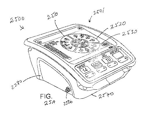
[0080] Figs. 25 A and 25B show aspects of an intrathoracic pressure regulation
systems
according to embodiments of the present invention.
DETAILED DESCRIPTION OF THE INVENTION
[0081] Embodiments of the present invention encompass techniques for
regulating
intrathoracic pressure, airway pressure, or endotracheal pressure. In some
cases, a positive end
expiratory pressure (PEEP) can be provided prior to application of a vacuum.
In some cases, a
PEEP can be provided subsequent to application of a vacuum. The addition of
PEEP may
provide additional oxygenation for a diseased or compromised lung, more than
just the positive
pressure breath would. In some cases, PEEP is provided via mechanical
ventilation, and can
refer to pressure greater than atmospheric pressure that is present in the
airway at the end of the
expiratory cycle. PEEP can improve gas exchange by preventing alveolar
collapse, recruiting
more lung units, increasing functional residual capacity, and redistributing
fluid in the alveoli.
In some cases, the use of ITPR can upregulate the autonomic nervous system.
And in some
cases, the combination of IPR and an intra-aortic balloon pump (IABP) can
provide an even
bigger effect on enhancing circulation than either provides alone.
[0082] In a broad sense, the invention provides devices and techniques for
lowering
intracranial and intraocular pressures and increasing cerebral perfusion
pressures. Such devices
and techniques may be particularly helpful with patients who have suffered a
traumatic brain
injury and those with low blood flow states and low blood pressure. Examples
of conditions
that may be treated include hypotension, shock secondary to hypovolemia,
sepsis, heart failure,
and the like. One way to lower the pressure within the head but maintain or
increase systemic
pressures is by using a valve system that is coupled to a person's airway and
that is used to
lower intrathoracic pressures. In so doing, the valve systems may be used to
accelerate the
removal of venous blood from the brain, thereby decreasing intracranial and
intraocular
pressures. At the same time, the systemic pressures increase due to
enhancement of venous
return to the heart. Other techniques may be used as well, such as by creating
a vacuum
intermittently within the thorax and/or by repeatedly compressing and/or
decompressing the
CA 02766064 2011-12-19
WO 2010/148412 PCT/US2010/039391
patient's chest using an external thoracic positive pressure source. By
reducing intracranial
pressures, movement of cerebral spinal fluid is also enhanced. In so doing,
intracranial
pressures are further reduced thereby providing further treatment for those
suffering from head
trauma. In some cases, the valve systems may also be used to treat the brain
function in a
person suffering from a heart condition (atrial fibrillation, heart failure,
cardiac tamponade, and
the like) that results in elevated intracranial pressures. Such heart
conditions may include, for
example, atrial fibrillation or heart failure. By reducing intracranial
pressures, cerebral spinal
fluid movement and translocation is increased to help improve brain function.
[0083] Intracranial pressures are regulated by the amount the cerebral
perfusion pressure,
which is determined by the arterial blood pressure to the head, the pressures
within the skull,
and the pressures within the venous system that drains blood flow from the
brain. The devices
and methods of the invention may be used to enhance the egress of venous blood
out of the
brain, thereby lowering intracranial pressures. The devices and methods can be
used in patients
that are breathing spontaneously and those that require assisted ventilation.
To do so, the
devices and methods may be used to augment the intrathoracic vacuum effect
each time a
patient inhales (or in the case of a non-breathing patient, each time the
pressure within the chest
is manipulated to fall below atmospheric pressure), thereby lowering the
pressures in the thorax
and in the venous blood vessels that transport blood out of the brain. The
vacuum effect is
transduced back into the brain, and as a result, intracranial pressures are
lowered with each
inspiratory effort. This in turn causes more venous blood to flow out of the
head than would
otherwise be possible, resulting in lower intracranial pressures and lower
intraocular pressures.
In addition, circulation to the vital organs is increased as the increase in
venous return to the
heart each time a negative intrathoracic pressure is generated results in an
increase in cardiac
output and improved vital organ perfusion. As such, this invention may be used
to help
patients suffering from low cardiac output states and low blood pressure.
[0084] To prevent or impede respiratory gases from flowing to the lungs, a
variety of
impeding or preventing mechanisms may be used, including those described in
U.S. Patent
Nos. 5,551,420; 5,692,498; 6,062,219; 5,730,122; 6,155,257; 6,234,916 and
6,224,562, and in
U.S. Patent Application Nos. 10/224,263, filed on August 19, 2002 ("Systems
and Methods for
Enhancing Blood Circulation", Attorney Docket No. 16354-000115), 10/401,493,
filed March
26
CA 02766064 2011-12-19
WO 2010/148412 PCT/US2010/039391
28, 2003 ("Diabetes Treatment Systems and Methods", Attorney Docket No. 16354-
000116),
09/966,945, filed September 28, 2001 and 09/967,029, filed September 28, 2001,
the complete
disclosures of which are herein incorporated by reference. The valve systems
may be
configured to completely prevent or provide resistance to the inflow of
respiratory gases into
the patient while the patient inspires. For valve systems that completely
prevent the flow of
respiratory gases, such valves may be configured as pressure responsive valves
that open after a
threshold negative intrathoracic pressure has been reached.
[0085] For example, the resistance to the inflow of respiratory gases may be
set between
about 0 cm H2O and about -25 cm H2Oand may be variable or fixed. More
preferably, the
valve system may be configured to open when the negative intrathoracic
pressure is in the
range from about -2 cm H2O to about -20 cm H20. In addition, the valve system
may used
continuously or on a variable basis. For example, the valve system may be used
for every other
spontaneous breath.
[0086] Although not intended to be limiting, specific kinds of impedance
valves that may be
used to reduce intracranial and intraocular pressures include those having
spring-biased
devices, automated/electronic and mechanical means to occlude and open a valve
lumen, duck
bill valves, ball valves, and other pressure sensitive valve systems capable
of opening and
closing when subjected to low pressure differentials triggered either by
spontaneous breathing
and/or external means to manipulate intrathoracic pressure (such as
ventilators, phrenic nerve
stimulators, an iron lung, and the like).
[0087] In the past, such threshold valve systems have been used to increase
the venous
preload on the heart and to increase cardiac output, stroke volume and blood
pressure because
of the augmented effects of the intrathoracic vacuum on the subsequent cardiac
contraction. In
contrast, the techniques of the invention function by facilitating the removal
of blood from the
venous side of the brain. Although there may be an increase in blood flow out
of the heart to
the vital organs (including to the brain) when using such valve systems, the
effect of the valve
systems on lowering of intracranial pressures was quite unexpected because of
the known
increase in blood flow to the brain. Remarkably, however, the reduction of
venous blood
pressures from the brain remains substantial when using the valve systems.
Thus, despite the
27
CA 02766064 2011-12-19
WO 2010/148412 PCT/US2010/039391
increase in blood flow to the brain, the net effect of the valve system is a
decrease in
intracranial pressures.
[0088] With the valve system coupled to the person's airway, the negative
intrathoracic
pressure may be enhanced by inspiring through the valve system. If the person
is
spontaneously breathing, the person may simply breath through the valve
system. If the person
is not breathing, artificial inspiration may be induced using a variety of
techniques, including
electrical stimulation of the diaphragm, a negative pressure ventilator such
as a body cuirass or
iron lung, or a positive pressure ventilator capable of also generating a
vacuum between
positive pressure ventilations.
[0089] The valve systems may have a fixed actuating pressure or may be
variable so that
once a desired negative intrathoracic pressure is reached, the resistance to
flow may be
lessened. Further, the valves of the invention may be configured to be
variable, either manually
or automatically. The extent to which the resistance to flow is varied may be
based on
physiological parameters measured by one or more sensors that are associated
with the person
being treated. As such, the resistance to flow may be varied so that the
person's physiological
parameters are brought within an acceptable range. If an automated system is
used, such
sensors may be coupled to a controller which is employed to control one or
more mechanisms
that vary the resistance or actuating pressure of the inflow valve as
generally described in the
references that have been incorporated by reference.
[0090] Hence, the valve systems of the invention may also incorporate or be
associated with
sensors that are used to detect changes in intrathoracic pressures or other
physiological
parameters. In one aspect, the sensors may be configured to wirelessly
transmit their measured
signals to a remote receiver that is in communication with a controller. In
turn the controller
may use the measured signals to vary operation of the valve systems described
or incorporated
by reference herein. For example, sensors may be used to sense blood pressure,
pressures
within the heart, intrathoracic pressures, positive end expiratory pressure,
respiratory rate,
intracranial pressures, intraocular pressures, respiratory flow, oxygen
delivery, temperature,
blood pH, end tidal C02, tissue C02, blood oxygen, cardiac output or the like.
Signals from
these sensors may be wirelessly transmitted to a receiver. This information
may then be used
28
CA 02766064 2011-12-19
WO 2010/148412 PCT/US2010/039391
by a controller to control the actuating pressure or the resistance of an
inflow valve as described
in the references incorporated herein by reference.
[0091] The techniques for reducing intracranial pressures may be used in a
variety of
settings. For example, the techniques may be used in person's who are
spontaneously
breathing, those who are not breathing but whose hearts are beating, and those
in cardiac arrest.
In the latter case, the techniques may use some means to create a vacuum
intermittently within
the thorax during the performance of CPR. This could be by using a valve
system or some
other type of pressure manipulation system. Further, such systems may be used
in other
settings as well, including when the person is breathing.
[0092] Fig. 1 is flow diagram illustrating one method for reducing
intracranial or intraocular
pressures. As shown in step 10, the process proceeds by coupling a valve
system to the
person's airway. Any kind of coupling mechanism may be used, such as by a
mouthpiece, an
endotracheal tube, a face mask, or the like. Further, any of the valve systems
described or
incorporated herein by reference may be used. In step 20, the person's
negative intrathoracic
pressure is repetitively decreased (either artificially or by spontaneous
breathing). Examples of
techniques to artificially reduce the negative intrathoracic pressure include
use of an iron lung
cuirass device, a ventilator that is capable of generating negative pressures,
a ventilator that is
capable of providing high frequency oscillations at a rate of about 200 to
about 2000 per
minute, a phrenic nerve stimulator, or the like. As the person's negative
intrathoracic pressure
is repeatedly decreased while the valve system is coupled to the airway, the
pressures in the
venous vessels that transport blood out of the head are also lowered. In so
doing, intracranial
and intraocular pressures are reduced.
[0093] As shown in step 30, various physiological parameters of the person may
optionally
be measured. Examples of such parameters include respiratory rate,
intrathoracic pressure,
intertracheal pressure, intracranial pressure, intracranial blood flow,
intraocular pressure, blood
pressure, heart rate, end tidal COz, oxygen saturation, and the like. Further,
as shown in step
40, the valve system's actuating threshold level may optionally be varied
based on the measured
physiological parameters. This may be done to maximize the amount of blood
drawn out of the
brain or simply to monitor the patient's condition to insure that the patient
remains stable.
29
CA 02766064 2011-12-19
WO 2010/148412 PCT/US2010/039391
[0094] Fig. 2 illustrates one embodiment of a facial mask 100 to which is
coupled a valve
system 200. Mask 100 is configured to be secured to a patient's face so as to
cover the mouth
and nose. Mask 100 and valve system 200 are examples of one type of equipment
that may be
used to lower intrathoracic pressures and thereby lower intracranial and
intraocular pressures.
However, it will be appreciated that other valve systems and other coupling
arrangements may
be used including, for example, those previously referenced. As such the
invention is not
intended to be limited to the specific valve system and mask described below.
[0095] Referring also to Figs. 3-5, valve system 200 will be described in
greater detail.
Valve system 200 includes a valve housing 202 with a socket 204 into which a
ball 206 of a
ventilation tube 208 is received. In this way, ventilation tube 208 may rotate
about a horizontal
axis and pivot relative to a vertical axis. A respiratory source, such as a
ventilation bag, may be
coupled to tube 208 to assist in ventilation. Disposed in ventilation tube 208
is a filter 210 that
is spaced above a duck bill valve 212. A diaphragm holder 214 that holds a
diaphragm 216 is
held within housing 202. Valve system 200 further includes a patient port 218
that is held in
place by a second housing 220. Housing 220 conveniently includes tabs 222 to
facilitate
coupling of valve system 200 with facial mask 100. Also held within housing
220 is a check
valve 224 that comprises a spring 224a, a ring member 224b, and an o-ring
224c. Spring 224a
biases ring member 224b against patient port 218. Patient port 218 includes
bypass openings
226 that are covered by o-ring 224c of check valve 224 until the pressure in
patient port 218
reaches a threshold negative pressure to cause spring 224a to compress.
[0096] When the patient is actively ventilated, respiratory gases are forced
through
ventilation tube 208. The gases flow through filter 210, through duck bill
valve 212, and forces
up diaphragm 214 to permit the gases to exit through port 218. Hence, at any
time the patient
may be ventilated simply by forcing the respiratory gases through tube 208.
[0097] During the exhalation phase of a breathing cycle, expired gases flow
through port 218
and lift up diaphragm 214. The gases then flow through a passage 227 in
ventilation tube 208
where they exit the system through openings 229 (see Fig. 3).
[0098] During the inhalation phase of a breathing cycle, valve system 200
prevents
respiratory gases from flowing into the lungs until a threshold negative
intrathoracic pressure
level is exceeded. When this pressure level is exceeded, check valve 224 is
pulled downward
CA 02766064 2011-12-19
WO 2010/148412 PCT/US2010/039391
as springs 224a are compressed to permit respiratory gases to flow through
openings 226 and to
the patient's lungs by initially passing through tube 208 and duck bill valve
212. Valve 224
may be set to open when the negative intrathoracic pressure is in the range
from about 0 cm
H2O to about -25 cm H20, and more preferably from about -2 cm H2O to about -20
cm H20.
Hence, the magnitude and duration of negative intrathoracic pressure may be
enhanced during
patient inhalation by use of valve system 200. Once the intrathoracic pressure
falls below the
threshold, recoil spring 224a again close check valve 224. In this way,
pressure within the
venous blood vessels that transport blood out of the brain are also lowered.
In so doing, more
blood is drawn out of the brain to reduce intracranial and intraocular
pressures.
[0099] Any of the valve systems described herein may be incorporated into a
treatment
system 300 as illustrated in Fig. 6. System 300 may conveniently include
facial mask 100 and
valve system 200, although any of the valve systems or interfacing mechanisms
described
herein or the like may be used, including but not limited to the valve system
of Fig. 14. Valve
system 200 may conveniently be coupled to a controller 310. In turn,
controller 310 may be
used to control the impedance level of valve system 200 in a manner similar to
any of the
embodiments described or incorporated herein. The level of impedance may be
varied based
on measurements of physiological parameters, or using a programmed schedule of
changes.
System 300 may include a wide variety of sensors and/or measuring devices to
measure any of
the physiological parameters described herein. These sensors or measuring
devices may be
integrated within or coupled to valve system 200 or facial mask, or may be
separate.
[0100] For example, valve system 200 may include a pressure transducer for
taking pressure
measurements (such as intrathoracic pressures, intracranial pressures,
intraocular pressures), a
flow rate measuring device for measuring the flow rate of air into or out of
the lungs, or a CO2
sensor for measuring expired CO2.
[0101] Examples of other sensors or measuring devices include a heart rate
sensor 330, a
blood pressure sensor 340, and a temperature sensor 350. These sensors may
also be coupled
to controller 310 so that measurements may be recorded. Further, it will be
appreciated that
other types of measuring devices may be used to measure various physiological
parameters,
such as oxygen saturation and/or blood levels of 02, blood lactate, blood pH,
tissue lactate,
tissue pH, blood pressure, pressures within the heart, intrathoracic
pressures, positive end
31
CA 02766064 2011-12-19
WO 2010/148412 PCT/US2010/039391
expiratory pressure, respiratory rate, intracranial pressures, intraocular
pressures, respiratory
flow, oxygen delivery, temperature, end tidal C02, tissue C02, cardiac output
or the like.
[0102] In some cases, controller 310 may be used to control valve system 200,
to control any
sensors or measuring devices, to record measurements, and to perform any
comparisons.
Alternatively, a set of computers and/or controllers may be used in
combination to perform
such tasks. This equipment may have appropriate processors, display screens,
input and output
devices, entry devices, memory or databases, software, and the like needed to
operate system
300.
[0103] A variety of devices may also be coupled to controller 310 to cause the
person to
artificially inspire. For example, such devices may comprise a ventilator 360,
an iron lung
cuirass device 370 or a phrenic nerve stimulator 380. Ventilator 360 may be
configured to
create a negative intrathoracic pressure within the person, or may be a high
frequency ventilator
capable of generating oscillations at about 200 to about 2000 per minute.
[0104] Example 1
[0105] The following is a non-limiting example illustrating how intracranial
pressures may
be lowered according to the invention. In this example, 30 kg pigs were
anesthetized with
propofol. Using a micromanometer-tipped electronic Millar catheter inserted
below the dura,
intracranial pressures were measured continuously in the spontaneously
breathing pigs.
Intrathoracic pressures (ITP) were recorded using a Millar catheter placed in
the trachea at the
level of the carina. After stabilizing the pigs blood pressure, heart rate,
and ventilation rate,
intracranial pressures (ICP) and intrathoracic pressures were recorded, with 0
cmH2O
inspiratory impedance and then with inspiratory impedances of 5,10,15, and 20
cm H20.
Inspiratory impedance was achieved using an impedance threshold valve (ITV) as
described in
Figs. 2-5.
[0106] At base, the intracranial pressure was approximately 8/4 mmHg. With
increasing
amounts of inspiratory impedance, the intracranial pressure was lowered
proportionally as
shown in Figure 7. The intracranial pressure was 6/-2 mmHg when the pig
breathed through
an impedance of 20 cm H20. These findings were observed in multiple pig
studies and were
reproducible. Next, the Millar catheter was inserted 3 cm into the pig's
brain. The intracranial
32
CA 02766064 2011-12-19
WO 2010/148412 PCT/US2010/039391
pressure increased secondary to the trauma associated with the insertion of
the probe. The
intracranial pressure increased to 25/22 mmHg at the new baseline. Next, the
impedance
threshold valve was evaluated at different levels of resistance (Fig. 8).
Again, there was a
decrease in intracranial pressure proportional to the degree of inspiratory
impedance.
[0107] Example 2
[0108] In this example, intracranial pressures were increased in the setting
of recovery from
cardiac arrest. The example used a pig model with ventricular fibrillation for
6 minutes
followed by cardiopulmonary resuscitation for 6 minutes, followed by
defibrillation.
Spontaneous breathing resulted in an up to 50% decrease in intracranial
pressures when the
animals breathed through an inspiratory impedance of 10 cm H2Ousing a valve
system similar
to Example 1.
[0109] In all examples above, the intrathoracic pressure decreased relative to
the rest of the
body, creating a suction effect that reduced the pressure in the venous blood
vessels draining
the brain, thereby reducing intracranial pressures.
[0110] The invention further provides techniques and devices for reducing
intracranial
pressure (ICP) by facilitating movement of cerebral spinal fluid (CFS). There
are a number of
causes of increased ICP including: head injury, ischemia, osmolar imbalance,
cerebral edema,
tumors, complications of dialysis, infections, stroke, hypertensive crises.
Each can result in a
slow, and in some cases, an acute rise in the ICP. The solid matter of the
brain contents makes
up about 80-85% of the material enclosed by the skull. Cerebral blood volume
accounts for 3-
6% and CSF for 5-15%. See, Anesthesia, Third Edition Editor, Ron Miller.
Chapter authors:
Shapiro and Drummond. Chapter 54 (1990), the complete disclosure of which is
herein
incorporated by reference. CSF moves within the brain from its site of
production to its site of
reabsorption in the brain in an unimpeded manner under normal physiological
states. Since the
contents in the brain are practically incompressible, a change in volume of
any one of the three
major components (brain matter, blood volume, CSF volume) results in a
reciprocal change in
one or both of the other brain components. When the volume of the brain
expands, secondary to
an increase in the non-CSF component(s), some of the CSF is forced to other
locations,
including through the foramen magnum (hole in skull connecting skull to space
where the
spinal cord is located) and into the CSF fluid space surrounding the spinal
cord. When the non-
33
CA 02766064 2011-12-19
WO 2010/148412 PCT/US2010/039391
CSF components expand in volume or size, the intracranial pressure rises.
Normal ICP levels
are 10-15 mmHg when supine. At levels greater than 15-20 mmHg, damage to the
brain can
occur secondary to compression and resultant tissue ischemia (lack of adequate
blood flow). A
reduction in ICP levels can be achieved by a number of clinical interventions
including water
restriction, diuretics, steroids, hyperventilation, a reduction of cerebral
venous pressure,
hypothermia, CSF drainage, and surgical decompression.
[0111] Increased ICP results in reduced CSF fluid movement and translocation.
CSF fluid
production generally remains constant (about 150 ml/day) despite elevated ICP.
CSF fluid
reabsorption is can be slowed by elevated ICP. By using the valve systems
described herein,
central venous pressures may be reduced. In turn, this results in a decrease
in ICP and results
in an increase in CSF fluid movement or translocation and reabsorption. This
results in a
further reduction in ICP.
[0112] The valve systems of the invention maybe used in spontaneously
breathing
individuals, in patients ventilated with negative pressure ventilation or in
patients ventilated
with a ventilator that causes a decrease in central venous pressures for at
least a portion of the
respiratory cycle. Each time the intrathoracic pressure is reduced with the
valve systems of the
invention, there is a concomitant reduction in ICP and an increase in the
movement of CSF. In
other words, there is an increase in the difference between the peak and
trough of the ICP wave
form when using the valve systems. The sinusoidal movement occurs in
spontaneously
breathing people because of the change in pressure in the thorax that is
transmitted to the brain
via the venous blood vessels. The normally fluctuating CSF pressures (the
pressure increases
and decreases with each inspiration) are altered by the valve systems. More
specifically, the
valve systems create a lower trough value thereby creating an overall created
change in the ICP
with each inspiration. In the non-breathing patient, a similar effect can be
produced with the
valve systems when used with a variety of ventilator devices, including an
iron lung, a phrenic
nerve stimulator (such as those described in U.S. Patent Nos. 6234985;
6224562; and 6312399,
incorporated herein by reference), a suction cup on the chest that is used to
periodically expand
the chest and the like.
[0113] Increased CSF fluid movement results in an overall improved metabolic
state for the
brain. This is shown schematically in Figs. 9A and 9B. In Fig. 9A, the brain
400 is shown
34
CA 02766064 2011-12-19
WO 2010/148412 PCT/US2010/039391
under normal conditions. The brain 400 is surrounded by CSF 402 which is
produced at a site
404. The CFS in turn is surrounded by the skull 406. Blood enters brain 400
through an artery
408 and exits through a vein 410. Vein 410 also includes a site 412 of CFS
drainage. Shown
in Fig. 9A is an arrow showing the direction of CFS flow when draining.
Extending from brain
400 is the spinal cord 414 that is surrounded by the foramen magnum 416.
[0114] In Fig. 9B, the brain 400 is significantly swollen which reduces the
space 402 where
the CFS is located. The swelling of the brain 400 can cause blockage of CSF to
the spinal cord
414 as shown by arrow 418. Also, movement of CSF to site 412 is reduced to
hinder
movement of CSF out of the skull 406.
[0115] By treating the elevated ICP associated with all of the conditions
noted above using
the valve systems described herein, brain swelling can be reduced. In so
doing, CFS movement
and fluid translocation is increased under those same conditions. This results
in a further
decrease in intracranial pressure as the CSF is able to relocate.
[0116] Referring now to Fig. 10, the effects of contracting the atria of the
heart on ICP will
be described. As shown, contraction of the atria results in a phasic movement
in ICP. This can
be most clearly demonstrated during cardiac ventricular fibrillation. In that
setting, the atria
often beat spontaneously and the pressure of each contraction and relaxation
waveform is
transmitted immediately to the brain and is reflected in nearly identical
fluctuations in ICP.
The inventor has discovered that the fluid systems (venous blood vessels and
CSF) are so
closely linked, that subtle changes in the heart rhythm result in immediate
changes in CSF
pressure. Thus, in some patients with significant heart rhythms, or
significant heart failure, the
rise in right heart pressures as a result of these conditions results in an
increase in ICP. Such
rises in ICP can lead to a decrease in cerebral perfusion, since cerebral
perfusion is determined
by the pressure of the blood entering the brain (mean arterial pressure) minus
the pressure of
the blood leaving the brain (ICP and central venous pressure). Use of the
valve and
intrathoracic vacuum systems described herein will result in a decrease in
intrathoracic
pressure. As shown in Fig. 10, the downwardly pointing arrows represent the
timing of each
inhalation through the valve system. In the baseline state, before the onset
of atrial fibrillation,
each inspiration (small arrows) results in a reduction in ITP, a reduction of
right atria pressure,
a reduction in central venous pressures, and then an immediate reduction in
ICP. With the
CA 02766064 2011-12-19
WO 2010/148412 PCT/US2010/039391
onset of atrial fibrillation, the intracranial pressure rises and the
sinusoidal pattern of ICP
amplitude changes becomes dampened. As soon as the animal begins to inspire
through an
inspiration impedance of -10 cm H2O there is an immediate decrease in
intrathoracic pressure
(ITP), an immediate decrease in right atrial (RA) pressures , and an immediate
decrease in
intracranial pressure (ICP) along with the restoration of a sinusoidal
fluctuation in ICP with
each inspiration. With elevated ICP, inspiration through the impeding means
results in a
decrease in ICP, increased cerebral spinal fluid flow, and a decrease in
cerebral ischemia
secondary to increased cerebral perfusion. As such, the valve systems can used
in patients with
heart rhythms, such as atrial fibrillation, or patients with heart failure who
have increased ICP
in order to reduce their ICP, increase CSF fluid movement and translocation,
and ultimately
help them to improve their brain function.
[0117] Hence, the amount of inspiratory resistance, or the amount of negative
intrathoracic
pressure generation (which may be generated using a variety of techniques) can
be controlled
or regulated by feedback from measurement of ICP, blood pressure, respiratory
rate, cardiac
output, or other physiological parameters. Such a system could include a
closed loop feedback
system.
[0118] Fig. 11 is a flow chart illustrating another method for treating a
person suffering from
head trauma associated with elevated intracranial pressures. In so doing, it
will be appreciated
that such techniques may also be used to treat those suffering from low blood
pressure or those
in cardiac arrest, among others. The techniques are particularly useful in
cases where the
person is not breathing, although in some cases they could be used for
breathing patients as
well.
[0119] Ina broad sense, when treating a person suffering from head trauma, a
person's
intrathoracic pressure is lowered to decrease intracranial pressures. In turn,
this assists in
reducing secondary brain injury. As shown in step 500, equipment may be
coupled to the
person to assist in lowering the person's intrathoracic pressure. A wide
variety of equipment
and techniques may be used to decrease the intrathoracic pressure, including
using a
mechanical ventilator capable of extracting respiratory gases, such as the one
described in U.S.
Patent No. 6,584,973, a phrenic nerve or other muscle stimulator (with or
without the use of an
impedance mechanism, such as those described in U.S. Patent Nos. 5,551,420;
5,692,498;
36
CA 02766064 2011-12-19
WO 2010/148412 PCT/US2010/039391
6,062,219; 5,730,122; 6,155,257; 6,234,916 and 6,224,562) such as those
described in U.S.
Patent Nos. 6234985; 6224562; 6312399; and 6463327, an iron lung device, a
thoracic vest
capable of pulling outward on the chest wall to create an intrathoracic vacuum
similar to the
effect of an iron lung, a ventilatory bag, such as the one described in
copending U.S.
Application No. 10/660,366, filed 09/11/2003 (attorney docket no. 16354-
005400), and the
like. The complete disclosures of all these references are herein incorporated
by reference. For
breathing patients, a threshold valve as described above and that is set to
open when about 5 cm
H2O is generated during an inhalation may be used to enhance the person's
negative
intrathoracic pressure.
[0120] When the person is not breathing, a positive pressure breath is
delivered to the person
as illustrated in step 502. This may be done with a mechanical ventilator, a
ventilatory bag,
mouth to mouth, and the like. This is followed by an immediate decrease in
intrathoracic
pressure. This may be done by extracting or expelling respiratory gases from
the patient's lungs
as shown in step 504. Any of the techniques described above may be used to
lower the
intrathoracic pressure. Such a reduction in intrathoracic pressure also lowers
central venous
pressure and intracranial pressure.
[0121] The vacuum effect during the expiratory phase maybe constant, varied
over time or
pulsed. Examples of different ways to apply the vacuum are described later
with respect to
Figs. 12A-12C. The initial positive pressure breath may be supplied for a time
of about 250
milliseconds to about 2 seconds, and more preferably from about 0.75 seconds
to about 1.5
seconds. The respiratory gases may be extracted for a time that is about 0.5
to about 0.1 to that
of the positive pressure breath. The positive pressure breath may be delivered
at a flow rate in
the range from about 0.1 liters per second to about 5 liters per second, and
more preferably
from about 0.2 liters per second to about 2 liters per second. The expiratory
flow (such as
when using a mechanical ventilator) may be in the range from about 0.1 liters
per second to
about 5 liters per second, and more preferably from about 0.2 liters per
second to about 2 liters
per second. The vacuum may be maintained with a negative flow or without any
flow. The
vacuum may be in the range from about 0 mmHg to about -50 mmHg, and more
preferably
from about 0 mmHg to about -20 mmHg.
37
CA 02766064 2011-12-19
WO 2010/148412 PCT/US2010/039391
[0122] As shown in step 506, the process of delivering a positive pressure
breath and then
immediately lowering intrathoracic pressures may be repeated as long as
necessary to control
intracranial pressures. Once finished, the process ends at step 508.
[0123] The manner in which positive pressure breaths and the vacuum are
created may vary
depending upon a particular application. These may be applied in a variety of
waveforms
having different durations and slopes. Examples include using a square wave,
biphasic (where
a vacuum is created followed by positive pressure, decay (where a vacuum is
created and then
permitted to decay), and the like. Three specific examples of how this may
occur are illustrated
in Figs. 12A-12C, although others are possible. For convenience of discussion,
the time during
which the positive pressure breath occurs may be defined in terms of the
inspiratory phase, and
the time during which the intrathoracic pressure is lowered may be defined in
terms of the
expiratory phase. The positive pressure breaths may occur at about 10 to about
16 breaths per
minute, with the inspiratory phase lasing about 1.0 to about 1.5 seconds, and
the expiration
phase lasing about 3 to about 5 seconds. As shown in Fig. 12A, respiratory
gases are quickly
supplied up to a pressure of about 22 mmHg. This is immediately reversed to a
negative
pressure of about -10 mmHg. This pressure is kept relatively constant until
the end of the
expiratory phase where the cycle is repeated.
[0124] In Fig. 12B, the positive pressure is more slowly applied. When
reaching a pressure
of about 10 to about 15 mmHg, the pressure is rapidly reversed to a negative
pressure of about -
20 mmHg. The negative pressure gradually declines to about 0 mmHg at the end
of the
expiratory phase. The cycle is then repeated. Hence, in the cycle of Fig. 12B,
the positive
pressure is reduced compared to the cycle in Fig. 12A, and the negative
pressure is initially
lower, but allowed to gradually increase. The technique is designed to help
reduce a possible
airway collapse.
[0125] In Fig. 12C, the positive pressure is brought up to about 20 mmHg and
then
immediately brought down to about 0 mmHg. The negative pressure is then
gradually
increased to about -20 mmHg toward the end of the expiratory phase. This cycle
is designed to
help reduce a possible airway collapse.
[0126] Figs. 13A and 13B schematically illustrate one embodiment of a device
500 that may
be used to lower intrathoracic pressures in a non-breathing patient. Device
500 comprises a
38
CA 02766064 2011-12-19
WO 2010/148412 PCT/US2010/039391
housing 502 having an interface opening 504 that may be directly or indirectly
coupled to the
patient's airway using any type of patient interface. Housing 502 also
includes a vacuum
source interface 506 that may be in fluid communication with any type of
device or system
capable of producing a vacuum. Also coupled to housing 502 is a means to
regulate the
vacuum, such as a pressure responsive valve system 508. Device 500 further
includes a
ventilation interface 510 that may be used to provide a breath to the patient,
if needed, when the
vacuum is not applied.
[0127] In this embodiment, the vacuum may be provided by essentially any type
of a vacuum
source, and the regulator may comprise an impedance valve, such as those
described in U.S.
Patent Nos. 5,551,420; 5,692,498; 6,062,219; 5,730,122; 6,155,257; 6,234,916;
6,224,562;
6,234,985; 6,224,562; 6,312,399; and 6,463,327 as well as others described
herein. To supply
a breath, a variety of ventilation sources may be used, such as, for example,
a bag valve
resuscitator, that is coupled to interface 510. Device 500 may further include
a mechanism 512
to inhibit the vacuum when delivering a breath to the patient from the bag
valve resuscitator.
Once the breath is delivered, mechanism 512 operates to permit the vacuum
within the thorax
to be reapplied. The mechanism 512 used to turn off and on the vacuum source
can include a
slider switch that moves to close off the branch in housing 500 having the
vacuum source as
illustrated in Fig. 13B. However, other types of switches or mechanisms may be
used. In
some cases, the vacuum source may have a controller that is configured to shut
off the vacuum
when the breath is administered so that mechanism 512 is not needed. Also, a
controller and
appropriate sensors could be used to sense when the breath is delivered and
stopped so that
mechanism 512 may be appropriately operated by the controller. After the
breath is delivered,
mechanism 512 moves back to the position illustrated in Fig. 13A so that the
vacuum may be
supplied to the patient. When the vacuum reaches a threshold amount, regulator
508 operates
to maintain the level of vacuum at about the threshold amount.
[0128] Figs. 14A and 14B illustrate another embodiment of a device 530 that
may be used to
treat a patient. Device 530 operates using similar principles as device 500
illustrated in Figs.
13A and 13B. Device 530 comprises a housing 532 having a patient interface 534
that may be
coupled to the patient's airway and a vacuum interface 536 that may be coupled
to a vacuum
source. Housing 532 also includes a ventilation interface 538 through which a
positive
39
CA 02766064 2011-12-19
WO 2010/148412 PCT/US2010/039391
pressure breath may be supplied. Also coupled to housing 532 is a vacuum
regulator 540 that
regulates the amount of vacuum supplied to the patient. One example of a flow
regulator that
may be used is described below with references to Figs. 15A and 15B. However,
it will be
appreciated that any of the flow regulators described herein may be used.
Disposed within
housing 532 is a flow control device 542 that is used orchestrate gas flows
through housing
532. Flow control device 542 comprises a cylindrical member 544 that may slide
within
housing 532 and includes a flow path 546 that permits gas flow between
interfaces 534 and 536
when flow control device 542 is in the position illustrated in Fig. 14A.
Conveniently, a spring
548 or other biasing mechanism is used to hold flow control device 542 in the
home position
illustrated in Fig. 14A. Flow control device 542 also includes a flow path 550
illustrated by the
arrow in Fig. 14A to permit gas flows between regulator 540 and interface 536.
Hence, when
in the home position, a vacuum may be supplied through interface 536 which
lowers the
person's intrathoracic pressure. If the vacuum becomes too great, gas flows
are permitted
through regulator 540 to lower the amount of vacuum.
[0129] As illustrated in Fig. 14B, flow control device 542 also includes a
flow path 552 that
passes from interface 538 to interface 534. This permits a positive pressure
breath to be
supplied to the patient through interface 538. More specifically, as gasses
are injected through
interface 538, they flow into flow control device 542 causing it to move
within housing 532
and compress spring 548. In so doing, flow path 546 closes as it becomes
blocked by housing
532. Flow path 550 also closes, leaving only flow path 552 opened to permit
the respiratory
gases to flow to the patient. When the positive pressure breath stops, spring
548 forces flow
control device back to the home position where the vacuum is once again
supplied to the
patient.
[0130] Hence, when a vacuum is applied from interface 536, air is pulled out
of the patient
through interface 534 until the cracking pressure of the impedance valve 540
is reached. At
that point air passes through impedance valve 540 from the ventilation source
at interface 538,
thereby setting the limit of the vacuum achieved in the patient. When positive
pressure
ventilation is delivered from the ventilation source at interface 538, the
internal slider switch
cylinder 542 moves downward to close off the vacuum source, allowing for
delivery of a
positive pressure volume to provide a breath to the patient. Flow control
device 542 may
CA 02766064 2011-12-19
WO 2010/148412 PCT/US2010/039391
include a cup-shaped opening 556 which helps to move the device 542 along with
minimal
force applied. Once the breath has been delivered, and there is no positive
force delivered from
the ventilation source to the device 542, spring 548 pushes upwards, re-
exposing the patient to
the vacuum source.
[0131] Device 530 may also include an optional pressure pop-off regulator 560.
In the event
that the vacuum source is too great, the pop-off regulator 560 opens allowing
for pressure
relief above the desired vacuum pressure. The pop-off regulator 560 may be
configured to
open for pressures greater than about 20 to about 100 mmHg.
[0132] Although the devices illustrated in Figs. 13 and 14 are shown with
mechanical
switching mechanisms (to turn the vacuum off and on), others may also be used,
such as
magnetic, electronic, or electrical. Other kinds of possible switches include
a ball valve,
flapper valve, fish mouth valve, or other mechanical means as well as electric
or electronic
valving systems, including a solenoid, to allow for temporary inhibition of
the vacuum once the
positive pressure breath is delivered from the ventilation source. Additional
regulators can also
be used on the vacuum source to limit the flow or force of the vacuum. For
example, the
vacuum source could be configured to provide a constant vacuum once a
threshold level has
been achieved. In addition, the vacuum regulator and impedance valves 508 and
530 may be
variable or set at a fixed level of impedance. The vacuum source may also be a
suction line or
come from a venture device attached to an oxygen tank that could both provide
oxygen to the
patient and a vacuum source. Further, the invention is not limited to using an
impedance valve,
as shown, to regulate the vacuum. Multiple switching and regulating means may
be used
instead. The ventilation source is similarly not limiting and may include
sources such as
mouth-to-mouth, a bag-valve resuscitator, an automatic ventilator, and the
like.
[0133] Figs. 15A and 15B illustrate flow regulator 540 in greater detail.
Regulator 540
comprises a housing 570 having a patient port 572 and a ventilation port 574.
Optionally, a
supplemental oxygen port 576 may also be provided. Gas may flow through
housing 570
(between ports 572 and 574) through one of two flow paths. The first flow path
is blocked by a
one way check valve 578 that comprises a check valve gasket 580 and a spring
582. The
second flow path is blocked by a diaphragm 584.
41
CA 02766064 2011-12-19
WO 2010/148412 PCT/US2010/039391
[0134] In operation, a vacuum is experienced at patient port 572 as the vacuum
source draws
a vacuum at port 536 (See Fig. 14A). When the vacuum reaches a threshold
level, spring 582
compresses to move gasket 580 downward, thereby creating a flow path as
illustrated in Fig.
15B. As the vacuum is pulled, diaphragm 584 closes to prevent air from flowing
through the
other flow path. Gasket 580 remains spaced apart from the opening as long as
the vacuum is at
the threshold level. In this way, regulator 540 is able to maintain the vacuum
at a constant
level.
[0135] When ready to ventilate the patient, the vacuum is stopped and
respiratory gases are
injected into port 574 and/or port 576. These gasses lift diaphragm 584 to
permit the gases to
flow to the patient.
Example 3
[0136] Example 3 is another non-limiting example illustrating how intracranial
pressures and
intrathoracic pressures may be lowered and systolic arterial pressure may be
increased
according to one aspect of the invention. In this example, 30 kg pigs were
anesthetized with
propofol. Using a micromanometer-tipped electronic Millar catheter inserted 2
cm below the
dura, intracranial pressures were measured in non-breathing pigs.
Intrathoracic pressures (ITP)
were recorded using a Millar catheter placed in the trachea at the level of
the carina. Systolic
aortic blood pressures (SBP) were measured in the aorta with a Millar
catheter. To regulate
intrathoracic pressures, a system similar to that illustrated in Figs. 14A,
14B, 15A and 15B was
used, with inspiratory impedance (-8 cm H2O with a flow rate of 30L/min).
Positive pressure
ventilation was provided at a rate of 10 breaths/min with a tidal volume of
approximately 400
ml delivered over 1.0 seconds with an automatic transport ventilator. The
objectives, methods,
results, and conclusions describing these novel cardiopulmonary-cranial
interactions are
summarized below.
[0137] An objective of this example was to evaluate the acute use of a novel
inspiratory
impedance threshold device (ITD) attached to a controlled but continuous
vacuum (CV) source
to decrease intrathoracic pressure (ITP) and intracranial pressure (ICP) but
simultaneously
increase mean arterial pressure (MAP), coronary perfusion pressure (CPP) and
cerebral
perfusion pressure (CerPP) in an apneic pig model of sequential insults of
cardiac arrest and
42
CA 02766064 2011-12-19
WO 2010/148412 PCT/US2010/039391
fixed-bleed hemorrhage hypotensive shock. This animal model is associated with
both
elevated ICP after cardiac arrest and significant hypotension after
hemorrhage.
[0138] This example used 6 female farm pigs (28-32kg) that were anesthetized
with propofol,
intubated and ventilated to maintain normocarbia and 02 saturation >90%.
Ventricular
fibrillation was induced and followed by 6 min of no treatment, 6 min of
standard CPR, and
then defibrillation. After return of spontaneous circulation and while
ventilated mechanically at
breaths/min, 35% of blood volume was removed with a rate of 60 cc/min. Five
min later
ITD-CV was applied for 5 min along with positive pressure ventilation with
100% oxygen at a
rate of 10 bpm. The ITD-CV was then removed and positive pressure ventilation
at a rate of 10
10 breaths/min was reapplied. Hemodynamic parameters and arterial blood gases
were assessed
before, during, and after ITD-CV application. Statistical analysis was
performed with a paired
t-test and ANOVA to compare +/- ITD-CV use.
[0139] The results are summarized in the Table below. As shown, by regulating
thoracic
pressures, use of the ITD-CV causes an instantaneous decrease in ITP and ICP
as well as a
rapid rise in MAP and a marked increase in CerPP. Hence, the ITD-CV may be
used to treat
hypotension, shock, and cerebral hypertension.
Table
Before ITD-CV During ITD-CV After ITD-CV p value
ITP 0.5 0.1 -12.0 1.1 0.1 0.2 0.001
MAP 46.7 5.2 54.7 7.7 38. 4.1 0.03
3
ICP 14.1 3.9 6.1 4.5 15. 3.9 0.001
4
CerPP 32.7 4.2 48.6 5.9 23. 4.5 0.01
0
CPP 40.1 4.5 58.4 7.7 31. 3.4 0.008
1
[0140] In one particular embodiment, a person may have his or her
intrathoracic pressure
manipulated using multiple techniques, alone or in combination. For example,
some type of
external thoracic positive pressure source may be used to increase and then
decrease the
person's intrathoracic pressure to move blood out of and then into the heart
and lungs in a
repetitive fashion. Examples of such an external thoracic positive pressure
source include a
43
CA 02766064 2011-12-19
WO 2010/148412 PCT/US2010/039391
mechanical extrathoracic vest, a body cuirass, a compression piston, a
compression cup, or the
like. Such devices may function as non-invasive hemodynamic support devices
for
maintenance of increase blood pressure and circulation in hypotensive
patients.
[0141] While the person's intrathoracic pressures are being externally
manipulated (e.g.,
being increased and decreased), the person may also have his or her
intrathoracic pressures
manipulated by applying positive pressure breaths and a vacuum using any of
the techniques
described herein. Further, any of the valve systems described herein may be
used in
combination as well. Hence, while the person's chest is being compressed and
relaxed, positive
pressure breaths followed by a vacuum may be applied at the same time. In this
way, non-
invasive techniques are provided for improving blood flow to the vital organs
for an indefinite
period of time, and may be used in cases where the patient is in shock, has
very low blood
pressure, those in cardiac arrest, and the like. Also, such techniques may be
used to circulate a
preservative solution, equivalent to cardioplegic agents, until more
definitive care is available.
[0142] The timing of each of these steps may be controlled to correlate in any
manner, such
as, for example, applying the vacuum while the force on the patient's chest is
relaxed. Also, the
timing of chest compressions could be tied to other variables, such as timing
the compressions
and/or decompressions with intrinsic cardiac rhythm (i.e., ECG activity).
Further, the positive
pressure breaths may be performed only as needed and not in association with
every chest
compression. Further, the chest may be decompressed only after a certain
number of chest
compressions.
[0143] As with other embodiments, the patient may also be supplied with
periodic positive
pressure ventilation or an extracorporeal oxygenator to provide adequate
respiration. Negative
pressure ventilation may also be used to provide proper ventilation. For
example, the chest
may be decompressed with an unimpeded airway to provide the negative pressure
ventilation.
Also, the techniques just described could also be used alone or in combination
with invasive
ways to also maintain blood pressure. For instance, a greater effect on
intracranial pressure
may be produced if some of the patient's blood is removed from the body.
[0144] One particular arrangement of a system that may be used with such
techniques is set
forth in Fig. 6 (previously described) where element 370 (an iron lung cuirass
device) may also
schematically represent any of the external thoracic positive pressure sources
described herein.
44
CA 02766064 2011-12-19
WO 2010/148412 PCT/US2010/039391
Further, controller 310 may also include some type of energy source for
operating the positive
pressure source, such as pneumatic, electronic, combustion or the like. In
this way, a variety of
energy sources may be used to compress the chest and then release the
compression in an
alternating manner. Ventilator 360 may be used to apply the positive pressure
breath followed
by a vacuum using any of the techniques described herein, as well as to
provide proper
ventilation. Further, although shown with valve system 200, it will be
appreciated that any of
the other valve systems described herein may be used as well. Also, it will be
appreciated that
temperature sensor 350 may be substituted with other types of sensors and/or
monitors, such as
an ECG monitor, so that chest compressions and/or decompressions may be timed
with ECG
activity.
[0145] Intrathoracic Pressure Regulation and Positive End Expiratory Pressure
[0146] In an intrathoracic pressure regulation (IPR) technique that involves
PEEP, during
ventilation, in an inhale/exhale cycle in an apneic person or person needing
assisted ventilation,
it is possible to provide a positive pressure breath or ventilation (PPV),
then provide positive
end expiratory pressure (PEEP), and then pull a vacuum. Aspects of such a
technique are
illustrated in Fig. 16A. Alternatively, it is possible to first provide a
positive pressure breath or
ventilation, then pull a vacuum, and then supply PEEP. Aspects of such a
technique are
illustrated in Fig. 16B. According to some embodiments, these treatments may
be effected, at
least in part, by use of a push/pull ventilator. In some cases, these
treatments can be performed
in conjunction with a cardiopulmonary resuscitation (CPR) procedure or other
approach for
treating low blood pressure or low circulation. The duration of PPV, PEEP, and
generation of a
vacuum within the thorax may vary, depending upon the physiological needs of
the patient. In
the graphs provided in Figs. 16A and 16B, pressure vs. time curves are
illustrated for novel
intrathoracic pressure regulation techniques. Pressure is illustrated in terms
of intrathoracic
pressure (ITP), airway pressure, or endotracheal pressure, in units of cm H20.
Timing may
depend on what the inspiratory: expiratory (I:E) ratio is set to on a
ventilator and the setting for
the respiratory rate. In the examples depicted here, the I:E ratio is 1:3, or
one in three, with a
respiratory rate of 10 breaths per minute. In some cases, the I:E ratio can be
anywhere within a
range from about 1:1 to about 1:4. In some cases, the respiration rate can be
within a range
from about 6 to about 30 breathes per minute. The addition of PEEP may provide
additional
CA 02766064 2011-12-19
WO 2010/148412 PCT/US2010/039391
oxygenation for a diseased or compromised lung, more than just the positive
pressure breath
does. In some cases, PEEP is provided via mechanical ventilation and the
degree and duration
of PEEP may be variable or fixed, and may be regulated with a closed loop
control system. In
some cases PEEP can refer to pressure greater than atmospheric pressure that
is present in the
airway during or at the end of the expiratory cycle. The delivery of a
positive pressure breath
can be performed using a mechanical ventilator or anesthesia machine. The
value of time t,
which corresponds to the duration of the PEEP administration, can be within a
range from
about 0.1 second to about 1.5 seconds, for example. In some cases, the
positive pressure breath
can be delivered to the patient for a time period within a range from about
250 milliseconds to
about 2 seconds. In some cases, the positive pressure breath can be delivered
to the patient a
rate within a range from about 0.1 liters per second to about 5 liters per
second. The time that
the positive pressure breath is supplied relative to the time in which PEEP
and/or vacuum can
be within a range from about 0.5 to about 0.1. When the person is not
breathing, a positive
pressure breath can be delivered to the person. This may be done with a
mechanical ventilator,
a ventilatory bag, mouth to mouth, and the like. Any of the inspiratory
impedance threshold
device (ITD) techniques encompassed by the instant application can be used in
conjunction
with this method. It is also understood that approaches encompassed by the
instant application
can be used in conjunction with diabetes treatment modalities, such as those
described in U.S.
Patent Application Nos. 10/401,493 filed March 28, 2003 and 11/735,320 filed
April 13, 2007,
the contents of which are incorporated herein by reference. Approaches
encompassed by the
instant application can be used in conjunction with treatment modalities for
heart failure and
other conditions, such as those described in U.S. Patent Nos. 5,551,420,
5,692,498, 6,062,219,
6,526,973, 6,604,523, 7,210,480, and 6,986,349, the contents of which are
incorporated herein
by reference. The pressure curves shown in Figs. 16A and 16B may in some cases
be achieved
by incorporating the use of a ventilator or anesthesia machine. Relatedly, in
some cases such
curves may be achieved without the use of a ventilator. Any of a variety of
mechanisms or
procedures may be used to decrease ITP or achieve negative ITP, including
without limitation a
vacuum source, a suction device, a push/pull ventilator, or an active
compression-
decompression device.
[0147] Fig. 17 provides a schematic for an exemplary system for administering
a treatment to
a patient. As shown here, system 1700 includes a process controller 1710 in
operative
46
CA 02766064 2011-12-19
WO 2010/148412 PCT/US2010/039391
association with a vacuum delivery apparatus 1720, a ventilation control valve
apparatus 1730,
and a PEEP delivery apparatus 1740. Vacuum delivery apparatus 1720 is in
operative
association with a vacuum source apparatus 1722, optionally via a vacuum
regulation apparatus
1724. PEEP delivery apparatus 1740 is in operative association with a gas
pressure source
blended apparatus 1742, optionally via a pressure regulation apparatus 1744.
Ventilation
control valve apparatus 1730 is in operative association with a ventilator
apparatus 1750,
optionally via a breathing circuit apparatus 1751 having an expiratory limb
apparatus 1752 and
an inspiratory limb apparatus 1754. PEEP delivery apparatus 1740 can be
configured to deliver
an adjustable amount of PEEP as desired. As shown here, system 1700 can also
include a
pressure transducer 1731. System 1760 also includes a patient connection
apparatus 1760
which can be coupled to a patient, for example via an endotracheal tube or
other patient airway
device.
[0148] According to some embodiments, a treatment method may include a first
step that
involves releasing the ventilation control valve apparatus 1730 to deliver
positive pressure
ventilation. The treatment method may also include a second step that involves
activating the
ventilation control valve apparatus 1730 and the vacuum delivery apparatus or
valve 1720, at
the end of the positive pressure breath. The PEEP delivery apparatus or valve
1740 can be
released, delivering positive end expiratory pressure to the patient from
internal gas blender
apparatus 1742 at a regulated pressure. The treatment method may further
include a third step
that involves energizing the PEEP valve 1740 and deenergizing the vacuum
delivery valve
1720 at the end of the PEEP stage, to generate a regulated vacuum to the
patient's airway. The
treatment method may also include repeating the first, second and third steps
described above.
In some cases, ventilator 1750 can be used to deliver a positive pressure
ventilation or breath, a
vacuum, or both, to the patient. According to some embodiments, a manual
resuscitator can be
used to deliver a positive pressure breath to the patient. Additional
operational aspects of a
ventilator are discussed elsewhere herein, for example, with in conjunction
with Figs.
19A-19F.
[0149] Intrathoracic Pressure Regulation Effect on Sympathetic Tone
[0150] An intrathoracic pressure regulator (ITPR) can combine an inspiratory
impedance
threshold device (ITD) with a vacuum source for the generation of vacuum, for
example in the
47
CA 02766064 2011-12-19
WO 2010/148412 PCT/US2010/039391
trachea during cardiopulmonary resuscitation (CPR) while allowing positive
pressure
ventilation. Use of an ITPR can modulate the autonomic system. During
inhalation a valve
system can function to produce a vacuum within the thorax to transiently
decrease intrathoracic
pressure and thereby modulate the person's autonomic function. More
specifically, by lowering
the intrathoracic pressure, the person experiences enhanced venous return of
blood to the heart,
and this causes an increase in cardiac output, an increase in blood pressure,
and increase in
blood flow to the brain, a decrease in intracranial pressure, and an autonomic
nervous system-
modulated decrease in sympathetic tone resulting in a decrease in peripheral
arterial resistance.
The resultant increase in venous blood flow back to the right heart and then
into the lungs
increases cardiac preload and facilitates the refilling of the right and left
chambers of the heart.
The subsequent cardiac contract results in an increase in cardiac stroke
volume and cardiac
output. This causes the body's receptors, such as the carotid baroreceptors in
the neck, to sense
the increase in blood pressure and circulation and alter the autonomic nervous
system balance.
This can be demonstrated by the shift from lower frequency power spectra from
electrocardiograms recorded from skin electrodes that are analyzed using
standard heart rate
variability analytic methods. Approaches encompassed by the instant
application can be used
in conjunction with treatment modalities such as those described in U.S.
Patent No. 7,195,013,
the content of which is incorporated herein by reference.
[0151] Hence, the use of intrathoracic pressure regulation (IPR) can modulate
the autonomic
nervous system. In some cases, when IPR therapy is applied when the thorax has
been opened,
for example during open heart surgery, the lungs are filled with respiratory
gases during the
positive pressure phase (inspiration) and during the expiratory phase
respiratory gases are
actively extracted from the lungs. This results in the rapid displacement of
blood within the
lungs into the left atrium, thereby priming the left heart with blood. By
alternately filling the
lungs with respiratory gases and providing space concurrently for blood from
the right heart,
and then extracting respiratory gases and propelling the blood within the lung
reservoir
forward, the lung serves as a peristaltic sponge to both suck up blood from
the right heart and
deliver it to the left heart. By `wringing out the sponge' the expansion and
contraction of the
lung parenchyma provides a novel means to propel blood forward in the setting
of low or
reduced blood circulation. The addition of PEEP either before or after this
`wringing out'
process provides a means to help maintain oxygenation and preserve and protect
lung function.
48
CA 02766064 2011-12-19
WO 2010/148412 PCT/US2010/039391
During this process the delivered tidal volume during the inspiratory phase
may vary and the
rate of respiratory gases removal by the method or device may vary, either
directly or indirectly
with the tidal volume delivered, thereby providing a means to achieve the
desired target airway
pressures and/or intrathoracic pressures. Methods and devices such as these
that provide IPR
therapy can therefore be used to enhance circulation and increase blood
pressure, even when
the thorax is open to atmospheric pressure such as during or after open heart
surgery. It can be
applied to both lungs or just one lung, as long as the method and device is
allowed to move
respiratory gases in and out of the lung(s).
[0152] The changes in pressures in the lung achieved with IPR therapy are a
direct result of
changes in lung respiratory gas volume. With each positive pressure
ventilation the gas volume
is increased and when it is actively extracted it is reduced. In the process
blood is squeezed out
of the lungs and blood can only move forward due to the intact one-way valves
within the heart
(pulmonic and mitral in this case). Thus blood is pumped out of the lungs,
which served as a
giant reservoir, during the gas extraction phase and when the lungs are
inflated respiratory
gases fill the alveoli of the lungs and indirectly restore the arterial and
venous bed architecture
so that blood from the right heart rushes into the lung blood reservoir as
soon as the lungs are
inflated. The active infusion and removal of respiratory gases by the IPR
therapy provides a
novel means to pump blood into the left heart. When the chest is open to
atmospheric pressure,
then changes in lung volumes do not alter intracranial pressures as the
pressures within the
non-lung structures in the thorax no longer vary with changes in airway or
lung pressures.
[0153] Embodiments of the invention can therefore be used to treat patients
suffering from a
number of disease states including but not limited to those suffering from
elevated intracranial
pressures, intra-ocular pressures, shock, hypotension, circulatory collapse,
cardiac arrest, heart
failure, intra-operative hypotension, and those in dialysis. It can also lower
venous pressures
within the abdomen during surgical procedures such as operations on the liver
or intestines, and
simultaneously provide greater blood flow to these and other vital organs such
as the kidneys,
brain, and heart. By lowering venous pressures it can help to reduce blood
loss during surgical
procedures. By the aforementioned described mechanisms the novel methods and
devices can
also treat hypotension and poor circulation associated with sepsis, poly-
traumatic organ
damage, and acute respiratory disease syndrome (ARDS). Embodiments of the
intention may
49
CA 02766064 2011-12-19
WO 2010/148412 PCT/US2010/039391
also be used to reduce venous pressure in `compartment syndrome' and therefore
help to
circulate more blood and preserve tissue viability and function. Embodiments
of the invention
can be based upon the discovery that reductions in intrathoracic pressure
result in a decrease in
intracranial pressures and enhancement of blood flow to the heart. In patients
with an open
thorax, device embodiments can lower pressure in the airway and in the lungs,
thereby
removing respiratory gases from the lungs. This results in a `wringing out' of
the lungs much
like a wet sponge with each application of the vacuum and this forces the
blood in the lungs
into the left heart as the pulmonic valve prevent reverse transpulmonary flow.
With the next
inspiration, respiratory gases fill the lungs and blood rushes into the lungs.
It is squeezed out
with the next application of the low level vacuum. As such, the changes in
airway pressure
provide a pulmonary pump to squeeze blood out of the lungs and with each
positive pressure
breath provide an empty vascular reservoir within the lungs that is rapidly
refilled from blood
within the right heart.
[0154] When the thorax is not intact device embodiments may also include a
mechanism for
varying the level of resistance of the valve system. For example, embodiments
may include
adding positive expiratory pressure. This device may be used in combination
with at least one
physiological sensor that is configured to monitor at least one physiological
parameter of the
person. In this way, the mechanism for varying the pressures and/or volume of
respiratory
gases within the lungs may be configured to receive signals from the sensor
and to vary the
level of impedance of the valve system based on the signals. This in turn
regulates the amount
of respiratory gas volume and/or pressure and the speed at which the gases are
actively infused
into and extracted from the lungs. Examples of sensors that may be used
include those that
measure, airway pressure, intratracheal pressure, blood pressure, right heart
pressure, heart rate,
end tidal C02, oxygen level, and left heart pressures.
[0155] As noted elsewhere herein, embodiments of the present invention are
well suited for
use in decreasing intracranial or intraocular pressures when the patient's
thorax is intact. Such
techniques can be employed with the open chest. Lung volume and pressure can
change
without a change in intrathoracic pressure, as the circuit is open. When the
chest is open this
approach typically does not lower intracranial pressures.
CA 02766064 2011-12-19
WO 2010/148412 PCT/US2010/039391
[0156] In some cases PEEP can be applied either before or after the extraction
of the gases.
With this approach, the method and device provide a 3-phase means to modulate
airway
pressures and when the thorax is intact intrathoracic pressure: the lungs are
inflated, the gases
are removed from the lungs, and the lungs are partially inflated by PEEP to
reduce atelectasis
and help preserve lung integrity.
[0157] As discussed elsewhere herein, the delivery of the positive pressure
breaths and the
extraction of gases can be performed using a mechanical ventilator, and the
respiratory gases
may be extracted with a constant extraction or a pulsed extraction. The speed
and volume and
pressure of gas infusion and extraction may vary depending upon the patient's
condition and
needs. For example, when the tidal volume is increased, the speed which the
large gas volume
is extracted may be varied. This can be important in order to maximize the
duration of negative
intrathoracic pressure (when the thorax is intact) and airway pressure and
lung pressure when
the thorax is open. Figs. 18A-18C show aspects of tidal volume and airway
pressure changes.
[0158] The top charts of Figs. 18A-18C show intrathoracic pressure (ITP) in
mmHg, as a
function of time. The IPR therapy is delivered to generate an intrathoracic
vacuum of -7.0
mmHg and the positive pressure breath provides a maximum intrathoracic
pressure of 14
mmHg. The middle charts of Figs. 18A-18C show blood flow in the carotid artery
in mL/min
(e.g. common carotid blood flow), as a function of time. In Fig. 18A, the
bottom chart
provides three tracings (top tracing, middle tracing, and bottom tracing). The
top tracing of the
bottom chart corresponds to blood pressure as a function of time, the middle
tracing of the
bottom chart corresponds to intracranial pressure as a function of time, and
the bottom tracing
of the bottom chart corresponds to right atrial pressure as a function of
time. Fig. 18B is from a
segment around minute 138 in the study represented by Fig. 18A, when the tidal
volume was
10 ml/kg, and Fig. 18C is from a segment around minute 140 in the same
experiment
represented by Fig. 18A. Fig. 18B depicts inspiratory tidal volume (TV) of
about 276m1, and a
target of about 27.2kg x 10 ml/kg. Fig. 18C depicts inspiratory tidal volume
(TV) of about
192m1, and a target of about 27.2kg x 7.5 ml/kg. When the tidal volume was
reduced but there
is no change in the speed at which respiratory gases are removed (as shown in
Fig. 18C), then
the amount of time the airway pressures are at the target level of -7.0 mmHg
is greater, thereby
51
CA 02766064 2011-12-19
WO 2010/148412 PCT/US2010/039391
increasing the overall effectiveness of the delivered therapy when compared
with the results
shown in Fig. 18B.
[0159] In this IPR therapy experiment with a 27.6 kg anesthetized pig having
an open thorax,
initially the pig was ventilated with a positive pressure ventilation at 14
breaths per minute
(bpm) and an inspiratory: expiratory ratio (I:E) of 1:3. The IPR therapy was
delivered as
indicated by the decrease in airway pressures, shown in Figs. 18A and 18B (top
charts).
According to these chart tracings, the intrathoracic pressure (ITP) decreases
from 14.0 mmHg
to -7.0 mmHg at each ventilated breath. The time required to lower the airway
pressure to the
target value of -7.0 mmHg was 0.84 seconds.
[0160] By decreasing the tidal volume (TV) from 276 ml (10 ml/kg) in this 28
kg
anesthetized pig (e.g. Fig. 18B) to 192 ml (-7ml/kg) (e.g. Fig. 18C) as shown
by arrow A in
Fig. 18A (near minute 139.4 in the bottom chart), the time required to lower
airway pressures
to the target value of -7.0 mmHg was reduced to 0.64 seconds. This reduced
time span is
illustrated in Fig. 18C (top chart). With the longer duration of negative
airway pressure the
blood pressure increased from about 75/42 mmHg (e.g. Fig. 18B, bottom chart,
top tracing) to
about 95/55 mmHg (e.g. Fig. 18A, bottom chart, top tracing). Blood flow in the
carotid artery
similarly increased.
[0161] According to some embodiments, Figs. 18A-18C graphically illustrate the
removal of
respiratory gases (e.g. application of vacuum) that depends upon the amount of
tidal volume
delivered (e.g. the greater the tidal volume the more slowly the gas can be
removed).
[0162] As noted above, an IPR device which is well suited for use with such
methods is
described in Figs. 19A-19F. Exemplary IPR devices provide a threshold valve
that can
regulate vacuum. Shown from one perspective, Fig. 19A-1 depicts an IPR system
1900 having
a manometer or pressure sensor 1910, a ventilator port 1920, an inlet cap
1930, a body 1940, a
patient port 1950, a vacuum stem 1960, and a valve (not shown). Shown from
another
perspective, Fig. 19A-2 depicts IPR system 1900 having a manometer or pressure
sensor 1910,
a ventilator port 1920, an inlet cap 1930, a body 1940, a patient port 1950, a
vacuum stem
1960, and a valve (not shown). Fig. 19B-19F depict system 1900 in various
operational
configurations, shown in cross-section. As represented in Fig. 19B, vacuum
stem 1960 is fully
inserted or pushed in. Vacuum stem 1960 can include a vacuum port or lumen
1961. Valve
52
CA 02766064 2011-12-19
WO 2010/148412 PCT/US2010/039391
1970 includes a piston 1971, a valve face 1972, and a rolling diaphragm 1973.
A first portion
1962 of vacuum stem 1960 urges valve face 1972 away from rolling diaphragm
1973, thus
providing an opening 1975 in valve 1970 through which air, gas, or fluid may
flow, as
indicated by arrow A. As shown here, inspiratory gases can flow from a
ventilator (not shown)
through ventilator port 1920, through opening 1975 of valve 1970, through
diaphragm aperture
or opening 1976 and into or toward the patient (not shown) via patient port
1950. Optionally, a
vacuum stem spring 1963 can push vacuum stem 1960 against valve face 1972,
thus sealing the
vacuum stem 1960 so that no air flows through via vacuum port 1961, while
simultaneously
opening valve 1970 to vent. In some embodiments, first portion of valve stem
1962 and valve
face 1972 form an on/off switch at a vacuum juncture, such that when first
portion of valve
stem 1962 and valve face 1972 are in contact with each other, the vacuum is
closed off due to a
vent seal formed between first portion 1962 and valve face 1972. In the
operational
configuration shown in Fig. 19B, vacuum stem 1960 can be locked or held in
place, thus
sealing off the vacuum and maintaining an open connection between a ventilator
and a patient.
A vacuum stem can be sealed off while holding a vent seal open, allowing
inspiratory and
expiratory gases to pass in both directions.
[0163] Fig. 19C depicts IPR system 1900 with vacuum stem 1960 in a partially
withdrawn
configuration. As vacuum stem 1960 is withdrawn, valve face 1972 is allowed to
seat against
the edge of rolling diaphragm 1973 and vacuum stem 1960 simultaneously, thus
sealing both
the diaphragm and the stem. When diaphragm 1973 is sealed, air is prevented
from flowing
through valve 1970. As show here, fluid cannot flow through diaphragm aperture
or opening
1976 because valve face 1972 is sealed against diaphragm 1973. When first
portion 1962 of
stem 1960 is sealed, air or fluid is prevented from flowing through vacuum
port 1961 of stem
1960. According to some embodiments, system 1700 presents a 3-way valve having
2
positions. Accordingly, valve 1970 can be open, therefore providing fluid
communication
between a patient and a ventilator (for example as shown in Fig. 19B) or
between patient and
vacuum (for example as shown in Fig. 19D). As illustrated in Fig. 19C, the
connection or
passage between the patient and the ventilator can be broken or interrupted
before the
connection or passage between the patient and the vacuum is made or
established. According
to some embodiments, this series of events occurs as the user selectively
moves vacuum stem
1960 to the vacuum therapy position. An identical or similar sequence of
events can occur as a
53
CA 02766064 2011-12-19
WO 2010/148412 PCT/US2010/039391
positive pressure breath is administered through the ventilator port 1920
while vacuum therapy
is being administered through the vacuum stem 1960. In the latter case, the
positive pressure,
rather than the vacuum stem, forces the valve face 1972 against the end or
first portion 1962 of
the vacuum stem 1960, closing off the vacuum prior to opening the pathway
between patient
and ventilator. Hence, this dual-sealing of the valve and stem can occur, for
example, for an
instant while vacuum stem 1960 is being moved to a position which allows
vacuum therapy to
be administered, for example by a CirQlator or similar device, as depicted in
Fig. 19D.
[0164] As shown in Fig. 19D, a diaphragm can be in a fully closed position,
sealing off a
ventilator port, with a vacuum step open to the patient port. A vacuum can
pull a manometer
piston downward indicating to a physician or operator that vacuum is applied
to the patient. An
opening between the manometer and main body of the device allows pressure to
actuate the
manometer. A physician or operator can pull the vacuum stem, locking it in a
therapeutic
position, enabling the valve mechanism to administer therapy.
[0165] Fig. 19D shows vacuum stem 1960 fully or substantially withdrawn, and
optionally
locked in place. For example, a physician or operator can pull the vacuum
stem, locking it into
a vacuum therapy position, which enables system 1900 to facilitate vacuum
therapy. Here, first
portion 1962 of vacuum stem is no longer sealed against valve face 1972, thus
allowing
vacuum therapy to be administered as shown by arrow A. As depicted here, gas
is withdrawn
from the patient, through patient port 1950, between first portion 1962 and
valve seat 1972,
through valve stem lumen 1961, and toward a vacuum source or mechanism (not
shown).
Rolling diaphragm 1973 is in the closed position, sealing off the ventilator
port 1920. Hence,
no fluid flows through diaphragm aperture or opening 1976. Vacuum stem 1960 is
open to the
patient port 1950. When administered, a vacuum can pull a manometer piston
1911 of
manometer 1910 downward, or into or toward manometer body 1942. When piston
1911 is in
this position, it can provide an indicator to the physician or operator that
vacuum is being
applied to the patient. For example, the manometer can provide a mechanical
signal to the
physician. An opening 1912 between manometer 1910 and the main body 1940
allows
pressure or vacuum to actuate manometer 1910.
[0166] The rolling diaphragm 1973 depicted in Figs. 19B-19E is shown in the
extended
position, extending from or away from the body 1940 of IPR system 1900.
Rolling diaphragm
54
CA 02766064 2011-12-19
WO 2010/148412 PCT/US2010/039391
1973 is held in or urged toward this extended position by spring 1974 until
positive pressure
from ventilator provided via ventilation port 1920 overcomes spring 1974 as
shown in Fig.
19E, causing diaphragm 1973 to move in the direction indicated by arrow A,
downward into or
toward body 1940. In some cases, this action can be aided by the upper spring.
Hence, due to
the initiation of the positive pressure breath, fluid from ventilator enters
ventilator port 1920 as
indicated by arrow B, and positive pressure pushes diaphragm 1973 and face
valve 1972
toward body 1940. A half-stroke configuration is show in Fig. 19E, such that
passages to both
the vacuum and the ventilator are closed for an instant. As shown here,
diaphragm aperture or
opening 1976 is closed. The manometer can indicate the pressure condition to
which the
patient is exposed, regardless of the position of the diaphragm.
[0167] As shown in Fig. 19E, upon initiation of a positive pressure breath,
positive pressure
can push the diaphragm and vent seal down until the seal contacts the vacuum
stem. The
device is shown at half-stroke, and both the vacuum and the ventilator are
closed for an instant.
[0168] Fig. 19F illustrates the effect of a positive pressure breath.
Specifically, a positive
pressure breath forces piston 1971, valve face 1972, and diaphragm 1973 toward
or into body
1940, until valve face 1972 seals first portion 1962 of vacuum stem 1960. The
vacuum is
sealed off due to seal between 1972 and 1962. Fig. 19F illustrates the effect
of continued
translation of the diaphragm 1973 toward or into body 1940, as indicated by
arrow B, thus
moving relative to vacuum stem 1960. However, as valve face 1972 contacts
vacuum stem
1960, piston 1971 and valve face 1972 no longer translate along the length of
vacuum stem
1960. Hence, positive pressure breath continues to force the diaphragm
downward or toward
body 1940, thus opening a gas flow path to allow a breath to be delivered to
the patient, while
the valve face 1972 remains seated on the first portion 1962 of the valve
stem, sealing the
vacuum from the patient. Positive pressure breath or gas passes through
ventilation port 1920,
between valve face 1972 and diaphragm 1973, through diaphragm aperture or
opening 1976,
and out patient port 1950 toward the patient as indicated by arrow A. Hence, a
pathway is open
that allows for the positive pressure breath to pass through, across, or past
the diaphragm 1973
to the patient while the vacuum is sealed off as a result of the seal between
valve face 1972 and
first portion 1962 of valve stem 1960. As depicted here, positive pressure
from the breath
forces the manometer 1910 upward or away from manometer body 1942 as indicated
by arrow
CA 02766064 2011-12-19
WO 2010/148412 PCT/US2010/039391
C, which can provide an indication to a physician or operator that positive
pressure is being
applied to the patient. Upon release, cessation, or sufficient reduction of
positive pressure, the
diaphragm return spring 1974 forces the diaphragm 1973 back into its resting
position, where it
extends or is urged away from body 1940 as depicted in Figs. 19B-19E, thereby
sealing off the
pathway from the ventilator prior to opening the pathway to the vacuum.
[0169] As shown in Fig. 19F, a ventilator seal can remain seated on a vacuum
stem, sealing
the vacuum from the patient. Positive pressure breath can continue to force
the diaphragm
downward, opening the gas flow path to allow a breath to be delivered to the
patient. Positive
pressure from the breath can force manometer upward indicating a positive
pressure application
to the patient.
[0170] As the positive pressure is released at the end of the delivered
breath, the valve moves
in reverse motion as shown in Fig. 19G. Cessation of positive pressure can
allow the valve to
revert to the position shown in Fig. 19D. The connection between patient and
ventilator
becomes sealed and the vacuum becomes opened in the same manner as that shown
in Fig.
19E, however in reverse order. According to some embodiments, the target
opening pressure
to administer a breath is 8 cm H20. Vacuum may be limited to 12 cm H2O and can
be
insufficient to open the valve. The pressure required to overcome the
secondary valve, as if a
patient were to spontaneously exhale, can be minimized. IPR device embodiments
such as
those depicted in Figs. 19A-19G can be supplemented by or combined with PEEP
mechanisms,
thus providing treatment systems such as those shown in Fig. 17. In some
cases, IPR device
embodiments such as those depicted in Figs. 19A-19G can be used without the
addition of a
PEEP procedure.
[0171] Intrathoracic Pressure Regulation and Intra-Aortic Balloon Pump
[0172] The combined use of intrathoracic pressure regulation (IPR) and an
intra-aortic
balloon pump (IABP), or another assisted device, can provide a greater effect
on enhancing
circulation to the heart and brain and other vital organs than either approach
taken alone. In
some cases, this combined technique can incorporate aspects of cuff
treatments, such as those
described in U.S. Patent Nos. 6,234,985, 6,224,562, 6,312,399, 6,463,327, and
6,587,726, and
in U.S. Patent Application Nos. 12/165,366 filed June 30, 2008 and 12/119,374
filed May 12,
2008, the contents of which are incorporated herein by reference. In some
embodiments, an
56
CA 02766064 2011-12-19
WO 2010/148412 PCT/US2010/039391
IABP device can decrease myocardial oxygen and increase cardiac output. An
IABP device
may include a counterpulsating expandable balloon positioned in the aorta,
actively deflating in
systole and actively inflating in diastole. The expandable element or balloon
can be controlled
by a computer, optionally coupled with an ECG or pressure transducer.
[0173] Recycling Anesthesia
[0174] Embodiments of the present invention encompass techniques for recycling
anesthetic
gases when an intrathoracic pressure regulator apparatus (ITPR) is used with
an anesthesia
machine. For example, as depicted in Fig. 20, it is possible to recycle within
an anesthesia
machine the anesthesia gases. Hence, IPR can be used without excessive
consumption of
anesthesia. It is also possible to capture the expiratory gases in a separate
chamber/scrubber
system. Advantageously, such approaches can help to reduce the overall
consumption of
anesthesia gases. Fig. 20 illustrates aspects of systems and methods for
generating negative
airway pressure with an anesthesia machine, according to embodiments of the
present
invention. Treatment system 2000 includes an endotracheal (ET) tube or mask
2010, which
can be coupled with an intrathoracic pressure regulator apparatus (ITPR) 2020.
According to
some embodiments, ITPR 2020 can incorporate one or more elements of an IPR
device such as
that depicted in Figs. 19A-19G. As shown in Fig. 20, a patient Wye 2030 is
coupled with ITPR
apparatus 2020. An ITPR vacuum line 2040 couples ITPR apparatus 2020 with a
negative
pressure generator 2050. A vacuum return to circuit apparatus 2060 is coupled
with negative
pressure generator apparatus 2050 via a conduit or passageway 2065. Vacuum
return apparatus
2060 is also coupled with an anesthesia machine 2090 and an expiratory limb
2070 of the
circuit. In some cases, anesthesia machine 2090 can incorporate or be replaced
with a
ventilator. Anesthesia machine 2090 is also coupled with an inspiratory limb
2080. Patient
wye 2030 is coupled with expiratory limb 2070 and inspiratory limb 2080. As
shown here,
bulk flow mechanics can be employed to generate negative pressure in the
expiratory limb
2070 of the anesthesia machine. Relatedly, the amount of fresh make up gas
when using ITPR
therapy is reduced. The negative pressure generator 2050 pulls a vacuum on the
ITPR vacuum
line 2040 to provide ITPR therapy and pushes all or most of the expiratory gas
back into the
anesthesia machine 2090 through a circuit tee on the expiratory limb 2070 of
the circuit. By
routing the expiratory gases through a negative pressure generator 2050 and
then back into the
57
CA 02766064 2011-12-19
WO 2010/148412 PCT/US2010/039391
anesthesia circuit the semi-closed circuit is maintained along with
corresponding low flow
makeup gas and anesthetic agent. Anesthesia circuits can be considered semi-
closed, for
various reasons. For example, the gas exhaled by the patient often needs to
have the expired
carbon dioxide removed from the gas stream. Further, the oxygen and anesthetic
agent that is
metabolized by the patient is not exhaled and needs to be replaced. The
replacing of
metabolized oxygen is performed by adding a low flow of gas, or makeup Gas,
into the circuit..
In some cases a low flow may be preferred so that anesthetic agent is saved.
[0175] Ventilator and Anesthesia with ITPR
[0176] Fig. 21 illustrates aspects of an IPR system 2100 according to
embodiments of the
present invention. In some cases, system 2100 may embody aspects of a
push/pull ventilator.
When a patient is being ventilated with a mechanical ventilator, the IPR
method can be
practiced to periodically lower airway pressures to enhance circulation, and
when the thorax is
intact, to lower intracranial pressure. In some cases the IPR method and
device can be
incorporated into a mechanism that provides positive pressure ventilation
(e.g. a resuscitator
bag, a mechanical ventilator, an anesthesia machine, or other means to provide
positive
pressure ventilation). In some embodiments, IPR therapy can be applied when
the patient is
being treated with different inspiratory: expiratory (I:E) ratios with the
mechanical ventilator.
For example, a patient may be treated with a higher I:E ratio (1:2 - 1:5) and
after each
inspiration the IPR will reduced airway pressures and/or intrathoracic
pressures to between -1
to -20 mmHg for a duration of time varying between 100 milliseconds and 2
seconds prior to
the resumption of the positive pressure. By this means respiratory gases can
be rapidly
extracted from the patient's lungs and circulation can be increased.
[0177] As shown in the pneumatic diagram of Fig. 21, IPR system 2100 includes
an input for
ambient air 2102 having a filter 2104, and an input for oxygen 2106 having a
filter 2108.
Ambient air input 2102 can be in fluid communication with an exhaust manifold
2110 having a
positive inspiratory pressure (PIP) mechanism or blower 2112. Exhaust manifold
2110 is
coupled with flow meter 2114, which in turn is coupled with a first inhalation
check valve
2116. Oxygen input 2106 can be in fluid communication with a first voltage
sensitive orifice
(VSO) oxygen valve 2120 and a second VSO oxygen valve 2122. VSO valves 2120,
2122, in
turn can be coupled with a flow meter 2124. Check valve 2116 and flow meter
2124 are
58
CA 02766064 2011-12-19
WO 2010/148412 PCT/US2010/039391
coupled with a first control valve 2126, which in turn is coupled with a PS2,
or second pressure
sensor 2128 and a positive pressure delivery mechanism 2129. As shown here,
PS2 is in
operative association with both control valve 2126 and positive pressure
delivery mechanism
2129.
[0178] System 2100 also includes an N-Exp pressure mechanism, vacuum source,
or blower
2130 coupled with a second control valve 2132, and a continuous positive
airway pressure
(CPAP) control mechanism 2134 in operative association with second control
valve 2132 and
an exhalation check valve 2136. Control valve 2132 is coupled with check valve
2136, which
in turn is coupled with a PSI, or first pressure sensor 2138. In some cases,
operation of blower
2130 can be based on pressure conditions sensed by first pressure sensor 2138.
Exhalation
check valve 2136 and PSI 2138 are in operative association with vacuum line
2140, which in
turn is coupled with vomit filter 2142. Positive pressure delivery mechanism
2129 is also
coupled with vomit filter 2142. As shown here, vomit filter 2142 is coupled
with a connector
mechanism 2144, such as an endotracheal tube or mask. Connector mechanism 2144
in turn
can be in operative association with a patient or individual.
[0179] In an inhalation configuration, second control valve 2132 is turned off
or open, and
first control valve 2126 is turned on or closed. PIP blower 2112 is turned on,
and may start
ahead. For example, the blower may have some inertia, and it is possible to
start running the
blower prior to starting a breath via the control valve so that when the
control valve is opened
flow can initiate immediately. N-Exp blower mechanism 2130 is turned off.
According to
some embodiments, it can be helpful to close off either one or the other
control valve, which
can facilitate the capability of the device to a) deliver a breath or b)
deliver ITPR therapy. The
sequence for turning off blowers may vary in some instances. Further, in some
cases the
inhalation configuration events may occur quite closely together, for example,
within a period
of less than 20 mSec.
[0180] In an exhalation configuration according to some embodiments, second
control valve
2132 is turned on or closed, and first control valve 2126 is turned off or
opened. PIP blower
2112 is turned off. N-Exp blower mechanism 2130 is turned on, and may start
ahead. In some
cases the exhalation configuration events may occur quite closely together,
for example, within
a period of less than 20 mSec. Fig. 21 may include or involve features related
to a ventilator or
59
CA 02766064 2011-12-19
WO 2010/148412 PCT/US2010/039391
ventilator operation. For example, in some cases all items except N-Exp blower
mechanism
2130 may be ventilator related. Aspects of Fig. 21 may be related to the
ventilator shown in
Fig. 17. From a pnuematic perspective according to some embodiments,
ventilator apparatus
1750 on Fig. 17 may be similar to Fig. 21 internally, with the exception of N-
Exp blower
mechanism 2130. Hence, it may be possible to substitute ventilator apparatus
1750 with the
features of Fig. 21, less N-Exp blower mechanism 2130.
[0181] Medical Conditions and Replacement Therapy
[0182] Embodiments of the present invention are well suited for use in
treating patients that
are suffering from or at risk of developing conditions such as sepsis, shock,
heart failure, acute
respiratory distress syndrome, polytrauma, head disease, elevated hepatic or
portal vein
pressures, bleeding during abdominal, head and neck surgery, or insufficient
circulation during
open heart surgery. What is more, exemplary techniques can be used to reduce
fluid
requirement in a patient during a treatment for low blood circulation or low
blood pressure. In
some cases, systems and methods can be employed to increase microcirculation
in a patient or
to treat a patient having low microcirculation. Optionally, systems and method
can be used to
enhance drug circulation in a patient. Exemplary techniques can be used in
conjunction with
pharmacological therapy. According to some approaches, a CPR protocol is
administered to
the patient in combination with or in addition to administration of an IPR
protocol.
[0183] Embodiments of the present invention further encompass methods to
evaluate fluid
status in a patient that involve applying an IPR protocol to the patient and
evaluating the effect
on blood pressure. If the blood pressure goes up rapidly, then the patient may
benefit from
intravenous volume replacement therapy. In some cases, such replacement
therapy includes
deliver of a crystalloid. In some cases, replacement therapy includes deliver
of a colloid.
[0184] According to some embodiments, IPR can enhance circulation and thus
provide a
means to more effectively and safely circulate more blood and drugs
administered during low
flow states. Because of the increased circulation provided in low blood states
with IPR
therapy, drugs circulate faster and lower doses can be given in many cases.
Thus, the
combination of IPR and drug therapy may be particularly helpful clinically. By
example,
during CPR use of IPR therapy to enhance circulation provides a means to
deliver drugs that
might normally lower blood pressure to dangerous levels, such as sodium
nitroprusside. In
CA 02766064 2011-12-19
WO 2010/148412 PCT/US2010/039391
patients experiencing states of shock, drugs such as vasopressin or
epinephrine can be
administered in lower doses to further enhance circulation. Higher doses of
vasopressin and
epinephrine can have significant untoward effects. In another example, the
efficacy of estrogen
and progesterone administration during treatment of hypotension is augmented
by IPR therapy.
Greater circulation, especially to the brain, results in greater efficacy.
[0185] Pressure Sensor Location and Blower
[0186] Embodiments of the present invention provide unique pressure sensor
locations for
breath control and unique blower configurations for a vacuum mode that allows
control of
expiratory resistance by turning a blower on for priming, optionally with the
use of feedback
control loops.
[0187] With continued reference to Fig. 21, intrathoracic pressure regulation
(IPR) system
2100 can include pressure sensors at various locations for use in breath
control. Optionally,
pressure sensors may provide a level of redundancy to the system. IPR system
2100 can be
configured to provide pressure monitoring of both inspiratory and expiratory
limb pressures,
and active control of end exhale pressures to sub-atmospheric levels when in a
circulatory assist
mode. IPR system 2100 can incorporate the use of pressure sensor redundancy to
protect
against patient injury which may be caused by a faulty sensor. It is possible
to accomplish the
goals of safety redundancy and monitoring of an expiratory limb 2141E and an
inspiratory limb
1241I of a patient circuit 2141 by careful placement of pressure transducers
in the device. As
shown in Fig. 21, a first pressure sensor 2138 can operate to monitor pressure
in the expiratory
line 2141E, and a second pressure sensor 2128 can operate to monitor pressure
in the
inspiratory line 21411. In some cases, pressure sensor 2138 placed on or in
communication
with the expiratory limb 2141E can be used to monitor and control the active
exhalation
function in a circulatory assist mode or procedure. To maintain redundant
safety monitoring of
the patient airway at all times the pressure sensors are placed in such a way
that when the
breathing circuit 2141 is connected to the manifold or device, each pressure
sensor or
transducer is monitoring a particular side of the breathing circuit (e.g.
exhalation side 2141E or
inhalation side 2141I) so that one transducer can be used for a feedback
control loop and the
other transducer can be used as a redundant safety monitoring feature. This
placement of the
transducers allows for the use of two transducers for control and safety
redundancy rather than,
61
CA 02766064 2011-12-19
WO 2010/148412 PCT/US2010/039391
for example, a system that includes four transducers wherein two transducers
are on the
inspiratory side of the circuit and two transducers are on the expiratory side
of the circuit. The
two transducer system described herein allows direct communication of the
pressure transducer
location and the connection point of the breathing circuit. In comparison, if
the transducers are
placed in such a manner as to not allow direct communication of the pressure
transducer
location and the connection point of the breathing circuit, four transducers
may be required.
The pressure sensor location is detailed in the pneumatic schematic shown in
Fig. 21. The
sensors are labeled PSI and PS2, with PSI monitoring the expiratory line 2141E
and PS2
monitoring the inspiratory line 2141I.
[0188] The expiratory phase of the ventilatory cycle is only a minor focus of
mechanical
ventilation. The primary focus for mechanical ventilation is the delivery of
air to the patient's
lungs with a lesser focus on how air is allowed out of the lungs. The
expiratory limb of a
mechanical ventilator can be designed with a goal of reducing airflow
resistance to the extent
possible to allow passive expiratory flow to eliminate the inhaled tidal
volume. One other
feature commonly found in mechanical ventilators that effects expiratory flow
is the addition of
positive end expiratory pressure (PEEP). Outside of PEEP and design of low
resistance
pathways, expiratory flow has been largely ignored in mechanical ventilation.
Currently some
ventilators have a limited capability for generating a negative expiratory
pressure to augment
the natural release of the delivered tidal volume. Treatment systems according
to embodiments
of the present invention provide for richer control of expiratory flow by use
of a blower to
generate a negative pressure to enhance expiratory flow, which may in some
cases be related to
a priming procedure. The use of a servo controlled expiratory pressure source
allows a wide
range of control of expiratory flow. With servo controlled expiratory
pressure, the device can
generate a thoracic vacuum at a variety of levels of end expiratory pressure
and varying
pressure profiles from end inhalation pressure to end expiratory pressure.
[0189] Embodiments of the present invention provide a treatment system having
a unique
two-limb circuit. As depicted in Fig. 21, Inspiratory and expiratory flow
paths travel from the
treatment system or manifold to the patient through a dual limb patient
circuit 2141. The two
limbs 21411, 2141E can be concentric. For example, tube assembly can include
an inner lumen
providing the inspiratory path and an outer lumen providing the expiratory
path. The
62
CA 02766064 2011-12-19
WO 2010/148412 PCT/US2010/039391
connection at the patient end can include a standard 22mm female conical ISO
fitting. The
connection at the treatment device can affix directly to a manifold system
housed within the
device. The connection at the treatment device can include a pair of conical
fittings,
concentrically oriented. This connection configuration can allow a caregiver
to make both
Inspiratory and expiratory circuit limb connections simultaneously, with a
single motion.
Additionally, this arrangement can prevent the caregiver from inadvertently
mixing up the
inspiratory and expiratory limbs. As further described elsewhere herein,
inspiratory and
expiratory flow paths can be controlled via two solenoid valves (one for each
direction of flow)
mounted on a 2-plane manifold system. Through the valve, the flow path can
enter by an outer
ring of openings and exit the valve by a centrally located lumen. Inspiratory
and expiratory
pressures can be monitored through pneumatic ports located on each plane of
the manifold. As
further described elsewhere herein, the inspiratory plane can collect and
combine fresh air from
a positive pressure blower and oxygen from a separate valved manifold which
controls the flow
rate of oxygen. Check valves can be located at both fresh air and oxygen inlet
locations to
prevent flow in the reverse direction. When the valve opens, the flow path
allows the
combined oxygen and air to pass through to the center lumen connected to the
patient circuit.
Because the patient circuit is often in a concentric orientation and that the
inspiratory and
expiratory gasses typically do not mix, the inspiratory path travels through
the expiratory plane
of the manifold before connecting to the patent circuit. This is accomplished
by a sliding seal
where one component telescopes into the other, compressing an O-ring radially
between them.
Expiratory gasses enter the manifold through the outer lumen of the patient
circuit. A check
valve is located at the entrance of the expiratory path to prevent expiratory
gasses from being
re-breathed by the patient. In a fashion similar to that of the inspiratory
flow, a valve opens and
closes to control the flow of expiratory gases. Gases enter the valve through
an outer ring of
openings and exit through a central lumen. When the valve is open, this allows
the flow to pass
from the expiratory plane of the manifold to a negative pressure blower which
exhausts to the
atmosphere. The connection between the expiratory plane of the manifold and
negative
pressure blower utilizes a similar O-ring seal mechanism as was described for
inspiratory gases.
[0190] In some embodiments, a treatment system can include a communication
module that
communicates with an external medical device. The communication module can
include a blue
tooth assembly, a radiofrequency assembly, or a communication assembly that
communicates
63
CA 02766064 2011-12-19
WO 2010/148412 PCT/US2010/039391
at a selected or desired bandwidth. The external medical device may be a
defibrillator or
automated chest compressor, or the like. Such communications can be used to
time the
delivery of changes in positive and negative intrathoracic pressure with
either a defibrillation
shock and/or chest compression and release.
[0191] Manifold Systems and Methods
[0192] Figs. 22A to 22G show aspects of manifold systems and methods according
to
embodiments of the present invention. Exemplary manifold systems can provide a
2-plane
manifold that segregates inspiratory and expiratory gases. As depicted in Fig.
22A, manifold
system 2200 includes a distal interface 2210, a proximal interface 2230, and a
central interface
2220 disposed between the distal interface 2210 and the proximal interface
2230. Manifold
system 2200 also includes an inspiratory control valve assembly 2240 coupled
with distal
interface 2210, and an expiratory control valve assembly 2250 coupled with
proximal interface
2230. Actuation of inspiratory control valve assembly 2240 operates to control
the flow of
inspiratory gases into manifold system 2200. For example, the opening of
inspiratory control
valve assembly 2240 facilitates the entry of inspiratory gases into manifold
system 2200, and
the closing of inspiratory control valve assembly 2240 inhibits the entry of
inspiratory gases
into manifold system 2200. In some cases, inspiratory control valve assembly
2240 includes a
solenoid valve. Actuation of expiratory control valve assembly 2250 operates
to control the
flow of expiratory gases into manifold system 2200. For example, the opening
of expiratory
control valve assembly 2250 facilitates the entry of expiratory gases into
manifold system
2200, and the closing of expiratory control valve assembly 2250 inhibits the
entry of expiratory
gases into manifold system 2200. In some cases, expiratory control valve
assembly 2250
includes a solenoid valve. Expiratory control valve assembly 2250 can operate
in many ways,
for example to facilitate delivery of a negative pressure treatment in
conjunction with a vacuum
mechanism or negative pressure blower, or to facilitate the delivery of a PEEP
treatment. In
some instances, if the expiratory control valve assembly 2250 is in an open
configuration, the
vacuum mechanism can pull a negative pressure so as to reduce the airway
pressure.
Alternatively, the expiratory control valve assembly 2250 can be closed with
an amount of
positive pressure remaining in the airway, thus providing a PEEP protocol.
Hence, the same
control valve assembly 2250 can operate to provide two different functions.
64
CA 02766064 2011-12-19
WO 2010/148412 PCT/US2010/039391
[0193] Inspiratory gases for delivery to the patient can enter manifold system
2200 in a
variety of ways. As shown in Fig. 22A, central interface 2220 may include an
oxygen inlet
2222 that receives oxygen from an oxygen source, and distal interface 2210 may
include an air
inlet port 2212 that receives air from an air source, such as a positive
pressure blower. Hence,
air inlet port 2212 can be in fluid communication with an air source for
example via a fluid
passage means such as a tube, and oxygen inlet 2222 can be in fluid
communication with an
oxygen source for example via a fluid passage means such as a tube.
Inspiratory gases for
delivery to the patient can be emitted from manifold system 2200 toward the
patient, for
example via a patient circuit interface 2232 of proximal interface 2230.
Expiratory gases from
the patient can enter manifold system 2200 at, for example, patient circuit
interface 2232 of
proximal interface 2230. Expiratory gases can pass through manifold system
2200 out of an
expiratory gas outlet port 2224 of central interface 2220, for example, and
toward a negative
pressure blower. Expiratory gas outlet port 2224 can be in fluid communication
with a
negative pressure blower for example via a fluid passage means such as a tube.
[0194] Manifold system 2200 may also include one or more sampling ports for
evaluating
pressure at various locations throughout the manifold system. As shown in Fig.
22A, central
interface 2220 includes an expiratory sampling port 2226 for use in sampling
expiratory
pressures. Similarly, distal interface 2210 includes an inspiratory sampling
port 2214 for use in
sampling inspiratory pressures.
[0195] In some cases, a manifold system 2200 may also include a continuous
positive airway
pressure (CPAP) assembly 2260. As shown in Fig. 22A, central interface 2220
includes a
CPAP port 2228 coupled with the CPAP assembly 2260. Operation of CPAP assembly
2260
can facilitate the administration of adjustable levels of continuous positive
airway pressure to a
patient.
[0196] Fig. 22B shows another view of manifold system 2200. As depicted here,
manifold
system 2200 includes a distal interface 2210, a proximal interface 2230, and a
central interface
2220 disposed between the distal interface 2210 and the proximal interface
2230. Manifold
system 2200 also includes an inspiratory control valve assembly 2240 coupled
with distal
interface 2210, and an expiratory control valve assembly 2250 coupled with
proximal interface
2230. Central interface 2220 may include an oxygen inlet 2222 that receives
oxygen from an
CA 02766064 2011-12-19
WO 2010/148412 PCT/US2010/039391
oxygen source. Inspiratory gases for delivery to the patient can be emitted
from manifold
system 2200 toward the patient, for example via a patient circuit interface
2232 of proximal
interface 2230. Expiratory gases from the patient can enter manifold system
2200 at, for
example, patient circuit interface 2232 of proximal interface 2230. As shown
in Fig. 22B,
patient circuit interface 2232 presents a concentric configuration having an
inner or inspiratory
lumen 2232a and an outer or expiratory lumen 2232b. Inner lumen 2232a operates
to carry
inspiratory gases out of the manifold and toward the patient, and outer lumen
2232b operates to
carry expiratory gases away from the patient and into the manifold. Typically,
these inspiratory
and expiratory gases are transmitted between the patient and patient circuit
interface 2232 via a
tube assembly having a first passage for inspiratory gases and a second
passage for expiratory
gases. For example, inspiratory and expiratory gases can be transmitted
between the patient
and patient circuit interface 2232 via a concentric tube assembly. The
concentric tube
assembly can include inner passage that fluidly communicates with inner lumen
2232a and an
outer passage that fluidly communications with outer lumen 2232b.
[0197] Fig. 22C shows an exploded perspective view of manifold system 2200
according to
embodiments of the present invention. Manifold system 2200 includes a distal
interface 2210,
a proximal interface 2230, and a central interface 2220 disposed between the
distal interface
2210 and the proximal interface 2230. Manifold system 2200 also includes an
inspiratory
control valve assembly 2240 coupled with distal interface 2210, and an
expiratory control valve
assembly 2250 coupled with proximal interface 2230. Actuation of inspiratory
control valve
assembly 2240 operates to control the flow of inspiratory gases into manifold
system 2200. For
example, the opening of inspiratory control valve assembly 2240 facilitates
the entry of
inspiratory gases into manifold system 2200, and the closing of inspiratory
control valve
assembly 2240 inhibits the entry of inspiratory gases into manifold system
2200. In some
cases, inspiratory control valve assembly 2240 includes a solenoid valve.
Actuation of
expiratory control valve assembly 2250 operates to control the flow of
expiratory gases into
manifold system 2200. For example, the opening of expiratory control valve
assembly 2250
facilitates the entry of expiratory gases into manifold system 2200, and the
closing of
expiratory control valve assembly 2250 inhibits the entry of expiratory gases
into manifold
system 2200. In some cases, expiratory control valve assembly 2250 includes a
solenoid valve.
66
CA 02766064 2011-12-19
WO 2010/148412 PCT/US2010/039391
[0198] Inspiratory gases for delivery to the patient can enter manifold system
2200 in a
variety of ways. As shown in Fig. 22C, central interface 2220 may include an
oxygen inlet
2222 that receives oxygen from an oxygen source, and distal interface 2210 may
include an air
inlet 2212 that receives air from an air source, such as a positive pressure
blower. Inspiratory
gases for delivery to the patient can be emitted from manifold system 2200
toward the patient,
for example via a patient circuit interface 2232 of proximal interface 2230.
Expiratory gases
from the patient can enter manifold system 2200 at, for example, patient
circuit interface 2232
of proximal interface 2230. Expiratory gases can pass through manifold system
2200 out of an
expiratory gas outlet port 2224 of central interface 2220, for example, and
toward a negative
pressure blower. Expiratory gas outlet port 2224 can be coupled with a
negative pressure
blower via a fluid passage means such as a tube.
[0199] Manifold system 2200 may also include one or more sampling ports for
evaluating
pressure at various locations throughout the manifold system. As shown in Fig.
22C, central
interface 2220 includes an expiratory sampling port 2226 for use in sampling
expiratory
pressures. For example, expiratory sampling port 2226 can be used to sample
expiratory
pressures present within an expiratory plane or chamber 2202 defined between
proximal
interface 2230 and central interface 2220. Similarly, distal interface 2210
includes an
inspiratory sampling port 2212 for use in sampling inspiratory pressures. For
example,
inspiratory sampling port 2212 can be used to sample inspiratory pressures
present within an
inspiratory plane or chamber 2204 defined between distal interface 2210 and
central interface
2220.
[0200] According to some embodiments, pressures or flow rates sensed at
inspiratory
sampling port 2214 or expiratory sampling port 2226 can be used to determine
fluid flow rates
throughout the manifold.
[0201] Manifold system 2200 may also include one or more check valves for
modulating or
controlling fluid flow at various locations throughout the manifold system. As
shown in Fig.
22C, manifold system 2200 includes an oxygen check valve 2223 that operates to
prevent or
inhibit reverse flow through oxygen inlet 2222, such that oxygen can flow into
manifold system
2200 via inlet 2222 in the direction indicated by arrow 2222A, but fluid is
prevented or
inhibited from flowing out of manifold system 2200 via inlet 2222 in the
reverse direction
67
CA 02766064 2011-12-19
WO 2010/148412 PCT/US2010/039391
indicated by arrow 2222B. Similarly, manifold system 2200 includes an air
check valve 2213
that operates to prevent or inhibit reverse flow through air inlet 2212, such
that air can flow into
manifold system 2200 via inlet 2212 in the direction indicated by arrow 2212A,
but fluid is
prevented or inhibited from flowing out of manifold system 2200 via inlet 2212
in the reverse
direction indicated by arrow 2212B. Further, manifold system 2200 includes a
patient circuit
or expiratory check valve 2233 that operates to prevent or inhibit reverse
flow through patient
circuit interface 2232, such that fluid can flow into manifold system 2200 via
outer or
expiratory lumen 2232b in the direction indicated by arrow 2232b(i), but fluid
is prevented or
inhibited from flowing out of manifold system 2200 via outer or expiratory
lumen 2232b in the
reverse direction indicated by arrow 2232b(ii).
[0202] In some cases, a manifold system 2200 may also include a continuous
positive airway
pressure (CPAP) assembly 2260. As shown in Fig. 22C, central interface 2220
includes a
CPAP port 2228 coupled with the CPAP valve assembly 2260. The inspiratory
plane or
chamber 2204 can operate to collects and combine fresh air from a positive
pressure blower
and oxygen from a separate valved manifold which controls the flow rate of
oxygen. Check
valves 2213 and 2223 are located at both fresh air and oxygen inlet locations,
respectively, to
prevent or inhibit flow in the reverse direction. When control valve 2242
opens, the flow path
allows the combined oxygen and air to pass through to the center lumen 2221
connected to the
patient circuit as indicated by arrow A. According to some embodiments, the
patient circuit is
in a concentric orientation, the inspiratory and expiratory gasses are not
allowed to mix, and the
inspiratory path travels through the expiratory plane of the manifold before
connecting to the
patent circuit. Such objectives can be achieved by use of a sliding seal where
one component
telescopes into the other, compressing an O-ring radially between them. As
depicted in Fig.
22C, an O-ring can be located between center lumen 2221 of central interface
2220and
centrally located lumen 2242 of distal interface 2210. As further explained
elsewhere herein,
expiratory gasses enter the manifold through the outer lumen 2232b of the
patient circuit. A
check valve 2233 is located at the entrance of the expiratory path to prevent
expiratory gasses
from being re-breathed by the patient. In a fashion similar to that of the
inspiratory flow, a
valve 2250 opens and closes to control the flow of expiratory gasses. Gases
enter the valve
2250 through an outer ring of openings 2251 and exit through a central lumen
2252. When the
valve 2250 is open, this allows the flow to pass from the expiratory plane
2202 of the manifold
68
CA 02766064 2011-12-19
WO 2010/148412 PCT/US2010/039391
to a negative pressure blower which exhausts to the atmosphere. The connection
between the
expiratory plane 2202 of the manifold and a negative pressure blower can use
an O-ring seal
mechanism similar to the one described for inspiratory gases. For example, an
O-ring cal be
located between orifice 2252 and the orifice inner diameter of flange 2224.
[0203] Fig. 22D shows an exploded perspective view of a portion of manifold
system 2200
according to embodiments of the present invention, in addition to aspects of
an inspiratory flow
path 2201 provided by the manifold system. Manifold system 2200 includes a
distal interface
2210, a proximal interface (not shown), and a central interface 2220 disposed
between the
distal interface 2210 and the proximal interface. Manifold system 2200 also
includes an
inspiratory control valve assembly 2240 coupled with distal interface 2210.
Actuation of
inspiratory control valve assembly 2240 operates to control the flow of
inspiratory gases into
manifold system 2200 from various fluid sources. For example, the opening of
inspiratory
control valve assembly 2240 facilitates the exit of inspiratory gases from the
inspiratory plane
or chamber 2204 as indicated by arrows 2212ii and 2222ii, the return of
inspiratory gases
toward distal interface 2210 as indicated by arrows 2212iii and 2222iii, and
the entry of
expiratory gases into an inspiratory delivery port 2221 of central interface
2220 as indicated by
arrows 2212iv and 2222iv. Conversely, the closing of inspiratory control valve
assembly 2240
inhibits the entry of inspiratory gases into manifold system 2200 from, for
example, oxygen
and air sources. In some cases, inspiratory control valve assembly 2240
includes a solenoid
valve.
[0204] Inspiratory gases for delivery to the patient can enter manifold system
2200 in a
variety of ways. As shown in Fig. 22D, central interface 2220 may include an
oxygen inlet
2222 that receives oxygen from an oxygen source, and distal interface 2210 may
include an air
inlet 2212 that receives air from an air source, such as a positive pressure
blower. Manifold
system 2200 may also include one or more check valves for modulating or
controlling fluid
flow at various locations throughout the manifold system. For example,
manifold system 2200
includes an oxygen check valve 2223 that operates to prevent or inhibit
reverse flow through
oxygen inlet 2222, such that oxygen can flow into manifold system 2200 via
inlet 2222 in the
direction indicated by arrows 2222i and 2222ii, but fluid is prevented or
inhibited from flowing
out of manifold system 2200 via inlet 2222 in the reverse direction.
Relatedly, control valve
69
CA 02766064 2011-12-19
WO 2010/148412 PCT/US2010/039391
2240 operates to control flow out from inspiratory plane or chamber 2204, as
indicated by
arrows 2222ii and 2212ii, through valve 2240 as indicated by arrows 2222iii
and 2212iii, and
through an inspiratory delivery port 2221 of central interface 2220 toward the
patient as
indicated by arrows 2222iv and 2212iv, via an inspiratory or internal lumen of
a patient circuit
interface of a proximal interface. In this way, selective opening and closing
of inspiratory
control valve 2240 modulates the flow of air and oxygen to the patient. As
illustrated in Fig.
22D, through the valve 2240, the flow path can enter by an outer ring of
openings 2241 and exit
the valve by a centrally located lumen 2242. Manifold system 2200 also
includes an air check
valve 2213 that operates to prevent or inhibit reverse flow through air inlet
2212, such that air
can flow into manifold system 2200 via inlet 2212 in the direction indicated
by arrows 2212i
and 2212ii, but fluid is prevented or inhibited from flowing out of manifold
system 2200 via
inlet 2212 in the reverse direction. During operation, air and oxygen can mix
within the
inspiratory chamber 2204, optionally at desired air:oxygen ratios, pass
through inspiratory
control valve 2240 and inspiratory delivery port 2221, and to the patient via
the inner or
inspiratory lumen of the patient circuit interface. In some cases,
introduction of air and oxygen
into the manifold can be independently controlled. Systems may include sensors
which
measure the flow rate or pressure, or both, of air or oxygen prior to mixing.
Control of
inspiratory gas administration to the patient can be based upon any
combination of such flow
rates or pressures.
[0205] Fig. 22E shows an exploded perspective view of a portion of manifold
system 2200
according to embodiments of the present invention, in addition to aspects of
an expiratory flow
path 2203 provided by the manifold system. Manifold system 2200 includes a
distal interface
(not shown), a proximal interface 2230, and a central interface 2220 disposed
between the
distal interface and the proximal interface 2230. Manifold system 2200 also
includes an
expiratory control valve assembly 2250 coupled with proximal interface 2230.
Actuation of
expiratory control valve assembly 2250 operates to control the flow of
expiratory gases into
manifold system 2200 from the patient. For example, the opening of expiratory
control valve
assembly 2250 facilitates the entry of expiratory gases into manifold system
2200 as indicated
by arrows 2232i and 2232ii, and the closing of expiratory control valve
assembly 2250 inhibits
the entry of expiratory gases into manifold system 2200. In some cases,
expiratory control
valve assembly 2250 includes a solenoid valve.
CA 02766064 2011-12-19
WO 2010/148412 PCT/US2010/039391
[0206] Expiratory gases from the patient can be routed through manifold system
2200 by first
passing through an expiratory or external lumen 2232b of patient circuit
interface 2232 of
proximal interface 2230, as indicated by arrow 2232i. Manifold system 2200 may
also include
a check valve for modulating or controlling fluid flow at proximal interface.
For example,
manifold system 2200 includes an expiratory check valve 2233 that operates to
prevent or
inhibit reverse flow through circuit interface 2232, such that expiratory
gases can flow from the
patient and into manifold system 2200 via expiratory lumen 2232b of circuit
interface 2232 in
the direction indicated by arrows 2232i and 2232ii, but fluid is prevented or
inhibited from
flowing out of manifold system 2200 via expiratory lumen 2232b of circuit
interface 2232 in
the reverse direction. Relatedly, control valve 2250 operates to control flow
out from
expiratory plane or chamber 2202, as indicated by arrow 2232ii, through valve
2250 as
indicated by arrow 2232iii, and through expiratory gas outlet port 2224 of
central interface
2220 as indicated by arrow 2232iv. In this way, selective opening and closing
of expiratory
control valve 2250 modulates the flow of expiratory gases from the patient. As
illustrated in
Fig. 22E, through the valve 2250, the flow path can enter by an outer ring of
openings 2251
and exit the valve by a centrally located lumen 2252.
[0207] Fig. 22F shows aspects of a patient circuit interface according to
embodiments of the
present invention. Patient circuit interface 2232 presents a concentric
configuration having an
inner or inspiratory lumen 2232a and an outer or expiratory lumen 2232b. Inner
or inspiratory
lumen 2232a operates to carry inspiratory gases toward the patient as
indicated by arrow A, and
outer or expiratory lumen 2232b operates to carry expiratory gases away from
the patient as
indicated by arrow B. Typically, these inspiratory and expiratory gases are
transmitted
between the patient and patient circuit interface 2232 via a tube assembly,
having an inner or
inspiratory passage that fluidly communicates with inner or inspiratory lumen
2232a and an
outer or expiratory passage that fluidly communications with outer or
expiratory lumen 2232b.
According to exemplary embodiments, treatment systems may include tube
connections having
concentric, conical fittings that engage with mating conical fittings on a
patient circuit, thus
providing quick and intuitive attachment of both Inspiratory and Expiratory
limbs of patient
circuit simultaneously. Concentric arrangement allows a caregiver to engage a
patient circuit
with a patient in a single motion and can be performed one-handed.
71
CA 02766064 2011-12-19
WO 2010/148412 PCT/US2010/039391
[0208] Fig. 22G shows aspects of a case or bonnet 2270 having a handle 2280
according to
embodiments of the present invention. Case 2270 can be configured to hold or
receive a
manifold system as described elsewhere herein. As shown here, handle 2280 can
wrap around
a back side 2272 of case 2270. This arrangement can strengthen the attachment
point and
provide impact protection on an upper-back corner 2274 of case 2270. According
to some
embodiments, handle 2280 wraps around the back side of the case by a distance
of about one
inch. Handle 2280 may include an attachment point 2282 having an underside
2284. Case
2270 may include an intake port 2286 that is configured to receive fluid into
the case. For
example, intake port 2286 can be configured to receive cooling air into the
case. In some cases,
handle 2280 may provide a retainment or recess 2288 for holding or receiving
one or more
cooling air filters (not shown). Optionally, handle 2280 may be configured as
an elastomeric
flap disposed on case 2270. In some cases, handle 2280 can include a semi-
elastomeric
material and can be attached to the underside of an arced cutout detail in a
side of the case.
According to some embodiments, the handle material can be flexible enough to
bend outward
when grasped for carrying. Where a handle includes soft material, the soft
material can provide
impact protection to the case or other structural elements associated with or
contained within
the case, such as elements of a manifold system. In some embodiments, the
protective case is
removable. The case can also provide protection against impact and against
water intrusion,
thus shielding the manifold from unwanted forces, shock, and water damage.
[0209] In some instances, a case may have multiples handles. For example, a
case may a first
handle on the left side of the case, and a second handle on the right side of
the case. The
handles may be made of moderately soft plastic, and lie flat against the sides
of the device
when not in use. When used as a handle or for an attachment point for a tie-
down, the handle
material flexes sufficiently to grasp easily. The handles can also conceal and
retain filters at
inlets for cooling air to be circulated inside the enclosure of the device.
Due to the locations of
the inlets, they can be protected from moisture ingress (e.g. rain) when the
device is in an
upright position, but in some cases may not protect if immersed or allowed to
lay face-down.
[0210] Fig. 23 illustrates aspects of a user interface 2300 for use with a
treatment system
according to embodiments of the present invention. What is more, additional
details regarding
the use and operation of a treatment system can be understood with reference
to Fig. 23. As
72
CA 02766064 2011-12-19
WO 2010/148412 PCT/US2010/039391
shown here, user interface 2300 includes several sub-interfaces that
correspond to various
modes of operation or use of the treatment system. For example, interface 2300
includes a
circulatory assist mode sub-interface 2310, a ventilation mode sub-interface
2320, and a
continuous positive airway pressure (CPAP) mode sub-interface 2330. When in
the circulatory
assist mode, the treatment system is configured to provide adjustable levels
of negative
pressure. When in the CPAP mode, the treatment system is configured to provide
adjustable
levels of continuous positive airway pressure, and when in the ventilation
mode, the treatment
system is configured to provide positive pressure ventilation with or without
positive end
expiratory pressure (PEEP). In some cases, a treatment system can be
configured to provide a
Bilevel Positive Airway Pressure (BIPAP) treatment to administer two levels of
pressure,
including an Inspiratory Positive Airway Pressure (IPAP) and a lower
Expiratory Positive
Airway Pressure (EPAP) for easier exhalation. Hence, user interface 2300 may
also include a
BIPAP mode sub-interface (not shown).
[0211] User interface 2300 presents a unique design with several innovative
features. As
depicted here, the mode sub-interfaces 2310, 2320, and 2330 are presented in a
circular layout.
User interface 2300 facilitates a two step start process, as follows. For the
circulatory assist
mode, the user can first press one of the size icons 2312a, 2312b, 2312c,
2312d depending on
the size of the person being treated (i.e. large size adult, medium size
adult, small size adult, or
child, respectively), and then press the confirm icon 2340 to start operation
of the system mode.
When determining which size icon to select, the user can refer to a patient
size legend 2350
provided on the interface. As shown here, patient size legend 2350 indicates
that when treating
a person having a height of 5' l 0" to 6'3" it is appropriate to select the
Large size icon, when
treating a person having a height of 5'4" to 5'9" it is appropriate to select
the Medium size
icon, when treating a person having a height of 4'8" to 5'3" it is appropriate
to select the Small
size icon, and when treating a person having a height of 4' to 4'7" it is
appropriate to select the
Child size icon. For the ventilation mode, the user can first press one of the
size icons 2322a,
2322b, 2322c, 2322d depending on the size of the person being treated (i.e.
large size adult,
medium size adult, small size adult, or child, respectively), and then press
the confirm icon
2340 to start operation of the system mode. For the CPAP mode, the user can
first press one of
the pressure amount icons 2332a, 2332b, 2332c, 2332d depending on the amount
of pressure
desired (e.g. 5 cmH2O, 7.5 cmH2O, 10 cmH2O, or 15 cmH2O), and then press the
confirm icon
73
CA 02766064 2011-12-19
WO 2010/148412 PCT/US2010/039391
2340 to start operation of the system mode. Hence, the interface is intuitive
and simple to
understand, thus providing a favorable usability and allowing the user to
obtain the desired
objective. User interface 2300 can also include a pressure indicator 2302 that
can display
real-time positive and negative airway pressures as determined within the
patient's airway with
one or more pressure sensors.
[0212] User interface 2302 can also be configured to provide a basic mode sub-
interface
2304 and an advanced mode sub-interface 2306. As shown here, the basic mode is
represented
by the upper portion of the display (e.g. where the circle shape is shown) and
the advanced
mode is represented by the lower portion of the display. According to some
embodiments
involving the basic mode, the operator makes a decision regarding which of the
treatment
modes (e.g. circulatory assist, ventilation, or CPAP) to use, and a decision
regarding the size of
the patient (e.g. Large, Medium, Small, or Child). In the basic mode, other
treatment system
parameters such as respiratory rate, tidal volume, level of PEEP, and level of
negative pressure
can be pre-programmed as default values. According to some embodiments
involving the
advanced mode, the operator can make decisions and adjustments regarding the
implementation
of certain treatment parameters, respiratory rate, tidal volume, level of
PEEP, and level of
negative pressure (circulatory assist level), optionally via manual controls.
For example, the
user can adjust the respiratory rate (bpm) by adjusting the respiratory rate
control 2306a(i), and
the respiratory rate can be displayed on the respirator rate display
2306a(ii). Similarly, the user
can adjust the tidal volume (ml) by adjusting the tidal volume control
2306b(i), and the tidal
volume can be displayed on the tidal volume display 2306b(ii). Likewise, the
user can adjust
the positive end expiratory pressure (PEEP) (cmH2O) by adjusting the positive
end expiratory
pressure (PEEP) control 2306c(i), and the positive end expiratory pressure
(PEEP) can be
displayed on the positive end expiratory pressure (PEEP) display 2306c(ii).
Further, the user
can adjust the circulatory assist (cmH2O) by adjusting the circulatory assist
control 2306d(i),
and the circulatory assist can be displayed on the circulatory assist display
2306d(ii). In some
cases, interface 2300 includes a lock-out mechanism 2308, whereby the operator
or another
individual can activate the mechanism 2308 and thereby lock-out use of the
advanced mode.
[0213] In some embodiments, a treatment system can be configured to use a
measured patient
parameter (e.g. end tidal carbon dioxide or ETCO2, cardiac output,
transthoracic impedance,
74
CA 02766064 2011-12-19
WO 2010/148412 PCT/US2010/039391
muscle oxygenation, muscle pH, or the like) as an indicator of increased
circulation and allow
the device to regulate the level of negative pressure automatically with a
feedback loop control.
In some cases, a treatment system can be configured to have a weight less than
12 pounds. The
treatment system can incorporate or be controlled by custom software.
[0214] In some cases, the exterior user interface surfaces may be covered with
a clear, plastic
membranous material that can serve multiple purposes. This membrane can
protect the user
interface from moisture, and can present a surface that is easier to clean
than an unprotected
control panel. This cover may also be constructed of a material which may
provide cushioning
around the perimeter of the device.
[0215] A low pressure 02 sub-interface 2380 can include an input for operator
selection of a
low oxygen procedure, for example when the treatment system is coupled with a
low pressure
oxygen source. Relatedly, a fraction inspired 02 sub-interface 2390 can
include an input for
operator selection of a fraction inspired oxygen procedure, for example when
the treatment
system is coupled with a high pressure (e.g. 15 psi) oxygen source. During a
fraction inspired
oxygen protocol, the system can operate to control a percentage of oxygen
administered to the
patient. For example, the system can be selected to deliver 100% oxygen, a
blend of 40%
oxygen and 60% air, a blend of 21 % oxygen and 79% air, or the like.
Optionally, the
percentage can be selected based on the patient's needs.
[0216] Fig. 24 is a simplified block diagram of an exemplary module system
that broadly
illustrates how individual system elements for a module system 2400 may be
implemented in a
separated or more integrated manner. Module system 2400 may be part of or in
connectivity
with a treatment system according to embodiments of the present invention.
Module system
2400 is well suited for receiving input or information from an operator, a
patient, or both, and
for displaying output or information as part of an intrathoracic pressure
treatment. Module
system2 400 as shown here includes hardware elements that are electrically
coupled via a bus
subsystem 2402, including one or more processors 2404, one or more input
devices 2406 such
as user interface input devices, one or more output devices 2408 such as user
interface output
devices, a network interface 2410, and a load system interface 2440 that can
receive signals
from and transmit signals to load system 2442.
CA 02766064 2011-12-19
WO 2010/148412 PCT/US2010/039391
[0217] In some embodiments module system 2400 also comprises software
elements, shown
as being currently located within working memory 2412 of memory 2414,
including an
operating system 2416 and other code 2418, such as a program designed to
implement methods
of the invention.
[0218] Likewise, in some embodiments module system 2400 may also include a
storage
subsystem 2420 that can store the basic programming and data constructs that
provide the
functionality of the various embodiments of the present invention. For
example, software
modules implementing the functionality of the methods of the present
invention, as described
herein, may be stored in storage subsystem 2420. These software modules are
generally
executed by the one or more processors 2404. In a distributed environment, the
software
modules may be stored on a plurality of computer systems and executed by
processors of the
plurality of computer systems. Storage subsystem 2420 can include memory
subsystem 2422
and file storage subsystem 2428. Memory subsystem 2422 may include a number of
memories
including a main random access memory (RAM) 2426 for storage of instructions
and data
during program execution and a read only memory (ROM) 2424 in which fixed
instructions are
stored. File storage subsystem 2428 can provide persistent (non-volatile)
storage for program
and data files, and may include tangible storage media which may optionally
embody patient,
treatment, assessment, or other data. File storage subsystem 2428 may include
a hard disk
drive, a floppy disk drive along with associated removable media, a Compact
Digital Read
Only Memory (CD-ROM) drive, an optical drive, DVD, CD-R, CD RW, solid-state
removable
memory, other removable media cartridges or disks, and the like. One or more
of the drives
may be located at remote locations on other connected computers at other sites
coupled to
module system 2400. The modules implementing the functionality of the present
invention
may be stored by file storage subsystem 2428. In some embodiments, the
software or code will
provide protocol to allow the module system 2400 to communicate with
communication
network 2430. Optionally, such communications may include dial-up or internet
connection
communications.
[0219] It is appreciated that system 2400 can be configured to carry out
various aspects of
methods of the present invention. For example, processor component or module
2404 can be a
microprocessor control module configured to receive physiological, device, or
treatment
76
CA 02766064 2011-12-19
WO 2010/148412 PCT/US2010/039391
parameter signals from sensor input device or module 2432 or user interface
input device or
module 2406, and to transmit treatment signals to output device or module
2436, user interface
output device or module 2408, network interface device or module 2410, or any
combination
thereof. Each of the devices or modules according to embodiments of the
present invention can
include one or more software modules on a computer readable medium that is
processed by a
processor, or hardware modules, or any combination thereof. Any of a variety
of commonly
used platforms, such as Windows, MacIntosh, and Unix, along with any of a
variety of
commonly used programming languages, may be used to implement embodiments of
the
present invention.
[0220] User interface input devices 2406 may include, for example, a touchpad,
a keyboard,
pointing devices such as a mouse, a trackball, a graphics tablet, a scanner, a
joystick, a
touchscreen incorporated into a display, audio input devices such as voice
recognition systems,
microphones, and other types of input devices. User input devices 2406 may
also download a
computer executable code from a tangible storage media or from communication
network
2430, the code embodying any of the methods of the present invention. It will
be appreciated
that terminal software may be updated from time to time and downloaded to the
terminal as
appropriate. In general, use of the term "input device" is intended to include
a variety of
conventional and proprietary devices and ways to input information into module
system 2400.
[0221] User interface output devices 2406 may include, for example, a display
subsystem, a
printer, a fax machine, or non-visual displays such as audio output devices.
The display
subsystem may be a cathode ray tube (CRT), a flat-panel device such as a
liquid crystal display
(LCD), a projection device, or the like. The display subsystem may also
provide a non-visual
display such as via audio output devices. In general, use of the term "output
device" is
intended to include a variety of conventional and proprietary devices and ways
to output
information from module system 2400 to a user.
[0222] Bus subsystem 2402 provides a mechanism for letting the various
components and
subsystems of module system 2400 communicate with each other as intended. The
various
subsystems and components of module system 2400 need not be at the same
physical location
but may be distributed at various locations within a distributed network.
Although bus
77
CA 02766064 2011-12-19
WO 2010/148412 PCT/US2010/039391
subsystem 2402 is shown schematically as a single bus, alternate embodiments
of the bus
subsystem may utilize multiple busses.
[0223] Network interface 2410 can provide an interface to an outside network
2430 or other
devices. Outside communication network 2430 can be configured to effect
communications as
needed or desired with other parties. It can thus receive an electronic packet
from module
system 2400 and transmit any information as needed or desired back to module
system 2400.
In addition to providing such infrastructure communications links internal to
the system, the
communications network system 2430 may also provide a connection to other
networks such as
the internet and may comprise a wired, wireless, modem, and/or other type of
interfacing
connection.
[0224] It will be apparent to the skilled artisan that substantial variations
may be used in
accordance with specific requirements. For example, customized hardware might
also be used
and/or particular elements might be implemented in hardware, software
(including portable
software, such as applets), or both. Further, connection to other computing
devices such as
network input/output devices may be employed. Module terminal system 2400
itself can be of
varying types including a computer terminal, a personal computer, a portable
computer, a
workstation, a network computer, or any other data processing system. Due to
the ever-
changing nature of computers and networks, the description of module system
2400 depicted in
Fig. 24 is intended only as a specific example for purposes of illustrating
one or more
embodiments of the present invention. Many other configurations of module
system 2400 are
possible having more or less components than the module system depicted in
Fig. 24. Any of
the modules or components of module system 2400, or any combinations of such
modules or
components, can be coupled with, or integrated into, or otherwise configured
to be in
connectivity with, any of the treatment system embodiments disclosed herein.
Relatedly, any
of the hardware and software components discussed above can be integrated with
or configured
to interface with other medical assessment or treatment systems used at other
locations.
[0225] In some embodiments, the module system 2400 can be configured to
receive a
physiological parameter of the patient at an input module. Physiological
parameter data can be
transmitted to an assessment module where a physiological profile is
determined. The profile
can be output to a system user via an output module. In some cases, the module
system 2400
78
CA 02766064 2011-12-19
WO 2010/148412 PCT/US2010/039391
can determine a treatment protocol for the patient, based on a physiological
parameter or
profile, for example by using a treatment module. The treatment can be output
to a system user
via an output module. Optionally, certain aspects of the treatment can be
determined by an
output device, and transmitted to a treatment system or a subdevice of a
treatment system. Any
of a variety of data related to the patient can be input into the module
system, including age,
weight, sex, treatment history, medical history, and the like. Parameters of
treatment regimens
or diagnostic evaluations can be determined based on such data.
[0226] Figs. 25A and 25B show aspects of an intrathoracic pressure regulator
system 2500
according to embodiments of the present invention. According to some
embodiment, system
2500 presents a fully automatic device that incorporates both an internal
vacuum source and a
positive pressure ventilator. Intrathoracic pressure regulator system 2500 can
include a
processor that accepts an operator selection input designating a circulatory
assist mode, a
ventilation mode, or a continuous positive airway pressure mode, for example
via a circulatory
assist mode sub-interface 2510, a ventilation mode sub-interface 2520, and a
continuous
positive airway pressure (CPAP) mode sub-interface 2530. When in the
circulatory assist
mode, the treatment system is configured to provide adjustable levels of
negative pressure.
When in the CPAP mode, the treatment system is configured to provide
adjustable levels of
continuous positive airway pressure, and when in the ventilation mode, the
treatment system is
configured to provide positive pressure ventilation with or without positive
end expiratory
pressure (PEEP). In some cases, a treatment system can be configured to
provide a Bilevel
Positive Airway Pressure (BIPAP) treatment to administer two levels of
pressure, including an
Inspiratory Positive Airway Pressure (IPAP) and a lower Expiratory Positive
Airway Pressure
(EPAP) for easier exhalation. Hence, user interface 2500 may also include a
BIPAP mode sub-
interface (not shown). Intrathoracic pressure regulator system 2500 can
include other interface
or system features such as those described elsewhere herein with regard to
Figs. 21 to 24, for
example.
[0227] In some cases, intrathoracic pressure regulator system 2500 encompasses
a blower
based transport ventilator with multiple modes, which may include a positive
pressure
ventilation mode (optionally with adjustable PEEP), a CPAP mode, and a
circulatory assist
mode. System 2500 may be battery powered. In some cases, system 2500 can be
used with or
79
CA 02766064 2011-12-19
WO 2010/148412 PCT/US2010/039391
without oxygen treatment. System 2500 may be pre-programmed with desired tidal
volume
and respiratory rate information based on a body icon selected per a height
chart, optionally
based on a predicted body weight calculation. In some cases, system 2500 can
be used to
administer multiple Fi02 levels. System 2500 presents multiple deployment
modes which can
be activated or deployed with a one button press. A manual mode (on/off) can
be disabled at a
medical director level. System 2500 may embody integrated CPAP with blending,
descending
breath waveforms (biomimetic), oxygen or battery power, and auto switching in
low oxygen
situations.
[0228] System 2500 may include a case 2570 having a handle 2580. System 2500
may also
include an intake port 2586 that is configured to receive fluid into the case.
For example,
intake port 2586 can be configured to receive cooling air into the case.
System 2500 may also
include a patient circuit interface 2590 having an inspiratory lumen 2592 that
transmits air,
oxygen, or both toward the patient and an expiratory lumen 2594 that transmits
expired gas
away from the patient. System 2500 may include a manifold assembly which is at
least
partially contained within case 2570. System 2500 may further include a fixed
or adjustable
negative pressure mechanism that delivers a negative pressure treatment to the
patient via the
expiratory lumen, for example when the system is in a circulatory assist mode.
System 2500
may also include a positive pressure ventilation mechanism that delivers a
positive pressure
ventilation treatment to the patient via the inspiratory lumen, for example
when the system is in
a ventilation mode. System 2500 may also include an adjustable continuous
positive airway
pressure mechanism that delivers an adjustable continuous positive airway
pressure treatment
to the patient via the expiratory lumen, for example when the system is in a
continuous positive
airway pressure mode. System 2500 may include a user display or interface 2501
that displays
information to a system user based on patient feedback information received
from one or more
sensor mechanisms in operative association with the system. Display
information may relate to
CPR quality during administration of a CPR treatment. Relatedly, display
information may
relate to circulation parameters or conditions occurring within the patient
during administration
of a non-CPR treatment (e.g. a treatment for patient shock).
CA 02766064 2011-12-19
WO 2010/148412 PCT/US2010/039391
[0229] The invention has now been described in detail for purposes of clarity
and
understanding. However, it will be appreciated that certain changes and
modifications may be
practiced within the scope of the appended claims.
81