Note: Descriptions are shown in the official language in which they were submitted.
CA 02766506 2016-10-25
1
SURGICAL INSTRUMENTS FOR CUTTING CAVITIES IN
INTRAMEDULLARY CANALS
TECHNICAL FIELD
Surgical instruments and procedures are disclosed for selectively forming
cavities in
intramedullary (114) canals of bones.
BACKGROUND
For purposes of this disclosure, the anatomy of a bone of a human or mammal
can be
divided into three principal segments: (1) the outer cortical bone that
provides a rigid outer
structure and weight bearing capabilities of the bone; (2) cancellous bone
tissue disposed
between the cortical bone and the intramedullary (IM) canal; and (3) the IM
canal that passes
axially through the cortical bone and cancellous bone tissue. Cancellous bone
is substantially
weaker than cortical bone. The boundary between the cancellous bone and the
outer cortical
bone structure is often referred to as the cortical wall.
Certain bone fractures are repaired surgically by clearing a cavity in the IM
canal of
the fractured bone that traverses the fracture site and installing a filler
material and/or other
structures in the cavity. Surgical instruments are available for forming such
cavities in
vertebrae. For example, some instruments include an expandable body or balloon
for
forming a cavity in the cancellous bone tissue of vertebrae. The expandable
body or balloon
compresses the cancellous bone to form the cavity. "f he cavity receives the
filler material,
which provides interior structural support for cortical bone while the
cortical bone heals.
Because such devices are not intended to cut bone, at least a small cavity
must be cut or
otherwise formed in the cancellous bone in a separate procedure in order to
initially insert the
balloon-like device.
CA 02766506 2011-12-22
WO 2011/011664
PCT/US2010/043018
2
It is frequently desirable to form a larger cavity in an IM canal and
cancellous bone
than can be formed with devices designed to compress and/or displace
cancellous bone or
material disposed in the IM canal, rather than cutting and removing such
material. However,
the concept of cutting and removing cancellous bone without damaging the
cortical wall or
cortical bone structure is problematic. Specifically, the diameters of IM
canals and cortical
walls are not constant, but highly irregular and non-circular. The IM canal
and cortical wall
often have oblong profiles that vary in dimension and geometry not only from
individual to
individual, but also along the length of a bone axis. As a result, drilling
cancellous bone with
a conventional surgical drill or a rotating cutting tool can cause damage to
the cortical wall,
especially along narrower portions of an IM canal and cortical wall.
Further, as cancellous bone is much weaker than cortical bone, conventional
drilling
instruments used in the IM canal have the potential to quickly drill through
cancellous bone
before unintentionally reaching the cortical wall and surrounding cortical
bone. While one
advantage of the above-described balloon compression devices is that the
danger of damaging
the cortical wall is minimal because cancellous bone is not cut, the above-
described
compression devices provide no means for forming larger cavities by cutting
cancellous bone
tissue safely without damaging the surrounding cortical wall. Further, the
above-described
balloon compression devices provide no means for removing cancellous bone
tissue, which
may be necessary for the formation of larger cavities within the IM canal.
In contrast, conventional drilling/reaming devices may be used to form the
cavity.
However, when using a conventional drilling/reaming device, the surgeon must
be concerned
with the pre-selected drill/reamer being too large for any part of the IM
canal. If the
drill/reamer is not properly selected, the cortical bone along an area where
the cortical wall
inner diameter is smaller than that of the drill/reamer may be unintentionally
cut. Further,
due to variations in the inner diameter of the cortical bone, the surgeon may
be forced to
select a drill bit or reamer size that is smaller than desired to avoid
cutting cortical bone. As a
result, the cavity may be smaller than desired.
Finally, another disadvantage to the prior art drilling/reaming devices is
that an entry
port for providing access to the IM canal must be axial with the IM canal.
Typically, the
entry port is drilled at the end of the bone through the joint. Often, this
results in the removal
of significant amounts of healthy cortical bone to reach the IM canal, and
breaching an
articular surface, which leads to joint pain. Further, if the fracture site is
at an axial mid-point
of the bone, more than half of the IM canal must be traversed to complete the
procedure.
CA 02766506 2011-12-22
WO 2011/011664
PCT/US2010/043018
3
Thus, it would be advantageous to provide a surgical instrument for forming
cavities in IM
canals that can utilize non-traditional entry port locations with an angled
trajectory relative to
the bone axis.
Accordingly, a need exists for an IM canal cavity forming device and method
that can
safely form cavities in IM canals without causing damage to cortical walls.
There is also
need for such devices that can remove cancellous bone tissue, marrow and other
materials
from the IM canal so that larger cavities can be formed. A need also exists
for an IM canal
cavity forming device that is of relatively simple construction and
inexpensive to
manufacture, that can be operated either manually or by a powered surgical
drill, and that
provides the surgeon with increased ability to create a cavity safely within
the IM canal
without damaging the surrounding healthy cortical bone. Further, it would be
advantageous
for such a device to be flexible and capable of entering the IM canal through
an angled entry
port, as opposed to an axial entry port at the end of the bone, i.e., through
a joint.
SUMMARY OF THE DISCLOSURE
Surgical instruments and procedures are disclosed that enable the injection of
an
optimal amount of curable resin or putty and/or the placement of an internal
fixation device
including balloon/expandable devices in an IM canal of a fractured bone. The
disclosed
instruments enable a surgeon to clear at least a portion of the IM canal of
cancellous bone and
marrow across the fracture site. As a result, the surgeon can safely create a
cavity for
injecting or placing curable resin, putty, and/or an internal fixation device
without damaging
the cortical wall. The disclosed surgical instruments are able to cut
cancellous bone in the IM
canal without substantially damaging or cutting the cortical wall regardless
of profile
irregularities of the IM canal. Flexible cutting arms of the disclosed
instruments are
sufficiently resilient to cut cancellous material while being sufficiently
elastic to deform
when contacting cortical bone. The disclosed instruments may be used through
an entry
portal that it is not coaxial with the bone shaft or IM canal. For example,
the disclosed
instruments can be used with an angled trajectory of up to 45 degrees or up to
90 degrees
relative to the bone axis.
In a general aspect, a surgical instrument for cutting a cavity in an
intramedullary
canal of a bone includes a shaft having a proximal end and a plurality of
flexible cutting
arms, and a distal nose section. The flexible cutting arms are formed from a
shape memory
material and define a relaxed effective outer diameter that is greater than
effective outer
CA 02766506 2011-12-22
WO 2011/011664
PCT/US2010/043018
4
diameters of the shaft and the distal nose section, the flexible cutting arms
are compressible
radially to a compressed effective outer diameter about equal to or less than
the effective
outer diameters of the shaft and distal nose section.
In another general aspect, a surgical instrument for cutting a cavity in an
intramedullary canal of a bone includes a shaft comprising a proximal end and
a distal end.
The distal end of the shaft is coupled to a plurality of flexible helical
cutting arms. The
plurality of flexible helical cutting arms couple the shaft to a distal nose
section. The flexible
helical cutting arms are formed from a shape memory material and define a
relaxed effective
outer diameter that is greater than effective outer diameters of the shaft and
the distal nose
section. The flexible helical cutting arms are compressible radially to a
compressed effective
outer diameter about equal to or less than effective outer diameters of the
shaft and distal
nose section.
Implementations can include one or more of the following features. For
example, the
distal nose section includes a drill tip. The shape memory material is a shape
memory alloy.
The flexible cutting arms have a width, a thickness, and are characterized by
a ratio of width
to thickness ranging from about 5:1 to about 2:1. The flexible cutting arms
are configured to
cut cancellous bone and are configured to substantially not cut cortical bone.
An expansion
force exerted by the cutting arms when the cutting arms are released from the
compressed
effective outer diameter to the relaxed effective outer diameter ranges from
about 1.0 lbf to
about 8.0 lbf. Each flexible cutting arm is helical and rotates at an angle
from between about
negative 60 degrees to about 60 degrees from a longitudinal axis of the
instrument. The
flexible cutting arms are left-hand helical. The shaft comprises at least one
of a
biocompatible polymer, a steel cable and a twisted wire.
In another general aspect, a surgical instrument for cutting a cavity in an
intramedullary canal of a bone includes a shaft and a plurality of flexible
and helical cutting
arms. The flexible and helical cutting arms are formed from a shape memory
alloy and
define a relaxed effective outer diameter that is greater than an effective
outer diameter of the
shaft. The flexible cutting arms are compressible radially to a compressed
effective outer
diameter about equal to or less than the effective outer diameter of the
shaft. An expansion
force exerted by the flexible and helical cutting arms is from about 1.0 lbf
to about 8.0 lbf.
In another general aspect, a method of repairing a bone fracture, the bone
comprising
a cortical wall, an intramedullary canal and a fracture site, includes
drilling an entry port in
the bone that is spaced apart from a fracture site, the entry port providing
access to an
CA 02766506 2011-12-22
WO 2011/011664
PCT/US2010/043018
intramedullary canal of the fractured bone, the entry port having a diameter
greater than
effective outer diameters of a shaft and a distal nose section of a surgical
instrument for
forming a cavity in the intramedullary canal, compressing flexible cutting
arms of the
surgical instrument, inserting at least a portion of the surgical instrument
into the
5 intramedullary canal through the entry port, and forming a cavity in the
intramedullary canal
proximate the fracture site.
Implementations can include one or more of the following features. For
example, the
distal nose section comprises a drill tip and drilling the entry port in the
bone comprises
rotating the surgical instrument while the drill tip engages the bone. Forming
the cavity
comprises rotating the surgical instrument so that the flexible cutting arms
cut cancellous
bone, the flexible cutting arms being configured to substantially not cut
cortical bone.
Allowing the flexible cutting arms to expand towards a relaxed effective outer
diameter
within the intramedullary canal due to an expansion force applied, at least in
part, by a spring
effect of the material of the flexible cutting arms, the expansion force being
from about 1.0
lbf to about 8.0 lbf. The expansion force is insufficient to allow the
flexible cutting arms to
substantially cut cortical bone. Removing material from the intramedullary
canal through a
lumen disposed in the shaft of the surgical instrument. Irrigating the
intramedullary canal by
dispensing irrigation fluid through a lumen disposed in the shaft of the
surgical instrument.
Withdrawing the surgical instrument through the entry port, injecting a
curable resin through
the entry port into the cavity, and allowing the resin to cure. The entry port
is drilled in a
non-articular surface of the bone, and inserting at least a portion of the
surgical instrument
comprises bending the shaft of the surgical instrument.
In another general aspect, a method of forming a cavity in a bone, the bone
having
cortical wall, cancellous bone, an intramedullary canal, and a fracture site,
includes drilling
an entry port in the bone that is spaced apart from the fracture site, the
entry port providing
access to the intramedullary canal of the fractured bone, inserting a surgical
instrument
through the entry port to the intramedullary canal by compressing flexible
cutting arms of the
surgical instrument, rotating the surgical instrument to remove cancellous
bone without
substantially damaging the cortical wall, and moving the surgical instrument
within the
intramedullary canal to create a cavity. The cavity can substantially follow
the shape of the
cortical wall.
CA 027 6650 6 2011-12-22
WO 2011/011664
PCT/US2010/043018
6
The details of one or more implementations are set forth in the accompanying
drawings and the description below. Other features will be apparent from the
description and
drawings, and from the claims.
BRIEF DESCRIPTION OF THE DRAWINGS
FIG. 1 is a cross-sectional view of a surgical instrument for cutting
cancellous bone in
an IM canal.
FIG. 2 is a side view of the surgical instrument of FIG. 1.
FIG. 3 is an end view of the surgical instrument of FIG. 1.
FIGS. 4-6 are enlarged detail views of an end section of the surgical
instrument of
FIG. 1.
FIG. 7 is a partial perspective view of the surgical instrument of FIG. 1.
FIGS. 8 and 9 are partial side views of a surgical instrument.
FIGS. 10 and 11 illustrate a leaf spring structure.
FIG. 12 is a side view of a surgical cutting device.
FIG. 13 is an end view of the device of FIG. 12.
FIG. 14 is a sectional view taken along line 14-14 of FIG. 12.
FIG. 15 is a perspective view of the surgical instrument of FIG. 12.
FIG. 16 is a partial side view of the surgical instrument of FIG. 12.
FIG. 17 illustrates a surgical instrument coupled to a surgical drill.
FIGS. 18 and 19 illustrate use of a surgical instrument.
FIG. 20 illustrates a surgical instrument with multiple lumens for delivering
irrigation
fluid and removing cuttings.
FIG. 21 is a partial view of a surgical instrument.
DETAILED DESCRIPTION
Turning to FIG. 1, a surgical instrument 20 is shown that includes a flexible
shaft 21
with a proximal end 22 and a distal end 23. The proximal end 22 of the shaft
21 may be
CA 02766506 2011-12-22
WO 2011/011664
PCT/US2010/043018
7
coupled to a connector for connecting the shaft 21 to surgical drilling
instrument, such as the
drill 24 of FIG. 17. Alternatively, the proximal end 22 of the shaft 21 may be
coupled to a
handle or other suitable device for assisting or allowing a surgeon to rotate
the instrument 20.
Any of these components can also be made as an integral part of the
instrument. The distal
end 23 the shaft 21 may be coupled directly or indirectly to an expandable
cutting device 25
which, as shown in FIGS. 1-3, includes four flexible cutting arms 26. The
number of cutting
arms 26 may vary but two or more cutting arms 26 are preferred. The cutting
arms 26 may
be coupled directly or indirectly to a distal nose section 27. For example, a
distal shaft or
collar section 28 may be disposed between the cutting arms 26 and the distal
nose section 27.
The distal nose section 27 comprises a drill tip with a brad point tip.
Exemplary details of a
suitable drill tip 27 for use with the instrument 20 are illustrated in FIGS.
4-6. A variety of
designs for the drill tip 27 may be employed as will be apparent to those
skilled in the art.
The design specifics of the drill tip 27 are not essential to an understanding
of this disclosure.
The drill tip 27 may be used to drill an entry port 41 (FIGS. 18-19) through
cortical bone
which allows the expandable cutting device 25 to enter the IM canal. While the
drill tip 27 is
primarily used to drill an entry port 41, the drill tip 27 may also be used to
remove initial
amounts of cancellous bone and marrow prior to forming a cavity by rotating
the instrument
and flexible cutting arms 26. In some implementations, the distal nose can
include a
trocar, spade drill, diamond point spade drill, or a half round drill.
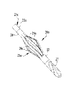
20 In FIG. 7, the shaft 21a is coupled to a collar 31 at its distal end
23a. The cutting
arms 26a couple the collar 31 to a distal collar 32, which, in turn, couples
the expandable
cutting device 25a to the distal nose section or drill tip 27. Thus, in the
device 20a illustrated
in FIG. 7, the shaft 21a and cutting device 25a may be fabricated or formed
separately and
coupled together during assembly.
FIGS. 12-16 illustrate a surgical instrument 20b that has a cutting device 25b
with
helical arms 26b. FIG. 21 illustrates a shaft 21c that passes through the
cutting arms 26c and
collars 3Ic and 32c. Regardless of the shaft construction and the cutting
device construction,
the surgical instruments 20-20c include flexible shafts 21-21c that are
coupled to an
expandable cutting device 25-25c at distal ends 23-23c of the shafts 21-21c
and a drill
attachment connector or handle is coupled to the proximal ends 22 of the
shafts 21-21c.
The shafts 21-21c, cutting arms 26-26c, optional collars 31, 32, 31c, 32c,
optional
distal shaft section 28 and optional drill tip 27 may be fabricated from a
single piece of
flexible material, such as a shape memory material. For example, the shaft 21
and cutting
CA 02766506 2011-12-22
WO 2011/011664
PCT/US2010/043018
8
arms 26 are fabricated from a single piece of nitinol (nickel-titanium shape
memory alloy
(SMA)). Other suitable shape memory materials include, but are not limited to,
alloys of
titanium-palladium-nickel, nickel-titanium-copper, gold-cadmium, iron-zinc-
copper-
aluminum, titanium-niobium-aluminum, uranium-niobium, hafnium-titanium-nickel,
iron-
manganese-silicon, nickel-titanium, nickel-iron-zinc-aluminum, copper-aluminum-
iron,
titanium-niobium, zirconium-copper-zinc, and nickel-zirconium-titanium. The
shape
memory alloys may be suitable for the fabrication of surgical instruments for
cutting
cancellous bone without cutting cortical bone. Other suitable shape memory
materials other
than metallic alloys and polymers are possible as will be apparent to those
skilled in the art.
Furthermore, in some implementations with different requirements, such as
where substantial
radial collapse of the cutting device 25-25c and cutting arms 26-26c is not
required, the arms
26-26c could be made from other metals or plastics.
The flexibility of the shafts 21-21c, is provided by a small shaft diameter
and by
selecting a material having a modulus of elasticity falling within a desired
range. In addition
to fabricating the shafts 21-21c from a shape memory alloy as described above,
the shafts 21-
21c may also be fabricated from a high-strength biocompatible polymer, such as
polyetheretherketone (PEEK), polyethereketone (PEK), high density polyethylene
(HDPE),
or a polyamide such as nylon. As will be apparent to those skilled in the art,
other suitable
polymers are available.
The expandable cutting device 25 illustrated in FIGS. 1-3 and 7 comprises two
or
more expandable elongated cutting arms 26. Referring to FIGS. 1-2, the cutting
arms 26 are
disposed between the distal end 23 of the shaft 21 and the optional distal
shaft section 28 or
the distal nose section or drill tip 27. As shown in FIG. 7, the cutting arms
26 may be
disposed between a pair of collars 31, 32. Alternatively, the cutting arms 26
can be coupled
to a pair of collars 31a, 32a that are slidably received over the distal end
23a of a continuous
shaft 2 lb, as illustrated in FIG. 21. In the device 20c of FIG. 21, one or
more pins or other
attachment mechanisms may hold the collars 31c, 32c in place on the shaft 21c.
The cutting arms 26-26c may form a cage-like structure. For some applications,
the
shape memory material or alloy used to fabricate the arms 26-26c should
exhibit elastic
properties. The designs illustrated in FIGS. 1-3, 7, 12-16, and 21 exploit the
elastic
properties of shape memory alloys to allow the cutting arms 26-26c expand
outward upon
entry in the IM canal to their original shape. The cutting arms 26-26c are
also designed to be
sufficiently flexible so that harder cortical bone will cause the arms to
deflect in a radially-
CA 02766506 2011-12-22
WO 2011/011664
PCT/US2010/043018
9
inward direction and to not cut cortical bone. In contrast, the arms 26-26c
are sufficiently
resilient to cut cancellous bone and other weaker materials disposed within
the cortical wall.
The cutting arms 26-26c can be machined using traditional techniques such as
chemical etching, laser cutting, or milling, among other techniques. The cage
structure of the
expandable cutting devices 25-25c can be formed by placing a cutting device
into a fixture
that compresses the cutting arms 26-26c axially and causes the cutting arms 26-
26c to expand
radially outward to the desired relaxed profile or relaxed diameter (compare
FIGS. 8 and 9).
The fixture and cutting devices 25-25c may then be placed in an oven at a
temperature of
about 842 F (450 C) for about 15 minutes, followed by water quenching shortly
after
removal from the oven. This process causes the cutting arms or elements 26-26c
to be
shaped into a desired profile. The cutting arms 26-26c may be sharpened on at
least one
lateral surface 33 (FIG. 3), 33b (FIG. 16) to enable cutting of cancellous
bone material. The
benefit of the sharpening the cutting arms 26-26c is to provide a smoother
cutting operation
by reducing chatter or vibration when cutting, and by requiring a lower
cutting torque.
To selectively cut cancellous bone material and not cut cortical bone
material, the
cutting arms must have the appropriate combination of resilience, or strength,
and elasticity.
Generally, the flexible cutting arms 26 should have a ratio of width (w) to
thickness (t)
ranging from about 5:1 to about 2:1 and ratio of length (L) to width (w)
ranging from about
20:1 to about 6:1. In one example, the material of the cutting arms 26 is
nitinol and the
elements have a cross-sectional thickness (t) of about 0.014 in (0.356 mm), a
width (w) of
about 0.056 in (1.42 mm) and a length (L) of about 0.75 in (19.05 mm) (see
also FIG. 8).
These dimensions are an example that allow the cutting arms 26-26c to be
strong enough to
cut cancellous material as the cutting device 25 rotates while being flexible
enough to
compress radially when the arms 26-26c engage cortical bone. The dimensions
will vary
depending upon the anatomy or size of IM canal in which a cavity is to be
formed.
Additional methodologies for calculating other appropriate dimensions of the
cutting
arms 26-26c include consideration of moment of inertia (I), expansion force
(P) and the
deflection (3) of the cutting arms 26-26c. Specifically, the behavior of the
cutting arms 26-
26c of the expandable cutting device 25-25c can be predicted by treating the
arms 26-26c as a
leaf spring 35, illustrated in FIGS. 10 and 11. The body of leaf spring 35 has
a length (L), a
width (w), and a thickness (t). Using a traditional beam deflection
calculation, the amount of
deflection (6) can be expressed as Equation 1.
= PL3/48EI (1)
CA 02766506 2011-12-22
WO 2011/011664
PCT/US2010/043018
In equation 1, (I) is the moment of inertia and (E) is the modulus of
elasticity. For
nitinol, E can range from about 5.8 x 10⁶ psi (40.0 GPa) to about 10.9 x 10⁶
psi (75.2 GPa).
Referring to FIG. 11, the moment of inertia (I) can be calculated from
Equation 2.
I = wt3/12 (2)
5 To allow for ease of insertion of the instruments 20-20c into an IM
canal, the
expansion force (P) of the arms 26-26c should not be excessive. However, to
expand
adequately in the IM canal, the expansion force (P) must be above a minimum
value.
Therefore, the design of the arms 26-26c should provide an optimal expansion
force (P).
Through laboratory experimentation, the expansion force can range from about
1.0 lbf to
10 about 8.0 lbf (from about 4.45 N to about 35.59 N).
By substituting Equation 2 into Equation 1 and solving for P, the expansion
force (P)
can be expressed as equation 3.
5 = PL3/4Ewt3, and therefore P = 4SEwt3/L3 (3)
As another example, if L = 0.65 in (15.61 mm), w = 0.060 in (1.52 mm), t =
0.018 in
(0.457 mm), and S = 0.085 in (2.16 mm), then an expansion force of P = 2.51
lbf is provided
by equation 3, which falls within the range of from about 1.0 lbf to about 8.0
lbf (from about
4.45 N to about 35.59 N). As 5 and P are proportional when w, t, and L, are
fixed, the
deflection 1 can be increased by about 300% by changing the size of the
fixture used during
the heat treatment process before P approaches the 8.0 lbf upper limit for the
dimensions
recited immediately above. The value of deflection 5 desired in a give
implementation will
be dependent upon the particular bone being treated and the size of the IM
canal. In other
implementations, the dimensions and parameters discussed above can vary
greatly, as will be
apparent to those skilled in the art.
FIGS. 12-16 illustrate another surgical instrument 20b with a flexible shaft
21b
having a proximal end 22 and a distal end 22. The distal end 23b of the shaft
2Ib is coupled
to an expandable cutting device 25b with helical cutting arms 26b. The helical
cutting arms
26b also include opposing sides or cutting edges 33b. The helical cutting arms
26b reduce
tensile and shear stresses at the bases 29 (FIG. 16) of the cutting arm 26b so
as to reduce the
possibility of device failure. The helix formed by the helical cutting arms
26b can be
designed to optimize the ease of cutting. The helix can be left-hand helical
or right-hand
helical and can be formed at an angle from about negative 60 degree to about
60 degrees
CA 02766506 2011-12-22
WO 2011/011664
PCT/US2010/043018
11
from a longitudinal axis of the surgical instrument. For example, left-hand
helical cutting
arms in a right-hand cut may be used.
The optional brad drill tip 27 can have a diameter that is slightly larger
than a
diameter of the shaft 21-21c or that is larger than a diameter of the cutting
arms 26-26c when
the cutting arms 26-26c are compressed. A slightly larger diameter of the
drill tip 27 enables
the drill tip 27 to create an entry portal 41 in cortical bone 42 to allow for
passage of the
remainder of the instrument 20-20c into the IM canal 46, as illustrated in
FIGS. 18 and 19.
The drill tip 27 will also prove useful in reaming an IM canal 46 that is
smaller than expected
or has an endosteal surface profile that is smaller than expected.
Incorporating a drill tip 27
on the device allows for the user to create the non-axial pilot/entry hole 41
in the cortical wall
42 to gain an access portal to the IM canal 46 and fracture site 47. Thus, a
separate drilling
tool may not be needed to create the entry portal 41 as the proximal end 22 of
the shaft 21-
21c may be coupled to a surgical drill 24 as shown in FIGS. 17, 19, and 20.
The tip 27 also
allows for cutting a pathway in the IM canal where a minimum diameter in
desired. For
example, to accommodate a specific sized implant, such as a nail, the tip 27
can be used to
drill a hole in the 1M canal for receiving the nail.
The shafts 21-21c may include a lumen 43 (FIGS. 18-20) to allow for suction
and
debris removal or, alternatively, for the delivery of irrigation fluid. As
shown in FIG. 20, the
shaft 21 may be disposed within an outer lumen 51 that can be used for suction
or for the
delivery of irrigation fluid. In the embodiment illustrated in FIG. 20, the
shaft 21 may also
accommodate an inner lumen 43 and be disposed axially within an outer lumen
51. The outer
lumen 51 and the inner lumen 43 may each be connected to a reservoir of
irrigation fluid or a
suction pump shown schematically at 52, 53 respectively. The bi-directional
arrows 54, 55
are intended to indicate that the outer lumen 51 and inner lumen 43 can be
used for either
suction or irrigation or both if only a single lumen 43, 51 is utilized. A
surgical drill 24 is
also shown schematically in FIG. 20 that is coupled to the proximal end 22 of
the shaft 21.
The components of the instruments 20-20c can be coupled to one another by a
variety
of means such as welding, pinning, adhesive bonds, mechanical locks (retaining
ring), etc.
The cutting arms 26-26c, in addition to having at least one sharpened edge 33,
33c may
include serrations, relief angles, and dual sharpened edges. Further, a series
of the
expandable cutting devices 25-25c may be disposed along the length of the
shaft 21-21c. As
noted above, the cage structure of the expandable cutting device 25-25c and/or
the drill tip 27
can be an integral with the shaft 21-21c.
CA 027 6650 6 2011-12-22
WO 2011/011664
PCT/US2010/043018
12
The arms 26-26c of the disclosed cutting devices 25-25c are designed to have a
high
moment of inertia Tin the direction of rotation and a lower moment of inertia
Tin the
transverse radially inward direction. The disclosed designs for the arms 26-
26c permit the
arms 26-26c to be strong enough to cut cancellous bone in an IM canal 46 when
rotating, but
elastic enough in a radial direction such that when the arms 26-26c encounter
a hard tissue
such as cortical bone, the arms 26-26c will be deflected in a radially inward
direction thereby
causing no or minimal trauma to the cortical bone 42. As a result, cancellous
bone in the
non-symmetrical non-circular cross-sectional IM canal 46 is cut without
substantial trauma or
removal of cortical bone 42.
FIG. 17 illustrates the flexibility of the shaft 21 connected to the drill 24.
The use of
flexible but adequately stiff shafts 21-21c allows for advancement of the
devices 20-20c
through an IM canal 46 towards a fracture site 47 and the creation of non-
traditional (i.e.,
non-axial) entry ports such as the one shown at 41 in FIGS. 18-19. Using a
material such as
reinforced PEEK or other biocompatible polymer for the shafts 21-21c, or other
structures
such as steel cable or twisted wire, offers an inexpensive solution as
compared to other
flexible shafts fabricated from nitinol, other shape memory alloys or laser
cut metal shafts.
While only certain embodiments have been set forth, alternatives and
modifications
will be apparent from the above description to those skilled in the art. These
and other
alternatives are considered equivalents and within the spirit and scope of
this disclosure and
the appended claims.