Note: Descriptions are shown in the official language in which they were submitted.
CA 02767032 2012-02-01
TITLE: METHODS FOR CONTROLLING ONE OR MORE PARAMETERS OF A FLOW
CYTOMETER TYPE MEASUREMENT SYSTEM
BACKGROUND OF THE INVENTION
1. Field of the Invention
This invention generally relates to methods for controlling one or more
parameters of a flow cytometer type
measurement system. Certain embodiments relate to methods that include
altering one or more parameters of a flow
cytometer type measurement system in real time based on monitoring of the
parameter(s).
2. Description of the Related Art
The following descriptions and examples are not admitted to be prior art by
virtue of their inclusion within
this section.
Generally, flow cytometers provide measurements of fluorescence intensity of
laser excited polystyrene
beads or cells as they pass linearly through a flow chamber. However, flow
cytometers can also be used to provide
measurements of one or more properties of other particles. Some systems are
configured to perform measurements
of the level of light scattered by particles at 90 or 180 to the excitation
source, two or more measurements of
fluorescence used to determine classification, which is the particle
"identity," and additional fluorescence
measurements known as "reporters," typically used to quantify chemical
reactions of interest. Each of the
fluorescent measurements is made at different wavelengths.
As the measurement capability of flow cytometer type measurement instruments
has improved, the
applications in which flow cytometers can provide useful measurements has
increased drastically. For example,
flow cytometers have become increasingly useful in providing data for
applications such as biological assays (e.g.,
displacement or competition assays, non-competition assays, enzyme assays),
nucleic acid analysis, and
combinatorial chemistry. In particular, the popularity of flow cytometer
measurements has dramatically increased
due to the speed with which assays can be performed particularly in comparison
to other assay methods (e.g.,
conventional enzyme linked immunosorbent assay "ELISA" format).
Under normal circumstances, calibration of flow cytometers occurs as one or
more preliminary steps in
preparing instruments for proper use and measurement to ensure accurate and
reliable assay results. In addition,
unless the fluorescence channels of each flow cytometer are calibrated to read
the same, there is no assurance as to
the source of variation among samples. It is likely that one instrument will
give different readings on the same
sample on different days if robust and complete calibration methods are not
employed. Similarly, if there is no
assurance that any two instruments will provide the same results even if
properly set up, although flow cytometry
may provide a better measure of identifying and distinguishing between cells
in a sample, its use as a clinical
instrument may be diminished.
Accordingly, many different methods for calibrating a flow cytometer have been
developed. Initially,
significant work was done to develop calibration methods that reduced the
level of involvement of the operator in
calibration to increase the accuracy of the calibration. This work led, in
large part, to the automation of many steps
of the calibration of flow cytometers. In addition, significant work was done
to improve the accuracy of the
calibration in other ways. For example, this work has led to advancement in
calibrations such as using calibration
1
CA 02767032 2012-02-01
standards that have uniform and constant properties. In particular, since the
properties of biological samples can
change over time, biological calibration standards for flow cytometers have
generally been replaced with synthetic
calibration standards (e.g., polymeric microspheres or particles) that have
more stable properties. In addition,
typically the calibration microspheres have properties (e.g., size, volume,
surface characteristics, granularity
properties, refractive index, fluorescence, etc.) that are substantially
similar (i.e., as close as possible) to the
properties of the test microspheres. Such calibration microspheres were
believed to increase the accuracy of the
flow cytometer by performing calibration at values that are as close as
possible to the values that were expected
during testing.
Attempts to improve the calibration of flow cytometers have also led to
increasing the number of
parameters of the flow cytometer that are accounted for by calibration. For
example, the laser excitation, detectors,
and electronics of flow cytometer measurement systems vary over time, which
affects the final measurement.
Therefore, these, and sometimes other, parameters of flow cytometers are
typically accounted for by calibration
methods.
Other parameters, which are more difficult to control, also affect the
measurements of a flow cytometer. One such
parameter is sample velocity. One example of a method for measuring sample
velocity is illustrated in U.S. Patent
No. 6,532, 061 to Ortyn et al. In this method, objects are entrained in a flow
of fluid, which is caused to flow
through the sensitive or measurement volume. In each of these embodiments,
optical gratings having a substantially
uniform pitch are employed to modulate light received from the moving objects.
The modulated light is converted
into an electrical signal, which is digitized and then processed using a Fast
Fourier Transform (FFT) to determine
the velocity of the object. There are, however, several disadvantages to the
methods and systems described by
Ortyn et al. for measuring sample velocity. For example, the methods require
fairly complex optical gratings and
software. In addition, due to the precision required for the optical gratings
and the complexity of manufacturing, the
optical gratings may be fairly expensive. Furthermore, the sample velocity
measurements may be somewhat
inaccurate due, for example, to the optical distortion of the detected light
by the moving objects.
However, the most significant error contribution in flow cytometer
measurements is generally caused by
temperature variance. In addition, it has been found that the effect of
temperature variance on the measurements
performed by a flow cytometer is not adequately accounted for by the presently
available calibration methods. For
example, the methods and systems described by Ortyn et al., although
attempting to correct for a number of
parameters, do not take into account temperature variations and how they
affect the measurements of a flow
cytometer. Therefore, although many different calibration methods are
available, additional improvements to each
of these methods can be made by more accurately accounting for temperature
variations during different flow
cytometer measurements or during individual flow cytometer measurements.
Accordingly, it may be advantageous to develop methods for controlling at
least the major error
contributing components of flow cytometer measurement systems, which could be
combined to produce a real time
calibration scheme.
2
CA 02767032 2012-02-01
SUMMARY OF THE INVENTION
As set forth in detail above, the most significant error contribution in flow
cytometers is generally caused
by temperature variance. Since the temperature may be a measured quantity, and
the physics behind its effects are
known, it is possible to reduce, and even nullify, the most critical of these
error sources.
Several measurement error contributors and real time correction techniques for
the measurement error
contributors have been identified. In addition, a real time fine-tuning method
using calibration microspheres
uniquely identifiable via a diameter at least slightly different from those
being measured, which may be included in
microsphere sample mixes, has been created. Added features of the fine-tuning
process may include real time
identification of system health, correction of non-linearities in one or more
channels, and/or the significant extension
of a flow cytometer measurement system's useful reporter dynamic range. The
described embodiments are useful to
compensate for system variations primarily due to temperature, thus extending
the calibrated range of operation.
In addition, it is to be noted that several different embodiments of methods
for controlling one or more
parameters of a flow cytometer type measurement system are described herein.
It is to be understood that each of
the methods may be used and performed separately. In addition, two or more of
the methods may be used or
performed in combination depending on, for example, the variability in various
components of the measurement
system and/or the desired accuracy of the measurement system.
One embodiment of the present invention relates to a method for controlling
one or more parameters of a
flow cytometer type measurement system. The method includes monitoring the one
or more parameters of the flow
cytometer type measurement system during measurements of sample microspheres
by the measurement system. The
method also includes altering the one or more parameters in real time based on
the monitoring.
In one embodiment, monitoring the one or more parameters may include
monitoring the one or more
parameters using measurements of calibration microspheres. The calibration
microspheres have diameters that are
different than (e.g., less than) diameters of the sample microspheres. In some
embodiments, the one or more
parameters may include output signals produced by detectors of the measurement
system. The output signals are
responsive to light scattered by the sample microspheres.
In another embodiment, monitoring the one or more parameters may include
monitoring the one or more
parameters using measurements of calibration microspheres. In this embodiment,
the calibration microspheres have
diameters that are different than (e.g., less than) diameters of the sample
microspheres, and at least some of the
calibration microspheres have different spectral addresses. In one such
embodiment, the one or more parameters
may include a dynamic range of the measurement system. In another embodiment,
altering the parameter(s) may
include extending a linear dynamic range of one or more channels of the
measurement system. In an additional
embodiment, the one or more parameters may include a measurement of system
health. The measurement of system
health may include health of a classification channel, health of a reporter
channel, or a combination thereof. In some
embodiments, the one or more parameters may include linearity in the
measurements of the sample microspheres. In
such an embodiment, the measurements may include measurements of a
classification channel, measurements of a
reporter channel, or a combination thereof. In another such embodiment,
altering the parameter(s) may include
substantially correcting any non-linearity in the measurements.
3
CA 02767032 2012-02-01
In some embodiments, the parameter(s) may include a parameter of an avalanche
photo diode of the
measurement system. In one such embodiment, the method may also 'include
determining a correction factor to be
used in altering the parameter(s) using empirically derived data. In another
embodiment, the parameter(s) may
include a parameter of a photomultiplier tube of the measurement system.
In a further embodiment, the parameter(s) may include a velocity of the sample
microspheres. In one such
embodiment, monitoring the parameter(s) may include monitoring a temperature
of a fluid in which the sample
microspheres are disposed and determining the velocity of the sample
microspheres from the temperature. In some
embodiments, the method may also include calibrating the one or more
parameters prior to the measurements of the
sample microspheres. Each of the embodiments of the method described above may
include any other step(s)
described herein.
Another embodiment relates to a different method for controlling one or more
parameters of a flow
cytometer type measurement system. This method includes monitoring a
temperature proximate to the flow
cytometer type measurement system. The method also includes altering a bias
voltage of an avalanche photo diode
of the measurement system in response to the temperature using empirically
derived data to substantially correct for
variation in a gain of the avalanche photo diode due to the temperature.
In one embodiment, the method may also include generating the empirically
derived data by applying a
substantially constant light level to the avalanche photo diode at one or more
temperatures and recording a current
output of the avalanche photo diode for multiple bias voltages at the one or
more temperatures. In another
embodiment, altering the parameter(s) is performed before sample measurements
are performed by the measurement
system. In such an embodiment, the bias voltage may be substantially constant
throughout the sample
measurements. In a different embodiment, monitoring the parameter(s) and
altering the parameter(s) are performed
in real time.
In some embodiments, the method may also include varying the bias voltage of
the avalanche photo diode
while calibration microspheres that emit light of known intensity are measured
by the measurement system until a
predetermined signal level is obtained from the avalanche photo diode. In one
such embodiment, the method may
further include determining a corresponding relative current for the avalanche
photo diode from a reverse bias
voltage for the avalanche photo diode, the bias voltage at the predetermined
signal level, and the temperature. This
embodiment of the method may also include determining the bias voltage using
the corresponding relative current,
the temperature, the reverse bias voltage, and the empirically derived data.
Each of the embodiments of the method
described above may include any other step(s) described herein.
An additional embodiment relates to yet another method for controlling one or
more parameters of a flow
cytometer type measurement system. This method includes monitoring a
temperature proximate to the flow
cytometer type measurement system. The method also includes altering an output
signal of a photomultiplier tube of
the measurement' system in response to the temperature using a characteristic
curve for the photomultiplier tube to
substantially correct for variation in a gain of the output signal of the
photomultiplier tube. The gain of the
photomultiplier tube varies approximately linearly in response to the
temperature. In some embodiments, the
photomultiplier tube is part of a reporter channel of the measurement system.
In another embodiment, the
characteristic curve for the photomultiplier tube varies with detection
wavelength and cathode construction of the
photomultiplier tube. Each of the embodiments of the method described above
may include any other step(s)
described herein.
4
CA 02767032 2012-02-01
Another embodiment relates to yet a different embodiment of a method for
controlling one or more
parameters of a flow cytometer type measurement system. This method includes
setting a voltage of a
photomultiplier tube of the measurement system at a first value and a second
value. The method also includes
measuring an output current of the photomultiplier tube at the first and
second values. In addition, the method
includes determining a calibration voltage of the photomultiplier tube from a
log of the first and second values
versus a log of the output currents at the first and second values. The method
further includes applying the
calibration voltage to the photomultiplier tube. The method also includes
testing the photomultiplier tube to
determine if one or more parameters of the photomultiplier tube are within
predetermined tolerances. Each of the
embodiments of the method described above may include any other step(s)
described herein.
An additional embodiment relates to another method for controlling one or more
parameters of a flow
cytometer type measurement system. This method includes determining a
calibration voltage of a detector of the
measurement system using successive approximation. The method also includes
applying the calibration voltage to
the detector. In one embodiment, the detector may include an avalanche
photodiode. In a different embodiment, the
detector may include a photomultiplier tube.
In one embodiment, the method may include comparing the calibration voltage to
a breakdown voltage of
the detector and repeating the determination of the calibration voltage if the
calibration voltage exceeds the
breakdown voltage. A different embodiment of the method includes collecting
and processing detector samples to
determine a detector signal level. In one such embodiment, the method may
include comparing the detector signal
level to a calibration target signal level and if the detector signal level is
above the calibration target signal level,
then reducing a bias voltage of the detector, and repeating the determination
of the calibration voltage. In another
such embodiment, the method may include comparing the detector signal level to
a calibration target signal level and
if the detector signal level is not within a predetermined range of the
calibration target signal level, then repeating
determination of the calibration voltage until all desired detector voltage
levels have been attempted. Each of the
embodiments of the method described above may also include any other step(s)
described herein.
A further embodiment relates to a different method for controlling one or more
parameters of a flow
cytometer type measurement system. This method includes monitoring a
temperature of a fluid that will flow
through the flow cytometer type measurement system. Sample microspheres are
disposed in the fluid. The method
also includes determining a velocity of the sample microspheres in the
measurement system from a viscosity of the
fluid at the temperature.
In one embodiment, the method may also include determining a length of time
that one of the sample
microspheres will be present in a detection window of the measurement system
based on the velocity. In some
embodiments, the method may include determining a length of time in which one
of the sample microspheres will
travel from one detection window of the measurement system to another
detection window of the measurement
system based on the velocity. In another embodiment, the method may include
determining when one of the sample
microspheres will be present in a detection window of the measurement system
based on the velocity. In yet another
embodiment, the method may include controlling a sampling interval for one or
more detection windows of the
measurement system to compensate for the velocity.
In an additional embodiment, monitoring the parameter(s) and determining the
velocity are performed prior
to performing measurements of the sample microspheres with the measurement
system. In some embodiments, the
method niay include determining one or more properties of output signals of
the measurement system from the
CA 02767032 2012-02-01
velocity. In one such embodiment, the method includes correcting the output
signals for error due to the velocity
using correction factors. The correction factors are determined using
empirical measurements. In another
embodiment, the measurement system is configured to maintain a substantially
constant pressure of the fluid during
measurements of the sample microspheres.
In one embodiment, determining the velocity may include determining the
velocity from a table, from
Poiseuille's equation, or from predetermined values of velocity versus
temperature. In some such embodiments, the
method may also include controlling a pressure of the fluid during
measurements of the sample microspheres based
on the velocity. Each of the embodiments of the method described above may
include any other step(s) described
herein.
A different embodiment relates to another method for controlling one or more
parameters of a flow
cytometer type measurement system. This method includes measuring a time in
which a microsphere travels from a
first detection window of the flow cytometer type measurement system to a
second detection window of the
measurement system. The method also includes altering an applied pressure of
the measurement system such that
the time is substantially constant. In one embodiment, the time is an average
time. The microsphere may be a
sample microsphere or a calibration microsphere. Measuring the time may
include measuring light scattered by the
microsphere in the first and second detection windows. In another embodiment,
measuring the time may include
measuring light scattered by the microsphere in the first and second detection
windows with one detector. The light
scattered by the microsphere in the first and second detection windows may be
directed to the one detector by one
beamsplitter. The method may or may not be performed in real time. Each of the
embodiments of the method
described above may include any other step(s) described herein.
A further embodiment relates to a different method for controlling one or more
parameters of a flow
cytometer type measurement system. This method includes measuring an average
time in which microspheres travel
from a first detection window of the flow cytometer type measurement system to
a second detection window of the
measurement system. The microspheres may include sample microspheres,
calibration microspheres, or calibration
and sample microspheres. The method also includes comparing the average time
to a reference time in which a
reference microsphere traveled from the first detection window to the second
detection window. In addition, the
method includes altering an applied pressure of the measurement system if a
difference between the average time
and the reference time is larger than a predetermined value.
In one embodiment, altering the applied pressure includes increasing the
applied pressure if the average
time is larger than the reference time. Alternatively, altering the applied
pressure includes decreasing the applied
pressure if the average time is smaller than the reference time. In some
embodiments, the predetermined value is
selected to compensate for known time variation mechanisms of the measurement
system. This method may or may
not be performed in real time. Each of the embodiments of the method described
above may include any other
step(s) described herein.
6
CA 02767032 2012-02-01
BRIEF DESCRIPTION OF THE DRAWINGS
Other objects and advantages of the invention will become apparent upon
reading the following detailed
description and upon reference to the accompanying drawings in which:
Fig. 1 is a schematic diagram illustrating one example of a measurement system
that may be used to carry
out the methods described herein;
Fig. 2 is a graph illustrating one example of multiple bias curves showing the
response of an APD, having
a reverse bias voltage (V60) of 130 volts, as a function of temperature;
Fig. 3 is a graph illustrating the response of various PMTs as a function of
temperature;
Fig. 4 is a graph illustrating one example of the log of gain of a PMT as a
function of the log of the PMT
bias voltage;
Fig. 5 is a flow chart illustrating one embodiment of a method for controlling
one or more parameters of a
flow cytometer type measurement system;
Fig. 6 is a schematic diagram illustrating a cross-sectional view of one
embodiment of a portion of a
measurement system that may be used to carry out at least one of the methods
described herein; and
Fig. 7 is an illustration of the pulse train (i.e., scattered light measured
at different times) that may be
measured in one of the embodiments of the methods described herein.
While the invention is susceptible to various modifications and alternative
forms, specific embodiments
thereof are shown by way of example in the drawings and will herein be
described in detail. It should be
understood, however, that the drawings and detailed description thereto are
not intended to limit the invention to
the particular form disclosed.
DETAILED DESCRIPTION OF THE PREFERRED EMBODIMENTS
Several different embodiments of methods for controlling one or more
parameters of a flow cytometer
type measurement system are described herein. As noted above, each of the
methods may be used and performed
separately. In addition, two or more of the methods may be used or performed
in combination depending on, for
example, the variability in various components of the measurement system
and/or the desired accuracy of the
measurement system.
Although embodiments are described herein with respect to microspheres or
polystyrene beads, it is to be
understood that the measurement systems and methods may also be used with
microparticles, gold nanoparticles,
beads, microbeads, latex particles, latex beads, fluorescent beads,
fluorescent particles, colored particles, colored
beads, and cells. The microspheres may serve as vehicles for molecular
reactions. Examples of appropriate
microspheres, beads, and particles are illustrated in U.S. Patent Nos.
5,736,330 to Fulton, 5,981,180 to Chandler et
al., 6,057,107 to Fulton, 6,268,222 to Chandler et al., 6,449,562 to Chandler
et al., 6,514,295 to Chandler et al.,
6,524,793 to Chandler et al., and 6,528,165 to Chandler. The measurement
systems and methods described herein
may be used with any of the microspheres, beads, and particles described in
these patents. In addition, microspheres
for use in flow cytometry may be obtained from manufacturers such as Luminex
Corp., Austin, Texas. The terms
"beads" and "microspheres" are used interchangeably herein.
7
CA 02767032 2012-02-01
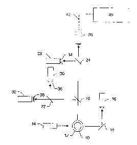
Fig. 1 illustrates one example of a measurement system that may be used to
perform the methods described
herein. In particular, one or more parameters of the measurement system
illustrated in Fig. 1 may be determined,
monitored, altered, and/or controlled according to the methods described
herein. It is noted that the figures
described herein are not drawn to scale. In particular, the scale of some of
the elements of the figures are greatly
exaggerated to emphasize characteristics of the elements. Some elements of the
measurement systems have not been
included in the figures for the sake of clarity.
In Fig. 1, the measurement system is shown along a plane through the cross-
section of cuvette 12 through
which microspheres 10 flow. In one example, the cuvette may be a standard
quartz cuvette such as that used in
standard flow cytometers. Any other suitable type of viewing or delivery
chamber, however, may also be used to
deliver the sample for analysis. The measurement system includes light source
14. Light source 14 may include any
appropriate light source known in the art such as a laser. The light source
may be configured to emit light having
one or more wavelengths such as blue light or green light. Light source 14 may
be configured to illuminate the
microspheres as they flow through the cuvette. The illumination may cause the
microspheres to emit fluorescent
light having one or more wavelengths or wavelength bands. In some embodiments,
the system may include one or
more lenses (not shown) configured to focus light from the light source onto
the microspheres or the flowpath. The
system may also include more than one light source. In one embodiment, the
light sources may be configured to
illuminate the microspheres with light having different wavelengths or
wavelength bands (e.g., blue light and green
light). In some embodiments, the light sources may be configured to illuminate
the microspheres at different
directions.
Light scattered forwardly from the microspheres may be directed to detection
system 16 by folding mirror
18 or another suitable light directing component. Alternatively, detection
system 16 may be placed directly in the
path of the forwardly scattered light. In this manner, the folding mirror or
other light directing components may not
be included in the system. In one embodiment, the forwardly scattered light
may be light scattered by the
microspheres at an angle of about 180 from the direction of illumination by
light source 14, as shown in Fig. 1.
The angle of the forwardly scattered light may not be exactly 180 from the
direction of illumination such that
incident light from the light source may not impinge upon the photosensitive
surface of the detection system. For
example, the forwardly scattered light may be light scattered by the
microspheres at angles less than or greater than
180 from the direction of illumination (e.g., light scattered at an angle of
about 170 , about 175 , about 185 , or
about 190 ).
Light scattered by the microspheres at an angle of about 90 from the
direction of illumination may also be
collected. In one embodiment, this scattered light may be separated into more
than one beam of light by one or
more beamsplitters or dichroic mirrors. For example, light scattered at an
angle of about 90 to the direction of
illumination may be separated into two different beams of light by
beamsplitter 20. The two different beams of light
may be separated again by beamsplitters 22 and 24 to produce four different
beams of light. Each of the beams of
light may be directed to a different detection system, which may include one
or more detectors. For example, one of
the four beams of light may be directed to detection system 26. Detection
system 26 may be configured to detect
light scattered by the microspheres.
Scattered light detected by detection system 16 and/or detection system 26 may
generally be proportional
to the volume of the particles that are illuminated by the light source.
Therefore, output signals of detection system
16 and/or output signals of detection system 26 may be used to determine a
diameter of the particles that are in the
8
CA 02767032 2012-02-01
illumination zone or detection window. In addition, the output signals of
detection system 16 and/or detection
system 26 may be used to identify more than one particle that are stuck
together or that are passing through the
illumination zone at approximately the same time. Therefore, such particles
may be distinguished from other sample
microspheres and calibration microspheres. Furthermore, the output signals of
detection system 16 and/or detection
system 26 may be used to distinguish between sample microspheres and
calibration microspheres as described
herein based on size.
The other three beams of light may be directed to detection systems 28, 30,
and 32. Detection systems 28,
30, and 32 may be configured to detect fluorescence emitted by the
microspheres. Each of the detection systems
may be configured to detect fluorescence of a different wavelength or a
different range of wavelengths. For
example, one of the detection systems may be configured to detect green
fluorescence. Another of the detection
systems may be configured to detect yellow-orange fluorescence, and the other
detection system may be configured
to detect red fluorescence.
In some embodiments, spectral filters 34, 36, and 38 may be coupled to
detection systems 28, 30, and 32,
respectively. The spectral filters may be configured to block fluorescence of
wavelengths other than that which the
detection systems are configured to detect. In addition, one or more lenses
(not shown) may be optically coupled to
each of the detection systems. The lenses may be configured to focus the
scattered light or emitted fluorescence
onto a photosensitive surface of the detectors.
The detector's output current is proportional to the fluorescent light
impinging on it and results in a current
pulse. The current pulse may be converted to a voltage pulse, low pass
filtered, and then digitized by an A/D
converter. Processor 40 such as a DSP integrates the area under the pulse to
provide a number which represents the
magnitude of the fluorescence. In addition, the processor may perform
additional functions described herein (e.g.,
monitoring one or more parameters of the flow cytometer type measurement
system, altering the one or more
parameters in real time based on the monitored parameter(s), etc.). As shown
in Fig. 1, processor 40 may be
coupled to detector 26 via transmission medium 42. Processor 40 may also be
coupled to detector 26 indirectly via
transmission medium 42 and one or more other components (not shown) such as
the A/D converter. The processor
may be coupled to other detectors of the system in a similar manner.
In some embodiments, the output signals generated from fluorescence emitted by
the microspheres may be
used to determine an identity of the microspheres and information about a
reaction taking place on the surface of the
microspheres. For example, output signals of two of the detection systems may
be used to determine an identity of
the microspheres, and output signals of the other detection system may be used
to determine a reaction taking place
on the surface of the microspheres. Therefore, the selection of the detectors
and the spectral filters may vary
depending on the type of dyes incorporated into or bound to the microspheres
and/or the reaction being measured
(i.e., the dye(s) incorporated into or bound to the reactants involved in the
reaction).
The detection systems that are used to determine an identity of the sample
microspheres (e.g., detection
systems 28 and 30) may be APDs, a PMT, or another photodetector. The APDs may
be corrected in real time for
gain variation as a function of temperature as described herein. The detection
system that is used to identify a
reaction taking place of the surface of the microspheres (e.g., detection
system 32) may be a PMT, an APD, or
another form of photodetector. The PMT may be corrected using a simple
multiplier derived from PMT
characteristic curves that can be applied to the output signals of the PMT as
described herein. The detectors and the
measurement system may be further configured as described herein.
9
CA 02767032 2012-02-01
Although the system of Fig. I is shown to include two detection systems having
two different detection
windows for distinguishing between microspheres having different dye
characteristics, it is to be understood that the
system may include more than two such detection windows (i.e., 3 detection
windows, 4 detection windows, etc.).
In such embodiments, the system may include additional beamsplitters and
additional detection systems having other
detection windows. In addition, spectral filters and/or lenses may be coupled
to each of the additional detection
systems.
In another embodiment, the system may include two or more detection systems
configured to distinguish
between different materials that are reacted on the surface of the
microspheres. The different reactant materials may
have dye characteristics that are different than the dye characteristics of
the microspheres.
Additional examples of measurement systems that may be used to perform the
methods described herein
are illustrated in U.S. Patents Nos. 5,981,180 to Chandler et al., 6,046,807
to Chandler, 6,139,800 to Chandler,
6,366,354 to Chandler, 6,411,904 to Chandler, 6,449,562 to Chandler et al.,
and 6,524,793 to Chandler et al. The
measurement system described herein may also be further configured as
described in these patents.
In flow cytometer type measurement systems, scattered light and bead identity
detection are generally
performed using avalanche photo diodes (APDs) as the light sensors. APDs are
advantageous over other detectors
since the output current level or"gain"of an APD may be varied over a wide
range through application of a reverse
bias voltage. The gain, which may be expressed in terms of the electrons that
flow as a result of a constant number
of input photons, is proportional to the magnitude of the applied bias
voltage. Unfortunately, the conversion from
input photons to output electrons is highly temperature dependent. Therefore,
an APD is highly temperature
dependent and much more so than any other element in flow cytometer type
measurement systems.
Accordingly, one embodiment of a method for controlling one or more parameters
of a flow cytometer
type measurement system includes monitoring a temperature proximate to the
flow cytometer type measurement
system. The method also includes altering a bias voltage of an APD of the
measurement system in response to the
temperature.
Each APD is rated by the manufacturer in terms of the reverse bias voltage
(V60) that will achieve an
output current 60 times greater than that of a silicon diode under
substantially identical illumination. Depending on
the individual device, V60 can range from tens of volts to more than 100
volts.
Since an APD's output is nonlinear with respect to temperature, a constant
compensation factor cannot be
used across the entire operating range of the APD. Empirical measurements of
current output vs. temperature can
be utilized in developing a comprehensive compensation method. In other words,
a correction factor to be used in
altering parameter (s) of the APD may be determined using empirically derived
data. In particular, the bias voltage
of the APD can be altered using empirically derived data to substantially
correct for variation in a gain of the
avalanche photodiode due to the temperature.
To characterize the APD's response with empirically derived data, a
substantially constant light level is
applied to the APD at one or more temperatures. At one or more given
temperatures, the current output of the APD
is recorded for multiple bias voltages. The temperature is changed (e. g., in
whole degree increments), and current
measurements are again repeated at multiple bias voltages. The resultant data
collection (such as that shown in Fig.
2) fully describes the illumination vs, current profile of that particular V60
device over temperature. To capture the
CA 02767032 2012-02-01
response of a plurality of different devices, these measurements may be
repeated for APDs with different V60
ratings.
In one embodiment, the bias curve tables may be utilized to correct for
temperature in the following
manner. During initial system calibration, calibration microspheres that emit
light of known intensity are introduced
to the system. The calibration microspheres flow through the system, and while
the calibration microspheres are
measured by the measurement system, the bias voltage is varied until a
predetermined signal level is obtained from
the APD. The V60 for the detector, the bias voltage at the predetermined
signal level, and temperature are then used
as an index into the APD response tables in order to insert the APD's current
reading into the table (the R value).
In another embodiment, the bias curve tables may be generated in the following
manner. A source of
constant light output, such as a light emitting diode (LED), could be used to
illuminate the photosensitive area of the
APD remotely via a fiber optic cable. The API) could then be placed in an
environmental chamber that has the
capability to change the ambient temperature to which the APD is exposed. A
measurement system would then
record the current output of the APD (R value) while both the temperature, and
the bias voltage to the APD, are
varied.
During a normal sample run, a temperature proximate to the flow cytometer type
measurement system may
be monitored. The bias voltage may then be determined using the desired
relative current, the temperature, and the
empirically derived data. For example, the R value, measured temperature, and
V60 parameters can be used as
inputs to the APD response table to find the corresponding bias voltage. If
the measured temperature lies between
table entries, the readings corresponding to the closest temperature entries
can be interpolated to find the best bias
voltage. The bias voltage obtained from the table is applied to the APD to
correct for its gain variation with
temperature. Since the sample run is typically less than two minutes in
duration, and the temperature varies little
over this amount of time, it is generally sufficient to make a single bias
correction at the beginning of a sample run
and hold this bias for the duration of the run. In other words, the bias
voltage may be altered before sample
measurements are performed by the measurement system, and the bias voltage may
be substantially constant through
the sample measurements. However, it is possible that the temperature
proximate the measurement system is
monitored over time during the sample run, and the bias voltage of the APD may
be altered accordingly. In this
manner, monitoring the temperature and altering the bias voltage of the APD
may be performed in real time.
The reporter channel of some flow cytometer measurement systems includes a
photo-multiplier tube (PMT)
as the photosensitive detector. The reporter channel may be generally defined
as the channel that is used to identify
a material involved in a reaction taking place on the surface of the
microspheres or a material bound to the surface
of the microspheres. PMTs generate electrical current in proportion to the
quantity of light illuminating the
photocathode, the applied bias voltage, and the number of internal dynodes in
the PMT. In a flow cytometer, the
PMT's bias voltage is typically used as a "control" point to normalize the
current output for a given level of
fluorescent light. The method used currently to find the normalized voltage
during a calibration procedure is
empirical in that a measurement is taken, and an educated guess is made as to
a PMT bias setting that is likely to
result in an output closer to the desired value. Often, many iterations are
required before the output error level is
within an acceptable range. It would, therefore, be advantageous to shorten
the calibration time, and thus reduce the
quantity of calibration reagents used to find the best PMT voltage. Several
different methods are described below
that will accelerate the calibration process beyond what is currently
available.
11
CA 02767032 2012-02-01
Due to a substantially linear response to temperature, PMTs are much simpler
to compensate for
temperature variations than APDs. For example, one embodiment of a method for
controlling one or more
parameters of a flow cytometer type measurement system includes monitoring a
temperature proximate to the flow
cytometer type measurement system. The temperature is typically measured as
close as possible to the PMT,
although the precise location is not critical due to the PMT's relatively mild
temperature variation rate. The method
also includes altering an output signal of a PMT of the measurement system in
response to the temperature using a
characteristic curve for the PMT to substantially correct for variation in a
gain of the output signal of the PMT due
to temperature. The gain of the PMT will vary approximately linearly in
response to the temperature. In addition,
the characteristic curve for the PMT will vary with detection wavelength and
cathode construction. In this manner,
for a given detection wavelength and cathode construction, the response of a
PMT with respect to temperature can
be expressed via a simple linear relationship, as shown in Fig. 3, which is
taken from "Photomultiplier tube-
Principal to Application", Hamamatsu Photonics K. K., 1994.
Since the PMT's gain varies with temperature much less than that of the APD
discussed previously, it is
generally not necessary to compensate the device by changing the gain or
determining the bias voltage. Instead, it is
sufficient to use a simple multiplier derived from PMT characteristic curves,
such as those shown in Fig. 3, which
can be applied to the final PMT reading via the reporting software.
In order to calibrate the PMT, calibration microspheres with a known quantity
of florescence are presented
to the instrument, and flow through the system just as a normal sample would
be acquired. While the calibration
microspheres are being measured by the measurement system, the bias voltage is
varied until a predetermined signal
level is obtained.
This method is an iterative process where statistics of a set of microsphere
readings are computed and used
to terminate the process if the desired tolerance has been met. If the error
is not small enough, then the results from
the two previous iterations may be used to predict the next PMT bias setting.
The equation of a line, y = m*x + b is
employed in the process, where the slope m is defined by the previous bias and
resultant fluorescent measurements.
If the transfer function of the PMT's bias voltage to current gain was linear,
the final solution could be attained
directly and tested with one additional measurement. However, since the PMT's
bias to current gain transfer function
increases exponentially with increasing bias voltage, the linear method only
works over a relatively small segment of
the curve, thus requiring several iterations to meet final tolerance
requirements.
Interestingly, when the PMT voltage versus gain is plotted on a log-log graph
(see Fig. 4), the transfer
function appears as a straight line. The data in Fig. 4 was taken from
"Photomultiplier Tube-Principal to
Application", Hamamatsu Photonics K. K., 1994.
As stated earlier, the internal dynode count and the applied bias voltage
govern the current amplification
of a PMT. For a fixed level of light, as shown in Equation 1, the output
current is proportional to V raised to the
Nth power, where V is the applied bias voltage, N is the number of dynodes,
and A is a constant of proportionality
that encompasses several physical aspects of the PMT.
12
CA 02767032 2012-02-01
i=A*Vr' (1)
Taking the logarithm of each side of Equation 1 results in the following
equation:
log(i) = N * log(V) + log(A) (2)
that can be rewritten as a simple and familiar first order linear equation:
y=nt*x+b (3)
where y = log(i), m = N, x = log(V), and b = log(A). Using this logarithmic
transformation, it is now possible to
perform a shortened calibration operation with as few as three sample
measurements.
For example, in one embodiment, a method for controlling one or more
parameters of a flow cytometer
type measurement system includes setting a voltage of a PMT of the measurement
system at a first value and a
second value. The method also includes measuring an output current of the PMT
at the first and second values. In
addition, the method includes determining a calibration voltage of the PMT
from a log of the first and second values
versus a log of the output currents at the first and second values. The method
further includes applying the
calibration voltage to the PMT, and testing the PMT to determine if the one or
more parameters of the PMT are
within predetermined tolerances.
One specific example of such a method is outlined in steps 1 through 7 below.
1. Set the PMT voltage to a value proximate or at the low end of its range (V
= VL) and obtain a measurement
(i=ic).
2. Set the PMT voltage to a value proximate or at the high end of its range (V
= VH) and obtain a
measurement (i = iH).
3. Take the log of all four values.
4. Compute the slope m and intercept b.
5. Solve for the target PMT setting (in log space) xcaj.
6. Take the anti-log of x,Ri to obtain the PMT calibration voltage Vcaj.
7. Apply V,,,,, and test to determine if the desired tolerance has been met.
This method has been tested and has successfully converged each time well
within tolerance. If the
tolerance has not been met, an acceptable answer would likely result by
generating a new slope and intercept in log
space using the previous computed V,Qi, icai and VH, iH. The point Vcai, ico1
is likely to be relatively close to the final
PMT voltage, and only a short traversal along the new line may be required
produce an acceptable answer. In this
case, four sample measurements would be used to find the proper calibration
voltage.
Another method for calibrating a detector of a flow cytometer type measurement
system advantageously
decreases the calibration iterations by using successive approximation. In one
embodiment, a method for
controlling one or more parameters of a flow cytometer type measurement system
includes determining a calibration
voltage of a detector of the measurement system using successive
approximation, as shown in step 50 of Fig. 5.
When all possible calibration voltages have been applied to the detector
without achieving a successful calibration,
the method may exit calibration with a failure, as shown in step 52. Since the
detector may be an APD, a PMT, or
any other detector suitable for the measurement system, each detector voltage
may be compared against a detector
voltage limit, as shown in step 54. If the calibration voltage exceeds the
voltage limit, a different calibration voltage
may be determined by repeating at least step 50.
13
CA 02767032 2012-02-01
As shown in steps 56, 58, and 60, the method applies the calibration voltage
to the detector, collects data
from the detector, and may include building a histogram of the collected data,
computing the peak value of the
histogram, and comparing the histogram peak value to a calibration target peak
value. If the histogram peak value is
sufficiently close to the calibration target peak value, calibration may be
ended, as shown in step 62.
The method may also include determining if the histogram peak value is above
the calibration target peak
value, as shown in step 64. The output of step 64 may be used to modify the
next calibration voltage generated by
the successive approximation method in step 50.
Although the method is described above with respect to histograms, it is to be
understood that the method
may be performed using any appropriate statistical measurements. For example,
any suitable method of determining
detector signal level may be used, which may, but need not, include
statistical methods of determining the
measurement from a collection of bead samples such as mean, median, etc.
In particular, successive approximation merely tries up to N times to make the
measured value equal the
target value by setting and clearing bits in a command word. In one
embodiment, the method may include collecting
and processing detector samples to determine the detector signal level. In one
such embodiment, the method may
include comparing the detector signal level to a calibration target signal
level and if the detector signal level is above
the calibration target signal level, then reducing the detector bias voltage
and repeating the determination of the
calibration voltage. In another such embodiment, the method may include
comparing the detector signal level to a
calibration target signal level and if the detector signal level is not within
a predetermined range of the calibration
target signal level, then repeating the determination of the calibration
voltage until all desired detector voltage levels-
have been attempted.
One particular example of such a method may include the following steps.
1. Initialize a bit mask and a DacCmd value to 2N. For a 12 bit Dac ("Digital-
to-Analog Converter"), N=12.
In this example, the bit mask = 4096, and the DacCmd value = 4096. The Dac may
include any suitable Dac such as
those commercially available from Analog Devices, Inc., Norwood,
Massachusetts.
2. Use the current mask bit to clear the corresponding bit in DacCmd. We are
either driving beyond the target
or beyond the detector maximum voltage limit.
3. Shift the mask one bit to the right (e.g., to move to the next most
significant bit).
4. If the mask is 0, then all possible bits have been tested and a sufficient
calibration has not been achieved.
The method may proceed to step 12.
5. Or mask into DacCmd to set the next most significant bit.
6. Determine the detector voltage corresponding to this DacCmd binary value.
Compare the detector voltage
to the detector breakdown or maximum voltage. If the voltage exceeds the
detector breakdown voltage, go back to
step 2.
7. Send the DacCmd value (e.g., the voltage) to the measurement system.
8. Wait for the voltage change to take effect.
9. Compare the new histogram peak value to the calibration target peak value
for this channel. If the
histogram peak is above the calibration target, go back to step 2.
10. If the histogram peak is not close enough to the desired target, go to
step 3.
11. Calibration passed. Method complete.
12. Calibration failed. Method complete.
14
CA 02767032 2012-02-01
The example method described in steps 1-12 may include any other step(s)
described herein.
Some flow cytometer measurement systems use a hydrostatic focusing technique
to separate the beads for
individual measurement as they pass through two detection windows. The
detection windows have a fixed size and
physical separation. For example, the distance between the illuminated spots
of light sources in the measurement
system defines the separation.
Variations in the velocity of the underlying fluid transport will vary the
length of time that the bead is
present in a detection window and the separation time to pass from one window
to the next. The final reading is
proportional to the length of time that the bead is present in each detection
window. In addition, the system also
uses the intra-window transit time to determine when the second detection
window is active (i.e., when a bead is
located in the second detection window for measurement). If the time-wise
alignment of the sample measurement to
the actual bead presence differs from the value obtained during calibration,
or the duration (dwell) time in the
illumination window differs, measurement accuracy will be degraded.
If the measurement system is configured to maintain a substantially constant
pressure of the fluid during
measurements of the sample microspheres, the effect of temperature is the
greatest contributor to velocity variation
through changes in the fluid's velocity. The definition of viscosity is the
measure of a fluid's resistance to flow.
The volume of fluid that flows per unit time through a tube of radius R and
length L at pressure P can be expressed
using Poiseuille's equation:
V/T = (x*R4*P)/(8*N*L) (4)
where V/T is volume per unit time (proportional to velocity), and N is
viscosity in units of poise. The flow
chamber's capillary, while having rectangular rather than round dimensions,
can be treated as a simple tube. Thus,
bead velocity is inversely proportional to the viscosity of the fluid
transport as defined in Poiseuille's equation
above.
The major component of the fluid used as a flow cytometer measurement system's
bead transport is water.
Over the 15 C to 30 C operating temperature range, the viscosity changes
from 1.139 to 0.7975 centipoise, which
is a significant 43 % variation. The above viscosity values were obtained from
the Handbook of Chemistry &
Physics, 61st edition, "The Viscosity of Water 0 to 100 C." The velocities of
the sheath and sample fluid also
change by about 43 % as does the velocity of the bead. Therefore, the
operating temperature may be measured and
may be used to determine the viscosity of the fluid. Accordingly, the velocity
of the fluid may be determined from a
table, from Poiseuille's equation, or from predetermined values of velocity
versus temperature. In such
embodiments, the method may include controlling a pressure of the fluid during
measurements of sample
microspheres based on the velocity.
In addition, the viscosity of the fluid may be used to determine the bead
velocity. As such, the transit time
can be extracted and corrected in real time. If the temperature of the fluid
does not substantially change during
sample measurements, monitoring the temperature and determining the velocity
may be performed prior to
performing measurements of the sample microspheres with the measurement
system. However, the steps of the
method may also be performed in real time.
Accordingly, one method for controlling one or more parameters of a flow
cytometer type measurement
system includes monitoring a temperature of a fluid that will flow through the
flow cytometer type measurement
system. Sample microspheres are disposed in the fluid. The method also
includes determining a velocity of the
sample microspheres in the measurement system from a viscosity of the fluid at
the temperature. In some
CA 02767032 2012-02-01
embodiments, the method may also include determining a length of time that one
of the sample microspheres will be
present in a detection window of the measurement system based on the velocity.
In another embodiment, the
method may include determining a length of time in which one of the sample
microspheres will travel from one
detection window of the measurement system to another detection window of the
measurement system based on the
velocity. In addition, the method may include determining when one of the
sample microspheres will be present in a
detection window of the measurement system based on the velocity. Furthermore,
the method may include
controlling a sampling interval for one or more detection windows of the
measurement system to compensate for the
velocity.
The infra-window transit time may be measured and saved to the system's non-
volatile memory or to a
computer that controls the system during the initial calibration procedure.
The measured transit time may then be
used during subsequent sample runs to properly time the sampling interval of
the second detection window. The
infra-window transit time can be shortened or lengthened to compensate for
viscosity changes. The temperature at
which the system was calibrated versus the current temperature can be used to
determine the amount of correction to
be applied. A simple table of temperature vs. viscosity factors could be
stored either in the computer that controls
the system or in the system's non-volatile memory. In either case, the transit
time correction factor may be
computed from the table and applied before a sample run commences.
Alternatively, any other suitable method
known in the art can be used to determine the correction factor.
The method may also include determining one or more properties of output
signals of the measurement
system from the velocity. For instance, the length of time that the bead is
present in the detection windows
determines the amplitude and shape of the detectors' output electrical pulses.
The pulses then pass through an
analog low pass filter, which has a significant effect on both amplitude and
shape tending to reduce amplitude and
stretch the pulse width. The post-filter pulse is digitized, and the area
under the pulse is measured resulting in a
value approximately proportional to the light level.
In addition, the method may include correcting the output signals for error
due to the velocity using
correction factors. The correction factors may be determined using empirical
measurements. It stands to reason that
a table of correction factors for pulse width changes due to flow rate
variations may be constructed using empirical
measurements. The table could be stored in either the system's memory or on a
controlling computer coupled to the
system.
Another method to compensate for velocity changes due to temperature
variations is to change the applied
fluid pressure in proportion to the viscosity change. This will result in the
velocity remaining constant, therefore the
time within each or between measurement windows will not change significantly.
The method may be performed
using Poiseuille's equation directly in real time or at the beginning of a
sample run, or via a predetermined table
computed from Poiseuille's equation, or via another method, in order to set
the proper pressure dynamically.
These methods have proven to provide a great improvement over the constant
pressure scheme, but
additional compensation for temperature variations may be desirable. Thus,
another method is described herein,
which may be used separately from the above described method or in combination
with the above described method
to provide a fine-tuning mechanism. Unlike the method described above, this
method employs an optical
mechanism. In addition, the method may use a measurement and control
algorithm. However, as described herein,
despite the added optical mechanism and the algorithm, the method is
relatively inexpensive and quick.
16
CA 02767032 2012-02-01
The distance between illumination spots (e.g., laser spots) is initially set
when optical elements of a flow
cytometer type measurement system are assembled. As the distance between the
illumination spots (e.g., or light
beams) decreases, the effect of velocity changes on bead transport time is
minimized, since the bead has a shorter
distance to travel between detection windows.
The minimum separation distance is further defined by the vertical
illumination profile of each light beam
(i.e., the profile of each beam in a direction substantially parallel to the
direction in which microspheres flow
through the measurement system). For example, if the beam intensities fall off
rapidly from peak to shoulder, and
there are no secondary maxima, it is possible to place the beams relatively
close together, since light from one light
source will not tend to spill over into the illumination spot of the other.
Care should be taken to avoid overlapping
light beams since such an overlap would necessitate a complex compensation
scheme between the classification and
reporter channels thereby resulting in a sensitivity loss.
As described previously, it is important to keep the bead transit time between
illumination spots
substantially constant, which, in turn, substantially fixes the velocity and
the time which a microsphere spends in the
respective illumination windows.
One method for maintaining a substantially constant bead transit time involves
measuring the average time
it takes for a bead to transit the two detection windows in real time and to
control the applied pressure as necessary
to keep the transit time constant. According to one embodiment, a method for
controlling one or more parameters of
a flow cytometer type measurement system includes measuring a time in which a
microsphere travels from a first
detection window of the flow cytometer type measurement system to a second
detection window of the measurement
system. In one embodiment, the time may be an average time. The microsphere
may be a sample microsphere or a
calibration microsphere. Measuring the time may include measuring light
scattered by the microsphere in the first
and second detection windows. In another embodiment, measuring the time may
include measuring light scattered
by the microsphere in the first and second detection windows with one
detector. In one such embodiment, the light
scattered by the microsphere in the first and second detection windows is
directed to the one detector by one
beamsplitter. The method also includes altering an applied pressure of the
measurement system such that the time is
substantially constant. The method may be performed in real time. The
embodiments described above may include
any other step(s) described herein.
Unfortunately, the current optical design of most flow cytometer type
measurement systems makes it
impossible to detect every bead that passes through a second detection window
where typically just the reporter
fluorescence is measured because the fluorescence emission, which is not known
in advance, may not be constant
from bead to bead, and could very well be zero for some beads. The obvious
solution would be to add an additional
optical detector to measure the second illumination source's light scattered
by the bead, but this adds significant cost
to the system as an additional electronics and digital processing chain must
also be added to process the new signal.
The proposed solution is both simple and inexpensive since it involves using
the same scattered light
detector to measure scatter in both detection windows. Since the current
optics layout prevents scattered light in the
second (reporter) window from reaching the scatter detector, it is necessary
to reposition the detector such that it
receives all light emitted or reflected from the bead. If this is done, a
distinct peak approximately proportional to the
scatter from each light source will be separately discernable by the
downstream electronics.
17
CA 02767032 2012-02-01
Fig. 6 illustrates one embodiment of a measurement system that can be used to
perform the methods
described herein. As shown in Fig. 6, the measurement system includes light
sources 70 and 72. Light source 70
may be, for example, a laser,that emits light having a wavelength of about 639
run. This laser may be suitable for
providing illumination for a classification channel of the measurement system.
Light source 72 may be, for
example, a laser that emits light having a wavelength of about 532 nm. This
laser may be suitable for providing
illumination for a reporter channel of the measurement system. Note that the
illumination zones of each laser are not
coincident along the axis of bead flow (not shown). Other light sources may be
used in place of the examples
described above. For example, the light sources and the wavelengths of the
light sources may vary depending on the
samples to be measured.
As shown in Fig. 6, both light sources 70 and 72 illuminate cuvette 74. In
particular, light sources 70 and
72 are configured to illuminate bead 76 as it flows through cuvette 74. As
further shown in Fig. 6, light sources 70
and 72 may be configured to illuminate the bead at substantially opposite
angles of illumination. However, it is to
be understood that the light sources may illuminate the bead at any suitable
angles of illumination.
Light scattered by the bead due to illumination by both light sources may be
collected by lens 78. Lens 78
may include any suitable lens(es) known in the art. In addition, lens 78 may
be replaced by a reflective collector or
may not be included in the system. Although lens 78 is shown to collect light
at a collection angle of about 90
(with respect to light sources 70 and 72), it is to be understood that the
lens may be arranged at any suitable
collection angle with respect to the light sources.
Light collected by lens 78 is directed to beamsplitter 80. Beamsplitter 80 may
include any suitable optical
component known in the art such as a glass plate or dichroic filter.
Beamsplitter 80 is configured to direct a portion
of the light collected by the lens to detector 82. Detector 82 may be
configured to detect light scattered by the bead
due to illumination by both (or multiple) light sources. In this manner, with
respect to the examples of the light
sources provided above, detector 82 may be configured to detect light
scattered by the bead, which has a wavelength
of about 532 nm and about 639 nm. The detector may include any suitable
detector known in the art such as a CCD
device.
Detector 82 will, therefore, detect two different scatter signals for a single
bead. The scatter signals will be
detected at different wavelengths, which will be determined based on the
wavelengths of the light sources. Since
each light source will illuminate the bead at a different time as the bead
passes through the cuvette, the times at
which the different scatter signals are detected can be used to measure the
time in which a bead, or microsphere,
travels from a first detection window of the measurement system to a second
detection window of the measurement
system.
In addition, beamsplitter 80 is configured to transmit the other portion of
the light collected by the lens.
The transmitted portion of the light may be directed by optical component 84
to classification portion 86 of the
detection subsystem of the system. Optical component 84 may include, for
example, a folding mirror, a dichroic
beamsplitter, a partially transmissive mirror, or any other suitable component
known in the art. Alternatively,
optical component 84 may not be included in the system depending on, for
example, the placement of the
classification portion of the detection subsystem. The classification portion
of the detection subsystem may include
any suitable components known in the art. In some embodiments, the
classification portion of the detection
subsystem may be configured as described and shown in Fig. 1. Another portion
of the light that is transmitted by
beamsplitter 80 may be directed to a reporter channel (not shown) of the
detection subsystem. While this system
18
CA 02767032 2012-02-01
uses the first illumination zone for classification, and the second for the
reporter signal, use in a device that employs
this technique is not restricted to these measurements. The florescent or
scattered light could be used for another
purpose, such as measurement of fluorescent reporter or other dyes within a
cell, bead, or other particle.
The fluorescent emissions, if any, that are directed to detector 82 by
beamsplitter 80 will add to the scatter
signal, but will be of no consequence, since their magnitudes are well below
that of the scattered light. As described
above, the implementation shown in Fig. 6 employs beamsplitter 80, which may
be a wavelength dependent
beamsplitter, to redirect scattered light into the repositioned detector and
does not modify the spectra applied to
classification detectors. Obviously, other embodiments are possible. For
example, it is conceivable to arrange the
detectors such that no additional parts would be included. The system shown in
Fig. 6 may be further configured as
described herein.
Another embodiment of a method for controlling one or more parameters of a
flow cytometer type
measurement system includes measuring an average time in which microspheres
travel from a first detection window
of the flow cytometer type measurement system to a second detection window of
the measurement system. The
microspheres may include sample microspheres, calibration microspheres, or a
combination thereof. The method
also includes comparing the average time to a reference time in which a
reference microsphere traveled from the
first detection window to the second detection window. The method may or may
not include measuring the
reference time. In addition, the method includes altering an applied pressure
of the measurement system if a
difference between the average time and the reference time is larger than a
predetermined value. In some
embodiments, the predetermined value may be selected to compensate for known
time variation mechanisms of the
measurement system. In one embodiment, altering the applied pressure includes
increasing the applied pressure if
the average time is larger than the reference time. In a different embodiment,
altering the applied pressure may
include decreasing the applied pressure if the average time is smaller than
the reference time. This method may also
be performed in real time.
The method described above provides a technique to directly control the system
pressure such that the time
between successive scatter pulses is substantially constant. This technique
could be implemented using electronic
hardware (e.g., counters, digital comparators, etc.) or software using the
sampled signals measured by a digital
signal processor or another suitable processor. In either embodiment, the
methods are analogous, and the same
results are obtained. A high level description of the algorithm is provided
below in steps 1-6, and an example of a
pulse train is illustrated in Fig. 7.
1. When the system is calibrated at a known pressure and temperature, the
average transit time between
successive scatter pulse peaks is measured and saved for later reference.
2. During a normal sample acquisition, the first scatter pulse from the red
laser (or any other light source
which first illuminates the bead) starts a timer. For example, as shown in
Fig. 7, at ti, a scatter pulse corresponding
to illumination by a laser having a wavelength of 639 nm is detected.
Accordingly, the timer is started at t1.
3. When the second scatter pulse arrives, the timer is stopped. For example,
when a scatter pulse
corresponding to illumination by a laser having a wavelength of 532 nm is
detected at t2 as shown in Fig. 7, then the
timer is stopped.
4. The value of the timer is then compared to the transit time that was
measured during the calibration
operation.
19
CA 02767032 2012-02-01
5. If the timer value is significantly larger than the calibration time, then
one or more parameters of the
pressure source (e.g., pump) are altered to increase its pressure. The
parameter(s) of the pressure source may be
altered by a processor. Alternatively, if the difference between t2 and t, is
larger than tt,,, then the pressure of the
pressure source may be increased. t,,, may be a predetermined value that
defines acceptable variation in the transit
time of the beads.
6. If the timer value is significantly smaller than the calibration time, then
one or more parameters of the
pressure source may be altered to reduce its pressure. The one or more
parameters may be altered by a processor.
Alternatively, if the difference between t2 and t1 is smaller than tc,,, then
the pressure of the pressure source may be
decreased. tc,l used in steps 5 and 6 may have the same value.
To keep this "control system" relatively stable, there are several things that
can be taken into consideration.
For instance, the method may be performed such that the system is not
controlled to try and make positive or
negative pressure corrections for every bead event that passes through the
system. Some averaging method may be
employed to compensate for a known time variation mechanism called "bead
jitter" which is believed to result, as
least in part, from a velocity gradient in the sample core. Also, the
threshold of the time error that causes a pressure
correction should be carefully chosen. The magnitude of the error may be best
used as input to the controller that
determines the amount of pressure correction. It is quite possible that a
classic integral-differential controller can be
used for well behaved operation.
While the correction factors listed above can be used to correct a major
portion of the measurement error
prior to measurements of sample microspheres, a fine correction may also be
made during a measurement process
that will compensate for residual errors that may be present after the above
techniques are implemented. For
example, one method for controlling one or more parameters of a flow cytometer
type measurement system includes
monitoring the one or more parameters of the measurement system during
measurements of sample microspheres by
the measurement system. The method also includes altering the one or more
parameters in real time based on the
monitored parameter(s). For example, as described above, the one or more
parameters that are monitored and
altered may include a parameter of a PMT of the measurement system. In
addition, error sources other than those
identified in this description may also be eliminated using this procedure.
A flow cytometer type measurement system identifies microspheres that pass
through the system based on
the measured intensity of two or more dyes internal to the microspheres. This
identification technique can also be
used to identify a calibration microsphere that contains known quantities of
fluorescent intensity in all channels
(both reporter and classification). After the calibration microsphere
measurement is known, a fine correction factor
can be applied to the reporter and/or classification channels for sample
microsphere measurements.
A complication of this technique may arise when distinguishing the calibration
microspheres from sample
microspheres. For example, a new spectral address for the calibration
microspheres could be created based on the
dye level combinations, but this would reduce the multiplexing capabilities of
the system by N-1. Another technique
is to identify the calibration microspheres by making their diameters larger
or smaller than those of the sample
microspheres.
The measurement system may measure light scattered by the microspheres at 90
to the illumination plane.
The level of the scattered light is used to identify multiple microspheres
that may be stuck together in a group or
may be passing substantially simultaneously through the illumination zone. For
example, the scattered light is
generally proportional to the volume of all particles that exist in the
illumination zone; thus, multiple microspheres
CA 02767032 2012-02-01
will have greater scatter signals than single microspheres. Since the majority
of the microspheres will usually pass
through the illumination zone as single objects, by looking at the population
events, it is easy to identify those events
that do not belong to single beads. Generally, two and sometimes three
microspheres aggregate and produce a
scatter signal that is higher than that produced by single microspheres. The
scatter signal level for single
microspheres is typically measured during assay development as the assay
format can have an effect on the scatter
signal.
Using calibration microspheres having a diameter that is smaller, rather than
larger, than a diameter of the
sample microspheres is desirable since it will be easier to identify the
calibration microspheres from any multiple
microsphere combinations that may pass through the illumination zone.
Accordingly, monitoring the parameter(s)
of the measurement device may be performed using measurements of calibration
microspheres, which have
diameters that are less than diameters of the sample microspheres. In
addition, the one or more parameters that are
monitored and altered may include output signals produced by detectors of the
measurement system, which are
responsive to light scattered by the sample microspheres. For example, if the
ratio of calibration microsphere
diameter to sample microsphere diameter is known, it is also possible to use
the calibration microspheres to fine-
tune the scatter measurement level as well.
At least some of the calibration microspheres may also have different spectral
addresses. In this manner, a
series of different calibration microspheres may be used to enhance the above
calibration method. For example, by
using diameter as the first discriminator, the spectral address of the
calibration microspheres may be the secondary
discriminator in the calibration space just as it is in the sample space.
Having multiple calibration levels, which are
separated sufficiently in the classification space to discriminate the
microsphere's identities, could be used in the
following implementations.
For example, the one or more parameters that may be monitored and altered may
include linearity in the
measurements of the sample microspheres. The measurements during which
parameter(s) of the measurement
device are monitored and altered may include measurements of a classification
channel of the measurement system.
Altering the parameter(s) of the measurement device, in this embodiment,
preferably corrects any non-linearity in
the measurements. In this manner, the multiple calibration levels may be used
to detect and correct non-linearity in
the classification space. The current measurement system uses-a single point
calibration, and as such errors due to
system non-linearity cannot be corrected. In a two-dye bead system, expressed
in two dimensions, this non-linearity
could be thought of as a morphing of the classification space in a plane based
on the observed locations of the
classification microspheres. Correcting the non-linearity improves the
classification accuracy of microspheres in
that plane. This technique can be extended to any number of dimensions with
similar effects.
The multiple calibration levels may also be used to detect and correct non-
linearity on the reporter signal.
Similar to the technique described above, the reporter channel may also
undergo a single calibration point in current
measurement systems. Detecting and correcting non-linearity in the reporter
channel may be performed as described
above. For example, the measurements during which parameter(s) of the
measurement system are monitored and
altered may include measurements of a reporter channel of the measurement
system. Furthermore, the
measurements during which parameter(s) of the measurement system are monitored
and altered may include
measurements of a reporter channel and a classification channel of the
measurement system. In this manner, non-
linearity in the classification and reporter channels can be monitored and
corrected substantially simultaneously.
21
CA 02767032 2012-02-01
In another example, the one or more parameters of the measurement system that
may be monitored and
altered include a dynamic range of the measurement system. For example, the
multiple calibration levels may also
be used for real time determination of the system's dynamic range. The
measurement system has a finite linear
range. By utilizing a different reporter calibration level on one or more
uniquely identified calibration
microspheres, it is possible to identify the lower limit of detection and/or
the upper limit of detection where the
system becomes nonlinear due to signal clipping.
In some embodiments, the multiple calibration levels may be used for
determination of classification
system health. In this manner, the one or more parameters that are monitored
and altered may include measurement
system health. The measurement system health may include health of a
classification channel, health of a reporter
channel, or a combination thereof. For example, if the collection of
individual solutions described above fails to
compensate the system for temperature or other effects, the calibration
microsphere fluorescence classification
levels will tend to be farther from their expected values. A threshold level
could be set, and the calibration
microsphere fluorescence classification levels may be compared to the
threshold level. If the calibration
microsphere fluorescence classification levels fall on a selected side of the
threshold level, a warning may be
presented to the system operator or may be sent to a computer coupled to the
measurement system that the results
are questionable. The warning may be a visual output signal and/or an audible
output signal. In a similar manner,
the multiple calibration levels may be used for determination of the reporter
system health. Similar to determination
of classification system health, uncorrectable errors in the reporter system
could be identified and reported to the
system operator or a computer coupled to the measurement system.
Furthermore, the multiple calibration levels may be used to extend the linear
dynamic range of the reporter
channel. In this manner, altering the parameter(s) of the measurement system
may include extending a linear
dynamic range of a reporter channel of the measurement system. By including
several levels of bright calibration
microspheres that exist in the nonlinear region, it is possible to map actual
measured fluorescent levels to their linear
equivalents. A smooth mapping from measured to expected curve could be
constructed from the calibration data by
interpolating between individual calibration microsphere values. Thus, the
linear, useful measurement range of the
system may be significantly extended if the sample microspheres in the
nonlinear region are adjusted using the
curve.
In the above description, several measurement error contributors and real time
correction techniques for
each have been identified. In addition, a real time fine-tuning method using
small diameter calibration microspheres
that could be included in microsphere sample mixes has been created. Added
features of the fine-tuning process
include real time identification of system health, correction of non-
linearities in one or more channels, as well as the
significant extension of the measurement system's useful reporter dynamic
range.
Program instructions implementing methods such as those described herein may
be transmitted over or
stored on the carrier medium. The carrier medium may be a transmission medium
such as a wire, cable, or wireless
transmission link, or a signal traveling along such a wire, cable, or link.
The carrier medium may also be a storage
medium such as a read-only memory, a random access memory, a magnetic or
optical disk, or a magnetic tape.
In an embodiment, a processor may be configured to execute the program
instructions to perform a
computer-implemented method according to the above embodiments. The processor
may take various forms,
including a dedicated processing board employing digital signal processing
chips or field programmable gate arrays,
a personal computer system, mainframe computer system, workstation, network
appliance, Internet appliance,
22
CA 02767032 2012-02-01
personal digital assistant ("PDA"), television system or other device. In
general, the term "computer system" may
be broadly defined to encompass any device having one or more digital signal
processing elements or other
processing elements.
The program instructions may be implemented in any of various ways, including
procedure-based
techniques, component-based techniques, and/or object-oriented techniques,
among others. For example, the
program instructions may be implemented using ActiveX controls, C++ objects,
JavaBeans, Microsoft Foundation
Classes ("MFC"), or other technologies or methodologies, as desired. In the
case of a FPGA implementation, the
use of high level languages such as VHDL may be employed to design the signal
processing circuit embedded
within the device.
It will be appreciated to those skilled in the art having the benefit of this
disclosure that this invention is
believed to provide methods for controlling one or more parameters of a flow
cytometer type measurement system.
Further modifications and alternative embodiments of various aspects of the
invention will be apparent to those
skilled in the art in view of this description. Accordingly, this description
is to be construed as illustrative only and
is for the purpose of teaching those skilled in the art the general manner of
carrying out the invention. It is to be
understood that the forms of the invention shown and described herein are to
be taken as the presently preferred
embodiments. Elements and materials may be substituted for those illustrated
and described herein, parts and
processes may be reversed, and certain features of the invention may be
utilized independently, all as would be
apparent to one skilled in the art after having the benefit of this
description of the invention.
23