Note: Descriptions are shown in the official language in which they were submitted.
CA 02767521 2012-01-06
WO 2011/006165 PCT/US2010/041731
1
DETECTING MULTINUCLEOTIDE REPEATS
REFERENCE TO RELATED APPLICATIONS
[001] This application claims priority to U.S. Provisional Patent Application
Serial Nos.
61/224,651, filed July 10, 2009 and 61/288,518, filed December 21, 2009, the
entire content of
both of which is incorporated herein by reference.
FIELD OF THE INVENTION
[002] Methods described generally relate to assays for determining the extent
of
multinucleotide repeat regions in a target nucleic acid. In specific aspects,
described methods
relate to assays for determining the extent of multinucleotide repeats in a
target nucleic acid in
which amplified target nucleic acids are labeled with a first label which is
independent of the
number of multinucleotide repeats and a second label which is proportional to
the number of
multinucleotide repeats in order to determine a ratio between signals detected
from the labels
which is indicative of the number of multinucleotide repeats.
BACKGROUND OF THE INVENTION
[003] Several constitutional disorders in humans are characterized by an
expanded region of
trinucleotide repeats in a particular locus of an individual's genome. The two
best-known
disorders of this type are Fragile X syndrome and Huntington's disease. The
number of
trinucleotide repeats present at the locus of an individual's genome
correlates with the severity of
the disorder. Thus, various methods have been developed for determining the
length of a
trinucleotide repeat region of certain disorder-related genes. In Fragile X,
the repeat motif is
CGG. The established clinical method for diagnosis of Fragile X is the
Southern blot, in which
genomic DNA from an individual is digested by a restriction enzyme to excise
the trinucleotide
repeat region from the genomic DNA. This trinucleotide repeat region is then
size-separated by
electrophoresis on an agarose gel, blotted onto a membrane, and the membrane
probed with a
labeled probe specific to the Fragile X locus. This method utilizes the size
separation capability
of electrophoresis to measure the size of the repeat region, and is labor
intensive, time
consuming, and requires subjective interpretation of fragment size.
[004] Other published methods for determining the length of a trinucleotide
repeat region
involve amplifying the target region by PCR followed by size evaluation by
capillary
electrophoresis using a sequencing instrument. PCR of the target region is
performed using
CA 02767521 2012-01-06
WO 2011/006165 PCT/US2010/041731
2
primers that straddle the repeat region. The PCR has been optimized to amplify
up to 1,000 or
more repeats with the best of these methods.
[005] Interpreting electrophoresis results on a conventional planar gel can be
challenging.
The size reading is somewhat subjective and involves comparing excursion
distances between a
standard and a sample, assuming insignificant distortion across the gel. One
PCR - gel method
utilizes repeat primers, in which the primers are full or partial complements
of the repeat motif of
the target. Electrophoresis of PCR products made with repeat primers results
in a smear; the
products are a mixture of many different products of different lengths.
Interpreting these repeat-
primer PCR electrophoresis images is subjective.
[006] Another published method utilizes repeat primers as an alternate to the
straddle
primers, with high-resolution evaluation on a capillary sequencing instrument.
While resolution
and clarity of results are improved vs. the planar electrophoresis
interpretation can be
challenging, particularly in cases of PCR stutter.
[007] All of the methods that utilize capillary sequencing instruments as the
reading
mechanism are limited by the high cost of those instruments, and by the fact
that their operation
(such as ambient temperature range) and maintenance requirements are quite
stringent.
SUMMARY OF THE INVENTION
[008] Methods of determining the length of a multinucleotide repeat region in
a target
nucleic acid are provided herein which include amplifying a target nucleic
acid containing a
multinucleotide repeat region to produce amplified target nucleic acids;
labeling the amplified
target nucleic acids with a first label and a second label, the first label
independent of the number
of multinucleotide repeats and the second label proportional to the number of
multinucleotide
repeats, wherein the first and second labels are each independently
incorporated in the amplified
target nucleic acids during the amplifying or after the amplifying; binding
the amplified target
nucleic acids to a capture probe specific for the amplified target nucleic
acids; detecting the first
label associated with the capture probe to produce a first signal; detecting
the second label
associated with the capture probe to produce a second signal; and determining
a ratio of the first
signal and the second signal, wherein the ratio is indicative of the length of
the multinucleotide
repeat region in the target nucleic acid.
[009] According to embodiments of described methods, binding of the amplified
target
nucleic acids comprises specific hybridization of the amplified target nucleic
acids to
complementary nucleic acid capture probes.
CA 02767521 2012-01-06
WO 2011/006165 PCT/US2010/041731
3
[0010] Optionally, the first label is incorporated in a straddle primer used
in amplifying the
target nucleic acid
[0011] In further options, the second label is present in nucleotides used in
amplifying the
target nucleic acid to produce the amplified target nucleic acids or present
in probes which
specifically bind to multinucleotide repeats in the amplified target nucleic
acids. For example,
probes which specifically bind to multinucleotide repeats in the amplified
target nucleic acids are
nucleic acid probes complementary to the multinucleotide repeats in the
amplified target nucleic
acids.
[0012] The target nucleic acid is isolated from a biological sample according
to
embodiments of methods provided herein. The term "isolated" refers to
separation of the nucleic
acids from at least some components of the environment in which they are
naturally found.
Thus, for example, isolated nucleic acids may be separated from cellular
debris.
[0013] Methods are described herein in which the target nucleic acid is
genomic DNA.
[0014] A biological sample is obtained from an individual subject, such as,
but not limited
to, a human subject for use in methods described herein.
[0015] For example, a biological sample used according to embodiments
described herein is
obtained from individual subject having or is at risk of having a
trinucleotide repeat expansion
disorder selected from the group consisting of: Dentatorubropallidoluysian
atrophy, Huntington's
disease, spinobulbar muscular atrophy, spinocerebellar ataxia type 1,
spinocerebellar ataxia type
2, spinocerebellar ataxia type 3, spinocerebellar ataxia type 6,
spinocerebellar ataxia type 7,
spinocerebellar ataxia type 17, fragile X syndrome; fragile XE mental
retardation; Friedreich's
ataxia; myotonic dystrophy; spinocerebellar ataxia type 8 and spinocerebellar
ataxia type 12.
[0016] Methods described herein include amplifying a reference nucleic acid
multinucleotide
repeat region to produce amplified target reference nucleic acids according to
some
embodiments. Such methods further include labeling the amplified target
reference nucleic acids
with a first label and a second label, the first label independent of the
number of multinucleotide
repeats and the second label proportional to the number of multinucleotide
repeats, wherein the
first and second labels are each independently incorporated in the amplified
target reference
nucleic acids during the amplifying or after the amplifying; binding the
amplified target
reference nucleic acids to a capture probe specific for the amplified target
reference nucleic
acids; detecting the first label associated with the capture probe to produce
a third signal;
detecting the second label associated with the capture probe to produce a
fourth signal;
determining a ratio of the third signal and the fourth signal, wherein the
ratio is indicative of the
CA 02767521 2012-01-06
WO 2011/006165 PCT/US2010/041731
4
length of the multinucleotide repeat region in the target reference nucleic
acid; and comparing
ratio of the first signal and the second signal with the ratio of the third
signal and the fourth
signal. Comparison of the ratio of the first signal and the second signal with
the ratio of the third
signal and the fourth signal allows for detection of differences between a
first nucleic acid
multinucleotide repeat region, such as a sample genomic DNA containing a
multinucleotide
repeat region from individual subject having or is at risk of having a
trinucleotide repeat
expansion disorder and a reference.
[0017] Optionally, further included is amplifying a second target nucleic acid
containing a
second multinucleotide repeat region to produce amplified second target
nucleic acids; labeling
the amplified second target nucleic acids with a first label and a second
label, the first label
independent of the number of multinucleotide repeats and the second label
proportional to the
number of multinucleotide repeats; binding the amplified second target nucleic
acids to a capture
probe specific for the amplified second target nucleic acids; detecting the
first label associated
with the capture probe to produce a first signal; detecting the second label
associated with the
capture probe to produce a second signal; and determining a ratio of the first
signal and the
second signal, wherein the ratio is indicative of the length of the
multinucleotide repeat in the
second target nucleic acid.
[0018] Optionally, the second encoded substrate is a plurality of encoded
particles,
producing a second particle set.
[0019] The first and second particle sets are present together in a reaction
vessel during
binding of the amplified first and second target nucleic acids to the first
and second encoded
substrates according to some embodiments.
[0020] Methods of determining the length of a multinucleotide repeat region in
a target
nucleic acid which include amplifying a target nucleic acid to produce
amplified target nucleic
acids; labeling the amplified target nucleic acids with a first label, the
first label independent of
the number of multinucleotide repeats; binding the amplified target nucleic
acids to a first
capture probe specific for the amplified target nucleic acids; amplifying the
target nucleic acid to
produce multinucleotide repeat region nucleic acids; labeling the
multinucleotide repeat region
nucleic acids with a second label, the second label proportional to the number
of multinucleotide
repeats; binding the multinucleotide repeat region nucleic acids to a second
capture probe
specific for the multinucleotide repeat region nucleic acids; detecting the
first label associated
with the first capture probe to produce a first signal; detecting the second
label associated with
the second capture probe to produce a second signal; and determining a ratio
of the first signal
CA 02767521 2012-01-06
WO 2011/006165 PCT/US2010/041731
and the second signal, wherein the ratio is indicative of the length of the
multinucleotide repeat
in the target nucleic acid. Optionally, the first and second capture probes
are the same. In
further embodiments, the first and second capture probes are different.
[0021] Methods of screening an individual for a genetic condition
characterized by an altered
5 multinucleotide repeat region in a target nucleic acid which include
amplifying from a sample
obtained from the individual a target nucleic acid to produce amplified target
nucleic acids,
wherein the amplified target nucleic acids contain a first label, the first
label independent of the
number of multinucleotide repeats and the second label proportional to the
number of
multinucleotide repeats; binding the amplified target nucleic acids to a
capture probe specific for
the multinucleotide repeat of the amplified target nucleic acids; detecting
the first label
associated with the capture probe to produce a first signal; detecting the
second label associated
with the capture probe to produce a second signal; determining a ratio of the
first signal to the
second signal, wherein the ratio is indicative of the length of the
multinucleotide repeat in the
target nucleic acid, and comparing the determined ratio with that from a
reference sample to
determine the presence of an altered multinucleotide repeat region in the
individual.
[0022] Assay compositions are provided according to embodiments described
herein which
include amplified target nucleic acids containing a multinucleotide repeat
region, the amplified
target nucleic acids including a first label and a second label, the first
label independent of the
number of multinucleotide repeats and the second label proportional to the
number of
multinucleotide repeats, wherein the first and second labels are each
independently incorporated
in the amplified target nucleic acids during the amplifying or after the
amplifying, and wherein
the amplified target nucleic acids are bound to a capture probe specific for
the amplified target
nucleic acids.
BRIEF DESCRIPTION OF THE DRAWINGS
[0023] Figures 1A, 1B and 1C are schematic process flow charts depicting
exemplary
methods for determining the length of multinucleotide repeats in a target DNA
molecule.;
[0024] Figure 2 is a schematic drawing depicting an exemplary configuration of
a target-
straddling primer pair;
[0025] Figure 3 is a schematic drawing depicting an exemplary configuration of
a repeat-
specific primer pair, in which the non-repeat primer is upstream from the
repeat-specific primer;
CA 02767521 2012-01-06
WO 2011/006165 PCT/US2010/041731
6
[0026] Figure 4 is a schematic drawing depicting an exemplary configuration of
a repeat-
specific primer pair, in which the non-repeat primer is downstream from the
repeat-specific
primer;
[0027] Figure 5A is a schematic drawing depicting an amplified target DNA
molecule
prepared using a target-straddling primer pair as depicted in Figure 2;
[0028] Figure 5B is a schematic drawing depicting an amplified repeat-specific
DNA
molecule prepared using a primer pair as depicted in Figure 3;
[0029] Figure 5C is a schematic drawing depicting an amplified repeat-specific
DNA
molecule prepared using a primer pair as depicted in Figure 4;
[0030] Figure 6 is a schematic drawing of an amplified target DNA molecule
specifically
bound to an oligonucleotide capture molecule immobilized on an encoded
particle;
[0031] Figure 7 is a schematic drawing of an amplified repeat-specific DNA
molecule
specifically bound to an oligonucleotide capture molecule immobilized on an
encoded particle;
[0032] Figure 8 is a schematic drawing of an amplified target DNA molecule
hybridized
with two repeat-detector probes and specifically bound to an oligonucleotide
capture molecule
immobilized on an encoded particle;
[0033] Figure 9 is a data plot of assay results generated from Coriell Fragile
X cell line
reference samples with known repeat lengths using the exemplary method
depicted in Figure 1A;
[0034] Figure 10 is a plot of assay results generated from Coriell Fragile X
cell line reference
samples with known repeat lengths using the exemplary method depicted in
Figure 1B;
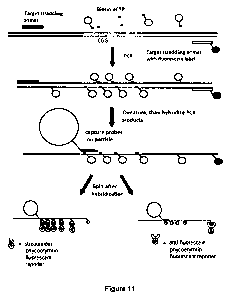
[0035] Figure 11 is a schematic drawing depicting an exemplary method for
determining the
length of a multinucleotide repeat region present in a target DNA molecule;
[0036] Figure 12 is a data plot of assay results using the exemplary method
depicted in
Figure 11 for male reference samples (Figure 12A) and female reference samples
performed
under a first set of hybridization conditions(Figure 12B) and a second set of
set of hybridization
conditions (Figure 12C); and
[0037] Figure 13 is a schematic drawing depicting an exemplary method for
determining the
length of a multinucleotide repeat region present in a target DNA molecule.
[0038] Schematic drawings provided herewith are not drawn to scale.
DETAILED DESCRIPTION OF THE INVENTION
[0039] Described herein are methods for determining the length of a
multinucleotide repeat
region present in a target nucleic acid.
CA 02767521 2012-01-06
WO 2011/006165 PCT/US2010/041731
7
[0040] As used herein, the term "length of a multinucleotide repeat region"
means the
number of multicleotide motifs, typically containing 3 or 4 nucleotides,
repetitively present in a
segment of target nucleic acid.
[0041] Methods of determining the length of a multinucleotide repeat region in
a target
nucleic acid, are provided which include amplifying a target nucleic acid to
produce amplified
target nucleic acids. Encoded particles which include capture probes specific
for the amplified
target nucleic acids are provided. The amplified target nucleic acids are then
bound to encoded
particles via specific binding to the capture probes. The amplified target
nucleic acids are
labeled with a first label and a second label. The first label is independent
of the number of
multinucleotide repeats and is also termed a "target-detection label" herein.
The second label is
proportional to the number of multinucleotide repeats and is also termed a
"repeat-detection
label" herein. The first label is detected to produce a first signal and the
second label is detected
to produce a second signal. The ratio of the first signal to the second signal
is determined and
the ratio is indicative of the length of the multinucleotide repeat in the
target nucleic acid.
[0042] In particular embodiments, the first label, that is, the "target-
detection label," is
incorporated in a straddle primer used in amplifying the target nucleic acid.
[0043] In some embodiments, the second label, that is, the "repeat-detection
label" is present
in a repeat-specific primer used in amplifying the target nucleic acid.
[0044] In some embodiments, the "repeat-detection label" is present in
nucleotides used in
amplifying the target nucleic acid to produce the amplified target nucleic
acids.
[0045] In some embodiments, the "repeat-detection label" is present in probes
which
specifically bind to multinucleotide repeats in the amplified target nucleic
acids. For example,
the probes which specifically bind to multinucleotide repeats in the amplified
target nucleic acids
are nucleic acid probes which have a complementary nucleic acid sequence
according to
embodiments of the present invention.
[0046] The term "nucleic acid" as used herein refers to RNA or DNA molecules
having more
than one nucleotide in any form including single-stranded, double-stranded,
oligonucleotide or
polynucleotide.
[0047] The target nucleic acid is DNA in particular embodiments and the DNA
can be in any
form, such as chromosomal DNA, mitochondrial DNA, cDNA, microdissected
chromosomal
DNA, an insert in a vector illustratively including a bacterial artificial
chromosome, yeast
artificial chromosome, human artificial chromosome, cosmid, plasmid, phagemid,
phage DNA,
and fosmid.
CA 02767521 2012-01-06
WO 2011/006165 PCT/US2010/041731
8
[0048] The target nucleic acid can be obtained from any source, including, but
not limited to,
a human, a non-human mammal, a vertebrate, an invertebrate, a microorganism,
or a plant. The
target nucleic acid can be obtained from one or more cells ex vivo or in
vitro. For example, the
target nucleic acid can be obtained from cultured cells, including, but not
limited to, cell lines,
primary cells or laboratory manipulated cells such as recombinant cells.
[0049] The target nucleic acid is typically contained within a biological
sample, which can
be obtained from an individual, such as from a bodily sample, for example,
blood, buccal swab,
skin tissue, urine, saliva, tissue, and the like, and cell lines derived
therefrom. A prenatal sample
can be obtained from amniotic fluid, products of conception, blastocysts and
blastomeres,
corionic villi, fetal cells and fetal DNA circulating in maternal blood.
Archived samples
extracted from formalin-fixed, paraffin-embedded (FFPE) pathology samples can
be used in the
methods described herein. Samples also be obtained from in vitro sources such
as cell lines.
[0050] Biological samples can be obtained from any source, including, but not
limited to, a
human, a non-human mammal, a vertebrate, an invertebrate, a microorganism, or
a plant.
Biological samples can be obtained from one or more cells ex vivo or in vitro.
For example,
biological samples can be obtained from cultured cells, including, but not
limited to, cell lines,
primary cells or laboratory manipulated cells such as recombinant cells.
[0051] Target nucleic acid, such as target DNA, is obtained by methods known
in the art, for
instance, as described in J. Sambrook and D.W. Russell, Molecular Cloning: A
Laboratory
Manual, Cold Spring Harbor Laboratory Press; 3rd Ed., 2001 or F.M. Ausubel,
Ed., Short
Protocols in Molecular Biology, Current Protocols; 5th Ed., 2002. Target
nucleic acid, such as
target DNA, may also be obtained commercially and/or using commercial kits for
isolation of
target nucleic acid, such as target DNA.
[0052] Scientific and technical terms used herein are intended to have the
meanings
commonly understood by those of ordinary skill in the art. Such terms are
found defined and
used in context in various standard references illustratively including J.
Sambrook and D.W.
Russell, Molecular Cloning: A Laboratory Manual, Cold Spring Harbor Laboratory
Press; 3rd
Ed., 2001; F.M. Ausubel, Ed., Short Protocols in Molecular Biology, Current
Protocols; 5th Ed.,
2002; B. Alberts et al., Molecular Biology of the Cell, 4th Ed., Garland,
2002; D.L. Nelson and
M.M. Cox, Lehninger Principles of Biochemistry, 4th Ed., W.H. Freeman &
Company, 2004;
and Herdewijn, P. (Ed.), Oligonucleotide Synthesis: Methods and Applications,
Methods in
Molecular Biology, Humana Press, 2004.
CA 02767521 2012-01-06
WO 2011/006165 PCT/US2010/041731
9
[0053] Methods for determining the length of a multinucleotide repeat region
present in a
target nucleic acid are useful, for example, when determining whether a
disease locus, such as
the Fragile X locus, contains a number of trinucleotide repeats that
correlates with a disease
phenotype. The presently described methods employ detectable labels that
generate signals
correlating with the length of a multinucleotide repeat, and thus allow
determination of the
length of a multinucleotide repeat region without the need for assessing the
molecular weight of
the repeat region or parts thereof.
[0054] Disorders associated with multinucleotide repeats include trinucleotide
repeat
expansion disorders such as, but not limited to Dentatorubropallidoluysian
atrophy (DRPLA),
Huntington's disease, spinobulbar muscular atrophy (SBMA), spinocerebellar
ataxia type 1
(SCA1), spinocerebellar ataxia type 2 (SCA2), spinocerebellar ataxia type 3
(SCA3),
spinocerebellar ataxia type 6 (SCA6), spinocerebellar ataxia type 7 (SCAT),
spinocerebellar
ataxia type 17 (SCA17), fragile X syndrome; fragile XE mental retardation;
Friedreich's ataxia;
myotonic dystrophy; spinocerebellar ataxia type 8 (SCAB), spinocerebellar
ataxia type 12
(SCA12). All of these trinucleotide repeat expansion disorders are well-
characterized. The gene
affected by trinucleotide repeat expansion in each disorder is known and the
location of the
trinucleotide repeat expansion in each of the affected genes is well-known.
[0055] The gene involved in DRPLA is on Chromosome 12 and is designated
"DRPLA."
Asymptomatic individuals have about 6 to35 copies of CAG in the DRPLA
trinucleotide repeat
locus. Symptomatic individuals have about 49 to 88 copies or more of the CAG
repeat. The gene
affected in Huntington's Disease is designated "huntingtin." Asymptomatic
individuals have
about 10 to 35 copies of CAG in the huntingtin trinucleotide repeat locus.
Symptomatic
individuals have about 40 or more copies of the CAG repeat. The gene affected
in SBMA is the
Androgen Receptor gene located on the X chromosome. Asymptomatic individuals
have about 9
to 36 copies of CAG in the Androgen Receptor trinucleotide repeat locus.
Symptomatic
individuals have about 38 to 62 copies. The gene involved in SCA1 is on
Chromosome 6 and is
designated "SCA1." Asymptomatic individuals have about 6 to 44 copies of CAG
in the SCA1
trinucleotide repeat locus. Symptomatic individuals have about 39 to 81 copies
of CAG. The
gene involved in SCA2 lies on Chromosome 12 and is designated "SCA2."
Asymptomatic
individuals have about 14 to 31 copies of CAG in the SCA2 trinucleotide repeat
locus.
Symptomatic individuals have about 36 to 64 copies. The gene involved in SCA3
lies on
Chromosome 14 and is designated "SCA3." Asymptomatic individuals have about 12
to 43
copies of CAG in the SCA3 trinucleotide repeat locus. Symptomatic individuals
have about 56 to
CA 02767521 2012-01-06
WO 2011/006165 PCT/US2010/041731
86 copies. The gene involved in SCA6 lies on Chromosome 19 and is designated
"SCA6."
Asymptomatic individuals have about 4 to 18 copies of CAG in the SCA6
trinucleotide repeat
locus. Symptomatic individuals have about 21 to 33 copies. The gene involved
in SCAT lies on
Chromosome 3 and is designated "SCAT." Asymptomatic individuals have about 4
to 19 copies
5 of CAG in the SCAT trinucleotide repeat locus. Symptomatic individuals have
about 37 to 306
copies. The gene involved in SCA17 is on Chromosome 6 and is designated
"SCA17."
Asymptomatic individuals have about 29 to 42 copies of CAG in the SCA17
trinucleotide repeat
locus. Symptomatic individuals have about 47-55 copies or more of the CAG
repeat. The
affected gene in Fragile X Syndrome, is designated "FMR1" which is on the X
chromosome.
10 Asymptomatic individuals have about 6 to 53 CGG repeats in the FMR1
trinucleotide repeat
locus. Symptomatic individuals have about 230 repeats or more. The affected
gene in Fragile
XE Mental Retardation is designated "FMR2" which is on the X chromosome.
Asymptomatic
individuals have about 6 to 35 copies of GCC in the FMR2 trinucleotide repeat
locus.
Symptomatic individuals have about 200 copies or more. The affected gene in
Friedreich's
Ataxia is designated "X25." Asymptomatic individuals have about 7 and 34 GAA
repeats in the
X25 trinucleotide repeat locus. Symptomatic individuals have about 100 or more
repeats. The
affected gene in Myotonic Dystrophy, is designated "myotonic dystrophy protein
kinase gene"
(DMPK). Asymptomatic individuals have about 5 and 37 CTG repeats in the DMPK
trinucleotide repeat locus. Symptomatic individuals have about 50 repeats or
more. The affected
gene in SCA8 is designated "SCAB." Asymptomatic individuals have about 16 to
37 repeats of
CTG in the SCA8 trinucleotide repeat locus. Symptomatic individuals have about
110 to 250
repeats. The affected gene in SCA12 is designated "SCA12." Asymptomatic
individuals have
about 7 to 28 repeats of CAG in the SCA12 trinucleotide repeat locus.
Symptomatic individuals
have about 66 to 78 repeats.
[0056] Methods of screening an individual for a genetic condition
characterized by an altered
multinucleotide repeat region in a target nucleic acid are provided according
to embodiments
described herein.
[0057] The term "altered multinucleotide repeat region" refers to a
multinucleotide repeat
region containing a number of multinucleotide repeats which differs from a
normal number of
multinucleotide repeats in the multinucleotide repeat region. An altered
multinucleotide repeat
region can be detected using a normal multinucleotide repeat region as a
reference in
embodiments of methods described herein. Thus, according to embodiments of
methods
CA 02767521 2012-01-06
WO 2011/006165 PCT/US2010/041731
11
described herein, a target nucleic acid which is a "reference" sample, that
is, a target nucleic acid
having a known number of multinucleotide repeats, is included.
[0058] An altered multinucleotide repeat region can be detected by comparison
of the
number of multinucleotide repeats detected using methods described herein with
known number
of multinucleotide repeats present in the normal multinucleotide repeat
region. The term
"normal" refers to the predominate number of multinucleotide repeats present
in the particular
analog multinucleotide repeat region found in healthy subjects.
[0059] Methods of screening an individual for a genetic condition
characterized by an altered
multinucleotide repeat region in a target nucleic acid include amplifying a
target nucleic acid
from a sample obtained from the individual to produce amplified target nucleic
acids containing
a first label, the first label independent of the number of multinucleotide
repeats in the target
nucleic acid and a second label, the second label proportional to the number
of multinucleotide
repeats in the target nucleic acid. The amplified target nucleic acids are
bound to a capture probe
specific for the multinucleotide repeat of the amplified target nucleic acids.
The first label
associated with the capture probe is detected to produce a first signal and
the second label
associated with the capture probe is detected to produce a second signal. A
ratio of the first
signal to the second signal is determined wherein the ratio is indicative of
the length of the
multinucleotide repeat in the target nucleic acid. The determined ratio
relating to the sample
from the individual is compared with a reference. According to embodiments of
described
methods, the reference is a control sample of nucleic acids obtained from a
normal individual.
[0060] Methods of screening an individual for a genetic condition
characterized by an altered
multinucleotide repeat region in a target genomic locus containing a
multinucleotide repeat
region include amplifying a target genomic locus continaing a multinucleotide
repeat region
from a sample obtained from the individual to produce amplified target genomic
DNA
containing a first label, the first label independent of the number of
multinucleotide repeats in the
target genomic DNA and a second label, the second label proportional to the
number of
multinucleotide repeats in the target genomic DNA. The amplified target
genomic DNA is
bound to capture probes specific for the multinucleotide repeat of the
amplified target genomic
DNA. The first label associated with the capture probe is detected to produce
a first signal and
the second label associated with the capture probe is detected to produce a
second signal. A ratio
of the first signal to the second signal is determined wherein the ratio is
indicative of the length
of the multinucleotide repeat in the target genomic locus. The determined
ratio relating to the
sample from the individual is compared with a reference. According to
embodiments of
CA 02767521 2012-01-06
WO 2011/006165 PCT/US2010/041731
12
described methods, the reference is a control sample of genomic DNA obtained
from a normal
individual.
[0061] LABELS
[0062] As described herein, the amplified target nucleic acids are labeled
with a first label,
the "target-detection label" and a second label, the "repeat-detection label."
[0063] The target-detection label is independent of the number of
multinucleotide repeats in
the multinucleotide repeat region of the target nucleic acid. Any number of
target-detection
labels can be incorporated into the amplified target nucleic acids as long as
the number of target-
detection labels incorporated does not vary significantly among the individual
amplified nucleic
acid molecules. According to embodiments described herein, each amplified
nucleic acid
molecule contains a label incorporated in at least one primer of a primer pair
used in amplifying
the target nucleic acids to produce the amplified target nucleic acids.
[0064] Thus, in particular embodiments, the "target-detection label," is
incorporated in at
least one straddle primer of a straddle primer pair used in amplifying the
target nucleic acid.
Optionally, both primers of a straddle primer pair used in amplifying the
target nucleic acid are
labeled.
[0065] The repeat-detection label" is proportional to the number of
multinucleotide repeats
in the multinucleotide repeat region of the target nucleic acid.
[0066] In some embodiments, the "repeat-detection label" is present in a
repeat-specific
primer used in amplifying the target nucleic acid.
[0067] In some embodiments, the "repeat-detection label" is present in
nucleotides used in
amplifying the target nucleic acid to produce the amplified target nucleic
acids.
[0068] In some embodiments, the "repeat-detection label" is present in probes
which
specifically bind to multinucleotide repeats in the amplified target nucleic
acids. For example,
the probes which specifically bind to multinucleotide repeats in the amplified
target nucleic acids
are nucleic acid probes which have a complementary nucleic acid sequence
according to
embodiments of the present invention.
[0069] The term "label" refers to a substance that can be measured and/or
observed, visually
or by any appropriate direct or indirect method illustratively including, but
not limited to,
spectroscopic, optical, photochemical, biochemical, enzymatic, electrical
and/or
immunochemical methods of detection, to indicate presence of the label. Non-
limiting examples
of labels that can be used in conjunction with methods described herein
illustratively include a
CA 02767521 2012-01-06
WO 2011/006165 PCT/US2010/041731
13
fluorescent moiety, a chemiluminescent moiety, a bioluminescent moiety, a
magnetic particle, an
enzyme, a substrate, a radioisotope and a chromophore.
[0070] For example, nucleotides, nucleotide analogs and/or primers can be
labeled with a
dye, such as a fluorophore, a chromophore, a radioactive moiety or a member of
a specific
binding pair such as biotin. The term "member of a specific binding pair"
refers to a substance
that specifically recognizes and interacts with a second substance exemplified
by specific
binding pairs such as biotin-avidin, biotin-streptavidin, antibody-antigen,
and target-aptamer.
Non-limiting examples of labels that can be used include fluorescent dyes such
as fluorescein,
fluorescein isothiocyanate, rhodamine, rhodamine isothiocyanate, Texas Red,
cyanine dyes such
as Cyanine 3 and Cyanine 5, and ALEXA dyes; chromophores such as horseradish
peroxidase,
alkaline phosphatase and digoxigenin; and radioactive moieties such as 32P,
35S, 3H, 1251 or
14C; and binding partners such as biotin and biotin derivatives. Methods for
detectably labeling
nucleotides, nucleotide analogs and/or primers are well-known in the art.
[0071] Nucleotides, including, but not limited to, deoxynucleotide
triphosphates (dNTPs)
and analogs thereof, labeled or unlabeled, can be included in primers and/or
amplification
reaction mixtures according to methods described herein. The term "nucleotide
analog" in this
context refers to a modified or non-naturally occurring nucleotide,
particularly nucleotide
analogs which can be polymerized, with naturally occurring nucleotides or non-
naturally
occurring nucleotides, by template directed nucleic acid amplification
catalyzed by a nucleic acid
polymerase. Nucleotide analogs are well-known in the art. Particular
nucleotide analogs are
capable of Watson-Crick pairing via hydrogen bonds with a complementary
nucleotide and
illustratively include, but are not limited to, those containing an analog of
a nucleotide base such
as substituted purines or pyrimidines, deazapurines, methylpurines,
methylpyrimidines,
aminopurines, aminopyrimidines, thiopurines, thiopyrimidines, indoles,
pyrroles, 7-
deazaguanine, 7-deazaadenine, 7-methylguanine, hypoxanthine, pseudocytosine,
pseudoisocytosine, isocytosine, isoguanine, 2-thiopyrimidines, 4-thiothymine,
6-thioguanine,
nitropyrrole, nitroindole, and 4-methylindole. Nucleotide analogs include
those containing an
analog of a deoxyribose such as a substituted deoxyribose, a substituted or
non-substituted
arabinose, a substituted or non-substituted xylose, and a substituted or non-
substituted pyranose.
Nucleotide analogs include those containing an analog of a phosphate ester
such as
phosphorothioates, phosphorodithioates, phosphoroamidates,
phosphoroselenoates,
phosophoroanilothioates, phosphoroanilidates, phosphoroamidates,
boronophosphates,
phosphotriesters, and alkylphosphonates such as methylphosphonates.
CA 02767521 2012-01-06
WO 2011/006165 PCT/US2010/041731
14
[0072] CAPTURE PROBES
[0073] Capture probe specific for the amplified nucleic acid are present on a
solid or semi-
solid substrate for attachment of the amplified nucleic acid to the substrate.
Capture probes can
be in any form which allows for attachment to the substrate and specific
capture of the amplified
nucleic acid.
[0074] According to some embodiments, capture probes are nucleic acids which
include a
nucleic acid sequence complementary to the amplified target nucleic acids.
Capture probes
attached to a substrate can be single-stranded and/or double-stranded nucleic
acids. In particular
embodiments, where double-stranded nucleic acids capture probes are bound to
the substrate,
they are denatured and rendered single stranded after immobilization to the
substrate for
preparation for use in certain embodiments of assay methods. Optionally,
double stranded
nucleic acid probes are denatured prior to immobilization and the single
stranded nucleic acids
are then bound to the substrate.
[0075] The term "complementary" as used herein refers to Watson-Crick base
pairing
between nucleotides and specifically refers to nucleotides hydrogen bonded to
one another with
thymine or uracil residues linked to adenine residues by two hydrogen bonds
and cytosine and
guanine residues linked by three hydrogen bonds. In general, a nucleic acid
includes a
nucleotide sequence described as having a "percent complementarity" to a
specified second
nucleotide sequence. For example, a nucleotide sequence may have 80%, 90%, or
100%
complementarity to a specified second nucleotide sequence, indicating that 8
of 10, 9 of 10 or 10
of 10 nucleotides of a sequence are complementary to the specified second
nucleotide sequence.
For instance, the nucleotide sequence 3'-TCGA-5' is 100% complementary to the
nucleotide
sequence 5'-AGCT-3'. Further, the nucleotide sequence 3'-TCGA- is 100%, or
completely,
complementary to a region of the nucleotide sequence 5' -TTAGCTGG-3' .
Determination of
particular hybridization conditions relating to a specified nucleic acid is
routine and is well
known in the art, for instance, as described in J. Sambrook and D.W. Russell,
Molecular
Cloning: A Laboratory Manual, Cold Spring Harbor Laboratory Press; 3rd Ed.,
2001; and F.M.
Ausubel, Ed., Short Protocols in Molecular Biology, Current Protocols; 5th
Ed., 2002. High
stringency hybridization conditions are those which only allow hybridization
of substantially
complementary nucleic acids. Typically, nucleic acids having about 85-100%
complementarity
are considered highly complementary and hybridize under high stringency
conditions.
Intermediate stringency conditions are exemplified by conditions under which
nucleic acids
having intermediate complementarity, about 50-84% complementarity, as well as
those having a
CA 02767521 2012-01-06
WO 2011/006165 PCT/US2010/041731
high degree of complementarity, hybridize. In contrast, low stringency
hybridization conditions
are those in which nucleic acids having a low degree of complementarity
hybridize. The terms
"specific hybridization" and "specifically hybridizes" refer to hybridization
of a particular
nucleic acid to a complementary nucleic acid without substantial hybridization
to nucleic acids
5 other than the complementary nucleic acid in a sample.
[0076] SUBSTRATES
[0077] A solid substrate, which includes semi-solid substrate, for attachment
of a capture
probe can be any of various materials such as glass; plastic, such as
polypropylene, polystyrene,
nylon; paper; silicon; nitrocellulose; or any other material to which a
nucleic acid can be attached
10 for use in an assay. The substrate can be in any of various forms or
shapes, including planar,
such as silicon chips and glass plates; and three-dimensional, such as
particles, microtiter plates,
microtiter wells, pins, fibers and the like.
[0078] A substrate to which a capture probe is attached is encoded according
to
embodiments of methods and compositions of the present invention. Encoded
substrates are
15 distinguishable from each other based on a characteristic illustratively
including an optical
property such as color, reflective index and/or an imprinted or otherwise
optically detectable
pattern. For example, the substrates can be encoded using optical, chemical,
physical, or
electronic tags.
[0079] In particular aspects, a solid substrate to which a capture probe is
attached is a
particle.
[0080] Particles to which a capture probe is attached can be any solid or semi-
solid particles
which are stable and insoluble in use, such as under hybridization and label
detection conditions.
The particles can be of any shape, such as cylindrical, spherical, and so
forth; size, such as
microparticles and nanoparticles; composition; and have various physiochemical
characteristics.
The particle size or composition can be chosen so that the particle can be
separated from fluid,
e.g., on a filter with a particular pore size or by some other physical
property, e.g., a magnetic
property.
[0081] Microparticles, such as microbeads, used can have a diameter of less
than one
millimeter, for example, a size ranging from about 0.1 to about 1,000
micrometers in diameter,
inclusive, such as about 3-25 microns in diameter, inclusive, or about 5-10
microns in diameter,
inclusive. Nanoparticles, such as nanobeads used can have a diameter from
about 1 nanometer
(nm) to about 100,000 nm in diameter, inclusive, for example, a size ranging
from about 10-
CA 02767521 2012-01-06
WO 2011/006165 PCT/US2010/041731
16
1,000 nm, inclusive, or for example, a size ranging from 200-500 nm,
inclusive. In certain
embodiments, particles used are beads, particularly microbeads and nanobeads.
[0082] Particles are illustratively organic or inorganic particles, such as
glass or metal and
can be particles of a synthetic or naturally occurring polymer, such as
polystyrene,
polycarbonate, silicon, nylon, cellulose, agarose, dextran, and
polyacrylamide. Particles are latex
beads in particular embodiments.
[0083] Particles used include functional groups for attaching nucleic acid
capture probes in
particular embodiments. For example, particles can include carboxyl, amine,
amino,
carboxylate, halide, ester, alcohol, carbamide, aldehyde, chloromethyl, sulfur
oxide, nitrogen
oxide, epoxy and/or tosyl functional groups. Functional groups of particles,
modification thereof
and binding of a chemical moiety, such as a nucleic acid, thereto are known in
the art, for
example as described in Fitch, R. M., Polymer Colloids: A Comprehensive
Introduction,
Academic Press, 1997. U.S. Pat. No. 6,048,695 describes an exemplary method
for attaching
nucleic acid capture probes to a substrate, such as particles. In a further
particular example, 1-
Ethyl- 3-[3-dimethylaminopropyl]carbodiimide hydrochloride, EDC or EDAC
chemistry, can be
used to attach nucleic acid capture probes to particles.
[0084] Particles to which a capture probe is attached are encoded particles
according to
embodiments of methods and compositions of the present invention. Encoded
particles are
particles which are distinguishable from other particles based on a
characteristic illustratively
including an optical property such as color, reflective index and/or an
imprinted or otherwise
optically detectable pattern. For example, the particles may be encoded using
optical, chemical,
physical, or electronic tags. Encoded particles can contain or be attached to,
one or more
fluorophores which are distinguishable, for instance, by excitation and/or
emission wavelength,
emission intensity, excited state lifetime or a combination of these or other
optical
characteristics. Optical bar codes can be used to encode particles.
[0085] In particular embodiments, the code is embedded, for example, within
the interior of
the particle, or otherwise attached to the particle in a manner that is stable
through hybridization
and analysis. The code can be provided by any detectable means, such as by
holographic
encoding, by a fluorescence property, color, shape, size, light emission,
quantum dot emission
and the like to identify particle and thus the capture probes immobilized
thereto. In some
embodiments, the code is other than one provided by a nucleic acid.
[0086] One exemplary platform utilizes mixtures of fluorescent dyes
impregnated into
polymer particles as the means to identify each member of a particle set to
which a specific
CA 02767521 2012-01-06
WO 2011/006165 PCT/US2010/041731
17
capture probe has been immobilized. Another exemplary platform uses
holographic barcodes to
identify cylindrical glass particles. For example, Chandler et al. (U.S. Pat.
No. 5,981,180)
describes a particle-based system in which different particle types are
encoded by mixtures of
various proportions of two or more fluorescent dyes impregnated into polymer
particles. Soini
(U.S. Pat. No. 5,028,545) describes a particle-based multiplexed assay system
that employs time-
resolved fluorescence for particle identification. Fulwyler (U.S. Pat. No.
4,499,052) describes an
exemplary method for using particle distinguished by color and/or size. U.S.
Patent Application
Publications 20040179267, 20040132205, 20040130786, 20040130761, 20040126875,
20040125424, and 20040075907 describe exemplary particles encoded by
holographic barcodes.
U.S. Pat. No. 6,916,661 describes polymeric microparticles that are associated
with nanoparticles
that have dyes that provide a code for the particles
[0087] While an embodiment described in detail herein utilizes the Luminex
encoded bead
platform, other types of encoded particle assay platforms may be used, such as
the VeraCode
beads and BeadXpress system (Illumina Inc., San Diego CA), xMAP 3D (Luminex)
and the like.
Magnetic Luminex beads can be used which allow wash steps to be performed with
plate
magnets and pipetting rather than with filter plates and a vacuum manifold.
Each of these
platforms are typically provided as carboxyl beads but may also be configured
to include a
different coupling chemistry, such as amino-silane.
[0088] Particles are typically evaluated individually to detect encoding. For
example, the
particles can be passed through a flow cytometer. Exemplary flow cytometers
include the
Coulter Elite-ESP flow cytometer, or FACScan.TM. flow cytometer available from
Beckman
Coulter, Inc. (Fullerton Calif.) and the MOFLO.TM. flow cytometer available
from Cytomation,
Inc., Fort Collins, Colo. In addition to flow cytometry, a centrifuge may be
used as the
instrument to separate and classify the particles. A suitable system is that
described in U.S. Pat.
No. 5,926,387. In addition to flow cytometry and centrifugation, a free-flow
electrophoresis
apparatus may be used as the instrument to separate and classify the
particles. A suitable system
is that described in U.S. Pat. No. 4,310,408. The particles may also be placed
on a surface and
scanned or imaged.
[0089] Provided are assays according to embodiments of the present invention
using more
than one type of encoded particles. In particular embodiments, a "particle
set" is provided
wherein each particle of the particle set is encoded with the same code such
that each particle of
a particle set is distinguishable from each particle of another "particle
set." In further
embodiments, two or more codes can be used for a single particle set. Each
particle can include
CA 02767521 2012-01-06
WO 2011/006165 PCT/US2010/041731
18
a unique code, for example. In certain embodiments, particle encoding includes
a code other than
or in addition to, association of a particle and a nucleic acid capture probe
specific for a target
nucleic acid.
[0090] Methods including two or more particle sets can be used in multiplex or
separate
assay formats.
[0091] BINDING OF CAPTURE PROBES TO SUBSTRATE
[0092] Binding of the nucleic acid capture probes to a substrate is achieved
by any of various
methods effective to bond a nucleic acid to a solid or semi-solid substrate,
illustratively including
adsorption and chemical bonding. The nucleic acids can be bonded directly to
the material of the
encoded particles or indirectly bonded to the encoded particles, for example,
via bonding to a
coating or linker disposed on the particles. Nucleic acids can be synthesized,
and/or modified
once synthesized, to include a functional group for use in bonding the nucleic
acids to particles.
For example, the nucleic acids sequences used as probes can include carboxyl,
amine, amino,
carboxylate, halide, ester, alcohol, carbamide, aldehyde, chloromethyl, sulfur
oxide, nitrogen
oxide, epoxy and/or tosyl functional groups.
[0093] In particular embodiments of assays described herein, amplified target
nucleic acids
are captured by the capture probes attached to the encoded particles by
hybridization.
[0094] The terms "hybridization" and "hybridized" refer to pairing and binding
of
complementary nucleic acids. Hybridization occurs to varying extents between
two nucleic
acids depending on factors such as the degree of complementarity of the
nucleic acids, the
melting temperature, Tm, of the nucleic acids and the stringency of
hybridization conditions, as
is well known in the art. The term "stringency of hybridization conditions"
refers to conditions
of temperature, ionic strength, and composition of a hybridization medium with
respect to
particular common additives such as formamide and Denhardt's solution.
[0095] AMPLIFICATION
[0096] Amplification of a target nucleic acid is achieved using an in vitro
amplification
method. The term "amplification method" refers to a method for copying a
template target
nucleic acid, thereby producing nucleic acids which include copies of all or a
portion of the
template target nucleic acid.
[0097] Amplification methods included in embodiments of the present invention
are those
which include template directed primer extension catalyzed by a nucleic acid
polymerase using a
pair of primers which flank the target nucleic acid, illustratively including,
but not limited to,
Polymerase Chain Reaction (PCR), reverse-transcription PCR (RT-PCR). ligation-
mediated PCR
CA 02767521 2012-01-06
WO 2011/006165 PCT/US2010/041731
19
(LM-PCR), phi-29 PCR, and other nucleic acid amplification methods, for
instance, as described
in C.W. Dieffenbach et al., PCR Primer: A Laboratory Manual, Cold Spring
Harbor Laboratory
Press, 2003; and V. Demidov et al., DNA Amplification: Current Technologies
and
Applications, Taylor & Francis, 2004.
[0098] The terms "amplified nucleic acid" and "amplified DNA" as well as
plurals thereof
refer to the product of a process of copying a target nucleic acid template.
[0099] PRIMERS
[00100] The term "primer" refers to an oligonucleotide nucleic acid that is
capable of acting
as a site of initiation of synthesis of a template directed primer extension
product under
appropriate reaction conditions. An oligonucleotide primer is typically about
10 - 30 contiguous
nucleotides in length and may be longer or shorter. An oligonucleotide primer
is completely or
substantially complementary to a region of a template nucleic acid such that,
under hybridization
conditions, the oligonucleotide primer anneals to the complementary region of
the template
nucleic acid. Appropriate reactions conditions for synthesis of a primer
extension product
include presence of suitable reaction components including, but not limited
to, a polymerase and
nucleotide triphosphates. Design of oligonucleotide primers suitable for use
in amplification
reactions is well known in the art, for instance as described in A. Yuryev et
al., PCR Primer
Design, Humana Press, 2007.
[00101] Primer design for amplification of a target nucleic acid is well-known
to those of skill
in the art. Primers for amplification of a target nucleic acid are designed
according to well-
known methods and criteria. For instance, the annealing temperature of the
primers should be
about the same, within a few degrees, the primers should not form dimers with
each other and
the primers should not form secondary structures, such as hairpins. Methods
and considerations
for primer design and amplification procedures are described in detail in
Yuryev, A., PCR
Primer Design, Methods in Molecular Biology, vol. 42, Human Press, 2007; C.W.
Dieffenbach
et al., PCR Primer: A Laboratory Manual, Cold Spring Harbor Laboratory Press,
2003; and V.
Demidov et al., DNA Amplification: Current Technologies and Applications,
Taylor & Francis,
2004.
[00102] Amplified nucleic acids optionally contain additional materials such
as, but not
limited to, nucleic acid sequences, functional groups for chemical reaction
and detectable labels,
present in the primers and not present in the original DNA template. Such
primer-derived
materials add functionality such as primer binding sites for additional
amplification reactions
and/or a functional group for chemical bonding to a substrate.
CA 02767521 2012-01-06
WO 2011/006165 PCT/US2010/041731
[00103] In embodiments of the present invention a pair of primers used in
amplification
includes primers which flank a multinucleotide repeat region, that is, one of
the primers has a
nucleotide sequence complementary to a region of the target nucleic acid
upstream of the
multinucleotide repeat region and a second primer of the primer pair has a
nucleotide sequence
5 complementary to a region of the target nucleic acid downstream of the
multinucleotide repeat
region. Such primers are termed "straddle primers" herein. Numerous straddle
primer pairs
designed to amplify a target nucleic acid containing a multinucleotide repeat
and which flank the
multinucleotide repeat region are known in the art, any of which can be used
in conjunction with
methods and compositions of the present invention. Alternatively, straddle
primers can be
10 designed and used with only routine experimentation.
[00104] Exemplary straddle primers and their use to amplify a target nucleic
acid including a
multinucleotide repeat region are described in Examples detailed herein.
[00105] Straddle primers to amplify a target nucleic acid including a
multinucleotide repeat
region present in the FMR1 gene are described in Wilson, JA et al., J. Molec.
Diagnostics,
15 10(1):2-12, 2008 and include: 1) Forward primer 5' -
GGAACAGCGTTGATCACGTGACGTGGTTTC - 3' (SEQ ID No.1); reverse primer 5' -
GGGGCCTGCCCTAGAGCCAAGTACCTTGT - 3' (SEQ ID No.2) (Chong, SS. et al., Am. J.
Med. Genet., 1994, 51:522-526.); 2) Forward primer 5' - GACGGAGGCGCCCGTGCCAGG -
3' (SEQ ID No.3); reverse primer 5' - TCCTCCATCTTCTCTTCAGCCCT - 3' (SEQ ID
No.4)
20 (Pergolizzi, RG. et al., Lancet, 1992, 339:271-272); 3) Forward primer 5' -
TGACGGAGGCGCCGCTGCCAGGGGGCGTGC - 3' (SEQ ID No.5); reverse primer 5' -
GAGAGGTGGGCTGCGGGCGCTCGAGGCCCA - 3' (SEQ ID No.6) (Wang Q., et al., J. Med
Genet., 1995, 32:170-173.); 4) Forward primer 5' -
AGGCGCTCAGCTCCGTTTCGGTTTCACTTC - 3' (SEQ ID No.7); reverse primer 5' -
GTGGGCTGCGGGCGCTCGAGG - 3' (SEQ ID No.8) (Tarlton, J., Neurogenetics: Methods
and Protocols (Methods in Molecular Biology, v. 217, Potter, N., Ed., Humana
Press Inc.,
Totowa, NJ, 2003), pp 29-39.); 5) Forward primer 5' -
GCTCAGCTCCGTTTCGGTTTCACTTCCGGT - 3' (SEQ ID No.9); reverse primer 5' -
AGCCCCGCACTTCCACCACCAGCTCCTCCA - 3' (SEQ ID No.10) (Verkerk, AJ. et al.,
Cell, 1991, 65:905-914; Fu, YH et al., Cell, 1991, 67:1047-1058.); 6) Forward
primer 5' -
GACGGAGGCGCCGCTGCCAGG - 3' (SEQ ID No.11); reverse primer 5' -
GTGGGCTGCGGGCGCTCGAGG - 3' (SEQ ID No.12) (Verkerk, AJ. et al., Cell, 1991,
65:905-914.); and 7) Forward primer 5' - GTGACGGAGGCGCCGCTGCCA - 3' (SEQ ID
CA 02767521 2012-01-06
WO 2011/006165 PCT/US2010/041731
21
No.13); reverse primer 5' - AGCTCCTCCATCTTCTCTTCAGCCCTGCTA - 3'(SEQ ID
No.14) (Fu, YH et al., Cell, 1991, 67:1047-1058.)
[00106] In embodiments of the present invention a pair of primers used in
amplification
includes a straddle primer and a repeat-specific primer. The straddle primer
has a nucleotide
sequence complementary to a region of the target nucleic acid upstream of the
multinucleotide
repeat region or complementary to a region of the target nucleic acid
downstream of the
multinucleotide repeat region. The repeat-specific primer has a nucleotide
sequence
complementary to a portion of the multinucleotide repeat region of the target
nucleic acid.
Exemplary primer pairs including a straddle primer and a repeat-specific
primer are described
herein. Alternatively, such primers can be designed and used with only routine
experimentation.
[00107] In one embodiment, the method involves amplifying the target nucleic
acid molecule
using a target-straddling primer pair, wherein one primer contains a target-
detection label;
hybridizing the amplified target DNA molecule to a set of encoded particles,
the particles
comprising a capture molecule selective for the amplified target nucleic acid
molecule; detecting
a signal produced by the target-detection label; amplifying segments of the
multinucleotide
repeat region using a repeat-specific primer pair, wherein one primer of the
repeat-specific
primer pair is specific for the multinucleotide repeat motif, and the other
primer of the repeat-
specific primer pair is specific for a target nucleic acid molecule sequence
outside of the
multinucleotide repeat region and contains a repeat-detection label, to
produce amplified repeat-
specific nucleic acid molecules; and hybridizing the amplified repeat-specific
nucleic acid
molecules to a set of encoded particles, the particles comprising a capture
molecule selective for
the amplified repeat-specific nucleic acid molecules; detecting a signal
produced by the repeat-
detection label; determining a ratio of signals produced by the target-
detection label and repeat-
detection label; determining the length of the multinucleotide repeat region
based on the
determined ratio.
[00108] As is described herein below, also hybridized to the amplified target
DNA molecules
are one or more repeat-detector probes, which contain a repeat-detection
label. The repeat-
detector probe molecules are complementary to the repeat region (i.e. the
multinucleotide repeat
motif) and thus a plurality of repeat-detector probes can hybridize to the
repeat region. The
repeat-detector probes can be hybridized with the amplified target DNA
molecules together with
or after the amplified target DNA molecules have been captured by the
particles.
[00109] In another embodiment, the method for determining the length of a
multinucleotide
repeat region present in a target nucleic acid molecule involves amplifying
the target nucleic acid
CA 02767521 2012-01-06
WO 2011/006165 PCT/US2010/041731
22
molecule using a target-straddling primer pair, wherein one primer contains a
target-detection
label; hybridizing a first portion of the amplified target nucleic acid
molecules to a first set of
encoded particles, the particles including a capture molecule selective for
the amplified target
nucleic acid molecules; detecting a signal produced by the target-detection
label; hybridizing a
second portion of the amplified target nucleic acid molecules with a
detectable probe specific for
the multinucleotide repeat motif and a second set of encoded particles, the
particles including a
capture molecule selective for the amplified target DNA molecules; detecting a
signal produced
by the probe (i.e., the repeat-detection probe containing the repeat-detection
label); determining a
ratio of signals produced by the target-detection label and probe; and
determining the length of
the multinucleotide repeat region based on the determined ratio.
[ 00110 ] In a further embodiment, the method for determining the length of a
multinucleotide
repeat region present in a target nucleic acid molecule involves amplifying
the target nucleic acid
molecule using a target-straddling primer pair, wherein one primer contains a
target-detection
label, in the presence of at least one type of deoxynucleotide comprising a
repeat-detection label;
hybridizing the amplified target nucleic acid molecule to a set of encoded
particles, the particles
comprising a capture molecule selective for the amplified target nucleic acid
molecule, and
detecting a signal produced by the target-detection label. The method can
further include
detecting a signal produced by the repeat-detection label; determining a ratio
of signals produced
by the target-detection label and repeat-detection label; and determining the
length of the
multinucleotide repeat region based on the determined ratio. There is a repeat-
detection label on
a detectable probe specific for the multinucleotide repeat motif.
[00111] In an embodiment, the method involves amplifying the target nucleic
acid molecule
using a target-straddling primer pair, in the presence of deoxynucleotide
comprising a repeat-
detection label, wherein a primer of the pair contains a target-detection
label, binding the
amplified target DNA molecule to a set of encoded particles, each particle
comprising a binding
element selective for the multinucleotide repeat of the amplified target
nucleic acid molecule;;
detecting a signal corresponding to an amount of the target-detection label
present in amplified
target nucleic acid molecules that are particle-bound; detecting a signal
corresponding to an
amount of the repeat-detection label present in amplified target nucleic acid
molecules bound to
the particles; determining a ratio of signals from the target-detection label
and repeat-detection
label; and determining the length of the multinucleotide repeat region based
on the determined
ratio.
CA 02767521 2012-01-06
WO 2011/006165 PCT/US2010/041731
23
[00112] In another embodiment, the method involves amplifying the target
nucleic acid
molecule using a target-straddling primer pair, in the presence of
deoxynucleotide comprising a
repeat-detection label, wherein a primer of the pair contains a target-
detection label, binding the
amplified target DNA molecule to a set of encoded particles, each particle
comprising a binding
element selective for the amplified target nucleic acid molecule; contacting a
portion of the
particle-bound amplified target nucleic acid with a reporter that renders
detectable the target-
detection label; detecting a signal corresponding to an amount of the target-
detection label
present in amplified target nucleic acid molecules that are particle-bound;
contacting another
portion of the particle-bound amplified target nucleic acid molecules with a
reporter that renders
detectable the repeat-detection label; detecting a signal corresponding to an
amount of the
repeat-detection label present in amplified target nucleic acid molecules
bound to the particles;
determining a ratio of signals from the target-detection label and repeat-
detection label; and
determining the length of the multinucleotide repeat region based on the
determined ratio. An
exemplary implementation of this method is illustrated in Fig. 11. In this
specific example, the
binding element selective for the amplified target nucleic acid molecule is
present in the
multinucleotide repeat region. Other portions of an amplified target nucleic
acid molecule also
can be used.
[00113] Methods described herein involve using encoded particles for
determining the length
of a multinucleotide repeat region present in a target nucleic acid molecule.
In an embodiment,
the determination is based on both the number or relative amount of amplified
target nucleic acid
molecules and the number or relative amount of mutinucleotide repeats within
the amplified
nucleic acid molecule. The number or relative amount of amplified target
nucleic acid molecules
and the number of multinucleotide repeats can be determined using separate
pools of amplified
nucleic acid as is illustrated in Figure 1A, or can be determined from a
common pool of
amplified target nucleic acid, as is illustrated in Figures 1B and 11. The
methods can proceed
using various strategies, which can be selected by the user based, for
example, on assay format
and detectable label preferences or requirements imposed by available
instrumentation.
[00114] Figure 13 illustrates an embodiments of methods described herein in
which a target
nucleic acid containing a multinucleotide repeat region (labeled "CGG repeats"
is amplified
using a pair of straddle primers, wherein one of the pair has a target
detection label. In this case
the target detection label is a fluorescein label. Following amplification,
the target detection
label is incorporated into the amplified nucleic acids, as illustrated.
CA 02767521 2012-01-06
WO 2011/006165 PCT/US2010/041731
24
[00115] The amplified nucleic acids are bound to encoded particles via
hybridization with
complementary nucleic acid capture probes and the resulting particle set is
split into two
portions.
[00116] A first portion is bound to repeat detection probes, in this case by
specific
hybridization to labeled oligonucleotides having a sequence complementary to a
portion of the
multinucleotide repeat region. Biotin labeled oligonucleotides in this case
are the repeat-
detection label. A streptavidin reporter is used to detect the label and
generate a first signal.
[00117] A second portion is bound to a reporter specific for the target
detection label
fluorescein, in this casean anti-fluorescein antibody, and generate a second
signal.
[00118] The signal from the repeat detection label is compared to the signal
detected from the
target detection label to determine the length of the multinucleotide repeat
region.
[00119] Methods described herein involve amplifying DNA molecules. The
amplification can
be performed using any suitable polynucleotide amplification technique.
Polymerase chain
reaction is described herein, and other published amplification methods can be
adapted for use
with the methods described herein.
[00120] Methods described herein involve detection of labels. Any of a variety
of labels and
their complementary detection modes can be used when practicing the described
methods. The
Examples below describe use of the fluorescent label phycoerythrin in
conjunction with a
fluorescence reader of a Luminex 200 instrument, although other assay reading
platforms such as
the Illumina BeadExpress, microplates, microarrays, etc. could be used with
their appropriate
labels. Platforms that utilize 2 or more labels could accomplish the assay
without the need for
spliiting the intermediate assay product into two vessels prior to reading, as
the single-label
Luminex examples incorporated herein require.
[00121] Any appropriate method, illustratively including spectroscopic,
optical,
photochemical, biochemical, enzymatic, electrical and/or immunochemical is
used to detect a
label in an assay described herein. The ratio of the first signal to the
second signal can be
determined by manual, machine or automated methods and the resulting ratio is
indicative of the
length of the multinucleotide repeat in the target nucleic acid.
[00122] A method of assaying sample nucleic acid is provided which includes
two or more
encoded particle sets encoded such that each particle of each encoded particle
set is detectably
distinguishable from each particle of each other encoded particle set. The
encoded particles of a
first particle set include attached capture probes which specifically capture
amplified target
nucleic acids corresponding to a first target nucleic acid containing a
multinucleotide repeat
CA 02767521 2012-01-06
WO 2011/006165 PCT/US2010/041731
region. The encoded particles of a second particle set include attached
capture probes which
specifically capture amplified target nucleic acids corresponding to a second
target nucleic acid
containing a multinucleotide repeat region.
[00123] Methods described can be performed in any suitable container. In
particular
5 embodiments, for example, where multiple samples are to be assayed, a multi-
chamber container
can be used. Multi-chamber containers illustratively include multi-depression
substrates such as
slides, silicon chips or trays. In some embodiments, each sample is disposed
in a different well
of a multi-well plate. For example, a multi-well plate can be a 96-well, 384-
well, 864-well or
1536-well assay plate.
10 [00124] Kits for determining the length of multinucleotide repeats in a
target nucleic acid are
provided. In particular embodiments, a kit is provided which includes an
encoded particle set
and/or a mixture of two or more encoded particle sets, wherein each particle
set includes attached
capture probes specific for a target nucleic acid. Instructional material for
use of the encoded
particle set and/or multiplex reagent including two or more encoded particle
sets is optionally
15 included in a kit. An ancillary reagent such as buffers, enzymes, washing
solutions,
hybridization solutions, detectable labels, detection reagents and the like
are also optionally
included.
[00125] Assay compositions are provided according to embodiments described
herein which
include amplified target nucleic acids containing a multinucleotide repeat
region, the amplified
20 target nucleic acids including a first label and a second label, the first
label independent of the
number of multinucleotide repeats and the second label proportional to the
number of
multinucleotide repeats, wherein the first and second labels are each
independently incorporated
in the amplified target nucleic acids during the amplifying or after the
amplifying, and wherein
the amplified target nucleic acids are bound to a capture probe specific for
the amplified target
25 nucleic acids.
[00126] Compositions and kits described herein are useful, for example, in
performing
methods for determining the length of a multinucleotide repeat region
substantially as described
herein.
[00127] Embodiments of inventive compositions and methods are illustrated in
the following
examples. These examples are provided for illustrative purposes and are not
considered
limitations on the scope of inventive compositions and methods.
[00128] Example 1
CA 02767521 2012-01-06
WO 2011/006165 PCT/US2010/041731
26
[00129] In an exemplary method, sample DNA 1 is divided into two aliquots,
each of which is
processed independently through the assay until fluorescent data from each
path is processed to
generate one or more ratios. A first process of one aliquot of the sample 1
starts with repeat-
specific primer PCR amplification 3. The repeat-specific primer pair 2
includes one primer that
hybridizes to the target DNA outside of the multinucleotide repeat region and
a second primer
that hybridizes to a plurality of regions within the repeat region. The non-
repeat primer in this
example is end-labeled to facilitate subsequent detection of the PCR product
molecules. In this
case, the end-label is biotin. The reaction product in this scenario is a
heterogenous population
of end-labelled nucleic acids containing variable numbers of multinucleotide
repeat motifs. Later
in the process, biotin will be bound to streptavidin labeled with a detectable
tag. Alternative
end-labels can be used, including molecules that, like biotin, become
detectable upon binding to
a partner as well as molecules that are inherently detectable. The PCR
reagents kit 7 includes a
polymerase enzyme, nucleotides and buffers.
[00130] Amplified repeat-specific product DNA 4, is then specifically captured
and subjected
to an encoded particle hybridization assay 5. The specific capture can be, for
example, based on
a complementary nucleotide sequence, or other binding-partner interactions.
Hybridization
reagents 8 include a hybridization buffer, a wash buffer, and a fluorescent
reporter. In this
specific example, the fluorescent reporter is streptavidin-phycoerythrin, the
standard reporter for
Luminex assays. Amplified repeat-specific DNA molecules are specifically
captured onto the
encoded particle set with immobilized oligonucleotide capture molecules 16.
The hybridization
assay 5 results in generation of amplified repeat-specific DNA fluorescent
signal data 6.
[00131] The second aliquot of the sample 1 is processed in parallel using the
same process but
different PCR primers. Target amplification 10 is performed using a target-
straddling PCR
primer pair 9. Target amplification and repeat-specific amplification are
shown as simultaneous
processing in Figure 1A. It is understood that in practice, the amplifications
can be performed
simultaneously, consecutively, in an overlapping manner, according to user
preference. This
produces amplified target DNA 11 that is hybridized with capture molecules.
The encoded
particle set 17 can be the same as that in 16, for example, when the capture
region of the two
amplified DNA samples is the same. The hybridization 12 produces amplified
target DNA
fluorescent signal data 13.
[00132] The amplified repeat-specific DNA fluorescent signal data is then
ratioed to the target
DNA fluorescent signal data. By using a ratio of the two signals variations in
the yield of the
two PCR processes are compensated. Figure 9 shows that the ratio of
fluorescent signals is
CA 02767521 2012-01-06
WO 2011/006165 PCT/US2010/041731
27
substantially proportional to the repeat content of male Fragile X samples
from 25 to about 500
repeats.
[00133] Thus, the methods described herein can be used to determine the length
of the
multinucleotide repeat region that is between about 25 and about 500 repeats.
For example, the
length of the multinucleotide repeat region can be between about 200 and about
500 repeats, and
about 300 and about 500 repeats.
[00134] Figure 2 depicts an exemplary PCR configuration for a target-
straddling primer pair
and target DNA. The target sequence 21 includes a multinucleotide repeat
sequence 24 that can
be of variable length, depending on the genetic locus of the individual. The
repeat region is
flanked by non-repeat sequences 22 and 23 on the 5' and 3' ends, respectively.
Target-straddling
primers 25 and 26 are complementary to the non-repeat sequences that flank the
repeat region.
In this example the 5' primer is end-labeled with biotin 27 to facilitate
subsequent detection of
the PCR products (also referred to as amplified target DNA molecules) with a
fluorescent
streptavidin-phycoerythrin reporter. Also in this example the primer on the 5'
end is located
such that there is a capture 28 sequence between the end of the primer and the
beginning of the
repeat region. This sequence is used for the subsequent hybridization capture
of PCR products.
[00135] Figure 3 depicts an exemplary PCR configuration for a repeat-specific
primer pair
and repeat-specific DNA. The target sequence 31 includes a multinucleotide
repeat sequence 34
can be of variable length, depending on the genetic locus of the individual.
The repeat region is
flanked by non-repeat sequences 32 and 33 on the 5' and 3' ends, respectively.
The 5' non-repeat
primer 36 is located 5' of the repeat region 34, and in this example the
primer is end-labeled with
biotin 37 to facilitate detection of the resulting PCR product DNA (also
referred to as amplified
repeat-specific DNA molecules). The non-repeat primer is defined so that there
is a region of
non-repeat DNA 38 between the non-repeat primer and the repeat region, where
this non-repeat
region can be used for subsequent capture or detection of the resulting PCR
products. This non-
repeat primer can be the same as a 5' target-straddling primer described
above. The second
primer 35 is repeat-specific primer, meaning that it hybridizes to the repeat
region. Such a repeat
primer can hybridize at any of a large number of locations along the repeat
region.
[00136] Figure 4 depicts an exemplary alternate PCR configuration similar to
that of Figure 3
except that the relative locations of the non-repeat and repeat primers are
reversed. The target
sequence 41 includes a repeat sequence 44 that can have variable length. The
repeat region is
flanked by non-repeat sequences 42 and 43 on the 5' and 3' ends, respectively.
The 3' non-repeat
primer 46 is located 3' of the repeat region 44, and in this example the
primer is end-labeled with
CA 02767521 2012-01-06
WO 2011/006165 PCT/US2010/041731
28
biotin 47 to facilitate detection of the resulting PCR product DNA (also
referred to as amplified
repeat-specific DNA molecules). The non-repeat primer is defined so that there
is a region of
non-repeat DNA 48 between the non-repeat primer and the repeat region, where
this non-repeat
region can be used for subsequent capture or detection of the resulting PCR
products. This non-
repeat primer can be the same as the 5' target straddling primer described
above. The second
primer 46 is repeat-specific primer, meaning that it hybridizes to the repeat
region. Such a
repeat-specific primer can hybridize at any of a large number of locations
along the repeat
region.
[00137] Figure 5A depicts an amplified DNA molecule 52 produced by the PCR
configuration of Figure 2. This amplified target DNA molecule contains a
biotin label 50 on the
5' end, a 5' non repeat sequence 53, a repeat region of variable length 51,
and a 3' non-repeat
region 54. Figure 5 B depicts an amplified DNA molecule 58 produced by the PCR
configuration of Figure 3. This amplified repeat-specific DNA molecule
contains a biotin label
55 on the 5' end, a 5' non-repeat sequence 56, and a repeat sequence 57 of
variable length.
Figure 5C depicts an amplified DNA molecule produced by the PCR configuration
of Figure 4.
This amplified repeat-specific DNA molecule contains a biotin label 61 on the
3' end, a 3' non-
repeat sequence 59, and a repeat sequence 60 of variable.
[00138] Figure 6 depicts, schematically, a single oligonucleotide capture
molecule
immobilized on an encoded particle, with a bound amplified target DNA molecule
from PCR
configurations such as that shown in Figure 5A. An encoded particle 77, such
as Luminex
xMAP bead, in this example, has a large number of oligonucleotide capture
molecules 74
coupled to its surface (only one molecule is shown in this figure). The
capture oligonucleotide
74 is designed to be complementary to a non-repeat region 72 of a target DNA
molecule 76
produced by the approach described above. The captured amplified target DNA
molecule
contains a detectable label 71, biotin in this example, on one end and
contains a repeat region 75
and a non-repeat region 73.
[00139] Figure 7 depicts, schematically, a single oligonucleotide capture
molecule
immobilized on an encoded particle with a bound amplified repeat-specific DNA
molecule. An
encoded particle 86, such as a Luminex xMAP bead in this example, has a large
number of
oligonucleotide capture molecules 83 coupled to its surface (only one molecule
is shown in this
figure). The capture oligonucleotide 83 is designed to be complementary to a
non-repeat region
82 of the repeat-specific DNA molecules produced by a repeat-specific primers
such as those
CA 02767521 2012-01-06
WO 2011/006165 PCT/US2010/041731
29
described by Figures 5B or 5C. The captured repeat-specific DNA molecule has a
detectable
label 81, biotin in this example, on one end, and a repeat region 84.
[00140] Figure 8 depicts, schematically, a single oligonucleotide capture
molecule
immobilized on an encoded particle, and a single amplified target DNA molecule
from one of the
PCR configurations such as those shown in Figure 5. An encoded particle 97,
such as a Luminex
xMAP bead in this example, has a large number of oligonucleotide capture
molecules 94 coupled
to its surface (only one molecule is shown in this figure). The capture
oligonucleotide 94 is
designed to be complementary to a non-repeat region 92 of an amplified target
DNA molecule
produced by target straddling primer pair as described in Figure 5A. The
captured amplified
target DNA molecule has a detectable label 91, biotin in this example, on one
end, and has a
repeat region 98.
[00141] Also hybridized to the amplified target DNA molecule 96 are one or
more repeat-
detector probes 99. One or more detectable labels 100, biotin in this example,
are incorporated
into each repeat-detector probe. The repeat-detector probe molecules are
complementary to the
repeat region 98 and a plurality of repeat-detector probes can hybridize to
the repeat region. The
repeat-detector probes can be hybridized with the amplified target DNA
molecules together with
or after the amplified target DNA molecules have been captured by the beads.
[00142] Notably, the longer the repeat region in the captured amplified target
DNA molecule,
the more labeled repeat-detector probes will hybridize to it and the larger
the signal that will be
generated and detected. Accordingly, the present method produces fluorescent
signals in
proportion to the length of the multinucleotide repeat region contained in the
amplified target
DNA molecule.
[00143] Figure 9 is example data from a first example assay according to the
process outlined
in Figure 1. The values along the horizontal axis are the number of CGG
repeats according to
the supplier of cell line DNA samples (Coriell Institute for Medical Research,
Trenton NJ). The
samples represent males with Fragile X repeat lengths of 25, 83, 110 and 477.
The values along
the vertical axis of the plot are the ratio as calculated according to the
first example, the ratio of
the fluorescent signal generated by repeat-specific DNA divided by the
fluorescent signal of
amplified target DNA of the same sample. The ratio data increases
monotonically and
approximately linearly with repeat region length through this range.
[00144] Example 2
[00145] Figure 1B is a schematic process flow chart showing another exemplary
method for
determining the length of a multinucleotide repeat region in a target DNA
molecule.
CA 02767521 2012-01-06
WO 2011/006165 PCT/US2010/041731
[00146] Referring to Figure 1B in detail, sample DNA 114 is PCR amplified 102
using target-
straddling primers 111 and a PCR reagents kit 101. Amplified target DNA
molecule 103 is
produced, and two aliquots of this product are then directed through two assay
paths. A first
aliquot is subjected to a repeat-detection probe assay 106 as is shown in
Figure 8. Repeat-
5 detection probes 105, an encoded particle set with immobilized capture
oligonucleotides 115 and
hybridization reagents 106 are combined and hybridized. The hybridization
reagents include
hybridization buffers, wash buffers, and a streptavidin-phycoerythrin
fluorescent reporter that
specifically binds to the biotin labels previously incorporated.
Alternatively, the encoded
particle set with immobilized probes and the repeat-detector probes can be
hybridized
10 sequentially in either order. The repeat-detection probes hybridize to the
amplified target DNA
molecule in approximate proportion to the repeat region length, plus one extra
label from the end
label on the amplified target DNA molecule. When the product of this assay is
read in the
appropriate detection instrument, a Luminex 200 in this example, repeat-
detection probe
fluorescent signal data 107 is produced.
15 [00147] The second aliquot of target DNA is processed with a similar assay
that omits the
repeat-detection probe. The encoded particle straddling primer assay 112 has
inputs of the
amplified target DNA molecule 103, the same encoded particle set with
immobilized probes 115,
and the hybridization reagents 110. Using the same assay protocol, this
version produces
amplified target DNA molecule fluorescent signal data 113 from one biotin
label on each PCR
20 product molecule regardless of the repeat region length.
[00148] For each sample, a ratio of the amplified repeat-specific DNA molecule
fluorescent
signal data 107 to the amplified target DNA molecule fluorescent signal data
113 is calculated.
By calculating the ratio with the amplified target DNA molecule fluorescent
signal data in the
denominator, sample repeat data is generated, a close representation of the
length of the
25 multinucleotide repeat region as is shown in the example data below.
[00149] Figure 10 is example data from a second example assay according to the
process
outlined in Figure 1B. The values along the horizontal axis are the number of
CGG repeats
according to the supplier of cell line DNA samples (Coriell Institute for
Medical Research,
Trenton NJ). The samples represent males with Fragile X repeat lengths of 25,
83, 110, 477 and
30 1,200. The values along the vertical axis of the plot are the ratio as
calculated according to the
second example, the ratio of the fluorescent signal generated by hybridized
repeat -detector
probes divided by the fluorescent signal of amplified target DNA of the same
sample. The ratio
CA 02767521 2012-01-06
WO 2011/006165 PCT/US2010/041731
31
data increases monotonically and approximately linearly with repeat length
throughout this
range.
[00150] Example 3
[00151] The example shows a protocol for determining the length of a
multinucleotide repeat
region of a Fragile X gene target DNA molecule.
[00152] PCR Materials
PCR reagent kit: Fast Start TAQ Polymerase (Cat # 12 032 902 001 or
12032937001,
Roche Molecular, Indianapolis, IN)
5 M betaine (Sigma-Aldrich, St Louis, MO)
Normal Male DNA (Promega, Madison, WI) Normal Female DNA (Promega)
[00153] Straddle Primers (Eurofins MWG Operon, Huntsville, AL) (the source for
all oligo
nucleotides in this disclosure)
5' Primer: [Biotin-5]GCTCAGCTCCGTTTCGGTTTCACTTCCGGT (SEQ ID No. 16)
3' Primer: AGCCCCGCACTTCCACCACCAGCTCCTCCA (SEQ ID No. 17)
[00154] CCG repeat primers
5' Primer: [Biotin-5]GCTCAGCTCCGTTTCGGTTTCACTTCCGGT (SEQ ID No. 16) (same
as 5' straddle primer above)
3'CCGPRIMER - CTCGAGGCCCAGCCGCCGCCGCCG (SEQ ID No. 18)
[00155] PCR of sample DNA
[00156] Make up PCR mix using Roche Fast Start
[00157] On ice make up PCR premix as follows
[00158] Premix for 1 reaction, volume 25 L:
9 L dH2O
10 L 5M Betaine
2.5 L 10X PCR reaction buffer (with 20mM MgC12)
1.25 L 10mM dNTP mix
0.75 L 10 M Primer 1
0.75 L 10 M Primer 2
0.25 L 5U/ L Taq DNA Polymerase
0.5 L Template (sample DNA) at 100-300 ng/ L
[00159] Or, premix for 10 reactions, volume 50 L:
180 L dH2O
200 L 5M Betaine
CA 02767521 2012-01-06
WO 2011/006165 PCT/US2010/041731
32
L 10X PCR reaction buffer (with 20mM MgC12)
2.5 L 10mM dNTP mix
L 10 M Primer 1
15 L 10 M Primer 2
5 5 L 5U/ L Taq DNA Polymerase
[00160] Dispense 49 L of premix + sample DNA into thin wall PCR tubes or plate
on ice
[00161] Add 1 l of Male and Female reference at 100-300ngs/ L to each well
with straddle
primers and CGG Repeat Primers.
[00162] Cap tubes or plate appropriately.
10 [00163] Place tubes on Cycler and run FMR1 PCR profile (estimated time 6
hours).
[00164] Remove tubes from cycler. Store at -20 C or continue to Gel Analysis
[00165] PCR cycling (PTC100, MJ Research, Watertown, MA):
98 C for 10 min
10 cycles at:
15 97 C for 35 sec,
64 C for 35 sec,
68 C for 4 min.
cycles at:
97 C for 35 sec,
20 64 C for 35 sec,
68 C for 4 min, plus 20 sec incremental extension for each cycle.
68 C for 10 min
4 C hold
[00166] Luminex Bead Hybridization
25 Immobilized capture molecule 5' CTGGCAGCGGCGCCTCCGTCAC (SEQ ID No. 19) Bead
Code 27 and/or 28
Oligo to bead coupling per standard Luminex EDC protocol
[00167] Hybridization Reagents
1.5 X TMAC Final Amount/
Hybridization Buffer Catalog Number Concentration 250 mL
250 mL Reagent
5 M TMAC Sigma T3411 4.5 M 225 mL
20% Sarkosyl solution Sigma L7414 0.15% 1.88 mL
1 M Tris-HC1, pH 8.0 Sigma T3038 75mM 18.75 mL
CA 02767521 2012-01-06
WO 2011/006165 PCT/US2010/041731
33
0.5 M EDTA, pH 8.0 Invitrogen 6mM 3.0 mL
15575-020
H2O ------ ------ 1.37 mL
Streptavidin-phycoerythrin reporter PJRS34 DH23 012 2.02mgs/mL (Prozyme, San
Leandro,
CA)
Wash Buffer 1X PBS, 0.01% Tween-20
[00168] Make up Hybridization Premix
Premix for 1 sample, 33 L / PCR-product sample :
32 L 1.5M TMAC hybridization buffer
1 L 5' Capture CTGGCAGCGGCGCCTCCGTCAC (SEQ ID No. 19) Bead at 1000 beads/ L
Premix for 10 samples:
320 L 1.5X TMAC
lOuL capture molecule_CTGGCAGCGGCGCCTCCGTCAC (SEQ ID No. 19) beads at 1000
beads/ L
330 L Total
Vortex premix vigorously immediately before dispensing.
[00169] Hybridization and Analysis
[00170] Dispense 33 l hybridization premix/well to be hybridized into PCR
tubes or plate.
Add 2 L of each PCR product (straddle primer or repeat primer PCR product).
Add 15 L dH2O
Final volume 50 L. Seal tubes with caps or plate with foil sealer. Place on
thermal cycler and
denature at 95 C for five minutes. Cool to 50 C, hold for 15 minutes.
[00171] Remove hybridization reactions from cycler and add 100 L wash buffer.
Transfer
buffer and hybridization solution to 0.45umicron Multiscreen filter plate
MSHVN4510
(Millipore, Bedford MA). Apply vacuum to filter plate bottom to aspirate
liquid. Add 100 L
wash buffer. Apply vacuum. Repeat step 7 one time.
[00172] Add 100 L streptavidin-phycoerythrin reporter at 4ug/ml in BSA diluent
to each
well. Wash wells with 100 L buffer. Apply vacuum to remove liquid.
[00173] Dry Filter plate bottom with absorbant pad.
[00174] Add 100 L wash buffer to resuspend beads and read on Luminex 200
instrument
with appropriate template.
[00175] Calculate ratio of repeat primer PCR product to straddle PCR product
fluorescence
signals for each sample. Compare sample ratios to those produced by standards
in the run.
[00176] Example 4
CA 02767521 2012-01-06
WO 2011/006165 PCT/US2010/041731
34
[00177] This example shows a protocol for determining the length of a
multinucleotide repeat
region of a Fragile X gene target DNA molecule.
PCR Materials
PCR reagent kit: Fast Start TAQ Polymerase (Cat # 12 032 902 001 or
12032937001, Roche
Molecular, Indianapolis, IN)
5 M betaine (Sigma-Aldrich, St Louis, MO)
Normal Male DNA (Promega, Madison, WI)
Normal Female DNA (Promega)
Straddle Primers (Eurofins MWG Operon, Huntsville AL) (the source for all
oligo nucleotides
in this disclosure):
5' Primer: [Biotin-5]GCTCAGCTCCGTTTCGGTTTCACTTCCGGT (SEQ ID No. 16)
3' Primer: AGCCCCGCACTTCCACCACCAGCTCCTCCA (SEQ ID No. 17)
[00178] PCR of sample DNA
Make up PCR mix using Roche Fast Start
On ice make up PCR premix as follows:
Premix for 1 reaction, volume 25 L:
9 L dH2O
10 L 5M Betaine
2.5 L 10X PCR reaction buffer (with 20mM MgC12)
1.25 L 10mM dNTP mix
0.75 L 10 M Primer 1
0.75 L 10 M Primer 2
0.25 L 5U/ L Taq DNA Polymerase
0.5 L Template (sample DNA) @ 100-300 ng/ L
[00179] Or, premix for 10 reactions, volume 50 L:
180 L dH2O
200 L 5M Betaine
5 L 10X PCR reaction buffer (with 20mM MgC12)
2.5 L 10mM dNTP mix
15 L 10 M Primer 1
15 L 10 M Primer 2
5 L 5U/ L Taq DNA Polymerase
CA 02767521 2012-01-06
WO 2011/006165 PCT/US2010/041731
[00180] Dispense 49 L of premix + sample DNA into thin wall PCR tubes or plate
on ice.
Add 1 J of Male and Female reference at 100-300ngs/ L to each well with
straddle primers and
CGG Repeat Primers. Cap tubes or plate appropriately. Place tubes on Cycler
and run FMR1
PCR profile (estimated time 6 hours). Remove tubes from cycler. Store at 20 C
or continue to
5 Gel Analysis
[00181] PCR cycling (PTC100, MJ Research, Watertown, MA):
98 C for 10 min
10 cycles at:
97 C for 35 sec,
10 64 C for 35 sec,
68 C for 4 min.
25 cycles at:
97 C for 35 sec,
64 C for 35 sec,
15 68 C for 4 min, plus 20 sec incremental extension for each cycle.
68 C for 10 min
4 C hold
[00182] Luminex Bead Hybridization
20 Immobilized capture molecule 5' CTGGCAGCGGCGCCTCCGTCAC (SEQ ID No. 19) Bead
Code 27 and/or 28
Oligo to bead coupling per standard Luminex EDC protocol
Biotin repeat motif reporter probe Biotin-CCGCCGCCGCCG (SEQ ID No. 20)
Hybridization Reagents
1.5 X TMAC Final Amount/
Hybridization Buffer Catalog Number Concentration 250 mL
250 mL Reagent
5 M TMAC Sigma T3411 4.5 M 225 mL
20% Sarkosyl solution Sigma L7414 0.15% 1.88 mL
1 M Tris-HC1, pH 8.0 Sigma T3038 75mM 18.75 mL
0.5 M EDTA, pH 8.0 Invitrogen 6mM 3.0 mL
15575-020
H2O ------ ------ 1.37 mL
Streptavidin-phycoerythrin reporter PJRS34 DH23 012 2.02mgs/mL (Prozyme, San
Leandro,
CA)
CA 02767521 2012-01-06
WO 2011/006165 PCT/US2010/041731
36
Wash Buffer 1X PBS, 0.01% Tween-20
Make up Hybridization Premixes 1 & 2
X Hybridization premix 1 (for measuring straddle primer product)
320 L 1.5X TMAC
5 10 L Capture-CTGGCAGCGGCGCCTCCGTCAC (SEQ ID No. 19) Bead at 1000 beads/ L
330 L Total
Vortex vigorously immediately before dispensing.
[00183] 10 X Hybridization premix 2 (for measuring repeat length with repeat
motif reporter
probe)
10 319 L 1.5X TMAC
10 L Capture CTGGCAGCGGCGCCTCCGTCAC (SEQ ID No. 19) Bead at 1000 beads/ L
1 L Biotin reporter CCGCCGCCGCCG (SEQ ID No. 20) 100 M
330 L Total
Vortex vigorously immediately before dispensing.
[00184] Hybridization procedure
Dispense 33 d/well Hybridization Premix 1 and Premix 2 for each PCR product
into separate
PCR tubes or plate wells.
Add 2uL of each straddle primer PCR product to one well with Hyb Premix 1 and
one well with
Hyb Premix 2. Add 15 L dH2O to all hybridization wells. Mix by pipetting up
and down. Seal
tubes with caps or plate with foil plate sealer. Place on thermal cycler and
denature @ 95 C for
five minutes. Cool to 50 C and hold for 15 minutes. Remove hybridization
reactions from
cycler and add 100 L wash buffer. Transfer wash buffer plus hybridization
mixture to
0.45umicron Multiscreen filter plate MSHVN4510 (Millipore, Bedford MA). Apply
vacuum to
filter plate bottom to aspirate liquid. Add 100 L wash buffer. Apply vacuum.
Repeat step 7 one
time. Add 100 L streptavidin-phycoerythrin reporter at 4 g/ml in BSA diluent
to each well.
Wash wells with 100 L buffer. Apply vacuum to remove liquid. Dry Filter plate
bottom with
absorbent pad. Add 100 L wash buffer to resuspend beads and read on Luminex
200 instrument
with appropriate template. Calculate ratio of repeat motif reporter probe to
straddle PCR product
fluorescence signals for each sample. Compare sample ratios to those produced
by standards in
the run.
[00185] Example 5
[00186] Figure 1C is a schematic process flow chart showing another exemplary
method for
determining the length of a multinucleotide repeat region in a target DNA
molecule.
CA 02767521 2012-01-06
WO 2011/006165 PCT/US2010/041731
37
[00187] Referring to Figure 1C, sample DNA is PCR amplified using target-
straddling
primers and a labeled nucleotide mix. The labeled nucleotide mix can contain
any
deoxynucleotide contained in the multinucleotide repeat sequence, and can have
any direct or
indirect label so long as the label can be attached to a deoxynucleotide
without affecting the
function of the deoxynucleotide, the ability for the deoxynucleotide to be
incorporated into a
nucleic acid by a DNA polymerase, or the function of the label. In an
exemplary case, the
labeled nucleotide mix contains biotinylated dCTP. The target-straddling
primer paid contains a
primer containing a target-detection label. The target-detection label and the
repeat-detection
label can be any label that is detectable or can be rendered detectable by a
treatment or by
binding to a reporter. In an exemplary case, the target-detection label is
fluorescein. A reporter
useful for rendering fluorescein detectable is anti-fluorescein antibody
conjugated with
phycoerythrin. Amplified target DNA molecule is produced, and hybridized to
encoded
particles. Hybridization can be between a multinucleotide repeat sequence in
the amplified
target nucleic acid molecule, and a complementary sequence attached to the
encoded particles.
The hybridized encoded particles are aliquoted. One aliquot is incubated with
a reporter probe
that binds to the biotin moieties of the amplified target nucleic acid (for
example, streptavidin
conjugated to a detectable moiety, such as streptavidin conjugated to
phycoerythrin), and another
aliquot is incubated with a reporter probe that binds to a label imparted by
one of the target-
straddling primers.
[00188] Thus, as is exemplified in Figure 11, detectable labels incorporated
or bound at
regular intervals along the length of the repeat region will indicate the
length via the aggregate
signal strength. To summarize in brief, a PCR amplification of the repeat
region of the target
DNA is performed. During this process, a detectable label (biotin) is
incorporated into PCR
products. The signal ultimately produced by the detectable label is
proportional to the length of
the repeat region amplified. There is also a detectable label (fluorescein as
a hapten) on one
primer used to amplify the PCR product. The signal ultimately produced by this
detectable label
is essentially 1 signal per PCR product. The PCR products are captured onto
encoded particles
such as Luminex beads and the two labels are separately detected. The
incorporated label
corresponding to the repeat-detection label corresponds to an average number
of CGG repeats.
The incorporated label corresponding to the target-detection label (i.e Primer
label) corresponds
to the number of PCR product molecules. It is possible to then calculate the
ratio of the two
detectable labels. The ratio approach allows for normalizing for the decrease
in PCR yields as
repeat lengths increase.
CA 02767521 2012-01-06
WO 2011/006165 PCT/US2010/041731
38
[00189] An additional feature of the above described embodiment is that the
fluorescein label
on one primer, while used as a hapten in the present hybridization assay, can
also be used as a
directly detected label in capillary electrophoresis. This allows the
alternative of using a first
portion the PCR products made according to this example to be used in the
hybridization assay,
for example in a screening assay setting, while utilizing a second portion to
be evaluated by
capillary electrophoresis as a second test for samples that tested positive in
a hybridization assay
as is described herein.
[00190] Figures 12A and 12B are data from a first example assay according to
the process
outlined in Figures 1C and 11. The values along the horizontal axis are the
number of CGG
repeats according to the supplier of cell line DNA samples (Coriell Institute
for Medical
Research, Trenton, NJ). The samples represent males with Fragile X repeat
lengths of between
about 20 to about 650. The values along the vertical axis of the plot are the
ratio of the
fluorescent signal generated by the repeat-detection label divided by the
fluorescent signal of
amplified target DNA (derived from target-detection label) of the same sample.
The ratio data
increases monotonically and approximately linearly with repeat length
throughout all or most of
this range.
[00191] Below are exemplary protocols for experimental steps useful when
determining the
length of a multinucleotide repeat region of a Fragile X gene target DNA
molecule, using labeled
deoxynucleotides during amplification of the target DNA.
[00192] CGG Repeat Straddle Primers:
5' Primer 1: GCTCAGCTCCGTTTCGGTTTCACTTCCGGT (SEQ ID No. 16)
3' Primer 2: Fluor 5' AGCCCCGCACTTCCACCACCAGCTCCTCCA (SEQ ID No. 17)
100 M Biotin dCTP
Genomic DNA 20-30ngs/ L
[00193] PCR of genomic DNA
Make Up PCR Mix Abbott GPR reagents & Protocol
On ice make up PCR premix as follows:
Abbott TR Enzyme & High GC Buffer PCR
1x Reaction Volume 25 L
8.15 L dH2O
13 L High GC PCR Buffer
0.8 L 10uM Primer 1
0.8 L I OuM Primer 2
CA 02767521 2012-01-06
WO 2011/006165 PCT/US2010/041731
39
1.25 L TR Enzyme Mix
1 L Biotin dCTP 'I OOuM
1 L Template at 10-30 ng/ L
Dispense 22 L into thin wall PCR tubes or plate on ice. Add 1 J of at 10-
30ngs/ L to
each well with straddle primers. Cap tubes or plate appropriately. Start
Abbott FRX PCR
profile (estimated time 6 hours). Place tubes on Cycler and place tubes in
cycler as temp reaches
98.5 C. Remove tubes from cycler. Store at -20 C or continue to Gel Analysis
PCR cycling (ABBO FRX progr): Amplification Conditions
Temperature Time (min: sec) Cycles
98.5 C 0:10 15
58.0 C 1:00
75.0 C 6:00
98.5 C Auto extend
0.1 C/cycle* 0:10 15
56.0 C 1:00
75.0 C 6:00
4.0 C Hold
*0.1 each cycle.
[00194] Gel analysis of PCR products:
10X Blue Juice Loading Buffer Invitrogen
2 Log DNA Ladder NEB
2.0% Agarose eGel, Invitrogen Cat G5618-02
1) Pre- run 2.0% agarose eGel as manufacturer recommends.
2) Make up 0.5X blue juice sufficient for the number of sample to be run or
use current
stock.
3) Make up DNA markers using 2 log DNA ladder from NEB at 300ngs/1O l in 0.5X
Blue or use current dilution.
4) Add 5 J of each PCR sample reaction Straddle and CGG repeat primer product
to a
separate tubes.
5) Add 15 l of 0.5X Blue juice to each tube for gel analysis. Mix samples by
vortexing
6) Add 10 l of 2 Log ladder to a well of 2.0% agarose eGel.
7) Add 20 l of each PCR in loading buffer.
8) Run gel for 30 minutes using preset command on eGel apparatus.
Capture Gel image on Kodak Image Station
Filter setting 4 F stop 1.2, Zoom 25, Focus 5(1.5)
Exposure; 20 sec 8 times
CA 02767521 2012-01-06
WO 2011/006165 PCT/US2010/041731
Save image for documentation.
Hybridization
1) Dispense 23 d/well of Hyb premix into PCR tubes or plate.
5 2) Add 2 L (1:3 Dilution) of each PCR product into individual wells.
3) Seal tubes with caps or plate with plate foil.
4) Place on thermal cycler and denature at 100 C for five minutes
5) Cycle to 50 C for 30minutes.
6) Remove hybridization reactions from cycler and add 100 L Wash
10 7) Split the hybridized PCR by transferring 50 l Wash Hyb Mix into 2 wells
of
0.4umicro Millipore filter plate. Apply vacuum to remove liquid.
8) Add 100 L Wash 2. Apply vacuum.
9) Repeat step 7 one time.
10) Add 100 L SA PE @ 2ug/ml in Diluent to one well of each Hybridized PCR.
15 11) Add 100 L of Antifluorescien PE at 2 g/ L in Diluent into the other
well of
Hybridized PCR.
12) Incubate 30 minutes with shaking.
13) Apply vacuum to remove liquid.
14) Wash wells with 100 L Wash. Apply vacuum to remove liquid.
20 15) Dry Filter plate bottom with absorb pad.
16) Add 100 L Wash and read on Luminex with appropriate template.
FRX Bead 83 & 91 CGGCGGCGGCGGCGG (SEQ ID No. 21) Capture Bead at 2000
beads/ L
1.5 X TMAC Hybridization
Solution (MICROSPHERE Final Amount/
DILUENT) 250 mL Reagent Catalog Number Concentration 250 mL
5 M TMAC Sigma T3411 4.5 M 225 mL
20% Sarkosyl solution Sigma L7414 0.15% 1.88 mL
1 M Tris-HC1, pH 8.0 Sigma T3038 75mM 18.75 mL
0.5 M EDTA, pH 8.0 Invitrogen 6mM 3.0 mL
15575-020
H2O ------ 1.37 mL
Prozyme SA PE PJRS34 DH23 012 2.02mgs/mL
Invitrogen Antifluorescien PE: Cat A21250, Lot 41973A 2mgs/mL
CA 02767521 2012-01-06
WO 2011/006165 PCT/US2010/041731
41
Anti Fluorescein & SA PE Diluent 1X PBS 0.01% Tween 0.1% BSA
Wash 1X PBS 0.01% Tween
0.4umicro Millipore filter plate
PCR reaction Biotin Fluorescein Method 2
Hybridization:
25 L/ Sample
X Premix
160 L 1.5X TMAC
10 80 L dH2O
2.5 L Capture 83&91 Bead at 2000 beads/ L
250 L Total
Vortex vigorously immediately before dispensing.
[00195] Example 6
[00196] Figure 12C is data from a second example trinucleotide enumeration
assay according
to the process outlined in Figures 1C and 11. The values along the horizontal
axis are the
number of CGG repeats according to the supplier of cell line DNA samples. The
samples
represent females with Fragile X repeat lengths in their longer alleles of
between about 29 to
about 650. The values along the vertical axis of the plot are the ratio of the
fluorescent signal
generated by the repeat-detection label divided by the fluorescent signal of
amplified target DNA
(derived from target-detection label) of the same sample. The ratio data
increases monotonically
with repeat length well into the full mutation (repeats greater than 200)
range.
[00197] Below are protocols for experimental steps useful when determining the
length of a
multinucleotide repeat region of a Fragile X gene target DNA molecule, using
labeled
deoxynucleotides during amplification of the DNA, and utilizing a second
exemplary assay
process.
[00198] The PCR was performed using the Asuragen Human FMR1 PCR Reagents
(Asuragen, Austin TX) using their standard protocol, with the addition of 1 l
of 100 M biotin-
dCTP into 20 l of the kit's PCR reaction mix. The enumeration assay was
performed using the
same materials and methods of Example 5 above, except for the hybridization
buffer was made
as described below.
CA 02767521 2012-01-06
WO 2011/006165 PCT/US2010/041731
42
Component Amount Final Concentration
50% Dextran Sulfate 2.15g 8.3%
Formamide 5 ml 50%
20x SSC 1 ml 2x
H2O 2.37 ml
Total 10 ml
[00199] The dextran sulfate was Millipore S4030 (Millipore, Bedford MA), the
formamide
was Millipore S4117, and the SSC was Sigma-Aldrich S6639. This hybridization
was also
performed at 60 C for 60 minutes.
[00200] Any patents or publications mentioned in this specification are
incorporated herein by
reference to the same extent as if each individual publication is specifically
and individually
indicated to be incorporated by reference. U.S. Provisional Patent Application
Serial Nos.
61/224,651, filed July 10, 2009 and 61/288,518, filed December 21, 2009, are
hereby
incorporated herein by reference in their entirety.
[00201] The compositions and methods described herein are presently
representative of
preferred embodiments, exemplary, and not intended as limitations on the scope
of the invention.
Changes therein and other uses will occur to those skilled in the art. Such
changes and other
uses can be made without departing from the scope of the invention as set
forth in the claims.