Note: Descriptions are shown in the official language in which they were submitted.
CA 02769205 2016-10-26
HUMERAL HEAD FIXATION DEVICE FOR OSTEOPOROTIC BONE
Priority Claim
[0001] The present application claims priority to U.S. Provisional
Application
Serial No. 61/242,093 filed on September 14, 2009 and entitled "Humeral Head
Fixation
Device for Osteoporotic Bone."
Field of the Invention
[00021 The present invention relates to the treatment of a bone and, in
particular, to
a system and method for treating an osteoporotic bone using pre-shaped wires
that
penetrate the bone when a force is exerted on proximal ends of the wires.
Background
[0003] Treatment of proximal and distal bone fractures can be very
challenging in
elderly patients with osteoporotic bones since implants cannot be fixed to the
bone in a
stable manner. Operative techniques used for treating proximal and distal bone
fractured
often result in complications such as malunion, non-union, osteonecrosis of
the epiphysis,
loosening of screws and/or loss of reduction.
Summary of the Invention
[0004] The present invention relates to a device for treating a bone,
comprising an
elongated body configured to be coupled to a bone plate so that, when the bone
plate is
mounted to a target bone in a desired position, the elongated body extends
away from the
bone plate at an angle selected so that the elongated body passes into a
target portion of
bone along a desired path. The elongated body defines a lumen therein
extending to an
opening in a distal end of the elongated body. In addition, the device
includes a deploying
member housed within the lumen of elongated
1
CA 02769205 2012-01-26
WO 2011/031416
PCT/US2010/045598
body for movement between a first position and a second position. The device
also includes a
plurality of wires coupled to the deploying member so that movement of the
deploying member
through the lumen moves the wires between an insertion position in which
distal ends of the
wires are housed within the lumen and a deployed position in which the distal
ends of the wires
extend distally out of the opening in the distal end of the elongated body to
penetrate a portion of
bone adjacent to the distal end of the elongated body. The wires are biased to
assume an
anchoring shape when extended out of the elongated body.
Brief Description of the Drawings
10005] Fig. 1 shows a perspective view of a system according to an exemplary
embodiment of
the present invention;
Fig. 2 shows a cross-sectional side view of the system of Fig. 1, in a first
configuration;
Fig. 3 shows a cross-sectional side view of a device of the system of Fig. 1,
in the first
configuration;
Fig. 4 shows a cross-sectional side view of the system of Fig 1, in a second
configuration;
Fig. 5 shows a cross-sectional side view of a distal portion of the device of
Fig. 3, in the
second configuration;
Fig. 6 shows a cross-sectional side view of a device according to an alternate
embodiment of the present invention, in a first configuration; and
Fig. 7 shows a cross-sectional side of the device of Fig. 6, in a second
configuration.
2
CA 02769205 2012-01-26
WO 2011/031416
PCT/US2010/045598
Detailed Description
[0006] The present invention may be further understood with reference to the
following
description and the appended drawings, wherein like elements are referred to
with the same
reference numerals. The present invention relates to the treatment of a bone,
particularly, the
treatment of an osteoporotic bone that may be difficult to fix. Exemplary
embodiments of the
present invention describe a system and method for treating the bone using pre-
shaped wires that
penetrate the bone when a force is exerted on proximal ends of the wires.
Although exemplary
embodiments described specifically relate to the treatment of fractures of the
upper humerus, it
will be understood by those of skill in the art that the present invention may
be used to treat any
bone in the body. It should also be noted that the terms proximal and distal,
as used herein, are
intended to describe a direction towards (proximal) and away from (distal) a
surgeon or other
user of the device.
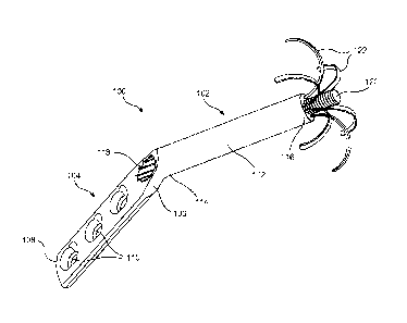
100071 As shown in Figs. 1 - 5, a system 100 according to an exemplary
embodiment of the
invention comprises a device 102 coupleable to or integrally formed with a
longitudinal plate
104 for fixing a fracture of a bone, such as a femur or humerus. As shown in
Fig. 1, the device
102 is coupled to a first end 106 of the plate 104 and extends away from the
end 106 at an angle
relative to a longitudinal axis of the plate 104 selected so that, when the
plate 104 is mounted at a
desired position on a target bone, the device 102 extends along a desired path
into a head portion
of the target bone. The plate 104 extends from the first end 106 to a second
end 108 and
includes a plurality of openings 110 along a length thereof for receiving bone
fixation elements
to fix the plate 104 to the target bone. Although the device 102 is shown
integrally formed with
the plate 104, it will be understood by those of skill in the art that in an
alternative embodiment,
a device 102', as shown in Figs. 6 - 7, may be formed as a separate component
configured to be
coupled to the plate 104, a nail or other anchoring element. For example, the
device 102' may be
inserted into the bone via an opening in the plate 104. Since the device 102'
is a separate
component, it will be understood by those of skill in the art that a proximal
end 114' of the
device 102' is configured to be coupled to the plate 104. The device 102' will
be described in
further detail below. It will also be understood by those of skill in the art
that although the
3
CA 02769205 2012-01-26
WO 2011/031416
PCT/US2010/045598
device 102 is shown as attached to the first end 106 of the plate 104, the
device 102 may be
attached or engaged to any point along a length of the plate 104 so long as,
when the plate 104 is
coupled to the target bone in a desired position, the device 102 extends into
the head of the bone
along a desired path.
[0008] The device 102 further comprises an elongated body 112 housing a bolt
120 and a
plurality of wires 122 which, in an insertion configuration, are received
within the body 112 and
which are movable to a deployed configuration in which they extend out of the
body 112 into the
head portion of the target bone. The elongated body 112 extends from a
proximal end 114 to a
distal end 116 and includes a lumen 118 extending therethrough. The proximal
end 114 of the
elongated body 112 is attached or engaged to the plate 104 such that the plate
104 may be
positioned externally to the target bone while the elongated body 112 extends
into the bone. The
lumen 118 is sized and shaped to accommodate the bolt 120 and the plurality of
wires 122 such
that the bolt 120 and the plurality of wires 122 are longitudinally movable
relative to the
elongated body 112. In the insertion configuration, shown in Figs. 2 - 3, the
wires 122 are
housed substantially within the lumen 118. Rotation of the bolt 120 about a
longitudinal axis of
the device 102 moves the plurality of wires 122 along the longitudinal axis,
distally relative to
the elongated body 112 into the deployed configuration shown in Figs. 4 - 5.
[0009] As shown in Figs. 2 - 5 specifically, each of the wires 122 extends
longitudinally from
a proximal end 146 including a protrusion 150 extending radially inward to
couple to the groove
142 of the head 138 of the bolt 120 such that the plurality of wires 122 are
spaced
circumferentially about the bolt 120. The wires 122 and the bolt 120 are
rotatably coupled to one
another such that the wires 122 do not rotate as the bolt 120 is rotated
through the elongated
body 112. Distal ends 148 of the wires 122 preferably include a bone
penetrating tip 152 which
may be sharpened or otherwise treated to facilitate penetration of the
spongious bone by the
wires 122. As seen in Fig 5, in the deployed configuration, distal portions of
wires 122 extend
distally out of the distal end 116 of the elongated body 112 to penetrate the
head portion of a
target bone into which the device 102 has been inserted. In this embodiment,
the wires 122 are
4
CA 02769205 2012-01-26
WO 2011/031416 PCT/US2010/045598
biased so that, upon exiting the body 112, they move toward a memorized shape
in which they
extend distally away from the distal end 116 for a distance and then bend back
proximally to
anchor the device 102 in the target bone. For example, the wires 122 may be
formed of a shape
memory material such as nitinol. Wires 122 formed of such a material may be
pre-shaped into
the bent configuration, in which a distal portion 154 of the wire is curved
through between
approximately 900 and 1800. In the insertion configuration, the wires 122 are
held in a
substantially straight configuration within the elongated body 112. However,
as the wires 122
are moved to the deployed configuration, the distal portions 154 extend past
the distal end 116 of
the elongated body 112 and revert to the bent configuration as the sharp tip
152 pierces through
the bone. The number of wires 122 and the circumferential orientation of the
wires 122 may
vary depending on specific patient issues and a load carrying capacity of the
device 102.
[0010] As the wires 122 move to the deployed configuration along these curved
paths, a
length of the wires 122 within the bone is increased while preventing the
wires 122 from too
closely approaching an outer surface of the head portion of the target bone.
It will be understood
by those of skill in the art that this bending of the wires 122 away from the
outer surface of the
head portion results in a high load-bearing, umbrella-shaped fixation
resisting movement of the
device 102 after the wires 122 have been implanted in the target bone. The
sharp tip 152 pierces
through the bone to form a recess that is substantially the same shape and
size as the wires 122,
resulting in a stable interface. Furthermore, the bent configuration guides
the wires 122 away
from cartilage in a joint area of the bone, preventing damage to the joint
surfaces. The bent
configuration also facilitates the dynamic behavior necessary for a smooth
transition of
compression and shear forces. Thus, it will be understood by those of skill in
the art that moving
the plurality of wires 122 from the insertion configuration to the deployed
configuration
enhances the stability and fixation of the device 102 within the target bone.
[0011] A thickness of a wall of the distal portion 124 of the elongated body
112 is greater than
a thickness of a wall of a proximal portion 126 of the elongated body 112 such
that a portion of
the lumen 118 extending through the distal portion 124 is greater in diameter
than a portion of
CA 02769205 2012-01-26
WO 2011/031416
PCT/US2010/045598
the lumen 118 extending through the proximal portion 126. The distal portion
124 includes a
threading 128 along at least a portion of an inner surface 130 thereof for
engaging with a shaft
136 of the bolt 120. The distal portion 124 may further include a plurality of
openings 132
extending along a length of the distal portion 124 and longitudinally aligning
with an inner
surface 134 of the proximal portion 126 of the elongated body 112 such that
each of the wires
122 may extend along the inner surface 134 into one of the openings 132. Thus,
it will be
understood by those of skill in the art that the openings 132 are sized and
shaped to
accommodate the wires 122.
[0012] The bolt 120 includes ahead 138 and a shaft 136, extending distally
therefrom. The
head 138 includes a driving element 140 such as a hex-recess for engaging a
driving tool to drive
the bolt 120 distally through the lumen 118 of the elongated body 112 to move
the wires between
the insertion and deployed configurations. The head 138 further includes a
groove 142 about a
circumference thereof for coupling with proximal ends 146 of the wires 122.
The shaft 136
includes a threading 144 along a length of the shaft 136 for engaging the
threading 128 of the
distal portion 124 of the elongated body 112. Thus, rotation of the bolt 120
about the
longitudinal axis of the device 102 moves the device 102 between the insertion
and deployed
configurations. In the insertion configuration, the bolt 120 is substantially
housed within the
lumen 118 of the elongated body 112 while, in the deployed configuration, the
distal end of the
bolt 120 extends distally from the body 112. Furthermore, the bolt 120 may
include a head 138
configured so that, when the device 102 moved to the deployed configuration,
the head 138 abuts
a proximal end 156 of the distal portion 124 of the elongated body 112 such
that the bolt 120
cannot move further distally.
[0013] It will also be understood by those of skill in the art that the device
102 may be
removed from the bone by rotating the bolt 120 about the longitudinal axis in
a direction
opposite the direction used to move the device 102 to the deployed
configuration. This moves
the bolt 120 proximally back into the elongated body 112 retracting the wires
122 along the
curved paths through which they were deployed back into the lumen 118 to the
insertion
6
CA 02769205 2012-01-26
WO 2011/031416
PCT/US2010/045598
configuration. At this point, the device 102 is no longer anchored in the head
portion of the
target bone and may be removed therefrom as would be understood by those of
skill in the art.
[0014] As shown in Figs. 6 - 7, the device 102' may be substantially
similar to the device 102,
as described above in regard to the system 100. The device 102' may be a nail,
screw or other
anchoring element that is insertable into the head portion of the target bone
via an opening in the
plate 104. Similarly to the device 102, the device 102' includes an elongated
body 112' that
extends from a proximal end 114' to a distal end 116' with a lumen 118'
extending therethrough.
The lumen 118' is sized and shaped to accommodate a bolt 120' and a plurality
of wires 122' such
that the bolt 120' and the wires 122' are longitudinally movable relative to
the lumen 118',
between an insertion configuration, shown in Fig. 6, and a deployed
configuration, shown in Fig.
7. The proximal end 114' of the elongated body 112' includes a coupling
mechanism for
coupling to the plate 104. For example, the proximal end 114' may include a
threading 115'
along an outer surface thereof for engaging a threading of the opening of the
bone plate 104.
[0015] In the insertion configuration, the wires 122' are substantially
housed within the
elongated body 112'. Similarly to the bolt 120, the bolt 120' is movable
relative to the elongated
housing 112' to move the wires 122' into the deployed configuration in which
distal portions 154'
extend distally past the distal end 116' of the elongated housing 112' and
into a head portion of
the bone, following a curved path of the distal portions 154'. The bolt 120',
however, is not
rotated relative to the elongated housing 112' to deploy the wires 122'. The
bolt 120' is non-
rotatably coupled to a proximal end 146' of the wires 122'. The bolt 120' may
further include a
pin 158' at a proximal end thereof such that the pin 158' may be used to push
the bolt 120' and
the wires 122' distally through the lumen 118' and into the deployed
configuration. To remove
the device 102' from the bone, the bolt 120' may be drawn proximally through
the lumen 118' via
the pin 158', retracting the wires 122' back into the insertion configuration
along the curved paths
through which they were deployed. At this point, the device 102' is no longer
anchored to the
head portion of the bone such that the device 102' may be removed therefrom.
7
CA 02769205 2012-01-26
WO 2011/031416
PCT/US2010/045598
[0016] It will be apparent to those skilled in the art that various
modifications and variations
may be made in the structure and the methodology of the present invention,
without departing
from the spirit or scope of the invention. Thus, it is intended that the
present invention cover the
modifications and variations of this invention provided that they come within
the scope of the
appended claims and their equivalents.
8