Note: Descriptions are shown in the official language in which they were submitted.
CA 02769282 2012-01-26
WO 2010/020876 PCT/IB2009/006726
CELL CULTURE METHOD
The present invention relates to cell and tissue culture. More particularly,
the present
invention provides methods for culturing cells to form aggregates, including
stem cells and
primary cells.
Research in human developmental biology has led to the discovery of human stem
cells
(precursor cells that can give rise to multiple tissue types), including
embryonic stem (ES)
cells, embryonic germ (EG) cells, fetal stem cells, and adult stem cells.
An enormous amount of interest has been generated in the use of embryonic and
adult stem
cells for cell replacement therapy and the treatment of disease. ES cells,
whose pluripotent
potential enables them to become any tissue in the body, have therapeutic
potential. Adult
stem cells are multipotent, rather than pluripotent. In other words, they are
capable of
transforming into a variety of tissue types. Like ES cells they have potential
uses such as
for cell replacement therapy and treatment of disease.
In order to study stem cells, and to use them for clinical therapies, one
prerequisite is the
supply of an adequate number of cells for the relevant clinical application. A
number of
different culture methods are known in the art which allow the proliferation
and
differentiation of stem cells (Ikeda et al., (2005), Vanderlaan et al.,
(2003), Amit et al.,
(2004), Bentzl (2006)).
Once proliferation has occurred, cultures of ES cells differentiate and
generate three
embryonic germ layers (mesoderm (muscle, bone, etc), ectoderm (neurons, skin,
etc) and
endoderm (hepatocytes, pancreatic beta cells, etc)) when the factors
maintaining stem cells
as stem cells are removed. (Keller G., 1995, Curr. Opin. Cell. Biol, 7:862).
Cells making
up these germ layers are multipotent and can differentiate only into cells of
one tissue of
the germ layer. Such cells are known as progenitor cells.
There are three methods know in the art which are capable of initiating stem
cell
differentiation: i) aggregation of ES cells for embryonic bodies (EBs); ii) co-
culture on
stromal cells (Nakano et al., 1994, Science, 265:1098); and iii) monolayer
culture on
extracellular matrix proteins (Keller G. 2005, Genes Dev., 19:1129).
Although co-culture and monolayer culture on ECM are simpler and more
convenient than
EB aggregation, the number of specific lineages that can be obtained by
differentiating
these cell types is still limited. In contrast, the three dimensional
structure of EB is
CA 02769282 2012-01-26
WO 2010/020876 PCT/IB2009/006726
2
analogous to embryonic development, and EB can form almost any kind of cell.
Hence, EB
formation is a general experimental protocol used for differentiating stem
cells into
specific cells (Marcel et al., 2003, Cardiovasc. Res., 58:292)
Various standard methods to aggregate ES cells into EB are known in the art
such as
hanging drop (HD) culture (Konno et al., 2005, J. Biosci. Bioeng., 100:88;
Dang et al.,
2002, Biotechnol. Bioeng. 78:442), liquid suspension culture (LSC) (Kovno et
al., 2005, J.
Biosci. Bioeng., 100:88; Oh et al., 2005, Biotechnol. Bioeng., 91:521);
Gerecht-Nir, 2004,
Biotechnol. Bioeng, 86:493) and attached culture (AC) (Konno et al., 2005, J.
Biosci.
Bioeng., 100:88; Dang et al., 2002, Biotechnol. Bioeng. 78:442).
Although HD is preferable to the other methods of forming EB because the
number of cells
in a single drop is controllable by the concentration of the cell suspension,
the method is
practically cumbersome and once formed, the EB must be transferred from the
hanging
drop to a separate culture dish to allow the cultures to differentiate
further. LCS and AC
methods of aggregation also involve a transfer step. Transferring the EB in
the known
methods is detrimental to the subsequent culturing steps as the integrity of
the EB is
potentially disturbed in the transfer step, resulting in a reduced efficiency
in the later
differentiating of the EB. Furthermore, a necrotic core has been observed in
EB grown
using known techniques for culturing stem cells in suspension.
Furthermore, the single EB formation efficiency is only around 70% due to the
cell
spreading in the hanging drop that generates satellite small clusters over the
inner surfaces
(Kurosawa et al., 2003, Biosci. Bioeng., 96:409). In addition, the size of the
EB in a
hanging drop is not always uniform due to the satellite aggregation of ES
cells and
irregular oval shapes of the drops. An additional method for generating EBs is
described in
Guo et al. (2006)
In addition, the HD methods known in the art require a relatively high degree
of manual
dexterity to manipulate. In particular, the transfer of EB from hanging drop
to a separate
culture to allow further differentiation requires a skill careful pipetting of
the stem cell
culture solution.
There is, therefore, a clear need in the art for an improved production and
culture method
for the formation of aggregated stem cell bodies, including both EB and
progenitor cell
bodies.
CA 02769282 2012-01-26
WO 2010/020876 PCT/IB2009/006726
3
The formation of aggregates is also an important part of primary cell culture.
It is
particularly useful in bringing together cells to allow them to form
organotypic cultures.
There is also a need for methods to improve the formation of aggregates of
primary cells.
At the moment primary cell aggregates are formed by spinning in flasks, but
this has the
disadvantage that the size of the aggregates of cells cannot be controlled.
Furthermore, it is
difficult to record electrophysiological activities from floating aggregates
generated from
primary cells grown using methods known in the art.
Disclosure of invention
The invention provides a method for culturing cells comprising the steps of:
(i) incubating cells in a hanging drop on the underside of a porous
membrane to form aggregates of cells;
(ii) inverting the membrane so that the aggregates of cells are located on the
upperside of the membrane; and
(iii) incubating the aggregates of cells on the upperside of the membrane.
Typically when the cell culture is incubated on the upperside of the membrane,
the
underside of the membrane is supplied with liquid medium. Preferably step
(iii) comprises
incubating the aggregates of cells at the air-liquid interface.
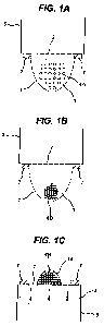
Figure 1 shows a schematic representation of the method of the invention.
Figures 1 A
shows a hanging drop on the underside of a porous membrane immediately after
application of the cells in suspension. Incubation of the cells forms
aggregates as shown in
Figure 113. Once inverted, the aggregates of cells are incubated on the
upperside of the
porous membrane as shown in Figure I C.
The methods of the invention overcome the problems in the prior art associated
with
transferring aggregates of cells from a hanging drop to a second culture dish.
The removal
of the transfer step means that any downstream use to which the aggregates of
cells are put
is more efficient and the cultures produced have a higher homogeneity and
structural
integrity.
Furthermore, the methods of the invention greatly reduce the level of manual
dexterity
required for culture methods for aggregate formation. Importantly, the methods
of the
CA 02769282 2012-01-26
WO 2010/020876 PCT/IB2009/006726
4
invention lend themselves to automation and therefore to high throughput
production of
aggregates of cells.
Step (i)
Step (i) of the cell culture method involves incubating cells as a hanging
drop on the
underside of a porous membrane to form aggregates of cells.
Cell culture and medium
The cells used in step (i) of the method of the invention may be primary
cells, embryonic
stem cells, adult stem cells, or progenitor cells. The various cell types
which can be used
with the methods of the invention are discussed below under the heading "Cell
type".
The cells may be a cell solution, i.e. cells suspended in a suitable medium.
The medium
may be any solution known to be capable of supporting the survival and/or
growth of the
cells. The medium will normally contain nutrients, a buffer and salts. The
type of medium
used will differ according to the type of cells being cultured and the
variations in the
constituents of the medium are discussed below under the heading "Medium".
The cells may be at any concentration within the medium solution. For example
the
concentration will usually be in the range of 1 to 50'000 cells/ l. More
preferably the
concentration is 5 to 10'000 cells/ l. The concentration of the cells may be
varied
depending on the type of cell being cultured, the use to which the aggregates
of cells are to
be put and/or the type of membrane being used.
The type of aggregates of cells formed will depend on the type of cells being
cultured. If
the cells being cultured are embryonic stem cells, the aggregates of cells are
embryoid
bodies (EB). If the cells being cultured are progenitor cells or primary
cells, then the
aggregates of cells will generate tissue-like structures.
Hanging drop
The cell culture is located on the underside of the membrane as a hanging drop
to allow for
the formation of the aggregates of cells. By "underside" is meant the lower
surface of the
membrane, so that the membrane is above the cells. The cell solution placed on
the
underside of the membrane forms a droplet due to the effect of gravity and the
attraction of
the liquid to the membrane via surface tension. The cells contained within the
droplet are
initially randomly distributed throughout the solution, but over time, under
the influence of
CA 02769282 2012-01-26
WO 2010/020876 PCT/IB2009/006726
gravity, will sediment to the bottom of the drop. In doing so, the cells
become compacted
and form aggregates of cells.
The size of the hanging drops used is limited by the amount of liquid which
can be retained
on the underside of the membrane by surface tension. The drop size may vary in
5 accordance with the composition of the medium and the type of membrane being
used, but
will usually be in the range of 0.1 l to 100 l, e.g. 0.1, 0.2, 0.5, 1, 2, 5,
10, 15, 20, 25, 30,
35, 40, 45, 50, 60, 70, 80, 90 or 100 l. More preferably the drop is 30 l to
4O 1.
Incubation period
The incubation period is limited by the amount of nutrients present in the
liquid medium.
As discussed above, the size of the hanging drop culture is limited by the
composition of
the medium and the type of membrane being used and there is a finite amount of
nutrients
in the medium. Therefore step (i) of the cell culture method comprises
incubation for a
finite period of time in the range of about 1 to 72 hours, e.g. about 1, 12,
14, 16, 18, 20, 24,
30, 36, 40, 44, 48, 60, 72 or 84. Most preferably step (i) is incubated until
aggregates of
cells have formed, which usually takes about 12 to 48 hours.
The method of the invention may comprise the preliminary step of applying the
cells onto
the membrane. This is usually achieved by manual pipetting of a cell solution.
It may also
be achieved by automated pipetting, for example the use of a robotic arm. Such
devices are
well known to the person skilled in the art. The cells can be applied to the
membrane in
any orientation. For example the cells can be applied to the upperside of the
membrane
which may then immediately be inverted so that the cells may be incubated in a
hanging
drop on the underside of the membrane as required by step (i). Alternatively
the cells can
be applied directly to the underside of the membrane.
Step (ii)
Once step (i) of the cell culture method is completed the membrane is inverted
so that the
aggregates of cells are located on the upperside of the membrane. This
inversion forms
step (ii) of the cell culture method. By "upperside" is meant the top of the
membrane, so
that the aggregates of cells are above the membrane.
Inversion of the membrane can be achieved manually, e.g. by a person inverting
the
membrane. Alternatively the inversion may be automated and carried out by
machine.
CA 02769282 2012-01-26
WO 2010/020876 PCT/IB2009/006726
6
Step (iii)
Step (iii) of the stem culture method involves incubating the aggregates of
cells on the
upperside of the membrane. By "upperside" is meant the top of the membrane, so
that the
aggregates of cells are above the membrane.
Preferably step (iii) comprises incubating the aggregates of cells at the air-
liquid interface.
The air-liquid interface is formed due to the porous nature of the membrane
and the
gravitational force exerted on the liquid medium surrounding the aggregates of
cells. Once
the membrane comprising the hanging drop and aggregates of cells is inverted
such that the
aggregates of cells are on the upperside of the membrane, i.e. step (ii),
gravity acts to draw
any excess liquid medium contained in the drop through the porous membrane and
away
from the aggregates of cells. At the same time, surface tension in the liquid
medium means
that not all of the medium is drawn away, but instead a layer of medium is
left coating the
aggregates of cells. The point where the aggregates of cells, the medium and
the air are in
close proximity due to the above effect is termed the "air-liquid-interface".
Gas transfer to
the aggregates of cells, both for the uptake of oxygen and the removal of
carbon dioxide, is
much more efficient than when the aggregates of cells is fully immersed in
culture
medium.
In order to ensure continued survival of the aggregates of cells, the
underside of the
membrane is supplied with liquid medium which is retained in contact with the
membrane.
As the aggregates of cells are resting on a porous membrane the liquid medium
is drawn
through the pores in the membrane by capillarity.
In some aspects of the invention, the liquid medium is retained in contact
with the
underside of the membrane by capillarity. Examples of devices that allow the
retention by
capillarity are described in W02006/134432. If the medium is retained by
capillarity this
acts to compact the cells on top of the membrane. The compaction of primary
cells in
particular acts to promote cell-cell contact and the formation of organotypic
cultures.
Incubation period
The incubation period referred to in step (iii) will vary depending on the
type of cells in the
culture. The period will usually be for a finite period of time in the range
of about 1 to 960
hours, e.g. about 1, 2, 5, 10, 20, 24, 48, 72, 96, 120, 150, 200, 250, 300,
400, 500, 600,
700, 800, 900, 960. Most preferably step (iii) comprises incubation for 24 to
400 hours.
CA 02769282 2012-01-26
WO 2010/020876 PCT/IB2009/006726
7
The time period may also vary depending on the intended use of the culture.
The method
of the invention may be used to produce proliferating cultures of primary
cells or stem
cells. By "proliferating culture" is meant a culture in which the number of
cells is
increasing by the division of single cells into two identical daughter cells.
If the cell culture method is intended to provide a proliferating culture then
the incubation
period will usually be from 24 to 400 hours depending on the cell types and
the species of
origin.
The method of the invention may be used to produce differentiating cultures of
stem cells.
By "differentiating culture" is meant a culture in which undifferentiated stem
cells are
acquiring the features of specialised cells.
If the cell culture method is intended to provide a differentiating culture
then the
incubation period will usually be from 24 to 400 hours depending on the cell
types and the
species of origin.
Cell types
The cells used in the cell culture methods of the invention may be primary
cells, embryonic
stem cells, adult stem cells, or progenitor cells
Stem cells
In one embodiment of the methods of the invention the cell culture is a stem
cell culture.
By "stem cell" is meant a multipotent cell. The term "stem cell" includes
"embryonic stem
cells", "adult stem cells", "progenitor cells" and "induced pluripotent stem
cells".
By "embryonic stem cell" is meant a pluripotent stem cell capable of
differentiating into
the three somatic germ layers that comprise an organism: mesoderm (muscle,
bone, etc),
ectoderm (neurons, skin, etc) and endoderm (hepatocytes, pancreatic beta
cells, etc).
By "adult stem cells" is meant a stem cell which is found in different tissues
of the
developed, adult organism which remains in an undifferentiated, or
unspecialized form.
These stem cells can give rise to specialized cell types of the tissue from
which they came,
i.e., a neural stem cell can give rise to a functional nervous tissue-like
parenchyma
comprising the different cell types (neuronal and glial cells). The degree of
self renewal
and differentiation potential of adult stem cells is more restricted when
compared to
embryonic stem cells. Adult stem cells are multipotent, not pluripotent.
CA 02769282 2012-01-26
WO 2010/020876 PCT/IB2009/006726
8
By "progenitor cell" is meant a multipotent cell which can differentiate only
into cells of
one tissue or germ layer. A progenitor cell is an early descendant of a stem
cell that can
only differentiate, but can only partially renew itself for a determined
period of time.
By "induced pluripotent stem cell (iPS)" is meant a type of pluripotent stem
cell artificially
derived from a non-pluripotent cell, typically an adult somatic cell, by
inducing a "forced"
expression of certain genes. iPS cells are believed to be identical to natural
pluripotent
stem cells, such as embryonic stem cells in many respects, such as the
expression of certain
stem cell genes and proteins, chromatin methylation patterns, doubling time,
embryoid
body formation, teratoma formation, viable chimera formation, and potency and
differentiability.
The methods of the invention include culturing any of the known types of stem
cells,
including embryonic stem cells, adult stem cells, induced pluripotent stem
cells (iPS cells)
from adult somatic cells and progenitor cells.
Primary cells
In another embodiment, the methods of the invention are suitable for culturing
primary
cells. By "primary cells" is meant that the cells are fully differentiated and
specialised into
a particular cell type. For example cells taken from the central nervous
system, blood
(e.g.monocytes), spleen, thymus, heart, mammary glands, liver, pancreas,
thyroid, skeletal
muscle, kidney, lung, intestine, ovary, bladder, testis, uterus or connective
tissue.
Primary cell cultures may be formed from dissociated cells or microexplants
taken from
organs. As used herein, the term "dissociated cell" refers to a single cell
that has been
isolated from an organ. The term "microexplant" refers to a small group from
400 cells to
up to few thousands cells isolated from the organ. Where the method of the
invention
refers to primary cells the culture comprises more than one dissociated cell,
or more than
one microexplant. Preferably, the method of the invention involves the culture
of many
dissociated cells, or many microexplants, isolated from an organ.
Where the methods of the invention relate to culturing primary cell, the
methods further
include the preliminary step of isolating the cells from the organ.
Methods for isolating dissociated cells from organs are known in the art. The
dissociated
cells may be isolated from the organ of interest by mechanical or enzymatic
dissociation of
tissue, or both. For example the dissociated cells may be obtained by
dissociation of the
CA 02769282 2012-01-26
WO 2010/020876 PCT/IB2009/006726
9
organ using the proteolytic enzyme trypsin 0.25% (w/w) in Hank's Balanced Salt
Solution
(HBSS) without calcium and magnesium. After the addition of trypsin inhibitor
to stop the
enzymatic dissociation, the cells may be incubated briefly in suspension to
allow
undissociated cells to fall to the bottom, leaving the dissociated cells in
suspension.
The microexplants and explants used in the methods of the invention may be
obtained by
mechanical reduction of the organ of interest to small pieces of tissue. For
example, the
microexplants may obtained by repeated aspiration, usually of post-natal
tissue, in a
disposable pipette tip, or by maceration with a scalpel blade. Preferably, the
tissue is
neonatal tissue.
The methods of the invention may be used to produce an organotypic culture
from a wide
variety of organs and the nature of the cells that are used in the process
will depend on the
organotypic culture that is desired. Preferably, the organ from which the
cells are obtained
is an animal organ, preferably a mammalian organ, preferably a human organ.
The cells may be obtained from any organ in the animal including, but not
limited to the
central nervous system, bone marrow, blood (e.g.monocytes), spleen, thymus
heart,
mammary glands, liver, pancreas, thyroid, skeletal muscle, kidney, lung,
intestine, ovary,
bladder, testis, uterus or connective tissue. Preferably, the dissociated
cells, explants or
microexplants are from the central nervous system, heart, liver or kidney.
Where the
dissociated cells, explants or microexplants are from the central nervous
system, they may
be from the brain or from the spinal cord. Preferably, the cells are from the
brain,
preferably from the hippocampus or the cortex.
The cells may be obtained from a particular region of the organ. For example,
where the
organ is brain, the cells may be obtained from the hippocampus or from the
cortex. As
demonstrated in the examples herein, dissociated cells from the cortical
region can be used
to produce an organotypic culture that shows the typical cell composition and
intercellular
connections of hippocampus. Where the organ is heart, the cells may be
obtained from the
myocardium.
The cells may be obtained from more than one organ and cultured together. For
example,
the cells may be derived from two, three, four or more different organs. The
co-culture of
cells obtained from more than one organ allows the generation of models of
interactions of
tissues derived from different organs. Preferably, where cells from more than
one organ are
used, the organs will be organs that naturally exist in contact in vivo so
that the organotypic
CA 02769282 2012-01-26
WO 2010/020876 PCT/IB2009/006726
culture resulting from co-culture of cells from these organs will provide a
model for the in
vivo situation. For example, immune cells, particularly white blood cells,
could be co-
cultured with cells from various organs to study inflammation. Tumor cells
might also be
co-cultured with cells from various organs to study cancer development. Stem
cells could
5 be co-cultured with other cell types to produce mixed cultures. Skeletal
muscle cells could
be co-cultured with cells from the central nervous system, including
hippocampus, cortex,
cerebellum and spinal cord, to produce a model of a neuro-muscular junction.
Endothelial
cells that line blood vessels could be co-cultured with brain cells to form a
model of the
blood-brain barrier.
10 The cells used in the methods of the invention may be derived from healthy
organisms or
from diseased organism. The ability of the methods of the of the invention to
generate
aggregates of cells quickly and easily means that the methods will have
extensive
applications in the production of cell cultures for the study of disease links
and for drug
screening. Comparison of aggregates of cells obtained by the methods of the
invention
from healthy organisms and diseased organisms will further current knowledge
of disease
states and allow the identification of biomarkers and drug targets which are
indicative of
disease states.
The cells used in the methods of the invention may be genetically altered. For
example, the
cells may be genetically altered to modulate expression of a drug target or a
biomarker. A
biomarker is a molecular marker, the presence of which at a certain level or
in a certain
molecular form indicates the presence of a diseased state. A drug target is a
molecular
species that can be modulated to affect a disease process, i.e. a molecule
through which a
drug acts. Changing the nature or level of function of the drug target must
have a positive
impact on disease outcome, and the target should be of a molecular type that
is amenable
to modulation. In many cases, information about drug targets is obtained from
genetic and
other biological studies, and classes of compounds that are known to interact
with those
targets are available. It is often desirable to modulate the levels of these
biomarkers and
drug targets in biological systems, and to study the biological consequences.
Alternatively, the cells may be genetically altered to express a visual
marker, such as a
fluorescent marker, that allows the cells to be tracked visually.
Technologies to express cloned genes and to ablate the expression of cloned or
endogenous
genes are known in the art. These technologies may be used to increase or
decrease
CA 02769282 2012-01-26
WO 2010/020876 PCT/IB2009/006726
11
expression of a marker, such as a drug target or biomarker, in the cells used
in the methods
of the invention.
Techniques to increase expression of a cloned or endogenous gene are based on
the
introduction of heterologous DNA in a form which recruits the cellular
expression system,
and many different approaches are well known to those skilled in the art. In
some cases
naked DNA may be used with a lipophilic transfection reagent, the DNA
including a
strong promoter co-linear with the gene to be expressed and a replication
origin that
enables cytoplasmic replication of the introduced DNA. In other cases a viral
vector may
be used to increase the efficiency of DNA introduction. Similarly, means to
ablate gene
expression that are well known to those skilled in the art including antisense
DNA
oligonucleotides, peptide nucleic acid and double-stranded RNA interference.
In some
cases, naked nucleic acid may be used. In other cases, especially for the use
of small
interfering RNA, expression vectors may be used to express the molecule in a
self-
assembling hairpin form. It has also been shown that proteins can be
introduced directly
into cells provided that they are attached to an entity that encourages
transport from the
exterior to the interior of the cell. The Tat protein of human
immunodeficiency virus (HIV)
is one such entity, and proteins to be transferred may be produced as fusion
proteins with
HIV-Tat and introduced into cells (Becker-Hapak M. et al, 2001).
It will also be clear to those skilled in the art that, instead of
transforming or transfecting
the cells as described above, the cells used in the method of the invention
may be from a
transgenic animal. For example, the cells may be from a transgenic animal
expressing a
visual marker, such as a fluorescent marker, of from a transgenic animal in
which
expression of a particular drug target or biomarker has been increased or
decreased.
The cells used in the methods of the invention may be derived from healthy
organisms or
from diseased organism.
Media
The medium may be any solution known to be capable of supporting the survival
and/or
growth of cells.
The selection of medium will vary depending on the type of cells being
cultured and the
intended use of the aggregates of cells. For example, stem cells require a
number of
different media components than primary cell cultures. In turn, embryonic stem
cell
CA 02769282 2012-01-26
WO 2010/020876 PCT/IB2009/006726
12
cultures require different components to progenitor cultures. The components
of the
medium in each case are intended to be varied accordingly and such variation
is within the
knowledge of the skilled person.
The medium will normally contain nutrients, a buffer and salts. For example ES
cell
medium comprises 80% DMEM/F12, 20% KnockOut-Serum Replacement, 2 mM L-
glutamine, 1% non-essential amino acids, 0.1 mM (3-mercaptoethanol, 4 ng/ml
basic
Fibroblast Growth Factor (bFGF) for the cell proliferation. Examples of
suitable liquid
media are described, for example, in Stoppini L. et al (1991) and Muller et al
(2001).
Where the cell culture is a stem cell culture the composition of the medium
may also be
varied depending on whether the culture is to be allowed to proliferate or
differentiate. By
"proliferate" is meant the expansion of the number of cells by the division of
a single cells
into two identical daughter cells. By "differentiate" is meant the process
whereby an
undifferentiated stem cell acquires the features of a specialised cell such as
heart, liver or a
muscle cell.
For example, a stem cell culture which is intended to be allowed to
proliferate will require
the presence of embryonic growth factor (EGF) and/or foetal growth factor
(FGF) in order
to prevent differentiation. On the other hand, if a stem cell culture is
required to
differentiate, then EGF and FGF should not be present.
The composition of the medium may thus be different in steps (i) and (iii).
For example, it
may be advantageous to prevent differentiation of a stem cell culture during
step (i), in
which case EGF or FGF will be present in the medium. If the culture is then to
be allowed
to differentiate in step (iii), the EGF or FGF will be removed from the medium
so that
differentiation can occur.
Membrane
The porous membrane on which the cells are incubated will depend on the nature
of the
cells being cultured and the intended use to which the aggregates of cells are
to be put. For
example, certain cell types grow more effectively on different membranes.
The porous membrane will usually comprise pores with a size of 0.4 m up to 12
m.
Membranes suitable for use in the cell culture method include but are not
limited to the
hydrophilic polytetrafluoroethylene (PTFE, also known under the DuPont trade
name
Teflon ) membrane produced by Millipore Corporation which is optically
transparent,
CA 02769282 2012-01-26
WO 2010/020876 PCT/IB2009/006726
13
membranes made of polycarbonate, PET (polyethylene terephthalate), or
AnoporeTM
(inorganic aluminium oxide, a trademark of Whatman Corp).
Preferably, the porous membrane is optically transparent. This feature enables
the cells or
aggregates of cells to be accessible at all times to microscopic examination
and sampling
for biochemical assays. Preferably, the porous membrane produces low
background
fluorescence at the wavelengths used for excitation, usually in the range of
400-750nm.
Preferably, the porous membrane is composed of hydrophilic
polytetrafluoroethylene
(PTFE) membrane.
Hydrophobic barrier
The methods of the invention described above confer a number of advantages
over the
methods known in the prior art. The introduction of a hydrophobic barrier
adapted to
contain the cell culture during step (i) and subsequently through the
inversion of step (ii)
and culturing the aggregates of cells of step (iii) extends these advantages
even further.
The presence of a hydrophobic barrier adapted to contain the culture on the
membrane
confers a number of advantages over the methods known in the prior art.
Methods known
in the art for, for example those described in W02006/136953, involve growing
cell
cultures on the upperside of a porous membrane. If a cell culture is grown in
this way it is
grown at the air-liquid interface. However, the methods in W02006/136953 do
not provide
a means for containing the growth of the culture, i.e. the edges of the
membrane are not
designed to restrict cell growth. Therefore, proliferation of the culture can
result in it
growing beyond the edges of the membrane and onto the device itself. This
prevents the
correct supply of medium to the culture and makes further handling of culture
difficult.
The inventors have surprisingly found that a hydrophobic barrier can be used
to delimit the
boundaries of the cell culture and prevent "over growth" of the culture beyond
the
membrane and onto the device in step iii) of the method.
Cultures grown using the method of W02006/136953 grow at the air-liquid
interface. The
air-liquid interface is formed due to the porous nature of the membrane and
the
gravitational force exerted on the liquid medium surrounding the cell culture.
Gravity acts
to draw any excess liquid medium contained in the cell culture through the
porous
membrane and away from the cell culture. At the same time, surface tension in
the liquid
medium means that not all of the medium is drawn away, but instead a layer of
medium is
CA 02769282 2012-01-26
WO 2010/020876 PCT/IB2009/006726
14
left coating the aggregates of cells. The growth of cell cultures at the air-
liquid interface is
advantageous to the cell cultures.
If no barrier is placed around the culture, once the culture has grown to the
edge of the
membrane, then capillarity may cause the medium to be drawn over the edges of
the
membrane. In this case, the culture will no longer be at the air-liquid
interface, but will
instead by submerged in the excess medium being drawn onto the upperside of
the
membrane. The culture will, in such a case, be flooded.
The inventors have surprisingly realised that the hydrophobic barrier used in
the methods
of the present invention also prevents such flooding in step iii) of the
method.
The inventors have found that the prevention of flooding is more effective
when the height
that the hydrophobic barrier projects above the membrane is below 100 m, e.g.
about 1, 2,
5, 10., 15, 20, 25, 30, 35, 40, 50, 60, 70, 80, 90 or 100 m. More preferably,
the
hydrophobic barrier projects no further than 50 m above the surface of the
membrane.
As well as preventing flooding, the hydrophobic barrier allows the boundaries
of the cell
culture to be controlled. Therefore, the shape and size of the cell culture
can be altered as
desired. The barrier can be of any shape, for example it may be circular,
elliptical,
triangular or square. The barrier can also be of more complex shapes such as a
dumbbell.
Figure 9 shows examples of different shapes which may be used for the
hydrophobic
barrier.
The shape of the barrier may be chosen based on the type of cells being
cultured. For
example, it may be desirable to grow neuronal cells within dumbbell shaped
hydrophobic
barriers, while it may be desirable to grow cells from pancreas or liver cells
within circular
shapes hydrophobic barriers.
The area contained within the hydrophobic barrier can also be altered. If the
barrier is
circular, then the radius will usually be in the range of 0.5mm to 5.0mm, e.g.
0.5, 0.75, 1.0,
2.0, 3.0, 4.0 or 5.0mm. If the hydrophobic barrier is any other shape, then
the area
contained within the hydrophobic barrier will usually be in the range of
0.5mm2 to 80mm2,
e.g. 0.5, 1.0, 2.0, 5.0, 10, 15, 20, 25, 30, 35, 40, 45, 50, 60, 70, 80mm2.
The use of the hydrophobic barrier also allows culture conditions to be
changed when the
culture reaches a specific predetermined shape or size. For example, a stem
cell culture
may be grown using the methods of the invention with a hydrophobic barrier.
The medium
CA 02769282 2012-01-26
WO 2010/020876 PCT/IB2009/006726
used to sustain the culture can be controlled so that the culture is allowed
to proliferate,
e.g. the inclusion of embryonic growth factor (EGF) or foetal growth factor
(FGF).
Proliferation can be continued until the cell culture fills the area within
the hydrophobic
barrier. At this stage, the medium can altered, e.g. by removing EGF or FGF,
and the
5 culture can be allowed to differentiate. Such control means that cell
cultures of precise size
and shape can be consistently generated. The generation of multiple cultures
in this way
improves the repeatability of experiments conducted using the cell cultures
generated with
the device of the invention.
Providing a drop of constant size and shape also allows the cell culture
volume and
10 concentration to be optimised. This allows for the number of aggregates of
cells which are
formed to be controlled and in turn reduces the number of satellite small
clusters formed.
Furthermore, the size of the aggregates of cells formed in the hanging drop
can be
controlled by altering the size of hydrophobic barrier and the number of cells
within the
drop.
15 The hydrophobic barrier also allows the precise location of the cell
culture to be known.
This increases the efficiency with which the cultures produced by the methods
of the
invention can be located. The efficiency can be further increased by using a
hydrophobic
barrier which is coloured in such a way so that it contrasts with the colour
of the porous
membrane. For example, the hydrophobic barrier may be red, blue, green, black,
grey,
yellow, orange, or any shade of these colours.
Furthermore, the hydrophobic barrier also confers advantages to the automation
of the cell
culture. It is currently known to use robotic arms to apply cell cultures to
multi-well plates.
However, as it is important to avoid damage of the membrane caused by the
pipette, the
pipette tip is not allowed to advance into contact with the membrane. This in
turn can
cause a slight variation in the location that the initial culture is pipetted
onto the membrane.
Therefore, subsequent automated procedures become increasingly difficult as
the exact
location of the culture is not known.
The methods of the invention using a hydrophobic barrier overcome this
disadvantage by
allowing the location of the cell culture to be known precisely, i.e. it is
always within the
boundaries of the hydrophobic barrier. Therefore, the minor variations in the
initial
location of the pipetting step are negated and the precise location of the
culture is known
CA 02769282 2012-01-26
WO 2010/020876 PCT/IB2009/006726
16
for further automated steps. In particular, this confers an advantage to the
automated
visualisation of the culture.
The hydrophobic barrier may be made of any material which is capable of
preventing the
movement of the liquid culture across a porous membrane and, thus, retaining
the cell
culture. In one embodiment the hydrophobic barrier is made of a hydrophobic
ink. The
hydrophobic ink can be drawn onto the porous membrane in the desired shape and
size, or
more usually will be printed onto the membrane with the desired size and
shape. Examples
of such inks include carbon commonly used as an ink source for laser printers
and
photocopiers, silicone inks and acrylic inks.
In an alternative embodiment the hydrophobic barrier is a laminated layer
which is
pre-shaped before application to the porous membrane. The laminated layer is a
sheet of
hydrophobic material, for example plastic polymers from which one or more
sections have
been removed. The removal of one or more sections from the laminate layer
creates one or
more voids. The void may be circular, dumbbell shaped or any other shape
depending on
the shape of the hydrophobic barrier required. The laminated layer is applied
to the porous
membrane such that the edges of the void in the laminate layer act as the
hydrophobic
barrier when the cell culture is placed on the porous membrane within the void
area.
Preferably, the laminated layer will be fused to the membrane by gluing, by
heat-sealing or
by ultra-sonic sealing.
Screening methods
The ability of the methods of the of the invention to generate aggregates of
cells quickly
and easily means that the methods will have extensive applications in the
production of cell
cultures for the study of disease links and for drug screening. Furthermore,
the methods of
the invention will have extensive applications for the study of stem cells. In
particular, the
methods of the invention allow for the screening of compounds which promote
differentiation of stem cells into different cell types.
Comparison of aggregates of cells obtained by the methods of the invention
from healthy
organisms and diseased organisms will further current knowledge of disease
states and
allow the identification of biomarkers and drug targets which are indicative
of disease
states.
CA 02769282 2012-01-26
WO 2010/020876 PCT/IB2009/006726
17
As described above, biomarkers are molecular markers which at a certain level
or in a
certain molecular form indicate the presence of a diseased state. A drug
target is a
molecular species that can be modulated to affect a disease process. One
application of the
cell cultures of the invention is in the identification of biomarkers and drug
targets.
Screening of several molecular classes, such as proteins and lipids, in cell
cultures that
express a disease state or the corresponding non-diseased state may be used to
identify
biomarkers. Validated biomarkers are currently used both to identify carriers
of a disease
state and to monitor their progress towards normality that may be assisted by
a therapeutic
regime such as a drug. It is necessary to establish a statistically
significant association
between a candidate biomarker and a disease state to validate the biomarker
for use in
clinical trials. The cell cultures of the present invention are ideally suited
to biomarker
discovery and validation due to the fact that they replicate organ function
and physiology
and can be generated quickly and easily by the methods of the invention such
they are
applicable to high throughput assays. The cell cultures of the invention could
thus be used
much more rapidly and cheaply than whole animals currently used for the
identification
and validation of biomarkers.
According to a further aspect of the invention, there is therefore provided a
method for the
identification and validation of biomarkers and drug targets comprising
screening the cell
cultures produced by the methods of the invention. Assays for identifying
biomarkers and
drug targets include the use of transcriptional profiling, proteomics, mass
spectrometry, gel
electrophoresis, gas chromatography and other methods for molecular profiling
known to
those skilled in the art.
Surrogate markers are a sub-set of biomarkers that can be used to assess the
presence or
progression of a disease state, but that do not measure directly a clinical
outcome of the
disease. The cell cultures of the invention may be used to identify and
validate surrogate
markers in the same way as other biomarkers.
The cell cultures produced by the methods of the invention are not only useful
in the
identification of biomarkers and drug targets associated with disease states
but are also
useful in screening to identify drugs that alleviate these disease states.
Cell cultures are
particularly useful in the screening of candidate drugs because it is
important for such
screening that the target culture has biochemical and physiological properties
that match as
closely as possible those features of the target organ in vivo. It must be
possible, however,
CA 02769282 2012-01-26
WO 2010/020876 PCT/IB2009/006726
18
for the cell culture to be used at high throughput to enable screening of
sufficiently large
numbers of drug candidates for a high probability of successful identification
of lead drugs.
Additional large-scale assays are often necessary to validate the inclusion of
a lead drug in
a preclinical and clinical drug development programme.
The methods of the invention may be used to generate many thousands of cell
cultures
simultaneously and are thus uniquely suited to high throughput applications
involving
multiple assays for each culture. In one embodiment, the methods for producing
a cell
culture according to the invention further comprises the step of screening
using the
resulting cell culture in a method of screening and pre-clinical validation of
candidate
drugs. As discussed above, one particularly useful aspect of the method of the
invention is
that it facilitates the high-throughput formation of cell cultures in which
the cells have been
genetically altered to modulate the expression of a biomarker or drug target.
These
modified organotypic cultures will also be useful in the screening of
candidate drugs.
The field of toxicology is a further application area for the present
invention that will
benefit greatly by the enhanced flexibility and throughput provided by the
methods of the
invention. Organotypic response is crucially important in this field, because
different
tissues differ greatly in their response to toxins, with different clinical
consequences.
Different tissues can contain different enzymes systems, notably of the
cytochrome P450
class, that metabolise different classes of exogenous compounds. The degree
and type of
metabolism of a compound can profoundly affect its toxicity. Large-scale
screening of
toxicity in a wide variety of tissues is so expensive at present that many
chemicals in
common use have never been tested adequately. Increasing awareness of
potential toxicity
has brought pressure to carry out such tests without the means to do so at
acceptable cost.
The invention therefore also includes a method of assessing the toxicity of a
chemical
using the cell cultures of the present invention.
High throughput
The methods of the invention described above can also be used in a high-
throughput
format that involves preparing and maintaining multiple cell cultures
simultaneously.
Accordingly the invention also provides a high-throughput method for the
preparation of a
collection of cell cultures comprising preparing multiple cell cultures
according to the
methods as described above.
CA 02769282 2012-01-26
WO 2010/020876 PCT/IB2009/006726
19
Preferably, the methods of the invention are carried out in a device which
allows multiple
parallel cultures per device, preferably 2, 4, 8, 16, 24, 96, 384, 1536 or
more parallel
cultures per device. A preferred device for carrying out the methods of the
invention is
described below.
Device
The methods of the invention as described above may be carried out on a device
adapted
for the purpose. Accordingly, the invention therefore provides a device for
carrying out the
methods of cell culture of the invention, said device comprising:
(i) a medium conduit having one open end and one end closed by a porous
membrane fused across it; and
(ii) a frame holding the medium conduit in a substantially vertical
orientation;
wherein the medium conduit is adapted to permit retention by capillarity of a
sufficient volume of liquid culture medium in the medium conduit to contact
the surface of
the porous membrane and thus supply nutrients to cells that may be grown on
the porous
membrane, characterised in that the surface of the membrane contralateral to
the surface of
said porous membrane sealed to said medium conduit comprises a hydrophobic
barrier
adapted to contain the culture.
The presence of a hydrophobic barrier adapted to contain the culture on the
membrane
confers a number of advantages over the devices known in the prior art as
described above.
During cell culture, the culture is maintained on the surface of the porous
membrane that is
disposed at one end of the conduit. One key feature of the device is that the
conduit is
designed such that, during cell culture, the force of capillarity maintains
contact between
the surface of the porous membrane contralateral to the cell culture, i.e. the
surface of the
membrane in the conduit, and the culture medium.
The use of the force of capillarity to maintain the culture medium in the
conduit enables
the removal and replacement of the culture medium by a pipetting step. When
supplying
the medium, the pipette tip should be positioned as closely as practicable to
the surface of
the membrane.
Preferably, the conduit is adapted such that it retains a sufficient volume of
liquid culture
medium by capillarity to maintain contact between the surface of the porous
membrane in
CA 02769282 2012-01-26
WO 2010/020876 PCT/IB2009/006726
the conduit and the culture medium when the device is in either the upright or
inverted
position. Said conduit may be referred to herein as the medium conduit.
By upright position is meant that the frame holds the conduit substantially
vertically with
the end sealed by the porous membrane positioned uppermost so that, when the
device is in
5 use, the cell culture is grown on the upper surface of the membrane. By
inverted position is
meant that the frame holds the conduit substantially vertically with the open
end positioned
uppermost and the end closed by the porous membrane lowermost so that, when
the device
is in use, the cell culture in the lower surface of the membrane. In contrast
to the devices
that are available in the art, the device of the invention thus allows
incubation of the cell
10 culture and change of the medium for the cell culture with the device in
either the upright
or inverted position. This flexibility in orientation of the culture and the
device means that
either microscopes with their objective lenses facing upwards or microscopes
with their
objective lenses facing downwards can be used interchangeably for studying the
culture,
and that liquid handling devices can be used in either orientation to add or
remove the
15 medium.
Preferably, the conduit is a cylinder, a cone or is frustoconical. Where the
medium conduit
is a cone the porous membrane is sealed across the narrowest radius of the
cone. The
conduit may also be of rectangular or asymmetrical cross-section. The exact
dimensions
and composition of the conduit are selected such that, during cell culture, it
retains a
20 sufficient volume of liquid culture medium by capillarity to maintain
contact between the
surface of the porous membrane in the conduit and the culture medium,
preferably
irrespective of whether the device is in the upright or inverted position. The
volume of
liquid retained should be sufficient such that in use, adequate nutrients are
supplied to the
cell culture without requiring the medium to be changed at unreasonably short
intervals.
Capillarity is dependent on several parameters. The force of capillarity is an
inverse
function of the diameter of a cylindrical vessel or the width or breadth of a
conduit of
rectangular section. The force of capillarity on an aqueous solution also
depends on the
surface tension of the solution being held by that force which can be weakened
by the
presence in solution of surfactants such as detergents. Capillarity is
affected by the degree
of attraction between the molecules of the liquid and the molecules of the
surface. In the
case of an aqueous liquid, capillarity is affected by the degree of
hydrophilicity of the
surface of the conduit. A further factor affecting the retention of liquid
culture medium in a
CA 02769282 2012-01-26
WO 2010/020876 PCT/IB2009/006726
21
conduit is the volume of the culture medium. These factors therefore need to
be taken into
account to ensure that the device of the invention can retain a volume of
liquid media in
contact with the surface of the porous membrane by capillarity.
In the device of the present invention, two different capillary forces act to
retain the liquid
medium in the conduit in contact with the porous membrane. The force of
capillarity
exerted by attraction between the liquid medium and the tube is one force. The
other force
is exerted by attraction between the liquid medium and the walls in the pores
of the
membrane. If sufficiently strong, the former will counteract gravity to keep
the liquid in
the conduit irrespective of whether it is upright or inverted, and the latter
will keep the
liquid in contact with the membrane. At a certain threshold, the force of
gravity on the
culture medium will exceed the force of capillarity and culture medium not
restrained by
an additional force will fall from the conduit.
Where the conduit is a cylinder, the mass of the liquid contained in the
cylinder and thus
the gravitational force acting to remove the liquid from the cylinder is
directly proportional
to the square of the radius of the cylinder, whereas the capillary force
acting to retain the
liquid in the cylinder is inversely proportional to the radius. Thus for a
given liquid and
cylinder length there is a maximum radius above which the liquid in a cylinder
of a given
surface composition will not be retained against the force of gravity, but
there is no
minimum radius below which liquid will not be retained against the force of
gravity.
Preferably, the conduit is a cylinder having a radius of 0.5cm or less,
preferably 0.3cm or
less, preferably 0.25cm 0.2cm, 0.15cm or less or is a cone having a maximum
radius of 0.8
cm. Preferably, the cylinder has a radius of approximately 0.3cm, 0.15cm or
0.075 cm. It
has been found that cylindrical conduits having a radius of 0.5cm or less or
cones having a
maximum radius of 0.5cm or less are adapted to maintain a l cm column of a
standard
liquid culture medium, such as Dulbecco's Minimum Essential Medium, in contact
with
the surface of the porous membrane in the conduit, irrespective of whether the
device is in
an upright or inverted position. Preferably, the conduit, preferably a
cylinder or cone, is
about 1 cm in length, to allow it to retain a 1 cm column of liquid.
Preferably, the conduit is
slightly greater than 1 cm in length, preferably approximately 1.1 cm or 1.2
cm in length.
Preferably, the conduit is made of a hydrophilic material, preferably a
hydrophilic
polymer, to increase the force of capillarity exerted on the liquid medium
when it is in the
conduit. Hydrophilic polymers will be known to the person skilled in the art.
The
CA 02769282 2012-01-26
WO 2010/020876 PCT/IB2009/006726
22
hydrophilicity of polymers from which the conduit is made may be increased
further, for
example by inclusion of polyethylene glycol groups.
The invention is not limited to cylinders or cones with a maximum radius of
less than
0.5cm as it will be well within the skilled person's ability to determine the
dimensions of
other conduits which may be used in the device. Specifically, the skilled
person will be
able to calculate the forces of capillarity and gravity exerted on a given
volume of liquid
culture medium in conduits of different dimensions and thus determine what
dimension of
conduit should be employed in the device to ensure that the forces of
capillarity exceed the
forces of gravity such that the liquid is retained in the conduit.
Furthermore, constrictions,
platforms or other obstructions may be included in the conduit to increase
resistance to the
force of gravity acting to remove the medium from the conduit.
According to the Laplace-kelvin equation,
force of capillarity = surface tension / (R1-R2),
where R1 = the radius of the tube or pore (in this case the conduit) in cm and
R2 = the
thickness of the meniscus layer in contact with the wall of the tube or pore.
1 dyne is the force required to accelerate 1 gram at 1 cm sec-2. The surface
tension of an
aqueous medium is about 73 dyne cm -2 unless surfactants such as detergents
are included.
It is not common practice to include detergents in culture media but proteins
can also affect
surface tension and proteins are commonly included in media particularly in
the form of
2
.
serum. Generally, the surface tension of a liquid culture medium is at least
50 dyne cm-
The total force of gravity acting on a given volume of liquid culture medium
is 98 x
(volume in cm3) dyne. The thickness of the meniscus layer (R2) generally need
not be
taken into consideration when calculating capillarity for the purpose of the
present
invention. When R2 is small, it has a negligible effect on capillarity and as
R2 approaches
Rl, the capillary force becomes greater. As it is only necessary to determine
whether the
minimum capillary force requirements are met for a given conduit and aqueous
medium
for the purpose of the present invention, measurement of R2 is not therefore
necessary. It
is, however, of course possible to measure R2 if it is desired to calculate
the force of
capillarity more precisely.
For a cylinder of length lcm and a radius of 0.5cm, a total capillary force of
at least 77
dyne would therefore be required to counteract the force of gravity and
maintain a Icm
CA 02769282 2012-01-26
WO 2010/020876 PCT/IB2009/006726
23
column of liquid with surface tension 50 dyne cm-2 in the cylinder by
capillarity when
inverted. If the hydrophilicity of the cylinder surface is sufficiently high,
the force of
capillarity can apply a force of greater than 100 dyne to such a column of
liquid.
For a cylinder of length lcm and a radius of 0.3cm, a total capillary force of
at least 28
dyne would be required to counteract the force of gravity and maintain a l cm
column of
liquid with surface tension 50 dyne cm-2 in the cylinder when inverted. If the
hydrophilicity of the cylinder surface is sufficiently high, the force of
capillarity can apply
a force of greater than 170 dyne to such a column of liquid.
These forces of capillarity are sufficient to retain such a column of liquid
when inverted
provided that the device is not moved or vibrated, because accelerations
caused by
movement or vibration change the momentum of the column of liquid and can
overcome
the restraining force. Preferably, the dimensions of the conduit are such that
no reasonable
changes in momentum such as may be caused by normal manual or robotic
manipulations
result in the loss of liquid from the conduit.
Preferably, the dimensions of the conduit are selected such that the capillary
force acting to
retain a given volume of liquid medium at the surface of the porous membrane
is at least 6
times the gravitational force acting to release the medium. A capillary force
of 6 times the
gravitational force has been found to be adequate to ensure retention of
liquid media in the
conduit of the device under normal handling, even when the medium contains
protein
components such as those in serum that diminish the surface tension of the
medium.
Preferably, the porous membrane is fused across one end of the conduit by
gluing, by heat-
sealing or by ultra-sonic sealing. The porous membrane applies a capillary
force to the
liquid in the conduit according to the Laplace-Kelvin equation (see above),
depending on
the radius and surface composition of the pores in the membrane. This
capillary force
exerted by the membrane should be sufficient to wet the membrane and keep the
liquid in
contact with the membrane.
Preferably, the porous membrane in the device of the invention comprises pores
with a size
of 0.4 m. Membranes suitable for use in the device of the invention include
but are not
limited to the hydrophilic polytetrafluoroethylene (PTFE, also known under the
DuPont
trade name Teflon ) membrane produced by Millipore Corporation which is
optically
transparent, membranes made of polycarbonate, PET (polyethylene
terephthalate), or
AnoporeTM (inorganic aluminium oxide, a trademark of Whatman Corp).
CA 02769282 2012-01-26
WO 2010/020876 PCT/IB2009/006726
24
Preferably, the porous membrane is optically transparent. This feature enables
the test
cultures to be accessible at all times to microscopic examination and sampling
for
biochemical assays. Preferably, the porous membrane produces low background
fluorescence at the wavelengths used for excitation, usually in the range of
400-750nm.
Preferably, the porous membrane is composed of hydrophilic
polytetrafluoroethylene
(PTFE) membrane.
The culture device of the invention may further incorporate one or more
electrode for the
measurement of electrophysiological response in the cell cultures produced.
The
electrode(s) may be located in the membrane, below the membrane, above the
membrane,
or in a combination of any of these locations.
Furthermore, the culture device of the invention may further incorporate one
or more
electrodes for the stimulation of the cell cultures produced. For example,
cardiomyocyte
cells cultures may be stimulated with an electric current from the electrodes.
As with the
electrodes for measuring an electrophysiological response, the electrode(s)
may be located
in the membrane, below the membrane, above the membrane, or in a combination
of any of
these locations.
Examples of membranes containing electrodes are known, for example from
European
patent EP1133691.
The electrodes may be located in the membrane within the area defined by the
hydrophobic
barrier. The use of the device of the invention in combination with electrodes
for the
measurement of electrophysiological response or for stimulating the cell
culture is
advantageous as it concentrates the cells being studied into a specific area
thereby allowing
improved electrophysiological measurements to be taken from the cells or
improved
stimulation of the cells. In addition, as discussed above, the hydrophobic
barrier allows the
precise location of the cells to be known, and therefore the electrodes can be
located more
accurately in contact with the cells.
Preferably, the frame holds the conduit in a vertical orientation such that
neither the end of
the conduit closed by the membrane nor the open end of the conduit is in
contact with any
surface. Preferably, the device further comprises a sealing ring which ensures
that the
frame is held firmly in contact with the conduit. Preferably, the device
comprises two such
sealing rings. The device may further comprise additional means to ensure that
the frame is
held firmly in contact with the conduit so that the conduit is not released
when it is
CA 02769282 2012-01-26
WO 2010/020876 PCT/IB2009/006726
inverted. Such additional means may comprise, for example, friction means such
as springs
between the frame and the conduit.
Preferably, the device further comprises a chamber enclosing the open end of
the conduit.
The chamber may form part of the frame holding the conduit in a vertical
orientation.
5 When the device is in use, the chamber contains an atmosphere of suitable
gaseous
composition that contacts the medium in the conduit to maintain optimum
acidity and
oxygen levels in the medium. The chamber is preferably sealed to ensure that
the liquid
medium is not exposed to the external atmosphere during use. The chamber may
further
comprise a gas inlet and a gas outlet to allow control of the atmospheric
conditions in the
10 chamber.
Preferably, the sealed chamber further comprises an opening to allow the
culture medium
to be changed. Preferably the opening is designed to minimise exposure of the
culture
medium to the atmosphere when the medium is changed. The opening may be sealed
by a
septum or valve that it is normally sealed but may be penetrated by a pipette
tip to
15 withdraw the medium and introduce new medium. The septum may be made of
rubber or
neoprene. The opening may also be used to introduce specific components to the
existing
medium, such as growth factors or antibiotics or toxins, rather than to change
the medium
completely. Preferably, the pipetting step is conducted without subjecting the
culture to a
significant change in hydrostatic pressure.
20 It will be apparent to those skilled in the art of manual and robotic
pipette construction that
to withdraw liquid from the conduit, a negative pressure must be applied that
is greater
than the pressure retaining the liquid in the conduit. It will be important to
avoid damage
by the pipette tip to the porous membrane, and for this reason the pipette tip
will not be
advanced into contact with the said membrane. It may not therefore be possible
to remove
25 all of the liquid medium from a conduit with a single pipetting step.
Liquid may be
retained in the conduit in the region of the conduit between the point of
furthest travel by
the pipette tip and the membrane. Such retention of liquid in the conduit by
capillary force
is most likely to apply with very small cylinder radius, although it will also
depend upon
the precise properties of the pipette tip and the liquid. If retention of
liquid does occur, it
will not, in most cases, affect the health of the culture.
In some circumstances, however, for example if exposure of the culture to a
toxic
substance is being tested, retention of liquid could potentially influence
experimental data.
CA 02769282 2012-01-26
WO 2010/020876 PCT/IB2009/006726
26
In this case the pipetting steps of liquid removal and replacement with fresh
liquid may be
repeated as many times as necessary to remove the toxic substance by dilution.
For
example, if the cylinder is l cm long and the pipette tip can be safely
advanced to within
0.1 cm of the membrane, then at most 10% of the volume may be retained in the
cylinder.
The addition of fresh liquid to the full l cm length would dilute the toxin to
10% of its
original concentration. Repetition of this process would dilute the toxin to
1% of its
original concentration. The time programming of pipetting steps would take
into account
the need to allow equilibration of the toxin to maximise the efficiency of
removal by
dilution.
Preferably, the device further comprises a lid that covers the surface of the
porous
membrane outside the conduit. The lid covers the surface of the porous
membrane on
which the culture is located when the device is in use. Where the device
comprises a lid,
the chamber and the frame preferably comprise additional ports to allow gas
flow between
the chamber and space above the membrane enclosed by the lid, allowing the
atmosphere
surrounding the culture to be controlled over periods of several weeks or
more.
The device of the first aspect of the invention is preferably adapted for use
in high-
throughput methods that involve preparing and maintaining multiple cell
cultures
simultaneously. According to a second aspect of the invention, there is
therefore provided
a device for high-throughput cell culture comprising multiple devices
according to the first
aspect of the invention. Preferably, the device for high-throughput cell
culture comprises
96, 384, 1536 or more devices according to the first aspect of the invention.
The device of the second aspect of the invention may thus contain thousands of
medium
conduits and each medium conduit can be supplied independently with culture
medium and
for which the culture medium can be changed independently. Preferably, the
medium
change is carried out by a multichannel pipette or robot as described above.
Preferably, the high-throughput device comprises a single lid covering all of
the individual
conduits within the device.
Preferably, the chambers enclosing the open ends of each medium conduit in the
high-
throughput device are connected by an opening, allowing gas flow between the
chambers
so that gas flow to all of the chambers within the device may be controlled by
a single gas
flow inlet and outlet in the high-throughput device.
CA 02769282 2012-01-26
WO 2010/020876 PCT/IB2009/006726
27
The multiple devices in the high-throughput device may be fabricated as a
single unit.
Alternatively, the high-throughput device may be supplied as individual
devices according
to the first aspect of the invention each containing a single medium conduit
that can be
assembled into a high-throughput device containing the desired number of
conduits by the
user. The high-throughput device may also be supplied as strips of individual
devices
according to the first aspect of the invention, for example, in batches of 2,
4, 8, or 12 that
can be assembled into a high-throughput device containing the desired number
of conduits,
optionally by the user. High-throughput devices comprising strips containing a
set number
of wells are known in the art for cell culture, although they do not confer
the advantages
that the device of the invention does. A multiwell device of this type has
been described by
Dynatech in Thorne A. (1979) in United States Patent 4,154,795.
Preferably, for high-throughput devices, the overall size of the device and
the position of
the individual conduits within the device should match the size of a standard
microtitre
plate to enable the device to be use with robotics designed for standard
microtitre plates.
For example, in a high-throughput device comprising 96 devices according to
the first
aspect of the invention, the devices are preferably arranged in an array of 8
by 12 devices,
resembling a standard 96 well microtitre plate. The conduits in the 96 devices
making up
the high-throughput device are preferably cylinders. Preferably each cylinder
or cone
.comprising a medium conduit has a radius of approximately 0.3 cm which is the
radius of a
well in a standard 96 well microtitre plate. The capillary and gravitational
forces acting in
such a cylinder have been described above.
In a high throughput device comprising 384 devices according to the first
aspect of the
invention, the medium conduit in each device is preferably a cylinder or cone
and the
cylinder or cone radius is preferably approximately 0.15cm, the radius of a
well in a
standard 384 well microtitre plate. The weight of the liquid in this cylinder
or cone of the
same 1 cm length is only 25% of the corresponding weight with a cylinder or
cone diameter
of 0.3cm, but the capillary force is doubled compared to the aforesaid larger
cylinder or
cone. In a high throughput device comprising 1536 devices according to the
first aspect of
the invention, the medium conduit in each device is preferably a cylinder or
cone and the
cylinder or cone radius is preferably approximately 0.075cm, the radius of a
well in a
standard 1536 well microtitre plate. In this case the weight of liquid in the
cylinder or cone
of the same 1 cm length is only 6.25% of the corresponding weight with a
cylinder or cone
diameter of 0.3cm, but the capillary force is four-fold higher. Thus, devices
of 96, 384 or
CA 02769282 2012-01-26
WO 2010/020876 PCT/IB2009/006726
28
1536 medium conduits made according to the invention to the overall size of a
standard
microtitre plate all retain liquid in the medium conduits in the inverted
position.
Figures:
Figure 1: Scheme of the hanging drop method on porous membranes (2). In A, a
drop of
dissociated cells (4A) suspended in medium (3) is deposited onto the membrane
(2) and
flipped over to form a hanging drop (1). The cells progressively settle down
at the bottom
of the meniscus to either form an aggregate or an embryoid body (4B) (as shown
in B). A
few hours up to a few days latter, the membrane is inverted (as shown in C) to
end up with
the aggregate or the embryoid body (4B) at the upper surface of the membrane
at the
air/liquid interface. Gravitational force (indicated by the arrows in C)
causes the medium
(3) to flow through the porous membrane (2). However, capillarity causes a
film of culture
medium (IA) to be formed over the aggregate or embryoid body (4B) such that
the
aggregate or embryoid body (4B) sits at the air/liquid interface. Medium (3)
is supplied to
the underside of the membrane (2) in the well (5). The surface of the membrane
(2) is
delimitated by a ring of hydrophobic ink (7).
Figure 2: Different volumes of PO cortical dissociated cell solutions were
deposited as
drops onto PTFE membranes (A: lul; B: 2u1; C: 3 ul) either without (A,B) or
with (C) a
2mm ring (2mm internal diameter) of hydrophobic black ink. Pictures A and B
show
irregular and flat aggregates of cells while a regular dome like structure can
be seen using
the ring of hydrophobic ink method. By using hydrophobic ink, we can thus
control the
volume and the cell density of the Hi-Spot which critically affects the evoked
responses
that can be recorded from the Hi-Spots.
Figure 3: Picture A shows a PO cortical aggregate that was laid down onto a
multi-
electrode array. We can take advantage of the constraints induced by the
hydrophobic
property to generate thicker structures with a lower surface of contact. This
phenomenon
can be interesting to obtain an important density of neurons that will
generate, for example,
good extracellular electrophysiological signals as illustrated by the
input/output curve
showed in B. The amplitude of the evoked field potential signal is depending
on the
density of cell per square mm that can be achieved by building a thick 3D
structure.
Figure 4 A and B: scheme of the dumbbell design of hydrophobic ink use to
generate 3D
co-cultures. Cell bodies (17), axons (19) and a connecting chamber (cells or
gels) are
CA 02769282 2012-01-26
WO 2010/020876 PCT/IB2009/006726
29
shown. Figure C shows a co-culture of neurons from GFP transduced neural stem
cells
with Sin-1 promoter to specifically visualize neurons. Neural cells were grown
at both
extremities where the soma of neurons is located (see C 1 and C3) separated by
a gap filled
with hydrogel or matrigel. Outgrowth axons from neurons from both sides were
observed
crossing the entire gap to connect neurons from the controlateral side. Note
the growth
cone in C2 indicated by the arrow (13). Microphotographs were taken using one
week old
cultures.
Figure 5 A: microphotography of a cortical PO co-culture laid down onto a
dedicated
multi-electrode array to fit to the dumbbell shape where neuron cell bodies
and axons are
located. B: extracellular evoked field potentials where recorded by using
stimulating
electrodes (25) close to the recording electrodes (27) (131) inducing a signal
after only I ms
or after 6ms when the stimulating electrodes are located at distance (B2).
Electrophysiological recordings were performed on 10 day-old cultures.
Figure 6 A: microphotography of cardiomyocyte cultures derived from human ESCs
at low
(Al) or high density (B 1). When cultures from 5 days up to 2 month old were
placed onto
multi-electrode arrays, electophysiological signals were recorded at
frequencies generally
between 0.5 up to 2Hz (131). At higher magnification (insert B2), we can
clearly see the
repolarising potentials (29)
Figure 7 A: Electrophysiological signals recorded before (Al) and after the
addition of 30
uM of lidocaine, a sodiun ion channel blocker (A2) B: Kinetic of the amplitude
of the
signals after the addition of 30 uM of lidocaine (arrow). A 50 %decrease of
the amplitude
can be seen after 6 minutes.
Figure 8: Electrophysiological signals recorded before (control) and after the
addition of 30
nM of the HERG potassium ion channel blocker E4031 after 5 and 15min. Note the
progressive shift of the repolarising potentials from 140 ms in control
recordings to 155 ms
after 5min of treatment and finally 173 ms after 15 min in presence of the
molecule.
Figure 9: Examples of designs of rings made with black hydrophobic ink used
for
individual cultures as single spots (A) or dumbbell shapes dedicated to co-
culture
monotypic cells or cells from different origins as co-spots (B).
Figure 10: The bright field micro-photography (X4) in A shows the resulting
aggregate
from neural stem cells Sin-1-GFP transduced on top of the membrane at the
air/liquid
CA 02769282 2012-01-26
WO 2010/020876 PCT/IB2009/006726
interface. In B, the sample was taken at under the fluorescent light at low
magnification
(X4) and at higher magnification in C (X10) and insert showed in D (X20) where
the
neuronal cell bodies as well as neurites can be visualized (arrow).
Examples:
5 Example 1 - Cell culture Hi-Spot Protocols
1) Neural primary cells
Decapitate Wistar rat pups (New Born PO) and dissect the cortices quickly into
cold EBSS
supplemented with MK801. (Roughly 3-4mls of EBSS per animal and 1 l of 10mM
MK801 per ml of EBSS is needed). Triturate the tissue using flame polished
pipettes and
10 pass the suspension through a BD cell strainer to remove debris. Count the
cells and
centrifuge at 1500rpm for 5minutes. Remove any supernatant and re-suspend the
cells to
50,000cells/ l in cortical Media.
Plate 5 l of cell suspension onto millicell inserts (5 per well) and maintain
the cultures at
37 degrees/5% CO2 for the required duration.
15 Maintenance medium consists of cortical media (1) for initial 7 days
followed by
Neurobasal (2) for the remainder. Media is changed twice a week.
CORTICAL MEDIA
500mIs Final Conc.
1. Ham's F12 50m1 10% Sigma 51651C
2. Fetal Bovine Serum 100ml 20% Invitrogen
3. Horse Serum 25m1 5% Invitrogen
4. Hepes 5ml I OrM PAA I M Hepes S 11-024
5. Glutamine 5m1 2mM PAA 200mM M 11-004
6. DMEM + glucose Up to PAA E15-009
20 CULTURE MEDIUM NEUROBASAL (-A) -2% B27
400mis Final Conc.
CA 02769282 2012-01-26
WO 2010/020876 PCT/IB2009/006726
31
1. Glutamine 200mM 4ml 2mM PAA Ml 1-004
2. B27 Neuromix 50X 8ml PAA FO1-002
3. Hepes IM 0-200 l 0.5 M PAA SI 1-024
4. Neurobasal - A Up to 400m1 PAA U 15-024
2) Neural progenitors from ESC D3 line
Neuronal differentiation of ES cells was induced by co-culture for 7 days with
murine bone
marrow-derived stromal feeder (MS5) cell line. Purification and propagation of
neural
precursors cells was then performed by subsequent culture for 2 days in N2
medium
supplemented with bFGF (l Ong/ml). At that point, cells were frozen in liquid
nitrogen.
Hispot neural culture were prepared from rapidly thawed D3-ES neural
precursors cells,
washed with N2 medium and plated at high density onto PTFE membrane disks (3
l;
10'000 cells / l). The hispots were then cultured for 7 days at interface
air/N2 medium added
with bFGF (1 Ong/ml). Then neuronal differentiation of D3 ES neural precursors
was
induced by culturing hispots at interface air/MEM plus 25% horse serum, for at
least 10
days before experiments were carried out.
3) Adult neural stem cells
Terms and Abbreviations
Approximately
bFGF basic fibroblast growth factor
cAMP adenosine 3',5'-cyclic monophosphate, N6,02 ~-dibutyryl-, sodium salt
CO2 carbon dioxide
DMEM Dulbecco's modified essential medium
EGF epidermal growth factor
ES embryonic stem cells
GDNF glial derived neurotrophic factor
min Minutes
CA 02769282 2012-01-26
WO 2010/020876 PCT/IB2009/006726
32
Chemicals - Accutase (PAA), B27 supplement (PAA), bFGF (Peprotech), cAMP
(Calbiochem), CO2 (BOC), DMEM (PAA), EGF (Sigma), Ethanol (Calbiochem), F-12
(PAA), GDNF (Calbiochen), Heparin sodium salt (Sigma), Laminin (Invitrogen), L-
Glutamine (Invitrogen), Trypan blue (Sigma), Vircon
Materials - 1-200 L pipette tips (Star labs), 15 mL centrifuge tubes
(Greiner), 2-20 L
pipette (Star labs), 10-100 L pipette (Star labs), 6 well plate (Greiner),
Cell scraper
(Greiner), Confetti membrane (BioCell, Switzerland), Fine curved forceps,
Haemocytometer (Marienfeld), Incubator (Heraeus), Millicell-CM 0.4 m culture
plate
insert 30 mm diameter (Millipore)
P2 Hood (Heraeus), Pipette pump (Star labs), Serological 5, 10 mL pipettes
(Greiner),
SteriCup filters (Millipore), T75 flask with vented lid (Greiner),
Method
Incubator settings - 37 C, 5% CO2.
Preparation of the flask
- cover the surface of the flask with 20 g/mL laminin in DMEM and incubate
for at
least 2 hours
- wash with sterile DMEM before replacing it with 20 mL of culture medium
- keep in the incubator until use
Preparation of the plate
- pipette 1 mL of differentiating medium in each well
- transfer the Millicell inserts
- place four confetti membranes on each insert
- keep in the incubator until use
Preparation of the cell counting
- pipette appropriate volume of trypan blue in an Eppendorf reaction tube
- place cover slip on an haemocytometer
Lifting and splitting the cells
remove all the medium from the T75 flask containing the confluent RenVM cells
add sufficient Accutase to cover the surface of the cell layer (- 3,5 mL)
CA 02769282 2012-01-26
WO 2010/020876 PCT/IB2009/006726
33
- incubate for 5-10 min until the cells lift
- if they still stick to the plastic scrape them gently using a sterile cell
scraper
- inoculate new culture (prepared T75 flask from the incubator). Split to a
ratio of
1:10
- transfer the remaining cell suspension to a 15 mL centrifuge tube
- add an equal volume of culture medium to inactivate the Accutase
- aseptically remove a sample of the cell suspension to trypan blue for cell
counting
- centrifuge remaining cell suspension at 1000 rpm for 5 min
- remove the supernatant
- resuspend the cell pellet in appropriate volume of differentiating medium to
give
50,000cells/ l
- pipette 3-5 L per Hi-spot (50,000 cells/ L on confetti using the 2-20 L
pipette
- transfer plate into incubator
- change medium twice a week
Calculation of the amount of cells in suspension
- count all trypan blue-negative cells in an appropriate number of lmm3
squares
- calculate the total amount of cells
counted cells x dilution factor x volume of cell suspension(ml) x 104 = total
number of
cells
number of squares counted
- calculate the amount of medium needed to re-suspend the cells to 30'000 to
50000/ l
total number of cells = volume needed to resuspend the 30'000 to 50,000 cells
Media:
culture differentiating
DMEM:F12 (1:l) DMEM:F12 (l:l)
1 x B27 1 x B27
10 U/mL heparin sodium 10 U/mL heparin sodium
2 mM glutamine 2 mM glutamine
10 ng/mL bFGF 1 mM cAMP
CA 02769282 2012-01-26
WO 2010/020876 PCT/IB2009/006726
34
20 ng/mL EGF 2 ng/mL GDNF
Sterilise the media using a filter with a pore size of 0.02 m.
4) Cardiomyocyte derived from hESCs
Beating aggregates are maintained at room temperature in cryo-vials full of
culture
medium (Human cardiovascular progenitor cells develop from a KDR+ embryonic-
stem-
cell-derived population. Yang L et al., (2008).
Chemicals - CO2 (BOC), Collagenase IV (Sigma), DNAse I (Sigma), EBSS, w Mg, w
Ca,
Ethanol (Calbiochem), Trypan blue (Sigma), Trypsin/EDTA, Virkon
Materials - 1-200 L pipette tips (Star labs), 12 m pore size polycarbonate
membranes
(Watman), 15 mL centrifuge tubes (Greiner), 2-20 L pipette (Star labs), 10-
100 L
pipette (Star labs), 6 well plate (Greiner), Confetti membrane (BioCell,
Switzerland), Fine
curved forceps, Haemocytometer (Marienfeld), Incubator (Heraeus), Millicell-CM
0.4 m
culture plate insert 30 mm diameter (Millipore), P2 Hood (Heraeus), Pipette
pump (Star
labs), Serological 5, 10 mL pipettes (Greiner), SteriCup filters (Millipore)
Method
Incubator settings 37 C, 5% CO2.
- Beating aggregates are plated on a 6 well plate and left in the incubator
for a few
hours
- Suspension of EBs are transferred in a 15 mL tube
- Centrifuged at 1000rpm for 1 min
- Discharged supernatant
- 0.2 collagenase
20 ng/mL DNAse Incubation 1 h at 37 C;
EBSS
- Centrifuged at 1000rpm for I min
- Discharged supernatant
- Washed with EBSS
- Centrifuged at 1000rpm for 1 min
- Discharged supernatant
- Incubated in o.o5% trypsin/EDTA at 37 C for 10 min;
- Centrifuged at 1000rpm for I min
Discharged supernatant
Inactivated trypsin with EBSS + 50 % FCS + 20 ng/mL DNAse
CA 02769282 2012-01-26
WO 2010/020876 PCT/IB2009/006726
- Centrifuged at 1000rpm for 1 min
- Discharged supernatant completely
- + 500 L of culture medium
- triturated with a yellow pipette
5 - trypan blue cell counting
- plated 3 L as CardioSpot - 30,000 cells/Hi-spot
Culture Medium - StemPro 34 and 10 ng/mL VEGF
5) CardioSpot from rat primary cardiomyocytes
10 = Coat confetti in collagen type I overnight and allow to dry in the hood
before putting
on membranes.
= Dissect out rat E16/17 foetus into dissecting solution on ice.
= Dissect out whole hearts into EBSS at room temp.
= Transfer hearts into EBSS with 2.5mg/ml trypsin and 200U Dnase I.
15 = Incubate at 37C for 1 hour.
= Wash x3 in EBSS.
= Triturate gently using graded flame polished pipettes.
= Allow debris to settle to bottom of tube then remove cell suspension into a
fresh tube.
= Count cells and spin at 2000rpm for 5 mins.
20 = Resuspend cells in whole heart media to 10'00 to 70,000cells/ l.
= Carefully drop 5 l of cell suspension into the centre of each confetti.
= Change media twice weekly.
= Whole heart media
= 20mis FCS
25 = 30mis horse serum
= 37mis M199
= 1 ml pen/strep
= make up to 200mls with DMEM
30 Example 2 - Electrophysiological recordings
1) Nervous tissues
Electrophysiological recordings were obtained using a perfusable
multielectrode array of
electrodes, at 37 C, in a Hepes-buffered extracellular saline solution (HBS)
containing
in mM: NaCl 140, KC1 1.6, MgCl2 1.5, glucose 10, CaC12 2.5, D-glucose 10,
Hepes 10
35 (pH:7.4, adjusted with NaOH).
Eight recording electrodes and 2 stimulating electrodes (200 .is bipolar
stimulation) were
selected for any given HiSpot. Paired-pulse evoked field potentials were
recorded in
response to stimulation of typically 2-3000 mV every 30 s., with a paired
pulse interval of
30ms. Data used for input-output curves construction were obtained in response
to stimuli
CA 02769282 2012-01-26
WO 2010/020876 PCT/IB2009/006726
36
ranging from 0 to 4000mV (one paire-pulse every 5 s.). Spontaneous recordings
were
typically obtained-over 5 minute using the same electrode set as the one used
to record
evoked activity
2) Cardiac tissues
CardioSpot made using primary or stem cell derived cardiomyocytes are placed
onto the
porous MEAs. Electrophysiological recordings were performed either through the
supporting membrane or from under the membrane in order to get a direct
contact of the
tissues with recording electrodes. Control recording were performed using the
culture
medium as well a the solution used for reference molecules (E403 1, and
lidocaine)
Example 3 - Results and conclusion
We found that the control of the cell growth within specific designs of
hydrophobic
barriers, illustrated in Figures 1 and 9, allowed us to engineer homogenous
three-
dimensional structures (figures IA and 2C) that differentiate and mature
overtime with
characteristic features similar to those observed in vivo.
The scheme of the hanging drop method on porous membrane showed in Figure 1
describes the .process to generate tissue-like structures as well as embryoid
bodies by
aggregating either primary cells or by aggregating and inducing the
proliferation of
embryonic stem cells respectively. A drop containing dissociated cells is
deposited onto
the surface of a porous membrane. The membrane is then flipped over to
generate a
hanging drop where cells will progressively settle down at the bottom of the
meniscus
(Figure 1A) to aggregate within several hours (Figure 1B). Finally, the
membrane is
flipped over so that formed aggregates end up on top of the porous membrane.
The
remaining medium of the drop is quickly drawn out by capillarity leaving only
a film of
culture medium on the surface of the aggregates (Figure 1 C). The culture
medium which is
present on the other side of the membrane ensures the provision of nutrients
and moisture
by continuously renewing the film covering cell aggregates.
Differentiation and maturation of cell aggregates will develop tissue-like
structures with
properties mimicking in vivo situation. For example, when aggregates from
neural stem
cells Sin- 1-GFP transduced obtained with the hanging drop method are grown on
top of the
membrane at the air-liquid interface, they differentiate in nervous parenchyma
with
CA 02769282 2012-01-26
WO 2010/020876 PCT/IB2009/006726
37
neuronal cytoarchitecture to form neural networks which are similar to native
nervous
tissues (Figure IOC and I OD).
Interestingly, this method enable the use of the minimum of cells needed to
recreate
functional tissues in vitro. As an example, when nervous tissues are
reconstituted either by
growing dissociated primary or neural progenitor cells, the input/output curve
(obtained by
stimulating the tissues with progressive increase of depolarizing voltages)
shows that by
concentrating a lower number of cells on a smaller surface delimitated by a
hydrophobic
barrier made of hydrophobic ink, higher amplitudes of evoked field potentials
could be
recorded (Figure 3, the line with triangular dots).
Similar functional results were obtained on heart tissue reconstituted with
either primary or
derived cardiomyocytes from embryonic stem cells (Figure 6). Low density of
spread
cardiomyocytes cultures (Figure 6A-1) give desynchronised electrophysiological
signals
whereas, the cultures of high densities of cardiomyocytes on a delimitated
surface of
membrane forms tissue-like structures (figure 6A-2) that beat synchronously
with
frequencies varying from 0.5 to 2 Hz (Figure 6B). Rhythmic signal with high
amplitudes
(0.5 to 5 mV) could be spontaneously recorded when tissues were placed on
porous
multi-electrode arrays (Figure 6B-1) as well as repolarising potentials with
lower
amplitudes 0.05 to 0.15 mV as seen in the insert (Figure 6B-2).
Pharmacological experiments were carried out using reference compounds to
characterise
and validate the cardiac tissue generated from cardiomyocytes derived from
human ESCs
and engineered using the method of invention. For example, we used lidocaine,
sodium
channel blocker, to verify that the amplitude of the first signal (which is
predominantly
dependent of sodium channel opening) was effectively diminished. As expected,
we could
observe a dramatic decrease of the amplitude of the signals within few minutes
(Figure 7).
Another reference compound E4031 (a HERG potassium ion channel blocker), that
causes
a shift in the repolarising potential so that it become delayed (Q-T
prolongation) as it does
in humans was used in a subsequent series of experiments (Figure 8). The
repolarising
potential shifts progressively from 140ms (delay measured from the first
signal) in control
medium to 155ms after 5min to 173ms after 15 min of incubation with a
concentration of
30nM of the molecule E4031.
In order to mimic even more closely the in vivo situation, where tissues and
organs interact
and communicate constantly, a specific design of the invention comprising a
dumbbell
CA 02769282 2012-01-26
WO 2010/020876 PCT/IB2009/006726
38
shaped hydrophobic barrier (Figure 4 A) was tested. The device was shown to
promote the
relationships between target tissues or different regions within the same
organ. We
characterized and validated this approach by co-culturing two nervous tissues
both placed
in the rings of the dumbbell shaped hydrophobic barrier. The gap between the
two target
tissues was filled either with different types of hydrogel . (agarose,
matrigel) or cells
(scheme of the whole mount, Figure 4 B).
In a series of experiments, neural cells were previously transfected or
transduced with
specific neuronal promoters with GFP as a tag to visualize neurites (axons and
dendrites)
as well as neuron cell bodies (Figure 4 Q. Outgrowth of axons through the
filled gap could
be observed after 48 hours and extensions of new fibres were still detected
after 10 days in
culture (Figure 4C-2). Functional activities were _ confirmed by carrying out
electrophysiological experiments using a dedicated multi-electrode array
design where the
different areas of the dumbbell shape can be stimulated and recorded (Figure 5
A).
In one experiment (Figure 5 B), electrophysiological recordings were performed
in only
one tissue whereas stimulating electrodes where located either close to the
recording area
or on the controlateral side. By varying the distance of the stimulating
trigger, we could
obtain evoked field responses with shorter (1 ms; Figure 5B-1) or longer (6ms;
Figure
5B-2) latency which would be consistent with monosynaptic responses.
The hanging drop method of the present invention combines the advantages of
reconstituted multicellular organotypic cultures and air-liquid interface
cultures that allow
high throughput tissue approaches and can also take advantage of recent
developments in
human stem cell technologies.
For example, even if rodent tissues are used to make 3D cultures there is a
potentially
50-100 fold reduction in the numbers of animals needed as several engineered
tissues can
be made from each animal. Since human embryonic stem cells have the potential
to renew
themselves in culture, it is possible theoretically to have a virtually
limitless supply of
human tissue available without the need for any animal sacrifice (Sundstrom L,
(2005&
2007).
With the recent discovery that it is possible to make induced pluripotent stem
cells (iPS
cells) from human adult skin (Takahashi et al. 2006), the need for embryonic
tissue as a
cellular source may also become reduced. In the latter case it may also be
possible to
generate test batteries of cellular systems from genetically diverse
individuals (Yamanka S.
CA 02769282 2012-01-26
WO 2010/020876 PCT/IB2009/006726
39
2007) allowing some degree of surrogate clinical trials to be tested in vitro
prior to running
human trials. In this case it may be possible to identify specific genotypes
of patients that
respond to particular drugs. If the promise of iPS technology is taken to its
extreme it may
even become possible in the future to generate a variety of tissues from any
individual and
predict individual drug responses or to appropriately mix drug cocktails to
suit individual
responders.
In conclusion, the in vitro system disclosed in the present invention offers a
better, cheaper
and more reliable source of material for toxicological screenings and drug
discovery.
CA 02769282 2012-01-26
WO 2010/020876 PCT/IB2009/006726
References
Amit, M., Shariki, C., Margulets V.and Itskovitz-Eldor J., Biology of
Reproduction 70,
837-845 (2004). Feeder Layer- and Serum-Free Culture of Human Embryonic Stem
Cells.
Bentzl Kristine, Molcanyi Marek, He13 Simone, Schneider Annette, Hescheler
Juergen,
5 Neugebauer Edmund and Schaefer Ute, Cell Physiol Biochem 2006;18:275-286.
Neural
Differentiation of Embryonic Stem Cells is Induced by Signalling from Non-
Neural Niche
Cells
Guo XM, Wang CY, Tian XC, Yang X. Methods Enzymol. 2006;420:316-38.
Engineering
cardiac tissue from embryonic stem cells.
10 Ikeda H, Osakada F, Watanabe K, Mizuseki K, Haraguchi T, Miyoshi H, Kamiya
D,
Honda Y, Sasai N, Yoshimura N, Takahashi M, Sasai Y. Proc Natl Acad Sci U S A.
2005
Aug 9;102(32):11331-6. Generation of Rx+/Pax6+ neural retinal precursors from
embryonic stem cells.
Sundstrom L, Morrison B 3rd, Bradley M, Pringle A. Organotypic cultures as
tools for
15 functional screening in the CNS. Drug Discov Today. (2005); 10(14):993-
1000. Review
Sundstrom LE. Thinking inside the box. To cope with an increasing disease
burden, drug
discovery needs biologically relevant and predictive testing systems. EMBO
Rep. (2007);
8:Spec No:S40-3.
Takahashi K, Tanabe K, Ohnuki M, Narita M, Ichisaka T, Tomoda K, Yamanaka S.
20 Induction of pluripotent stem cells from adult human fibroblasts by defined
factors. Cell.
(2007); 131(5):861-72
Vanderlaan Rachel D., Oudit Gavin Y., Backx Peter H. Circulation Research.
2003;93:1.
Electrophysiological Profiling of Cardiomyocytes in Embryonic Bodies Derived
From
Human Embryonic Stem Cells.
25 Yamanaka S. Strategies and new developments in the generation of patient-
specific
pluripotent stem cells. Cell Stem Cell. (2007); l(1):39-49
Yang L, Soonpaa MH, Adler ED, Roepke TK, Kattman SJ, Kennedy M, Henckaerts E,
Bonham K, Abbott GW, Linden RM, Field LJ, Keller GM. Nature. 2008 May
22;453(7194):524-8. Epub 2008 Apr 23