Note: Descriptions are shown in the official language in which they were submitted.
CA 02769316 2012-02-27
SYSTEM AND METHOD FOR DETECTING AND SUPPRESSING ARC FORMATION
DURING AN ELECTROSURGICAL PROCEDURE
BACKGROUND
1. Technical Field
[0001] The present disclosure relates to an electrosurgical system and method
and, more
particularly, to arc detection and suppression for electrosurgical tissue
treatment procedures such
as vessel sealing and tissue ablation.
2. Background of Related Art
[0002] Energy-based tissue treatment is well known in the art. Various types
of energy
(e.g., electrical, ohmic, resistive, ultrasonic, microwave, cryogenic, laser,
etc.) are applied to
tissue to achieve a desired result. Electrosurgery involves application of
high radio frequency
electrical current to a surgical site to cut, ablate, coagulate or seal
tissue. In monopolar
electrosurgery, a source or active electrode delivers radio frequency energy
from the
electrosurgical generator to the tissue and a return electrode carries the
current back to the
generator. In bipolar electrosurgery, one of the electrodes of the hand-held
instrument functions
as the active electrode and the other as the return electrode. The return
electrode is placed in
close proximity to the active electrode such that an electrical circuit is
formed between the two
electrodes (e.g., electrosurgical forceps). In this manner, the applied
electrical current is limited
to the body tissue positioned between the electrodes.
1
CA 02769316 2012-02-27
[0003] Electrical arc formation is a discharge of current that is formed when
a strong
current flows through normally nonconductive media such as air (e.g., a gap in
a circuit or
between two electrodes). Electrical are formation is problematic when
occurring at the site of
tissue being treated during an electrosurgical procedure. The arcing results
in increased current
being drawn from the electrosurgical generator to the electrical arc, thereby
increasing the
potential of damage to tissue due to the presence of increased levels of
current and to the
electrosurgical generator due to overcurrent conditions.
SUMMARY
[0004] According to an embodiment of the present disclosure, a method for
suppressing
arc formation during an electrosurgical tissue treatment procedure includes
the steps of supplying
pulsed current from an energy source to tissue and measuring the pulsed
current supplied from
the energy source. The method also includes the steps of comparing an
instantaneous measured
pulse to a predetermined threshold and controlling the pulsed current supplied
from the energy
source based on the comparison between the instantaneous measured pulse and
the
predetermined threshold.
[0005] According to another embodiment of the present disclosure, a method for
suppressing are formation during an electrosurgical tissue treatment procedure
includes the steps
of supplying pulsed current from a current source to tissue and measuring each
pulse of the
supplied pulsed current. The method also includes the steps of comparing each
measured pulse
to a predetermined threshold and terminating each measured pulse if the
measured pulse exceeds
the predetermined threshold.
2
CA 02769316 2012-02-27
[0006] According to another embodiment of the present disclosure, an
electrosurgical
system includes an electrosurgical generator adapted to supply pulsed current
to an
electrosurgical instrument for application to tissue and a current limiting
circuit operably coupled
to the electrosurgical generator and configured to measure each pulse of the
pulsed current for
comparison with a predetermined threshold. Each pulse is controlled based on
the comparison.
BRIEF DESCRIPTION OF THE DRAWINGS
[0007] Various embodiments of the present disclosure are described herein with
reference to the drawings wherein:
[0008] Fig. IA is a schematic block diagram of a monopolar electrosurgical
system in
accordance with an embodiment of the present disclosure;
[0009] Fig. 1B is a schematic block diagram of a bipolar electrosurgical
system in
accordance with an embodiment of the present disclosure;
[0010] Fig. 2 is a schematic block diagram of a generator in accordance with
an
embodiment of the present disclosure;
[0011] Fig. 3 illustrates a relationship between current source voltage vs.
time for tissue
undergoing treatment in accordance with two contrasting scenarios; and
[0012] Fig. 4 is a circuit diagram of a current limiting circuit according to
an
embodiment of the present disclosure.
3
CA 02769316 2012-02-27
DETAILED DESCRIPTION
[0013] Particular embodiments of the present disclosure are described
hereinbelow with
reference to the accompanying drawings. In the following description, well-
known functions or
constructions are not described in detail to avoid obscuring the present
disclosure in unnecessary
detail.
[0014] The generator according to the present disclosure can perform monopolar
and
bipolar electrosurgical procedures, including vessel sealing procedures. The
generator may
include a plurality of outputs for interfacing with various electrosurgical
instruments (e.g., a
monopolar active electrode, return electrode, bipolar electrosurgical forceps,
footswitch, etc.).
Further, the generator includes electronic circuitry configured for generating
radio frequency
power specifically suited for various electrosurgical modes (e.g., cutting,
blending, division, etc.)
and procedures (e.g., monopolar, bipolar, vessel sealing).
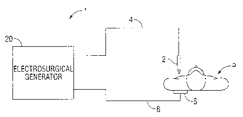
[0015] Fig. 1A is a schematic illustration of a monopolar electrosurgical
system
according to one embodiment of the present disclosure. The system includes an
electrosurgical
instrument 2 (e.g., monopolar) having one or more electrodes for treating
tissue of a patient P
(e.g., electrosurgical cutting, ablation, etc.). More particularly,
electrosurgical RF energy is
supplied to the instrument 2 by a generator 20 via a supply line 4, which is
connected to an active
terminal 30 (see Fig. 2) of the generator 20, allowing the instrument 2 to
coagulate, seal, ablate
and/or otherwise treat tissue. The energy is returned to the generator 20
through a return
electrode 6 via a return line 8 at a return terminal 32 of the generator 20
(see Fig. 2). The active
terminal 30 and the return terminal 32 are connectors configured to interface
with plugs (not
4
CA 02769316 2012-02-27
explicitly shown) of the instrument 2 and the return electrode 6, which are
disposed at the ends
of the supply line 4 and the return line 8, respectively.
[0016] Fig. 113 is a schematic illustration of a bipolar electrosurgical
system according to
the present disclosure. The system includes a bipolar electrosurgical forceps
10 having one or
more electrodes for treating tissue of a patient P. The electrosurgical
forceps 10 includes
opposing jaw members 11 and 13 having an active electrode 14 and a return
electrode 16,
respectively, disposed therein. The active electrode 14 and the return
electrode 16 are connected
to the generator 20 through cable 18, which includes the supply and return
lines 4, 8 coupled to
the active terminal 30 and return terminal 32, respectively (see Fig. 2). The
electrosurgical
forceps 10 is coupled to the generator 20 at a connector 21 having connections
to the active
terminal 30 and return terminal 32 (e.g., pins) via a plug disposed at the end
of the cable 18,
wherein the plug includes contacts from the supply and return lines 4, 8.
[0017] The generator 20 includes suitable input controls (e.g., buttons,
activators,
switches, touch screen, etc.) for controlling the generator 20. In addition,
the generator 20 may
include one or more display screens for providing the user with variety of
output information
(e.g., intensity settings, treatment complete indicators, etc.). The controls
allow the user to adjust
power of the RF energy, waveform parameters (e.g., crest factor, duty cycle,
etc.), and other
parameters to achieve the desired waveform suitable for a particular task
(e.g., coagulating,
tissue sealing, intensity setting, etc.).
[0018] Fig. 2 shows a schematic block diagram of the generator 20 having a
controller
24, a DC power supply 27, and an RF output stage 28. The power supply 27 is
connected to a
CA 02769316 2012-02-27
conventional AC source (e.g., electrical wall outlet) and is adapted to
provide high voltage DC
power to an RF output stage 28 that converts high voltage DC power into RF
energy. RF output
stage 28 delivers the RF energy to an active terminal 30. The energy is
returned thereto via the
return terminal 32.
[0019] The generator 20 may include a plurality of connectors to accommodate
various
types of electrosurgical instruments (e.g., instrument 2, electrosurgical
forceps 10, etc.). Further,
the generator 20 may be configured to operate in a variety of modes such as
ablation, monopolar
and bipolar cutting coagulation, etc. The generator 20 may also include a
switching mechanism
(e.g., relays) to switch the supply of RF energy between the connectors, such
that, for example,
when the instrument 2 is connected to the generator 20, only the monopolar
plug receives RF
energy.
[0020] The controller 24 includes a microprocessor 25 operably connected to a
memory
26, which may be volatile type memory (e.g., RAM) and/or non-volatile type
memory (e.g., flash
media, disk media, etc.). The microprocessor 25 includes an output port that
is operably
connected to the power supply 27 and/or RF output stage 28 allowing the
microprocessor 25 to
control the output of the generator 20 according to either open and/or closed
control loop
schemes. Those skilled in the art will appreciate that the microprocessor 25
may be substituted
by any logic processor (e.g., control circuit) adapted to perform the
calculations discussed herein.
[0021] A closed loop control scheme or feedback control loop is provided that
includes sensor circuitry 22 having one or more sensors for measuring a
variety of tissue and
energy properties (e.g., tissue impedance, tissue temperature, output current
and/or voltage, etc.).
6
CA 02769316 2012-02-27
The sensor circuitry 22 provides feedback to the controller 24. Such sensors
are within the
purview of those skilled in the art. The controller 24 then signals the HVPS
27 and/or RF output
phase 28 which then adjust DC and/or RF power supply, respectively. The
controller 24 also
receives input signals from the input controls of the generator 20 or the
instrument 10. The
controller 24 utilizes the input signals to adjust power outputted by the
generator 20 and/or
performs other control functions thereon.
[00221 In particular, sensor circuitry 22 is adapted to measure tissue
impedance. This
is accomplished by measuring voltage and current signals and calculating
corresponding
impedance values as a function thereof at the sensor circuitry 22 and/or at
the microprocessor 25.
Power and other energy properties may also be calculated based on collected
voltage and current
signals. The sensed impedance measurements are used as feedback by the
generator 20. In
embodiments, sensor circuitry 22 may be operably coupled between RF output
stage 28 and
active terminal 30.
[0023) A current limiting circuit 40 is operably coupled between the power
supply 27
and the RF output stage 28 and is configured to instantaneously control the
pulsed current output
of power supply 27 on a pulse-by-pulse basis. More specifically, the current
limiting circuit 40
is configured to monitor the pulsed current output of power supply 27 and, for
each output pulse,
the current limiting circuit 40 measures the instantaneous current of the
pulse and compares the
measured instantaneous current to a predetermined threshold current. In some
embodiments, the
predetermined threshold current may be the maximum allowable current output of
the power
supply 27. If the measured instantaneous current exceeds the predetermined
threshold current,
7
CA 02769316 2012-02-27
the current limiting circuit 40 controls the pulsed current output of power
supply 27 and/or
terminates the measured pulse, as will be discussed in further detail below
with reference to Figs.
3 and 4.
[0024] In one embodiment, current limiting circuit 40 includes suitable
components
preconfigured to measure instantaneous current on a pulse-by-pulse basis and
control (e.g.,
adjust, terminate, suspend, etc.) output of generator 20 based on a comparison
between the
measured current and the predetermined threshold current, as will be discussed
in detail below.
For example, in some embodiments, the predetermined threshold current may be
between about
4.5A and about 6A. In another embodiment, the predetermined threshold current
may be, for
example, data stored in memory 26 and configured to be compared to each
current pulse
measured by current limiting circuit 40 for processing by microprocessor 25.
Based on this
comparison, the microcontroller 25 generates a signal to controller 24 to
control the pulsed
output current of generator 20.
[0025] Fig. 3 illustrates the relationship between a current source voltage
v,,s (e.g., the
voltage across power supply 27) vs. time "t" waveform 50, wherein current
limiting circuit 40 is
not utilized, and a corresponding current voltage source ves vs. time "t"
waveform 60 taken over
the same time range as waveform 50, wherein current limiting circuit 40 is
utilized in accordance
with embodiments described by the present disclosure. Waveforms 50 and 60
illustrate, by way
of example, the pulsed output current of generator 20 resulting from the
contrasting scenarios
discussed above. Waveform 50 is illustrated for purposes of contrast with
waveform 60
(described below) to show the benefit of using current limiting circuit 40 to
detect and suppress
8
CA 02769316 2012-02-27
electrical arc formation at the tissue site. Electrical are formation at the
site of the tissue being
treated results in increased current being drawn from the generator 20 to the
electrical are,
thereby increasing the potential of damage to tissue caused by the presence of
increased current
and to the generator 20 due to overcurrent conditions.
[0026] Waveform 50 illustrates the pulsed output current of RF output stage 28
including
an abnormal pulse 55 that represents a significantly increased voltage vcs
between time intervals
to and tb. As described above, the abnormal pulse 55 may be caused by the
occurrence of
electrical arcing at the tissue site that results in increased current drawn
from power supply 27 to
the electrical are and, thus, an increase in voltage v,s, for the duration of
pulse 55 (e.g., time
interval ta < time "t" < time interval tb).
[0027] Similar to waveform 50, waveform 60 illustrates the pulsed output
current of
power supply 27 including an abnormal pulse 65 that represents a significantly
increased voltage
vcs starting at time interval to similar to abnormal pulse 55 discussed above.
However, in this
instance, the abnormal pulse 65 is instantaneously detected by current
limiting circuit 40, and
terminated at time interval tb_1. More specifically, the abnormal pulse 65 is
measured and
compared to the predetermined threshold current, and terminated at time
interval tb_1 since the
measured pulse exceeds the predetermined threshold current. This is in
contrast to the duration
of abnormal pulse 55, which without the benefit of detection and termination
via current limiting
circuit 40 endures from time interval ta to time interval tb. Since the above
described comparison
is repeated for each pulse, the current limiting circuit 40 of the present
disclosure operates to
regulate the duty cycle and, thus, the RMS current of the pulsed current
output of the generator
9
CA 02769316 2012-02-27
20 to prevent or suppress arc formation, thereby minimizing potential damage
to tissue from
increased drawing of current to the electrical arc.
[00281 Current limiting circuit 40 may include any suitable components to
enable
operation as described hereinabove such as, for example, current sense
resistors, shunt resistors,
transistors, diodes, switching components, transformers, and the like.
Although shown in the
illustrated embodiments as being operably coupled between power supply 27 and
RF output
stage 28, current limiting circuit 40 may be integrated within power supply 27
or RF output stage
28 or may be operably coupled between RF output stage 28 and active terminal
30.
[0029] Fig. 4 shows, by way of example, a circuit schematic of current
limiting circuit 40
according to some embodiments of the present disclosure. In this example,
current limiting
circuit 40 includes a current sense resistor 42 that operates in conjunction
with an op-amp 44, a
comparator 46, a plurality of resistors 43a-f, and a microprocessor 48, to
control the pulsed
current output of power supply 27 that is supplied to instrument 2 or forceps
10 on a pulse-by-
pulse basis, as discussed hereinabove.
[00301 As power supply 27 supplies pulsed current to RF output stage 28, a
potential
difference V,S is generated across current sense resistor 42 and received, as
input, at the non-
inverting or positive input of op-amp 44. V,,S is proportional to the pulsed
current output of
power supply 27. Op-amp 44 generates an output voltage signal that is fed back
to the inverting
or negative input of op-amp 44 (e.g., negative feedback), causing op-amp 44 to
drive its output
voltage signal toward a level that minimizes the differential voltage between
its positive and
negative inputs. The output voltage signal of op-amp 44 is received, as input,
at the positive
CA 02769316 2012-02-27
input of comparator 46. A reference voltage VREF (e.g., 3.3 volts) that is
proportional to the
predetermined threshold current is applied to the negative input of comparator
46 for comparison
to the input voltage signal received from op-amp 44. In this way, the pulsed
current output of
power supply 27 is compared to the predetermined threshold current on a pulse-
by-pulse basis by
way of comparison, at comparator 46, between VREF and the input voltage signal
from op-amp
44. This comparison dictates the output of comparator 46, which will change
either from "high"
to "low" or from "low" to "high" as the input voltage signal from op-amp 44
exceeds VREF. The
"high" or "low" output of comparator 46 signals microprocessor 48 to control
the output of RF
output stage 28 accordingly. As the input voltage signal from op-amp 44
exceeds VIF,
comparator 46 signals the microprocessor 48 to cause RF output stage 28 to
interrupt or
terminate the instantaneous current pulse (e.g., pulse 65) being supplied to
instrument 2 or
forceps 10. For example, in some embodiments, RF output stage 28 includes one
or more
suitable switching components (not shown) such as a transistor that opens or
closes in response
to an output signal (e.g., turn-on voltage) received from microprocessor 48.
In this scenario,
when the switching component is closed, the pulsed current output of power
supply 27 is
delivered un-interrupted to instrument 2 or forceps 10 via RF output stage 28
and, when the
switching component is open, the pulsed current output of power supply 27 is
shunted to ground
through resistors 43a and 43b rather than being supplied to instrument 2 or
forceps 10. The
termination or interruption of an instantaneous current pulse being supplied
to instrument 2 or
forceps 10 operates to suppress or terminate electrical arcing that may be
formed at the tissue
site. By terminating the instantaneous current pulse, the voltage V,S across
the current sense
resistor 42 decreases below VREF such that the output voltage signal from op-
amp 44 is less than
11
CA 02769316 2012-02-27
VREF by comparison. The resulting output of comparator 46 operates to signal
the RF output
stage 28 to continue or re-establish (e.g., via closing of the switching
component) delivery of
pulsed current output from power supply 27 to instrument 2 or forceps 10 for
application to
tissue. In this way, current limiting circuit 40 controls on a pulse-by-pulse
basis whether or not
pulsed current is supplied to instrument 2 or forceps 10.
[0031] While several embodiments of the disclosure have been shown in the
drawings
and/or discussed herein, it is not intended that the disclosure be limited
thereto, as it is intended
that the disclosure be as broad in scope as the art will allow and that the
specification be read
likewise. Therefore, the above description should not be construed as
limiting, but merely as
exemplifications of particular embodiments. Those skilled in the art will
envision other
modifications within the scope and spirit of the claims appended hereto.
12