Note: Descriptions are shown in the official language in which they were submitted.
CA 02771169 2012-02-14
WO 2011/025775 PCT/US2010/046473
APPARATUS FOR TRANS-CEREBRAL ELECTROPHORESIS AND
METHODS OF USE THEREOF
[0001] This application claims priority benefit, under 35 U.S.C. 119(e), of
U.S.
Provisional Patent Application 61/236,303, filed August 24, 2009, the contents
of which
application are hereby incorporated by reference in their entirety.
1. INTRODUCTION
[0002] The present invention provides apparatus and methods for the delivery
of
therapeutic agents to target tissues through the use of electric fields. The
utilization of
electric fields according to the methods of the invention aids in the
distribution and targeting
of therapeutic agents, in particular, where standard means of agent
application is insufficient
or impractical to reach target tissues. In particular embodiments, the present
invention
utilizes a convective force in combination with the developed electric fields
to further
increase the flux or distribution of the therapeutic agent within the target
tissues.
2. BACKGROUND OF THE INVENTION
[0003] The ability to treat neurological and mental disorders is limited, in
part, by the
ability to deliver therapeutic compounds efficiently to the brain parenchyma.
Pharmaceuticals are delivered to the majority of bodily tissues via the oral
(PO),
intramuscular (IM) or intravenous (IV) routes. When these mechanisms prove
insufficient,
intra-arterial delivery via selective catheterization can be used to achieve
high local
concentrations of the desired agent while limiting the risks of systemic
toxicity. For bone
disorders, intramedullary delivery can achieve the same results. These
delivery routes are
relatively ineffective for disorders of the central nervous system (CNS)
because of the blood-
brain barrier (BBB). The BBB, which is specific to the brain and spinal cord
and is formed
by tight junctions that bind adjoining cerebral endothelial cells, protects
the CNS from toxic
substances that may enter the bloodstream from time to time. Unfortunately,
the BBB also
impedes the delivery of therapeutic agents via the blood, creating unique
difficulties for
treating most neurological disorders. Although numerous strategies have been
devised to
disrupt or bypass the BBB, the lack of clinically relevant methods indicates
that improved
methods are necessary.
[0004] As one method of bypassing the BBB, neuroscientists have turned to
direct
intracerebral infusion, which requires the surgical implantation of
microcatheters that effect
-1-
CA 02771169 2012-02-14
WO 2011/025775 PCT/US2010/046473
infusion of therapeutic agents into specific brain regions. The catheters are
connected to
pumps, which deliver the desired agent. The most promising form of parenchymal
infusion is
termed "convection enhanced drug delivery" or CEDD. Developed more than a
decade ago
at the NIH, CEDD employs steady positive pressure to push macromolecules
through the
neuropil slowly and atraumatically (see, e.g., Bobo, et al., 1994, PNAS USA
91:2076-80).
This technique has been demonstrated to be effective for infusions into
relatively small target
areas and these types of pumps are currently in use in a number of prospective
clinical trials.
However, even if CEDD proves effective in delivering therapeutic agents to
small tissue
volumes, the technique may not be scalable, leaving numerous brain disorders
that may
require treatment of much greater tissue volumes unaffected. For example,
reported failures
of CEDD infusions of glial derived neurotropic factor (GDNF) for the treatment
of
Parkinson's disease may have been caused by inadequate distribution of the
GDNF infusate
within the putamen rather than a failure of the GDNF to generate the desired
biological
effect. Thus, while CEDD represents significant progress toward a viable
intracerebral drug
delivery system, there is a great need for an improved drug delivery system so
that a greater
variety of neurological and psychiatric disorders can be treated.
3. SUMMARY OF THE INVENTION
[0005] The invention is directed to apparatus, and methods of use thereof, for
the
delivery of therapeutic, diagnostic or investigational agents to target
tissues of a subject in
need thereof. In specific embodiments, the invention is directed to apparatus
for the delivery
of agents to target tissue which apparatus may be used in combination with or
is itself part of
a second apparatus or system for providing an electric field within a target
tissue to effect the
distribution and/or targeting of agents within the tissue. The application of
an electric field to
target tissues as described herein is termed trans-cerebral electrophoresis
("TCE"). In
specific embodiments, the target tissue is tissue of the central nervous
system ("CNS") and,
in particular, tissue of the brain or spinal cord. Without being bound by a
particular
mechanism of action, it is believed that the application of an electric field
within the target
tissue results in an electromotive force (EMF) to the one or more agents
(e.g., one or more
therapeutic, investigational, and/or diagnostic agents) that improves or
modifies dispersive
forces normally present in the tissue, e.g., diffusive distribution. This
invention is also
directed to the methods for use of the apparatus described herein for
providing one or more
agents to target tissues. In specific embodiments, the one or more agent is an
agent for the
-2-
CA 02771169 2012-02-14
WO 2011/025775 PCT/US2010/046473
treatment or diagnosis of a disease or disorder of the CNS, and the target
tissue is the situs of
the disease or disorder. In other embodiments, the methods of the invention
are used in
connection with investigations of central nervous system function.
[0006] The invention provides for an integrated TCE cannula for delivery of a
fluid to
a tissue delivery site of a subject, which integrated TCE cannula comprises an
implantable
cannula having proximal and distal ends, a fluid delivery pathway through the
cannula and, at
its distal end, 1) one or more outlet ports for the fluid pathway through
which the agent is
administered to the delivery site and 2) one or more monopolar electrodes
having a region for
electrical connection to a power source. When the fluid delivery pathway of
the cannula is in
fluid connection or communication with a fluid delivery system, the fluid
flows through the
delivery pathway of the cannula and exits via the outlet ports into the tissue
delivery site. In
certain embodiments, the integrated TCE cannula comprises, at its proximal
end, a connector
for connecting the fluid delivery pathway with a fluid delivery system and/or
a connector for
connecting the monopolar electrode to a power source. The connectors may be
suitable to
allow temporary communication between the integrated TCE cannula and the fluid
delivery
system and/or power source (i.e., allowing the cannula to be readily
disconnected from the
fluid delivery system and/or power source, or allowing multiple integrated TCE
cannulas to
be used sequentially with a single fluid delivery system and/or power source)
or may be such
that the connection between the integrated TCE cannula and the fluid delivery
system and/or
power source is permanent. In preferred embodiments, the fluid comprises one
or more of a
therapeutic, diagnostic or investigational agent in a pharmaceutically
acceptable carrier. In
certain embodiments, the integrated TCE cannula further comprises thermocouple
in contact
with the electrode, cannula and/or surrounding tissue. In certain embodiments,
thermocouple
is in communication with a display device for display of the temperature of
the cannula,
electrode and/or surrounding tissue and/or may further be connected to a
processor for
automatic regulation of the parameters of the TCE or agent delivery as
described herein. The
integrated TCE cannula may be disposable in that it is designed for a single
use or may be
designed for repeated use. In embodiments where the integrated TCE cannula is
to be reused,
the materials of the cannula are suitable for sterilization by any method
known in the art.
[0007] The integrated TCE cannula of the invention is suitable for the direct
infusion
of fluids into the body tissues of a subject in need thereof, and, in specific
embodiments, is
suitable for convective enhanced drug delivery into the tissue of the central
nervous system.
In certain embodiments, the integrated TCE cannula is a reflux-free cannula.
To this end, in
certain embodiments, the TCE cannula is in communication with an agent
delivery system
-3-
CA 02771169 2012-02-14
WO 2011/025775 PCT/US2010/046473
suitable for delivery of one or more agents to the tissue of the patient via
the cannula. In
certain embodiments, the invention encompasses apparatus comprising, in
addition to the
integrated TCE cannula, an agent delivery system comprising one or more pumps
that
provide one or more agents, e.g., one or more therapeutic, investigational, or
diagnostic
agents, to the integrated TCE cannula and, thus, to the delivery area within
the subject's
tissues. In certain embodiments, the invention encompasses the use of one or
more integrated
TCE cannulas for introduction of one or more agents to the delivery area
within the tissues of
the subject, e.g., tissues of the CNS. The invention may further comprise an
agent delivery
system comprising one or more regulators that control the agent delivery via
the one or more
integrated TCE cannulas so as to supply a specified total dose and/or to
supply a specified
agent delivery rate. The one or more integrated TCE cannulas and/or agent
delivery systems
may be designed for temporary use or permanent implantation as is known in the
art. For
example, the integrated TCE cannulas may be designed to allow insertion into
the tissues of
the subject without external housings, e.g., in the manner of an syringe
needle, or may be
designed to comprise external housings that aid insertion, which housings are
withdrawn
leaving the cannula implanted within the delivery area. In certain
embodiments, the cannulas
may be pre-filled with a therapeutic, diagnostic, or investigational agent
and/or with a
pharmaceutical carrier prior to implantation/insertion. In certain
embodiments, the one or
more integrated TCE cannulas and agent delivery system (including, but not
limited to, any
pumps, agent reservoirs and regulators) may be fully implantable as is known
in the art. As
used herein, the term "cannula" and "integrated TCE cannula" encompasses the
device
through which an agent is provided to the delivery area within the tissue or
tissues of the
patient, and thus may encompass a variety of materials, designs and sizes
depending on the
delivery area, including, but not limited to, inflexible needles/tubing and
flexible tubing
devices, as is well known and routinely implemented in the art.
[0008] The monopolar electrode of the integrated TCE cannula may be used in
conjunction with one or more, e.g., an array or a plurality, of independently
polarizable
monopolar electrodes to generate an electric field that at least partially
encompasses the
target tissue. Accordingly, in certain embodiments, in addition to the
integrated TCE
cannula, the apparatus of the invention comprises one or more monopolar
electrodes, separate
from that of the integrated TCE cannula, each of which monopolar electrode is
independently
polarizable. The array of monopolar, polarizable electrodes, each in
connection with a power
source (e.g., a current and/or voltage source), when powered, effects the
generation of the
electric field at least partially encompassing the target tissue. In certain
embodiments the
-4-
CA 02771169 2012-02-14
WO 2011/025775 PCT/US2010/046473
electrodes comprises a connector suitable for connection to a power source,
which connection
may be temporary or permanent. The array includes at least two electrodes such
that when
connected to the power source and polarized, an electric field is generated
between the two or
more electrodes. In specific embodiments, the array of electrodes comprises
two or more or a
plurality of electrodes, one of which is the electrode of the integrated TCE
cannula. The
remaining electrodes that form the array may be surface or implantable
electrodes. In
specific embodiments, the invention provides a spatial arrangement of
electrodes, such that,
when the array is connected to the power source, an electric field is
generated that at least
partially encompasses the target tissue. Application or administration of one
or more agents
(e.g., one or more therapeutic or diagnostic agents) within a suitably
oriented electric field
will cause the agent(s) that respond to electric fields, i.e., charged or
ionized agents, to move
down the electric gradient, preferably, to or within the target tissue.
[0009] The array of electrodes comprises at least two electrodes, one of which
may be
the electrode of the integrated TCE cannula, and may comprise any number
sufficient for
development of the desired electric field within the target tissue. In certain
embodiments, the
array of electrodes comprises at least 2, at least 3, at least 4, at least 5,
at least 6, at least 7, at
least 8, at least 9, at least 10, at least 11 or at least 12 electrodes. In
other embodiments, the
array of electrodes comprises no more than 2, no more than 3, no more than 4,
no more than
5, no more than 6, no more than 7, no more than 8, no more than 9, no more
than 10, no more
than 11 or no more than 12 electrodes. In preferred embodiments, the array of
electrodes
comprises from 2 to 5 electrodes. In yet more preferred embodiments, the array
of electrodes
comprises 3 to 4 electrodes. In the preferred embodiments, at least one of the
electrodes in
the array is the electrode of the integrated TCE cannula.
[0010] The electrode array of the invention comprises a sufficient number of
electrodes placed in a suitable three-dimensional ("3D") orientation such
that, when
connected to the power source, an electric field is developed in the array,
which electric field
at least partially encompasses the target tissue. The electrodes may or may
not be in contact
with the subject, e.g., in certain embodiments, the apparatus of the invention
comprises
external electrodes which electrodes are not in direct contact with the
subject and/or target
tissue. In alternate embodiments, the apparatus of the invention comprises an
electrode array
in direct contact with the subject and/or target tissue (e.g., placed on or
placed/implanted
within the subject or target tissue). In a specific example in accordance with
this
embodiment, the apparatus of the invention comprises an array of implantable
and/or surface
electrodes. In certain embodiments, the apparatus of the invention comprises
an array of
-5-
CA 02771169 2012-02-14
WO 2011/025775 PCT/US2010/046473
surface electrodes. In other embodiments, the apparatus of the invention
comprises an array
of implantable electrodes. In yet other embodiments the apparatus of the
invention comprises
an array of surface and implantable electrodes.
[0011] In specific embodiments of the invention, the electric field developed
within
the electrode array encompasses the site of administration (i.e., the delivery
area) of the one
or more therapeutic or investigational agents. In other embodiments, the
electric field
developed by the electrode array does not encompass the delivery area and the
one or more
agents enter the electric field by dispersive forces within the target tissue
of the subject (e.g.,
via diffusion, active transport, bulk flow, blood flow, etc.).
[0012] In certain embodiments of the invention, the invention encompasses an
apparatus comprising a power source and, in some embodiments, further
comprising one or
more regulators for regulating and/or controlling the power provided to the
individual
electrodes within the electrode array. The power source and/or power source
and regulator
may provide a current and/or voltage to the array such that the developed
electric field
maintains a constant strength and/or polarity throughout the entirety of a TCE
session. In
alternate embodiments, the power source and/or power source and regulator
provide a current
and or voltage to the array such that the developed electric field is variable
in strength and/or
polarity over a single TCE session. The power source and/or power source and
regulator may
provide a direct or alternating current. The power provided to the electrode
array (e.g., the
current) may be continuous or pulsed.
[0013] The power supplied to the electrode array is sufficient to effect the
dispersion
of the therapeutic, investigational and/or diagnostic agent to or within the
target tissue. In
preferred embodiments, the power supplied to the electrode array is below the
threshold level
to effect electroporation of the agent within the target tissue. In a specific
example in
accordance with this embodiment, the developed electric gradient within the
array is less than
100 kV/cm. In other examples, the developed electric gradient is less than 10
kV/cm or less
than 5 kV/cm. In still other examples the developed electric gradient is less
than 95, 90, 85,
80, 75, 70, 65, 60, 55, 50, 45, 40, 35, 30, 25, 20, or 15 kV/cm.
[0014] In certain embodiments, invention comprises the use of one or more
biosensors. The biosensors may be separate components of the apparatus of the
invention or
may be integrated into one or more other components of the apparatus in
contact with the
subject, e.g., incorporated into the one or more electrodes or the one or more
integrated TCE
cannulas. The biosensors of the apparatus can monitor one or more performance
parameters
of the apparatus (e.g., agent delivery rate, electric field strength) and/or
one or more patient
-6-
CA 02771169 2012-02-14
WO 2011/025775 PCT/US2010/046473
specific parameters (e.g., temperature of the tissue surrounding the two or
more electrodes
within the array, or local pressure or pressure gradients within the tissue
surrounding the one
or more agent delivery cannulas). The biosensors of the apparatus of the
invention may
comprise connectors for connecting to external display devices allowing manual
regulation of
apparatus function in response to the displayed output of the one or more
biosensors. In other
embodiments, the biosensors of the apparatus of the invention comprise
connectors for
communication with a processor that automatically regulates apparatus
operation in response
to signals from the one or more biosensors.
[0015] The methods of the invention can be used with any method of agent
(e.g.,
drug) delivery known in the art that is suitable for administration of an
agent to the delivery
area within the tissues of the subject. In other embodiments, the methods of
the invention
encompass administration of an agent to the subject within the developed
electric field. In
alternate embodiments, the methods of the invention encompass administration
of an agent
external to the developed electric field, which agent then enters the electric
field by
dispersive forces acting at the site of administration other than the
developed EMF. In certain
embodiments, the invention encompasses the use of convection enhanced drug
delivery
("CEDD") for administration of an agent. Such convection enhanced methods are
well
known in the art and are routinely used to provide, for example, therapeutic
or diagnostic
agents to CNS tissues under pressure. It is believed that methods of the
invention combining
TCE and CEDD will not only improve agent delivery (i.e., therapeutic or
diagnostic agent
delivery) to target tissues (e.g., tissues of the CNS), but also allow
targeting of an agent that
is not possible using current methods known in the art. Because the methods of
the invention
use EMF to direct the one or more agents within the tissue, existing
limitations of CEDD may
be overcome. In certain embodiments, agent administration need not be direct
or near target
tissue, but can be in a more remote site. Such embodiments are advantageous,
particularly,
for example, wherein direct access via traditional administration methods
(e.g., injection,
cannulation, catheterization) would be impractical or impossible.
[0016] In certain embodiments, the integrated TCE cannulas of the invention
are
suitable for agent administration in accordance with the methods of CEDD as
known in the
art. The integrated TCE cannula of the invention combines the function of an
infusion
catheter for CEDD and one or more electrophoretic electrodes. Having at least
one of the
polarizeable electrodes of the electrode array at the site of agent
administration allows the
developed electric field to be modulated to better focus, direct and/or
regulate agent dispersal
to or within the target site. As described herein, in addition to the
integrated TCE cannula,
-7-
CA 02771169 2012-02-14
WO 2011/025775 PCT/US2010/046473
the invention comprises one or more monopolar electrodes separate from that of
the
implantable, integrated TCE cannula having a connector for connecting to a
power source.
When the array of electrodes is connected to the power source and separately
polarized, the
electric field is generated by the array. The invention also encompasses
methods of use of
the apparatus described herein for delivery of an agent to a target tissue
within the CNS of a
subject, e.g., for the treatment, prevention or amelioration of one or more
symptoms of a CNS
disease or disorder.
[0017] The invention also comprises a method for delivering an agent to or
within a
target tissue of the CNS of a subject, said method comprising the steps of A)
positioning an
array of electrodes such that, when powered and separately polarized, the
array is positioned
so as to provide an electric field of sufficient amplitude and polarity to
cause movement of
the agent from the delivery area (or point of entry of the agent within the
field) to or within
the target tissue of the subject; B) polarizing the array of electrodes and
thereby generating an
electric field in the array; and C) applying the agent to the delivery area.
In specific
embodiments, the methods of the invention comprise applying the agent to the
delivery area,
which delivery area is within the electric field developed by the powered
electrode array. In
certain embodiments, the agent is applied to the delivery area prior to,
concomitant with, or
subsequent to the powering of the electrode array. The spatial arrangement of
the electrodes
in the array causes the target tissue to be at least partially encompassed by
the electric field,
and the electric field provides an EMF to drive the one or more agents to or
within the target
tissue. In preferred embodiments, the target tissue is tissue of the CNS and
the electrode
array is positioned such that, when powered, the developed EMF provides a
dispersive force
within the CNS tissue, along the surface of the CNS tissue, within the
subcutaneous tissue
surrounding the CNS tissue, or on the surface/within the skin of the subject.
In specific
embodiments, the method of the invention encompasses the treatment, prevention
or
amelioration of one or more symptoms of a disease or disorder of the CNS in a
subject in
need thereof.
[0018] Therapeutic agents for use in accordance with the methods of the
invention
include any agent that will migrate along an electric potential gradient
(i.e., charged
molecules, dipoles). Such therapeutics may naturally respond to an EMF or can
be modified
to respond provided that the modification does not alter their desired
bioactivity
-8-
CA 02771169 2012-02-14
WO 2011/025775 PCT/US2010/046473
3.1 Terminology
[0019] As used herein, the term "about" or "approximately" when used in
conjunction
with a number refers to any number within 1, 5 or 10% of the referenced number
or within
the experimental error typical of standard methods used for the measurement
and/or
determination of said number.
[0020] As used herein, the term "central nervous system ('CNS') disorder" and
analogous terms refer to a disorder associated with the death and/or
dysfunction of a
particular neuronal or non-neuronal cell population (e.g., glial cells) in the
CNS and/or the
aberrant growth of cells within the CNS. The aberrantly growing cells of the
CNS may be
native to the CNS or may be derived from other tissues, and may be malignant
or non-
malignant. The disorder may be acute or chronic. Non limiting examples of CNS
disorders
include, but are not limited to, cancer, neoplastic growth, infection, head
trauma, spinal cord
injury, multiple sclerosis, dementia with Lewy bodies, ALS, lysosomal storage
disorders,
amyloidogenic diseases (e.g., Alzheimer's disease), neurodegenerative
diseases, autoimmune
disorders, stroke, epilepsy, psychiatric disorders, and disorders of hormonal
balance. Further
contemplated are methods for reducing inflammation that is associated with a
CNS disorder
characterized by neuronal death and/or dysfunction.
[0021] As used herein, the term "in combination" in the context of the
administration
of (a) therapy(ies) to a subject, refers to the use of more than one therapy
(e.g., more than one
prophylactic and/or therapeutic agent or method). The use of the term "in
combination" does
not restrict the order in which therapies (e.g., prophylactic and/or
therapeutic agents or
methods) are administered to a subject, but instead refers to the use of more
than one therapy
as part of an overall treatment regimen. A first therapy (e.g., a first
prophylactic and/or
therapeutic agent or method) can be administered prior to (e.g., at least 5
minutes, at least 15
minutes, at least 30 minutes, at least 45 minutes, at least 1 hour, at least 2
hours, at least 4
hours, at least 6 hours, or at least 12 hours before), concomitantly with, or
subsequent to (e.g.,
at least 5 minutes, at least 15 minutes, at least 30 minutes, at least 45
minutes, at least 1 hour,
at least 2 hours, at least 4 hours, at least 6 hours, or at least 12 hours
after) the administration
of a second therapy (e.g., a second prophylactic and/or therapeutic agent or
method) to a
subject.
[0022] As used herein, the terms "manage," "managing," and "management" refer
to
the beneficial effects that a subject derives from a therapy (e.g., a
prophylactic and/or
therapeutic agent or method), which does not result in a cure of the disease
or disorder, e.g., a
CNS disease or disorder. In certain embodiments, a subject is administered one
or more
-9-
CA 02771169 2012-02-14
WO 2011/025775 PCT/US2010/046473
therapies (e.g., prophylactic and/or therapeutic agents or methods) to
"manage" a condition or
symptom associated with a disease or disorder (e.g., a CNS disease or
disorder), so as to
prevent the progression or worsening of the disease/disorder.
[0023] As used herein, the terms "prevent", "preventing" and "prevention"
refer to
the prevention of onset of, the recurrence of, or a reduction in one or more
symptoms of a
disease/disorder (e.g., disorder of the CNS) in a subject as a result of the
administration of a
therapy (e.g., a prophylactic and/or therapeutic method of he invention).
[0024] As used herein, the terms "therapies" and "therapy" can refer to any
protocol(s), method(s), and/or agent(s) that can be used in the diagnosis,
prevention,
treatment, management, or amelioration of a disease/disorder, and/or a symptom
thereof (e.g.,
a CNS disease or disorder or a condition or symptom associated therewith). In
certain
embodiments, the terms "therapies" and "therapy" refer to diagnostic
procedures, biological
therapy, supportive therapy, and/or other therapies useful in diagnosis,
treatment,
management, prevention, or amelioration of a disease or condition, or of one
or more
symptoms associated therewith.
[0025] As used herein, the terms "treat," "treatment," and "treating" in the
context of
administration of a therapy to a subject for a disease or disorder refers to
the cure of the
disease or disorder, or may refer to the eradication, reduction or
amelioration of one or more
symptoms of said disease/disorder (e.g., CNS disease/disorder).
4. DESCRIPTION OF THE FIGURES
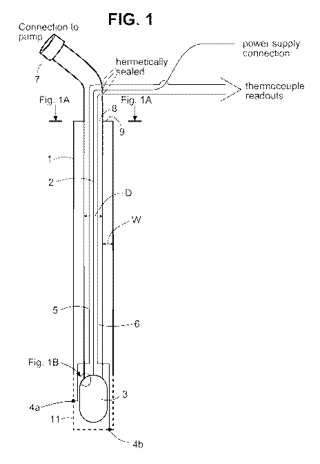
[0026] FIG. 1 Schematic of exemplary integrated TCE cannula
[0027] FIG. IA Schematic of a cross section of the exemplary integrated TCE
cannula of FIG 1.
[0028] FIG. 1B Schematic of an electrode portion of the exemplary integrated
TCE
cannula of FIG. 1.
[0029] FIG. 2 Schematic of exemplary distal end of the TCE cannula
[0030] FIG. 3 Schematic of exemplary arrangement of electrode array
[0031] FIG. 4 Schematic of exemplary arrangement of fluid regulating
components
of the TCE apparatus.
[0032] FIG. 5 Schematic of exemplary control circuit for an individual
electrode
and/or electrode lead.
-10-
CA 02771169 2012-02-14
WO 2011/025775 PCT/US2010/046473
5. DETAILED DESCRIPTION OF THE INVENTION
[0033] The invention provides for the use of an electric field to effect the
distribution
and/or the targeting of charged agents within a target tissue, such as that of
the CNS. The
application of an electric field within the tissue results in an electromotive
force (EMF) that
disperses or moves the agent to or within the target tissue. The movement or
dispersal
provided by the EMF according to the methods of the invention may also be used
to improve
or modify the movement associated with other dispersive forces, e.g., those
associated with
diffusive distribution or convective enhanced drug delivery ("CEDD").
[0034] In particular embodiments, the methods of the invention provide for the
use of
electrophoresis in combination with convective enhanced drug delivery
("CEDD"). The
addition of an electromotive force (EMF) to agents represents a major
improvement to
CEDD. Charged molecules, including proteins and nucleic acids, can be directed
along a
potential gradient so long as the appropriate electrical field is created
between or among the
two or more electrodes. Clinical use of CEDD has demonstrated that the tissue
of the CNS
is, in fact, a porous matrix that permits the flow of macromolecules through
the matrix
without damage to cytoarchitecture or induction of neurological deficits.
Application of a
low level electric field across or including the target tissue will create a
potential gradient
down which the applied or introduced agent(s) (e.g., therapeutic, diagnostic,
or investigative
agents) will migrate. Employed over a period of days, weeks, months or years,
the charge
gradient will enhance the treatment volume of parenchymal infusions,
dramatically
increasing their potential clinical applications.
[0035] The central nervous system can function well despite the application of
low-
level, therapeutic, exogenous electrical current. For example, chronic spinal
cord stimulation
has become a mainstay of chronic pain management, allowing patients with
otherwise
disabling pain syndromes to lead fuller lives without any untoward effects
from the
stimulation on normal spinal cord functions. Vagal nerve stimulation has
proven to be an
effective treatment for generalized epilepsy when medications fail to provide
adequate
seizure control and surgical resection of the seizure focus is not feasible.
Also, deep brain
stimulation ("DBS") has become the treatment of choice for movement disorders
such as
Parkinson's disease, Essential Tremor, and Idiopathic Torsion Dystonia when
medications fail
to provide adequate symptomatic relief. In all instances, the low level
electric fields
developed during these therapies are well-tolerated. However, unlike these
highly localized
therapies, the instant invention utilizes trans-cerebral electrophoresis
("TCE"): the creation of
-11-
CA 02771169 2012-02-14
WO 2011/025775 PCT/US2010/046473
a relatively larger electric field not to stimulate or lesion a discrete
region of tissue, e.g.,
excitable tissue, but to create an electric gradient of defined shape and
volume down which
therapeutic agents will migrate.
[0036] In certain embodiments, TCE according to the methods of the invention
enhances the efficacy of parenchymal infusion, e.g., CEDD, by broadening the
distribution of
an infused agent such as a therapeutic, investigational or diagnostic agent.
In other
embodiments, TCE according to the methods of the invention enhances the
efficacy of
parenchymal infusion, e.g., CEDD, by allowing targeting to specific tissues or
specific
volumes of tissue. For example, the methods of the invention allow the
parameters of the
parenchymal infusion and TCE to vary to achieve specific tissue distribution
goals. For
example, the methods of the invention allow a single application of a
therapeutic agent
directly to target tissues in conjunction with TCE to distribute the agent
over a larger volume
of target tissue than a standard single application (e.g., via diffusion or
CEDD) would allow.
In other embodiments, the methods of the invention allow application of an
agent at a site
remote to the target tissue (e.g., where direct application is impossible or
impractical), and the
use of TCE to establish an electric gradient that directs the agent to the
target tissue. In still
other embodiments, the methods of the invention allow the distribution of the
therapeutic
agent to be controlled such that only a desired volume, shape or area of
target tissue is
contacted by the agent.
[0037] In certain embodiments, the invention provides for the treatment,
management
or prevention of a CNS disease or disorder or for the treatment, management,
prevention or
amelioration of one or more symptoms of a CNS disease or disorder. In certain
examples in
accordance with this embodiment, the disease or disorder is a
neurodegenerative disease,
neurodegeneration associated with stroke, neurodegeneration associated with
cancer or a
disease or disorder associated with neuronal death and/or dysfunction. Non-
limiting
examples of CNS disorders include, but are not limited to, cancer, neoplastic
growth,
infection, head trauma, spinal cord injury, multiple sclerosis, dementia with
Lewy bodies,
ALS, lysosomal storage disorders, amyloidogenic diseases (e.g., Alzheimer's
disease),
neurodegenerative diseases, autoimmune disorders, tauopathies, stroke,
epilepsy, psychiatric
disorders, and disorders of hormonal balance. Further contemplated are methods
for reducing
inflammation that is associated with a CNS disorder characterized by neuronal
death,
infection, and/or dysfunction.
[0038] In certain embodiments, the invention provides for the treatment,
management
or prevention of a CNS cancer or for the treatment, management, prevention or
amelioration
-12-
CA 02771169 2012-02-14
WO 2011/025775 PCT/US2010/046473
of one or more symptoms of a CNS cancer in a subject in need thereof. The CNS
cancer may
be a cancer originating from CNS cells or may include tumor(s) derived from
cells of other
tissues of the body, e.g., a metastasized tumor(s) to the CNS. The methods of
the invention
encompass the direct application of therapeutic agents to the tumor(s) (e.g.,
at or on the
surface of, or within the tumor) or, alternatively, application at a site
distal to the tumor
wherein TCE is used to regulate, control, or direct the therapeutic agent to
and/or within the
tumor site.
[0039] In certain embodiments, the invention provides for the diagnosis or
investigation of a CNS disease or disorder comprising the administration of a
diagnostic or
investigational agent. In certain embodiments, the diagnostic agent or
investigational agent
may comprise a targeting moiety that targets the agent to specific cell types
or that causes the
preferential uptake of the agent within a specific cell population. In other
embodiments, the
diagnostic or investigational agent is a contrast agent suitable for use with
tissue visualization
modalities such as, but not limited to, X-ray, Computerized Tomography (CT),
magnetic
resonance imaging (MRI), optical imaging, positron emission tomography (PET
scanning) or
Single Photon Emission Computerized Tomography (SPECT). In specific examples,
the
diagnostic agent or investigational agent comprises an antibody or antigen
binding fragment
thereof specific for a tumor, neoplastic and/or malignant cell marker, which
antibody when
used in accordance with the methods of the invention allows the detection and
localization of
cells expressing the tumor, neoplastic and/or malignant marker. The methods of
the
invention encompass the use of diagnostic or investigational agents, for
example, to detect
the presence or absence of a disease, disorder or infection (or to detect
characteristic
indicators thereof), or to monitor the development or progression of a
disease, disorder or
infection as part of a clinical testing procedure to, e.g., determine the
efficacy of a given
treatment regimen. Diagnostic or investigational agents for use in accordance
with the
methods of the invention may respond themselves to the developed EMF (i.e.,
the agent is
itself a charged molecule or dipole), or may be conjugated to a molecule that
exhibits such
activity (i.e., acting as a carrier for the diagnostic or investigational
molecule and/or that
targets the diagnostic molecule to a tissue of interest). In specific
embodiments, the
diagnostic or investigational agent is coupled to a detectable substance to
aid in detection of
the agent. Non-limiting examples of detectable substances include, but are not
limited to,
various enzymes, including, but not limited to, horseradish peroxidase,
alkaline phosphatase,
beta-galactosidase, or acetylcholinesterase); prosthetic group complexes, such
as, but not
limited to, streptavidin/biotin and avidin/biotin; fluorescent materials, such
as, but not limited
-13-
CA 02771169 2012-02-14
WO 2011/025775 PCT/US2010/046473
to, umbelliferone, fluorescein, fluorescein isothiocyanate, rhodamine,
dichlorotriazinylamine
fluorescein, dansyl chloride or phycoerythrin; luminescent materials, such as,
but not limited
to, luminol; bioluminescent materials such as, but not limited to, luciferase,
luciferin, and
aequorin; radioactive material, such as, but not limited to, bismuth (213Bi),
carbon (14C),
chromium (51Cr), cobalt (57Co), fluorine (18F), gadolinium (153Gd, 159Gd),
gallium (68Ga,
67Ga), germanium (68Ge), holmium (166Ho), indium (115In, 113In, 112In, 111In),
iodine (1311, 1251,
1231, 121I), lanthanium (140La), lutetium (177Lu), manganese (54Mn),
molybdenum (99Mo),
palladium (103Pd), phosphorous (32P), praseodymium (142Pr), promethium
(149Pm), rhenium
(186Re, 188Re), rhodium (105Rh), ruthemium (97Ru), samarium (153Sm), scandium
(47Sc),
selenium (75Se), strontium (85Sr), sulfur (35S), technetium (99Tc), thallium
(20 'Ti), tin (113 Sn,
117Sn), tritium (3H), xenon (133Xe), ytterbium (169Yb, 175Yb), yttrium (90Y),
zinc (65Zn);
positron emitting metals using various positron emission tomographies, and
nonradioactive
paramagnetic metal ions.
[0040] Any type of agent that is or can be made responsive to an electric
field, e.g.,
ionized, may be used in accordance with the methods of the invention. Non-
limiting
examples of agents that may be used in accordance with the methods and
apparatus of the
invention include naturally occurring and/or synthetic nucleic acids,
peptides, peptide
mimetics, polypeptides, antibodies, antigen-specific antibody fragments, and
small
molecules. Agents that may be used in accordance with the methods of the
invention include
therapeutics, investigationals and diagnostics.
[0041] The nucleic acids for use in accordance with the methods of the
invention
include, but are not limited to, DNA molecules (e.g., cDNA or genomic DNA),
RNA
molecules (e.g., mRNA), combinations of DNA and RNA molecules or hybrid
DNA/RNA
molecules, and analogs of DNA or RNA molecules. Such analogs can be generated
using,
for example, nucleotide analogs, which include, but are not limited to,
inosine or tritylated
bases. Such analogs can also comprise DNA or RNA molecules comprising modified
backbones that lend beneficial attributes to the molecules such as, for
example, nuclease
resistance or an increased ability to cross cellular membranes. The nucleic
acids or
nucleotide sequences can be single-stranded, double-stranded, may contain both
single-
stranded and double-stranded portions, and may contain triple-stranded
portions. In particular
embodiments, the nucleic acid for use in accordance with the methods of the
invention is a
therapeutic nucleic acid as known in the art and/or described herein, e.g., an
antisense nucleic
acid, an siRNA, a short hairpin RNA, or an enzymatic nucleic acid.
-14-
CA 02771169 2012-02-14
WO 2011/025775 PCT/US2010/046473
[0042] The antibodies for use in accordance with the methods of the invention
include, but are not limited to, monoclonal antibodies, multispecific
antibodies, human
antibodies, humanized antibodies, chimeric antibodies, single-chain Fvs
(scFv), single chain
antibodies, Fab fragments, F(ab') fragments, disulfide-linked Fvs (sdFv),
intrabodies,
minibodies, diabodies and anti-idiotypic (anti-Id) antibodies (including,
e.g., anti-Id
antibodies to antibodies of the invention), and epitope-binding fragments of
any of the above.
In particular, antibodies include immunoglobulin molecules and immunologically
active
fragments of immunoglobulin molecules, i.e., molecules that contain an antigen
binding site.
[0043] In certain embodiments, the agent for use in accordance with the
methods of
the invention is a neuroactive agent, modulating the activity of one or more
types of CNS
cells. For example, the methods of the invention provide for the management,
treatment, or
prevention of a CNS disease or disorder, or the management, treatment,
prevention or
amelioration of one or more symptoms of a CNS disease or disorder by, e.g.,
promoting the
survival or death of a particular phenotype of a neuron or a particular region
of CNS tissue,
modulating synapse formation or activity (e.g., by the use of a
neurotransmitter uptake
inhibitor), modulating electrical activity of a neuron (e.g., by the use of
calcium ion channel
inhibitors), modifying the activity of a first neuron by effecting a response
or activity in a
second cell of the CNS, e.g., a microglial cell. Non-limiting examples of
neurotransmitter
uptake inhibitors that may be used in accordance with the methods of the
invention to
modulate the activity of CNS, e.g., neural tissue, include serotonin, dopamine
and
norepinephrine.
[0044] In one aspect, the invention also provides kits for the treatment of
CNS
disorders comprising the use of TCE, optionally in combination with CEDD,
which kits
comprise a delivery device useful for TCE or for combination TCE and CEDD,
preferably a
reflux-free cannula/catheter comprising a polarizable electrode, and one or
more separately
polarizable electrodes. The separately polarizable electrodes may be surface
style electrodes
that transmit the field through the surface of the skin (e.g., scalp) to the
target tissue (e.g.,
brain) or may be electrodes designed for implantation in or remote to target
tissues (e.g., the
brain surface or within the CNS parenchyma), or combinations thereof.
5.1 Trans-Cerebral Electrophoresis
[0045] The invention provides for the generation of an electric field within
or
encompassing the target tissue to effect the trans-tissue electrophoresis and
targeted delivery
of a therapeutic, diagnostic, or investigational agent. Multiple methods exist
for the
-15-
CA 02771169 2012-02-14
WO 2011/025775 PCT/US2010/046473
generation of such an electric field in vivo, in particular, within the brain
or parts of the CNS
of a subject in need thereof. Examples of such methods include, but are not
limited to, the
use of external plates surrounding, but not touching, the target tissue and/or
subject, surface
electrode arrays, penetrating electrode arrays, and combinations of surface
and penetrating
electrode arrays. The invention can also be practiced with any electrode
system suitable for
propagating the electrical signals within or encompassing the targeted region
of tissue. The
specific characteristics of the electrode systems will determine if that type
of electrode is
suitable for use in a given application.
[0046] The most common use of electrode systems in the CNS in current clinical
practice is the use of single, bipolar electrodes capable of stimulating or
lesioning a target
tissue. Because the target tissues of the CNS are comprised primarily of
closely packed
neural tissue, such electrodes are designed to affect a relatively small area
immediately
proximal to the electrode; stimulation or lesioning of larger areas would
result in unknown
and potentially undesirable side-effects. Accordingly, these electrodes are
primarily designed
as single lead bipolar or multichannel electrodes. In contrast, the apparatus
and methods of
the instant invention comprise an array of separately polarizeable electrodes.
In certain
embodiments, the apparatus of the invention comprises an array of at least two
separately
polarizable electrodes, one of which is optionally housed in a cannula or
catheter, e.g., an
integrated TCE cannula as described herein, for application of an agent, e.g.,
a therapeutic or
diagnostic agent. In another embodiment, the apparatus of the invention
comprises an array
of more than two separately polarizable electrodes (i.e., a plurality of
electrodes), at least one
of which is optionally housed in a cannula or catheter for application of the
agent to be
delivered, e.g., an integrated TCE cannula as described herein. In a specific
embodiment,
each electrode in the array of the apparatus of the invention is independent
from the means of
delivery of the therapeutic agent. In a specific example in accordance with
this embodiment,
the electrodes of the apparatus may be plates external to, but not touching,
the subject and
surrounding the target tissue.
[0047] The array of separately polarizable electrodes can be polarized by
independent
connection, for example, to a variable voltage power source, e.g., such as a
battery, and
activating the power source. Whether a specific polarizable electrode is
charged negatively
or positively will be a function of the location of agent application with
respect to the location
of the target tissue and the charge of the agent. The number, position, and
charge of the
polarizable electrodes can be determined by any method known in the art or
described herein
for estimation of agent response in vivo to a developed electric field, e.g.,
by use of computer-
-16-
CA 02771169 2012-02-14
WO 2011/025775 PCT/US2010/046473
based three-dimensional simulation (e.g., finite element analysis software
packages (e.g.,
COMSOL Multiphysics, (COMSOL, Inc., Burlington MA); FEMPRO (ALGOR, Inc., San
Rafael CA)) and/or other methods known in the art (see, e.g., Lee et al.,
2007, International
Journal of Control, Automation, and Systems 5:337-342., encompassed by
reference herein in
its entirety).
[0048] When using simulation procedures, the target tissue location can be
identified
using any method known in the art, e.g., magnetic resonance imaging ("MRI").
The target
location can then be simulated in three dimensional space using a computer
based system and
the effects of the electrical field and tissue composition on the charged
agent can be
simulated. The amount of the agent and the appropriate electrical field can
then be
determined to establish not only the desired concentration but also the
residency time of the
therapeutic agent within the target tissue.
[0049] In certain embodiments, the apparatus of the invention comprises
surface-style
electrodes, e.g., plates or meander-type electrodes (see e.g., U.S. Pat. No.
5,968,006, which is
incorporated herein by reference in its entirety). Surface-style electrodes
propagate the
electric field through the surface of the skin and into the target tissue. In
other embodiments,
the apparatus of the invention comprises implantable, or penetrating,
electrodes. Implantable
electrodes useful for the generation of an electric field within the tissues
of the CNS include,
but are not limited to, electrodes designed to be inserted beneath the surface
of the skin along
the cranium, those designed to be inserted in the epidural space of the
vertebral column or
cranium, or those implanted along the surface of the brain, brainstem, or
spinal cord.
Penetrating electrodes are conductive elements whose size and shape are
sufficient to enable
insertion through the matter covering a tissue of interest or through the
tissue of interest itself.
Penetrating electrodes are well known in the art, and have, for example, been
used to treat
chronic pain, symptoms of Parkinson's disease, epilepsy, hearing disorders,
depression,
obsessive/compulsive disorder, and muscle disorders.
[0050] Electrode design is a critical component of TCE. Electrode parameters
include diameter, conducting surface geometry, length, conductivity and
materials. In certain
embodiments, the electrodes are hollow, allowing for the administration of an
agent, e.g.,
diagnostic, therapeutic or anesthetic agent. In other embodiments, the
electrodes are coated
with anesthetics and/or lubricious agents for pain mitigation and ease of
insertion. The
design or selection of the electrode is determined by several treatment
factors, including
properties of the target tissue, tissue volume to be treated, and charge
injection/current
densities at the electrode-tissue interface. The inter-electrode spacing and
penetration depth
-17-
CA 02771169 2012-02-14
WO 2011/025775 PCT/US2010/046473
define the shape of the electric field, and thus the volume of tissue to be
treated. In certain
embodiments the spatial arrangement of the electrodes surrounding or within
the target tissue
may be based on computer simulation of the electric field and the agent
response thereto.
Such simulations may be developed by any method known in the art, e.g., using
finite
element analysis software such as, but not limited to, COMSOL Multiphysics
(COMSOL,
Inc., Burlington MA); FEMPRO (ALGOR, Inc., San Rafael CA) and/or IPlan
Software
(BrainLab, Inc., Munich Germany).
[0051] The electric field generated between or among the two or more
electrodes
creates an EMF that moves the charged agent in a controlled fashion so as to
achieve the
desired agent concentration/distribution for a specified time within the
target tissue, thereby
generating the desired effect. Because TCE is used to direct or regulate the
movement of the
agent to or within the target tissue, the delivery location of the agent need
not be directly to
the target location, but can be at a remote site. In such embodiments, to
effectuate the desired
movement of the agent within the tissue, the electrical field is preferably
adjustable/changeable. Moreover, the polarity of two or more of the
polarizable electrodes
can be switched to manipulate the direction of the movement of the charged
agent within the
tissue. The strength of the electrical field can also be adjusted to control
the rate of
movement of the charged therapeutic agent to and within the tissue.
[0052] It is preferred that the electrodes have a sufficiently inert surface
material that
is electrochemically stable and will not exhibit substantial oxidation-
reduction reactions
within the interstitial environment when exposed to the electric current. Non-
limiting
examples of such surfaces include gold, nickel, titanium, titanium nitride,
platinum, platinum-
iridium, iridium, iridium-oxide, silver, silver-plated copper, silver
tungsten, silver cadmium-
oxide, silver tin-oxide, indium-tin-oxide, and tin-oxide. Depending upon the
material chosen,
it may be desirable for cost and structural reasons to deposit these inert
metals to the surface
of a base metal. Appropriate base metals include, but are not limited to
titanium, tungsten and
stainless steel. As known in the art, the level of charge injection and
irreversible oxidation-
reduction reactions are parameters to be considered when choosing a
sufficiently inert
material and deposition thickness.
[0053] In certain embodiments, dielectric coatings are deposited on the
surface of the
electrode to avoid generation of non-homogeneous electrical fields. Such
dialectric coatings
are typically deposited at the level of tenths to hundreds of microns thick
Non-limiting
examples of suitable dielectric coatings include polytetrafluoroethylene
(PTFE), parylene,
and silicon carbide. In certain embodiments, the electrode is covered in a
biocompatible
-18-
CA 02771169 2012-02-14
WO 2011/025775 PCT/US2010/046473
insulating material except for a small region to allow a contact through which
charge may
flow.
[0054] The electric field encompassed by the invention is preferably less than
that
required to stimulate the excitable tissue(s) of the CNS. Stimulation or
lesioning methods
generally require high frequency AC current (up to approximately 200 tamps).
In contrast,
the instant invention comprises methods using low frequency AC, low frequency
DC pulses
or DC current. Without being bound by any particular mechanism of action, it
is believed
that the DC current or low frequency pulses establish an EMF sufficient to
effect transfer of
charged therapeutic agents through the target tissue. To avoid damage to the
CNS during
TCE, the invention uses a current of low amperage to establish the electric
field. In certain
embodiments, the electric current is no greater than 10 mA. In other
embodiments, the
current is no greater than 8 mA, 6 mA, 4 mA, 2 mA, 100 tA, 75 pA, 50 tA, 25
tA, 15 pA,
tA, 8 tA, 6 tA, 4 tA, 2 to or 1 tA. Because of the low amperage, it is
envisioned that,
for certain embodiments, the mobility effects of the methods of the invention
require the
subject to undergo prolonged TCE. In certain embodiments, the methods of the
invention
encompass one or more round of TCE of about 1 h, 2 h, 4 h, 6 h, 8 h, 10 h, 15
h, 20 h, 24 h,
1.5 days, 2 days, 5 days, 1 week or 2 weeks, duration. However, in certain
embodiments
involving full subcutaneous implantation of the apparatus, TCE may be
implemented as a
repeated or continuous treatment for months or years. In certain embodiments,
TCE
according to the methods of the invention may be effected by the use of
electrode plates
external to, but not touching, the patient and surrounding the target tissue.
Alternatively, for
TCE according to the methods of the invention the apparatus of the invention
may be
designed for acute implantation. In other embodiments of the invention, TCE is
chronically
applied to the target tissue, e.g., with chronic administration of therapeutic
agents, and,
accordingly, the apparatus of the invention is designed for permanent or
chronic implantation.
[0055] In certain embodiments, the electric field is generated by plates
external to, but
not touching, the subject and surrounding target tissue. In specific examples
in accordance
with this embodiment, the electric potential between the two external plates
is from 1-100 V,
1-80 V, 1-60 V, 1-40 V, 1-20 V, 1-10 V, 1-5 V, 5-600 V, 5-500 V, 5-400V, 5-300
V, 5-200
V, 5-100 V, 100-600 V, 100-500 V, 100-400 V, 100-300 V, or from 100-200 V. In
specific
embodiments, the electric potential between the two plates is 5 V, 8 V, 10 V,
15 V, 20 V, 25
V, 50 V, 100 V, 150 V, 200 V, 250 V, 300 V, 350 V, 400 V, 450 V, 500 V, 550 V
or 600 V.
[0056] In other embodiments, the electric field is generated by two or more
implantable electrodes, or a combination of two or more implantable and
surface electrodes.
-19-
CA 02771169 2012-02-14
WO 2011/025775 PCT/US2010/046473
In specific examples in accordance with this embodiment, the electric
potential between the
reference electrode any other of the array may be from 2-20 V, 5-20 V, 10 -20
V, 10-18 V,
10-16 V, 10-14 V or from 10-12 V. In specific embodiments, the electric
potential between
the reference electrode and any other of the array is 2 V, 4 V, 6 V, 8 V, 10
V, 12 V, 14 V, 16
V, 18 V, or 20 V. In embodiments of the invention comprising the use of more
than two
electrodes, the electric potential between any two electrodes within the array
may be the same
or different from that between any other two. A varied gradient within the
array may be
useful, e.g., for the creation of a concentration gradient of the applied
agent within the
developed electric field.
[0057] The power source to the electrodes is capable of delivering alternating
current
(AC) or direct current (DC). The current may be delivered by an andoal and a
cathodal
segment. In certain embodiments, the current is pulsed. In the AC embodiment,
the pulse
frequency is generally low, about 10 Hz or less. In specific examples in
accordance with this
embodiment, the pulse frequency is 5 Hz or less, 2 Hz or less, 1Hz or less,
0.5 Hz or less, 0.1
Hz or less, 0.05 Hz or less, 0.01 Hz or less, 0.005 Hz or less, 0.001 Hz or
less, 0.0005 Hz or
less, or 0.0001 Hz or less. For non-pulsed DC current, the pulse frequency is
0. Pulse width
may be varied to provide optimum dispersion of the administered agent. In
certain
embodiments, the pulse width, or signal duration, is from 1 microsecond (" s")
to 5 seconds
("s"), 1 s to 2 s, 1 s to 1 s, 1 s to 500 milliseconds ("ms"), 1 s to 200
ms, 1 s to 100 ms,
1 s to 50 ms, 1 s to 20 ms, 1 s to 10 ms, 1 s to 1 ms, 1 s to 500 s, 1
s to 100 s, 1 s
to 50 s, 1 s to 10 s, 1 ms to 200 ms, 1 ms to 100 ms, 1 ms to 5ms, or l ms
to 20 ms. In
other embodiments, the pulse width, or signal duration, is from 1 ms to 5 s, 1
ms to 2 s, 1 ms
to 1 s, 1 to 500 ms, 1 to 200 ms, 1 to 100 ms, 1 to 50 ms, 1 to 20 ms, 1 to 10
ms, 10 to 200
ms, 10 to 100 ms, 10 to 50 ms, or 10 to 20 ms. The pulse widths of the anodal
and cathodal
segments are either symmetric or can be asymmetric. It is believed that an
asymmetric wave
offer reduced pH changes at the electrode surface. Train length may be from
hours to days,
dependent on pulse width and frequency. The invention contemplates any pulse
shape, or
current waveform, including bipolar, monopolar, capacitive discharge, square,
sawtooth, or
any combination of the foregoing. In certain embodiments, the pulse shape is a
square
wave. It is believed that square waves may offer further improved agent
dispersion due to the
impulse nature of the developed EMF.
[0058] In certain embodiments, the invention encompasses the monitoring of one
or
more of the electrodes in the array, in particular the resistance of the one
or more electrodes,
such that the charge to the electrode can be varied and/or controlled to
generate the desired
-20-
CA 02771169 2012-02-14
WO 2011/025775 PCT/US2010/046473
field within the target tissue. In other embodiments, the one or more
electrodes comprise a
thermocouple to monitor the temperature of the tissue surrounding the
electrode. The
temperature at the electrode site may be monitored for levels of heat that may
lead to tissue
damage. Safety devices may be included as part of the apparatus of the
invention to
automatically stop TCE or switch off the apparatus when a set temperature is
reached or
exceeded. The safety temperature may vary depending on the length of time the
tissue is to
be exposed to the electric current, as high temperatures may be tolerated for
brief periods,
but, generally, the safety temperature is not greater than 40 C. In other
embodiments, the
one or more electrodes comprise a microtube or capillary through which cooled
fluid (for
example, saline) may be pumped to maintain the temperature of the electrode
and/or
surrounding tissue at or below the safety temperature. In preferred
embodiments, the
microtube or capillary forms a fluid path within the interior of the one or
more electrodes
such that the path does not disrupt the conductive surface of the electrode in
contact with the
tissue of the subject. In such embodiments, the microtube or capillary within
the electrode
has an inflow and outflow comprising suitable connectors for fluid connection
to a reservoir
or other source of cooling fluid to form a cooling system. The apparatus of
the invention may
comprise one or more pumps, valves or flow initiators/controllers in fluid
connection with a
cooling apparatus and the microtubes/capillaries of the one or more electrodes
to provide a
flow of fluid through the microtubes/capillaries to maintain or reduce the
temperature of the
electrode or tissue. In certain embodiments each electrode of the electrode
array comprises a
microtube/capillary as described herein and is fluid connection with cooling
system; in other
embodiments, only one, a minority, about half or more than half but not all of
the electrodes
of the array comprise a microtube/capillary as described herein in fluid
connection with the
cooling system. The cooling system may receive input from the thermocouples of
the
electrodes as described herein to automatically regulate the temperature of
the one or more
electrodes and/or surrounding tissue. The flow of fluid through the
microtube/capillaries of
the one or more electrodes of the electrode array need not be continuous, but
may be
regulated by manual or processor control. In preferred embodiments the fluid
path through
the microtubes/capillaries and other components of the cooling system is
closed such that the
cooling fluid does not come into contact with the tissue of the subject.
Because the cooling
fluid does not contact the subject, any suitable cooling fluid known in the
art may be used,
but is preferably biocompatible and/or non-toxic.
[0059] To ensure that the proper concentration of the administered agent is
reaching
the target location, the concentration of the agent in the subject tissue can
be measured at
-21-
CA 02771169 2012-02-14
WO 2011/025775 PCT/US2010/046473
certain points in/around the target location. The concentration of the charged
agent can be
measured using any technique known in the art and/or described herein, e.g., a
microdialysis
technique. The measured concentration can be compared with a desired
concentration. If the
measured concentration of the agent within the target location is not
approximately equal to
the desired concentration, the delivery of the agent to the target location
can be adjusted
accordingly. For example, the strength of the electrical field, an alteration
of the polarity of
one or more of the polarizable electrodes, and/or an adjustment to the rate of
application/delivery of the administered agent to the tissue may each be
independently or
concomitantly adjusted to obtain the desired concentration at the target
location.
5.2 Convection Enhanced Drug Delivery ("CEDD")
[0060] In certain embodiments, TCE is combined with Convection Enhanced Drug
Delivery ("CEDD") techniques. CEDD, also known as high-flow interstitial
infusion, is a
technique well known in the art, and involves the application of an agent
under pressure to a
tissue structure. The pressure generated by the delivery system is believed to
provide
convection assisted agent dispersion within the target tissue. CEDD generally
requires
positioning the tip of one or more infusion catheters or cannulas (e.g., an
integrated TCE
cannula as described herein), preferably a reflux-free catheter or cannula,
within a tissue
structure and provision of a solution comprising an agent to be administered
through the
catheter/cannula while maintaining a pressure gradient from the tip of the
catheter during the
infusion. Most commonly, the pressure gradient is created by connecting the
one or more
infusion catheters/cannulas to a pump after positioning in the tissue situs as
is well known in
the art (see, e.g., Saito et al., 2005, Exp Neurol, 196:381-389; Krauze et
al., 2005, Exp
Neurol, 196:104-111; Krauze et al., 2005, Brain Res Brain Res Protocol., 16:20-
26; Noble et
al., 2006, Cancer Res 66:2801-2806; Saito et al., 2006, J Neurosci Methods
154:225-232;
Hadaczek et al., 2006, Hum Gene Ther 17:291-302; Hadaczek et al., 2006, Mol
Ther 14:69-
78, U.S. Patent Application Publication No. 2006/0073101; and U.S. Pat. No.
5,720,720,
each of which is incorporated herein by reference in its entirety). The pumps
of the apparatus
of the invention may be implantable pumps (including but not limited to active
and passive
drug delivery systems (see, e.g., U.S. Patents 7,351,239; 4,629,147; 4,013,074
each of which
is hereby incorporated by reference in its entirety)) or external pumps (e.g.,
roller pumps or
syringe pumps) and may deliver one or more separate agents to one or more
delivery sites
within the tissues of the subject. In specific embodiments, the apparatus of
the invention
comprises a CEDD-compatible reflux-free step design cannula (see, e.g., Krauze
et al., 2005,
-22-
CA 02771169 2012-02-14
WO 2011/025775 PCT/US2010/046473
J Neurosurg 103:923-9, and U.S. Patent Application Publication Nos. US
2006/0135945 and
US 2007/0088295, each of which is incorporated herein by reference in its
entirety).
[0061] In specific embodiments, the distal end of the one or more infusion
catheters/cannulas and/or integrated TCE cannulas is a needle-like assembly,
such as a
stainless steel or stiff polymer tube having one or more elongated ports to
release the
therapeutic agent in a discrete location or over an extended site. The more
proximal portions
of the infusion catheter/cannula may be formed of one or more segments of any
biocompatible material of a suitable stiffness to dependably transmit
microdose or microflow
volumes of compositions comprising the therapeutic agent from the pump through
the
catheter/cannula, without loss of pressure. In certain embodiments, the
invention comprises
the use of more that one infusion catheter/cannula for application of the
therapeutic agent at
more than one tissue situs. In other embodiments, the infusion
catheter/cannula has one or
more sensors to monitor apparatus performance and/or method parameters, e.g.,
drug
concentration at the site of application, tissue condition (e.g.,
temperature). In still other
embodiments, the one or more infusion cannulas/catheters of the invention are
primed with a
pharmaceutically acceptable carrier and/or a pharmaceutical composition prior
to
implantation or insertion into the tissue situs.
[0062] The invention encompasses CEDD at any suitable flow rate such that the
intracranial pressure is not increased to levels injurious to tissues of the
brain. In certain
embodiments, the infusion flow rate is from 0.1 - 1000 L/hr, 0.1 - 900 L/hr,
0.1 - 800
L/hr, 0.1 - 700 L/hr, 0.1 - 600 L/hr, 0.1 - 500 L/hr, 0.1 - 400 L/hr, 0.1 -
300 L/hr, 0.1 -
200 L/hr, 0.1 - 100 L/hr, 0.1 - 80 L/hr, 0.1 - 70 L/hr, 0.1 - 60 L/hr,
0.1 - 50 L/hr, 0.1 -
40 L/hr, 0.1 - 30 L/hr, 0.1 - 25 L/hr, 0.2 - 20 L/hr, 0.1 - 15 L/hr, 0.1 -
10 L/hr, 0.1 - 5
L/hr, 0.1 - 2 L/hr, 0.1 -1 L/hr, 0.1 - 0.8 L/hr, 0.1 - 0.6 L/hr, 0.1 - 0.5
L/hr, 0.1 - 0.4
L/hr, 0.1 - 0.3 L/hr, 0.1 - 0.2 L/hr, 0.1 - 25 L/min, 0.5 - 20 L/min, 1 -
15 L/min, 1 - 10
L/min, 1 - 5 L/min, or 1 - 2 L/min. In specific embodiments, infusion flow
rate is about
0.1 l/hr or less, about 0.5 L/hr or less, about 0.7 L/hr or less, about 1
L/hr or less, 0.1
l/min or less, 0.5 L/min or less, about 0.7 L/min or less, about 1 L/min or
less, about 1.2
L/min or less, about 1.5 L/min or less, about 1.7 L/min or less, about 2
L/min or less,
about 2.2 L/min or less, about 2.5 L/min or less, about 2.7 L/min or less,
about 3 L/min
or less, about 5 L/min or less, about 7 L/min or less, about 10 L/min or
less, about 12
L/min or less or about 15 L/min or less. In preferred embodiments, the
infusion flow rate
is 5 gl/min or less. In other embodiments, the invention provides for CEDD
comprising
incremental increases in flow rate, referred to as "stepping", during
delivery.
-23-
CA 02771169 2012-02-14
WO 2011/025775 PCT/US2010/046473
[0063] The distal end of the catheter/cannula is implanted providing a fixed
site of
agent administration, and, in certain embodiments, extends such that one or
more ports of the
catheter open in or near the target site, which may, for example, be a tumor
site, a nerve, a
lesion or other targeted region of affected brain or other CNS tissue. Because
TCE used in
conjunction with CEDD will effect dispersion of the administered agent, there
is not a
requirement to have the one or more ports of the catheter/cannula open
directly in the target
tissue. Thus, in certain embodiments, the invention encompasses insertion of
the
catheter/cannula into a non-target tissue situs and use of TCE to ensure
contact between the
administered agent and the target tissue. The freedom of administration site
may allow
tissues to be targeted according to the methods of the invention that are
otherwise unsuited
for treatment using standard CEDD.
[0064] In certain embodiments, the distal end of the catheter/cannula is
stereotactically implanted into brain tissue through a cranial hole to deliver
the agent into the
parenchymal spaces. The remaining components of the CEDD system, e.g.,
infusion pump
and power supply, need not be near the one or more infusion catheters but may
be connected
via appropriate electrical connections and tubing. For long term infusions
according to the
methods of the invention, the invention encompasses chronic implantation of
the infusion
catheter. In such embodiments, the remaining components of the CEDD system
and/or TCD
apparatus as described herein may also be implanted subdermally. For example,
in certain
embodiments, the fluid supply to the inlet of the infusion pump is an
implanted reservoir or
other supply. In other embodiments, the fluid reservoir is implanted
subdermally and
possesses a cover or septum formed of a self-sealing polymer. Such reservoirs
are refillable
through the patient's skin by piercing the septum with a syringe to deliver a
refill volume of
the fluid comprising the therapeutic agent. In still other embodiments, the
reservoir is a
pressurized assembly, such as a pressure-driven bellows, in which case the
infusion pump
assembly may be implemented by simply providing one or more valves,
restrictors or other
elements that regulate the time and/or the rate at which fluid is allowed to
pass from the
reservoir. In still other embodiments, the infusion pump is an electrically
powered assembly,
having a power source and a controller.
[0065] In specific embodiments, the invention provides for one or more
chambers in
the pump assembly that contains one or more concentrated agents to be
administered, which
the assembly combines with one or more carrier fluids. A mixing chamber may be
provided
to allow mixing of the one or more agents and one or more carriers before they
are pumped to
the tissue site. This is especially advantageous, for example, in multidrug
regimens in which
-24-
CA 02771169 2012-02-14
WO 2011/025775 PCT/US2010/046473
several incompatible or mutually unstable drugs are to be delivered at once,
or in which
concentration must be closely controlled. In other embodiments, dispersion of
the
administered agent is further augmented by the use of a facilitating agent,
such as low
molecular weight heparin.
5.3 TCE Apparatus
[0066] In exemplary embodiments, the invention provides an integrated TCE
cannula/catheter that functions as both a cannula for infusion of an agent
into a tissue situs
and at least one monopolar electrode of a monopolar electrode array. The
integrated TCE
cannula may function as part of a TCE apparatus comprising two subsystems
according to the
methods of the invention: the first, a CEDD infusion system, e.g., including a
programmable
infusion pump and one or more infusion catheters/cannulas, through which an
agent may be
delivered under pressure into a targeted tissue situs and; the second, an
array of two or more
electrodes connected to a current source with which the trans-tissue, e.g.,
trans-cerebral,
electric potential gradient is created, one of electrodes is, optionally, the
at least one
monopolar electrode of the integrated TCE cannula. In alternate embodiments,
the invention
provides for a method encompassing the use of one or more infusion catheters
to administer
the agent and two or more electrodes to establish an electric field within the
target tissue. In
specific embodiments, the methods of the invention provide for the use of
separable systems,
e.g., wherein the one or more infusion catheters are not combined with one or
more
electrodes and/or are not connected to a current source. In other embodiments,
the invention
provides one or more integrated infusion catheters comprising one or more
polarizable
electrodes. In specific embodiments, the integrated TCE catheter of the
invention is
surgically implanted into the desired CNS site (e.g., parenchymal site) and
the remaining one
or more electrodes are placed along the surface of the skin covering or
surrounding the target
tissue site and/or are implanted into the CNS tissue. For example, in those
embodiments
wherein the CNS tissue is the brain, the remaining one or more electrodes may
be placed
along the brain surface, within the brain parenchyma, within the epidural
space, within the
skull, under the scalp, or along the surface of the scalp. The infusion
catheter and electrode
wires may be tunneled subcutaneously and connected, respectively, to a pump
and current
source, optionally a pulse generator, which may either be external to the body
or implanted
subcutaneously. In specific embodiments, both the infusion pump and the
implantable
current source may be programmed transcutaneously.
-25-
CA 02771169 2012-02-14
WO 2011/025775 PCT/US2010/046473
[0067] In certain embodiments the invention provides for a processor which is
in
communication with the electrodes of the array and can send and receive
signals to other
external components of the system, directing the activity of the other
components, e.g., the
infusion pump and the power source, in response to the electrode signals. The
electrode
signals may include determinations of electrode resistance or, for electrodes
comprising a
thermocouple, temperature. In certain embodiments, the processor can be used
independently
or concomitantly to regulate the flow of the therapeutic agent from the
infusion pump, to
regulate the intensity and shape of the electric field within the target
tissues by regulating the
output of the power source (e.g., current or voltage generator) or to
selectively power only a
subset of the electrodes in the array. In certain embodiments, the connection
between one or
more electrodes and the processor comprises resistors for modulating the
impedance of the
electrode, allowing electrodes with increased impedance to function as a
recording electrode,
i.e., used to provide information to the processor. The connection between a
recording
electrode and the processor may also include one or more preamplifiers for
amplifying signal
received from the recording electrode.
[0068] In certain embodiments, the apparatus of the invention comprises one or
more
thermocouples. In specific embodiments, the thermocouple is implanted in the
target tissue
and is separate from any other implantable or surface component of the
apparatus of the
invention, e.g., an electrode, infusion catheter/cannula. In other
embodiments, the
thermocouple is integrated into one or more implantable or surface components
of the
apparatus, e.g., electrode, infusion catheter/cannula. The thermocouple
monitors the
temperature of the tissue surrounding or in contact with the apparatus
component, including,
for example, the tissue of the target area, and may, optionally, be connected
to a processor
regulating the TCE apparatus of the invention. The output of the thermocouple
may be used
by the human operator or the control processor to adjust apparatus parameters,
e.g., current
output, voltage, so as to avoid tissue damage from exposure to the electric
current.
[0069] The invention provides for the use of biocompatible materials to form
the
components of the integrated TCE cannula and/or apparatus, which components
are
interconnected by one or more leads. The leads may extend from components such
as one or
more electrodes to, e.g., a power source and/or a processor for regulating the
function of the
apparatus. Where in contact with the tissue of the subject, leads are placed
within a
biocompatible, sterilizable, flexible or semi-flexible sheath. As used herein,
the term "source
devices for the electrode array" describes a device comprising a battery or
power source to
power the electrode array, a pump or delivery device for agent administration,
and,
-26-
CA 02771169 2012-02-14
WO 2011/025775 PCT/US2010/046473
optionally, a processor for providing instructions to the apparatus. In
certain embodiments,
the source devices are implantable and/or portable and/or self-regulating. In
other
embodiments, source devices are extracorporeal device and may be controlled by
the patient
and/or a health care worker.
[0070] FIGS. 1, IA and lB schematically illustrate the integrated TCE cannula,
which cannula comprises an integrated infusion catheter/cannula and electrode.
Throughout
the figures, like elements are identified by like numbers. The integrated TCE
cannula is a
hollow tube with an outer wall (1) formed from any biocompatible, sterilizable
material of
suitable stiffness to allow target tissue implantation and delivery of
microflows of solutions
comprising one or more therapeutic agents to be delivered under pressure. The
integrated
TCE cannula comprises a solid inner core (8) containing one or more electrical
leads (2, 5, 6)
that connect electrical components at the base or side of the cannula to
source devices of the
apparatus, e.g., a power source and/or processor. For example, the solid inner
core (8) may
comprise a plurality of wires or leads embedded in a solid plastic (e.g.,
polyurethane)
material. In certain embodiments, the electrical leads are hermetically
sealed. Each lead is
separated from the other by a region of insulating material such that there is
no electrical
cross-talk between the leads. For example, as shown in Figure 1B, each lead
(10) may
comprises a thin layer of insulating coating, e.g., thin plastic coating,
having a thickness of
about 0.01 to about 0.5 mm, preferably, the coating has a thickness of about
0.1 mm.
[0071] The central core maybe formed completely from the insulating material
or
may be formed from the material of the cannula and filled with the insulating
material. In
some embodiments, the solid inner, core (8) may have a diameter (D) of any
suitable size,
preferably from about 0.1 to about 10 mm, about 0.5 to about 5 mm, about 0.5
to about lmm
or about 0.lmm. Insulating materials are any materials having a dielectric
constant greater
than that of the lead metal. Non-limiting examples of insulating materials
include glass fiber,
silicon elastomers, TEFLON (PTFE), plastics, including, but not limited to,
polyurethane,
and like materials having high dielectric constants. Typically, the entire
lead is covered by
insulating material except for region(s) at the tip (e.g., of about 2 to 5 m)
where "open
contacts" or surfaces through which electrical current can pass are necessary,
e.g., as in a
thermocouple. The solid inner core creates a hollow space within the
integrated TCE cannula
(9) to allow flow of a solution through the cannula. In some embodiments, the
solid inner
core (8) may be concentric with the integrated TCE cannula (9). More
specifically, a
distance (W) between a radius of the solid inner core (8) and the integrated
TCE cannula (9)
may be of any suitable size, preferably from about 0.1 to about 10 mm, about
0.5 to about 5
-27-
CA 02771169 2012-02-14
WO 2011/025775 PCT/US2010/046473
mm about 0.8 to about 3 mm, about 1 to about 2 mm, or about 2 mm. The proximal
end of
the cannula is connected to an infusion pump to provide the solution
comprising the
therapeutic agent at a given flow rate (7). The distal tip of the outer wall
of the integrated
TCE cannula comprises one or more ports (i.e., holes) (12) through which the
solution may
exit the flow space (9) and flow into the tissue situs (see, FIG.2).
[0072] In one exemplary embodiment, as shown in Figure 2, the distal end of
the
cannula comprises a porous outer sheath (11), comprising a plurality of pores
(12). The
sheath (11) may be formed from any suitable bio-inactive plastic material,
preferably a
biocompatible polyurethane. The pores (12) may serve as ports to allow flow of
fluids and
drugs therethrough and into the tissue. The pores (12) may have any suitable
shapes or sizes.
For example, the pores (12) may have an irregular, circular or oval shape. In
some
embodiments, the pores (12) may have a diameter or largest transverse of about
0.01mm to
about 0.5 mm. In certain embodiments, the pores (12) may be variable in size.
In particular,
the pores (12) may be uniformly distributed throughout the sheath (11). In
some specific
embodiments, the pores (12) may occupy at least 10%, 25%, 30%, 40%, or 50% of
the
overall area of the sheath (11). In a particularly preferred embodiments, the
pores (12) may
be variable in size, uniformly distributed throughout the sheath (11) and
occupy at about 50%
of the overall area of the sheath (11).
[0073] In specific embodiments, the entire cannula is formed from an
insulating
material, e.g., polyurethane, and the ports or open contact areas at the
distal end of the
cannula (11, 12) allow electric current to flow from the electrode (3) into
the tissue. The
integrated TCE cannula comprises one or more polarizable electrodes (3). For
example, the
electrode (3) may comprise a Platinum/Iridium (Pt/Ir) electrode. The surface
area of the
electrode in electrical contact with the tissue of the subject is preferably
sufficiently large to
avoid high charge densities within the tissue. The one or more electrodes may
be of any size
or shape suitable for the generation of an electric field within the target
tissue of sufficient
strength to provide an EMF capable of dispersing the therapeutic agent through
the target
tissue while avoiding tissue damage from the presence of an electric current.
In some
embodiments, the electrode (3) may have a smooth or uniformly even surface to
minimize
areas for charge buildup on the surface. In certain embodiments, the electrode
is an oblate
spheroid to minimize variable charge densities over the surface of the
electrode (which also
provides a more uniform electric field within the tissue) with a longer axis
of about 1 to about
mm, including axis of about 1 mm, 2 mm, 3 mm, 4 mm, 5 mm, 6 mm, 7 mm, 8 mm, 9
mm, or 10 mm; and the shorter axis of about 0.3 to 3 mm, including axis of
about 0.3 mm,
-28-
CA 02771169 2012-02-14
WO 2011/025775 PCT/US2010/046473
0.6 mm, 0.9 mm, 1.2 mm, 1.5 mm, 1.8 mm, 2.1 mm, 2.4 mm, 2.7 mm or 3 mm. Other
sizes
and shapes, with either larger or smaller may also be used in accordance with
the methods of
the invention.
[0074] In other embodiments, one or more of the electrodes in the array is a
surface
electrode. The surface electrode may be of any shape suitable for the
generation of an
electric field within the target tissue of sufficient strength to provide an
EMF capable of
dispersing the administered agent through the target tissue while avoiding
tissue damage from
the presence of an electric current. In specific embodiments, the surface
electrode is a disc of
diameter about 0.5 to about 5 cm, including 0.5 cm, 1 cm, 1.5 cm, 2 cm, 2.5
cm, 3 cm, 3.5
cm, 4 cm or 5 cm. In certain embodiments, the integrated TCE cannula comprises
one or
more thermocouples that may be used to determine the temperature of the tissue
at one or
more contact points with the cannula (e.g., on the side (4a) or distal end
(4b) of the cannula).
In some embodiments, the thermocouples (4a and 4b) may be embedded into a
porous outer
sheath serving as open contact areas at the distal end of the cannula (11).
More particularly,
the thermocouples (4a and 4b) may also be coated with a thin layer of
insulating material,
such as plastic.
[0075] The integrated TCE cannula is configured to treat a target tissue in a
patient's
body, such as CNS tissue (including, but not limited to, the brain). In
certain embodiments,
one or more integrated TCE cannulas are implanted such that the fluid
comprising the agent
exits through one or more openings at the distal ends of the one or more
cannulas at or remote
to the target tissue site. The combination CEDD and TCE effected by the
apparatus and
methods of the invention as described herein effect a permeation of the one or
more
administered agents through targeted tissue to contact and effectively treat
the targeted
region(s). Thus, the region of effective tissue treatment is defined by both
the cannula
placement and the parameters of the electric field. The one or more cannulas
may be placed
at or near a brain tumor or diseased region of the brain, at or near a tumor
to deliver a
chemotherapeutic agent, at or near a nerve location to treat chronic pain, or
at another
suitable local site. In certain embodiments, one or more implantable
components of the
apparatus of the invention, e.g., one or more electrodes, one or more infusion
catheter/cannulas, an integrated TCE cannula, may be implanted using image-
guided,
electrophysiologically-guided or stereotactic techniques to ensure correct
spatial positioning
of the one or more components.
[0076] Electrical continuity is necessary between the two or more polarizeable
electrodes and the electrical signal generator/power source. Therefore, in
embodiments
-29-
CA 02771169 2012-02-14
WO 2011/025775 PCT/US2010/046473
where the electrode array is detachable, a continuous and reliable electrical
connection should
be readily achieved between the two or more electrodes within the array and
apparatus source
devices, e.g., the power generators and optional regulators.
[0077] The invention also includes an electrical signal generator in
conductive
communication with the electrodes and/or integrated TCE cannula. The nature of
the
electrical signal generator will depend on the desired application. In some
embodiments, the
electrical signal generator is located within or physically connected to, by
means other than
electrical leads, the integrated TCE cannula. In the case of external
electrical signal
generator, a cable between the generator and the two or more electrodes of the
apparatus is
provided with a suitable connector to the two or more electrodes in a manner
to minimize
interference with operator use. In certain embodiments, the apparatus
comprises a plurality
of electrodes. In such embodiments, the plurality of electrodes may be
organized into sets of
channels, each set comprising at least two, and preferably, at least four
channels for
connection to the apparatus source devices. FIG. 3 schematically illustrates
exemplary
components of one embodiment of the apparatus of the invention, which
embodiment
comprises a combination of surface and implantable electrodes. The plurality
of electrodes
represented in FIG. 3 comprises one single implanted electrode secured by a
locking cap on
the skull of the subject, e.g., an integrated TCE cannula as described herein,
(24) and a series
of surface electrodes (23). The electrode array is attached to a processor or
control
mechanism (28) and power source (26 and 27). In certain embodiments, each
electrode has
an independent circuit (29) within the processor or control mechanism (28) to
monitor the
impedance of each electrode, to monitor the parameters of the developed
electric field and to
provide safety switches for cutting power to the electrode should any
parameter exceed safety
levels.
[0078] In specific embodiments, the invention provides for one or more
infusion
reservoirs or sources of fluid(s) connected through a pump, valve or flow
initiator/controller.
The pump, valve or flow regulator is fluidly connected to the infusion
catheter of the
invention, e.g., an integrated TCE cannula, to provide a solution comprising
one or more
agents through the catheter into a tissue situs. FIG. 4 schematically
illustrates exemplary
components of one embodiment of the invention, which embodiment comprises an
infusion
reservoir (15) connected to a pump or flow initiator/controller (14). The
infusion reservoir
may be one or more containers for holding fluid to be introduced to one or
more tissue sites,
which containers are capable of maintaining the sterility of the fluid. In
specific
embodiments, the one or more fluid reservoirs are one or more syringes. The
pump or flow
-30-
CA 02771169 2012-02-14
WO 2011/025775 PCT/US2010/046473
initiator/controller (14) can independently control the one or more fluid
reservoirs and may
further comprise programmable safety switches to halt flow should the pressure
in any line
from the pump become too great. In certain embodiments, the pump or flow
initiator/controller is operated by a health care worker or the subject. In
other embodiments,
the pump or flow initiator/controller is operated remotely or by a processor
that has been
programmed to regulate pump functions and safety. The output of the pump is
fluidly
connected to the infusion catheter, e.g., the integrated TCE cannula (16, 18,
20). The fluid
connection may be made of any suitable sterilizable material capable of
maintaining both the
pressure generated by the pump and the sterility of the fluid from the pump.
In certain
embodiments, the fluid connection may also comprise additional components for
monitoring
pump performance such as a pressure or flow transducer (17 and 19,
respectively). The fluid
reservoirs need not necessarily contain the administered agent (e.g.,
therapeutic or diagnostic
agent), but, in certain embodiments, contain only one or more pharmaceutically
acceptable
carriers. In such embodiments, the one or more agents may be introduced at any
point in the
fluid connection between the pump or flow initiator/controller and the
infusion
catheter/cannula (21), e.g., introduced via a standard IV port in the fluid
connection. Such
embodiments are particularly useful wherein two or more agents are to be used
that have
varying stability, handling and/or storage requirements. The precise pump
structure may be
flexibly implemented with any suitable structure as known in the art, either
with an
electromechanically-actuated peristaltic or displacement pumping mechanisms,
or with a
pressurized reservoir or osmotically-driven source connected to a control
valve or restrictor
assembly to regulate the provision of fluid into the fluid delivery path. In
either case,
whether powered by pressure or electromechanically, the infusion pump assembly
produces
an accurately administered and sustainable flow of a total volume of fluid at
a suitable
infusion flow rate. In accordance with one aspect of the invention, the pump
or flow
initiator/controller provides a flow of fluid through a release device that is
effective to
increase the pressure locally at the region of the infusion catheter/cannula
distal outlet ports,
where the catheter/cannula is implanted in tissue, creating a pressure
gradient that drives bulk
transport of the drug into the target tissue site. For a typical implanted
brain catheter/cannula
delivery route, such pressure gradients are normally achieved with a flow rate
of about 0.5 to
about 20.0 l/min. The fluid flow may be set and the pump assembly actuated
based upon
modeled properties such as histological tissue traits, therapeutic agent and
carrier viscosity,
infusion catheter and port dimensions and the like, or the flow may be
governed by one or
more extrinsic inputs, e.g., by a controller operative on input signals from
sensors that detect
-31-
CA 02771169 2012-02-14
WO 2011/025775 PCT/US2010/046473
pressure or flow at relevant locations (e.g., a pressure transducer in the
fluid connection (17)),
or biosensors that provide other indicia relevant to selecting the rate for
achieving and
maintaining the desired drug delivery conditions. In certain embodiments, the
apparatus
comprises more than one infusion catheter, e.g., to treat independent target
tissues.
[0079] The invention also encompass the use of one or more sensors that
provide
output signals upon which the controller operates to determine a pumping, drug
release or
TCE regimen. The sensors may sense fluid pressure, detect the level or
presence of a
substance, a drug or a metabolite, electric field strength, tissue temperature
or detect a
physiologic condition to which the treatment is applied. Advantageously, an
array of sensors
may themselves be implanted at positions to determine the spatial distribution
in the target
tissue of the drug delivered by the delivery system, and a processor or
controller may operate
accordingly to achieve the delivery of the desired dose or concentration
distribution, or to
achieve the desired control of sensed conditions during changing metabolic and
tissue states.
The invention also encompasses an apparatus that has presettable procedure
parameters,
procedure automation, and/or closed loop systemic control. In certain
embodiments, the
control system and electrical signal generator are incorporated into a
portable or handheld
unit.
[0080] FIG. 5 schematically illustrates an exemplary component of one
embodiment
of the invention, which embodiment comprises an independent safety circuit for
each
electrode. The safety circuit comprises a lead to the electrode (37) and a
tuning resistor (31)
that can be manually or, under control of a processor, set to continually
regulate the current
flow to the circuit and lead. The circuit also comprises current and voltage
sensors ((32) and
(33), respectively) that monitor the current flow and voltage in the specific
lead/electrode.
The output of the current and voltage sensors can be used to set the tuning
resistor to modify
the current passing from the power supply to the lead/electrode. The circuit
may additionally
comprise voltage and current safety set points ((35) and (36), respectively)
such that a
voltage/current outside of a defined range will cut power and/or current to
the specific lead
and, thus, electrode. The output from any sensor in the circuit may be
displayed for operator
use or may serve as an input for processor control.
[0081] In certain aspects, one or more implantable components of the invention
(e.g.,
one or more electrodes, one or more infusion catheters, one or more integrated
TCE cannulas)
are provided in a housing or casing to facilitate its handling and/or
implantation. The housing
or casing is generally made of a biocompatible, sterilizable material and
further can comprise
one or more activating buttons connected to switches coupled to an interfacing
connector for
-32-
CA 02771169 2012-02-14
WO 2011/025775 PCT/US2010/046473
connection to a source device of the apparatus. In certain embodiments, the
buttons may be
used to manually activate one or more functions of the implanted device (e.g.,
imaging, drug
delivery, electrode operation and the like). In specific embodiments, the
housing for the
integrated TCE cannula is removed once the cannula is implanted.
[0082] The implantable component, or the housing and/or casing thereof, can
comprise one or more radiopaque markers. This may be useful, for example, for
positioning
the component during initial implantation and/or during routine assessments of
the apparatus
in the case of chronic implantation.
5.4 Therapeutic Applications
[0083] The methods of the invention encompass the use of two or more
electrodes
surrounding or within target tissue such as tissues of the CNS or any other
tissue or organ for
which administration of an agent, e.g., a therapeutic or diagnostic agent, to
a specific region
of the tissue or organ is desired and within which an electric field may be
generated in
accordance with the methods described herein. The placement of the electrodes
according to
the methods of the invention creates an electric field within or through the
target tissue that
provides an EMF of sufficient force to effect the movement of an agent to or
through the
target tissue.
[0084] In preferred embodiments, the target tissue comprises brain tissue, and
the
movement of the agent through the brain tissue in response to the electric
field is termed
trans-cerebral electrophoresis ("TCE"). In optional embodiments, TCE may be
combined
with CEDD using one or more infusion catheters or cannulas. In a specific
embodiment,
when TCE is combined with CEDD, one or more integrated TCE cannulas may be
used that
separately function as both an infusion catheter and an electrode. As
described herein, the
three dimensional spacing of two or more electrodes in the electrode array,
and the
parameters of the electric field developed between them, relative to the site
of therapeutic
agent application, will determine the volume of agent distribution. The phrase
" volume of
agent distribution" refers to a region of a solid tissue into which delivery
of a therapeutic
agent is desired and/or achieved. For example, the volume of distribution may
correspond
with the volume occupied by a tumor, or may be a particular region of the
brain that is
targeted for treatment. In certain aspects, the volume of distribution is
determined by the use
of a tracer compound (e.g., an MRI or X-ray contrast agent) and/or the use of
a modeling
system (e.g., a mathematical model or an animal model, e.g., cat). The volume
of distribution
also may be smaller or greater than the tracer's observed volume of
distribution, in which
-33-
CA 02771169 2012-02-14
WO 2011/025775 PCT/US2010/046473
case, a correlation between the volume of distribution of the tracer and the
volume of
distribution of the therapeutic agent may be used to convert the observed
tracer distribution to
a therapeutic agent distribution. When monitoring the distribution of a
tracer, infusion may
be stopped when the desired volume of distribution is reached, regardless of
the relative
mobilities of the tracer and therapeutic agent in the tissue. A determination
of whether or not
the specific tracer has a mobility that is equivalent to that of an agent to
be administered, or a
determination of how the volume of distribution of a tracer correlates to the
volume of
distribution of the desired agent may, for example, be determined by animal
studies which
compare the volume of distribution of, e.g., a radiolabeled agent (determined,
for example, by
QAR or PET scanning) to the volume of distribution determined by MRI or CT for
a co-
infused tracer. In specific embodiments, the tracing agent comprises a
liposome. In other
embodiments, the tracing agent comprises a liposome containing an MRI contrast
agent, e.g.,
a gadolinium chelate.
[0085] TCE is applicable to the delivery of a variety of classes of
therapeutic agents
for a variety of purposes and may be sustained in cycles lasting several
minutes, hours, days,
weeks, months, years, or may in some instances be continuous (i.e., chronic
treatment). In
other embodiments, the apparatus may only be used for relatively short
periods, for example,
in response to detection of a condition or as a diagnostic tool. In certain
embodiments, the
apparatus of the invention comprises chronically implanted components but the
apparatus is
only activated periodically. It will be understood that the precise parameters
and duration of
activation will depend upon a variety of factors, including the identity and
concentration of
the agent, drug or bioactive material and carrier, the size and tissue
properties of the target
site, the nature of the disease or disorder to be treated, and the manner of
agent application,
e.g., the dosing and total number of cycles of agent administration, e.g., via
an infusion
catheter.
[0086] It is envisioned that the methods of the invention using TCE provide a
more
homogeneous and far-reaching distribution and/or more directed and
controllable
administration of an agent than can currently be achieved with CEDD alone. The
invention
encompasses the delivery of agents that naturally posses or may be chemically
modified
(without losing their desired bioactive property(ies)) to have sufficient
charge to respond to
the EMF developed within the tissue according to the methods of the invention.
Any such
agent, e.g., therapeutic, investigational or diagnostic agent, possessing or
capable of being
modified to posses (without losing their desired bioactive property(ies)) such
EMF
responsive characteristics may therefore be used with the apparatus and
according to the
-34-
CA 02771169 2012-02-14
WO 2011/025775 PCT/US2010/046473
methods of the invention for the treatment of any CNS disease or disorder
and/or for delivery
of pharmaceuticals, gene therapies, peptides, proteins or any charged compound
for the
purpose of conducting diagnostic assays or neuroscience research. Non-limiting
examples of
such agents include proteins, peptides, polypeptides, neurotrophic factors,
gene therapies
(both virally and liposomally mediated) and small molecules, and non-limiting
examples of
such CNS diseases or disorders include malignant or benign tumors of the CNS,
amyloidogenic diseases (e.g., Alzheimer's disease), neurodegenerative
disorders (e.g.,
Parkinson's disease), inflammatory disorders (e.g., Multiple sclerosis,
Neurosarcoidosis),
infections (e.g. encephalitis, HIV cerebritis, PML, tuberculosis), lysosomal
storage diseases,
mitochondrial diseases or other genetically mediated central nervous system
disorders.
Additionally, the methods of the invention encompass the treatment of acute
CNS diseases or
disorders, for example, but not limited to, CNS trauma and stroke.
[0087] Treatment according to the methods of the invention may improve the
subject's condition to a clinical endpoint, which endpoint may be amelioration
of the disease
or disorder, complete or partial recovery from the disease or disorder, or
reduction or
amelioration of one or more symptoms of the disease or disorder. Once the
clinical endpoint
is reached, treatment according to the methods of the invention may be
stopped. However,
the methods of the invention also encompass the treatment of chronic diseases
or disorders
requiring chronic treatment. The methods of the invention for treating a
subject can be
supplemented with other forms of therapy. Supplementary therapies include drug
treatment,
radiation therapy, a change in diet, etc. Supplementary therapies can be
administered prior
to, contemporaneously with or following the invention methods of treatment.
The skilled
artisan can readily ascertain therapies that may be used in a regimen in
combination with the
treatment methods of the invention.
[0088] Agents that may be used in accordance with the methods of the invention
include, but are not limited to, antineoplastic agents, radioiodinated
compounds, toxins
(including protein toxins), cytostatic or cytolytic drugs, genetic and viral
vectors,
neurotrophic factors, cytokines, enzymes and agents for targeted lesioning of
specific sites.
Therapeutic agents also include any therapeutic molecule which is targeted
selectively to a
cell expressing a particular antigen, for example, antibodies and immunotoxins
(see, for
example, Laske et al., "Tumor regression with regional distribution of the
targeted toxin TF-
CRM107 in patients with malignant brain tumors," Nature Medicine, 3: 1362-
1368, 1997).
Non-limiting examples of agents that may be used according to the methods of
the invention
include Doxorubicin, Temozolomide, Carbusin, Carmustine, Bevacizumab,
Cisplatin,
-35-
CA 02771169 2012-02-14
WO 2011/025775 PCT/US2010/046473
Nitrosoureas (BCNU, CCNU), anti-angiogenesis factors, therapies targeted to
cell-surface
receptors, virally and liposomally mediated gene therapies, cDNA, plasmid DNA,
RNA
including siRNA, toxins directed to tumor antigens via specific antibodies,
growth factors
such as GDNF, BDNF, NGF, VGF, immunomodulating agents such as interleukins,
interferons, antiviral agents, and stem cells. But, as embodied herein, any
biologically active
compound that is sufficiently charged to respond to the electrical field
established by the
immediate invention, or may be modified to respond to an electrical field
without mitigating
its desired biological activity, and is therapeutically or diagnostically
useful for the CNS
disease, injury, or disorder to be treated or investigated, may be employed.
[0089] The specific dose of the one or more therapeutic agent is typically
calculated
according to the volume of distribution for the particular subject. The
calculations necessary
to determine the appropriate dosage for treatment involving pharmaceutical
formulations is
routinely made by those of ordinary skill in the art and is within the ambit
of tasks routinely
performed by them without undue experimentation.
[0090] The course of treatment according to the methods of the invention,
e.g., using
the TCE apparatus as described herein, may be continuous or may be provided in
one or more
repeated intervals until the desired therapeutic result or total dose to the
target tissue is
achieved. Treatment parameters are dictated by the nature of the disease or
disorder to be
treated, the bioactivity and biodistribution of the agent(s) to be
administered and the response
of the subject to the treatment. The efficacy of the therapeutic methods of
the invention will
de determined at intervals determined by the treating clinician. The
determination of the
appropriate length of treatment or the appropriate number of treatments, and
the methods and
times of assessment of therapeutic efficacy are routinely made by those of
ordinary skill in
the art and are within the ambit of tasks routinely performed by them without
undue
experimentation.
[0091] In specific embodiments, a course of treatment with the TCE apparatus
according to the methods of the invention is repeated at intervals of about 1-
2 days, about 1-4
days, about 1-5 days, about 1 week to about 2 weeks, about 1 week to about 3
weeks, about 1
week to about 4 weeks, about 1 week to about 1 month, about 1 week to about 2
months,
about 1 week to about 4 months, about 1 week to about 6 months, about 1 week
to about 8
months, about 1 week to about 9 months, about 1 week to about 10 months, about
1 week to
about 12 months, about 1 week to about 15 months, about 1 week to about 18
months, about
1 week to about 24 months, about 1 week to about 30 months, or about 1 week to
about 36
months. In other embodiments treatment with the TCE apparatus according to the
methods
-36-
CA 02771169 2012-02-14
WO 2011/025775 PCT/US2010/046473
of the invention is repeated daily, or at 2 day, 4 day, 5 day, 1 week, 2 week,
3 week, 4 week,
1 month, 2 month, 4 month, 6 month, 8 month, 9 month, 10 month, 12 month, 15
month, 18
month, 24 month, 30 month, or 36 month intervals. The repeat regimen may be
administered
as a matter of course, for example, when symptoms associated with the CNS
disease or
disorder recur after an improvement following the initial or previous therapy,
or when
symptoms associated with the CNS disease or disorder do not improve after the
initial
therapy according to methods of the invention.
[0092] Efficacy of the treatment may be determined as described herein or as
is
known in the art in between treatment intervals, during continuous treatment
or after
cessation of treatment according to the methods of the invention.
Determinations of
treatment efficacy will be made by the treating clinician according to
standard practices in the
art. In certain embodiments, the diagnostic determinations of treatment
efficacy may be
made at 1 hour, 2 hour, 5 hour, 12 hour, 24 hour, 48 hour, 72 hour, 96 hour,
110 hour, 1
week, 2 weeks, 3 weeks, 4 weeks, 1.5 months, 2 months, 4 months, 6 months, 9
months, 12
months, 15 months, 18 months, 24 months, 30 months, or 36 months after the
start of
treatment according to the methods of the invention. In other embodiments the
diagnostic
determination of treatment efficacy may be made in between treatments, for
example at 1
week, 2 weeks, 3 weeks, 4 weeks, 1.5 months, 2 months, 4 months, 6 months, 9
months, 12
months, 15 months, 18 months, 24 months, 30 months, or 36 months subsequent to
the initial
or previous treatment or 1 week, 2 weeks, 3 weeks, 4 weeks, 1.5 months, 2
months, 4 months,
6 months, 9 months, 12 months, 15 months, 18 months, 24 months, 30 months, or
36 months
prior to the beginning of the next course of treatment.
[0093] In another embodiment, the subject is provided TCE therapy according to
the
methods of the invention wherein the therapy is continuous administration over
about 1-2
hours, 1-4 hours, 1-6 hours, 1-8 hours, 1-12, hours, 1-18 hours, 1 hour to l
day, 1 hour to 2
days, 1 hour to 3 days, 1 hour to 4 days, 1 hour to 5 days, 1 hour to 6 days,
1 hour to 7 days, 1
hour to 8 days, 1 hour to 9 days, 1 hour to 10 days, 1 hour to 11 days, 1 hour
to 12 days, 1
hour to 13 days or 1 hour to 14 days. In other embodiments, the treatment with
the TCE
apparatus and according to the methods of the invention is continuous
administration over
about 30 min, 1 hour, 2 hours, 4 hours, 6 hours, 8 hours, 10 hours, 12 hours,
18 hours, 1 day,
2 days, 3 days, 4 days, 5 days, 6 days, 7 days, 8 days, 9 days, 10 days, 11
days, 12 days, 13
days or 14 days. In certain embodiments, the TCE therapy may be chronic.
[0094] In other embodiments, the invention provides for dose escalation,
wherein the
TCE therapy comprises increasing the dose of the therapeutic agent until the
daily
-37-
CA 02771169 2012-02-14
WO 2011/025775 PCT/US2010/046473
prophylactically or therapeutically effective amount of the therapeutic agent
is achieved.
Depending on the therapeutic agent, it may be desirable to increase the
effective amount of
the agent over time until the therapeutically or prophylactically total dose
is achieved. For
example, in certain embodiments, the dose of administered agent escalates over
the first
fourth, first half or first 2/3 of the treatment regimen. In other
embodiments, a subject is
administered a treatment regimen comprising an infusion of a therapeutic
agent, wherein the
prophylactically or therapeutically effective amount of the agent is increased
by a factor of
1.25, a factor of 1.5, a factor of 2, a factor of 2.25, a factor of 2.5, or a
factor of 5 per hour or
day until the daily prophylactically or therapeutically effective amount, or
until the total
desired dose, of the agent is achieved.
5.5 Pharmaceutical Compositions
[0095] The present invention provides compositions comprising therapeutic
agents
for the treatment, prophylaxis, and amelioration of one or more symptoms
associated with a
disease or disorder of the CNS. In certain embodiments, in addition to one or
more
therapeutic agents, the composition may also comprise an agent for modifying
osmotic
pressure in vivo and/or facilitating movement of the therapeutic agent, e.g.,
mannitol.
[0096] As recognized in the art, in certain embodiments, buffers, emulsifying
agents
or diluents may be required for the preparation of the therapeutic infusion.
For example,
emulsifying agents may be required in order to modify uncharged lipophilic
compounds such
that the compounds and/or resultant composition develop(s) a sufficient charge
to be
deliverable according to the methods of the invention. However, tissue
reactions with the
buffers, diluents and/or emulsifying agents should be considered and those
with potentially
toxic properties avoided. In preferred embodiments, the diluent is saline
and/or glucose. In
certain embodiments, the composition for use according to the methods of the
invention is
phosphate buffered.
[0097] In certain embodiments, the composition for use in accordance with the
methods of the invention is a pharmaceutical composition. Such compositions
may comprise
a prophylactically or therapeutically effective amount of one or more
therapeutic agents for
the treatment of CNS diseases or disorders, and a pharmaceutically acceptable
carrier. In a
specific embodiment, the term "pharmaceutically acceptable" means approved by
a
regulatory agency of the Federal or a state government or listed in the U.S.
Pharmacopeia or
other generally recognized pharmacopeia for use in animals, and more
particularly in
humans. The term "carrier" refers to a diluent, excipient, or vehicle with
which the
-38-
CA 02771169 2012-02-14
WO 2011/025775 PCT/US2010/046473
therapeutic is administered. In preferred embodiments, the carrier is suitable
for
administration to CNS tissue, e.g., the brain. Saline solutions are the
preferred carriers,
optionally comprising dextrose and glycerol, e.g., for modifying the viscosity
of the
composition.
[0098] The composition, if desired, can also contain minor amounts of wetting
or
emulsifying agents, or pH buffering agents, provided that the agents are
suitable for use with
the target tissue. These compositions can take the form of solutions,
suspensions, emulsions
and the like. Examples of suitable pharmaceutical carriers are described in
"Remington's
Pharmaceutical Sciences" by E.W. Martin. Such compositions will contain a
prophylactically or therapeutically effective amount of a prophylactic or
therapeutic agent
preferably in purified form, together with a suitable amount of carrier so as
to provide the
form for proper administration to the patient, e.g., via CEDD. In a preferred
embodiment, the
pharmaceutical compositions are sterile and in suitable form for CEDD
administration to a
subject, preferably an animal subject, more preferably a mammalian subject,
and most
preferably a human subject.
[0099] In specific embodiments, the pharmaceutical composition can be
delivered in
a vesicle, in particular a liposome (see Langer, Science 249:1527-1533 (1990);
Treat et at., in
Liposomes in the Therapy of Infectious Disease and Cancer, Lopez-Berestein and
Fidler
(eds.), Liss, New York, pp. 353- 365 (1989); Lopez-Berestein, ibid., pp. 3 17-
327; see
generally ibid.).
[00100] Generally, the ingredients of compositions of the invention are
supplied either
separately or mixed together in unit dosage form, for example, as a dry
lyophilized powder or
water free concentrate in a hermetically sealed container such as an ampoule
or sachette
indicating the quantity of active agent. Where the composition is to be
administered by
infusion, it can be dispensed with an infusion bottle containing sterile
pharmaceutical grade
water or saline. Where the composition is administered by injection, an
ampoule of sterile
water for injection or saline can be provided so that the ingredients may be
mixed prior to
administration.
6. EXAMPLES
6.1 Glioblastoma
[00101] A patient is diagnosed with a malignant tumor involving the basal
ganlia of
the right cerebral hemisphere. Biopsy indicates that the tumor is a
glioblastoma multiforme.
-39-
CA 02771169 2012-02-14
WO 2011/025775 PCT/US2010/046473
The risks of open resection of the tumor are great and the tumor is too large
for stereotactic
radiosurgery.
[00102] In a surgical setting, a combination infusion catheter/central
electrode is
stereotactically inserted into the center of the tumor and a series of burr
holes are made at
various sites along the ipsilateral hemicalvarium through which are inserted
plate electrodes.
The electrodes are left resting on the surface of the brain and are similar to
the recording
electrodes commonly employed for invasive EEG monitoring. The infusion
catheter and the
wires connected to the electrode array exit the scalp via sites other than the
incision created to
insert the various components. Incisions are closed according to standard
surgical
techniques.
[00103] The infusion catheter is primed with a solution containing the
therapeutic
agent and is connected to a convective infusion pump, which is programmed to
deliver the
agent at a specified rate/dose. The electrode wires are connected to an
electrical generator,
which will create the electrical field down which the therapeutic agent will
migrate. Polarity
of the central electrode, i.e., that of the combination infusion
catheter/electrode, and the
electrode array is set to effect dispersion of the therapeutic agent according
to the charge
profile of the agent. For negative agents, the central electrode is set as the
negative pole (i.e.,
the cathode) and the surface electrodes are set as positive poles (i.e., the
anodes). The
parameters of the electrode array are programmed into the electrical generator
and therapy is
initiated.
[00104] The combination convective infusion and electrical gradient distribute
the
therapeutic agent throughout the tumor and the surrounding white matter over
the course of
hours to a few days. The agent may be a chemotherapy encapsulated in
nanospheres that are
designed to release the chemotherapeutic agent slowly into the brain
parenchyma over the
ensuing weeks. The therapeutic agent is mixed with a tracer molecule such at
gadolinium to
enable evaluation of the distribution of the delivered compound. On completion
of the
therapy, the electrode wires and infusion catheter are disconnected from the
TCE machine,
electrical generator and any other accessory devices according to the methods
of the
invention and the patient is returned to the surgical setting for removal of
the electrodes and
catheter.
6.2 Alzheimer's Disease
[00105] A patient diagnosed with Alzheimer's disease is admitted to the
hospital for
TCE therapy. In a surgical setting one or more combination infusion
catheters/central
-40-
CA 02771169 2012-02-14
WO 2011/025775 PCT/US2010/046473
electrodes are inserted into the patient's frontal lobes bilaterally. A series
of burr holes are
made at various sites around the calvariusm through which are inserted plate
electrodes. The
electrodes are left resting on the surface of the brain and are similar to the
recording
electrodes commonly employed for invasive EEG monitoring. The catheters are
primed with
a solution containing a therapeutic neurotrophic compound and connected to
tubing tunneled
under the skin to the abdomen where the tubing is connected to a programmable
convective
pump. The pump is or has been previously implanted within a subcutaneous
pocket created
by a surgeon. The electrode wires are brought to a central point at the scalp
and connected to
an extension cable. Similar to the catheter tubing, the extension cable is
tunneled
subcutaneously to a multichannel, programmable stimulator that is placed
within a
subcutaneous pocket at the chest wall. All incisions are closed according to
standard surgical
techniques.
[00106] After discharge and a time sufficient for healing of the
surgical/incision sites,
the devices are activated. The pump is programmed to deliver the therapeutic
compound at
the desired rate and the generator is programmed to created the desired
electrical gradient.
The therapeutic reservoir and remaining power may be monitored by any trained
medical
staff and refilled in a hospital setting or, alternatively, in the patient's
home by a trained
health care provider.
[00107] Those skilled in the art will recognize, or be able to ascertain using
no more
than routine experimentation, many equivalents to the specific embodiments of
the invention
described herein. Such equivalents are intended to be encompassed by the
following claims.
[00108] All publications, patents and patent applications mentioned in this
specification are herein incorporated by reference into the specification to
the same extent as
if each individual publication, patent or patent application was specifically
and individually
indicated to be incorporated herein by reference.
-41-