Note: Descriptions are shown in the official language in which they were submitted.
CA 02772376 2017-02-08
COMPACT AUTOMATED CELL COUNTER
CROSS-REFERENCE TO RELATED APPLICATION
[0001] This application claims the benefit of United States Provisional Patent
Application
No. 61/238,534, filed August 31, 2009
BACKGROUND OF THE INVENTION
1. Field of the Invention
[0002] This invention lies in the field of hemocytometry and systems in
general for the
counting of biological cells suspended in fluids. The focus of this invention
is on automated
cell counting systems.
2. Description of the Prior Art
[0003] Cell counting is of interest in a variety of clinical and research
procedures, including
the counting of leukocytes and erythrocytes, which is of value in the
diagnosis of various
diseases or abnormal conditions and in the monitoring of patients that are
undergoing
treatment for such diseases or conditions. Cells can be counted manually by
placing a known
dilution of a sample between optically clear plates that are sufficiently
close to each other
(typically with a spacing on the order of 100 microns) to form the cells into
a single layer,
magnifying an area of the layer of designated dimensions to a known
magnification, and
counting the cells in the magnified area through a microscope. Manual cell
counters often
include a grid inscribed in the counting area to lessen the burden on the
user. A description
of such a grid and the procedure for its use is found in Qiu, I., United
States Patent No. US
= 7,329,537 B2, issued Febniary 12, 2008, "Micro-Pattern Embedded Plastic
Optical Film
Device for Cell-Based Assays." Regardless of how it is done, manual cell
counting is tedious
and highly vulnerable to user error. Counting is commonly aided by using a
high dilution of
the sample to lessen the number of cells in the counting area, but the
accuracy of the counting
declines with every decrease in the proportion of cells that are counted.
1
CA 02772376 2012-02-27
WO 2011/026029 PCT/US2010/047143
[0004] Automation of cell counting procedures has been made possible by the
use of digital
imaging systems. An example of such a system is ImageJ, a Java-based image
processing
program developed at the National Institutes of Health and reported by
Collins, T.J., "ImageJ
for microscopy," BioTechniques 43 (1 Suppl.): 25-30 (July 2007). The use of
ImageJ in
hematology systems is reported by Gering, T.E., and C. Atkinson, "A rapid
method for
counting nucleated erythrocytes on stained blood smears by digital image
analysis,"
Parasitot 90(4): 879-81 (2004). Further disclosures of automated cell counting
are Chang,
J.K., et al., United States Patent No. US 7,411,680 B2, issued August 12,
2008, "Device for
Counting Micro Particles," and Chang, J.K., et al., United States Patent
Application
Publication No. US 2006/0223165 Al, published October 5, 2006, "Device for
Counting
Cells and Method for Manufacturing the Same."
[0005] Automated cell counting systems themselves contain an inherent
statistical
uncertainty due to what is commonly referred to as "sampling error," which
refers to the error
inherent in selecting the area in which the automated counting is performed.
One of the
limitations of automated cell counters that are currently available is that
due to the limitations
of the optical components in the instruments, the area in which cells are
counted is of limited
size compared to the entire area occupied by the sample. Since this limits the
number of cells
accordingly, and the error increases with every decrease in the number of
cells being counted,
the typical instrument of the prior art is constructed with a long optical
path or a large
footprint (the surface area on a laboratory bench that the instrument
consumes), or both, to
achieve an acceptable level of accuracy. This presents disadvantages to the
user, particularly
when the instrument is to be used in a cell culture hood.
SUMMARY OF THE INVENTION
[0006] Disclosed herein is a fully self-contained instrument for highly
accurate cell
counting with minimal user intervention as well as a relatively small
footprint and limited
height. A cell suspension is placed in a consumable sample vessel whose size
and
dimensions can vary widely, one convenient example of which is a vessel whose
outer
dimensions are similar to those of a microscope slide. The vessel can thus be
similar in
construction and dimensions to the vessel described in US 2006/0223165 Al
referenced
.. above, with at least one flat, shallow internal chamber bounded on the top
and bottom by flat,
optically clear windows, which can be plastic sheets, whose spacing is close
enough that most
of the cells of the sample form a layer that is one cell deep. Appropriate
inlet and vent ports
can be included in the vessel to allow the chamber to be easily and completely
filled with the
2
CA 02772376 2012-02-27
WO 2011/026029 PCT/US2010/047143
sample. The vessel is then placed in the instrument where it intersects a
linear optical path.
The term "linear" as used herein denotes a path with no turns or other changes
in direction of
the light beams other than those caused by lenses. The vessel enters the
instrument through a
slot at a designated height in the optical path, and as described below in
greater detail, the
instrument in certain embodiments of the invention contains features that
automatically adjust
the height of the sample for purposes of focusing the sample image. Certain
embodiments
contain features that cause all instrument functions to begin operation upon
the insertion of
the sample vessel into the instrument.
BRIEF DESCRIPTION OF THE DRAWINGS
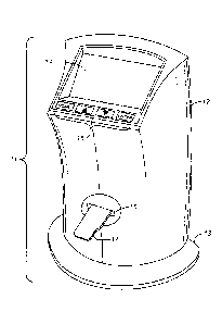
[0007] FIG. 1 is a perspective view of a cell counting instrument representing
an example
of an implementation of the concepts disclosed herein.
[0008] FIG. 2 is a diagram of the optical components of the instrument of FIG.
1.
[0009] FIG. 3 is a perspective view of optical components in the interior of
the instrument
of FIG. 1.
[0010] FIG. 4 is an exploded view, in perspective, of two plates constituting
a sample slide
for use in the instrument of FIG. 1.
[0011] FIG. 5A is a view of the upper surface of the upper plate of the sample
slide of FIG.
4. fig. 5B is a view of the lower surface of the upper plate of the sample
slide of FIG. 4.
DETAILED DESCRIPTION
[0012] The upper and lower optical windows between which the cell suspension
is retained
inside the sample vessel are close enough that the retained suspension is a
thin film whose
lateral dimensions, i.e., its exposed length and width, are at least an order
of magnitude
greater that its thickness. The entire exposed area (i.e., lateral dimensions)
of the sample
chamber or a laterally dimensioned portion thereof serves as a field of view
that is projected
onto a digital imaging sensor that contains at least about 4,000,000 (four
million) pixels, or in
certain embodiments from about 4,000,000 to about 10,000,000 pixels, with each
pixel being
no greater than about 2 x 2 pm (4 m2) in size, or from about 0.5 x 0.5 pm
(0.25 m2) to about
2 x 2 m (41=2) in certain embodiments, and in certain of the latter from
about 1 x 1 pm
(1 m2) to about 2 x 2 m (41=2). The field of view imaged by the sensor is at
least about 3
3
CA 02772376 2012-02-27
WO 2011/026029 PCT/US2010/047143
square millimeters, and often from about 3 mm2 to about 10 mm2. A
complementary metal
oxide semiconductor (CMOS) is one example of a digital imaging sensor useful
for this
purpose. Examples of CMOS sensors meeting these parameters are the 0V5620 and
0V5632 color imagers available from anniVision, Santa Clara, California, USA.
Other
examples are available from Aptina Imaging, a division of Micron Technology,
Inc., of San
Jose, California, USA. A color digital imaging sensor can also be used. Image
processing to
count the cells in the image generated by the CMOS sensor can be achieved by
known digital
counting methods, such as those mentioned above.
[0013] The image of the sample chamber can be magnified along the optical path
by a
magnification that is often within a range of from about 1.5 to about 6, or a
range of from
about 1.5 to about 3, with a magnification of about 2 as an example. This can
be achieved by
a two-lens achromat assembly. An example of such a lens assembly is a lens of
35-mm focal
length closest to the sample, a lens of 60-mm focal length closest to the
sensor, and an
aperture between the two lenses. The distance between the lens nearest the
sample and the
sample itself in this example is thus 35mm, and the distance between the lens
nearest the
sensor and the imager itself is 60mm. The magnification of the system is the
ratio of the
focal lengths of the two lenses, which in this case is 60mm/35mm = 1.7. The
two lenses can
each for example be 12.5mm in diameter, and the aperture can be 6mm in
diameter. Lenses
of other diameters and focal lengths that will produce the same or
approximately the same
results will be readily apparent to those skilled in the art. The footprint of
the instrument is
defined as the area projected by the larger of the instrument and its support
base on a plane
perpendicular to the optical path. As noted above, the instrument can be
constructed with a
small footprint, particularly one that is less than 300 cm2 in area.
100141 When a flat digital imaging sensor is used, a negative lens can be
positioned below
the sensor to intercept the optical signal immediately and to correct the
focus field curvature
of the achromat lens pair. This type of field curvature is common in optical
systems and is
also referred to as Petzval curvature. In an illustrative embodiment, a 6mm-
diameter lens
with a minus-18mm focal length is used. The lens thickness can vary but is
optimally
selected to correct the curvature without substantially reducing the field of
view.
[0015] Illumination of the sample can be achieved with a conventional light
source at the
base of the instrument and a collimating lens between the light source and the
sample. With
these components the sample is illuminated by trans-illumination without a
diffuser. A
preferred light source is a single white light-emitting diode (LED) with a
fluorescent coating.
An example of such a component is LUXEON Rebel White, part no. LXML-PWN1-
0050,
4
CA 02772376 2012-02-27
WO 2011/026029 PCT/US2010/047143
available from Philips Lumileds Lighting Company, San Jose, California, USA.
An example
of a collimating lens is one that is 9 mm in diameter with a focal length of
18 mm. With
these dimensions and those of the preceding paragraphs, an instrument can be
constructed
with the achromat lens pair approximately 35 mm above the sample, and the
sensor
approximately 60 mm above the achromat lens pair. With an achromat lens pair
having a
thickness of approximately 13 mm, the total distance between the sample and
the sensor can
be as little as 108 mm. In general, the optical path of the instrument, i.e.,
defined herein as
the arrangement of the components extending from the light source to the CMOS
or other
digital imaging sensor, can be 20 cm or less in height. In preferred
instruments within the
scope of this invention, the optical components are mounted to the housing
interior in a
floating manner using compliant counts, to avoid damage to, or misalignment
of, the optical
system upon jolts to the instrument, such as might occur when the instrument
is dropped or
mishandled, or collides with another instrument or piece of equipment.
[0016] As noted above, the sample vessel, which will be referred to henceforth
as a sample
slide in view of its similarity in size and shape to a microscope slide, is
received in the
instrument through a slot that is positioned at a location along the optical
path that is at a
distance from the nearest lens of the achromat lens pair equal to the focal
length of the lens.
In its preferred embodiments, the instrument as a whole is 30 cm or less in
height, and the use
of a digital imaging sensor as described above that employs a large number of
pixels of the
small sizes indicated permits the instrument to be constructed with the slot
at a sufficient
height to allow the user to comfortably insert the slot by hand, i.e.,
clearing the user's hand
from the table on which the instrument rests. The slot can thus be 60 mm or
more from the
base of the instrument, and preferably 70-80 mm from the base.
[0017] In preferred embodiments of the invention, the instrument provides
autofocusing of
the sample image by automatically adjusting the height of the slide following
its insertion.
One means of autofocusing involves the use of an image processor chip that
provides an
output of image contrast within an array of zones across the image from the
sensor. An
example of such a chip is the Freescale Semiconductor MC9328MX21, available
from KeilTM
- an ARM Company, Plano, Texas, USA; other examples will be apparent to those
skilled in
.. the art. The sum of the absolute differences of adjacent green pixels in a
particular zone of
the sensor array can be used as the image contrast value, and optimum focus is
achieved
when the image contrast value is at a maximum. The focus can then be adjusted
by a geared
motor connected to the slide mount within the receiving slot, i.e., the motor
when rotated will
move the slide mount up or down to change the focus of the image. The contrast
value is
5
CA 02772376 2012-02-27
WO 2011/026029
PCT/US2010/047143
detected at various positions of the motor which is then returned to the
position producing the
highest contrast value. In many embodiments of the instrument, this
autofocusing can occur
in 15 seconds or less.
[0018] An accessory that can be supplied with the instrument is a standard
slide for quality
control, such as verifying the accuracy of counting live and dead cells and
the ability of the
instrument to focus properly. The standard slide can have the same external
dimensions as a
sample slide, but instead of a sample chamber(s), the standard can have an
array of dark-
colored spots and rings printed on it, the spots simulating dead cells and
detected as such in
the digital imaging sensor and the rings simulating live cells and detected as
such in the
digital imaging sensor.
[0019] In certain embodiments of the concepts described herein, the functions
performed
by the instrument, including autofocusing and cell counting, are initiated by
the simple
insertion of the sample slide. This initiation can be achieved by the
inclusion of a non-
contact optical reflection sensor located within the slot or on the slide
mount within the slot.
An example of a suitable sensor is one that emits an infra-red beam and
detects objects within
approximately one millimeter of the sensor aperture by detecting a reflected
signal from the
beam. The reflected signal will rise to a maximum level when the slide is
fully inserted, and
the high signal will initiate the autofocusing and cell counting mechanisms.
One example of
a sensor that can serve this purpose is the QRE1113 Reflective Object Sensor,
available from
.. Fairchild Semiconductor Corporation, San Jose, California, USA. Other
examples will be
apparent to those skilled in the art.
[0020] A further feature that can be included in instruments embodying the
features
described herein is the automatic detection of cells in the sample that are
stained with a vital
stain. A vital stain is one that preferentially stains dead cells, and the
differentiation between
cells stained with such a stain and those that are not is achieved by the use
of differently
colored pixels. Trypan blue is one example of a vital stain; eosin and
propidium iodide are
other examples. Trypan blue transmits blue light and attenuates red light, and
by comparing
the intensities of blue and red pixels in the image sensor, the instrument can
determine
whether cells stained with a vital stain are present. Other dyes will afford
similar color
distinctions as appropriate to the dyes themselves. Image processing chips
that incorporate
this automatic detection feature include those referenced above and are
readily available. The
instrument can be programmed to eliminate any possible undercounting of viable
cells and
thereby detect viable cells to a particularly high degree of accuracy by
focusing on two or
more planes. The contrast between live cells and dead cells can be increased
further by using
6
CA 02772376 2012-02-27
WO 2011/026029 PCT/US2010/047143
optical filters to control the illumination bandwidth, or by selecting a
spectrally narrow light
source, such as an LED of a particular color instead of white. For example, a
585nm optical
filter with about 20nm bandwidth can be used to match the illumination to the
peak
absorption wavelength of the Trypan blue dye, whose peak absorption is 586nm.
The dead
cells will appear darker when the sample is illuminated through this filter.
[0021] In preferred instruments within the scope of this invention, all
functions that
contribute to the obtainment of a cell count in the sample are contained
within the instrument
housing, and the full operation of the instrument can thus be achieved without
the use of an
external machine or computer. Included among these functions are the automatic
focusing by
varying the height of the sample slide to find the best focal plane to
discriminate cells from
background, the determination of whether the sample has been stained with
Trypan blue or
other vital stain, a multi-focal plane analysis when a vital stain is detected
so that each cell is
scored on multiple focal planes to prevent undercounting of live cells, an
integrated dilution
counter to determine the volume of a cell suspension to use, the ability to
produce a visual
image of the cells on the display at the option of the user and to zoom in for
a detailed visual
inspection of the cells, and the ability and user option to export the results
to a USB flash
drive or to a thermal printer or other external printer. All of these
functions can be initiated
by the simple insertion of the sample slide by way of the non-contact optical
reflection sensor
described above, and in many cases, the execution of these functions is
completed in 30
seconds or less.
[0022] The Figures hereto depict an instrument that contains many of the
features described
above and serves as one example of an implementation of the concepts described
herein.
[0023] FIG. 1 depicts an automated cell counter instrument 11 in its upright
position as it
would be used on a laboratory bench. The visible parts of the instrument are a
housing 12, a
support base 13, a display screen 14, a control panel 15, and a slot 16 for
insertion of a
sample slide 17. The display screen shows the progress of the cell counting
analysis,
identifies the functions of the instrument as they are being performed, and
offers options to
the user for various functions and for showing an image of the cells in the
sample slide.
[0024] FIG. 2 depicts components of the optical path in the interior of the
instrument of
FIG. 1 with the sample slide 17 having been positioned in the optical path.
The sample slide
17 is horizontal and resides above an LED board 22 serving as the light
source. A
collimating lens 23 renders the light rays from the LED parallel as they
approach the sample
slide. The achromat lens pair 24 is positioned between the sample slide 17 and
the sensor 25.
7
CA 02772376 2012-02-27
WO 2011/026029 PCT/US2010/047143
The two lenses 26, 27 of the achromat lens pair are separated by an aperture
28. A field
flattening lens 29 is positioned immediately below the sensor 25.
[0025] FIG. 3 depicts the main optics assembly, showing the slide mount 31
with the
sample slide 17 partially inserted, the LED board 22, the illumination
(collimating) lens 23,
the geared motor 32 that adjusts the slide height to focus the image, and an
imaging lens tube
33 terminating in a fitting 34 to receive the CMOS sensor board. Also shown in
the Figure is
the main printed circuit board 35 that controls the functions of the
instrument and includes a
motor drive chip to control the motor 32. The board 35 resides within the
housing and the
position of the board in the Figure reflects its position relative to the
optics assembly,
[0026] A sample slide for use in the instrument of the preceding Figures is
shown in FIGS.
4, 5A, and 5B. The view in FIG. 4 is a perspective view, and the slide 17 is
formed of two
plates 42, 43 bonded together but shown separated in the Figure. The slide
contains two
sample chambers, as indicated by the indicia "A" and "B," respectively,
separated from each
other lengthwise along the slide and laterally offset from each other. The
areas 44, 45 of the
lower plate 43 that form the bottom surfaces of the sample chambers are made
of optically
transparent material, as are the corresponding areas of the upper plate 42
that are directly
above these areas on the lower plate and form the upper surfaces of sample
chambers. The
lower plate 43 in this embodiment is thicker than the upper plate 42 to
provide rigidity to the
slide, and the relative thinness of the upper plate 42 permits the upper
window of each sample
.. chamber to be thinner than the lower window, and indeed as thin as possible
to achieve a
highly focused image in the CMOS sensor. Each sample chamber is thus offset
from the
center plane of the slide and closer to the upper plate 42 than to the lower
plate 43.
[0027] FIGS. 5A and 5B are planar views of the top surface 51 and bottom
surface 52,
respectively, of the upper plate 42, the bottom surface 52 being the surface
that is bonded to
the lower plate 43. Each sample chamber is defined by a recess 53 (FIG. 5B) in
the bottom
surface of the upper plate, which further reduces the thickness of the area
forming the
optically clear window at the top of each sample chamber. In one example of
the dimensions
of the slide, the thickness of the upper plate in areas other than the recess
53 is 0.65mm and
the thickness of the lower plate is 1.00mm, for a total slide thickness of
1.65mm. The recess
53 is 0.100mm in depth, which thus forms a sample chamber that is 0.100mm in
depth, a
standard sample chamber thickness for manual hemocytometers. Each sample
chamber has
two loading or vent ports 54, 55, one at each of the two opposing longitudinal
ends of the
elongated chamber. Overflow areas 56, 57, 58, 59 that are open at the top of
the slide are
8
CA 02772376 2017-02-08
. ,
positioned at each of the four comers of each sample chamber to accommodate
excess sample
and thereby insure that the sample chamber is properly filled with sample.
(0028] Since each sample chamber is closer to the upper plate 42 than to the
lower plate 43,
the slide functions best when properly inserted into the cell counter with the
upper plate 42,
and hence the thinnest optical window, at the top. To ensure that the slide is
inserted in this
orientation, the slide is formed with notches 61, 62 in two diagonally
opposing corners of the
slide. The internal surfaces of the slot in the cell counter into which the
slide is inserted to
initiate the functions of the cell counter contains contour features that are
complementary to
these notches. The notches and complementary contours in the slot thereby
prevent the user
from inserting the slide upside down, i.e., with the upper plate 42 at the
bottom rather than
the top. The symmetrical arrangement of the notches also complements the
symmetrical
= arrangement of the two sample chambers and permits the slide to be
inserted with either end
first, while preventing the slide from being inserted in an inverted position
(upside down).
Since the slide is preferably a consumable item, it can thus be used for cell
counting
.. measurements on two independent samples at different times, and once both
chambers have
been used the slide can be disposed of and not used again.
[00291 Variations on the construction of the sample slide that still ensure
proper orientation
will be readily apparent to those skilled in the art. The arrangement, number,
and shapes of
the notches can thus be varied, as can the number of sample chambers and their
locations
relative to each other in the slide. The material of construction can vary
widely and can be
any material that can form an optically clear window, that is inert to the
sample, and that is
sufficiently rigid to be inserted into the cell counter. Poly(methyl
methacrylate) and
polycarbonate are examples of materials that are can be used. Others will be
readily apparent
to those skilled in the art. Likewise, the bonding of the plates can be
accomplished by
conventional means. Laser welding and ultrasonic welding are examples.
[00301 In the claims appended hereto, the terms "a" and "an" are intended to
mean "one or
more." The temi "comprise" and variations thereof such as "comprises" and
"comprising,"
when preceding the recitation of a step or an element, are intended to mean
that the addition
of further steps or elements is optional and not excluded.
Any discrepancy between any reference material cited herein
or any prior art in general and an explicit teaching of this specification is
intended to be
resolved in favor of the teaching in this specification. This includes any
discrepancy between
9
CA 02772376 2012-02-27
WO 2011/026029
PCT/US2010/047143
an art-understood definition of a word or phrase and a definition explicitly
provided in this
specification of the same word or phrase.