Note: Descriptions are shown in the official language in which they were submitted.
ANALYTE MEASUREMENT METHOD AND SYSTEM
[0001]
BACKGROUND
[0002] Electrochemical sensors have long been used to detect or measure
the presence of
substances in fluid samples. Electrochemical sensors include a reagent mixture
containing
at least an electron transfer agent (also referred to as an "electron
mediator") and an
analyte specific bio-catalytic protein (e.g. a particular enzyme), and one or
more
electrodes. Such sensors rely on electron transfer between the electron
mediator and the
electrode surfaces and function by measuring electrochemical redox reactions.
When used
in an electrochemical biosensor system or device, the electron transfer
reactions are
monitored via an electrical signal that correlates to the concentration of the
analyte being
measured in the fluid sample.
[0003] The use of such electrochemical sensors to detect analytes in
bodily fluids, such as
blood or blood derived products, tears, urine, and saliva, has become
important, and in
some cases, vital to maintain the health of certain individuals. In the health
care field,
people such as diabetics, for example, must monitor a particular constituent
within their
bodily fluids. A number of systems arc capable of testing a body fluid, such
as, blood,
urine, or saliva, to conveniently monitor the level of a particular fluid
constituent, such as,
CAN_DMS \10781340111 1
CA 2772738 2017-07-05
CA 02772738 2012-02-29
WO 2011/030093 PCT/GB2010/001683
cholesterol, proteins, and glucose. Patients suffering from diabetes, a
disorder of the
pancreas where insufficient insulin production prevents the proper digestion
of sugar, have
a need to carefully monitor their blood glucose levels on a daily basis.
Routine testing and
controlling blood glucose for people with diabetes can reduce their risk of
serious damage
to the eyes, nerves, and kidneys.
[0004] Electrochemical biosensors may be adversely affected by the presence
of certain
blood components that may undesirably affect the measurement and lead to
inaccuracies
in the detected signal. This inaccuracy may result in an inaccurate glucose
reading, leaving
the patient unaware of a potentially dangerous blood sugar level, for example.
As one
example, the blood hematocrit level (i.e. the percentage of the amount of
blood that is
occupied by red blood cells) can erroneously affect a resulting analyte
concentration
measurement.
[0005] Variations in a volume of red blood cells within blood can cause
variations in
glucose readings measured with disposable electrochemical test strips.
Typically, a
negative bias (i.e., lower calculated analyte concentration) is observed at
high hematocrit,
while a positive bias (i.e., higher calculated analyte concentration) is
observed at low
hematocrit. At high hematocrit, for example, the red blood cells may impede
the reaction
of enzymes and electrochemical mediators, reduce the rate of chemistry
dissolution since
there is less plasma volume to solvate the chemical reactants, and slow
diffusion of the
mediator. These factors can result in a lower than expected glucose reading as
less current
is produced during the electrochemical process. Conversely, at low hematocrit,
fewer red
blood cells may affect the electrochemical reaction than expected, and a
higher measured
current can result. In addition, the blood sample resistance is also
hematocrit dependent,
which can affect voltage and/or current measurements.
[0006] Several strategies have been used to reduce or avoid hematocrit
based variations on
blood glucose. For example, test strips have been designed to incorporate
meshes to
remove red blood cells from the samples, or have included various compounds or
formulations designed to increase the viscosity of red blood cell and
attenuate the affect of
low hematocrit on concentration determinations. Other test strips have
included lysis
agents and systems configured to determine hemoglobin concentration in an
attempt to
correct hematocrit. Further, biosensors have been configured to measure
hematocrit by
2
CA 02772738 2012-02-29
WO 2011/030093 PCT/GB2010/001683
measuring optical variations after irradiating the blood sample with light, or
measuring
hematocrit based on a function of sample chamber fill time. These methods have
certain
disadvantages.
SUMMARY OF THE DISCLOSURE
100071 Applicants have recognized a need for a system and method that can
be used to
determine an accurate glucose concentration that avoids the disadvantages in
the field.
f0008I In view of the foregoing and in accordance with one aspect, there is
provided a
method of operating an analyte measurement system having a meter and a test
strip. The
test strip may include a reference electrode, a first working electrode and a
second
working electrode in which the first and second working electrodes are coated
with a first
and second reagent layer, respectively. The respective first and second
reagent layers are
disposed on a matrix layer having a mediator. The meter may include an
electronic circuit
for applying a test voltage between the reference electrode and the first
working electrode
and for applying a second test voltage between the reference electrode and the
second
working electrode. The meter also may include a signal processor for measuring
a
plurality of test currents and for calculating a glucose concentration from
the test currents.
The method may be achieved by applying a test voltage between the reference
electrode
and the second working electrode; measuring a first test current, a second
test current and
a third test current at the working electrode with the meter after a blood
sample containing
an analyte is applied to the test strip; ascertaining the glucose
concentration from the first,
second and third test currents; and displaying the glucose concentration.
100091 In the exemplary method, the glucose concentration may be a value
obtained with
the following:
P
(-a-\ 13 ¨ interceptl
'2)
G ¨ __________________________
slope I
where:
G includes the hematocrit-corrected glucose concentration;
3
CA 02772738 2012-02-29
WO 2011/030093 PCT/GB2010/001683
II includes the first test current;
12 includes the second test current;
/3 includes the third test current;
p includes a power term;
interceptl includes an intercept value determined from a linear regression of
a plot
of [ r a/ _
.- 3 versus a reference glucose concentration; and
12
...
slopel includes a slope value determined from a linear regression of a plot of
[( ri P
L- 13 versus the reference glucose concentration.
/2
[0010] In such embodiment, the power term p depends on a threshold value of
the first test
current Ii and may be from about one to about four. If the first test current
I, includes
above the threshold value, then the above equation is used to calculate the
hematocrit-
corrected glucose concentration G. If the first test current II is at or below
the threshold
value, then the power term p is set to zero in the above equation and the term
¨1- ( / JP
12
becomes one. The threshold value of the first test current II may be from
about 4
microamperes to about 7 microamperes.
[0011] In another embodiment, the power term p may include a value obtained
with the
following:
p = a ¨ ¨b
/3
where a includes a first tuning parameter and b includes a second tuning
parameter.
[0012] In one embodiment, each of first and second tuning parameters a and
b is from
about zero to about five.
[0013] In another embodiment, batch-specific tuning parameters a and b may
be
determined by a calculating a first power term for a first combination of the
first tuning
parameter and the second tuning parameter with the following:
4
CA 02772738 2012-02-29
WO 2011/030093 PCT/GB2010/001683
pl = a ¨
13
where pl includes the first power term;
ascertaining the current for each of a plurality of samples tested with the
batch of test
strips with the following:
corrected ¨ r * "3
12
where 'corrected includes the hematocrit-corrected current;
computing a slope and intercept from a linear regression of a plot of
hematocrit-corrected
current versus a reference plasma glucose concentration;
estimating a hematocrit-corrected glucose concentration for each of the
plurality of
samples with the following:
¨ intercept2
Gcorrected = 'corrected
slope2
where Gcorrected includes the hematocrit-corrected glucose concentration,
intercept2
includes an intercept value determined from a linear regression of a plot of
/corrected
versus a reference glucose concentration and slope2 includes a slope value
determined from a linear regression of a plot of /corõc,ed versus a reference
glucose
concentration;
evaluating a bias for each of the hematocrit-corrected glucose concentrations
with
equations of the form:
BiaSabs Gcorrected Greference for Greference less than 75mg/dL and
Gcorrected Greference
Bias% = ___________________ for Greference greater than or equal to 75mg/dL
Greference
where Biasabs includes absolute bias, Bias % includes percent bias and
Greference
includes the reference glucose concentration;
estimating accuracy for the first combination of the first and second tuning
parameters
with the following:
Accuracy = ¨n15 *100
where n15 includes the number of data points within a bias criteria and n
includes
CA 02772738 2012-02-29
WO 2011/030093 PCT/GB2010/001683
the total number of data points;
computing a hematocrit slope from a linear regression of a plot of the bias
versus the
percent hematocrit;
establishing a standard deviation of the bias with the following:
s )2)2
n-1
where s includes the standard deviation, n includes the number of samples, xt
includes the sample and SE includes the mean of the sample;
repeating the previous steps for all combinations of the first and second
tuning parameters;
plotting an accuracy calibration space of the accuracy calibration space for
all
combinations of the first and second tuning parameters; plotting an accuracy
calibration
space of the hematocrit slope calibration space for all combinations of the
first and second
tuning parameters; generating a combined surface plot for all combinations of
the first and
second tuning parameters which meet an accuracy and hematocrit slope
acceptance
criteria; and determining batch-specific first and second tuning parameters
from the
combined surface plot.
[0014] In another embodiment, the method of determining batch-specific
tuning
parameters further may include determining a set of batch-specific calibration
parameters,
e.g., slope and intercept.
[0015] In yet another embodiment, the method of determining batch-specific
tuning
parameters further may include determining tuning parameters for multiple
batches of test
strips and then determining regions of overlap for all the batches in the
combined surface
plots of the accuracy calibration space and the hematocrit slope calibration
space.
[0016] In yet a further embodiment, a method for determining a hematocrit-
corrected test
current measurable with a system having a test strip and a meter is provided.
The method
can be achieved by applying a test voltage between a reference electrode and a
working
electrode coated with a reagent layer disposed on a matrix layer having a
mediator;
measuring a first test current, a second test current and a third test current
at the working
electrode with the meter after a blood sample containing an analyte is applied
to the test
strip; and ascertaining a hematocrit-corrected test current via a ratio of the
first test current
to the second test current raised to a power term and multiplying the ratio by
the third test
6
CA 02772738 2012-02-29
WO 2011/030093 PCT/GB2010/001683
current, in which the power term is a function of a first tuning parameter and
a second
tuning parameter.
[0017] In yet a further embodiment, an analyte measurement system to
measure at least
glucose concentration in physiological fluid of a user is provided. The system
includes a
test strip and a meter. The test strip includes a substrate having a reference
electrode and a
working electrode coated with a reagent layer, which is disposed on a matrix
layer having
a mediator. The electrodes are connected to corresponding contact pads. The
analyte
meter has a test circuit in connection with a test strip port that receives
the contact pads of
the test strip so that the meter is configured to apply a test voltage after
deposition of
physiological fluid on the electrodes and determine a hematocrit-corrected the
glucose
concentration from measured first, second and third test currents at first,
second, and third
discrete intervals after application of the test voltage by the meter.
[0018] These and other embodiments, features and advantages of the
invention will
become apparent to those skilled in the art when taken with reference to the
following
more detailed description of the exemplary embodiments in conjunction with the
accompanying drawings that are first briefly described.
BRIEF DESCRIPTION OF THE DRAWINGS
[0019] The accompanying drawings, which are incorporated herein and
constitute part of
this specification, illustrate presently preferred embodiments of the
invention, and,
together with the general description given above and the detailed description
given
below, serve to explain features of the invention (in which like numerals
represent like
elements), of which:
[0020] Figure 1 illustrates an exemplary embodiment of a top view of a
system for
measuring two analyte concentrations;
[0021] Figure 2 illustrates an exemplary embodiment of a perspective
exploded view of a
test strip;
[0022] Figure 3 illustrates an exemplary embodiment of a top view of the
test strip shown
in Figure 2;
7
[0023] Figure 4 illustrates an exemplary embodiment of a schematic of the
functional
components of the meter shown in Figure 1 forming an electrical connection
with the test
strip of Figures 2 and 3;
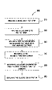
[0024] Figure 5A illustrates an exemplary embodiment of a flow chart of a
method of
estimating a hematocrit-corrected glucose concentration using the system shown
in Figure
1; and Figure 5B illustrates a method to determine batch-specific first and
second tuning
parameters in the embodiments herein;
100251 Figure 6 illustrates an exemplary embodiment of a chart showing
test voltages
applied by the meter to the test strip;
[0026] Figure 7 illustrates an exemplary embodiment of a chart showing
test currents
generated when the test voltages of Figure 6 are applied to the test strip;
[0027] Figure 8 illustrates an exemplary embodiment of a surface plot of
the accuracy
calibration space for all combinations of the first tuning parameter and the
second tuning
parameter for a batch of test strips having the embodiment shown in Figures 2
and 3;
[0028] Figure 9 illustrates an exemplary embodiment of a surface plot of
the hematocrit
slope calibration space for all combinations of the first tuning parameter and
the second
tuning parameter for a batch of test strips having the embodiment shown in
Figures 2 and
3;
[0029] Figure 10 illustrates an exemplary embodiment of a combined
surface plot for all
combinations of the first and second tuning parameters which meet an accuracy
and
hematocrit slope acceptance criteria and using the data in Figures 8 and 9;
[0030] Figures I IA and 11B illustrate Clarke Error Grid analysis showing
test glucose
concentration plotted as a function of reference glucose concentration prior
to and after
applying an exemplary embodiment to the test data, respectively. The test data
was
obtained with a batch of test strips having the embodiment shown in Figures 2
and 3; and
[0031] Figures 11C and 11D illustrate Parkes Error Grid analysis showing
test glucose
concentration plotted as a function of reference glucose concentration prior
to and after
applying an exemplary embodiment to the test data, respectively. The test data
in Figures
11A and 11B was used along with additional data and after applying a suitable
error
trapping.
CAN DMS \107813401\1 8
CA 2772738 2017-07-05
DETAILED DESCRIPTION OF ILLUSTRATIVE EMBODIMENTS
[0032] The following detailed description should be read with reference
to the drawings,
in which like elements in different drawings are identically numbered. The
drawings,
which are not necessarily to scale, depict selected embodiments and are not
intended to
limit the scope of the invention. The detailed description illustrates by way
of example,
not by way of limitation, the principles of the invention. This description
will clearly
enable one skilled in the art to make and use the invention, and describes
several
embodiments, adaptations, variations, alternatives and uses of the invention,
including
what is presently believed to be the best mode of carrying out the invention.
[0033] As used herein, the terms "about" or "approximately" for any
numerical values or
ranges indicate a suitable dimensional tolerance that allows the part or
collection of
components to function for its intended purpose as described herein. In
addition, as used
herein, the terms "patient,- "host," "user," and "subject" refer to any human
or animal
subject and are not intended to limit the systems or methods to human use,
although use of
the subject invention in a human patient represents a preferred embodiment.
[0034] Figure 1 illustrates a system 100 for measuring at least two
analyte concentrations
in which system 100 may include a meter 102 and a test strip 200. Meter 102
may include
a display 104, a housing 106, a plurality of user interface buttons 108, and a
strip port 110.
Meter 102 further may include electronic circuitry within housing 106 such as
a memory
120, a microprocessor 122, electronic components for applying a test voltage,
and also for
measuring at least two test current values (see Figure 4). A proximal portion
204 of test
strip 200 may be inserted into strip port 110. Display 104 may output at least
two analyte
concentrations, e.g., glucose and/or a ketone concentration, and may be used
to show a
user interface for prompting a user on how to perform a test. The plurality of
user
interface buttons 108 allow a user to operate meter 102 by navigating through
the user
interface software. Display 104 may optionally include a backlight.
[0035] An optional data port 114 accepts a suitable connector attached to
a connecting
lead, thereby allowing meter 102 to be linked to an external device such as a
personal
computer. Data port 114 may be any port that allows for transmission of data
(serial or
parallel) such as, for example, serial or parallel port in wired or wireless
form, A personal
CAN_DMS M 07813401 \I 9
CA 2772738 2017-07-05
CA 02772738 2012-02-29
WO 2011/030093 PCT/GB2010/001683
computer, running appropriate software, allows entry and modification of set-
up
information (e.g. the current time, date, and language), and may perform
analysis of data
collected by meter 102. In addition, the personal computer may be able to
perform
advanced analysis functions, and/or transmit data to other computers (i.e.
over the
intemet) for improved diagnosis and treatment. Connecting meter 102 with a
local or
remote computer facilitates improved treatment by health care providers.
[0036] Figures 2 and 3 are exemplary exploded perspective and top assembled
views,
respectively, of test strip 200, which may include seven layers disposed on a
substrate 205.
The seven layers disposed on substrate 205 may be a conductive layer 250, an
insulation
layer 216, a matrix layer 222, a first reagent layer 224 and a second reagent
layer 226, an
adhesive layer 260, a hydrophilic layer 270, and a top layer 280. Test strip
200 may be
manufactured in a series of steps where the conductive layer 250, insulation
layer 216,
matrix layer 222, first reagent layer 224, second reagent layer 226 and
adhesive layer 260
are sequentially deposited on substrate 205 using, for example, a screen-
printing process.
Hydrophilic layer 270 and top layer 280 may be disposed from a roll stock and
laminated
onto substrate 205 as either an integrated laminate or as separate layers.
Test strip 200 has
a distal portion 203 and a proximal portion 204, as shown in Figure 2.
[0037] Test strip 200 may include a sample-receiving chamber 292 through
which a blood
sample may be drawn. Sample-receiving chamber 292 may include an inlet at a
proximal
end of test strip 200. An outlet or air vent is included in hydrophilic layer
270, as will be
described below. A blood sample may be applied to the inlet to fill a sample-
receiving
chamber 292 so that at least two analyte concentrations may be measured. The
side edges
of a cut-out portion of adhesive layer 260 located adjacent to first and
second reagent
layers 224 and 226 define a wall of sample-receiving chamber 292, as
illustrated in Figure
2. A bottom portion or "floor" of sample-receiving chamber 292 may include a
portion of
substrate 205, conductive layer 250, and insulation layer 216. A top portion
or "roof' of
sample-receiving chamber 292 may include distal hydrophilic portion 232.
[0038] For test strip 200, as illustrated in Figure 2, substrate 205 may be
used as a
foundation for helping support subsequently applied layers. Substrate 205 may
be in the
form of a polyester sheet such as a polyethylene tetraphthalate (PET)
material. Substrate
CA 02772738 2012-02-29
WO 2011/030093 PCT/GB2010/001683
205 may be in a roll format, nominally 350 microns thick by 370 millimeters
wide and
approximately 60 meters in length.
[0039] A conductive layer is required for forming electrodes that may be
used for the
electrochemical measurement of glucose. Conductive layer 250 may be made from
a
carbon ink that is screen-printed onto substrate 205. In a screen-printing
process, carbon
ink is loaded onto a screen and then transferred through the screen using a
squeegee. The
printed carbon ink may be dried using hot air at about 140 C. The carbon ink
may include
VAGH resin, carbon black, graphite, and one or more solvents for the resin,
carbon and
graphite mixture. More particularly, the carbon ink may incorporate a suitable
ratio of
carbon black: VAGH resin in the carbon ink.
[0040] For test strip 200, as illustrated in Figure 2, conductive layer 250
may include a
reference electrode 210, a first working electrode 212, a second working
electrode 214, a
reference contact pad 211, a first contact pad 213, a second contact pad 215,
a reference
electrode track 207, a first working electrode track 208, a second working
electrode track
209, and a strip detection bar 217. In the embodiment shown in Figure 2,
reference
electrode 210 is located in between first working electrode 212 and second
electrode 214
such that cross-talk between first and second working electrodes 212 and 214
is
minimized.
100411 Conductive layer 250 may be formed from a carbon ink. Reference
contact pad
211, first contact pad 213 and second contact pad 215 may be configured to
electrically
connect to a test meter. Reference electrode track 207 provides an
electrically continuous
pathway from reference electrode 210 to reference contact pad 211. Similarly,
first
working electrode track 208 provides an electrically continuous pathway from
first
working electrode 12 to first contact pad 213. Similarly, second working
electrode track
209 provides an electrically continuous pathway from second working electrode
214 to
second contact pad 215. Strip detection bar 217 is electrically connected to
reference
contact pad 211. A test meter may detect that test strip 200 has been properly
inserted by
measuring a continuity between reference contact pad 211 and strip detection
bar 217.
[0042] Insulation layer 216 may include a rectangular aperture 218 that
exposes a portion
of reference electrode 210, first working electrode 212, and second working
electrode 214,
which may be wetted by a liquid sample. The area of first working electrode
212, second
11
CA 02772738 2012-02-29
WO 2011/030093 PCT/GB2010/001683
working electrode 214, and reference electrode 210 may be defined as the area
exposed to
the liquid sample. In addition to defining an electrode area, insulation layer
216 prevents
a liquid sample from touching the electrode tracks 207, 208, and 209. It is
believed that
the functional area of a working electrode should be accurately defined
because the
magnitude of the test current is directly proportional to the effective area
of the electrode_
As an example, insulation layer 216 may be Ercon E6110-116 Jet Black
InsulayerTM ink
that may be purchased from Ercon, Inc. The test strip at this point may be
treated with
plasma. The plasma is created by high-voltage alternating current (AC) between
two or
more plasma sources spaced about 100 millimeters apart and rotated about a
generally
vertical axis at ambient temperatures to define a plasma ring. The plasma ring
is
configured to be spaced apart from the substrate 205, which may include the
test strip
electrode, at a distance of approximately 5 millimeters to approximately 30
millimeters
and preferably from about 10 millimeters to about 20 millimeters. The voltage
utilized by
the plasma controller may be configured to be about 5kVA and the voltage
provided to the
plasma electrodes is preferably less than about 2kVA. The frequency of the AC
is about
16kHz to about 20kHz. The resulting ring of plasma, consisting of ionised,
highly
energetic particles is swept downstream towards the substrate 205 using
filtered and
generally contaminant free compressed air at about 1.2 bars or higher absolute
pressure,
preferably about 2.5 bars at a volumetric flow rate of less than 2 cubic meter
of air per
hour, towards the substrate 205, which may be moving orthogonally to the flow
of air at
about 5 meters per minute to about 15 meters per minute and preferably
approximately 10
meters per minute. The plasma ring may be arrayed adjacent to other plasma
rings along
the path of travel of the substrates. The number of plasma rings may be from
one to as
many as necessary along the path of travel of the substrate or transverse to
such path to
provide for surface modification of the substrate. The plasma is used to
modify the
surface of the screen printed carbon based electrodes. This surface
modification or plasma
treatment is believed to increase the electrochemical activity of the carbon
surface and
increase the surface energy of the printed layers allowing for better adhesion
between
them and subsequently printed layers. Plasma treatment is also believed to
improve the
electrochemistry of the carbon surface making the reaction with the mediator
more ideal.
12
[0043] Matrix layer 222 may include a mediator such as, for example,
ferricyanide and a
cofactor such as, for example, nicotinamide adenine dinucleotide (NADH). In
one
embodiment, matrix layer 222 may include potassium ferricyanide, NADH, Tris-
HCL
buffer, hydroxyethylcellulose, DC 1500 Antifoam, Cabosil TS 610, poly (vinyl
pyrrolidone vinyl acetate), Triton XIOOTM, calcium chloride and analar water.
100441 First and second reagent layers 224 and 226 are each disposed on
matrix layer 222,
as illustrated in Figure 2. First and second reagent layers 224 and 226 each
may include
chemicals such as an enzyme which selectivity reacts with an analyte of
interest such that
the analyte concentration may be determined. The reagent layer can include an
enzyme
and a mediator. Exemplary enzymes suitable for use in the reagent layer
include glucose
oxidase, glucose dehydrogenase (with pyrroloquinoline quinone co-factor,
"PQQ"), and
glucose dehydrogenase (with flavin adenine dinucleotide co-factor, "FAD"). An
exemplary mediator suitable for use in the reagent layer includes
ferricyanide, which in
this case is in the oxidized form. The reagent layer can be configured to
physically
transform glucose into an enzymatic by-product and in the process generate an
amount of
reduced mediator (e.g., ferrocyanide) that is proportional to the glucose
concentration.
The working electrode can then measure a concentration of the reduced mediator
in the
form of a current. In turn, glucose meter 102 can convert the current
magnitude into a
glucose concentration.
[0045] Exemplary analytes of interest for monitoring diabetes include
glucose and
ketones. In one embodiment, first reagent layer 224 may include at least one
enzyme that
selectively reacts with ketones and second reagent layer 226 may include an
enzyme that
selectively reacts with glucose. In another embodiment, first reagent layer
224 may
include an enzyme that selectively reacts with glucose and second reagent
layer 226 may
include at least one enzyme that selectively reacts with ketones.
[0046] In one embodiment, the components in the reagent layer used to
determine the
ketone concentration may include beta-hydroxybutyrate dehydrogenase (BHD),
Tris-HCL
buffer, hydroxyethylcellulose, potassium ferricyanide, DC 1500 Antifoam,
Cabosil TS
610, poly(vinyl pyrrolidone vinyl acetate), Triton XlOOTM, calcium chloride
and analar
water. In another embodiment, the reagent layer used to measure ketones may
include a
second enzyme such as, for example, diaphorase.
CAN_DMS \107813401\1 13
CA 2772738 2017-07-05
[00471 Examples of enzymes suitable for use in the reagent layer for
measuring glucose
may include either glucose oxidase or glucose dehydrogenase. More
specifically, the
glucose dehydrogenase may have a pyrrylo-quinoline quinone (PQQ) cofactor or a
flavin
adenine dinucleotide (FAD) cofactor. In one embodiment, the components in the
reagent
layer that is used to determine the glucose concentration may include glucose
oxidase,
Tris-HCL buffer, hydroxyethyleellulose, potassium ferricyanide, DC 1500
Antifoam,
Cabosil TS 610, poly(vinyl pyrrolidone vinyl acetate), Triton XlOOTM, calcium
chloride
and analar water.
[0048] First and second reagent layers 224 and 226 may be formed from a
reagent ink,
which is disposed onto matrix layer 222 and dried. Note that the reagent ink
may also be
referred to as an enzyme ink or reagent formulation. A reagent ink typically
contains a
liquid, such as a buffer, for dispersing and/or dissolving materials used for
the
electrochemical detection of an analyte such as glucose. In one embodiment,
first and
second reagent layers 224 and 226 may be screen-printed in two successive
steps onto
matrix layer 222. Reagent ink may be loaded onto a screen until it is flooded.
Next, a
squeegee may be used to transfer the reagent ink through the screen and onto
matrix layer
222. After the deposition, the reagent ink may be dried using hot air at about
50 C.
[0049] In one embodiment, the area of first reagent layer 224 and second
reagent layer
226 is sufficiently large to cover the entire area of first working electrode
212 and second
working electrode 214, respectively. Each of first and second reagent layers
224 and 226
include a width and a length that is sufficiently large to at least account
for the largest
electrode area that may be used in test strip 200. The width of first and
second reagent
layers 224 and 226 may be about 2 millimeters, which is more than double a
width of
rectangular aperture 218.
[0050] Adhesive layer 260 may be disposed on test strip 200 after the
deposition of first
and second reagent layers 224 and 226. Portions of adhesive layer 260 may be
aligned to
be immediately adjacent to, touch, or partially overlap with first and second
reagent layers
224 and 226. Adhesive layer 260 may include a water based acrylic copolymer
pressure
sensitive adhesive which is commercially available. Adhesive layer 260 is
disposed on a
portion of insulation layer 216, conductive layer 250, and substrate 205.
Adhesive layer
260 binds hydrophilic layer 270 to test strip 200.
CAN_DMS \107813401\1 14
CA 2772738 2017-07-05
[0051] Hydrophilic layer 270 may include a distal hydrophilic portion 232
and proximal
hydrophilic portion 234, as illustrated in Figure 2. A gap 235 is included
between distal
hydrophilic portion 232 and proximal hydrophilic portion 234. Gap 235 serves
as a side
vent for air as blood fills sample-receiving chamber 292. Hydrophilic layer
270 may be a
polyester having one hydrophilic surface such as an anti-fog coating, which is
commercially available from 3M.
[0052] The final layer to be added to test strip 200 is top layer 280, as
illustrated in Figure
2. Top layer 280 may include a clear portion 236 and opaque portion 238. Top
layer 280
is disposed on and adhered to hydrophilic layer 270. Top layer 280 may be a
polyester
that has an adhesive coating on one side. It should be noted that the clear
portion 236
substantially overlaps distal hydrophilic portion 232, which allows a user to
visually
confirm that sample-receiving chamber 292 may be sufficiently filled. Opaque
portion
238 helps the user observe a high degree of contrast between a colored fluid
such as, for
example, blood within sample-receiving chamber 292 and opaque portion 238.
[0053] In another embodiment, the system may include a meter and test
strip for
measuring one analyte, e.g., glucose, as is described in US patent numbers
5,708,247,
5,951,836, 6,241,862, and 7,112,265.
[0054] Figure 4 shows a simplified schematic of meter 102 interfacing
with test strip 200.
Meter 102 may include a reference connector 180, a first connector 182 and a
second
connector 184, which respectively form an electrical connection to reference
contact 211,
first contact 213 and second contact 215. The three aforementioned connectors
are part of
strip port 110. When performing a test, a first test voltage source 186 may
apply a test
voltage VwE2 between second working electrode 214 and reference electrode 210.
As a
result of test voltage VwE2, meter 102 may then measure a test current IwE2 at
second
working electrode. In a similar manner, a second test voltage source 188
applies a test
voltage VwEj between first working electrode 212 and reference electrode 210.
As a result
of test voltage VwEi, meter 102 may then measure a test current IwEi. In an
embodiment,
test voltage VwE2 and second test voltage VwEi may be about equal. For
simplifying the
description of the following sections, the set of instructions for determining
a hematocrit
corrected glucose concentration will be described for only one working
electrode and
CAN_DMS. \ 107813401 \ 1 15
CA 2772738 2017-07-05
reference electrode. It should be apparent that the embodiments should not be
limited to
one working electrode and reference electrode, but that multiple working
electrodes may
also be utilized.
[0055] Referring to Figure 5A, a method 300 for determining a hematocrit-
corrected
analyte concentration (e.g., glucose) that uses the aforementioned meter 102
and test strip
200 embodiments will now be described.
[0056] In exemplary step 310, meter 102 and test strip 200 are provided.
Meter 102 may
include electronic circuitry that can be used to apply at least one test
voltage to the test
strip and to measure current flowing through at least second working electrode
214. Meter
102 also may include a signal processor with a set of instructions for the
method of
determining at least one analyte concentration in a fluid sample as disclosed
herein. In
one embodiment, the analytes are blood glucose and ketone.
100571 Figure 6 is an exemplary chart of a test voltage applied to test
strip 200. Before a
fluid sample is applied to test strip 200, test meter 102 is in a fluid
detection mode in
which a test voltage of about 400 millivolts is applied between second working
electrode
214 and reference electrode 210. In exemplary step 320, the fluid sample is
applied to test
strip 100 at to and is allowed to react with first and second reagent layers
224 and 226 for a
reaction period tR. The presence of sample in the reaction zone of test strip
200 is
determined by measuring the current flowing through second working electrode
214. The
beginning of reaction period tR is determined to begin when the current
flowing through
second working electrode 214 reaches a desired value, typically about 0.150
nanoamperes
(not shown), at which point a test voltage of zero millivolts is applied
between second
working electrode 214 and reference electrode 210. Reaction period tR is
typically from
about 2 to about 4 seconds after initiation of the measuring and is more
typically about 3
seconds after initiation of the measuring, i.e., after ti. In exemplary step
330, after
reaction period tR, the test voltage in the subject method is applied to test
strip 200 at ti for
a total test time tT. In an alternative method (not shown), the reaction
period tR is omitted
such that the start of the test commences as soon as sufficient current is
flowing through
second working electrode 214.
[0058] Figure 7 is an exemplary chart of a current transient A (i.e., the
measured
electrical current response in nanoamperes as a function of time) that is
measured when
CAN_DMS Y107813401 \ 1 16
CA 2772738 2017-07-05
CA 02772738 2012-02-29
WO 2011/030093 PCT/GB2010/001683
the test voltage of Figure 6 is applied to test strip 200. Test currents li
obtained from
current transients A are generally indicative of the analyte concentration in
the sample as
will be described in exemplary step 350 below. Referring to Figures 6 and 7,
in
exemplary step 340, after the test voltage is applied between second working
electrode
214 and reference electrode 210 at time ti, a first test current //, a second
test current 12,
and a third (or end) test current /3 are measured at times /2, /3, and tr,
respectively. The test
voltage applied between second working electrode 214 and reference electrode
210 is
generally from about +100 millivolts to about +600 millivolts. In one
embodiment in
which second working electrode 214 is carbon ink and the mediator is
ferricyanide, the
test voltage is about +400 millivolts. Other mediator and electrode material
combinations
will require different test voltages. The duration of first test voltage is
generally from
about 4 and 6 seconds after a reaction period and is typically about 5 seconds
after a
reaction period. Typically, time ti is measured relative to time ti. In
practice, each test
current Ii is the average of a set of measurements obtained over a short
interval, for
example, five measurements obtained at 0.01 second intervals starting at ti+/,
where 1
ranges from 1 to 3.
[0059] Referring to Figure 5A in exemplary step 350, a hematocrit-corrected
glucose
concentration may be determined with the following:
[
r P (-' ) 13]- interceptl
/2
G= ___________________________
slope I
(1)
where:
G is the hematocrit-corrected glucose concentration;
// is the first test current;
12 is the second test current;
13 is the third test current;
p is a power term that determines the strength of the hematocrit correction:
the
greater the magnitude of p, the greater the hematocrit correction, i.e., the
larger is the term (1 ¨I in Equation 1;
/2
17
CA 02772738 2012-02-29
WO 2011/030093 PCT/GB2010/001683
intercept] is an intercept value determined from a linear regression of a plot
of
[ i P
-- /3 versus a reference glucose concentration; and
'2)slopel may be a slope value determined from a linear regression of a plot
of
[ i P
11- 13 versus the reference glucose concentration.
12
[0060] In one embodiment, first test current II may be from about 3 seconds
after a
reaction period to about 4 seconds after a reaction period ti, second test
current 12 may be
from about 4 seconds after a reaction period ti to about 5 seconds after a
reaction period
tl, and third test current /3 may be about 5 seconds after a reaction period
tl. In one
embodiment, first test current // may be measured at a time at which signal
noise is low.
For plasma treated test strip, the first test current may be measured at about
3.5 seconds,
the second test current may be measured at about 4.5 seconds and the third
test current at
about 5 seconds. For untreated test strip, the first current may be measured
at about 4
seconds; the second test current at about 4.5 seconds; and the third test
current at about 5
seconds.
[0061] In one embodiment, power term p depends on a threshold value of
first test current
1-1 and may be from about one to about four. If first test current Ii is above
the threshold
value, then Equation 1 is used to calculate the hematocrit-corrected glucose
concentration
G. If first test current II is at or below the threshold value, then power
term p may be set
( P
1
to zero in Equation 1 and the term - becomes one. In one embodiment, the
threshold
12
value of first test current II may be from about 4 microamperes to about 7
microamperes.
[0062] In another embodiment, power term p comprises a value obtained with
the
following:
b
p = a - ¨
13 (2)
where a is a first tuning parameter and b is a second tuning parameter.
18
10063] By subtracting the inverse of /3 from first tuning parameter a,
power term p is
increased for large values of 13 and is reduced for low values of /3,
corresponding to high
and low glucose concentrations, respectively. In one embodiment, each of first
and
second tuning parameters a and b is from about zero to about five. For low
glucose
values, e.g., less than about 75 mg/dL, the value of p is preferably about 1
while for other
glucose values, the value of p can be from about 1.5 to about 3.5. In
exemplary step 360,
the hematocrit-corrected glucose concentration may then be displayed on meter
102.
[0064] Referring to Figure 5B, a method 400 for determining batch-
specific first and
second tuning parameters a and b will now be described. In exemplary step 410,
a
plurality of combinations of first and second tuning parameters a and h are
provided. In
an embodiment in which each of the first and second tuning parameters may vary
from
about zero to about five in increments of 0.1, a total of 2601 tuning
parameter
combinations are possible. In exemplary step 420, a first power term p1 for a
first
combination of the first tuning parameter and the second tuning parameter may
be
determined with Equation 3.
[0065] In exemplary step 430, a hematocrit-corrected current for each of
a plurality of
samples tested with the batch of test strips may be determined with the
following:
\ PI
/I *1
I correcled = I ,
\ 2 ) (3)
where /
¨correctedis a hematocrit-corrected current and pl is the first power term.
[0066] In exemplary step 440, a s1ope2 and an intercept2 is determined
from a linear
regression of a plot of hematocrit-corrected current versus a reference plasma
glucose
concentration.
[0067] In exemplary step 450, a hematocrit-corrected glucose
concentration is determined
for each of the plurality of samples with the following:
G _ ICOrreC ¨ intercept2
(4)
corpecied
slope2
where:
Gcorreeted is a hematoerit-corrected glucose concentration;
CAN_DMS: m7813401\1 19
CA 2772738 2017-07-05
CA 02772738 2012-02-29
WO 2011/030093 PCT/GB2010/001683
intercept2 is the intercept value determined from a linear regression of a
plot of
'corrected versus a reference glucose concentration G reference; and
slope2 is the slope value determined from a linear regression of a plot of
'corrected
versus a reference glucose concentration;
[0068] In exemplary step 460, a bias for each of the hematocrit-corrected
glucose
concentrations is determined with equations of the form:
Biasabs = Gcorrected Greference for G reference less than 75mg/dL and (5)
(G corrected ¨ G reference)
Bias% _________________________ for tr reference to 75mg/dL (6)
Greference
where:
Bias abs is an absolute bias;
Bias% is a percent bias;
Gem-meted is defined above for Equation 4; and
G reference is the reference glucose concentration;
[0069] In exemplary step 470, an accuracy for the first combination of the
first and second
tuning parameters is determined with the following:
Accuracy= ¨n15 *100
(7)
where:
n15 is the number of data points within a bias criteria; and
n is the total number of data points;
[0070] In exemplary step 480, a hematocrit slope is determined from a
linear regression of
a plot of the bias versus the percent hematocrit.
[0071] In exemplary step 490, a standard deviation of the bias (which may
be a mean bias)
is determined with the following:
a
1
s = - )2)2 (8)
where:
s is the standard deviation;
n is the number of samples;
xi is the sample; and
CA 02772738 2012-02-29
WO 2011/030093 PCT/GB2010/001683
X is the mean of the sample.
The standard deviation of the bias (which may be a mean bias) is a measure of
the
noise introduced by the set of instructions.
[0072] In exemplary step 500, the previous steps for all combinations of
the first and
second tuning parameters are repeated. In exemplary step 510, a surface plot
800 (Fig. 8)
of the accuracy calibration space for all combinations of first tuning
parameter a and
second tuning parameter b is generated. A region 802 of acceptable accuracy
may be
determined from the accuracy calibration space. The region 802 indicates an
area of
greatest accuracy, approximately 15% or about 12 mg/dL for accuracy
requirement. The
data generated by plot 800 is calculated from a batch of plasma treated carbon
type test
strip. In one embodiment, a minimum accuracy of 95% is used as an acceptance
criterion.
[0073] In exemplary step 520, a surface plot 900 (Fig. 9) of the hematocrit
slope
calibration space for all combinations of first tuning parameter a and second
tuning
parameter b is determined. A maximum negative hematocrit slope may then be
determined from the hematocrit slope calibration space. In one embodiment, the
hematocrit slope acceptance criterion is greater than -0.6 % bias per %
hematocrit, which
is indicated by region 902 in plot 900.
[0074] In exemplary step 530, a combined surface plot 1000 (Fig. 10) of
both the accuracy
calibration space and the hematocrit slope calibration space for all
combinations of first
tuning parameter a and second tuning parameter b is determined.
[0075] In exemplary step 540, the batch-specific first tuning parameter and
second tuning
parameter is determined from the region in the combined surface plot in which
the
acceptance criteria for both accuracy and hematocrit slope are met. In one
embodiment,
the acceptance criterion for accuracy is greater than 95% and the hematocrit
slope
acceptance criterion is greater than -0.5 % bias per % hematocrit. The batch-
specific first
and second tuning parameters may then be used to determine a set of batch-
specific
calibration parameters, e.g., slope and intercept, by repeating steps 420, 430
and 440 in
method 400. To use the same set of tuning parameters for multiple batches of
test strips, a
set of tuning parameters may be determined for each batch by method 400 and
then
regions of overlap in the combined accuracy and hematocrit calibration space
for all the
batches may be determined. That is, combinations which pass suitable criteria
(e.g., with
21
CA 02772738 2012-02-29
WO 2011/030093 PCT/GB2010/001683
accuracy is greater than 95% and the slope greater than -0.6%bias per % hct)
in Figs. 8
and 9 are retained. The resulting calibration space is illustrated by the
elevated region in
Figure 10.
[0076]
EXAMPLE: Determination of hematocrit-corrected glucose concentration with a
test strip as shown in Figures 2 and 3.
[0077] A batch of test strips was tested with 432 whole blood samples
having at least
three different glucose concentrations (i.e., 55 mg/dL, 240 mg/dL and 450
mg/dL) and
hematocrit levels ranging from 30 to 55%. The hematocrit-corrected glucose
concentration was determined for each data point in the data mapping as
described
previously with methods 300 and 400. A surface plot 800 of the accuracy
calibration
space for all combinations of tuning parameters a and b was determined and is
illustrated
in Figure 8. The elevated region 802 in the center of the surface plot
indicates the area of
acceptable accuracy, e.g., greater than 95% of the values within an
International Standards
Organization (ISO) bias requirement of about +/- 15% for glucose values
greater than or
equal to about 75 mg/dL or about 12 mg/dL for glucose values less than about
75 mg/dL.
[0078] A surface plot 900 of the hematocrit slope calibration space for all
combinations of
tuning parameters a and b was also determined and is shown in Figure 9 for
glucose
concentration greater than about 100 mg/dL and less than about 300 mg/dL
because it is
believed that this range is the most resistant to hematocrit correction. The
region 902 in
the center of the plot meets the acceptance criteria for the hematocrit slope
of greater than
about -0.6 % bias per % hematocrit.
[0079] Figures 8 and 9 illustrate a large calibration space that
characterizes the effect of
all 2061 possible combinations of the tuning parameters on accuracy and
hematocrit slope.
Visualizing the data in this manner provides a method for reducing this large
calibration
space into a useful set of tuning parameters. Figure 8 suggests where there is
a region
(e.g., 802) of accuracy within the acceptance criteria. This region 802 may be
reduced
further by considering the hematocrit slope along with the accuracy. This may
be
achieved by setting acceptance criteria for both the accuracy and hematocrit
slope at each
22
combination of tuning parameters. Using an accuracy requirement of greater
than 95% of
the data within the ISO bias limits of +/- 15% for glucose values greater than
or equal to
75 mg/dL or 12 mg/dL for glucose values less than 75 mg/dL (Fig. 8) and a
hematocrit
requirement of greater than -0.6% bias per % hematocrit (Fig. 9), a
calibration space 1000
may determined, as illustrated by the shaded region in Figure 10. The
calibration space
can be reduced by using more narrow acceptance criteria, e.g., by increasing
the required
accuracy and by reducing the allowed hematocrit slope which results in a
smaller set of
batch-specific tuning parameters.
[0080] Once the preferred set of tuning parameters a and b are obtained
from the data
mapping, they can be applied to the data set and the above is repeated to
determine the
slopes and intercepts for the hematocrit compensated currents and the
reference glucose
values. The tuning and calibration parameters are now set for this batch. When
dealing
with multiple batches, all of the steps should be repeated for each individual
batch, and
areas in the calibration space which allow the same set of tuning parameters
to be used
should be found (e.g. by creating Figure 10 for each batch and looking for
areas of
overlap).
100811 Figures 11A and 11B illustrate Clarke Error Grid plots of test
glucose
concentration as a function of reference glucose concentration as determined
on a
reference instrument. A Clark's Error Grid analysis provides a method to
access the
clinical accuracy of a blood glucose monitoring device. The error grid of such
an analysis
categorizes a device's response against a reference value into one of five
clinical accuracy
zones (i.e., zones A-E). Zone A indicates clinically accurate results; zone B
indicates
results that are not clinically accurate but pose minimal risk to patient
health; and zones C
through E indicate clinically inaccurate results that pose increasing
potential risk to patient
health (see Clarke, William L. et al., Evaluating Clinical Accuracy of Systems
for Self-
Monitoring of Blood Glucose, Diabetes Care, Vol. 10 No. 5, 622-628 [1987]).
Specifications can be developed based on the percent of results falling within
the various
error grid zones. In the current example, it is desirable that at least 95% of
the data lie
within zone A and the rest of the data lie within zone B. Figure 11A
illustrates
uncorrected data from the given batch of test strips tested with 432 whole
blood samples.
Figure 11B illustrates the same set of data but with
CAN_DMS. \107813401\1 23
CA 2772738 2017-07-05
CA 02772738 2012-02-29
WO 2011/030093 PCT/GB2010/001683
the hematocrit-correction of the subject method applied to the data described
previously in
methods 300 and 400. A summary of the percent of data falling within each zone
is given
in Table 1 below for uncorrected data and corrected data.
Table 1: Summary of Clarke Error Grid Analysis
Zone Percent within Zone Percent within Zone
for Uncorrected Data for Corrected Data
A 92.2 98.6
6.7 1.2
0.1 0.1
0.9 0.0
0.0 0.0
[0082] The data in Table 1 illustrates an increase in the percent of data
points in Zone A
when the subject method is used to correct the data for the hematocrit effect.
[0083] The data may also be presented as a percent falling within different
ISO bias
criteria, as illustrated in Table 2 below. Steps 410 ¨470 of method 400 were
used to
determine the percent within each bias criteria.
Table 2: Summary of Bias Results
ISO Bias Criteria Percent within Percent within
(%) Bias Criteria for Bias Criteria for
Uncorrected Data Corrected Data
+/- 20 92.3 98.6
+/- 15 83.7 97.1
+/- 10 66.3 85.4
24
CA 02772738 2012-02-29
WO 2011/030093 PCT/GB2010/001683
[0084] The data in Table 2 indicates an increase in the percent of data
falling within each
ISO bias criteria when the subject method is used to correct the data for the
hematocrit
effect.
[0085] Figures 11C and 11D illustrate Parkes Error Grid plots of the same
data as shown
in Figures 11A and 11B with error trapping to remove outliers. The Parkes
Error Grid is a
successor to the Clarke Error Grid and differs from the latter (a) in
representing a
consensus of a larger number of physicians and (b) in changing risk boundaries
based on
advances in knowledge acquired since the original publication of Clarke, et
al. (see Parkes,
Joan L. et al., A New Consensus Error Grid to Evaluate the Clinical
Significance of
Inaccuracies in the Measurement of Blood Glucose, Diabetes Care, Vol. 23 No.
8, 1143-
1147 poop. The Parkes Error Grid eliminates the discontinuities of risk levels
(i.e.,
skipping risk categories in crossing from one zone boundary to another) of the
Clarke
Error Grid.
[0086] Figure 11C illustrates uncorrected data from the given batch of test
strips tested
with 761 whole blood samples and with outliers removed by error trapping.
Figure 11D
illustrates the same set of data as in Figure 11C but with the hematocrit-
correction of the
subject method applied to the data described previously in methods 300 and
400. It is
desirable that at least 95% of the data lie within zone A and the rest of the
data lie within
zone B. A summary of the percent of data falling within each zone is given in
Table 3
below for uncorrected data and corrected data.
Table 3: Summary of Parkes Error Grid Analysis
Zone Percent within Zone Percent within Zone
for Uncorrected Data for Corrected Data
A 96.8 99.2
3.2 0.8
0.0 0.0
0.0 0.0
0.0 0.0
CA 02772738 2012-02-29
WO 2011/030093 PCT/GB2010/001683
[0087] The data in Table 3 illustrates an increase in the percent of data
points in Zone A
when the subject method is used to correct the data for the hematocrit effect.
[0088] In conclusion, the system and methods described and illustrated
herein can be used
to determine a hematocrit-corrected glucose concentration. Thus, the glucose
result
obtained with the exemplary subject system and method is believed to be more
accurate.
[0089] While the invention has been described in terms of particular
variations and
illustrative figures, those of ordinary skill in the art will recognize that
the invention is not
limited to the variations or figures described. In addition, where methods and
steps
described above indicate certain events occurring in certain order, those of
ordinary skill
in the art will recognize that the ordering of certain steps may be modified
and that such
modifications are in accordance with the variations of the invention.
Additionally, certain
of the steps may be performed concurrently in a parallel process when
possible, as well as
performed sequentially as described above. Therefore, to the extent there are
variations of
the invention, which are within the spirit of the disclosure or equivalent to
the inventions
found in the claims, it is the intent that this patent will cover those
variations as well.
26