Note: Descriptions are shown in the official language in which they were submitted.
CA 02772946 2012-03-30
FORCE MEASUREMENT FOR LARGE BEND ANGLES OF METER
FIELD OF THE INVENTION
The present invention relates generally to invasive
medical devices, and specifically to methods and devices
for sensing displacement of a joint in a probe, such as a
catheter, that is applied to the body of a patient, and
for measuring force exerted on the distal end or tip of
the catheter, particularly force resulting in extreme or
large bend angles at the catheter distal end.
BACKGROUND OF THE INVENTION
In some diagnostic and therapeutic techniques, a
catheter is inserted into a chamber of the heart and
brought into contact with the inner heart wall. In such
procedures, it is generally important that the distal tip
of the catheter engages the endocardium with sufficient
pressure to ensure good contact. Excessive
pressure,
however, may cause undesired damage to the heart tissue
and even perforation of the heart wall.
For example, in intracardiac radio-frequency (RF)
ablation, a catheter having an electrode at its distal
tip is inserted through the patient's vascular system
into a chamber of the heart. The electrode is brought
into contact with a site (or sites) on the endocardium,
and RF energy is applied through the catheter to the
electrode in order to ablate the heart tissue at the
site. Proper
contact between the electrode and the
endocardium during ablation is necessary in order to
achieve the desired therapeutic effect without excessive
damage to the tissue.
A number of patent publications describe catheters
with integrated pressure sensors for sensing tissue
contact. As one
example, U.S. Patent Application
1
Publication 2007/0100332, describes systems and methods
for assessing electrode-tissue contact for tissue
ablation. An electro-
mechanical sensor within the
catheter shaft generates electrical signals corresponding
to the amount of movement of the electrode within a
distal portion of the catheter shaft. An output device
receives the electrical signals for assessing a level of
contact between the electrode and a tissue.
To date, there have been no known devices or methods
for accurately sensing displacement of a joint in a
device, such as a catheter, and for measuring force
exerted on the distal end or tip of the device,
particularly force resulting in extreme or large bend
angles at the distal end of the device.
SUMMARY OF THE INVENTION
The present invention is directed to a method for
calibrating a force measuring probe used in a medical
procedure performed on a body of a patient. The method
comprises the steps of providing a probe, comprising an
insertion tube, having a longitudinal axis and a distal
end, a distal tip disposed at the distal end of the
insertion tube and is configured to be brought into
contact with tissue of the body, a joint comprising a
resilient member, which is configured to deform in
response to force exerted on the distal tip when the
distal tip engages tissue, the joint coupling the distal
tip to the distal end of the insertion tube and a joint
sensor, contained within the probe, for sensing a
2
CA 2772946 2018-06-07
CA 02772946 2012-03-30
,
position of the distal tip relative to the distal end of
the insertion tube, the joint sensor comprising a first
subassembly and a second subassembly, which are disposed
within the probe on opposite, respective sides of the
joint, wherein the first subassembly and the second
subassembly comprise one or more magnetic transducers.
A processor is coupled to the probe for applying a
current to one of the first subassembly and the second
subassembly, thereby causing one of the first subassembly
and the second subassembly to generate at least one
magnetic field, and which is coupled to receive and
process one or more signals output by the other of the
first subassembly and the second subassembly responsively
to the at least one magnetic field so as to detect
changes in a position of the distal tip relative to the
distal end of the insertion tube, wherein the changes in
the position of the distal tip detected by the processor
comprise an axial displacement of the distal tip and an
angular deflection of the distal tip relative to the
distal end of the insertion tube, and wherein the
processor is configured to generate, responsively to the
detected changes in the position, an output that is
indicative of the force exerted on the distal tip, and a
memory having an axial displacement threshold for the
joint stored therein.
Force is applied to the distal tip and axial
displacement and the angular deflection of the distal tip
are measured. The next step is correlating the measured
axial displacement and the angular deflection of the
distal tip to the applied force at the distal tip and
3
CA 02772946 2012-03-30
storing the correlation in the memory until reaching the
axial displacement threshold.
Once this is achieved, force greater than the axial
displacement threshold is applied to the distal tip to
define a new force value and the position of the distal
tip in a plane transverse to the direction of force
exerted on the distal tip is measured.
The measured position of the distal tip in the plane
transverse to the direction of force is then correlated
to the new force value and the correlation is stored in
the memory until reaching a pre-established upper limit
for the new force value.
The present invention will be more fully understood
from the following detailed description of the
embodiments thereof, taken together with the drawings in
which:
BRIEF DESCRIPTION OF THE DRAWINGS
Fig. 1 is a schematic, pictorial illustration of a
catheter-based medical system, in accordance with an
embodiment of the present invention;
Fig. 2 is a schematic detail view showing the distal
tip of a catheter in contact with endocardial tissue, in
accordance with an embodiment of the present invention;
Fig. 3 is a schematic, sectional view showing
details of the distal end of a catheter, in accordance
with an embodiment of the present invention;
Fig. 4 is a schematic, sectional view showing
details of the distal end of the catheter in Fig. 3 at
4
its force threshold for compression, in accordance with
the present invention; and
Fig. 5 is a schematic, flow chart of the force
calibration method in accordance with the present
Invention.
DETAILED DESCRIPTION OF THE INVENTION
This application uses the technical disclosure of
commonly owned pending U.S. Patent Application No.
11/868,733 (publication no. 2009/0093806)filed October 8,
2007, and U.S. Patent Application No. 12/327,226
(publication no. 2009/0138007) filed December 3, 2008
which are assigned to the assignee of the present patent
application. Accordingly, like or similar features are
identified using the same reference numerals from U.S.
Patent Application No. 12/327,226.
The above-mentioned U.S. Patent Application No.
11/868,733 describes a catheter whose distal tip is
coupled to the distal end of the catheter insertion tube
by a spring-loaded joint, which deforms in response to
pressure exerted on the distal tip when it engages
tissue. A magnetic position sensing assembly within the
probe, comprising coils on opposite sides of the joint,
senses the position of the distal tip relative to the
distal end of the insertion tube. Changes in
this
relative position are indicative of deformation of the
spring and thus give an indication of the pressure.
Embodiments of the present invention that are
described hereinbelow provide a design for the sensing
assembly and method of calibrating and method of
5
CA 2772946 2018-06-07
CA 02772946 2012-03-30
operation, which facilitates precise measurement of tip
movement as well as more precise measurement of force.
The configuration of the coils in the design in
conjunction with the method of calibration and method of
operation permit precise sensing as well as precise force
measurement at very large deflections and maximum
compression of the joint connecting the catheter tip to
the insertion tube. Therefore, the pressure on the tip
can be accurately measured with enhanced accuracy even at
large bend angles for the catheter, thereby allowing the
catheter and its method of use to be more accurate and
predictive of actual force exerted on the catheter tip
even at large or extreme bend angles, i.e. those bend
angles resulting in complete or maximum compression of
the joint, which will be addressed in greater detail
below.
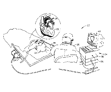
Fig. 1 is a schematic, pictorial illustration of a
system 20 for cardiac catheterization, in accordance with
an embodiment of the present invention. System 20 may be
based, for example, on the CART0' system, produced by
Biosense Webster Inc. (Diamond Bar, California). This
system comprises an invasive probe in the form of a
catheter 28 and a control console 34. In the embodiment
described hereinbelow, it is assumed that catheter 28 is
used in ablating endocardial tissue, as is known in the
art.
Alternatively, the catheter may be used, mutatis
mutandis, for other therapeutic and/or diagnostic
purposes in the heart or in other body organs.
An operator 26, such as a cardiologist, inserts
catheter 28 through the vascular system of a patient 24
6
CA 02772946 2012-03-30
so that a distal end 30 of the catheter enters a chamber
of the patient's heart 22. The operator
advances the
catheter so that the distal tip of the catheter engages
endocardial tissue at a desired location or locations.
Catheter 28 is typically connected by a suitable
connector at its proximal end to console 34. The console
may comprise a radio frequency (RF) generator, which
supplies high-frequency electrical energy via the
catheter for ablating tissue in the heart at the
locations engaged by the distal tip. Alternatively
or
additionally, the catheter and system may be configured
to perform other therapeutic and diagnostic procedures
that are known in the art.
Console 34 uses magnetic position sensing to
determine position coordinates of distal end 30 of
catheter 28 inside heart 22. For this purpose, a driver
circuit 38 in console 34 drives field generators 32 to
generate magnetic fields in the vicinity of the body of
patient 24. Typically,
the field generators comprise
coils, which are placed below the patient's torso at
known positions external to the patient. These coils
generate magnetic fields within the body in a predefined
working volume that contains heart 22. A magnetic field
sensor within distal end 30 of catheter 28 (shown in Fig.
3) generates electrical signals in response to these
magnetic fields. A signal processor 36 processes these
signals in order to determine the position coordinates of
the distal end, typically including both location and
orientation coordinates. This method of position sensing
is implemented in the above-mentioned CARTO system and is
7
described in detail in U.S. Patents 5,391,199, 6,690,963,
6,484,118, 6,239,724, 6,618,612 and 6,332,089, in PCT
Patent Publication WO 96/05768, and in U.S. Patent
Application Publications 2002/0065455 Al, 2003/0120150 Al
and 2004/0068178 Al.
Processor 36 typically comprises a general-purpose
computer, with suitable front end and interface circuits
for receiving signals from catheter 28 and controlling
the other components of console 34. The processor may be
programmed in software to carry out the functions that
are described herein. The software may be downloaded to
console 34 in electronic form, over a network, for
example, or it may be provided on tangible media, such as
optical, magnetic Or electronic memory media.
Alternatively, some or all of the functions of processor
36 may be carried out by dedicated or programmable
digital hardware components. Based on the
signals
received from the catheter and other components of system
20, processor 36 drives a display 42 to give operator 26
visual feedback regarding the position of distal end 30
in the patient's body, as well as regarding displacement
of the distal tip of the catheter, and status information
and guidance regarding the procedure that is in progress.
Alternatively or additionally, system 20 may
comprise an automated mechanism for maneuvering and
operating catheter 28 within the body of patient 24.
Such mechanisms are typically capable of controlling both
the longitudinal motion (advance/retract) of the catheter
and transverse motion (deflection/steering) of the distal
8
CA 2772946 2018-06-07
CA 02772946 2012-03-30
,
end of the catheter. Some mechanisms of this sort use DC
magnetic fields for this purpose, for example. In such
embodiments, processor 36 generates a control input for
controlling the motion of the catheter based on the
signals provided by the magnetic field sensor in the
catheter. These
signals are indicative of both the
position of the distal end of the catheter and of force
exerted on the distal end, as explained further
hereinbelow.
Fig. 2 is a schematic sectional view of a chamber of
a heart 22, showing distal end 30 of catheter 28 inside
the heart, in accordance with an embodiment of the
present invention. The
catheter comprises an insertion
tube 50, which is typically inserted into the heart
percutaneously through a blood vessel, such as the vena
cava or the aorta. An electrode 56 on a distal tip 52 of
the catheter engages endocardial tissue 58. Pressure
exerted by the distal tip against the endocardium deforms
the endocardial tissue locally, so that electrode 56
contacts the tissue over a relatively large area. In the
pictured example, the electrode engages the endocardium
at an angle, rather than head-on. Distal
tip 52
therefore bends at an elastic joint 54 relative to the
distal end of insertion tube 50 of the catheter. The
bend facilitates optimal contact between the electrode
and the endocardial tissue.
Because of the elastic quality of joint 54, the
angle of bending and the axial displacement of the joint
are proportional to the pressure exerted by tissue 58 on
distal tip 52 (or equivalently, the pressure exerted by
9
CA 02772946 2012-03-30
,
the distal tip on the tissue). Measurement of the bend
angle and axial displacement thus gives an indication of
this pressure. The pressure indication may be used by
the operator of catheter 20 is ensuring that the distal
tip is pressing against the endocardium firmly enough to
give the desired therapeutic or diagnostic result, but
not so hard as to cause undesired tissue damage.
Fig. 3 is a schematic, sectional view of distal end
30 of catheter 28, showing details of the structure of
the catheter in accordance with an embodiment of the
present invention. Insertion
tube 50 is connected to
distal tip 52 by joint 54, as noted above. The insertion
tube is covered by a flexible, insulating material 62,
such as Celcon , Teflon , or heat-resistant polyurethane,
for example. The area of joint 54 is covered, as well,
by a flexible, insulating material, which may be the same
as material 62 or may be specially adapted to permit
unimpeded bending and compression of the joint. (This
material is cut away in Fig. 3 in order to expose the
internal structure of the catheter.) Distal tip 52 may
be covered, at least in part, by electrode 56, which is
typically made of a conductive material, such as a
platinum/iridium alloy.
Alternatively, other suitable
materials may be used, as will be apparent to those
skilled in the art. Further
alternatively, for some
applications, the distal tip may be made without a
covering electrode. The
distal tip is typically
relatively rigid, by comparison with the flexible
insertion tube.
Joint 54 comprises a resilient coupling member 60.
In this embodiment, the coupling member has the form of a
tubular piece of an elastic material, with a helical cut
along a portion of its length. For example, the coupling
member may comprise a superelastic alloy, such as nickel
titanium (Nitinol). The helical cut causes the tubular
piece to behave like a spring in response to forces
exerted on distal tip 52. Further details regarding the
fabrication and characteristics of this sort of coupling
member are presented in U.S. Patent Application
12/134,592, filed June 6, 2008, which is assigned to the
assignee of the present patent application.
Alternatively, the coupling member may comprise a coil
spring or any other suitable sort of resilient component
with the desired flexibility and strength
characteristics.
The stiffness of coupling member 60 determines the
range of relative movement between tip 52 and insertion
tube 50 in response to forces exerted on the distal tip.
Such forces are encountered when the distal tip is
pressed against the endocardium during an ablation
procedure. The desired
pressure for good electrical
contact between the distal tip and the endocardium during
ablation is on the order of 20-30 grams. The coupling
member is configured to permit axial displacement (i.e.,
lateral movement along the axis of catheter 28) and
angular deflection of the distal tip in proportion to the
pressure on the tip. Measurement of the displacement and
deflection by processor 36 gives an indication of the
11
CA 2772946 2018-06-07
CA 02772946 2012-03-30
pressure and thus helps to ensure that the correct
pressure is applied during ablation.
A joint sensing assembly, comprising coils 64, 66,
68 and 70 within catheter 28, provides accurate reading
of the position of distal tip 52 relative to the distal
end of insertion tube 50, including axial displacement
and angular deflection. These coils
are one type of
magnetic transducer that may be used in embodiments of
the present invention. A "magnetic transducer," in the
context of the present patent application and in the
claims, means a device that generates a magnetic field in
response to an applied electrical current and/or outputs
an electrical signal in response to an applied magnetic
field. Although the
embodiments described herein use
coils as magnetic transducers, other types of magnetic
transducers may be used in alternative embodiments, as
will be apparent to those skilled in the art.
The coils in catheter 28 are divided between two
subassemblies on opposite sides of joint 54: One
subassembly comprises coil 64, which is driven by a
current via a cable 74 from console 34 to generate a
magnetic field. This field
is received by a second
subassembly, comprising coils 66, 68 and 70, which are
located in a section of the catheter that is spaced
axially apart from coil 64. (The term
"axial," as used
in the context of the present patent application and in
the claims, refers to the direction of the longitudinal
axis of distal end 30 of catheter 28, which is identified
as the Z-direction in Fig. 3. An axial plane is a plane
perpendicular to this longitudinal axis, and an axial
12
CA 02772946 2012-03-30
,
,
section is a portion of the catheter contained between
two axial planes.) Coils 66, 68 and 70 emit electrical
signals in response to the magnetic field generated by
coil 64.
These signals are conveyed by cable 74 to
processor 36, which processes the signals in order to
measure the axial displacement and angular deflection of
joint 54.
Coils 66, 68 and 70 are fixed in catheter 28 at
different radial or angular deflection locations.
(The
term "radial" or "angular" refers to coordinates relative
to the catheter axis, i.e., coordinates in an X-Y plane
in Fig. 3.)
Specifically, in this embodiment, coils 66,
68 and 70 are all located in the same axial plane at
different azimuthal angles about the catheter axis. For
example, the three coils may be spaced azimuthally 120
apart at the same radial distance from the axis.
The axes of coils 64, 66, 68 and 70 are parallel to
the catheter axis (and thus to one another, as long as
joint 54 is undeflected). Consequently, coils 66, 68 and
70 will output strong signals in response to the field
generated by coil 64, and the signals will vary strongly
with the distances of coils 66, 68 and 70 from coil 64.
(Alternatively, the axis of coil 64 and/or coils 66, 68
and 70 may be angled relative to the catheter axis, as
long as the coil axes have a sufficient parallel
component in order to give substantial signals.) Angular
deflection of tip 52 will give rise to a differential
change in the signals output by coils 66, 68 and 70,
depending on the direction and magnitude of deflection,
since one or two of these coils will move relatively
13
CA 02772946 2012-03-30
closer to coil 64. Compressive displacement of the tip
will give rise to an increase in the signals from all of
coils 66, 68 and 70.
Processor 36 analyzes the signals output by coils
66, 68 and 70 in order to measure the deflection and
displacement of joint 54. The sum of the changes in the
signals gives a measure of the compression, while the
difference of the changes gives the deflection. The
vector direction of the difference gives an indication of
the bend direction. A suitable calibration procedure may
be used to measure the precise dependence of the signals
on deflection and displacement of the joint.
Various other configurations of the coils in the
sensing subassemblies may also be used, in addition to
the configuration shown and described above. For
example, the positions of the subassemblies may be
reversed, so that that field generator coil is on the
proximal side of joint 54, and the sensor coils are in
the distal tip. As another alternative, coils 66, 68 and
70 may be driven as field generators (using time- and/or
frequency-multiplexing to distinguish the fields), while
coil 64 serves as the sensor. The sizes and numbers of
the coils in Fig. 3 are shown only by way of example, and
larger or smaller numbers of coils may similarly be used,
in various different positions, so long as one of the
subassemblies comprises at least two coils, in different
radial positions, to allow differential measurement of
joint deflection.
Prior calibration of the relation between pressure
on tip 52 and movement of joint 54 may be used by
14
CA 02772946 2012-03-30
processor 36 in translating the coil signals into terms
of pressure. By virtue of
the combined sensing of
displacement and deflection, this pressure sensing system
reads the pressure correctly regardless of whether the
electrode engages the endocardium head-on or at an angle.
The pressure reading is insensitive to temperature
variations and free of drift, unlike piezoelectric
sensors, for example. Because of the high sensitivity to
joint motion that is afforded by the arrangement of coils
64, 66, 68 and 70 that is shown in Fig. 3, processor 36
can measure small displacements and deflections with high
precision. Therefore,
coupling member 60 can be made
relatively stiff, and processor 36 will still be able to
sense and measure accurately the pressure on tip 52. The
stiffness of the coupling member makes it easier for the
operator to maneuver and control the catheter.
One or more of coils 64, 66, 68 and 70 may also be
used to output signals in response to the magnetic fields
generated by field generators 32, and thus serve as
position sensing coils. Processor 36
processes these
signals in order to determine the coordinates (position
and orientation) of distal end 30 in the external frame
of reference that is defined by the field generators.
Additionally or alternatively, one or more further coils
72 (or other magnetic sensors) may be deployed in the
distal end of the catheter for this purpose. The
position sensing coils in distal end 30 of catheter 28
enable console 34 to output both the location and
orientation of the catheter in the body and the
CA 02772946 2012-03-30
displacement and deflection of tip 52, as well as the
pressure on the tip.
Although the operation of a magnetic position
sensing assembly and its use in sensing pressure are
described above in the context of catheter-based
ablation, the principles of the present invention may
similarly be applied in other applications that require
accurate sensing of the movement of a joint, and
particularly in therapeutic and diagnostic applications
that use invasive probes, both in the heart and in other
organs of the body. As one
example, the devices and
techniques for position and pressure sensing that are
implemented in system 20 may be applied, mutatis
mutandis, in guiding and controlling the use of a
catheter insertion sheath. If the position of the sheath
is not properly controlled and excessive force is used in
its insertion, the sheath may perforate the heart wall or
vascular tissue. This
eventuality can be avoided by
sensing the position of and pressure on the distal tip of
the sheath. In this regard,
the term "distal tip" as
used herein should be understood to include any sort of
structure at the distal end of a probe that may be bent
and/or displaced relative to the main body of the probe.
As best illustrated in Fig. 4, a force F exerted on
the catheter tip 52 causes joint 54 (catheter spring) to
experience both axial compression and angular deflection.
As outlined above, both of these dimensions of movement
are taken into account for converting the tip position to
force F. Beyond a certain force limit, i.e. compression
threshold, however, the gaps between the windings of the
16
CA 02772946 2012-03-30
spring 54 close down, as illustrated in Fig. 4, and no
further compression is possible. Thus, the axial
compression limit for the spring 54 is achieved which is
measured at is about 30 grams for the spring design
illustrated in Figs. 3 and 4. Accordingly,
any
additional force F exerted on catheter tip 52 beyond the
given force limit can be expressed only in deflection (up
until now).
However, as best illustrated in Fig. 5, the
present invention is directed to a method of calibration
for the catheter 30 of Figs. 3 and 4. In addition to the
force calibration procedure described earlier, a further
calibration procedure (Fig. 5) is conducted in order to
enhance accuracy and force measurements for very large
(substantial) or extreme force F exerted on catheter tip
52. This additional calibration procedure of the present
invention schematically shown in Fig. 5 is directed
toward obtaining force measurements F after maximum axial
compression has already been achieved (Fig. 4).
Accordingly as part of the overall calibration for
the device or catheter 30 calibration is started in step
100 wherein force is applied to catheter tip 52 in step
105 wherein for various discrete force applications and
measurements, a corresponding axial compression and
angular deflection measurement is made and stored in
calibration memory as described earlier (step 110).
Step 120 is a logic step wherein the measured axial
compression is compared to the known/pre-established
compression threshold or limit, for example, about 30
grams for the catheter tip design shown in Figs. 3 and 4.
17
CA 02772946 2012-03-30
,
If the measured axial compression is below the
compression threshold, the force level F is increased in
step 105, for example at discreet force intervals, and
measurement and compare/logic steps of 110 and 120
respectively are repeated until the axial compression
level measured has achieved, i.e. equal to or greater
than, the compression threshold at step 120.
Once the axial compression threshold has been met in
step 120, additional force F (force greater than the
axial compression threshold limit as a new force value)
is applied to tip 52 (as shown in Fig. 4) in step 125,
for example at discreet force levels exceeding the
compression limit or threshold for spring 54, i.e. at
force levels greater than 30 grams in the embodiment of
Figs. 3 and 4.
For each discreet force application above the spring
axial compression threshold (in step 125), the position
coordinates of the tip 52 are measured in a plane
transverse to the direction of force F in step 130. Thus,
in the illustrated examples of Figs. 3 and 4, force F is
applied in X-Z axis direction and position coordinates
are measured for the X-Y transverse axis or plane in step
130. These position coordinates are six-dimensions of
location and orientation information, i.e. X,Y,Z axis
directions and pitch, yaw and roll orientations.
In step 135, the position coordinates measured in
the transverse plane, i.e. the X-Y transverse plane in
this example, are correlated directly to the new force
level or value F applied in step in 125 and stored in the
memory of system 34 (Fig. 1) in step 140.
18
CA 02772946 2012-03-30
A further logic step 140 is conducted wherein the
new force level F applied in step 125 is compared to a
pre-established upper limit for force F (by test design),
for example, the maximum limit above compression
threshold for force F tested. One example of the test
upper force limit is 60 grams of force, which is a
substantial amount of force to be exerted on a catheter
tip 52, especially when the axial compression threshold
for the spring 54 is about 30 grams in the example
provided. If the maximum force limit has not been
achieved, steps 125, 130, 135, 140 and 145 are repeated
until the upper limit (test limit) of force F has been
reached wherein the calibration is completed at step 150.
The present invention capitalizes on the discovery
that the force F on the catheter tip 52 is proportional
to the magnitude of the projection of the catheter tip 52
location in the plane transverse to the direction of
force (i.e., the X-Y plane in the coordinate system of
Fig. 3 and Fig. 4). Thus, by measuring the catheter tip
projection, i.e. measuring the position of tip 52 in the
transverse plane or transverse axial direction, based on
the signals output by the sensing coils 66, 68 and 70, it
is sufficient to give an accurate force reading when the
force F is above the spring compression threshold (in
this example about 30 grams).
Therefore, in calibrating the catheter 30, the
calibration model and parameters above the force
threshold F are correlated directly to a measurement of
the transverse axial projection of the tip (in this
example the transverse plane being the X-Y axis plane)
19
CA 02772946 2012-03-30
and computation of the proportionality and relationship
between the applied force F and the tip
projection/location (based on position coordinates).
Thus, during actual operation of the device/catheter 30
in a surgical procedure (Fig. 1), accurate force
measurements can be made based on the actual force being
exerted on the catheter tip 52 even after the spring
threshold has been achieved.
It will thus be appreciated that the embodiments
described above are cited by way of example, and that the
present invention is not limited to what has been
particularly shown and described hereinabove. Rather,
the scope of the present invention includes both
combinations and subcombinations of the various features
described hereinabove, as well as variations and
modifications thereof which would occur to persons
skilled in the art upon reading the foregoing description
and which are not disclosed in the prior art.