Note: Descriptions are shown in the official language in which they were submitted.
CA 02773425 2012-03-07
WO 2010/029360 PCT/GB2009/051161
1
Composition and Assay method for the detection of pathogenic bacteria
FIELD OF INVENTION
The invention relates to assay methods for use in detecting specific materials
derived from
microorganisms, particularly pathogenic microorganisms, in a test sample. The
invention further
relates to compositions and methods for the rapid growth of such
microorganisms enabling
detection of same significantly earlier than is currently possible.
BACKGROUND OF INVENTION
Because food products are biological in nature they are capable of supporting
the growth of a
variety of contaminating microorganisms. In the United States, an estimated 76
million cases of
foodborne illness occurs each year costing between $6.5 and $34.9 billion
dollars in medical
care and lost productivity (Buzby and Roberts, 1997; Mead et al, 1999). In
Europe it has been
estimated that the economic and health care costs of Salmonella are between
620 million and 3
billion Euro (David Byrne, European Commissioner for health and consumer
protection, 2000).
Salmonella, Listeria, Campylobacter, Escherichia coli 0157:H7 and Shigella are
responsible for
the majority of cases of foodborne illness. For example, Salmonella and
Listeria alone were
responsible for 31% and 28% respectively of food-related deaths (Mead et al,
1999) and in
Japan, salmonellosis accounted for over 14% of the total foodborne illness
outbreaks between
1981 and 1995 (Lee et al, 2001). In fact it has been estimated that bacteria
are the causative
agents of as much as 60% of the cases of foodborne illness requiring
hospitalisation. As a
result, one of the biggest contributors to waste is delay caused by
inefficient and slow testing of
products for microbial contamination. With current testing methods,
manufacturers must wait
from three to seven days for the results of microbial incubation. The costs
arising from such
delays are significant - reducing supply chain efficiency, tying up inventory
and increasing
spoilage.
The costs of inadequate or insufficient testing can be as, if not more,
costly. For example, in
1999, it cost Sara Lee an estimated $76 million in costs related to the recall
of 35 million pounds
of hot dogs and deli meats at its Bil Mar Foods unit, after the food was
linked to an outbreak of
L.isteria According to `The Scotsman', contamination of chocolate with
Salmonella in 2006 cost
Cadbury Schweppes an estimated 20 million in recall costs, advertising, lost
revenue and
subsequent improvements to its manufacturing operation. More recently in 2009,
the Peanut
CONFIRMATION COPY
CA 02773425 2012-03-07
WO 2010/029360 PCT/GB2009/051161
2
Corporation of America, a company with an estimated $25 million in sales in
2008, filed for
bankruptcy after being identified as the source of a major Salmonella outbreak
in peanuts in the
USA.
Therefore, detection of the presence of pathogenic microorganisms such as
Salmonella,
Shigella and Listeria in food, feed and environmental samples is of great
economic importance.
However, conventional culture methods for detection of such microorganisms are
both labour
intensive and time-consuming. Often such methods rely on standard processes
that have been
in use for more than 50 years.
In addition, pathogenic microorganisms can persist for long periods in an
environment in a
heavily stressed state known as `viable but not culturable (VNC)' or `not
immediately culturable
(NIC)'. Such heavily stressed microorganisms show only a weak metabolic
activity, often at the
limits of detection, and they lose the ability to form colonies on non-
selective plating media or to
grow in non-selective broth media (Reissbrodt et al, (2002). However, when
such nonculturable
colonies exist in food and animal feed, they may still be capable of causing
disease if ingested.
This poses particular problems with regard to detection since such stressed
microorganisms
may not be revived sufficiently to be detected.
As a result, additional cell culture steps are often included in any
diagnostic with the aim of
reviving such cells prior to further culture, plating and detection. Hence,
pre-enrichment in non-
selective culture media is an essential element of conventional methods
(Stephens et al, 2000).
For example, the detection of Salmonella requires several stages of culture
spread over as
many as five days; enrichment steps are often included in the analysis to
revive `sick' bacteria
and detection is often limited by the performance of such enrichment broths
and cultures.
Thus, for the recovery of microorganisms from clinical specimens, food and
other products that
potentially harbour a heterogenous population of bacteria, three general types
of culture media
are available: (1) non-selective media for primary isolation, (2) enrichment
broths and (3)
selective and/or differential agars.
The formulas for such media are generally complex and include ingredients that
not only inhibit
growth of certain bacterial species, i.e. they are selective, but also detect
several biochemical
CA 02773425 2012-03-07
WO 2010/029360 PCT/GB2009/051161
3
characteristics that are important in making a preliminary identification of
the micro-organisms
present in the specimen, i.e. they are differentiating. In order to make
rational selections,
microbiologists must know the composition of each formula and the purpose and
relative
concentration of each chemical compound included. Unfortunately the media
available are
often overly complex and the effect and amounts of the various components are
generally little
understood. Often the medium that is used is the same as that which has been
used for several
decades and may originally have been developed for an entirely different
organism. For
example, because of these inefficiencies, current detection rates of
Salmonella are less than
50% within 15 days and 90% within 28 days (King, 2009).
Hence, there is a need for culture media that are well defined, do not contain
surplus
ingredients that may have little to no or even negative effects and are
optimal for the growth and
rapid culture of even stressed microorganisms. Such culture media should
negate the need for
secondary/additional culture steps. There is also a need for new and better
detection methods
that enable the isolation and/or identification of pathogenic microorganisms
found in very low
numbers and in a heterogenous microflora environment. Further, any such
methods should be
equally applicable to detection of microorganisms from a wide variety of
sources such as
cosmetics, food products including frozen, lyophilised and liquid products,
clinical samples such
as urine, stool or blood samples and environmental samples.
SUMMARY OF THE INVENTION
In a first aspect of the invention there is provided a culture medium for the
growth of at least one
microorganism consisting essentially of:
(i) A base broth;
(ii) At least one growth inhibitor selected from the group consisting of
brilliant green,
nalidixic acid and lithium chloride; and
(iii) Optionally, at least one growth promoter selected from the group
consisting of sodium
tetrathionate, potassium tetrathionate, ammonium ferric citrate and sodium
citrate.
For the avoidance of doubt, the term `consisting essentially' as used herein
includes the
specified materials or steps only and additional components or elements to the
extent that they
do not materially affect the basic and novel characteristics of the invention.
CA 02773425 2012-03-07
WO 2010/029360 PCT/GB2009/051161
4
Media can be classified as simple, complex or defined. Base broths or basal
media are
basically simple media that support bacteria with minimal additional
components. Generally
such base broths simply need to provide a source of energy and maintain
correct osmolarity.
Peptone, tryptone, nutrient broth (peptone, meat extract, optionally yeast
extract and sodium
chloride), L-broth (tryptone, yeast extract and sodium chloride), gram
negative broth, tryptic soy
broth, tryptic soy broth with yeast and modified tryptic soy broth are
suitable base components
known in the art. Peptones are various water-soluble protein derivatives
obtained by partial
hydrolysis of a protein(s) by an acid or enzyme during digestion. Tryptic soy
broth generally
comprises tryptone (a pancreatic digest of casein), Soytone (a papaic digest
of soybean meal)
and sodium chloride, for example. Modified tryptic soy broth may further
comprise dextrose,
bile salts and dipotassium phosphate. Particularly the base broth is selected
from the group
consisting of tryptone, nutrient broth, L-broth, gram negative broth, peptone,
tryptic soy broth,
tryptic soy broth with yeast and modified tryptic soy broth. More particularly
the base broth is
selected from the group consisting of peptone, tryptic soy broth, tryptic soy
broth with yeast and
modified tryptic soy broth.
In particular embodiments the growth inhibitor is brilliant green, a
triarylmethane dye, (CAS
number 633-03-4).
Brilliant green is a dye known to inhibit Gram-positive bacteria and a
majority of Gram-negative
bacilli. It is used in varying amounts in the art, for example 25mg/L in
DifcoTM m Brilliant Green
Broth, 70mg/L in Brilliant Green Tetrathionate bile broth, 4.5-6mg/L in MLCB
agar and 10mg/L
in Muller Kauffmann tetrathionate broth. Despite being used for several
decades, the inventors
have now surprisingly discovered that such concentrations of brilliant green
are not optimal for
the growth of, for example Salmonella and Shigella. In fact such high levels
are believed to be
detrimental to the efficient and rapid growth of Salmonella and Shigella and
may also impede
the recovery of `sick' or `stressed' bacteria. Particular strains of
Salmonella such as Salmonella
typhi, Salmonella paratyphi amongst others are known as brilliant green
sensitive strains and
there are currently no suitable culture mediums which do not show a
differential inhibitory effect
between strains (Chau and Leung, 2008).
The inventors have now discovered a range of concentrations of brilliant green
that provide both
an inhibitory effect against, for example, gram-positive bacteria whilst
allowing the rapid
CA 02773425 2012-03-07
WO 2010/029360 PCT/GB2009/051161
recovery and growth of Salmonella (including S. typhi and S. paratyphi) and
Shigella. Thus, in
particular embodiments the culture medium comprises brilliant green in an
amount of between
about 0.05 to about 0.25mg/L or between about 0.1 mg/L to about 0.25mg/L, more
particularly
0.15mg/L.
5
These `low levels' are surprising in light of the levels seen in media already
known in the art. It
is believed that, due to the long, protracted culture methods known in the art
it has previously
been necessary to utilise high levels of brilliant green to inhibit the growth
of competing
microorganisms for the duration of culture which may be as long as 48 hours.
However, such is
the efficiency of growth in the media of the present invention that
microorganisms can be
cultured to suitable levels for detection in a single culture medium within 20
hours, particularly
about 4-15 hours, more particularly about 4-8 hours and yet more particularly
about 4-6 hours.
In other embodiments, and for example when used in surface swab testing this
may be reduced
further from between about 30 minutes to about 4 hours, particularly about 1,
1.5, 2, 2.5 or 3
hours. As a consequence it has been possible to utilise brilliant green at
surprisingly low levels
which still function to inhibit the growth of certain competing microorganisms
for up to 20 hours
but which are sufficiently low as to have no effect on growth of the
microorganism of interest,
such as Salmonella and/or Shigella for example.
Whilst amounts are generally referred to in mg/L or g/L it should be
understood that the
compositions may be provided pre-mixed in dry form, for example, as tablets,
powders,
granules or any other convenient dry form to be added to water separately or
sequentially. The
compositions may also be provided as separate components of a multi package
system, if
desired. In this case the amounts should be taken to refer to the final
concentration of a
component that would result once diluted with an appropriate volume of water.
For example, a
packet of dry powder containing 0.5mg of brilliant green for dilution in 2
litres of water would
have a resultant concentration of 0.25mg/L.
In other embodiments the medium contains nalidixic acid and/or lithium
chloride as growth
inhibitor(s).
Nalidixic acid (CAS number 389-08-2) is effective against both gram-positive
and gram-negative
bacteria. In lower concentrations, it acts in a bacteriostatic manner; that
is, it inhibits growth and
reproduction of bacteria. In higher concentrations, it is bactericidal,
meaning that it kills bacteria
CA 02773425 2012-03-07
WO 2010/029360 PCT/GB2009/051161
6
instead of merely inhibiting their growth. In particular embodiments the
medium contains
nalidixic acid in an amount of between about 1 to 3mg/L, more particularly
about 2mg/L.
Lithium chloride (CAS number 7447-41-8) inhibits the growth of gram-negative
bacteria without
affecting the growth of gram-positive bacteria. In particular embodiments the
medium contains
lithium chloride in an amount of between about 1 to 3g/L, more particularly
about 2g/L.
The use of nalidixic acid and/or lithium chloride as growth inhibitors is
beneficial in culture media
for the growth of Listeria spp.
In particular embodiments, the culture medium may optionally comprise a growth
promoter.
For Salmonella spp, when the base broth consists of peptone, it has been
discovered that the
inclusion of sodium tetrathionate, or salt thereof, is beneficial.
Surprisingly, the Inventors have
discovered that levels of sodium tetrathionate in a culture media of above
about 20g/L
significantly inhibit the growth of Salmonella spp. This is surprising because
levels above 20g/L
are routinely used in the art for positive selection and growth of Salmonella
spp. Thus,
preferably sodium tetrathionate is present in an amount of between about 1 to
about 20g/L,
more particularly about 4 to about 15g/L, about 6 to about 15g/L, yet more
particularly about 7
to 15g/L, about 8 to 12g/L or about 8g/L.
In alternative embodiments, in place of sodium tetrathionate suitable
quantities of sodium
thiosulphate and iodine may be used without departing from the spirit of the
invention. This is
because Iodine may react with sodium thiosulphate to produce sodium
tetrathionate (and
sodium iodide) in situ. In other embodiments potassium tetrathionate, barium
dithionate
dehydrate, salts thereof or compounds or mixtures of compounds that release
the tetrathionate
anion (54062-) may be utilised.
In other embodiments the culture medium comprises a growth promoter, wherein
the growth
promoter is ammonium ferric citrate.
CA 02773425 2012-03-07
WO 2010/029360 PCT/GB2009/051161
7
In particular embodiments, ammonium ferric citrate (CAS number 1185-57-5) is
used in an
amount of between about 200 to 1000mg/L, more particularly about 200 to about
500mg/L, yet
more particularly 200mg/L to about 300mg/L and still yet more particularly
about 250mg/L.
In yet another embodiment, the culture medium further comprises the growth
promoter sodium
citrate.
In particular embodiments, tri-sodium citrate (CAS number 68-04-2) is used in
an amount of
between about 10 to about 20g/L, about 12 to 18g/L and more particularly about
15g/L.
In particular embodiments, the culture medium is for the growth of Salmonella
spp. In other
embodiments, the culture medium is for the growth of Shigella spp. In yet
further embodiments
the culture medium is for the growth of Listeria spp.
According to a second aspect the invention provides a method of releasing the
core
oligosaccharide monomer from a cell of a microorganism comprising:
(i) adding a detergent to at least one culture sample containing said
microorganism to
provide a detergent-culture solution; and
(ii) heating the detergent-culture solution to a temperature sufficient to
release the core
oligosaccharide.
Bacterial lipopolysaccharides (LPS) are an essential component of all gram-
negative and some
gram-positive bacterial outer membranes. They are believed to be the principle
agents
responsible for inflammatory responses in patients infected with such
bacteria. Examples of
gram-negative bacteria include Escherichia coli, Salmonella, Shigella and
Campylobacter.
Listeria is a gram-positive bacterium.
Most of the characterised LPSs have the same principal structure; the
structure of the LPS has
been determined as consisting of three distinct regions: a lipid A region, a
core oligosaccharide
and an o-polysaccharide chain (Figure 12a). This structure is especially
conserved in the lipid A
and inner core parts of the LPS. Because of this structural conservation,
binding members,
CA 02773425 2012-03-07
WO 2010/029360 PCT/GB2009/051161
8
such as antibodies, to the lipid A region may not be specific to a particular
species leading to
false positives in any molecular detection steps. Further, the use of multiple
binding members
to, for example, the core region is unsatisfactory since such binding members
may compete for
the same epitope or, because of the close proximity of epitopes, may hinder
each other's
respective binding reaction. Thus, detection methods of the prior art have
relied on binding
members specific to the cell surface or flagellae of, for example, Salmonella,
since these are
easily accessible.
LPSs are generally isolated from bacteria by aqueous phenol extraction
followed by purification.
Isolated LPSs can then be characterised by, for example, SDS-PAGE, mass
spectrometry and
NMR (Raetz, 1996). The inventors have discovered that the core oligosaccharide
region may
be released or made accessible or available for detection, for example by
antibody binding
techniques, through use of a rapid method utilising a detergent and the
application of heat. Use
of such a simple methodology would not be suitable for detection of, for
example, cell surface
antigens or flagellae because detergents are known to interact with lipids and
would destroy or
disrupt lipid A epitopes with which binding members may react. Whilst
detergent alone could be
used, the use of heat is further advantageous since it breaks down the LPS
into detectable
monomers and has the added advantage of killing pathogenic bacteria.
Preferably the detergent is sodium dodecyl sulphate (SDS) or TWEEN 20, 40, 60
or 80.
Surprisingly the inventors have discovered that the use of SDS can enhance
binding between a
binding member, such as an antibody, and an epitope by as much as 10 fold in
the direct assay
described below. Similarly, whereas other detergents interfere with and
prevent antibody
binding in a direct assay (described below), surprisingly the inventors have
discovered that
TWEEN 20, 40, 60 or 80 has little or no such effect, for example, in a
competitive assay. This is
in direct contrast to the established teachings of the art, such as in
Qualtiere et al, 1977.
The detergent may be added to a culture sample as a liquid, for example,
dissolved in a solvent
such as water, or in the case of SDS as a solid. Particular detergent
concentrations for use in
the method are from about 0.1 % to about 2%, particularly about 0.5% to about
1 % (w/v or v/v).
CA 02773425 2012-03-07
WO 2010/029360 PCT/GB2009/051161
9
Preferably the detergent is dissolved or diluted in water and added as a
liquid resulting in
concentrations described above. Preferably the detergent solution is absent
further constituents
such as buffers and the like. Thus, in a preferred embodiment, the detergent
solution consists
essentially of the detergent, either sodium dodecyl sulphate or TWEEN 20, 40,
60 or 80,
dissolved in water.
In a next step of the method the detergent-culture solution is heated to a
temperature sufficient
to release the core oligosaccharide. Preferably the solution(s) is/are heated
to a temperature
sufficient to kill bacteria, particularly Salmonella, Shigella or Listeria,
that may be present in the
sample. Particular temperatures include from about 60 C to about 100 C,
particularly about 65,
70, 75, 80, 85, 90, 95 to about 100 C. It will also be apparent to one skilled
in the art that steps
(i) and (ii) may be carried out sequentially, at the same time, or. that the
culture sample and/or
detergent may be heated independently before being combined. The detergent-
culture solution
may be heated for about 30 seconds to about 20 minutes, particularly for about
2 minutes to
about 15 minutes, and more particularly for about 2, 3, 4, 5, 6, 7, 8, 9 or
about 10 minutes.
In a third aspect of the invention there is provided an assay method for
detecting the presence
or absence of a microorganism of interest in a test sample, the method
comprising:
(i) Culturing the test sample in a culture medium which allows for propagation
of the
microorganism of interest;
(ii) Treating the test sample sufficient to release one or more core
oligosaccharides from
any microorganisms present within the test sample;
(iii) Exposing the test sample to at least one binding member which has
binding specificity to
a core oligosaccharide of the microorganism of interest; and
(iv)Detecting any binding of the at least one binding member to a core
oligosaccharide of the
microorganism of interest.
The assay method may be direct or indirect. In a direct binding or non-
competitive assay (direct
or indirect), also referred to as a `sandwich assay', core oligosaccharides
are preferably bound
to a surface and a binding member, such as an antibody, is reacted with any
core
oligosaccharides of the microorganism of interest. Preferably the binding
member is a labelled
binding member. The amount of labelled binding member on the surface is then
measured.
CA 02773425 2012-03-07
WO 2010/029360 PCT/GB2009/051161
The results of the direct assay method are generally directly proportional to
the concentration of
core oligosaccharide in the sample. Clearly the labelled binding member will
not bind if the core
oligosaccharide is not present in the sample.
5 In a competitive assay, the core oligosaccharide in the test sample competes
with labelled core
oligosaccharide for binding to a binding member. The amount of labelled
binding member
bound to the core oligosaccharide is then measured. In this method, the
response will be
inversely proportional to the concentration of core oligosaccharide in the
sample. This is
because the greater the response, the less core oligosaccharide in the
`unknown' or test sample
10 was available to compete with the labelled core oligosaccharide.
Regardless of whether the assay is direct or indirect preferably either core
oligosaccharide or
labelled core oligosaccharide respectively is bound to a surface for
detection.
The surface to which the core oligosaccharide(s) are bound may be of a
material known in the
art, for example, organic polymers such as plastics, glasses, ceramics and the
like. Particular
organic polymers include polystyrene, polycarbonate, polypropylene,
polyethylene, cellulose
and nitrocellulose. A preferred polymer is polystyrene and more particularly
gamma-irradiated
polystyrene. The surface itself may be in the form, or part, of a sheet,
microplate or microtitre
plate, tray, membrane, well, pellet, rod, stick, tube, bead or the like.
In a particular embodiment LPSs or monomers comprising the core
oligosaccharide are
immobilised onto a surface without any modification. For example, the
hydrophobic lipid A
portion of the molecule may bind to a surface, such as a gamma-irradiated
polystyrene surface,
via non-covalent hydrophobic interactions. Such binding leaves the core
oligosaccharide region
accessible for interactions with binding members such as antibodies.
In alternative embodiments, the LPSs and/or core oligosaccharides are
immobilised onto a
surface through use of an intermediate binding member, such as an antibody,
conjugate or
other linkage. Suitable alternatives are disclosed in International patent
application publication
no. W003/36419.
CA 02773425 2012-03-07
WO 2010/029360 PCT/GB2009/051161
11
A first step of the method comprises culturing a test sample in a culture
medium which allows
for propagation of the microorganism of interest.
In certain embodiments, the method is used to detect microbial proteins or
fragments present in
food or a food product. In further embodiments, the sample is an environmental
sample, an
agricultural sample, a medical product, or a manufacturing sample. The test
sample may be a
food product such as meat, meat products including mince, eggs, cheese, milk,
vegetables,
chocolate, peanut butter and the like including processed, dried, frozen or
chilled food products.
Alternatively the test sample may be a clinical sample such as a biopsy
sample, faecal, saliva,
hydration fluid, nutrient fluid, blood, blood product, tissue extract,
vaccine, anaesthetic,
pharmacologically active agent, imaging agent or urine sample and the like.
The test sample
may also include swabs, such as skin-, coecum-, faecal, cloacal or rectal-
swabs or swabs of
surfaces, such as floors, doors and walls or swabs taken from food products
including animal
carcass swabs. The test sample may also include cosmetic samples such as
foundation
makeup, lip-balms, lotions, creams, shampoos and the like.
Preferably the test sample is cultured in a culture medium according to the
first aspect of the
invention.
In particular embodiments the test sample is cultured in a culture medium at
about 300C to
about 44 C, particularly about 37 C to 42 C, more particularly at about 37 C.
The test sample
may be cultured in a culture medium for about 4-15 hours, more particularly
about 4-8 hours
and yet more particularly about 4-6 hours. In other embodiments, the test
sample may be
cultured in a culture medium from between about 30 minutes to about 4 hours,
particularly
about 1, 1.5, 2, 2.5 or 3 hours.
A second step of the method comprises treating the test sample sufficient to
release one or
more core oligosaccharides from any microorganisms present within the test
sample.
The test sample may be treated in any way suitable to cause release of
bacterial LPSs and or
core oligosaccharide from the cell membrane of a microorganism. Preferably the
test sample is
treated according to the second aspect of the invention.
CA 02773425 2012-03-07
WO 2010/029360 PCT/GB2009/051161
12
Other suitable, although possibly less efficient, extraction methods exist in
the art and could
also be employed including sonication, use of a French press, use of enzymes,
`bead beating'
and the like. However, the use of detergent with high temperatures (such as
boiling or those
discussed above) is particularly useful when handling pathogenic bacteria such
as Salmonella
because high temperatures ensure that all of the bacteria have been killed.
More particularly,
when the assay is a direct binding assay SDS is preferably utilised whereas
when the assay is
in the competitive form , SDS is used to prepare the plate coating antigen
whilst either TWEEN
20, TWEEN 40, TWEEN 60 or TWEEN 80, particularly TWEEN 20, is employed
throughout the
rest of the procedure. Suitable heating/treatment time spans are provided in
relation to the first
aspect above. It will be apparent that the microorganism of interest may not
be present in the
test sample in which case LPSs and core oligosaccharides of the microorganism
of interest will
also not be present.
In a third step of the method, the test sample is exposed to at least one
binding member which
has binding specificity to a core oligosaccharide of the microorganism of
interest.
In particular embodiments the core oligosaccharides, LPSs or monomers within
the treated test
sample are immobilised to a surface prior to step (iii), being exposed to the
at least one binding
member which has binding specificity to a core oligosaccharide of the
microorganism of interest.
In such embodiments the core oligosaccharides, LPSs or monomers within the
sample may be
immobilised by bringing the treated test sample into contact with the surface
and incubating
and/or maintaining contact for about 10 minutes, 20 minutes, 30 minutes, 40
minutes, 50
minutes to about 60 minutes.
In other embodiments, for example a competitive assay, the test sample is
applied to or
contacted by a surface on which is already immobilised a known or standard
quantity of core
oligosaccharide, LPS or monomer. Core oligosaccharide, LPS or monomer from
both the
known or standard compete with core oligosaccharide, LPS or monomer from the
test sample
for binding to the at least one binding member.
Core oligosaccharides, LPSs or monomers may be directly immobilised to said
surface, for
example, by way of non-covalent hydrophobic interactions or indirectly as
described above.
CA 02773425 2012-03-07
WO 2010/029360 PCT/GB2009/051161
13
The test sample should be exposed to the at least one binding member for a
sufficient time to
allow for the core oligosaccharide, LPS or monomer to bind to the at least one
binding member
to form a complex, for example a core oligosaccharide/binding member complex.
Suitable
times include from about 1 minute to about 4 hours, particularly from about 30
minutes to about
2 hours, particularly about 45 minutes, 1 hour and 1.5 hours.
In certain embodiments, and in an optional step of the method, the complex is
exposed to a
secondary binding member which has binding specificity to the at least one
binding member for
a sufficient time to allow for the secondary binding member to form a
secondary complex, for
example a core oligosaccharide/binding member/secondary binding member
complex.
Preferably the binding member is an antibody, more particularly an affinity-
purified antibody and
yet more particularly a monoclonal antibody.
An antibody for use in the assay of the present invention may be a polyclonal,
monoclonal,
bispecific, humanised or chimeric antibody. Such antibodies may consist of a
single chain but
would preferably consist of at least a light chain or a heavy chain, but it
will be appreciated that
at least one complementarity determining region (CDR) is required in order to
bind a target such
as a core oligosaccharide or microbial contaminant to which the antibody has
binding specificity.
Methods of making antibodies are known in the art. For example, if polyclonal
antibodies are
desired, then a selected mammal, such as a mouse, rabbit, goat or horse may be
immunised
with the antigen of choice, such as bacterial endotoxin. The serum from the
immunised animal
is then collected and treated to obtain the antibody, for instance by
immunoaffinity
chromatography.
Monoclonal antibodies may be produced by methods known in the art, and are
generally
preferred. The general methodology for making monoclonal antibodies using
hybridoma
technology is well known (see, for example, Kohler, G. and Milstein, C, Nature
256: 495-497
(1975); Kozbor et al, Immunology Today 4: 72 (1983); Cole et al, 77-96 in
Monoclonal
Antibodies and Cancer Therapy, Alan R. Liss, Inc. (1985).
CA 02773425 2012-03-07
WO 2010/029360 PCT/GB2009/051161
14
An antibody, as referred to herein, should consist of an epitope-binding
region, such as CDR.
The antibody may of any suitable class, including IgE, IgM, IgD, IgA and, in
particular, IgG. The
various subclasses of these antibodies are also envisaged. As used herein, the
term "antibody
binding fragments" refers in particular to fragments of an antibody or
polypeptides derived from
an antibody which retain the binding specificity of the antibody. Such
fragments include, but are
not limited to antibody fragments, such as Fab, Fab', F(ab')2 and Fv, all of
which are capable of
binding to an epitope.
The term "antibody" also extends to any of the various natural and artificial
antibodies and
antibody-derived proteins which are available, and their derivatives, e.g.
including without
limitation polyclonal antibodies, monoclonal antibodies, chimeric antibodies,
humanized
antibodies, human antibodies, single-domain antibodies, whole antibodies,
antibody fragments
such as F(ab')2 and F(ab) fragments, Fv fragments (non-covalent heterodimers),
single-chain
antibodies such as single chain Fv molecules (scFv), minibodies, oligobodies,
dimeric or trimeric
antibody fragments or constructs, etc. The term "antibody" does not imply any
particular origin,
and includes antibodies obtained through non-conventional processes, such as
phage display.
Antibodies of the invention can be of any isotype (e.g. IgA, IgG, IgM i.e. an
a, y or heavy
chain) and may have a K (kappa) or a /\ (lambda) light chain.
The invention therefore extends to the use of antibodies and antibody derived
binding fragments
which have binding specificity to core oligosaccharides for use in the present
invention.
The term "specifically binds" or "binding specificity" refers to the ability
of an antibody or
fragment thereof to bind to a target microbial pathogen with a greater
affinity than it binds to a
non-target epitope. For example, the binding of an antibody to a target
epitope may result in a
binding affinity which is at least 10, 50, 100, 250, 500, or 1000 times
greater than the binding
affinity for a non-target epitope. In certain embodiments, binding affinity is
determined by an
affinity ELISA assay. In alternative embodiments, affinity is determined by a
BlAcore assay.
Alternatively, binding affinity may be determined by a kinetic method.
In certain embodiments, the binding member, such as an antibody, may be
immobilised on the
surface and after an optional washing step, the test sample, which may contain
the core
CA 02773425 2012-03-07
WO 2010/029360 PCT/GB2009/051161
oligosaccharide or microbial contaminant of interest can be exposed to the
surface-bound
antibody for a sufficient time for binding to take place and a surface bound
first binding member-
core complex to form. The assay may then involve a step of exposing the
surface bound first
binding member-core complex to a secondary binding member, such as an
antibody, which may
5 be covalently conjugated with means for light emission, for example, an
acridinium ester. In
such cases, the secondary binding member has binding specificity for an
epitope present on the
first binding member, or on the core oligosaccharide or microbial contaminant,
so that the
amount of signal generated corresponds to the amount of core oligosaccharide
or microbial
contaminant bound by the primary or secondary binding member.
Typically, an antibody is purified to prevent aggregation.
In certain embodiments the surface is, for example, a microtitre plate of
conventional design,
but an advantage can be gained by using a modified surface, for instance
having darkened side
walls and a white or transparent portion (e.g. on the base). This can
intensify any signal
generated and reduces the background light at the time of measurement. The
white portion
allows reflection of the light to intensify the generated signal. Thus, in
particular embodiments
the surface is a multi-well plate comprising a plurality of wells, wherein the
base of each well is
transparent or substantially transparent, while the walls of the wells are
opaque, or darkened to
prevent the passage of light, or coloured to provide a contrast against the
base portion of the
well which allows light to pass there through.
Yet more particularly the antibody is a species specific monoclonal antibody.
Use of the term `species specific' is intended to mean that such an antibody
will differentiate
between, for example, Salmonella, Shigella and Listeria with little or no
cross-reaction.
In particular embodiments, the binding member will interact with and bind to
the:
GIcNAc
1
a 1,2
GIcal -2 Galal
CA 02773425 2012-03-07
WO 2010/029360 PCT/GB2009/051161
16
epitope of the LPS core oligosaccharide. This epitope is species specific
differentiating
Salmonella from other bacteria such as, by way of non-limiting example,
Shigella, Listeria, E.
coli. In particular embodiments the assay method is a method for the
quantitative detection of
Salmonella. The assay method may also be utilised to detect for the presence
or absence of
Salmonella. In particular embodiments the binding member is a labelled binding
member
labelled by, for example, conjugation to a chemiluminescent or fluorescent
compound.
It will be apparent however, that the methods of the invention can be used for
identification and
quantitation of various target microbial contaminants. The assay methods of
the invention
involve analysis of samples for the presence or amount of a microbial
contaminant. It will be
understood that not all samples tested using the methods of the invention will
contain microbial
contaminants. In certain embodiments, the microbial contaminant is a protein
or protein
fragment derived from a pathogenic organism. In certain further embodiments,
the microbial
contaminant may be at least on of the group consisting of, but limited to: a
cell wall fragment, a
peptidoglycan, a glycoprotein, a lipoprotein, a glycolipoprotein, a small
peptide, a sugar
sequence and a lipid sequence. The methods of the present invention are
particularly suited for
detection of microbial proteins including structural proteins and/or toxins
derived from bacteria,
viruses and fungi.
A fourth step of the method comprises detecting any binding of the at least
one binding member
to a core oligosaccharide or microbial contaminant of the microorganism of
interest.
The detection method may be by any suitable method known in the art such as by
fluorescence
measurement, colourimetry, flow cytometry, chemiluminescence and the like. In
preferred
embodiments, detection of binding is by measurement/detection of a luminescent
signal, for
example, chemiluminescent light produced by a chemiluminescent compound.
Suitable
chemiluminescent compounds include acridinium esters, acridinium sulfonamides,
phenanthridiniums, 1,2-dioxetanes, luminol or enzymes that catalyse
chemiluminescent
substrates and the like.
In certain embodiments the binding member may be conjugated directly to a
light-emitting
moiety. In certain embodiments the binding member is conjugated to an
acridinium compound
or derivative thereof, such as an acridinium ester molecule or acridinium
sulphonamide which
acts as a luminescent label. In embodiments where the antibody or binding
fragment is
CA 02773425 2012-03-07
WO 2010/029360 PCT/GB2009/051161
17
conjoined to an acridinium ester or acridinium sulphonamide the assay method
may further
comprise the step of adding AMPPD to the test sample.
AMPPD may also be know by the synonyms: 3-(2'-spiroadamantane)-4-methoxy-4-(3"-
phosphoryloxy)phenyl-1,2-d i o x e t a n e ; 3-(4-methoxyspiro(1,2-dioxetane-
3,2'-
tricyclo(3.3.1.1(3,7))decan)-4-yl)phenyl phosphate; 4-methoxy-4-(3-
phosphatephenyl)spiro(1,2-
dioxetane)-3,2'-adamantane.
In certain further embodiments, the antibody may be indirectly associated with
a light- emitting
moiety, for example the acridinium ester molecule may be conjugated to a
second antibody
which is capable of binding to the first antibody. In certain embodiments, one
or more
luminescent or fluorescent moieties may be bound to avidin/streptavidin, which
in turn may be
bound to biotin chemically conjugated to an antibody. In certain further
embodiments, lectins
(Protein A/G/L) can be linked to a luminescent or fluorescent molecule which
may also be
attached to an antibody or other protein conjugate.
The stimulus to produce a detectable signal can be light, for example, of a
particular
wavelength, e.g. UV light, or may be some other stimulus such as an electrical
or radioactive
stimulus, a chemical or enzyme-substrate reaction.
Preferably the detection method should be capable of detecting/differentiating
1 colony forming
unit (cfu) of Salmonella, Shigella or Listeria in as many as 10,000cfu of
another microorganism
such as E. coli, for example, or per swab, starting sample, and the like.
Particular detection
limits are about 1000cfu, particularly about 500cfu, yet more particularly
from about 250cfu,
200cfu, 150cfu, 100cfu, 50cfu, 10cfu and about 1 cfu per unit of sample size
(mg, g and the like)
or volume (ml, L and the like). For liquid cultures a particular detection
limit is about 500cfu/ml.
In other embodiments the antibody may be indirectly associated with such a
light-emitting
moiety, for example, the acridinium ester molecule may be conjugated to a
second binding
member which is capable of binding to the first binding member.
CA 02773425 2012-03-07
WO 2010/029360 PCT/GB2009/051161
18
The assay methods may be qualitative or quantitative, and standard controls
can be run to
relate the average signal generated to a given quantity of, for example, core
oligosaccharide.
In certain embodiments, the method may be used for the determination in a
sample of a plurality
of core oligosaccharides or microbial contaminants, this being achieved by
providing a plurality
of binding members such as antibodies each of which having binding specificity
to a different
epitope or microbial contaminant. In certain embodiments, antibodies which are
bispecific may
be used.
It should be apparent that between or at each stage of the method, optional
washing, drying
and/or incubation steps may be included. The method may also optionally
include `blocking
steps' between one or more steps of the method wherein a concentrated solution
of a non-
interacting protein, such as bovine serum albumin (BSA) or casein, is added,
for example to all
wells of a microtitre plate. Particular blocking agents also include solutions
of milk powder and
the like. Such proteins block non-specific adsorption of other proteins to the
plate and may be
beneficial in reducing `background' artifacts which can interfere with the
sensitivity of the assay.
According to a fifth aspect of the invention there is provided the use of a
binding member which
has binding specificity to a core oligosaccharide for the specific detection
of a microorganism
selected from the group consisting of Salmonella, Shigella and Listeria.
According to a sixth aspect of the invention there is provided a kit for
carrying out the invention
according to the first, second, third, fourth and/or fifth aspect of the
invention. Such kits may
comprise culture media in liquid (ready-to-use or concentrated for dilution)
or dry (for example,
powder, granules, tablets, etc.) form, detergents or detergent solutions, wash
buffers, diluents,
pre-prepared plates, tubes or beads, one or more antibodies (i.e. primary,
secondary), detection
reagents, gloves, pipette tips, instruction manuals and the like. Wells of pre-
prepared plates or
tubes may be pre-coated with a known or standard amount of a core
oligosaccharide, LPSs or
monomer or a binding member such as an antibody. Such pre-prepared surfaces
may be
lyophilised.
BRIEF DESCRIPTION OF FIGURES
CA 02773425 2012-03-07
WO 2010/029360 PCT/GB2009/051161
19
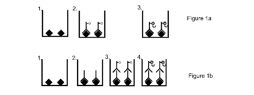
Figures 1(a) and (b) are schematics of a direct binding assay wherein =
represents the
bacterial core-oligosaccharide, LPSs or monomer, for example, of Salmonella.
Figure 1(a)
shows a direct immunoassay, Figure 1(b) shows an indirect immunoassay.
Figures 2(a) and (b) are schematics of a competitive binding assay wherein =
represents the
bacterial core-oligosaccha ride, LPSs or monomer, for example, of Salmonella.
Figure 2(a)
shows a direct competitive immunoassay, Figure 2(b) shows an indirect
competitive
immunoassay.
Figure 3 is a graph demonstrating the positive growth effect of tetrathionate
on Salmonella
whilst growth of other bacteria is inhibited.
Figure 4 is a graph demonstrating the effect of brilliant green on growth of
Salmonella, Shigella,
E. coli and staphylococcus. The graph exemplifies the optimum range of
concentrations of
brilliant green for growth of Salmonella with inhibition of competing
bacteria, particularly at
levels of 0.15mg/I brilliant green.
Figure 5 is a graph demonstrating the effect of ferric ammonium citrate on
growth of
Salmonella, Shigella, E. coli and staphylococcus. The graph exemplifies the
optimum range of
concentrations of ferric ammonium citrate for growth of Shigella particularly
at levels of 0.25g/l.
At levels above 0.25g/l, growth of Salmonella is unaffected.
Figure 6 is a graph demonstrating the effect of sodium citrate on growth of
Salmonella,
Shigella, E. coli and staphylococcus. Whilst growth of both Salmonella and
Shigella is
enhanced, growth of competing bacteria is inhibited.
Figure 7 is a graph demonstrating bacterial growth in Gram-Negative broth.
Figure 8 is a graph demonstrating bacterial growth in deoxycholate citrate
lactose sucrose
broth.
Figure 9 is a graph demonstrating bacterial growth in Peptone Broth.
Figure 10 is a graph demonstrating bacterial growth in modified Tryptic Soy
Broth. The growth
of both Salmonella and Shigella is enhanced demonstrating a doubling time of -
30 minutes.
Figure 11 is a graph demonstrating high growth of Listeria spp. With
inhibition of competing
bacteria in broths of the present invention.
Figure 12(a) illustrates the general structure of the LPS (0-antigen, core
polysaccharide
(oligosaccharide), lipid A) of certain bacteria of interest. Figure 12(b) is a
detailed illustration of
the Salmonella LPS monomer including the species specific antibody binding
epitope.
CA 02773425 2012-03-07
WO 2010/029360 PCT/GB2009/051161
DETAILED DESCRIPTION OF INVENTION
The assays of the present invention are preferably utilised to identify the
presence or absence
5 of core oligosaccharides of bacterial LPSs in a given sample. The assays of
the present
invention are capable of identifying samples containing, or contaminated, with
bacteria such as
Salmonella, Shigella or Listeria which have species-specific epitopes in the
core
oligosaccharide region of the LPS. The inventions may be better appreciated by
reference to
the following description and examples which are intended to be illustrative
of the methods of
10 the invention.
Figure la illustrates the steps of a direct binding assay utilising a labelled
primary antibody.
Figure 1 b illustrates the direct binding assay utilising unlabelled primary
antibody and a
secondary labelled antibody. A direct binding (direct or indirect antibody-
linked)
15 chemiluminescence-based immunosorbent assay for the detection of Salmonella
spp on animal
carcasses and in foodstuffs may be carried out as described below.
25g of a food sample were added to 225m1 culture medium according to the first
aspect of the
invention. Alternatively a surface swab may be taken from a 10x1 Ocm area on a
carcass and
20 cultured in 2-5m1 culture medium according to the first aspect of the
invention. Specifically the
culture medium comprised 1% peptone, 8g/L sodium tetrathionate and 0.15mg/L
brilliant green.
The sample was cultured for 5 hours at 37 C.
After 5 hours of culture, a 2m1 aliquot of the sample was removed and SDS was
added to a final
concentration of 0.5% (w/v). The sample was heated to 100 C for 5 minutes and
allowed to
cool. One hundred microlitres of each test sample was added directly to a well
of a solid white
96 well high binding microtitre plate (Greiner Bio One) and incubated at 37 C
for 30 minutes.
During incubation, the lipid A portion of the LPS binds to the surface of the
plate via non-
covalent hydrophobic interactions (Figure 1 a (1), Figure 1 b(1)). Following
incubation the plate
was emptied and the wells washed three times with a wash buffer comprising
0.01 M sodium
phosphate buffer, pH 7.4, containing 0.147M NaCl and 0.05 (v/v) Tween 20.
CA 02773425 2012-03-07
WO 2010/029360 PCT/GB2009/051161
21
One hundred microlitres of anti-Salmonella antibody conjugate, at a
concentration of 500ng/ml
in 0.01 M phosphate buffer, pH7.4, containing 0.147M NaCl, was added to each
well. The final
concentration of antibody per well was 50ng. The plate-bound sample
(Salmonella LPS/core
oligosaccharide) and antibody were incubated in the coated wells for 30
minutes as 37 C.
Following incubation, plates were washed three times in wash buffer and pat
dried prior to
detection (Figure 1 a(2), Figure 1 b(2)).
When the anti-Salmonella is directly labelled with acridinium ester, plates
were placed into a
luminometer. 30p1 of trigger solution A and 60p1 of trigger solution B was
added to each well of
the microtitre plate to initiate light output from conjugated acridinium ester
(Figure 1 a(3)). The
luminometer settings were as follows:
Delay Injection P (for solution A) - 1.6 seconds
Measurement Time Interval 1 - 0.0 seconds
Delay injection M (for solution B) - 0.0 seconds
Measurement Time Interval 2 - 1.0 seconds
Trigger solution A comprised: 63p1 70% (w/w) HNO3 and 165p1 30% (v/v) H202 in
a total
volume of 1 Oml distilled water. Trigger solution B comprises: 0.1 g NaOH and
75mg CTAC in 10
ml of distilled water.
Addition of goat anti-mouse IgG2b acridinium conjugate (if anti-Salmonella
monoclonal antibody
is unconiugated).
When the anti-Salmonella antibody is not labelled, a second binding member, a
goat anti-
mouse IgG2b conjugate is used. Post column IgG2b was diluted 1:100 in a
diluent comprising
3% (w/v) non-fat milk powder and 0.05% (v/v) Tween 20 and 100ul of this
solution was added to
each well of the plate (Figure 1 b(3)). Following incubation at 37 C for 60
minutes the plate was
washed four times in wash buffer, dried and read as above (Figure 1 b(4)).
A competitive (direct or indirect) chemiluminescence-linked immunosorbent
assay for the
detection of Salmonella spp in foodstuffs may be carried out as described
below. Salmonella
enteritidis LPS-coated microtitre plates were prepared as follows. S.
enteritidis was cultured in
a standard broth culture medium (2% (w/v) Buffered Peptone Water - Oxoid) not
according to
CA 02773425 2012-03-07
WO 2010/029360 PCT/GB2009/051161
22
the first aspect of the invention for 18 hours. The number of colony forming
units was quantified
and approximately 108cfu/ml were placed in a covered but unsealed
polypropylene boiling tube
containing NaEDTA and SDS to achieve final concentrations of 10mM and 0.5%
(w/v)
respectively. The culture was boiled at a temperature of 100 C for 2 minutes
thereby killing the
bacteria (and neutralising any biohazard associated) whilst also exposing the
bacterial LPS core
oligosaccharide or monomer epitope (see for example Figure 12b). The boiled
stock was
further diluted to a concentration of 106 cfu/ml by addition of a diluent
comprising 2% Buffered
Peptone Water (BPW).
One hundred microlitres of diluted boiled stock was added to each well of a
solid white 96 well
high binding microtitre plate (Greiner Bio One) and incubated at 37 C for 60
minutes. During
incubation, the lipid A portion of the LPS binds to the surface of the plate
via non-covalent
hydrophobic interactions. Following incubation the plate was emptied and the
wells washed
three times with a wash buffer comprising 0.01 M sodium phosphate buffer, pH
7.4, containing
0.147M NaCl and 0.05 (v/v) Tween 20. Washed coated plates were either used
immediately or
freeze-dried for storage (Figure 2a(1), Figure 2b(1)).
25g of a test sample of minced meat spiked with 10cfu of Salmonella was added
to 200m1 of
culture medium according to the first aspect of the invention. Specifically
the culture medium
comprised 1 % peptone, 8g/L sodium tetrathionate and 15mg/L brilliant green.
The sample was
cultured for 5 hours at 37 C. After 5 hours of culture, a 5m1 aliquot of the
sample was removed
and TWEEN 20 was added to a final concentration of 2% (v/v). The sample was
heated to
100 C for 2 minutes and allowed to cool. 80u1 aliquots of the boiled sample
were added to
each well of the coated microtitre plate.
Twenty microlitres of anti-Salmonella antibody conjugate at a concentration of
125ng/ml in
0.01 M phosphate buffer, pH7.4, containing 0.147M NaCl was added to each well
(Figure 2a(2),
Figure 2b(2)). The final concentration of antibody per well was 25ng/ml.
Competing sample
LPS/core oligosaccharide and antibody were incubated in the coated wells for
60 minutes as
37 C. Following incubation, plates were washed three times in wash buffer and
pat dried prior
to detection (Figure 2a(3), Figure 2b(3)).
When the anti-Salmonella is directly labelled with acridinium ester, plates
were placed into a
luminometer. 30p1 of trigger solution A and 60u1 of trigger solution B was
added to each well of
CA 02773425 2012-03-07
WO 2010/029360 PCT/GB2009/051161
23
the microtitre plate to initiate light output from conjugated acridinium ester
(Figure 2a(4)). The
luminometer settings were as follows:
Delay Injection P (for solution A) - 1.6 seconds
Measurement Time Interval 1 - 0.0 seconds
Delay injection M (for solution B) - 0.0 seconds
Measurement Time Interval 2 - 1.0 seconds
Trigger solution A comprised: 63u1 of 70% (w/w) nitric acid (HNO3) and 165u1
of 30% (v/v) H202
in a total volume of 10ml of distilled water. Trigger solution B comprises:
0.1g NaOH and 75mg
of CTAC in 10 ml of distilled water.
Addition of goat anti-mouse IgG2b acridinium conjugate (if anti-Salmonella
monoclonal antibody
is unconiugated)
When the anti-Salmonella is not labelled, a second binding member, a goat anti-
mouse IgG2b
conjugate is used. Post column IgG2b was diluted 1:100 in a diluent comprising
3% (w/v) non-
fat milk powder and 0.05% (v/v) Tween 20 and 100ul of this solution was added
to each well of
the plate (Figure 2b(4)). Following incubation at 37 C for 60 minutes the
plate was washed four
times in wash buffer, dried and read as above (Figure 2b(5)).
EXAMPLES
Example 1 - Preparation of culture media for growth of Salmonella
Figure 3 demonstrates the effect of sodium tetrathionate at concentrations of
between 0 and 16
g/L on the growth of Salmonella aberdeen, Shigella flexneri, Staphylococcus
aureus and E. coli.
0.1 ml inoculum (103 cells/m1) was added to a 100 ml conical flask containing
tryptic soy broth
with 0 to 16g/L of sodium tetrathionate. The flask was incubated at 37 C for
18 hours. After this
time, the A620 was measured. Each value represents the mean SD of three
separate
experiments. * shows p<0.05. At levels of between 2 to 16 g/L growth of
Shigella,
Staphylococcus and E. coli are inhibited in contrast to growth of Salmonella
which is un-affected
or promoted.
Concentration E. coli A620 Salmonella A620
(g/litre)
0 0.214 0.208 0.156 0.138
CA 02773425 2012-03-07
WO 2010/029360 PCT/GB2009/051161
24
4 0.096 0.104 0.187 0.179
8* 0.078 0.073 0.818 0.848
12 0.053 0.048 0.226 0.270
15 0.011 0.011 0.167 0.186
20 0.015 0.018 0.150 0.139
25 0.023 0.022 0.086 0.099
30 0.021 0.020 0.059 0.073
Not only does the Tetrathionate inhibit the growth of E. coli at levels of
>4g/litre but at a
concentration of 8g/litre it has a clear enhancing effect on the growth of
Salmonella. N.B. A620
measures turbidity and hence, the higher the value the higher the bacterial
growth. At levels
above 16g/L, growth of Salmonella is inhibited.
Figure 4 demonstrates the growth response of bacteria to brilliant green. 0.1
ml inoculum (103
cells/ml) was added to a 100 ml conical flask containing tryptic soy broth
with 0.05g to 5g/L of
brilliant green. The flask was incubated at 37 C for 18 hours. After this
time, the A620 was
measured. Each value represents the mean SD of three separate experiments. *
shows
p<0.05, **p<0.01.
Concentration E. coli A620 Salmonella A620
(mg/litre)
0 0.235 0.279 0.256 0.238
0.15 0.102 0.118 0255 ,&229
0.3 0.043 0.062 0.046 0.041
1 0.037 0.035 0.057 0.040
3 0.041 0.034 0.072 0.061
*5 0.250 0.230 0.282 0.256
*7 0.524 0.500 0.606 0.569
*10 0.624 0.665 0.614 0.607
A620 is employed as a measure of bacterial numbers by a turbidometric
method._*high
absorbance values due to absorbance of Brilliant Green; at these
concentrations the
spectrometer could not be blanked against the Brilliant Green solution. At
levels of Brilliant
CA 02773425 2012-03-07
WO 2010/029360 PCT/GB2009/051161
Green 0.3mg/L or higher the growth of both Salmonella and E. coli are limited
but at 0.1 5mg/L it
has an inhibitory effect on the E-coli but NOT the Salmonella.
Example 2 - Preparation of culture media for growth of Shigella
Figure 5 demonstrates the growth response of bacteria to ammonium ferric
citrate. 0.1 ml
5 inoculum (103 cells/ml) was added to a 100 ml conical flask containing
tryptic soy broth with
0.25 to 1.5 g/L of ammonium ferric citrate. The flask was incubated at 37 C
for 18 hours. After
this time, the A620 was measured. Each value represents the mean SD of three
separate
experiments. * shows p<0.05. At levels of ammonium ferric citrate of 0.25 g/L
or higher the
growth of both Staphylococcus and E. coli are limited.
Figure 6 demonstrates the growth response of bacteria to sodium citrate. 0.1
ml inoculum (103
cells/ml) was added to a 100 ml conical flask containing tryptic soy broth
with 5 to 25 g/L of
sodium citrate. The flask was incubated at 37 C for 18 hours. After this time,
the A620 was
measured. Each value represents the mean SD of three separate experiments. *
shows
p<0.05. At levels of sodium citrate of 5 g/L or higher the growth of both
Staphylococcus and E.
coli are limited. At levels of 15 g/L the growth response of Shigella is
significantly increased
over those of Staphylococcus and E. coli.
Example 3 - Generation study of different bacteria in peptone, tryptic soy
broth and
modified tryptic soy broth
Three strains of Shigella and other bacteria including Salmonella aberdeen, E.
coli and
Staphylococcus aureus were grown in conventional broth cultures to investigate
the generation
time. 0.1 ml inoculum (103 cells/ml) was added to a 100 ml conical flask
containing either
peptone (Figure 7), trypric soy broth (TSB) (Figure 8), modified tryptic soy
broth (mTSB) (Figure
9) or gram-negative broth (Figure 10).
Each flask was incubated at 37 C for 18 hours. After this time, the number of
viable cells was
determined by drop plate technique on nutrient agar. The values in parenthesis
are generation
times. Each value represents the mean SD of three separate experiments. *
shows p<0.05.
The doubling time was studied in peptone, tryptic soy and modified tryptic soy
broth. The
generation time of Shigella flexneri, Salmonella aberdeen, E. coli and
Staphylococcus aureus
was 36, 57, 41 and 44 min respectively when they were grown in Gram-negative
broth.
CA 02773425 2012-03-07
WO 2010/029360 PCT/GB2009/051161
26
The growth rate of all bacteria increased in TSB. As a result, TSB was used as
the basic
growth media in conjunction with other traditional selective agents, alone or
in combination to
selectively allow better growth of Shigella. The doubling time of Shigella
flexneri, Salmonella
aberdeen, E. coli and Staphylococcus aureus was 48, 46, 28 and 33 min in TSB.
Shigella
flexneri and Salmonella aberdeen grew significantly better (p<0.01) in the
mTSB, whereas, it
took longer for E. coli and Staphylococcus aureus to multiply in this base
broth. The growth rate
of E. coli could be delayed up to 68 min when grown in modified TSB.
Generation time of
Shigella flexneri could be shortened to 46 minutes in modified TSB.
Example 4 - Preparation of culture media for growth of Listeria
Listeria growth medium was prepared comprising a combination of lithium
chloride and Nalidixic
acid. 0.1 ml inoculum (103 cells/ml) was added to a 100 ml conical flask
containing according to
the following recipe: TSBYE - 3.3% tryptic soy broth with 0.5% yeast extract,
2g/l LiCI, 2mg/I
Nalidixic acid and 250mg/I ammonium ferric citrate. The flask was incubated at
37 C for 20
hours. After this time, the A620 was measured (Figure 11). Each value
represents the mean
SD of three separate experiments. L. monocytogenes and L. innocua were both
able to grow
efficiently in the media. The growth of E. coli. Lactobacillus acidophilus and
Erysipelothrix
rhusiopathiae were all significantly inhibited.
Example 5 - Preparation of culture media for the selective growth of
Salmonella, Shipella
or Listeria
Utilising the above data, selective culture media were prepared according to
the following
recipes:
Salmonella 1
2% peptone
0.15mg/I brilliant green
4-8g/l tetrathionate (All types)
Salmonella 2
3.3% (w/v) mTSB
0.15mg/I BG
1 g/l ammonium ferric citrate.
CA 02773425 2012-03-07
WO 2010/029360 PCT/GB2009/051161
27
Shigella 1
3.3% mTSB
0.1 mg/I Brilliant Green
250mg/I ammonium ferric citrate
15g/I trisodium citrate
Listeria 1
TSBYE - 3.3% tryptic soy broth with 0.5% yeast extract
2g/l LiCI
2mg/I Nalidixic acid
250mg/I ammonium ferric citrate
Example 6 - Preparation of antibodies and antibody fragments for conjugation
with
acridium ester for use in immunoassays
An improved preparation of antibodies can be produced by preparation of pure
IgG from ascites
by Protein A chromatography, followed by the optional step of cleaving the IgG
to give a Fab
fragment, and conjugation of the fragment or whole antibody to an ester and
subsequent
purification. Alternatively, other isotypes or isoforms of antibody can be
used unpurified.
Protein AIG separation
(i) Buffers and solutions
PBS: 0.1 M phosphate buffer, pH 8, containing 0.15M NaCl.
0.1 M citrate-acid buffers, pH 6, and pH 4.5: dissolve 29 g dry sodium citrate
in 800ml of distilled
water. Add 1 M citric acid solution (210 g/I) until a pH of 6 and 4.5,
respectively, is obtained.
Make up to 1 litre.
pH 3 buffer, 0.1 M acetic acid containing 0.15 M NaCl, to 800 ml of distilled
water, add 100 ml 1
M acetic acid and 100 ml 1.5 M NaCl.
CA 02773425 2012-03-07
WO 2010/029360 PCT/GB2009/051161
28
1.5 M glycine buffer, pH 8.9, containing 3M NaCl: dissolve 112g glycine and
174g NaCl in 700
ml of distilled water. Adjust pH to 8.9 with 5M sodium hydroxide solution and
make up to 1 litre
with distilled water.
(ii) Procedure
Allow 1.5g of Protein A-Sepharose (CL-4B, Pharmacia)(Protein G may also be
used) to swell in
0.1 M phosphate buffer, pH 8, for 30 minutes (1.5g of beads give 5ml of gel);
fill a 20x2cm
column and rinse with starting buffer. After dialysis against starting buffer,
load on to the
column 1 ml delipided ascites previously precipitated by ammonium sulphate at
40% (v/v)
saturation. Delipiding of ascites, if used, is carried out by centrifugation
at 100,000g for 45
minutes. Any pellet formed or floating 'lipid' is discarded. Wash the column
with 0.1 M
phosphate buffer, pH 8, until the A280 is <0.050. Add citrate buffer, pH6, and
wash until the A280
is <0.050. Elute the other immunoglobulins in the same manner by successively
employing the
pH4.5 and pH 3 buffers. Neutralise with phosphate buffer, pH 8, containing
0.02% sodium
azide, and store the Protein A-Sepharose in this buffer. After elution,
neutralise the antibody
with several drops of 1 M phosphate buffer, pH 8, and dialyse against PBS.
NO Preparation of Fab Fragments
Since enzymatic digestion never goes to completion, the action of papain on
IgG gives rise to
10% of undigested IgG in addition to the Fab and Fc fragments, which have the
same molecular
weight and are hence difficult to separate. Protein A is used to simplify
their purification. In the
first step, the antibody is treated with papain, and then the mixture is
passed over Protein A in
order to isolate the IgG and the Fab. The Fab fragments are then separated
from undigested
IgG by filtration on Sephadex or Protein A-Sepharose.
Materials
Chromatography column (2.5cm x 80cm).
Papain: Boehringer.
L-cysteine hydrochloride: Merck.
EDTA: ethylenediaminetetraacetic acid: Merck.
lodoacetamide: Merck
Buffers and solutions
CA 02773425 2012-03-07
WO 2010/029360 PCT/GB2009/051161
29
Phosphate-buffered saline - PBS.
0.1 M phosphate buffer, pH 7.4.
1 mg/ml papain solution prepared from a commercial stock solution.
0.2M L-cysteine: 35mg/ml of 0.1 M phosphate buffer, pH 7.4.
0.1 M EDTA: dissolved 3.6g EDTA in 100mi of 0.2M NaOH. Since EDTA dissolves
significantly
only at approximatelypH 8, it may be necessary to add a few drops of IM NaOH
in order to pH
the solution to obtain complete solubility of the EDTA.
0.4M iodoacetamide: 74mg/ml in 0.1 M phosphate buffer, pH 7.4.
Procedure
The immunoglobulin fraction was prepared from the antiserum by precipitation
with 40% (v/v)
saturated ammonium sulphate solution. The immunoglobulins were dialysed
against 0.1 M
phosphate buffer, pH 7.4. An approximate determination of the protein
concentration was made
(a 1 mg/ml solution of IgG gives an A280 of 1.4).
The concentration of the IgG was adjusted to 30mg/ml and the final volume (V)
required to give
a protein concentration of 20mg/ml was calculated. For affinity purified
antibodies, a final
concentration of 2.5mg/ml was used. A volume of V/20 of 0.04M EDTA (final
concentration:
0.002 M) was added. A volume of V/20 of 0.2M L-cysteine solution (final
concentration: 0.01 M)
was then added. A 1 mg/ml papain solution to give 1 mg of papain per 100mg
globulins was
added. The volume was adjusted to V ml with the 0.1 M phosphate buffer, pH
7.4. The
reaction was allowed to proceed for 2 hours at 37 C. A volume of V/10 of a
0.4M
iodoacetamide solution (final concentration: 0.04M) was added. This was left
for 30 minutes,
and then the preparation was dialysed against PBS overnight at +4 C.
IgG antibodies binding to the GIcNAc-Glc-Gal epitope (Figure 12) were isolated
and the Fab
fragments were isolated by Protein A chromatography. The mixture was
fractionated on a
column of Sephadex G-100 (2.5 x 80cm) and equilibrated with PBS. The first
peak
corresponded to IgG, and the second peak corresponded to the Fab fragments.
The Fab peak
was concentrated to 5mg/mi.
Example 7 - Conjugation of the antibody or antibody fragment with acridinium
ester
CA 02773425 2012-03-07
WO 2010/029360 PCT/GB2009/051161
a) Preparation
i) The acridindium ester e.g. (4-(2-succinimidyloxycarbonylethyl)phenyl-
10-dimethylacridinium-9-carboxylate fluorosulphate is weighed in a clean, dry
borosilicate vial.
Dry dimethyl formamide is added (volume depending on acridinium ester quantity
available) and
5 the solution aliquoted into vials at 5mg per vial normally.
ii) Antibody is dissolved in 0.2M sodium phosphate buffer, pH 8.0 at a
concentration of 0.5mg
IgG/ml.
iii) Add 5mg acridinium ester solution to 200ml antibody solution and mix
well.
iv) Incubate for 15 minutes at room temperature and then stop reaction by the
addition of 100ml
10 10% (w/v) lysine monohydrochloride followed by a further 5 minutes at room
temperature in the
dark.
v) Purify in accordance with (b) below.
b) Purification of Conjugate
15 (i) A gel filtration column may be used to purify the conjugate.
A column of 1.6 x 100 cm of Sephadex G200 (Pharmacia) is equilibrated with 0.1
M phosphate
buffer, pH 7.4, containing 0.147 M NaCl and 0.5% (w/v) bovine serum albumin
(Sigma). Up to
1.5ml of conjugate is placed on the column and separated for 18 hours at a
flow rate of 9ml/h.
The effluent is monitored at A280 and the peak corresponding to 45-55 K
daltons collected for a
20 Fab fragment or 140-170K daltons for a whole antibody - this is the
conjugate, which should be
diluted to a working strength before use.
(ii) The preferred alternative procedure employs the use of an FPLC. The
conjugate is purified
on a Pharmacia Superdex 200 HR 10/30 column. 50ml 0.007g/ml solution of bovine
serum
25 albumin (BSA) is added to the conjugate (to bring the BSA concentration of
the conjugate up to
that of the elution buffer).
Before applying the sample, the column is equilibrated with two column volumes
(50ml) of
elution buffer. The conjugate solution is then centrifuged at 10,000g for 10
minutes to remove
30 any particulate matter and applied to the FPLC column. The antibody is
eluted from the column
CA 02773425 2012-03-07
WO 2010/029360 PCT/GB2009/051161
31
in the elution, and storage buffer at a flow rate of 0.5m1/min. After the
first 5m1 has passed
through the column 0.5m1 fractions are collected.
The presence of antibody is detected though the use of an ultraviolet ( UV)
monitor and the
fractions spanning the antibody peak are collected and analyzed for
luminescent activity
(normally fractions 16-21).
Checking Luminescent Activity
The antibody fractions are diluted 1:500 in saline and 5 I samples of each
fraction spotted into
the wells of an assay plate. The fractions are then tested for luminescent
activity by reaction
with activating reagents 1 and 2. -15 I of activating reagent 1 is first added
to the sample well,
followed by 30 I of activating reagent 2. This is normally achieved by
automatic injectors in the
luminometer, which is then activated to read the light emission from the well
in question. The
results are recorded using a repeat for each sample. Samples containing high
levels of
luminescent activity can then be confirmed in a microbial assay, in this
example, a Salmonella
Assay.
Example 8 - AMPPD use with alkaline phosphatase-conjugated anti-salmonella
antibody
In order to generate a satisfactory luminescent signal, the antibody may be
conjugated to the
enzyme alkaline phosphatase and the substrate AMPPD employed in the
immunoassay
(AMPPD - 3-(2'-spiroad amantane)-4-methoxy-4-(3"-phosphoryloxy)phenyl-1,2-
dioxetane; 3-(4-
methoxyspiro(1,2-dioxetane-3,2'-tricyclo(3.3.1.1(3,7))decan)-4-yl)phenyl
phosphate). The
diluent for this substrate is 0.9g of CTAB (cetyltrimethyammonium bromide),
1.9m1 AMP (2-
amino-2-methyl-1-propanol), 14.5mg magnesium chloride.6H20, 1mM, pH 9.6, in
100ml distilled
water.
Reagents
Wash Buffer
0.2M Tris (24.228g/litre) 0.2M NaCl (11.688g/litre) + 0.05% (v/v) Tween
(0.5m1/litre).
Dissolve 24.228g of Tris and 11.688g of NaCl in 900m1 of dH2O. Add 0.5mis of
Tween 20.
Adjust the pH to 7.4 using HCI, make up to 1 litre and store at room
temperature.
CA 02773425 2012-03-07
WO 2010/029360 PCT/GB2009/051161
32
1 OX wash buffer concentrate (with preservative, sodium azide)
2M Tris (24.228g/IOOml) 2M NaCl (11.688g/IOOml) + 0.5% Tween (0.5mis/IOOml).
Dissolve
24.228g Tris and 11.688g of NaCl in 80mis distilled H20. Add 0.5m1 of Tween 20
and adjust the
pH to 7.4 with conc. HCI. Make up to 100ml with distilled H2O and store at
room temp. To
reconstitute the wash buffer, add 100ml of concentrate to 900m1 of dH2O and
store at room
temperature.
Elution and Storage Buffer
0.1 M sodium phosphate buffer pH 6.3 with 0.15M NaCl 0.1 % (w/v) bovine serum
albumin (BSA)
0.05% NaN3.
Make up a solution of O.1 M NaH2PO4 with 0.15M NaCl containing 0.1% w/v BSA
(A) and O.1 M
Na2HPO4 with 0.15M NaCl containing 0.1% w/v BSA (B). Add 100ml of A to 50m1 B.
Add
0.05% NaN3, filter through a 0.22mM filter and store at 4 C.
Assay Buffer
0.01 M NaH2PO4 (1.2g/litre) 0.15M NaCl (8.75g/litre) with 0.1% w/v NaN3 and
0.25% w/v BSA.
Dissolve 1.2g of NaH2PO4 and 8.75g of NaCl in 900m1 dH2O. Add 1.0g NaN3 and
2.5g BSA.
Allow to dissolve completely and adjust the pH to 7.4 with 1.OM NaOH. Make to
1 000ml with
distilled H20. Filter through a 0.22 M filter and store at 4 C.
Detergent Solution
20% (w/v) SDS solution: Dissolve 5g of SDS in 25m1 of dH2O. Store at room
temperature.
Growth Enhancer
8g sodium tetrathionate and 0.15mg Brilliant Green added to 1 litre of sterile
peptone broth. Mix
gently until evenly distributed.
Activating Reagent 1 (1 litre)
CA 02773425 2012-03-07
WO 2010/029360 PCT/GB2009/051161
33
6.3m1 of 70% nitric acid; 16.5m1 of 30% hydrogen peroxide; 977m1 of distilled
water.
Activating Reagent 2 (1 litre)
10.Og NaOH; 7.5m1 cetyltrimethyammoniumchloride; 983m1 distilled water
Testing fractions for Salmonella binding
The wells of an assay plate are coated with standard concentrations of
bacteria for 1 hour at
37 C. These standard concentrations are: 106, 105, 5 x 10⁴, 2.5 x 10⁴, 104 and
5 x 10³ and
blank wells containing 106 E. coli. The fractions to be tested are diluted
1:100 in assay buffer
and 50m1 is added to each well and incubated at 37 C for 20 minutes. The wells
are then read
on the luminometer, as above. Those fractions demonstrating good binding in
the assay are
pooled and the optimal dilution for the pooled conjugate determined - normally
1:100 to 1:1000.
Influence of Detergent on the Direct Binding Salmonella Assay
Novel black and white plate (Wallac) read on a tube luminometer (Berthold LB
9509) using the
detergent, sodium dodecyl sulphate (SDS)
1:50 dilution of conjugate
0% (w/v) SDS 0.5% (w/v) SDS
Peptone 1370+ 127 1198+ 112
E.coli 10 1039+ 59 958 +242
S.aberdeen 5 x10 1393+ 130 1622 + 21
10 1423+ 488 3199 +735
2.5 x 10 1347 +152 6697+ 1676
5x10 1582+333 12,231 +723
101, 2287 +248 22,245+529
10 16,860+ 131 59,070 1216
1:100 dilution of conjugate
0% SDS 0.5% SDS
Peptone 1006+ 203 974 +107
CA 02773425 2012-03-07
WO 2010/029360 PCT/GB2009/051161
34
E.coli 1110+ 127 922 + 78
S.aberdeen 5 x10 724 +24 1209 + 66
10 932 +231 1606+ 243
2.5 x 10 1094+ 110 3933+ 379
5x10 919+8 8721 +63
101, 1468+ 121 15,009+871
10 11,109+49 40,190+783
1:150 dilution of conjugate
0% SDS 0.5% SDS
Peptone 928+ 47 998+ 103
10 E.coli 659+ 10 707+ 11
S.aberdeen 5 x10 622 + 25 975+ 42
10 1104 + 22 1415 +132
2.5x10 1141+22 1615+132
5 x 10 1035+ 29 6576+ 549
101, 1927 +220 12,817+975
10 11,872 + 4303 31,571 + 2
Results expressed as mean standard deviation; n = 7
White Plate (Wallac), Read on Lucy I Plate Luminometer (1:100 conjugate
dilution)
5
0% (w/v) SDS 0.1% (w/v) SDS 0.2% (w/v) SDS
Peptone 4,143+ 107 4,288 + 1.653 3,904+59
10 E.coli 4,259+209 4,151 +256 3,322+479
10 S.aberdeen 35,532 56,046 356,444 102,877 365,496 + 12,729
10 S.aberdeen 79,334 + 1,248 143,796 + 4,297 112,096 + 3,841
5 x 10 38,834 + 2,624 51,909 + 3,036 75,900 + 5,798
2.5 x 10 17,891 + 3,422 28,622 + 2,162 27,339 + 2,635
CA 02773425 2012-03-07
WO 2010/029360 PCT/GB2009/051161
10 10,920+305 11,324 + 2,770 11,098+828
5 x 10 5,586+327 8,536 + 1,520 10,334 + 2,264
0.3% SDS 0.4% SDS 0.5% SDS
Peptone 3,026+473 3,499+569 3,103+267
10 E.coli 2,491 + 107 3,197+5 4,574 + 1,885
10 S.aberdeen 352,714 + 88,260 398,979 + 44,871 374,007 + 24,114
10 S.aberdeen 209,913 + 40,150 199,287 + 67,462 166,049 + 288
5 x 10 123,881 15,994 120,481 + 16,389 95,766 + 6,186
2.5 x 10 38,450 + 1,411 59,399 + 13,550 95,766 + 6,186
10 14,314+617 29,127 + 2,516 46,273 + 2,310
5 x 10 14,313 + 2,881 25,175 + 5,025 24,984 + 1,727
Effects of Various Levels of SDS on the Direct Binding Salmonella Assay (1:100
conjugate dilution) Using Black and White Plates
0% SDS 0.5% (w/v) SDS 0.1% SDS
Peptone 807 + 24 705+ 97 643 + 54
10 E.coli 764+ 399 684 + 88 579+ 37
10 S.aberdeen 5008+ 639 16,959+360 16,738 + 1051
10 S.aberdeen 867+ 100 2,259 + 103 2,386+263
5 x 10 897 +102 1,826+ 166 1,297+4
2.5 x 10 642 + 90 1094+ 156 1,277+211
10 927+ 266 796+ 23 1,662+531
5x10 891 +50 623+9 591 + 1
5
0% SDS 0.5% SDS 0.1% SDS
Peptone 643 + 54 631 + 38 503+ 76
10 E.coli 579+ 37 714 + 74 658+ 108
10 S.aberdeen 24,566 + 4,287 26,621 +843 28,169 + 1516
10 S.aberdeen 3,131 121 3,947+494 4,654+636
CA 02773425 2012-03-07
WO 2010/029360 PCT/GB2009/051161
36
x 10 2,027+206 2,213+ 170 2321+ 437
2.5 x 10 1,617+250 1,265+46 1,442+28
1,102+357 908+ 10 930 + 21
5 x 10 575+ 120 748 + 4 678 + 40
Effects of Various Levels of TWEEN 20 on the competitive binding Salmonella
Assay
(1:100 conjugate dilution) Using Black and White Plates
Competing 0.5% 0.5% 1%TWEEN, 1%TWEEN 2%TWEEN 2%TWEEN
S. TWEEN TWEEN 2min boil 20min boil 2min boil 20min boil
enteriditis 2min boil 20min boil
(cfu/ml)
10 4274 712 3531 154 4497 181 3216 130 6747 223 2074 42
101, 37803 2376 30413 2985 48390 2811 25614 1030 53067 701 6619 573
10 200745 12074 190752 6265 222178 3436 171024 2620 226168 24620 63938 4766
10 277066 20343 305744 27327 305343 15168 270782 15522 266820 24059 281305
20480
0 370245 16595 370245 16595 370245 16595 370245 16595 370245 16595 370245
16595
(Mean standard deviation, relative light units)
5
Effects of anti Salmonella mAB incubation times with 1:100 anti 2b conjugate
Monoclonal antibody (1:100 dilution) was incubated with either Salmonella or
Listeria to
determine optimum incubation times:
30 mins 40 mins 60 mins
10 S. aberdeen 598080 609383 593741
10 S. aberdeen 716821 644854 629340
10 S.aberdeen 276239 328483 371163
10 S.aberdeen 20117 19454 29194
10 S.aberdeen 5439 7204 17242
10 L.innocua 6376 8909 12151
(Read on Lucy I Plate Luminometer, results shown in relative light units)
10 Effects of anti Salmonella mAB incubation times with 1:200 anti 2b
conjugate
CA 02773425 2012-03-07
WO 2010/029360 PCT/GB2009/051161
37
Monoclonal antibody (1:200 dilution) was incubated with either Salmonella or
Listeria to
determine optimum incubation times:
30 mins 40 mins 60 mins
S. aberdeen 339224 340594 356500
10 S. aberdeen 344236 353391 372633
10 S. aberdeen 247657 243787 256889
10 S. aberdeen 14023 16848 22829
10 S. aberdeen 4869 6846 10817
10 L.innocua 11395 7775 11455
(Read on Lucy I Plate Luminometer, results shown in relative light units)
Effects of SDS Concentration on Food Cultures
5 White Plate read on Lucy 1 luminometer
- ve Chicken + ve Chicken*
0% SDS 1755 272 0% SDS 47,872 4509
0.1% SDS 3426+ 316 0.1% SDS 131,488 15,357
0.5% SDS 4494 904 0.5% SDS 36,770 2,020
- ve Mayonnaise + ve Mayonnaise*
0% SDS 1896 163 0% SDS 255,232 26,535
0.1% SDS 4180 610 0.1% SDS 746,670 86,449
0.5% SDS 3260 733 0.5% SDS 387,924 106,504
- ve Drinking + ve Drinking
Chocolate Chocolate
0% SDS 2047 +134 0% SDS 4051+ 136
0.1% SDS 1315 63 0.1% SDS 45,626 3204
0.5% SDS 3266 142 0.5% SDS 110,756 5737
*All +ve (bacteria positive) food cultures were contaminated with 10 C.F.U. of
S. aberdeen and
cultured for 18 hours (25g in 225m1) of Peptone broth + 8g/1 sodium
tetrathionate and 0.15mg/I
10 of Brilliant Green. The bacteria negative cultures (-ve) were contaminated
and also cultured for
CA 02773425 2012-03-07
WO 2010/029360 PCT/GB2009/051161
38
18 hours. 5m1 samples of all the food cultures (with or without SDS) were
heated for 20 minutes
in a boiling water bath prior to the assay.
Effect of different detergents on release and detection of core
oligosaccharide
CTAB CTAC TRITON TWEEN SODIUM 0.1% SDS 0.5% SDS NO
X -100 20 CHOLATE DETERGENT
BOILED
ONLY
866920 881712 455886 1025524 917476 895811 884352 36939
10 775871 792108 867745 573381 529553 717259 959119 19373
10 203215 205093 203602 16893 67710 65960 495603 9173
10 21541 22758 22570 9594 12088 12375 30399 7862
10 9746 12485 10494 8355 8921 9525 9987 9109
10 8901 10321 9981 8117 8858 10248 9918 9268
10 L.
innocua 7593 7892 7076 7226 7495 7863 8051 8454
5 (Mean of three separate experiments, relative light units)
SDS provides the most reliable and reproducible results for dissolution of
food sample-based
Salmonella LPS into monomers. However only the Tween can be used for this
purpose in the
competitive assay due to protein-detergent interactions with the other
detrgents.
10 Effects of varying anti-Salmonellaantibody levels on detection of
Salmonella in the
competive assay.
(cfu/ml) 50 ng/ml 25ng/ml 10 ng/ml 5ng/ml
106 9490 7828 8676 8198
105 23588 19974 14138 11554
104 91779 70822 32551 23628
103 149420 82361 42485 31153
102 151012 99182 55797 35750
101 151187 109799 71297 40671
No competing
bacteria 159874 125724 86091 45119
(mean of three separate experiments, relative light units)
CA 02773425 2012-03-07
WO 2010/029360 PCT/GB2009/051161
39
REFERENCES
Buzby, J.C. and Roberts, T. (1997) Economic costs and trade impacts of
microbial foodborne
illness. World Health Stat. Q. 50(1-2):57-66.
Chau, P.Y. and Leung, Y.K. (2008) Inhibitory action of various plating media
on the growth of
certain Salmonella serotypes. JApp. Microbiol. 45(3):341-345.
King, L. (2009) Salmonella Rapid detection interagency group meeting. FDA
executive
summary, 30th January 2009.
Lee, W.C., Lee, M.J., Kim, J.S. and Park, S.Y. (2001) Foodborne illness
outbreaks in Korea and
Japan studied retrospectively. J Food Prot 64:899-902.
Meade, P.S., Slutsker, L., Dietz, V., McCaig, L.F., Bresee, J.S., Shapiro, C.,
Griffin, P.M. and
Tauxe, R.V. (1999) Food related illness and death in the United States. Emerg.
Infect. Dis.
5:607-625.
Qualtiere, L.F., Anderson, A.G. and Meyers, P. (1977) Effects of ionic and non-
ionic detergents
on antigen-antibody reactions. J. Immunol. 119(5):1645-1651.
Raetz, C. R. H. (1996) Bacterial Iipopolysaccharides: a remarkable family of
bioactive
macroamphiphiles, in Escherichia coli and Salmonella, Vol. I (Neidhardt, F.,
ed.), Second
Edition, pp. 1035-1063, ASM Publications, Washington, DC.
Reissbrodt, R., Rienaecker, I., Romanova, J.M., Freestone, P.P.E., Haigh,
R.D., Lyte, M.,
Tschape, H. and Williams, P.H. (2002) Resuscitation of Salmonella enterica
serovar
Typhimurium and enterohemorrhagic Escherichia coli from the viable but
nonculturable state by
heat stable enterobacterial autoinducer. App. Env. Microbiol. 68(10):4788-
4794.
Stephens, P.J., Druggan, P., Nebe-von Caron, G. (2000) Stressed Salmonella are
exposed to
reactive oxygen species from two independent sources during recovery in
conventional culture
media. Int. J Food Microbiol. 60:269-285.