Note: Descriptions are shown in the official language in which they were submitted.
PROTEIN DETECTION VIA NANOREPORTERS
FIELD OF THE INVENTION
[0002] The present invention relates generally to field of protein detection,
quantification,
identification, and multiplex analysis using the tools of molecular biology to
generate unique
nanoreporter constructs and the methods for using them.
BACKGROUND OF THE INVENTION
[0003] With the recent completion of analysis of the human gcnome, much
attention is now
shifting to the field of proteomics, where gene products (proteins), their
variants, interacting
partners and the dynamics of their regulation and processing are the emphasis
of study. Such
studies are essential in understanding, for example, the mechanisms behind
genetic/and
environmentally induced disorders or the influences of drug mediated
therapies, as well as
potentially becoming the underlying foundation for further clinical and
diagnostic analyses.
Critical to these studies is the ability to qualitatively determine specific
variants of whole
proteins (e.g., splice variants, point mutations, post-translationally
modified versions, and
environmentally/therapeutically-induced modifications) and the ability to view
their quantitative
modulation. Moreover, it is becoming increasingly important to perform these
analyses from not
just one, but multiple biological fluids/extracts. There are limited methods
of multiplexed protein
measurement technologies due to the additional challenges inherent in protein
samples.
[0004] However, measurement of proteins in biological fluid is difficult due
to their inherent
properties. Accordingly, there is a pressing need for rapid, sensitive,
reproducible, and accurate
analytical approaches for the analysis of proteins and their variants.
[0005] In order to analyze proteins of interest from- and in- their native
environment, assays
capable of assessing proteins present in a variety of biological fluids and/or
extracts, both
qualitatively and quantitatively, are needed.
1
CA 2775613 2017-06-27
CA 02775613 2012-03-26
WO 2011/047087 PCT/US2010/052556
SUMMARY OF THE INVENTION
[0006] The invention provides method and compositions for analysis of
proteins. In some
embodiments, the invention provides methods and compositions for the detection
and/or
quantification of proteins in a sample. In some embodiments, the invention
provides methods
determining the concentration of at least one protein in a sample comprising
the steps of: (a)
providing: (i) at least one protein, (ii) a first protein probe specific for a
first region of said at
least one protein, where the first protein probe contains a capture region,
(iii) a second protein
probe specific for a second region of the at least one protein, where the
second protein probe
contains a nanoreporter comprising a plurality of different detectable labels,
and (iv) a matrix
having attached thereto a moiety which is capable of binding to the capture
region in the first
protein probe; (b) forming at least a complex comprising the at least one
protein, the first protein
probe, the second protein probe and the moiety, where the at least one protein
is bound to the
first and second protein probes, and where the moiety is bound to the capture
probe in the first
protein probe; and (c) individually detecting the complex or at least part of
the complex by a
method comprising individually counting the presence of one or more molecules
of the
nanoreporter where the presence of the one or more molecules is indicative of
the concentration
of the protein in the sample. In some embodiments, the individually detecting
further comprises
detecting a digital signal.
[0007] A moiety refers to and is also known as an entity. A moiety of the
invention is operably
linked to a matrix and binds with a capture region of a first protein probe.
The moiety is operably
linked to the matrix by a physical or chemical bond, including, but not
limited to, a covalent
bond, a non-covalent bond, an electron bond, a bent bond, an aromatic bond, a
metallic bond, a
hydrogen bond, an ionic bond, or van der Waals forces. The moiety binds with a
capture region
of a first protein probe through any of the physical or chemical bonds
described herein, receptor-
ligand interactions, hybridization events between two oligonucleotides, or
interactions between
an oligonucleotide and a polypeptide. For example, a capture region that
contains biotin binds to
a moiety containing streptavidin, forming a strong non-covalent bond, wherein
a matrix having
attached to the streptavidin, permits the matrix to bind to the capture region
of the first protein
probe (see, Figure 1). While all known receptor-ligand interactions are
contemplated, those
interactions with a dissociation constant (Kd) of between 0.1 fM and 1000 nM
are preferred.
Hybridization events occur between oligonucleotides having complementary
sequences,
2
CA 02775613 2012-03-26
WO 2011/047087 PCT/US2010/052556
however, perfect or complete complementarity is not required. The invention
encompasses those
hybridization events between oligonucleotides having 50%, 60%, 70%, 80%, 90%,
95%, 100%,
and any percentage complementarily in between. Furthermore, the association of
an aptamer
with a first protein probe provides a non-limiting example of a preferred
interaction between an
oligonucleotide and a polypeptide.
[0008] In some embodiments, the invention provides methods for determining the
concentration
of a plurality of target proteins by forming a plurality of complexes, each
complex comprising (i)
at least one target protein (ii) a first protein probe specific for a first
region of the at least one
protein, where the first protein probe comprises a capture region (iii) a
second protein probe
specific for a second region of the at least one protein, where the second
protein probe comprises
a nanoreporter comprising a plurality of different detectable labels and (iv)
a moiety attached to a
matrix, where the moiety is capable of binding to the capture region in the
first protein probe,
where each second protein probe comprises a different nanoreporter region. In
some
embodiments, each nanoreporter in the plurality of complexes has a detectable
signal that
distinguishes it from other nanoreporters in the population. In some
embodiments, the
dissociation constant of the first and the second protein probes is about
1.00x10⁻¹ to about
1.00x10-08. In some embodiments, the concentration of two or more target
proteins is
determined. In some embodiments, the concentration of 3, 4, 5, 10, 20, 30, 50,
100, 200, 300,
500, 600, 700, 800, 900, 1000 or more than 1000 different target proteins is
determined. In some
embodiments, the concentration of at least 972 different target proteins is
determined.
[0009] In some embodiments, the matrix is selected from the group consisting
of a bead and an
array. In some embodiments, the matrix is a bead. In some embodiments where a
plurality of
target proteins is analyzed, the matrix is a bead and each moiety in each
complex of the plurality
of complex is attached to a different bead. In some embodiments, the matrix is
a surface. In
some embodiments where a plurality of target proteins is analyzed, the matrix
is a surface and
each moiety in each complex of the plurality of complex is attached to a
different location of the
surface.
[00101 In some embodiments, the first protein probe and the second protein
probe are
independently selected from the group consisting of antibody, peptide, aptamer
and peptoid.
[0011] In some embodiments, the nanoreporter comprises a single-stranded
nucleic acid
backbone, the backbone comprising a plurality of label attachment regions
covalently attached
3
CA 02775613 2012-03-26
WO 2011/047087 PCT/US2010/052556
together in a linear combination, where each label attachment region is
hybridized to a
complementary polynucleotide sequence having attached thereto the detectable
label. In some
embodiments, the nanoreporter is attached to the second probe through
hybridization to a linker
oligo. In some embodiments, the nanoreporter is hybridized to the linker oligo
at a temperature
of about 32 degrees Celsius ( C) to about 40 C. In some embodiments, the
nanoreporter is
hybridized to the linker oligo at a temperature of about 37 C. In some
embodiments, the
nanorcportcr compriscs a portion that is complementary to the linker oligo. In
somc
embodiments, the complementary region is about 15 to about 20 bases.
[0012] In some embodiments, the invention provides methods for determining the
concentration
of at least one protein in a sample comprising the steps of: (a) providing:
(i) at least one protein,
(ii) a first protein probe specific for a first region of the at least one
protein, where the first
protein probe is attached to a first capture region or a first matrix, (iii) a
second protein probe
specific for a second region of the at least one protein, where the second
protein probe comprises
a signal oligo, and (iv) when the first probe is attached to a first capture
region: a second matrix
having attached thereto a moiety which is capable of binding to the capture
region in the first
protein probe; (b) forming at least a first complex comprising the at least
one protein, the first
protein probe, and the second protein probe, where the at least one protein is
bound to the first
and second protein probes, and where when the first probe is attached to a
first capture region the
capture probe is bound to the moiety in the second matrix; (c) releasing the
signal oligo from the
first complex; (d) forming a second complex comprising: (1) at least the
signal oligo and (2) at
least one oligo probe comprising a signal oligo-specific region and a region
comprising a
nanoreporter where the nanoreporter comprises a plurality of different
detectable labels; and (e)
individually detecting the second complex or at least part of the second
complex by a method
comprising individually counting the presence of one or more molecules of the
nanoreporter,
where the presence of the second one or more molecules is indicative of the
concentration of the
protein in the sample. In some embodiments, individually detecting further
comprises detecting
a digital signal.
[0013] In somc embodiments, thc first matrix is a bead or an array.
Preferably, thc first matrix is
a bead. In other embodiments, the second matrix is a bead or an array.
4
CA 02775613 2012-03-26
WO 2011/047087 PCT/US2010/052556
[0014] In some embodiments, the signal oligo is attached to a second capture
region. In some
embodiments, the releasing of the signal oligo further comprises capturing
directly or indirectly
the signal molecule into a third matrix.
[0015] In some embodiments, the nanoreporter further comprises a constant
region, where the
constant region comprises a plurality of repeat nucleotide sequences. In some
embodiments, the
constant region is bound to a second moiety in a third matrix, where the
second moiety is capable
of binding the constant region.
[0016] In some embodiments, the invention provides methods for determining the
concentration
of a plurality of target proteins by forming a plurality of complexes, each
complex comprising (i)
at least one target protein (ii) a first protein probe specific for a first
region of the at least one
protein, where the first protein probe is attached to a capture region or a
first matrix (iii) a second
protein probe specific for a second region of the at least one protein, where
the second protein
probe comprises a signal molecule, where when the first probe is attached to a
first capture
region the capture probe is bound to the moiety in the second matrix, and
where each second
protein probe in each the plurality of complexes comprises a different signal
oligo. In some
embodiments, the concentration of two or more target proteins is determined.
In some
embodiments, the concentration of 2, 3, 4, 5, 10, 20, 30, 50, 100, 200, 300,
500, 600, 700, 800,
900, 1000 or more than 1000 different target proteins is determined. In some
embodiments, the
concentration of at least 972 different target proteins is determined.
[0017] In some embodiments, the first matrix of the complex of the plurality
of complexes is a
bead and the bead comprises a plurality of identical first protein probes. The
term identical is
meant to describe a protein probe having the same sequence and either
containing or attaching to
the same capture region.
[0018] In some embodiments, the first protein probe and the second protein
probe are
independently selected from the group consisting of antibody, peptide, aptamer
and peptoid.
[0019] In some embodiments, the nanoreporter comprises a single-stranded
nucleic acid
backbone, the backbone comprising a plurality of label attachment regions
covalently attached
together in a linear combination, where each label attachment region is
hybridized to a
complementary polynucleotide sequence having attached thereto the detectable
label.
[0020] In some embodiments, the invention provides methods for determining the
concentration
of at least one protein in a sample comprising the steps of: (a) providing:
(i) at least one protein,
CA 02775613 2012-03-26
WO 2011/047087 PCT/US2010/052556
(ii) a first protein probe specific for a first region of the at least one
protein, where the first
protein probe is attached to a first oligo, and (iii) a second protein probe
specific for a second
region of the at least one protein, where the second protein probe is attached
to a second oligo;
(b) forming a first complex comprising the at least one protein, the first
protein probe and the
second protein probe, where the at least one protein is bound to the first and
second protein
probes; (c) ligating the first and the second oligo to form a signal oligo;
(d) forming a second
complex comprising: (1) the first signal oligo and (2) at least one oligo
probe comprising a signal
oligo-specific region and a region comprising a nanoreporter where the
nanoreporter comprises a
plurality of different detectable labels; and (e) individually detecting the
second complex or at
least part of the second complex by a method comprising individually counting
the presence of
one or more molecules of the nanoreporter, where the presence of the one or
more molecules is
indicative of the concentration of the protein in the sample. In some
embodiments, the
individually detecting further comprises detecting a digital signal.
[0021] In some embodiments, the signal oligo is released from the first
complex. In some
embodiments, the signal oligo comprises a capture region. In some embodiments,
the releasing
of the signal oligo further comprises capturing directly or indirectly the
signal oligo into a
matrix.
[0022] In some embodiments, the invention provides methods determining the
concentration of a
plurality of target proteins by forming a plurality of complexes, each complex
comprising (i) at
least one target protein, (ii) a first protein probe specific for a first
region of the at least one
protein, where the first protein probe is attached to a first oligo (iii) a
second protein probe
specific for a second region of the at least one protein, where the second
protein probe is attached
to a second oligo, where the ligation of the first oligo and the second oligo
form a signal oligo,
and where each complex in the plurality of complexes comprises a different
signal oligo. In
some embodiments, the concentration of two or more target proteins is
determined. In some
embodiments, the concentration of 2, 3, 4, 5, 10, 20, 30, 50, 100, 200, 300,
500, 600, 700, 800,
900, 1000 or more than 1000 different target proteins is determined. In some
embodiments, the
concentration of at least 972 different target proteins is determined.
[0023] In some embodiments, the first protein probe and the second protein
probe are
independently selected from the group consisting of antibody, peptide, aptamer
and peptoid.
6
[0024] In some embodiments, the nanoreporter comprises a single-stranded
nucleic acid
backbone, the backbone comprising a plurality of label attachment regions
covalently attached
together in a linear combination, where each label attachment region is
hybridized to a
complementary polynucleotide sequence having attached thereto the detectable
label.
[0025] In some embodiments, the invention provides a population of uniquely
labeled protein
probes, where each probe comprises: i) a target-specific region; and ii) a
region comprising a
nanoreporter comprising a plurality of different detectable molecules, where
the nanoreporter in
each protein probe has a detectable signal that distinguishes it from other
nanoreporters in the
population. In some embodiments, the target-specific region is selected from
the group
consisting of antibody, peptide, aptamer and peptoid.
[0026] In some embodiments, the nanoreporter comprises a single-stranded
nucleic acid
backbone, the backbone comprising a plurality of label attachment regions
covalently attached
together in a linear combination, where each label attachment region is
hybridized to a
complementary polynucleotide sequence having attached thereto the detectable
label. In some
embodiments, the dissociation constant of the target specific region is about
1.00x10⁻¹() to about
1.00x10-s.
[0027] In some embodiments, the nanoreporter is attached to the protein probe
through
hybridization to a linker oligo. In some embodiments, the nanoreporter is
hybridized to the
linker oligo at a temperature of about 32 C to about 40 C. In some
embodiments, the
nanoreporter is hybridized to the linker oligo at a temperature of about 37 C.
In some
embodiments, the nanoreporter comprises a portion that is complementary to the
linker oligo. In
some embodiments, the complementary region is about 15 to about 20 bases.
BRIEF DESCRIPTION OF THE DRAWINGS
[0029] The novel features of the invention are set forth with particularity in
the appended claims.
A better understanding of the features and advantages of the present invention
will be obtained
7
CA 2775613 2017-06-27
CA 02775613 2012-03-26
WO 2011/047087 PCT/US2010/052556
by reference to the following detailed description that sets forth
illustrative embodiments, in
which the principles of the invention are utilized, and the accompanying
drawings of which:
[0030] Figure 1 is a schematic diagram depicting one embodiment of the
invention in which two
antibodies specific for a target protein bind to the target protein in
solution. The first antibody is
attached to an affinity tag such as biotin (depicted as a circle labeled "W.),
while the second
antibody is attached to a partially double stranded nucleic acid probe. The
binding of the first
and second antibodies to the target protein forms a complex that is isolated
from the solution via
the affinity tag of the first antibody. One of the strands of the partially
double stranded nucleic
acid probe can be eluted to generate a signal oligo, which can then be
analyzed by any the
methods described herein.
[0031] Figure 2 is a graph depicting the results of a detection assay using
the IL-2 target protein
at different concentrations. Specifically, the detection of IL-2 within
solutions containing no
blocker, milk at 0.03%, bovine serum albumin (BSA) at 0.1%, or salmon sperm
(SS) at 98 ng/ml,
was measured as the total counts detected as a function of increasing IL-2
target protein molar
concentration ([IL2 target, MD.
[0032] Figure 3 is a graph depicting the efficiency of IL-2 detection in the
assay used in Figure
2. Total counts detected were normalized to 600 molecules per field of view
(FOY) and
expressed as a function of increasing concentration of IL2 target protein
molecules ([IL2 target,
molecules]). The efficiency of detection is the slope of the line depicted in
this graph.
[0033] Figure 4 is a schematic diagram depicting two alternate embodiments of
the invention
for solution tripartite binding. According to this method, two antibodies
specific for a target
protein bind to that target protein in solution. The first antibody is
attached to an affinity tag
such as biotin and contains a constant region, which, for example, contains F
repeats. The second
antibody is attached to a nanoreporter probe and a second constant region,
which, for example,
contains, G repeats. The binding of the first and second antibodies to the
target protein forms a
complex that can be isolated from the solution via the affinity tag of the
first antibody. "Normal"
elution of the complex is accomplished by melting off the G and F bead.
"Alternative" elution of
the complex is accomplished via digestion. The label monomers of the
nanoreporter (depicted as
circles) emit individual signals of qualitatively different wavelengths that
are spatially-
distinguishable and are, from left to right positions, red (R), yellow (Y),
green (G), blue (B), red
(R), and violet (V).
8
CA 02775613 2012-03-26
WO 2011/047087 PCT/US2010/052556
[0034] Figure 5 is a graph depicting the dissociation constant (Kd) and probe
concentration for
one of the embodiments of the invention, expressed as the fraction of target
bound versus the Kd
of the nanoreporter probe and the protein probe.
[0035] Figure 6 is a schematic diagram depicting an embodiment of the
invention in which a
capture antibody specific for a target protein binds to the target protein in
solution to form a
complex. The complex can then be isolated from the solution. The complex is
then contacted
with a second antibody, where the second antibody is attached to a partially
double stranded
nucleic acid probe. One of the strands of the partially double stranded
nucleic acid probe can be
eluted to generate a signal oligo that may be analyzed by any the methods
described herein.
[0036] Figures 7A and 7B are schematic diagrams depicting an embodiment of the
invention in
which two antibodies specific for a target protein bind to the target protein
in solution. The first
antibody is a capture antibody, while the second antibody is attached to a
partially double
stranded nucleic acid probe, where one of the strands in the probe is attached
to an affinity tag
such as biotin. The binding of the first and second antibodies to the target
protein formed a
complex that can be isolated from the solution via the capture antibody. One
of the strands of
the partially double stranded nucleic acid probe can be eluted to generate a
signal oligo
containing the affinity tag. The signal oligo can then be hybridized to a
nanoreporter to form a
nanoreporter-signal oligo complex that can be isolated and/or immobilized into
a solid surface.
The nanoreporter-signal oligo complex can be analyzed by any the methods
described herein.
The label monomers of the nanoreporter (depicted as circles) emit individual
signals of
qualitatively different wavelengths that are spatially-distinguishable and
are, from left to right
positions, red (R), yellow (Y), green (G), blue (B), red (R), and violet (V).
[0037] Figure 8A is a schematic diagram depicting certain embodiments of the
invention using
proximity ligation. A first and a second oligo are attached to a first and a
second antibody,
respectively, both antibodies being specific for a target protein. The first
and second antibodies
bind to the target protein, bringing the first and second oligo to close
proximity. A bridging
oligo and a ligase are added to the solution to connect the first and second
oligo to generate a
signal oligo. The signal oligo can then be analyzed by any the methods
described herein.
[0038] Figures 8B-D are schematic diagrams depicting methods by which the
signal oligo
shown in Figure 8A may be released and purified.
9
CA 02775613 2012-03-26
WO 2011/047087 PCT/US2010/052556
[0039] Figure 9 is a schematic diagram depicting certain embodiments of the
invention using
proximity ligation. The label monomers of the nanoreporter (depicted as
circles) emit individual
signals of qualitatively different wavelengths that are spatially-
distinguishable and are, from left
to right positions, red (R), yellow (Y), green (G), blue (B), red (R), and
violet (V).
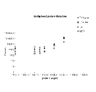
[0040] Figure 10 is a graph depicting the results of multiplexed protein
detection of TNFalpha,
ILl alpha, IL6, and VEGF, measured as total counts detected as a function of
increasing protein
target concentration ([protein target]). In this example, a sandwich detection
assay was used in
solution. A 4-plex measurement is shown.
[0041] Figure 11 is a graph depicting the data analyzed in Figure 10, plotted
by lane instead of
by concentration. Specifically, this figure demonstrates that two target
proteins were titrated in
whereas two other protein targets were titrated out.
[0042] Figure 12 is a graph depicting the results of a limit of detection
(LOD) experiment using
two protein targets, ILlalpha and IL6. Total counts detected were plotted as a
function of
increasing molar concentration of the target protein ([target] molar). The
experiment
demonstrated that the limits of detection were 26 and 38 picograms per
milliliter (pg/m1),
corresponding to 1.4x10⁻¹²M and 1.9x10⁻¹²M for ILlalpha (ILla) and IL6,
respectively. The
limit of detection was two standard deviations above background detection
levels. Six negative
controls were performed, resulting in average counts of plus or minus one
standard deviation, i.e.
3196 265 and 6703 585, respectively.
[0043] Figure 13 is a graph depicting the total counts retained by various
components of the
antibody reporter complex (PROX01, PROX03, PROX04, PROX05, and PROX06)
following
purification and a rinsing step using either water or SSPE buffer of various
fold concentrations
(0.01X, 0.03X, or 0.1X). At 0.03X SSPE the oligo representing a ligated
product, PROX05, was
retained.
[0044] Figure 14 is a graph showing the counts per field of view (FOY) for
each antibody probe
on a reporter that bound, stretched, and immobilized (S17, S8, S22, S14, S23,
S6, S13, S7, S18,
S9, S10, S11, S12, S15, S16, S19, S20, and S21). Counts are shown only of the
reporter with the
antibody probe that is bound to the surface.
DETAILED DESCRIPTION OF THE INVENTION
[0045] Reference will now be made in detail to particularly preferred
embodiments of the
invention. Examples of the preferred embodiments are illustrated in the
following Examples
section.
[0046] Unless defined otherwise, all technical and scientific terms used
herein have the same
meaning as is commonly understood by one of skill in the art to which this
invention belongs.
[0047] The present invention provides compositions and methods for detection
and
quantification of individual target molecules in biomolecular samples. In
particular, the
invention provides protein probes that are capable of binding individual
target molecules. The
invention also provides the use of nanoreporters. Through nanoreporters' label
codes, the
binding of the protein probes to target molecules results in the
identification of the target
molecules. Methods of making and using such protein probes and/or
nanoreporters are also
provided. The methods and compositions described herein can be used in a wide
variety of
applications such as diagnostic, prognostic, quality control and screening
applications.
[0048] Certain aspects of the invention relate to the detection of multiple
target molecules.
Multiplexing is the measurement of more than one target molecule within a
sample without
having to split the sample. The methods described herein provide potential
benefits in the areas
of multiplexing, quantification, and sensitivity. For example, in some
embodiments the target
molecule is a protein. Measurement of protein concentrations is challenging.
Proteins are sticky
and tend to aggregate. In addition proteins are unstable, and tend to unfold
easier than RNA or
DNA. Extremes in pH, temperatures, solute concentration, and the presence of
denaturants are
conditions that can interrupt protein stability and complicate measurement. In
some
embodiments, the invention provides methods and compositions for multiplexed
protein
measurements that are sensitive and reliable.
[0049] Multiplexing within a fluid sample is a key advantage of this approach.
Multiplexing
within one sample saves significant labor, reduces sample quantity
requirements proportional to
the number of measurements, and improves accuracy by elimination of errors
compounded by
separate sample handling and measurement steps. In some embodiments, the
methods described
herein allow for the pooling of different samples together during processing
to be analyzed at
11
CA 2775613 2017-06-27
CA 02775613 2012-03-26
WO 2011/047087 PCT/US2010/052556
once. This offers throughput advantages and can accelerate the analysis of
different samples,
e.g., up to eight times.
[0050] In some embodiments, the invention provides protein probes for the
analysis of target
molecules. In some embodiments, the invention provides a protein probe
population for use in a
multiplexed assay. Each protein probe in the population is specific for a
target molecule. The
binding of the target molecules to the proteins probes is then detected using
nanoreporters. Each
nanorcportcr comprises a unique label code that can be associated to a
specific target molecule.
[0051] In some embodiments, the nanoreporters are attached, directly or
indirectly, to the protein
probes. A unique nanoreporter's label code is then assigned to a specific
protein probe such that
each nanoreporter's label code can be associated to the target molecule bound
to the protein
probe.
[0052] In other embodiments, the protein probes are attached, directly or
indirectly, to a signal
oligo. Each protein probe is attached to a unique signal oligo. The
nanoreporters used for the
analysis of the signal oligo comprise a portion that is complementary to the
signal oligo. A
unique nanoreporter's label code is assigned to a specific signal oligo
sequence such that each
nanoreporter's label code can be associated to the target molecule via the
signal oligo sequence.
[0053] In other aspect of the invention, the invention provides methods for
detecting target
molecules by measuring signals digitally. Current technologies use analogue
fluorescent signals
to quantify the presence of target molecules. Quantification using
fluorescence can be error
prone for a variety of reasons. For example, fluorophores can photobleach.
There can be
changes in the spectra in the presence of proteins or due to local
environment, e.g., pH, salt. In
addition, the light sources can vary in intensity over time. For example, arc
lamps, a commonly
used light source, demonstrate a phenomenon called arc wander that can cause
significantly
different illumination levels over time. In embodiments of the invention, the
target molecules
are detected digitally. While fluorescence might be used to read the
nanoreporter's label code,
the signals are high and the spot is either present of not, thus the digital
detection. The digital
detection of target molecules leads to more accurate quantification.
Protein Probes
[0054] Protein probes are molecules or assemblies that are designed to bind
with at least one
target protein, at least one target protein surrogate, or both; and can, under
appropriate
12
CA 02775613 2012-03-26
WO 2011/047087 PCT/US2010/052556
conditions, form a molecular complex comprising the protein probe and the
target protein. The
terms "protein", "polypeptide", "peptide", and "amino acid sequence" are used
interchangeably
herein to refer to polymers of amino acids of any length. The polymer may be
linear or
branched, it may comprise modified amino acids, and it may be interrupted by
non amino acids
or synthetic amino acids. The terms also encompass an amino acid polymer that
has been
modified, for example, by disulfide bond formation, glycosylation, lipidation,
acetylation,
phosphorylation, or any other manipulation, such as conjugation with a
labeling component. As
used herein the term "amino acid" refers to either natural and/or unnatural or
synthetic amino
acids, including but not limited to glycine and both the D or L optical
isomers, and amino acid
analogs and peptidomimetics.
[0055] The methods of the invention also encompassed protein probes designed
to bind targets
other than proteins. Examples of target other than proteins include, but are
not limited to,
nucleic acids, lipids, carbohydrates, ions, small molecules, organic monomers,
and drugs. For
convenience only, most of the embodiments described herein are explained in
the context of
protein probes that bind to a target protein. However, these embodiments also
can be applied to
other target molecules.
[0056] Protein probes typically are part of at least one probe set, comprising
at least one first
probe and at least one second probe. In certain embodiments, however, at least
one probe set can
comprise only first probes or second probes, but not both first probes and
second probes. Probes
comprise at least one reaction portion that allow them to bind to or interact
with at least one
target protein, at least one part of at least one target protein, at least one
target protein surrogate,
at least part of a target protein surrogate, or combinations thereof;
typically in a sequence-
specific, a confirmation-specific manner, or both; for example but not limited
to antigen-
antibody binding, aptamer-target binding, and the like.
[0057] In certain embodiments, the protein probes comprise an identity portion
or at least part of
an identity portion, for example, a signal oligo, a nanoreporter and/or linker
oligo. In certain
embodiments, the protein probes comprise a capture region. In some
embodiments, the capture
region is used for the isolation of the protein probe and/or immobilization of
the protein probe
into a surface. The capture region can be an affinity tag as described below,
a bead, a slide or an
array.
13
CA 02775613 2012-03-26
WO 2011/047087 PCT/US2010/052556
[0058] In some embodiments, the protein probe is an antibody. As used herein,
the terms
antibody and antibodies are used in a broad sense, to include not only intact
antibody molecules,
for example but not limited to immunoglobulin A, immunoglobulin G and
immunoglobulin M,
but also any immunoreactive component(s) of an antibody molecule that
immunospecifically
bind to at least one epitope. Such immunoreactive components include but are
not limited to, Fab
fragments, Fab' fragments, F(ab')2 fragments, single chain antibody fragments
(scFv),
miniantibodics, diabodics, crosslinked antibody fragments, AffibodyTM,
cyclotidcs, molecules,
and the like. Immunoreactive products derived using antibody engineering or
protein engineering
techniques are also expressly within the meaning of the term antibodies.
Detailed descriptions of
antibody and/or protein engineering, including relevant protocols, can be
found in, among other
places, J. Maynard and G. Georgiou, Ann. Rev. Biomed. Eng. 2:339 76 (2000);
Antibody
Engineering, R. Kontermann and S. Dubel, eds., Springer Lab Manual, Springer
Verlag (2001);
U.S. Pat. No. 5,831,012; and S. Paul, Antibody Engineering Protocols, Humana
Press (1995).
[0059] The skilled artisan will appreciate that antibody can be obtained from
a variety of
sources, including but not limited to polyclonal antibody, monoclonal
antibody, monospecific
antibody, recombinantly expressed antibody, humanized antibody, plantibodies,
and the like; and
can be obtained from a variety of animal species, including rabbit, mouse,
goat, rat, human,
horse, bovine, guinea pig, chicken, sheep, donkey, human, and the like. A wide
variety of
antibody is commercially available and custom-made antibody can be obtained
from a number of
contract labs. Detailed descriptions of antibodies, including relevant
protocols, can be found in,
among other places, Current Protocols in Immunology, Coligan et al., eds.,
John Wiley & Sons
(1999, including updates through August 2003); The Electronic Notebook; Basic
Methods in
Antibody Production and Characterization, G. Howard and D. Bethel, eds., CRC
Press (2000); J.
Goding, Monoclonal Antibodies: Principles and Practice, 3d Ed., Academic Press
(1996); E.
Harlow and D. Lane, Using Antibodies, Cold Spring Harbor Lab Press (1999); P.
Shepherd and
C. Dean, Monoclonal Antibodies: A Practical Approach, Oxford University Press
(2000); A.
Johnstone and M. Turner, Immunochemistry 1 and 2, Oxford University Press
(1997); C.
Borrcbacck, Antibody Engineering, 2d ed., Oxford university Press (1995); A.
Johnstone and R.
Thorpe, Immunochemistry in Practice, Blackwell Science, Ltd. (1996); H. Zola,
Monoclonal
Antibodies: Preparation and Use of Monoclonal Antibodies and Engineered
Antibody
Derivatives (Basics: From Background to Bench), Springer Verlag (2000); and S.
Hockfield et
14
al., Selected Methods for Antibody and Nucleic Acid Probes, Cold Spring Harbor
Lab Press
(1993). Additionally, a vast number of commercially available antibodies,
including labeled or
unlabeled; polyclonal, monoclonal, and monospecific antibodies, as well as
immunoreactive
components thereof; custom antibody suppliers, and the like can be found on
the World Wide
Web at, among other places, the Antibody Search page at biocompare.com, the
Antibody
Resource Page at antibodyresource.com, and the Antibody Explorer page at
sigmaaldrich.com.
[0060] In some embodiments, the antibodies described herein are attached to a
nucleic acid, e.g.,
signal oligo, linker oligo and/or nanoreporter. Methods to attach nucleic
acids to antibodies are
known in the art. Any suitable method to attach nucleic acids to antibodies is
encompassed in the
methods of the invention. The antibodies described herein can be attached to a
nucleic acid by
the methods described in Gullberg et al., PNAS 101 (22): pages 228420-8424
(2004); and
Boozer et al, Analytical Chemistry, 76(23): pages 6967-6972 (2004). The
antibodies described
herein can be attached to a nucleic acid by random amine attachment. In some
embodiments, the
antibodies described herein can be attached to a nucleic acid by random amine
attachment using
a 10 to 1 ratio of nucleic acid to antibody. The antibodies described herein
can be attached to a
nucleic acid by the methods described in Kozlov et al., Biopolymers 5: 73 (5):
pages 621-630
(2004). The antibodies described herein can be attached to a nucleic acid by
hydrazine
chemistry. The antibodies described herein can be attached to a nucleic acid
using tadpoles as
described in Nolan, Nature Methods 2,11 - 12 (2005). The antibodies described
herein can be
attached to a nucleic acid by any suitable methods known in the art to
generate engineered
antibodies including the ones described herein.
[0061] In some embodiments, the protein probe is an aptamer. Aptamers include
nucleic acid
aptamers (i.e., single-stranded DNA molecules or single-stranded RNA
molecules) and peptidc
aptamers. Aptamers bind target molecules in a highly specific, conformation-
dependent manner,
typically with very high affinity, although aptamers with lower binding
affinity can be selected if
desired. Aptamers have been shown to distinguish between targets based on very
small structural
differences such as the presence or absence of a methyl or hydroxyl group and
certain aptamers
can distinguish between D- and L-enantiomers. Aptamers have been obtained that
bind small
molecular targets, including drugs, metal ions, and organic dyes, peptides,
biotin, and proteins,
including but not limited to streptavidin, VEGF, and viral proteins. Aptamers
have been shown
CA 2775613 2017-06-27
CA 02775613 2012-03-26
WO 2011/047087 PCT/US2010/052556
to retain functional activity after biotinylation, fluorescein labeling, and
when attached to glass
surfaces and microspheres.
[0062] Nucleic acid aptamers, including speigelmers, are identified by an in
vitro selection
process known as systematic evolution of ligands by exponential amplification
(SELEX). In the
SELEX process very large combinatorial libraries of oligonucleotides, for
example 1014 to 1015
individual sequences, often as large as 60 -100 nucleotides long, are
routinely screened by an
iterative process of in vitro selection and amplification. Most targets are
affinity enriched within
8 - 15 cycles and the process has been automated allowing for faster aptamer
isolation. Peptide
aptamers are typically identified by several different protein engineering
techniques known in the
art, including but not limited to, phage display, ribosome display, mRNA
display, selectively
infected phage technology (SIP), and the like. The skilled artisan will
understand that nucleic
acid aptamers and peptide aptamers can be obtained following conventional
procedures and
without undue experimentation. Detailed descriptions of aptamers, including
relevant protocols,
can be found in, among other places, L. Gold, J. Biol. Chem., 270(23):13581
84(1995); S.
Jayasena, Clin. Chem., 45:1628-50 (1999); V. Sieber et al., Nat Biotechnol. 16
(10):955-60
(1998); D. Wilson and J. Szostak, Ann. Rev. Biochem. 68:611-47 (1999); L.
Jermutus et al., Eur.
Biophys. J., 31:179-84 (2002); S S. Spada et al., Biol. Chem., 378:445-56
(1997); B. Wlotzka et
al., Proc. Natl. Acad. Sci., 99:8898-8902 (2002).
[0063] In some embodiments the aptamer will be ligated or hybridized to a
signal oligo, a linker
oligo and/or a nanoreporter. In some embodiments, the ligation of the aptamer
to a nanoreporter
is done before annealing segments with labels to the nanoreporters. The
hybridization or ligation
of aptamers can be done by any suitable method known in art. For example
ligation can be
performed enzymatically by at least one DNA ligase or at least one RNA ligase,
for example but
not limited to, T4 DNA ligase, T4 RNA ligase, Thermus thermophilus (Tth)
ligase, Thermus
aquaticus (Taq) DNA ligase, or Pyrococcus furiosus (Pfu) ligase. Ligation can
also be
performed by chemical ligation can, using activating and reducing agents such
as carbodiimide,
cyanogen bromide (BrCN), imidazole, 1-methylimidazole/carbodiimide/cystamine,
N-
cyanoimidazolc, dithiothreitol (DTT) and ultraviolet light.
[0064] In some embodiments, the protein probe is a peptoid. Peptoids are short
sequences of N-
substituted glycines synthetic peptides that bind proteins. In some
embodiments, small size
peptoids improve diffusion and kinetics of the methods described herein. Any
suitable method
16
known in the art to generate peptoids is encompassed in the methods described
herein. See
Simon et al., PNAS 15; 89(20): 9367-9371 (1992).
Target Proteins
[0065] Target proteins are the protein detected or measured by binding of a
protein probe whose
target-specific region(s) recognize thereto. However, the invention
encompasses detection of
other targets beyond proteins such as nucleic acid, a lipid, a carbohydrate, a
small molecule, an
organic monomer, or a drug. Nucleic acids that can be analyzed by the methods
herein include:
double-stranded DNA, single-stranded DNA, single-stranded DNA hairpins,
DNA/RNA hybrids,
RNA (e.g. mRNA or miRNA) and RNA hairpins. For convenience only, the methods
described
herein are explained mostly in the context of analyzing proteins. However, the
embodiments
described herein also can be used to detect non-protein targets.
[0066] A target protein can be part of a biomolecular sample that contains
other components or
can be the sole or major component of the sample. A target protein can be a
component of a
whole cell or tissue, a cell or tissue extract, a fractionated lysate thereof
or a substantially
purified molecule. The target protein can be attached in solution or solid-
phase, including, for
example, to a solid surface such as a chip, microarray or bead. Also the
target molecule can have
either a known or unknown structure or sequence.
[0067] The compositions, methods, and kits disclosed herein can also be used
in a wide variety
of applications to determine the presence of target proteins in a sample. For
example but without
limitation, the compositions, methods, and kits are useful for,
pharmacokinetic studies, including
but not limited to, drug metabolism, ADME profiling, and toxicity studies;
target validation for
drug discovery; protein expression profiling; proteome analyses; metabolomic
studies; post-
translation modification studies, including but not limited to glycosylation,
phosphorylation,
acetylation, and amino acid modification, such as modification of glutamate to
form gamma-
carboxy glutamate and hydroxylation of proline to form hydroxylation; analyses
of specific
serum or mucosal antibody levels; evaluation of non-nucleic acid diagnostic
indicators; foreign
antigen detection; and the like.
[0068] In certain embodiment, at least one first protein probe, at least one
second protein probe,
or both the first protein probe and the second protein probe of at least one
probe set comprise at
least one antibody, aptamer or peptoid that reacts specifically with at least
one target protein or at
least one target protein surrogate. In certain embodiments, at least one first
protein probe, at least
17
CA 2775613 2017-06-27
CA 02775613 2012-03-26
WO 2011/047087 PCT/US2010/052556
one second protein probe, or both the first protein probe and the second
protein probe of at least
one probe set comprise binding proteins that specifically interact with at
least one target protein
or at least one target protein surrogate.
[0069] The skilled artisan understands that with antibody probes, the reactive
portion typically
comprises the antigen binding site and related residues of the antibody
molecule; and the target
sequences comprise that portion of the analyte that includes the epitope,
whether such sequences
arc linear, conformational, or combinations thereof. The skilled artisan will
appreciate that the
molecular complexes and the at least part of the molecular complexes described
herein can be
individually detected while tethered or attached to a substrate or while in
solution, depending on,
among other things, the nature of the specific molecular complex or cleavable
component and
the SMD technique and detection apparatus employed.
[0070] Protein isolation techniques are also well known in the art and kits
employing at least
some of these techniques are commercially available. Protein isolation
techniques typically
employ one or more of the following: maceration and cell lysis, including
physical, chemical and
enzymatic methods; centrifugation; separations by molecular weight, such as
size exclusion
chromatography and preparative electrophoresis; selective precipitation, for
example, salting-in
and salting-out procedures; various chromatographic methods; and the like.
Detailed descriptions
of and relevant protocols for protein purification techniques can be found in,
among other places,
Marchak et al., Strategies for Protein Purification and Characterization: A
Laboratory Course
Manual, Cold Spring Harbor Press (1996); Essentials from Cells: A Laboratory
Manual, D.
Spector and R. Goldman, eds., Cold Spring Harbor Press (2003); R. Simpson,
Proteins and
Proteomics: A Laboratory Manual, Cold Spring Harbor Press (2003); and D.
Liebler,
Introduction to Proteomics, Humana Press (2002). Commercially available kits
can also be used,
for example but not limited to, ProteoExtract.TM. Partial Proteome Extraction
Kits (P-PEK) and
ProteoExtract.TM. Complete Proteome Extraction Kits (C-PEK), available from
CALB1OCHEM®, La Jolla, Calif. The skilled artisan will appreciate that non-
nucleic acid
analytes for use with the inventive compositions, methods, and kits can be
readily obtained
without undue experimentation using such purification techniques and
commercial kits.
Methods
18
CA 02775613 2012-03-26
WO 2011/047087 PCT/US2010/052556
[0071] The present invention provides methods for detection and quantification
of individual
target proteins in biomolecular samples. In particular, the invention provides
protein probes that
are capable of binding individual target proteins. The invention also provides
the use of
nanoreporters. Through nanoreporters label codes, the binding of the protein
probes to target
proteins results in the identification of the target proteins. Methods of
making and using such
protein probes and/or nanoreporters are also provided.
[0072] In some embodiments, the invention provides methods for detection
and/or quantification
of a target protein by binding a protein probe to a target protein. A protein
probe comprises at
least one reaction portion that allow the probe to bind to or interact with
the target protein or a
target protein surrogate or combinations thereof typically in a sequence-
specific, a confirmation-
specific manner, or both; for example but not limited to antigen-antibody
binding, aptamer-target
binding, and the like.
[0073] Protein probes typically are part of at least one probe set, comprising
at least one first
probe and at least one second probe. Thus, in some embodiments the invention
provides methods
for detection and/or quantification of a target protein by binding a protein
probe set to a target
protein, where the protein probe set comprises a first protein probe and a
second protein probe.
The first protein probe and the second protein probe comprise at least one
reaction portion that
allow the probes to bind to or interact with different regions of the target
protein or a target
protein surrogate or combinations thereof, e.g., in a sequence-specific
manner, a confirmation-
specific manner, or both.
[0074] In some embodiments, the methods described herein further comprise
protein probes
containing an identity portion or at least part of an identity portion, for
example, a signal oligo, a
nanoreporter and/or linker oligo. The identity portion allows for the
identification of the
presence or absence of the protein probe or probes bound to the target protein
in the detection
step of the methods described herein. Thus, in some embodiments the invention
provides
methods for detection and/or quantification of a target protein by binding the
protein probe or
protein probe set to a target protein, wherein the protein probe or at least
one of the protein
probes in the probe set contains an identity portion (e.g., a signal oligo, a
nanorcportcr and/or
linker oligo).
[0075] In some embodiments, the identity portion is a signal oligo. A signal
oligo comprises a
polynucleotide sequence. Each protein probe or protein probe set will have a
specific and/or
19
CA 02775613 2012-03-26
WO 2011/047087 PCT/US2010/052556
unique signal oligo in an assay, such that the signal oligo can be associated
with the target
protein. In certain embodiments, the signal oligo comprises about 4, 5, 6, 7,
8, 9, 10, 11, 12, 13,
14, 15, 16, 17, 18, 19, 20, 21, 22, 23, 24, 25, 26, 27, 28, 29, 30, 40, 50,
60, 70 or more nucleotide
bases In one embodiment, the signal oligo comprises between 40 to 120 bases,
or between 80
and 100 bases. In some embodiments, the signal oligo is bioatinylated and used
with a capture
probe and a nanoreporter as described below. The signal oligo can be attached
directly or
indirectly to the protein probe. Methods for attaching nucleic acid to
proteins probes arc known
in art including those described herein. Signal oligos can be a designed
synthetic nucleic acid
sequences or a natural sequence derived from a natural source such as sequence
from viral
genome, bacteriophages, or animal genomes.
[0076] In some embodiments, the signal oligo is attached indirectly to a
protein probe through
hybridization with a linker oligo attached to the protein probe. A linker
oligo comprises a
polynucleotide sequence. In the embodiments in which a linker oligo is used,
each linker oligos
will be specific and/or unique for a protein probe or protein probe set in an
assay such that the
complementary signal oligo can be associated to the target protein. The signal
oligo comprises a
portion that is complementary to the linker oligo attached to the protein
probe. In some
embodiments, the complementary portion of the signal oligo is 5, 6, 7, 8, 9,
10, 11, 12, 13, 14,
15, 16, 17, 18, 19, 20, 21, 22, 23, 24, 25, 26, 27, 28, 29, 30, 40, 50, 60, 70
or more nucleotide
bases. In some embodiments, the complementary portion of the signal oligo is
10-25 bases. In
some embodiments, the complementary portion of the signal oligo is in the
range of 15-20 bases.
In some embodiments, the complementary portion of the signal oligo is 40
bases. In some
embodiments, the complementary portion of the signal oligo is 30 bases. In
some embodiments,
the complementary portion of the signal oligo is 20 bases. The linker oligo
can be a designed
synthetic nucleic acid sequences or a natural sequence derived from a natural
source such as a
sequence from viral genome, bacteriophages, or animal genomes.
[0077] Figure 1 shows a schematic representation of one of the embodiments of
the invention in
which a signal oligo is used for the detection of the target protein. The
embodiment depicted in
Figure 1 is set up to separate the binding of the target protein from the
hybridization of the
nanoreporters. Figure 1 in step 1) shows a first protein probe comprising a
signal oligo attached
to the probe via hybridization with a linker oligo; and a second protein
attached to an affinity tag.
In the embodiment depicted in Figure 1 the protein probes are antibodies and
the affinity tag is
CA 02775613 2012-03-26
WO 2011/047087 PCT/US2010/052556
biotin. However, the embodiment depicted in this figure can utilize any of the
protein probes
and affinity tags described herein. Both the first and second protein probes
comprise a target
specific region capable of binding one or more portions of a target. In step
2) and 3), the target
protein is mixed with the first and second protein probes. In step 4), the
complex of target
protein and protein probes is purified. In the example depicted in Figure 1
the complex of target
protein and protein probes is purified using streptavidin-coupled magnetic
beads, such as
Dynabeadsg (Invitrogen). However, in this or any other embodiment described
herein, the
complex of target protein and protein probe (s) can be purified by any
suitable method known in
the art such as chromatography, including but not limited to HPLC, FPLC, size
exclusion (gel
filtration) chromatography, affinity chromatography, ion exchange
chromatography,
hydrophobic interaction chromatography, immunoaffinity chromatography, and
reverse phase
chromatography; ligand-receptor binding, such as biotin-avidin, maltose-
maltose binding protein
(MBP), calcium-calcium binding peptide; aptamer-target binding; zip code
hybridization; and
the like.
[0078] In step 5) of Figure 1, the signal oligo is eluted from the complex of
target protein and
protein probes and analyzed using nanoreporters as described below. Methods
for eluting the
signal oligos are know in the art including the ones depicted in Figure 1 and
described herein. In
some embodiments, the methods depicted in Figure 1 are used to detect and/or
quantify a
plurality of target proteins. Each target protein will be detected by a probe
set comprising a first
probe and a second probe as described in Figure 1. Each probe set will have a
specific and/or
unique signal oligo that can then be associated to the target protein of each
probe set.
[0079] In some embodiments, the protein probes comprise a capture region. In
some
embodiments, the capture region is used for the isolation of the protein probe
and/or
immobilization of the protein probe into a surface. The capture region can be
an affinity tag as
described below or a solid surface such as bead, a slide or an array.
[0080] Figure 6 shows a schematic representation of one of the embodiments of
the invention. In
this embodiment a protein probe is attached to a capture region, e.g. a
magnetic bead. Figure 6
depicts the use of an antibody. However, the embodiment depicted in this
figure can utilize any
of the protein probes and capture regions described herein. The protein probes
(e.g., antibodies)
can be attached to a capture region by any suitable method knows in the art
including the
methods described herein. The target protein is mixed with the protein probe
containing the
21
CA 02775613 2012-03-26
WO 2011/047087 PCT/US2010/052556
capture region. The complex of target protein and protein probe is then
contacted with a second
protein probe attached to a signal oligo via a linker oligo. The complex of
target protein and
protein probes are purified. In this example, the complex of target protein
and antibody is
purified using the magnetic bead in the capture antibody. However, in this or
any other
embodiment described herein, the complex of target protein and protein probes
can be purified
by any suitable method known in art such as the methods described above. If
the capture region
is a slide or an array, the complex of target protein and protein probes can
be purified by washing
off the excess of unbound sample and protein probes. The isolated target
protein/protein probes
complex is then washed and the signal oligo is eluted. The signal oligo is
analyzed using
nanoreporters as described below. Methods for eluting the signal oligos are
know in the art
including the methods described herein. In this embodiment, the proteins and
nanoreporters are
largely separate, which eliminates concerns about protein stickiness. In some
embodiments, the
methods depicted in Figure 6 are used to detect and/or quantify a plurality of
target proteins.
Each target protein will be detected by a probe set comprising a first probe
and a second probe as
described in Figure 6. Each probe set will have a specific and/or unique
signal oligo that can
then be associated to the target protein of each probe set.
[0081] In some embodiments, the signal oligo is attached to an affinity tag.
The affinity tag in
the signal oligo can be used to isolate and/or immobilized the signal oligo.
In any of the methods
described herein utilizing a signal oligo, the signal oligo can be attached to
an affinity tag.
[0082] Figure 7 shows a schematic representation of one of the embodiments of
the invention.
This embodiment can be used with any of the methods described herein. The
diagram is Figure
7 shows antibodies as protein probes, however, this example can be used with
any of the protein
probes described herein. Figure 7 shows an antibody attached directly or
indirectly (e.g. via
hybridization through an oligo) to a capture region (e.g. a magnetic bead) and
a second antibody
attached to a biotinylated signal oligo. However, the embodiment depicted in
this figure can
utilize any of the capture regions and affinity tags described herein. The
target protein is mixed
with the protein probes. The complex of target protein and antibodies is
purified using the
magnetic bead in the capture antibody. However, in this or any other
embodiment described
herein, the complex of target protein and protein probes can be purified by
any suitable method
known in art such as the methods described above. If the capture region is a
slide or an array, the
complex of target protein and protein probes can be purified by washing off
the excess of
22
CA 02775613 2012-03-26
WO 2011/047087 PCT/US2010/052556
unbound sample and protein probes. The isolated target protein/antibody
complex is then
washed and the signal oligo is eluted by any suitable method known in the art
including those
described herein. In the embodiment of Figure 7, the signal oligo is purified
using
oligonucleotide-coupled beads such as Dynabeads . However, the signal oligo
can be purified
by any suitable method according to the affinity tag attached to it. The
signal oligo is analyzed
using nanoreporters as described below. In some embodiments, the methods
depicted in Figure 7
arc used to detect and/or quantify a plurality of target proteins. Each target
protein will be
detected by a probe set comprising a first probe and a second probe as
described in Figure 7.
Each probe set will have a specific and/or unique signal oligo that can then
be associated to the
target protein of each probe set. The embodiments described in Figure 7
provide the advantage
that it requires only two bead purifications. In addition, in this embodiment,
proteins and
nanoreporters are largely separate, which eliminates concerns about protein
stickiness.
[0083] In some embodiments, the signal oligo is generated by ligating two
oligos that are in
close proximity, e.g., proximity ligation. A diagram of proximity ligation is
depicted in Figure 8.
In step 1) of Figure 8 probes containing the oligos are designed to bind
pairwise to a target
protein and to form a signal oligo by ligation when the probes are brought in
proximity. Figure 8
shows an embodiment using antibodies as protein probes. However, the method
described in
Figure 8 can be used with any of the protein probes described herein. The
probes containing the
oligos can be prepared and purified by any methods known in the art, for
example the methods
described in Gullberg et al, PNAS 101(22), p 8420-24 (2004). In step 2) of
Figure 8, the target
protein is then mixed with the probes containing the oligos and the bridging
oligos.
[0084] A bridging oligo comprises a polynucleotide sequence. The oligos
attached to protein
probes comprise a portion that is complementary to the bridging oligo. In some
embodiments
the complementary portions of the oligos are 5, 6, 7, 8,9, 10, 11, 12, 13, 14,
15, 16, 17, 18, 19,
20, 21, 22, 23, 24, 25, 26, 27, 28, 29, 30, 40, 50, 60, 70 or more nucleotide
bases. In some
embodiments the complementary portions of the bridging oligo with each of the
oligos attached
to the protein probe are 6 to 15 bases, with a total length of bridging oligo
is 12-30 bases. In
some embodiments, the complementary portions of the oligos arc 40 bases. In
some
embodiments, the complementary portions of the oligos are 30 bases. In some
embodiments, the
complementary portions of the oligos are 20 bases.
23
CA 02775613 2012-03-26
WO 2011/047087 PCT/US2010/052556
[0085] In step 4) of Figure 8, the components required for probe ligation are
added. The oligos
in the protein probes can be ligated by any suitable method known in art.
Ligation according to
the present invention comprises any enzymatic or chemical process wherein an
inter-nucleotide
linkage is formed between the opposing ends of nucleic acid sequences that are
adjacently
hybridized to the bridging oligo. Example of enzymes that can be used for
ligation include but
are not limited to DNA ligase, and RNA ligase such as T4 DNA ligase, T4 RNA
ligase, Thermus
thermophilus (Tth) ligase, Thcrmus aquaticus (Taq) DNA ligase, or Pyrococcus
furiosus (Pfu)
ligase. Chemical ligation can be performed using activating and reducing
agents such as
carbodiimide, cyanogen bromide (BrCN), imidazole, 1-
methylimidazole/carbodiimide/cystamine, N-cyanoimidazole, dithiothreitol (DTT)
and
ultraviolet light. Also within the scope of the invention are ligation
techniques such as gap-
filling ligation, including, without limitation, gap-filling OLA and LCR,
bridging oligonucleotide
ligation, and correction ligation. Descriptions of these techniques can be
found, among other
places, in U.S. Pat. No. 5,185,243, published European Patent Applications EP
320308 and EP
439182, and PCT Publication Nos. WO 90/01069 and WO 01/57268.
[0086] In step 5) of Figure 8, after ligation, the signal oligo is then
released via disulfide
reduction, uracil excision, restriction digest, proteinase K, or any other
suitable method know in
the art. Additionally, the signal oligo can be released by the methods
depicted in Figure 8B-8D.
Figure 8B, shows an embodiment in which the signal oligo has an affinity tag
such as biotin or a
sequence. The affinity tag can be used to isolate and/or immobilized the
signal oligo as described
herein. Figure 8C shows an embodiment in which the bridging oligo has an
affinity tag such as
biotin or a sequence. The affinity tag can be used to isolate and/or
immobilized the signal oligo
as described herein. Only the ligated oligo will have enough overlap to remain
hybridized to the
signal oligo during the isolation and/or immobilization process. Figure 8D
shows an
embodiment in which the embodiments of Figure 8B and 8C are combined. The
signal oligo is
analyzed using nanoreporters as described below. In some embodiments, the
methods depicted
in Figure 8 are used to detect and/or quantify a plurality of target proteins.
Each target protein
will be detected by a probe set comprising a first probe and a second probe as
described in Figure
8. Each probe set will have a specific and/or unique signal oligo that can
then be associated to
the target protein of each probe set. The embodiments described in Figure 8
have several benefits
around sensitivity, minimization of cross-reactivity, and multiplexing.
Proximity ligations have
24
CA 02775613 2012-03-26
WO 2011/047087 PCT/US2010/052556
shown high sensitivity and have the effect of lowering the apparent Kd by
essentially decreasing
the off-rate.
[0087] In some embodiments utilizing proximity ligation one of the oligos is
attached to a
nanoreporter. Figure 9 shows a diagram of one of such embodiments.
[0088] In step 1) of Figure 9 probes containing the oligos are designed to
bind pairwise to target
proteins. One of the oligos in one of the protein probes is attached to a
nanoreporter. Figure 9
shows an embodiment using antibodies as protein probes. However, the method
described in
Figure 9 can be used with any of the protein probes described herein. The
probes containing the
oligos can be prepared and purified as described above. In step 2) and 3) of
Figure 9, the target
protein is then mixed with the probes containing the oligos and the bridging
oligos. The bridging
oligo binds to the oligo in a first protein probe and a portion of the
nanoreporter attached to the
second protein probe.
[0089] The oligo attached to the first protein probe and the nanoreporter
comprise a portion that
is complementary to the bridging oligo. In some embodiments the complementary
portion is 5,
6, 7, 8, 9, 10, 11, 12, 13, 14, 15, 16, 17, 18, 19, 20, 21, 22, 23, 24, 25,
26, 27, 28, 29, 30, 40, 50,
60, 70 or more nucleotide bases. In some embodiments, the complementary
portion is 40 bases.
In some embodiments, the complementary portion is 30 bases. In some
embodiments, the
complementary portion is 20 bases. In some embodiments the complementary
portions of the
bridging oligo with each the oligos attached to the protein probe and the
nanorereporter is 6 to 15
bases, with a total length of bridging oligo is 12-30 bases. In step 4) of
Figure 9, the components
required for probe ligation are then added. The oligo in the first protein
probe and the
nanoreporter can be ligated by any suitable method known in art as described
above. In step 5)
of Figure 9, after ligation, the signal oligo can be optionally released via
disulfide reduction,
uracil excision, restriction digest, proteinase K, or any other suitable
method know in the art.
[0090] Additionally, the signal oligo can be released by the methods depicted
in Figure 8B-8D.
For instance, using the approach described in Figure 8C, a purification step
is performed to
separate ligated oligos from non-ligated oligos after release of the signal
oligo from, for instance,
an antibody. This purification step can be performed using magnetic beads or
any other method
known in the art for the physical separation of proteins. Importantly, if the
amount of antibody
used is higher than the amount of reporters used, then the resultant excess of
unligated oligos
may block the hybridization of the reporter to the oligo. As described in
Example 7, the
CA 02775613 2012-03-26
WO 2011/047087 PCT/US2010/052556
purification step further includes a rinsing step with a buffer solution.
Figure 13 demonstrates
how various components of an antibody reporter complex are purified and rinsed
in a variety of
buffer conditions. A preferred rinsing buffer is SSPE; however, other buffers
and all
concentrations having similar capacities for retaining counts of a reporter
complex or a
component thereof are encompassed by these methods.
[0091] The signal oligo is analyzed using nanoreporters as described below. In
some
embodiments, the methods depicted in Figure 9 are used to detect and/or
quantify a plurality of
target proteins. Each target protein will be detected by a probe set
comprising a first probe and a
second probe as described in Figure 9. Each probe set will have a specific
and/or unique signal
oligo that can then be associated to the target protein of each probe set. The
embodiments
described in Figure 9 take advantage of the decrease in the Koff via proximity
ligation. A lower
Koff means a lower Kd and the ability to work with lower concentrations of
protein probe. This
decrease in Kd makes it easier to work in concentrations required for
reporters, and thus to
contemplate direct detection approaches for multiplex analysis and lower
reagent costs. These
embodiments do not need a step for hybridization to reporters within the
assay. Thus, these
assays will be faster and have a shorter time to answer.
[0092] In some embodiments, the signal oligo is analyzed/detected using
nanoreporter(s) as
described in sections below. In these embodiments, the nanoreporter(s)
comprise a portion that
is complementary to the signal oligo. In some embodiments the complementary
portion is 5, 6,
7, 8, 9, 10, 11, 12, 13, 14, 15, 16, 17, 18, 19, 20, 21, 22, 23, 24, 25, 26,
27, 28, 29, 30, 40, 50, 60,
70 or more nucleotide bases. In some embodiments, the complementary portion is
40 bases. In
some embodiments, the complementary portion is 30 bases. In some embodiments,
the
complementary portion is 20 bases. In some embodiments, the complementary
portion 15-20
bases.
[0093] In some embodiments, the methods described herein further comprise
protein probes
containing a nanoreporter. Thus, in some embodiments the invention provides
methods for
detection and/or quantification of a target protein by binding a protein probe
or protein probe set
to a target protein, wherein the protein probe or at least one of the protein
probes in the probe set
contains a nanoreporter.
[0094] Figure 4 shows a schematic diagram of one of the embodiments of the
invention. In this
embodiment a nanoreporter is attached to one of the antibodies. The methods
described in
26
CA 02775613 2012-03-26
WO 2011/047087 PCT/US2010/052556
Figure 4 can be utilized using any of the protein probes described herein. In
some embodiments,
the nanoreporter can be directly attached to the protein probe. In other
embodiments, the
nanoreporter can be attached to a protein probe via hybridization through a
linker oligo. Thus,
the nanoreporter comprises a portion that is complementary to the linker oligo
in the protein
probe. In some embodiments the complementary portion is 5, 6, 7, 8, 9, 10, 11,
12, 13, 14, 15,
16, 17, 18, 19, 20, 21, 22, 23, 24, 25, 26, 27, 28, 29, 30, 40, 50, 60, 70 or
more nucleotide bases.
In some embodiments the linker oligo is 15-20 bases. In some embodiments, the
complementary portion is 40 bases. In some embodiments, the complementary
portion is 30
bases. In some embodiments, the complementary portion is 20 bases. In some
embodiments, the
complementary portion is 15 bases.
[0095] The hybridization of the nanoreporter to the linker oligo can occur at
different
temperatures depending of the length of the complementary portion. In some
embodiments, the
nanoreporter can be hybridized to a linker oligo attached to a protein probe
at a temperature in
the range of 32 C to 40 C. In some embodiments, the nanoreporter can be
hybridized to a
linker oligo attached to a protein probe at a temperature of 35 C. In some
embodiments, the
nanoreporter can be hybridized to a linker oligo attached to a protein probe
at a temperature of
37 C. In some embodiments, the nanoreporter can be hybridized to a linker
oligo attached to a
protein probe at a temperature of 45 C. In some embodiments, the nanoreporter
can be
hybridized to a linker oligo attached to a protein probe at a temperature of
52-57 C. In some
embodiments, the nanoreporter can be hybridized to a linker oligo attached to
a protein probe at
a temperature of 15-20 C below the melting temperature (Tm) of the
complementary portions of
the nanoreporter with the linker oligo. One of ordinary skilled in art will
understand that the
length of the complementary portions of the nanoreporter with the linker oligo
and their
hybridization temperature will depend on the type of protein probe used. In
some embodiments,
the protein probe is an antibody and the length of the complementary portions
of the
nanoreporter with the linker oligo is 15-20 bases, which gives a Tm of about
57 C or 15-20 C
above the ideal antibody temperature of 37 C. Thus in some embodiments, the
protein probe is
an antibody, the length of the complementary portions of the nanoreporter with
the linker oligo is
15-20 bases and the hybridizing temperature is 37 C.
[0096] Figure 4 shows that a complex of target protein and antibodies is
formed in which one of
the antibodies is bound to biotin and the other antibody has a nanoreporter
attached. The
27
CA 02775613 2012-03-26
WO 2011/047087 PCT/US2010/052556
methods described in Figure 4 can use any affinity tag described herein
besides biotin.
Purification of the target protein-antibodies complex can be performed by any
suitable method
known in the art including those described herein. Elution of the nanoreporter
can be
accomplished by melting off G and F beads, via digestion or any other suitable
method known in
the art. In the embodiments in which the protein-antibodies complex contain an
affinity tag, the
complex can be bound to a coverslip, e.g., coated with streptavidin
(Optichemg, Accelr8
Technology Corporation). The nanoreporter is analyzed as described below. In
some
embodiments, the methods depicted in Figure 4 are used to detect and/or
quantify a plurality of
target proteins. Each target protein will be detected by a probe set
comprising a first probe and a
second probe as described in Figure 4. Each probe set will have a specific
and/or unique
nanoreporter that can then be associated to the target protein of each probe
set.
[0097] Without intending to be limited to any theory or any specific
embodiments, the
embodiments of the inventions that utilize a signal oligo present several
advantages: (1) these
embodiments separate the target proteins and the protein probes from the
nanoreporters.
Separation of the proteins from the reporters eliminates the potential
problems of solubility and
stickiness associated with using nanoreporters to measure proteins. Separation
of the target
proteins from the nanoreporters avoids the Kd mismatch issues between DNA and
proteins,
allows for the use of ideal concentrations for both to get maximum signal and
lowest noise, and
allows for the use of low Kd antibodies if needed; (2) the indirect signal
oligo approach can be
run as a process upstream of the nanoreporter assay described below, thereby
taking advantage
of an optimized nanoreporter assay; (3) protein probe sets (e.g, antibody
pairs) can be used in
their normal configuration if needed, e.g., capture antibody on surface (on a
magnetic bead for
example), and detection antibody in solution. Some antibodies work best in
this configuration;
(4) with these embodiments problems associated with the protein probes coming
off the target
(Koff rate) are minimized, e.g., antibodies only have to stay bound to the
target during binding
and purification on the beads. This allows for use of a large range of
antibodies including
antibodies with lower binding affinity; and (5) proteins can be read in the
same lane as nucleic
acids, e.g., RNA or DNA. The sample is first split: part is run through the
protein detection
embodiments described herein (lyse cells with detergent then bind and purify
as described
herein), and part is split off and processed as nucleic acid samples (cells
are lysed with GITC).
The samples are then recombined and analyzed using nanoreporters as described
below,
28
CA 02775613 2012-03-26
WO 2011/047087 PCT/US2010/052556
potentially in the same lane. Measurement of both nucleic acids (e.g., RNA)
and proteins in the
same lane will minimize measurement differences, make protein and nucleic acid
expression
data more comparable, and eliminate the need for multiple measurement methods
to get the
required data.
[0098] In some embodiments, the methods described herein provide for the
measurement of
nucleic acids, e.g., RNA or DNA, in combination with the measurement of
proteins.
[0099[ Any of the embodiments described herein can be used in the detection of
multiple target
proteins. In some embodiments, the invention provides methods comprising
protein probes for
the analysis of target proteins. In some embodiments, the invention provides a
protein probe
population for use in a multiplexed assay. Each protein probe in the
population is specific for a
target molecule. The binding of the target proteins to the protein probes is
then detected using
nanoreporters. Each nanoreporter comprises a unique label code that can be
associated to a
specific target molecule as described below.
[00100] In some embodiments, the detection of the nanoreporters as
described below is
digital in nature in that one molecule at a time is counted. While
fluorescence is used to read the
code, the signals are high and the spot is either present of not, thus the
digital detection. Using
digital detection rather than an analogue fluorescent signal used to quantify
signal leads to more
accurate quantification. Thus the methods described herein allows for
multiplexing to levels
beyond currently possible, for more accurate quantification, and possibly
higher sensitivity.
Nan oreporters
[00101] A nanoreporter which provides a code of signals (the nanoreporter
label code)
associated with a specific target. In some embodiments, upon binding of the
nanoreporter to a
signal oligo or a linker oligo associated with a protein probe, the
nanoreporter code identifies the
signal oligo or the protein probe to which the nanoreporter is bound. Thus, in
some
embodiments the nanoreporters of the invention comprise two main portions: (i)
a sequence
specific for a signal oligo-specific or a linker oligo associated with a
protein probe; and (ii) a
labeled nanoreporter. In some embodiments, the nanoreporters are directly
attached to a protein
probe.
[00102] Nanoreporters are modular structures. In some embodiments, the
nanoreporter
comprises a plurality of different detectable molecules. In some embodiments,
a labeled
29
CA 02775613 2012-03-26
WO 2011/047087 PCT/US2010/052556
nanoreporter is a molecular moiety containing certain basic elements: (i) a
plurality of label
attachment regions attached in linear combination, and (ii) complementary
polynucleotide
sequences attached to the label attachment regions of the backbone. In some
embodiments, the
labeled nanoreporter comprises 2, 3, 4, 5, 6, 7, 8, 9, 10 or more unique label
attachment regions
attached in a linear combination, and complementary polynucleotide sequences
attached to the
label attachment regions of the backbone. In some embodiments, the labeled
nanoreporter
comprises 3 or more label attachmcnt rcgions attached in linear combination,
and
complementary polynucleotide sequences attached to the label attachment
regions of the
backbone. The term label attachment region includes a region of defined
polynucleotide
sequence within a given backbone that may serve as an individual attachment
point for a
detectable molecule.
[00103] The plurality of label attachment regions attached in linear
combination can
comprise uniquely designed sequences. In addition, the plurality of label
attachment regions
attached in linear combination in the nanoreporters can comprise at least one
template, for
example but not limited to, at least one nucleic acid sequence, such as at
least part of a linear or
linearizable viral genome, such as the genomes of adenovirus, hepatitis virus,
herpes virus,
rotavirus, and the like, or bacteriophages such as lambda, M13, (IA-174, T-
series bacteriophages,
and the like, including derivatives thereof comprising cloning cassettes,
polylinkers, and the like;
plasmids, such as pBR322 and pUC series plasmids, etc., including derivatives
thereof
comprising cloning cassettes, polylinkers, and the like; synthetic templates;
templates comprising
artificial sequences; and the like. The skilled artisan will understand that
virtually any piece of
nucleic acid can serve as a template for fabricating a nanoreporter provided
that it is large
enough to include at least two label attachment regions, or it can be combined
with at least one
other nucleic acid sequence so that the combined sequence is large enough to
include at least two
label attachment regions.
[00104] In some embodiments, the labeled nanoreporter also comprises a
backbone
containing a constant region. The constant region can be directly or
indirectly attached to the
nanoreporter. Thus, the constant region can covalently attached to a
nanoreporter or the constant
region can be bound to the nanoreporter later in the assay. The term constant
region includes
tandemly-repeated sequences of about 10 to about 25 nucleotides. The constant
region can be
attached at either the 5' region or the 3' region of a nanoreporter, and may
be utilized for capture
and immobilization of a nanoreporter for imaging or detection, such as by
attaching to a solid
substrate a sequence that is complementary to the constant region.
[00105] The elements of a nanoreporter can be found in a single molecular
moiety (a
singular nanoreporter), or two distinct molecular moieties (a dual
nanoreporter). Each molecular
moiety may be composed of one molecule or more than one molecule attached to
one another by
covalent or non-covalent means. In some embodiments, each component of a dual
nanoreporter
has a signal oligo-specific sequence that binds to a different site on the
same signal oligo
molecule. When using a dual nanoreporter system one of the nanoreporter probes
may be
unlabeled. In some embodiments, the unlabeled nanoreporter probe may comprise
a capture
region. In some embodiments, the unlabeled nanoreporter probe may comprise a
signal oligo-
specific region and a backbone that may be single stranded. In some
embodiments, the
unlabeled nanoreporter probe may comprise a signal oligo-specific region and a
backbone that
may be double stranded.
[00106] The complementary polynucleotide sequences attached to a
nanoreporter
backbone serve to attach detectable molecules, or label monomers, to the
nanoreporter backbone.
The complementary polynucleotide sequences may be directly labeled, for
example, by covalent
incorporation of one or more detectable molecules into the complementary
polynucleotide
sequence. Alternatively, the complementary polynucleotide sequences may be
indirectly
labeled, such as by incorporation of biotin or other molecule capable of a
specific ligand
interaction into the complementary polynucleotide sequence. In such instances,
the ligand (e.g.,
streptavidin in the case of biotin incorporation into the complementary
polynucleotide sequence)
may be covalently attached to the detectable molecule. Where the detectable
molecules attached
to a label attachment region are not directly incorporated into the
complementary polynucleotide
sequence, this sequence serves as a bridge between the detectable molecule and
the label
attachment region, and may be referred to as a bridging molecule, e.g., a
bridging nucleic acid.
[00107] In some embodiments, the invention uses the nanoreporters described in
US patent
7,473,767; US applications No. 10/542,458; 12/324,357; 11/645,270 and
12/541,131.
[00108] The nucleic-acid based nanoreporter, nanoreporter-signal oligo
complexes, or
nanoreporter-protein probe complexes of the present invention comprise nucleic
acids, which
may be affinity-purified or immobilized using a nucleic acid, such as an
oligonucleotide, that is
31
CA 2775613 2017-06-27
complementary to the constant region of the nanoreporter. As noted above, in
some
embodiments the nanoreporters comprise at least one constant region, which may
serve as an
affinity tag for purification and/or for immobilization (for example to a
solid surface). The
constant region typically comprises two or more tandemly-repeated regions of
repeat
nucleotides, such as a series of 15-base repeats. In such exemplary
embodiments, the
nanoreporter, whether complexed to a signal oligo, a target molecule or
otherwise, can be
purified or immobilized by an affinity reagent coated with a 15-base
oligonucleotide which is the
reverse complement of the repeat unit.
[00109] Nanoreporters, nanoreporter-signal oligo complexes, or nanoreporter-
protein probe
complexes can be purified in two or more affinity selection steps. For
example, in the
embodiments in which the nanoreporter is attached to a protein probe, the
nanoreporter can
comprise an affinity tag. In other embodiments when a signal oligo and dual
nanoreporters are
used, one nanoreporter probe can comprise a first affinity tag and the other
nanoreporter probe
can comprise a second (different) affinity tag. The nanoreporter probes are
mixed with the signal
oligos, and complexes comprising the two probes of the dual nanoreporters are
separated from
unbound materials (e.g., the signal oligo or the individual probes of the
nanoreporter) by affinity
purification against one or both individual affinity tags. In the first step,
the mixture can be
bound to an affinity reagent for the first affinity tag, so that only probes
comprising the first
affinity tag and the desired complexes are purified. The bound materials are
released from the
first affinity reagent and optionally bound to an affinity reagent for the
second affinity tag,
allowing the separation of complexes from nanoreporter probes comprising the
first affinity tag.
At this point only full complexes would be bound. The complexes are finally
released from the
affinity reagent for the second affinity tag and then preferably stretched and
imaged. The affinity
reagent can be any solid surface coated with a binding partner for the
affinity tag, such as a
column, bead (e.g., latex or magnetic bead) or slide coated with the binding
partner.
Immobilizing and stretching nanoreporters using affinity reagents is fully
described in U.S.
Provisional Application no. 60/753,816 by Sean M. Ferree and Dwayne L.
Dunaway, entitled
"Compositions Comprising Oriented, Immobilized Macromolecules and Methods for
Their
Preparation," filed on December 23, 2005, and US patent 7,473,767; US
applications No.
10/542,458; 12/324,357; 11/645,270 and 12/541,131.
32
CA 2775613 2017-06-27
[00110] The sequence of signals provided by the label monomers associated with
the various
label attachment regions of the backbone of a given nanoreporter allows for
the unique
identification of the nanoreporter. For example, when using fluorescent
labels, a nanoreporter
having a unique identity or unique spectral signature is associated with a
signal oligo-specific
sequence or a protein probe that recognizes a specific target molecule or a
portion thereof.
Detection of the nanoreporter signal, such as the spectral code of a
fluorescently labeled
nanoreporter, associated with the nanoreporter allows detection of the
presence of the target
molecule in the mixture (qualitative analysis). Counting all the label
monomers associated with a
given spectral code or signature allows the counting of all the molecules in
the mixture
associated with the signal oligo -specific sequence or the protein probe
coupled to the
nanoreporter (quantitative analysis). In the embodiments where a signal oligo
is used, the signal
oligos then can be correlated to the target molecule via the binding of target
molecule to the
protein probe associated with the signal oligo. Nanoreporters are thus useful
for the diagnosis or
prognosis of different biological states (e.g., disease vs. healthy) by
quantitative analysis of
known biological markers.
[00111] Moreover, the exquisite sensitivity of single molecule detection and
quantification
provided by the nanoreporters of the invention allows for the identification
of new diagnostic and
prognostic markers, including those whose fluctuations among the different
biological states is
too slight detect a correlation with a particular biological state using
traditional molecular
methods. The sensitivity of nanoreporter-based molecular detection permits
detailed
pharmacokinetic analysis of therapeutic and diagnostic agents in small
biological samples.
[00112] Nanoreporters' syntheses can be performed by any suitable methods
known in the art.
Examples of nanoreporters' syntheses are described in US patent 7,473,767; US
applications No.
10/542,458; 12/324,357; 11/645,270 and 12/541,131.
[00113] In one embodiment, the invention provides a nanoreporter further
comprising an
affinity tag attached to the nanoreporter backbone, such that attachment of
the affinity tag to a
support allows backbone stretching and resolution of signals provided by label
monomers
corresponding to different label attachment regions on the backbone.
Nanoreporter stretching
may involve any stretching means known in the art including but not limited
to, means involving
physical, hydrodynamic or electrical means. The affinity tag may comprise a
constant region.
33
CA 2775613 2017-06-27
CA 02775613 2012-03-26
WO 2011/047087 PCT/US2010/052556
[00114] The uniqueness of each nanoreporter probe in a population of probe
allows for the
multiplexed analysis of a plurality of target molecules. For example, in some
embodiments, each
nanoreporter probe can contain contains six label attachment regions, where
each label
attachment region of each backbone is different from the other label
attachment regions in that
same backbone. If the label attachment regions are going to be labeled with
one of four colors
and there are 24 possible unique sequences for the label attachment regions
and each label
attachment region is assigned a specific color, each label attachment region
in each backbone
will consist of one of four sequences. There will be 4096 possible
nanoreporters in this example.
The number of possible nanoreporters can be increased, for example, by
increasing the number
of colors, increasing the number of unique sequences for the label attachment
regions and/or
increasing the number of label attachment regions per backbone. Likewise the
number of
possible nanoreporters can be decreased by decreasing the number of colors,
decreasing the
number of unique sequences for the label attachment regions and/or decreasing
the number of
label attachment regions per backbone.
[00115] In certain embodiments, the methods of detection are performed in
multiplex assays,
whereby a plurality of target molecules is detected in the same assay (a
single reaction mixture).
In a preferred embodiment, the assay is a hybridization assay in which the
plurality of target
molecules is detected simultaneously. In certain embodiments, the plurality of
target molecules
detected in the same assay is, at least 2, at least 5 different target
molecules, at least 10 different
target molecules, at least 20 different target molecules, at least 50
different target molecules, at
least 75 different target molecules, at least 100 different target molecules,
at least 200 different
target molecules, at least 500 different target molecules, or at least 750
different target
molecules, or at least 1000 different target molecules. In other embodiments,
the plurality of
target molecules detected in the same assay is up to 50 different target
molecules, up to 100
different target molecules, up to 150 different target molecules, up to 200
different target
molecules, up to 300 different target molecules, up to 500 different target
molecules, up to 750
different target molecules, up to 1000 different target molecules, up to 2000
target molecules, or
up to 5000 target molecules. In yet other embodiments, the plurality of target
molecules detected
is any range in between the foregoing numbers of different target molecules,
such as, but not
limited to, from 20 to 50 different target molecules, from 50 to 200 different
target molecules,
34
from 100 to 1000 different target molecules, from 500 to 5000 different target
molecules, and so
on and so forth.
[00116] In addition to the qualitative analytical capabilities provided by the
nanoreporters of
the invention and the analytical techniques based thereon, the nanoreporters
of the invention are
uniquely suitable for conducting quantitative analyses. By providing a one to
one binding
between the nanoreporters (whether singular or dual nanoreporters) of the
invention and their
target molecules in a biomolecular sample, all or a representative portion of
the target molecules
present in the sample can be identified and counted. This individual counting
of the various
molecular species provides an accurate and direct method for determining the
absolute or relative
concentration of the target molecule in the biomolecular sample. Moreover, the
ability to
address each molecule in a mixture individually leverages benefits of
miniaturization including
high sensitivity, minimal sample quantity requirements, high reaction rates
which are afforded by
solution phase kinetics in a small volume, and ultimately very low reagent
costs.
Detectable Molecules or Label Monomers
[00117] The nanoreporters of the present invention can be labeled with any of
a variety of label
monomers, such as a radioisotope, fluorochrome, dye, enzyme, nanoparticle,
chemiluminescent
marker, biotin, or other monomer known in the art that can be detected
directly (e.g., by light
emission) or indirectly (e.g., by binding of a fluorescently-labeled
antibody). Generally, one or
more of the label attachment regions in the nanoreporter is labeled with one
or more label
monomers, and the signals provided by the label monomers attached to the label
attachment
regions of a nanoreporter constitute a detectable code that identifies the
target to which the
target-specific region of the nanoreporter binds. In certain embodiments, the
lack of a given
signal from the label attachment region (e.g., a dark spot) can also
constitute part of the
nanoreporter code.
[00118] Example of label monomers that can be used with the nanoreporters
described herein
and methods to incorporate the labels monomers into the nanoreporters are
described in US
patent 7,473,767; US applications No. 10/542,458; 12/324,357; 11/645,270 and
12/541,131.
Affinity Tags
CA 2775613 2017-06-27
=
[00119] A variety of affinity tags known in the art may be used, e.g., to
purify and/or
immobilize nanoreporters. In some embodiments, a biotin anchor is attached to
the nanoreporter,
allowing immobilization of the nanoreporter on a streptavidin coated slide.
[00120] In some embodiments, a labeled nanoreporter will contain an affinity
tag at each end,
Al and A2. The labeled nanoreporter can be immobilized to a surface through
the binding of Al
to an immobilized affinity partner. In the absence of an affinity binding
partner for A2, the A2
end of the nanoreporter remains in solution, but in the presence of an
affinity binding partner
(A2'), the A2 end of the nanoreporter is also immobilized. In some
embodiments, a labeled
nanoreporter will contain a single affinity tag, Al. Another affinity tag, A2,
can be attached to
the nanoreporter by direct binding of the nanoreporter to a molecule
containing A2 (e.g., if the
nanoreporter is or comprises a nucleic acid, it can hybridize directly with
another nucleic acid to
which A2 is attached). Alternatively, either affinity tag can be attached to
the labeled
nanoreporter via a bridging molecule, such as the bridging nucleic acid. In
some embodiments,
upon immobilization of Al, the nanoreporter can be stretched, or "elongated",
for example by
electrostretching, for separation of the label attachment regions in a manner
that permits
detection of the nanoreporter code. Optionally, while the nanoreporter is in
an elongated state,
A2 is introduced and binds the end of the nanoreporter that is complementary
to A2 down to the
surface.
[00121] In some embodiments, an affinity tag is attached to a protein probe,
e.g., to purify
and/or immobilize the protein probe.
[00122] An affinity tag can be used for attachment to beads or other matrixes
for a variety of
useful applications including but not limited to purification.
[00123] Examples of affinity tags and methods of making and/or attaching them
to the
nanoreporters described herein are described in US patent 7,473,767; US
applications No.
10/542,458; 12/324,357; 11/645,270 and 12/541,131.
Biomolecular Samples
[00124] The protein probe and nanoreporter systems of the invention can be
used to detect
target proteins in any biomolecular sample. As will be appreciated by those in
the art, the
sample may comprise any number of things, including, but not limited to:
biological samples,
such as cells (including both primary cells and cultured cell lines), cell
lysates, or extracts,
36
CA 2775613 2017-06-27
CA 02775613 2012-03-26
WO 2011/047087 PCT/US2010/052556
tissues and tissue extracts; bodily fluids (including, but not limited to,
blood, urine, serum,
lymph, bile, cerebrospinal fluid, interstitial fluid, aqueous or vitreous
humor, colostrum, sputum,
amniotic fluid, saliva, anal and vaginal secretions, perspiration and semen, a
transudate, an
exudate (e.g., fluid obtained from an abscess or any other site of infection
or inflammation) or
fluid obtained from a joint (e.g., a normal joint or a joint affected by
disease such as rheumatoid
arthritis, osteoarthritis, gout or septic arthritis) of virtually any
organism, with mammalian
samples being preferred and human samples being particularly preferred;
environmental samples
(including, but not limited to, air, agricultural, water and soil samples);
biological warfare agent
samples; research samples including extracellular fluids, extracellular
supernatants from cell
cultures, inclusion bodies in bacteria, cellular compartments, cellular
periplasm, mitochondria
compartment, etc.
[00125] The biomolecular samples can be indirectly derived from biological
specimens. For
example, where the target protein of interest is a kinase the biomolecular
sample of the invention
can be a sample containing isolated proteins from a cell lysate. In another
example, the
biomolecular sample of the invention is generated by subjecting a biological
specimen to
fractionation, e.g., size fractionation or membrane fractionation.
[00126] The biomolecular samples of the invention may be either native, e.g.,
not subject to
manipulation or treatment, or treated, which can include any number of
treatments, including
exposure to candidate agents including drugs, genetic engineering (e.g., the
addition or deletion
of a gene), etc.
[00127] Biomolecular samples may also include environmental samples, such as
those
containing bacteria or other organisms, such as diatoms, dinoflagellates,
algae, among others,
such as in certain marine or earth-based samples.
Detection of Nanoreporters
[00128] Nanoreporters are detected by any means available in the art that is
capable of
detecting the specific signals on a given nanoreporter. Where the nanoreporter
is fluorescently
labeled, suitable consideration of appropriate excitation sources may be
investigated. Possible
sources may include but are not limited to are lamp, xenon lamp, lasers, light
emitting diodes or
some combination thereof The appropriate excitation source is used in
conjunction with an
appropriate optical detection system, for example an inverted fluorescent
microscope, an epi-
37
fluorescent microscope or a confocal microscope. Preferably, a microscope is
used that can allow
for detection with enough spatial resolution to determine the sequence of the
spots on the
nanoreporter. For example in one embodiment an image of a dual nanoreporter
hybridized to a
target molecule can be obtained. If for example, the nanoreporters are labeled
with three different
colors, Alexa 488, Cy3 and Alexa 647 (labeled 1, 2 and 3, respectively).
Colors 1, 2 and 3 are
each acquired in different channels and the first and second registers, which
can be seen as rows
of spots, are shifted up by several pixels to be able to show each register
individually.
[00130] Examples of methods for detection of nanoreporters that can be used in
the methods of
the invention are described in US patent 7,473,767 entitled "Methods for
detection and
quantification of analytes in complex mixtures", US patent publication no.
2007/0166708
entitled "Methods for detection and quantification of analytes in complex
mixtures", US
application number 11/645,270 entitled "Compositions comprising oriented,
immobilized
macromolecules and methods for their preparation", PCT application no
US06/049274 entitled
"Nanoreporters and methods of manufacturing and use thereof", and US
provisional application
60/088,988 entitled "Stable nanoreporter".
Applications for Protein Detection via Nanoreporter Technology
[00131] The compositions and methods of the invention can be used for
diagnostic,
prognostic, therapeutic, patient stratification, drug development, treatment
selection and
screening purposes. The present invention provides the advantage that many
different target
proteins can be analyzed at one time from a single biomolecular sample using
the methods of the
invention. This allows, for example, for several diagnostic tests to be
performed on one sample.
[00132] The composition and methods of the invention can be used in
proteomics. The
methods described herein will typically provide an answer rapidly which is
very desirable for
this application. The methods and composition described herein can be used in
the process of
finding biomarkers that may be used for diagnostics or prognostics and as
indicators of health
and disease. The methods and composition described herein can be used to
screen for drugs,
e.g., drug development, selection of treatment, determination of treatment
efficacy and/or
identify targets for pharmaceutical development. The ability to test protein
expression on
screening assays involving drugs is very important because proteins are the
final gene product in
the body. In some embodiments, the methods and compositions described herein
will measure
38
CA 2775613 2017-06-27
CA 02775613 2012-03-26
WO 2011/047087 PCT/US2010/052556
both protein and gene expression simultaneously which will provide the most
information
regarding the particular screening being performed.
[00132] The present methods can be applied to the analysis of biomolecular
samples obtained
or derived from a patient so as to determine whether a diseased cell type is
present in the sample,
the stage of the disease, the prognosis for the patient, the ability to the
patient to respond to a
particular treatment, or the best treatment for the patient. The present
methods can also be
applied to identified biomarkcrs for a particular disease
[00133] In some embodiments, the methods described herein are used in the
diagnosis of a
condition. As used herein the term "diagnose" or "diagnosis" of a condition
includes predicting
or diagnosing the condition, determining predisposition to the condition,
monitoring treatment of
the condition, diagnosing a therapeutic response of the disease, and prognosis
of the condition,
condition progression, and response to particular treatment of the condition.
For example, a
blood sample can be assayed according to any of the methods described herein
to determine the
presence and/or quantity of markers of a disease or malignant cell type in the
sample, thereby
diagnosing or staging the a disease or a cancer.
[00134] In some embodiments, the methods and composition described herein are
used for the
diagnosis and prognosis of a condition.
[00135] Numerous immunologic, proliferative and malignant diseases and
disorders are
especially amenable to the methods described herein. Immunologic diseases and
disorders
include allergic diseases and disorders, disorders of immune function, and
autoimmune diseases
and conditions. Allergic diseases and disorders include but are not limited to
allergic rhinitis,
allergic conjunctivitis, allergic asthma, atopic eczema, atopic dermatitis,
and food allergy.
Immunodeficiencies include but are not limited to severe combined
immunodeficiency (SCID),
hypereosinophilic syndrome, chronic granulomatous disease, leukocyte adhesion
deficiency I
and II, hyper IgE syndrome, Chediak Higashi, neutrophilias, neutropenias,
aplasias,
Agammaglobulinemia, hyper-IgM syndromes, DiGeorge/Velocardial-facial syndromes
and
Interferon gamma-Till pathway defects. Autoimmune and immune dysregulation
disorders
include but are not limited to rheumatoid arthritis, diabetes, systemic lupus
crythematosus,
Graves' disease, Graves ophthalmopathy, Crohn's disease, multiple sclerosis,
psoriasis, systemic
sclerosis, goiter and struma lymphomatosa (Hashimoto's thyroiditis,
lymphadenoid goiter),
alopecia aerata, autoimmune myocarditis, lichen sclerosis, autoimmune uveitis,
Addison's
39
CA 02775613 2012-03-26
WO 2011/047087
PCT/US2010/052556
disease, atrophic gastritis, myasthenia gravis, idiopathic thrombocytopenic
purpura, hemolytic
anemia, primary biliary cirrhosis, Wegener's granulomatosis, polyarteritis
nodosa, and
inflammatory bowel disease, allograft rejection and tissue destructive from
allergic reactions to
infectious microorganisms or to environmental antigens.
[00136] Proliferative diseases and disorders that may be evaluated by the
methods of the
invention include, but are not limited to, hemangiomatosis in newborns;
secondary progressive
multiple sclerosis; chronic progressive myclodegenerative disease;
ncurofibromatosis;
ganglioneuromatosis; keloid formation; Paget's Disease of the bone;
fibrocystic disease (e.g., of
the breast or uterus); sarcoidosis; Peronies and Duputren's fibrosis,
cirrhosis, atherosclerosis and
vascular restenosis.
[00137] Malignant diseases and disorders that may be evaluated by the methods
of the
invention include both hematologic malignancies and solid tumors.
[00138] Hematologic malignancies are especially amenable to the methods of the
invention
when the sample is a blood sample, because such malignancies involve changes
in blood-borne
cells. Such malignancies include non-Hodgkin's lymphoma, Hodgkin's lymphoma,
non-B cell
lymphomas, and other lymphomas, acute or chronic leukemias, polycythemias,
thrombocythemias, multiple myeloma, myelodysplastic disorders,
myeloproliferative disorders,
myelofibroses, atypical immune lymphoproliferations and plasma cell disorders.
[00139] Plasma cell disorders that may be evaluated by the methods of the
invention include
multiple myeloma, amyloidosis and Waldenstrom's macroglobulinemia.
[00140] Example of solid tumors include, but are not limited to, colon cancer,
breast cancer,
lung cancer, prostate cancer, brain tumors, central nervous system tumors,
bladder tumors,
melanomas, liver cancer, osteosarcoma and other bone cancers, testicular and
ovarian
carcinomas, head and neck tumors, and cervical neoplasms.
[00141] The methods described herein can be used to diagnose pathogen
infections, for
example infections by intracellular bacteria and viruses, by determining the
presence and/or
quantity of markers of bacterium or virus, respectively, in the sample.
[00142] A wide variety of infectious diseases can be detected by the process
of the present
invention. Typically, these are caused by bacterial, viral, parasite, and
fungal infectious agents.
The resistance of various infectious agents to drugs can also be determined
using the present
invention.
CA 02775613 2012-03-26
WO 2011/047087 PCT/US2010/052556
[00143] Bacterial infectious agents which can be detected by the present
invention include
Escherichia coli, Salmonella, Shigella, Klebsiella, Pseudomonas, Listeria
monocytogenes,
Mycobacterium tuberculosis, Mycobacterium aviumintracellulare, Yersinia,
Francisella,
Pasteurella, Brucella, Clostridia, Bordetella pertussis, Bacteroides,
Staphylococcus aureus,
Streptococcus pneumonia, B-Hemolytic strep., Corynebacteria, Legionella,
Mycoplasma,
Ureaplasma, Chlamydia, Neisseria gonorrhea, Neisseria meningitides, Hemophilus
influenza,
Entcrococcus faccalis, Protcus vulgaris, Protcus mirabilis, Helicobacter
pylori, Trcponcma
palladium, Borrelia burgdorferi, Borrelia recurrentis, Rickettsial pathogens,
Nocardia, and
Acitnomycetes.
[00144] Fungal infectious agents which can be detected by the present
invention include
Cryptococcus neoformans, Blastomyces dermatitidis, Histoplasma capsulatum,
Coccidioides
immitis, Paracoccidioides brasiliensis, Candida albicans, Aspergillus
fumigautus, Phycomycetes
(Rhizopus), Sporothrix schenckii, Chromomycosis, and Maduromycosis.
[00145] Viral infectious agents which can be detected by the present invention
include human
immunodeficiency virus, human T-cell lymphocytotrophic virus, hepatitis
viruses (e.g., Hepatitis
B Virus and Hepatitis C Virus), Epstein-Barr Virus, cytomegalovirus, human
papillomaviruses,
orthomyxo viruses, paramyxo viruses, adenoviruses, corona viruses, rhabdo
viruses, polio
viruses, toga viruses, bunya viruses, arena viruses, rubella viruses, and reo
viruses.
[00146] Parasitic agents which can be detected by the present invention
include Plasmodium
falciparum, Plasmodium malaria, Plasmodium vivax, Plasmodium ov ale,
Onchoverva volvulus,
Leishmania, Trypanosoma spp., Schistosoma spp., Entamoeba histolytica,
Cryptosporidum,
Giardia spp., Trichimonas spp., Balatidium coli, Wuchereria bancrofti,
Toxoplasma spp.,
Enterobius vermicularis, Ascaris lumbricoides, Trichuris trichiura,
Dracunculus medinesis,
trematodes, Diphyllobothrium latum, Taenia spp., Pneumocystis carinii, and
Necator americanis.
[00147] The present invention is also useful for detection of drug resistance
by infectious
agents. For example, vancomycin-resistant Enterococcus faecium, methicillin-
resistant
Staphylococcus aureus, penicillin-resistant Streptococcus pneumoniae, multi-
drug resistant
Mycobacterium tuberculosis, and AZT-resistant human immunodeficiency virus can
all be
identified with the present invention
41
CA 02775613 2012-03-26
WO 2011/047087 PCT/US2010/052556
[00148] Thus, the target molecules detected using the compositions and methods
of the
invention can be either patient markers (such as a cancer marker) or markers
of infection with a
foreign agent, such as bacterial or viral markers.
[00149] Because of the quantitative nature of nanoreporters, the compositions
and methods of
the invention can be used to quantitate target protein whose abundance is
indicative of a
biological state or disease condition, for example, blood markers that are
upregulated or
downregulatcd as a result of a disease state.
[00150] In some embodiments, the methods and compositions of the present
invention can be
used for cytokine detection. The low sensitivity of the methods described
herein would be
helpful for early detection of cytokines, e.g., as biomarkers of a condition,
diagnosis or prognosis
of a disease such as cancer, and the identification of subclinical conditions.
Kits
[00151] The invention further provides kits comprising one or more components
of the
invention. The kits can comprise, for example, one or more protein probe sets
and/or one or more
nanoreporters. The kits can be used for any purpose apparent to those of skill
in the art, including
those described above.
[00152] In certain embodiments, the present invention also provides kits
useful for the
extension and selective immobilization of nanoreporters. The kits can comprise
a substrate for
immobilization and one or more binding partners to facilitate extension or
immobilization of a
nanoreporter. The binding partners could, in certain embodiments, comprise a
moiety useful for
extension of the nanoreporter in an appropriate force. In certain embodiments,
the binding
partners could facilitate immobilization or selective immobilization of the
nanoreporter to the
surface. In further embodiments, the kits could comprise a nanoreporter for
extension and
immobilization. In further embodiments, the kits could comprise a device
capable of extending
the nanoreporter.
[00153] The kits can contain a population of protein probes and/or
nanoreporters as described
herein.
[00154] The kits can contain pre-labeled nanoreporters, or unlabeled
nanoreporters with one or
more components for labeling the nanoreporters. Moreover, the nanoreporters
provided in a kit
42
CA 02775613 2012-03-26
WO 2011/047087 PCT/US2010/052556
may or may not have target-specific sequences pre-attached. In one embodiment,
the target
sequences are provided in the kit unattached to the nanoreporter backbone.
[00155] The kits can comprise other reagents such as signal oligos, linker
oligos and bridging
oligos. In some embodiments, the kits can separate the protein probe pairs
into different
premixes.
[00156] The kits can include other reagents as well, for example, buffers for
performing
hybridization reactions, linkers, restriction cndonucleases, and DNA I
ligascs.
[00157] The kits also will include instructions for using the components of
the kit, and/or for
making and/or using the labeled nanoreporters.
[00158] While preferred embodiments of the present invention have been shown
and described
herein, it will be obvious to those skilled in the art that such embodiments
are provided by way
of example only. Numerous variations, changes, and substitutions will now
occur to those
skilled in the art without departing from the invention. It should be
understood that various
alternatives to the embodiments of the invention described herein may be
employed in practicing
the invention. It is intended that the following claims define the scope of
the invention and that
methods and structures within the scope of these claims and their equivalents
be covered thereby.
EXAMPLES
Example 1. Detection of proteins using indirect measurements - Sandwich assay
in solution
[00159] A diagram of the protocol for this Example is depicted in Figure 1. In
this Example the
assay is set up to separate the binding of the protein target from the
hybridization of the reporters
to eliminate issues with the mismatch between low binding affinity of protein
probes and the
working concentration of reporters.
Label of detection antibody with oligo and purification
[00160] The linker oligos were attached to the antibody by random amine
attachment using a 10
to 1 ratio of linker oligos to antibodies. Briefly, bifunctional crosslinker
Sulfosuccinimidyl 44p-
maleimidophenyl]butyrate (SMPB) (ThermoFisher, Inc., Waltham, MA) was coupled
to anti-1L2
antibody A then reacted to thiolated oligo to crosslinker at an antibody:oligo
ratio of 1:3 at room
temperature. The antibody A linked to SMPB was purified by running the mixture
thru Zeba
column 2X, 10000, (ThermoFisher, Inc., Waltham, MA) and the yield is
determined.
43
CA 02775613 2012-03-26
WO 2011/047087 PCT/US2010/052556
[00161] To couple oligo to SMPB linked IL-2 antibody A the oligo was added to
purified
antibody linked to SMPB at 4 C.
[00162] The oligo linked to 1L-2 antibody A the antibody-SMPB-oligo and PBE
were added to
Pall Nanosepj-t) Centrifugal Device with Omega Membrane, (MWCO 100 kDa, Sigma-
Aldrich,
Inc., St. Louis, MO) was washed and centrifuged.
Hybridization of signal oligo to detection antibody
[00163] The signal oligo was preannealed to the oligo linked to 1L-2 antibody
A by adding oligo
linked 112 antibody A and signal oligo in a ratio of 3:2, signal
oligo:Antibody ratio Other ratios
are contemplated.
Formation of target and antibodies complex
[00164] IL-2 antibody A annealed to signal oligo was mixed with biotinylated
antibody B
(BAF202,R&D systems, Inc., Minneapolis, MN) at approximately 1x10-15 to lx10-8
M, and
blockers (salmon sperm), with room to add target solution. The target protein
IL-2 was added to
the desired dilution (<1x10⁻⁸M). Antibodies were at 10X concentration over an
estimated Kd of
100-15 0 10 0-8
t M. The mixture was incubated.
[00165] The complex of target protein and antibodies was purified using
Streptavidin-coupled
Dynabeads0 (Invitrogen) according to protocol.
Elution of Signal 01/go
[00166] The isolated target protein/antibody complex was washed and the signal
oligo was
eluted at greater than 45 C using 0.1X SSPE for 10 to 15 minutes. Shorter and
longer periods
are contemplated Detection of Signal Oligo
[00167] Detection of the signal oligo in each sample was carried out using a
dual nanoreporter
system having both labeled nanoreporter probes and unlabeled nanoreporter
probes. The signal
oligos from each sample were hybridized with final concentrations of the
hybridization reagents
as follows: unlabeled, biotinylated probe labeled reporter probe, 5X SSPE (pH
7.5), 5X
Denhardt's reagent (Sigma), sheared salmon sperm DNA (Sigma), and detergent.
Reagents were
mixed and incubated in a thermocycler block with a heated lid for 16 hours.
Post-hybridization purification
[00168] To remove unhybridized reporters, reactions were purified over
magnetic beads
(InvitrogenTM) coupled to oligonucleotides complementary to the 3'-repeat
sequence contained
on every biotinylated probe. Reactions were first diluted SSPE in 0.1%
detergent mixture/TE and
44
CA 02775613 2012-03-26
WO 2011/047087 PCT/US2010/052556
allowed to bind to beads at greater than 20 C with continuous rotation. The
beads were washed
three times in SSPE and detergent and the hybridized complexes eluted in of
0.1X SSPE/0.1%
/detergent mixture for 15 minutes at 45 C. After elution, samples were
purified a second time to
remove excess biotinylated probes by binding to magnetic beads coupled to
oligonucleotides
complementary to the 5'-repeat sequence contained on every reporter probe. The
elutions from
the anti-3'-repeat beads were brought to a final concentration of 1X SSPE and
bound for 15
minutes at 22.5 C with rotation. Beads wcrc washed as above and clutcd in of
0.1X SSPE/0.1%
/detergent mixture at greater than 40 C. The doubly-purified samples were then
prepared for
capture as described below.
NanoString reporter capture, stretching, and imaging
[00169] A solution of a custom-formulation of Tetraspeck fluorescent
microspheres
(InvitrogenTM) was added to each sample. Samples were loaded into a NanoString
fluidic
device processed and imaged. Results:
[00170] Results are shown in Figure 2. The results of this experiment showed
that IL-2 was
detected by the assay described herein (Figure 2). This experiment showed a
sensitivity of
approximately 110-11 to 110-1 M. The efficiency of detection is the slope
shown in the plot in
Figure 3. The efficiency observed was probably due to the binding affinity of
the antibody to
which the signal oligo was attached. This antibody seems to have a Kd of
approximately 1.3x10-
7 . It is expected that the efficiency can be increased 100X by replacing this
antibody with an
antibody having a Kd of approximately 10-9.
[00171] It is expected that with improvements in efficiency and modest
improvements in
background, sensitivity should reach levels of 1x10-13M but further
improvements are possible
with continued reduction of background
[00172] This technology allows for multiplexing to levels beyond currently
possible, has the
potential to allow for more accurate quantification, and possibly higher
sensitivity though only
further development will prove the last point.
Example 2. Detection of proteins using direct measurement ¨ Solution
Triparitide ¨ Protein
Probe attached to reporter
[00173] A diagram of the protocol for this Example is depicted in Figure 4. In
this Example the
nanoreporter is attached to one of the antibodies. Preparation of the
antibodies and binding to
the sample happens similar to the protocol described in Example 1. In the
assay described in
CA 02775613 2012-03-26
WO 2011/047087 PCT/US2010/052556
Figure 4 a complex of target protein and antibodies is formed in which one of
the antibodies is
bound to biotin and the other antibody has a nanoreporter attached.
[00174] This approach would work best with medium to low Kd antibodies, i.e.,
strong binding
affinity. Without intending to be limited to any theory, The Kd (dissociation
constant) of an
antibody is usually much higher than the working concentrations of
nanoreporters. In order to
work with antibodies attached to nanoreporters the Kd needs to be ten times
lower than the
working concentration of the reporters to insure >90% binding of target to
probe. Figure 5
shows a calculation of the ideal Kd for the probes needed in this assay.
Figure 5 shows the
fraction of target bound vs. the Kd of the nanoreporter probe and the protein
probe. Figure 5
shows that the ideal Kd for the protein probes would be approximately 1.0x10-
15 to 1.0x10 1 .
This Kd would allow for concentrations of multiplexed reporters below the
reporter
entanglement threshold of 1x10⁻⁷M.
Attachment of Nanoreporter to Antibody
[00175] One approach is to attach the antibody on the reporter before the
binding of the
antibody to the target protein. This approach requires very strong binding
affinity (Kd)
antibodies to allow for concentrations of multiplexed reporters significantly
below the reporter
entanglement threshold of 1x10⁻⁷M.
[00176] A linker oligo is added to the antibody as described in Example 1. The
nanoreporter is
the attached to the antibody by hybridization to the linker oligo at
temperatures between 37 ¨
45 C. Antibody: reporter ratio was 1:1. The labeled antibody and the reporter
were hybridized
at 0.05 nM, 37 C, 1X SSPE, overnight.
[00177] A second direct approach is to first bind the antibodies to the target
in solution and
purify the complex as described in Example 1. After the purification the
nanoreporter is
hybridized to the antibody (after target binding) using the protocol described
above. In this
approach, problems of mismatch between hybridization and protein binding are
avoided.
[00178] For both these approaches, strong binding affinities are also needed
to remain bound
during purifications and during imaging. Basically the Koff rate (antibody
dissociation from the
target protein) must be longer than the time for purification and reading of
the assay.
[00179] Purification of the target protein-antibodies complex can be performed
as described in
Example 1. Elution can be accomplished by melting off G and F beads or via
digestion.
46
CA 02775613 2012-03-26
WO 2011/047087 PCT/US2010/052556
However, one skilled in the art will understand that the melting off the
complex might require
optimization of conditions to allow for antibodies to remain bound but
affinity tags to release.
[00180] The protein-antibodies complex can be bound to a coverslip coated with
streptavidin
(OptichemX, Accelr8 Technology Corporation), stretched and image as described
in Example 1.
[00181] Stretching of a reporter hybridized to an antibody was tested to be
sure that the
presence of the antibody does not cause non-specific binding or stickiness to
the surface that
inhibited the normal binding, stretching, and imaging process (data not
shown).
Example 3. Detection of proteins using indirect measurement ¨ Sandwich assay
on a surface
[00182] A diagram of the protocol for this Example is depicted in Figure 6. In
this Example the
capture antibody is attached to a surface, e.g. a magnetic bead, and the
second antibody is
attached to a signal oligo. The antibodies can be prepared by any methods
knows in the art
including the methods described in Example 1. In this example, proteins and
nanoreporters are
largely separate, which eliminates concerns about protein stickiness. In this
assay the local
antibody concentrations on the surface can be high.
[00183] The target proteins are mixed with the capture antibody on the
magnetic beads (2 hours
to overnight, 1X PBS, and room temperature). The unbound protein sample is
washed away.
The labeled antibody signal oligo complex is added to the beads with blockers
(1X PBS and
room temperature). After a period of binding the excess labeled antibody
signal oligo complex
is washed away. The isolated target protein/antibody complex is then washed
and the signal
oligo is eluted and analyzed as described in Example 1.
Example 6. Detection of proteins using indirect measurement ¨ Biotinylated
signal oligos
[00184] A diagram of the protocol for this Example is depicted in Figure 7. In
this Example
the capture antibody is attached to a surface, e.g. a magnetic bead, and the
second antibody is in
solution or attached to a biotinylated signal oligo. This assay provides the
advantages that it
requires only two bead purifications. In addition, in this assay, like in
Example 2, proteins and
nanoreporters are largely separate, which eliminates concerns about protein
stickiness. The target
proteins are mixed with the capture antibody, labeled antibody signal oligo
complex, and
blockers as described in Example 3. The complex of target protein and
antibodies are purified
using the magnetic bead in the capture antibody as described in Example 3.
47
CA 02775613 2012-03-26
WO 2011/047087 PCT/US2010/052556
[00185] The isolated target protein/antibody complex is then washed and the
signal oligo is
eluted as described in Example 1. The signal oligo can then be purified using
Streptavidin-
coupled DynabeadsCk (Invitrogen) according to manufacturer's protocol. The
signal oligo is then
analyzed as described in Example 1.
Example 7. Detection of proteins using proximity ligation ¨ Indirect
Measurement
[00186] A diagram of the protocol for this Example is depicted in Figure 8. In
this assay two
physically close oligos are ligated. Probes containing the oligos are designed
to bind pairwise to
target proteins and to form a signal oligo by ligation when the probes are
brought in proximity.
[00187] This approach has several benefits around sensitivity, minimization of
cross-reactivity,
and multiplexing. Proximity ligations have shown high sensitivity and have the
effect of
lowering the apparent Kd by essentially decreasing the off-rate.
[00188] The probes containing the oligos are prepared and purified as
described in Gullberg et
al, PNAS 101(22), p 8420-24 (2004). The target proteins are then mixed with
the probes
containing the oligos and the bridging oligos by incubating samples for one
hour. The
components required for probe ligation are then added as described in Gullberg
et al. After five
minutes ligation at room temperature the signal oligo is then released as via
disulfide reduction,
uracil excision, restriction digest, proteinase K, or any other suitable
method know in the art.
Additionally, the signal oligo can be released by the methods depicted in
Figure 8B-8D
[00189] The signal oligo is the analyzed as described in Example 1.
[00190] Alternatively, the assay can be performed as depicted in Figure 9. In
this approach,
one of the oligos is attached to a nanoreporter.
[00191] This approach takes advantage of the decrease in the Koff via
proximity ligation. A
lower Koff means a lower Kd and the ability to work with lower concentrations
of protein probe.
This decrease in Kd makes it easier to work in concentrations required for
reporters, and thus to
contemplate direct detection approaches for multiplex analysis and lower
reagent costs. This
approach does not need a step for hybridization to reporters within the assay
as some of the other
methods proposed herein. Thus, it will be faster and have a shorter time to
answer.
[00192] Using the approach described in Figure 8C, purification conditions are
optimized to
eliminate ligated oligos from non-ligated oligos after release from the
antibodies. For example,
this purification step can be performed using magnetic beads. Importantly, if
the amount of
48
CA 02775613 2012-03-26
WO 2011/047087 PCT/US2010/052556
antibody used is higher than the amount of reporters used, then the resultant
excess of unligated
oligos may block the hybridization of the reporter to the oligo.
[00193] Antibody pairs were labeled with oligos using the methods described in
Example 8.
These oligos were designed to include abridging oligo having an overlap of 9
bases and melting
temperatures of 37 C in 1X PBS. The bridging oligo had 18 bases and a biotin
tag for
purification purposes. The ligated oligos have 18 bases of overlap with the
biotinylated bridge
and, thus, the ligated oligos arc more stable than the unligatcd oligos that
arc bound to the
antibodies. The biotinlyated oligos are isolated from the solution on magnetic
beads coated with
streptavidin. It was determined that only ligated oligos have sufficiently
high melting
temperatures to remain attached to the biotinylated oligos following a rinsing
step using stringent
buffer conditions.
[00194] Figure 13 demonstrates how various components are purified in a
variety of buffer
conditions. Components were present in solution at the concentrations used in
the assay. The
solutions were digested by protease prior to purification to release the
oligos from the antibodies,
as performed in the assay. As expected by melting temperature estimates, 0.03X
SSPE provided
the most efficient buffer. PROX05 represents a ligated product that is
retained following this
rinsing step.
Example 8. Detection of proteins using indirect measurements ¨ Multiplexed
assay in solution
[00195] This example is similar to Example 1, however, the detection is
multiplexed and
utilizes a different coupling chemistry between the antibodies and oligos.
Bioinforma tics
[00196] Signal oligos were designed with minimal cross-reactivity at a range
of temperatures,
typically from about 4 to about 37 C,and 1X PBS. Unique overlaps between
signal oligos and
labeling oligos had melting temperatures of 51 to 56 in 1X PBS to allow for
hybridizing the
signal oligo to the labeling oligo at 37C. These overlaps of 15 to 17 bases
had melting
temperatures of 41 to 45 C in 0.1X SSPE to allow for elution after a magnetic
bead purification.
Coupling of oligo to antibodies
[00197] Oligos were coupled to antibodies using aldehyde-hydrazine chemistry.
All antibodies
and target were purchased from R&D systems, Inc. (Minneapolis, MN). Each
antibody A (see,
Table 1) was desalted using a size exclusion spin column (0.5 ml Zeba Spin
Column, Fisher
Scientific, Pittsburgh, PA). Succinimidyl 6-hydrazinonicotinate acetone
hydrazone (Solulink,
49
CA 02775613 2012-03-26
WO 2011/047087 PCT/US2010/052556
San Diego, CA) was reacted to each antibody A. Each antibody was again
purified using a size
exclusion spin column.
[00198] Table 1. Antibodies
Part numbers
Protein Target Antibody A Antibody B
TNF alpha 210-TA-010 MAB610 BAF210
111 alpha 200-LA-002 MAB200 BAF200
116 alpha 206-TA-010 MAB206 BAF206
VEGF 293-V E-010 M4B293 BAF293
[00199] The amine oligos were desalted using a membrane spin column (5K MWCO
VivaSpin,
Fisher Scientific, Pittsburgh, PA). Twenty molar equivalents succinimidy1-4-
formyl benzoate
(Solulink, San Diego, CA) were reacted with each oligo. The oligos were again
purified using
membrane spin columns.
[00200] Each corresponding modified oligo was reacted with the corresponding
modified
antibody at a molar ration of 3 to 1. This was purified on a spin column (2 ml
Zeba Spin
Column, Fisher Scientific, Pittsburgh, PA). Table 2 shows that between 1-2
oligos were attached
to each antibody at the end of the counting process (Oligo:Ab).
[00201] Table 2. Molar substitution ratios showing quality control and
quantification
capabilities of antibody ¨ oligo coupling process.
Antibody MSR (molar substitution ratio)
Protein target 4FB:oligo HyNic:Ab Oligo:Ab
TNFalpha 1.46 7.3 1.48
IL1 alpha 2.31 11 1.53
VEGF 3.58 7.0 2.07
IL6 1.34 7.4 1.37
Hybridization of signal oligo to detection antibody
[00202] Each unique signal oligo was preannealed separately to the unique
oligo linked to each
antibody A by adding oligo linked antibody A and corresponding signal oligo in
a ratio of 4:1,
signal oligo:Antibody ratio. Other ratios are contemplated.
Formation of target and antibodies complex
CA 02775613 2012-03-26
WO 2011/047087 PCT/US2010/052556
[00203] A 2X master mix was created containing: each antibody A annealed to
signal oligo and
biotinylated antibody B (4 pairs) in a single tube at approximately 1x10-15 to
1x10⁻⁸ M, with
blockers (salmon sperm). The target proteins were added to the desired
dilution (<1x1 0-8M) to
aliquots of this master mix. Antibodies were at 10X concentration over an
estimated Kd of 10-15
to 10-8 M. The mixture was incubated.
[00204] The complex of target protein and antibodies was purified using
Streptavidin-coupled
Dynabcadsg (Invitrogcn) according to protocol.
Elution of Signal Oligo
[00205] The isolated target protein/antibody complex was washed and the signal
oligo was
eluted at greater than 45 C using 0.1X SSPE for 10 to 15 minutes. Shorter and
longer periods
are contemplated
Detection of Signal Oligo:
[00206] Detection of the signal oligo in each sample was carried out using a
dual nanoreporter
system having both labeled nanoreporter probes and unlabeled nanoreporter
probes. The signal
oligos from each sample were hybridized with final concentrations of the
hybridization reagents
as follows: unlabeled, biotinylated probe labeled reporter probe, 5X SSPE (pH
7.5), 5X
Denhardt's reagent (Sigma), sheared salmon sperm DNA (Sigma), and detergent.
Reagents were
mixed and incubated in a thermocycler block with a heated lid for 16 hours.
Post-hybridization purification
[00207] To remove unhybridized reporters, reactions were purified over
magnetic beads
(InvitrogenTM) coupled to oligonucleotides complementary to the 3'-repeat
sequence contained
on every biotinylated probe. Reactions were first diluted SSPE in 0.1%
detergent mixture/TE and
allowed to bind to beads at greater than 20C with continuous rotation. The
beads were washed
three times in SSPE and detergent and the hybridized complexes eluted in of
0.1X SSPE/0.1%
/detergent mixture for 15 minutes at 45 C. After elution, samples were
purified a second time to
remove excess biotinylated probes by binding to magnetic beads coupled to
oligonucleotides
complementary to the 5'-repeat sequence contained on every reporter probe. The
elutions from
the anti-3'-rcpcat bcads wcrc brought to a final concentration of 1X SSPE and
bound for 15
minutes at 22.5 C with rotation. Beads were washed as above and eluted in of
0.1X SSPE/0.1`)/0
/detergent mixture at greater than 40 C. The doubly-purified samples were then
prepared for
capture as described below.
51
CA 02775613 2012-03-26
WO 2011/047087 PCT/US2010/052556
NanoString reporter capture, stretching, and imaging
[00208] A solution of a custom-formulation of Tetraspeck fluorescent
microspheres
(InvitrogenTM) was added to each sample. Samples were loaded into a NanoString
fluidic
device processed and imaged.
Results
[00209] Results are shown in Figure 10. The results of this experiment showed
that 4 proteins
were simultaneously detected by the assay described herein (Figure 10). The
proteins detected
were TNFa, IL1cL, IL6, and VEGF. This experiment showed sensitivities of about
1x10-12 M.
Figure 11 shows the same data plotted versus fluid sample. A subsequent
experiment (Figure 12)
showed the limit of detection of two of these proteins was 26 and 38 pg/ml
(1.4x10⁻¹² and
1.9x10⁻¹²M for ILla and IL6, respectively).
[00210] Improvements in background, i.e. reduction of background detection or
the
improvement of background to target detection ratios, allow for increases in
sensitivities of
approximately 2 orders of magnitude, thus sensitivity would reach 1x10-14 M or
significantly <1
pg/ml.
Example 9. Anti-Streptavidin Probe Reporter
[00211] An anti-streptavidin antibody (Affinity Bioreagents, Rockford, IL) was
labeled with an
oligonucleotide (oligo), as described in Example 1.
[00212] In a particular example of this embodiment, an antibody-labeled oligo
was hybridized
to a reporter at concentration of 0.05 nM, at a temperature of 45 C, in 1X
SSPE buffer,
overnight. There was a 25-base overlap between the reporter and oligo that was
bound to the
antibody. This overlap may be shortened, if desired. In certain embodiments,
the overlap is
optionally 1, 5, 10, 15, 20, 25 bases or any length in between. A shorter
overlap of bases between
the reporter and oligo on the antibody allows for increased efficiency of
hybridization of the
antibody-oligo to the reporter at temperatures that produce antibody
stability. A reporter with an
anti-strcptavidin antibody probe was introduced into the flow chamber of the
cartridge (at a
concentration of 0.025 nM in 0.25X SSPE buffer) and a first end of the
reporter was allowed to
bind to the streptavidin surface for 10 minutes. The chamber was then washed
with TAE buffer.
The reporters were stretched and subsequently immobilized on the surface by
first using an
electric field of 200 volts (V)/centimeter (cm) and, second, introducing
biotinlyated oligos to
52
CA 02775613 2012-03-26
WO 2011/047087 PCT/US2010/052556
attach a second end of the reporter to the surface. The sample was washed
again with TAE, and
SlowFadeTM was introduced to stabilize the dyes. This sample was then imaged.
[00213] Figure 14 shows that only the reporter with the anti-streptavidin
probe (S16) was
detected.
OTHER EMBODIMENTS
[00214] While preferred embodiments of the present invention have been shown
and described
herein, it will be obvious to those skilled in the art that such embodiments
are provided by way
of example only. Numerous variations, changes, and substitutions will now
occur to those
skilled in the art without departing from the invention. It should be
understood that various
alternatives to the embodiments of the invention described herein may be
employed in practicing
the invention. It is intended that the following claims define the scope of
the invention and that
methods and structures within the scope of these claims and their equivalents
be covered thereby.
53