Note: Descriptions are shown in the official language in which they were submitted.
CA 02775761 2012-03-27
WO 2011/041613 PCT/US2010/051008
COMBINATION IMMUNOTHERAPY FOR THE TREATMENT OF CANCER
FIELD OF INVENTION
[0100] The present invention relates generally to methods and compositions
for the treatment
of cancer employing T cell inhibitory receptor blockade in conjunction with
ICOS stimulation.
BACKGROUND OF THE INVENTION
[0001] Optimal T cell activation requires contemporaneous signals through
the T cell
receptor and costimulatory molecules. CD28, the prototypical costimulatory
molecule, upon
interaction with its ligands B7-1 and B7-2, plays a crucial role in initial T
cell priming. Sharpe
et al., Nat. Rev. ImmunoL 2:203-209 (2002). CD28-mediated T cell expansion is
opposed by
another B7-1,2 counter receptor, cytotoxic T lymphocyte associated antigen 4
(CTLA-4), which
attenuates the proliferation of recently activated T cells. Krummel et aL, J.
Exp. Med. 183:2533-
2540 (1996); Leach et aL, Science 271:1734-1736 (1996). Temporal regulation of
CD28 and
CTLA-4 expression maintains a balance between activating and inhibitory
signals and ensures
the development of an effective immune response, while safeguarding against
the development
of autoimmunity. Blockade of the inhibitory signals mediated by CTLA-4 has
been shown to
enhance T cell responses and induce tumor rejection in a number of animal
models, and
monoclonal antibodies to human CTLA-4 have found modest success in ongoing
human clinical
trials, including durable complete responses in a small subset of patients
with metastatic disease.
See, e.g. Korman et al, Adv. ImmunoL 90:297-339 (2006).
[0002] The identification and characterization of additional CD28 and B7
family members
PD-1 (programmed death-1), PD-Li (programmed death ligand-1 or B7-H1), and PD-
L2 (B7-
DC) has added further complexity to the process of T-cell activation and
peripheral tolerance in
humans. Similar to the B7-1,2/CTLA-4 interaction, PD-1 interactions with PD-Li
and PD-L2
downregulate central and peripheral immune responses. Fife et al., ImmunoL
Rev. 224:166-82
(2008). Accordingly, antibody-based blockade of PD-1, like CTLA-4, is also
being explored in
human clinical trials for the treatment of cancer. See, e.g., Berger et =al.
Clin. Cancer
Res.14:3044-3051 (2008). Nevertheless, as with CTLA-4, improved therapies are
still needed.
CA 02775761 2012-03-27
WO 2011/041613 PCT/US2010/051008
[0003] Inducible costimulator (ICOS) is a T-cell-specific surface molecule
that is structurally
related to CD28 and CTLA-4. Hutloff et al., Nature 397:263-266 (1999); Dong et
al., Nature
409:97-101 (2001). Initially, the role of ICOS in immune responses was
strongly linked to the
production of Th2 cytokines, suggesting that ICOS-expressing T cells might
play a role in
suppressing immune responses. ICOS-deficient mice demonstrated decreased
production of the
Th2 cytokine interleukin 10, and IL-10 production by regulatory T cells has
been associated with
the suppression of effector T cell responses in a cell-extrinsic manner.
Yoshinaga et al., Nature
402:827-832 (1999); Kohyama et al., Proc. NatL Acad. ScL USA 101:4192-97
(2004).
Contrarily, however, more recent data suggested that ICOS-expressing T cells
might also be
involved in autoimmune responses, and CTLA-4 blockade in bladder cancer
patients was shown
to increase ICOS expression on CD4+ T cells, which cells then produced IFN-
gamma and
recognized tumor antigen. Yu et al. Nature 450:299-303 (2007); Liakou et al.,
Proc. NatL Acad.
Sci.USA 105:14987-992 (2008). Further, ICOS has also been shown to be
associated with
increased survival of both effector memory and regulatory T cells,
demonstrating that its
functional relevance may not be restricted to regulatory T cells. Burmeister
et al., J. ImmunoL
180:774-782 (2008). As such, the physiological role of ICOS signaling in the T
cell activation
process is still being unraveled. Due to this continuing uncertainty, the
potential impact of
modulating ICOS signaling in the context of cancer therapy is currently
unknown.
SUMMARY OF INVENTION
[0004] The present invention clarifies the role of ICOS signaling in the
progression or
treatment of cancer by demonstrating that the contemporaneous administration
of an ICOS
agonist in conjunction with T cell inhibitory receptor blockade can further
enhance the anti-
tumor effects of the blockade. Accordingly, compositions and methods are
provided combining
the blockade of a T cell inhibitory receptor (e.g.., CTLA-4 and/or PD-1) with
agonist-induced
ICOS signaling for the treatment of cancer. Function-activating ICOS
antibodies are provided as
well as ICOS-Ligand-expressing vaccines for use in the subject compositions
and methods.
- 2 -
CA 02775761 2012-03-27
WO 2011/041613 PCT/US2010/051008
BRIEF DESCRIPTION OF THE DRAWINGS
[0005] FIG. 1 shows the agonistic effect of anti-ICOS antibody (7E. 17G or
C398.4) on murine
CD4+ T cells in the absence or presence of anti-CD3 antibody.
[0006] FIG. 2 shows the inverse correlation in untreated animals or animals
treated with anti-
CTLA-4 antibody after three weeks between tumor volume (mm3; y-axis) and
percent (%) ICOS
expression by CD4+Foxp3" cells (x-axis).
[0007] FIG. 3 demonstrates the percent survival of B16 tumor bearing
ICOS+/ICOSL+ animals
that were untreated, ICOS+/ICOSL+- animals treated with GVAX and anti-CTLA-4
antibody
(9H10), ICOSVICOSL+ animals that were untreated, ICOSACOSL+ animals that were
treated
with GVAX and anti-CTLA-4 antibody (9H10), ICOS+/ICOSL" animals that were
untreated, and
ICOS+/ICOSL- animals treated with GVAX and anti-CTLA-4 antibody (9H10).
[0008] FIG. 4 shows tumor size (mm3; y-axis) 0-50 days after tumor challenge
(x-axis) in
animals treated with GVAX and anti-CTLA-4 antibody (aCTLA4) or animals treated
with
GVAX, anti-CTLA-4 antibody (aCTLA4) and anti-ICOS antibody (aIC0).
[0009] FIG. 5 shows percent survival of B16/13L6 tumor bearing animals that
were untreated,
treated with GVAX, treated with GVAX and anti-CTLA-4 antibody, or treated with
GVAX,
anti-CTLA-4 antibody and anti-ICOS antibody.
[0010] FIG. 6 shows percent survival of B16/BL6 tumor bearing animals that
were untreated,
treated with GVAX and anti-ICOS (7E.17G9) antibody, treated with GVAX and anti-
PD-Li
antibody (10F.9G2), or treated with GVAX, anti-PD-Li antibody and anti-ICOS
antibody.
[0011] FIG. 7 shows the individual tumor growth curves of each animal (left
column),
average tumor volumes in each treatment group (upper right corner), and
survival curves of each
treatment group (bottom right corner) of animals treated with GVAX and B16/BL6
cells
tranduced to express Thy1.1 (B16-Thy1.1) or B16/BL6 cells transduced to
express membrane-
bound ICOSL (B16-mICOSL). The numbers in the individual tumor growth curves
indicate the
percentage of tumor-free mice at the end of the experiment. For the survival
curves, a mouse
was considered dead when the tumor volume reached 300mm3.
[0012] FIG. 8 shows the individual tumor growth curves of each animal (left
column),
average tumor volumes in each treatment group (upper right corner) and
survival curves of each
- 3 -
CA 02775761 2012-03-27
WO 2011/041613 PCT/US2010/051008
treatment group (bottom right corner) of animals treated with GVAX and anti-
CTLA-4 antibody
(9H10) alone or in combination with B16/BL6 cells transduced to express
membrane-bound
ICOSL (mICOSL). The numbers in the individual tumor growth curves indicate the
percentage
of tumor-free mice at the end of the experiment. For the survival curves, a
mouse was
considered dead when the tumor volume reached 300mm3.
[0013] FIG. 9 shows individual tumor growth curves from a first experiment
of B16/BL6 in
mice that were untreated or treated with B16/BL6 cells transduced to express
Thy1.1 (B16-
Thy1.1) in the absence or presence of anti-CTLA-4 antibody (9H10) or B16/BL6
cells
transduced to express membrane-bound ICOSL (B16-mICOSL) in the absence or
presence of
anti-CTLA-4 antibody (91110). The numbers indicate the percentage of tumor-
free mice at the
end of the experiment.
[0014] FIG. 10 shows individual tumor growth curves from a second
experiment of B16/BL6
in mice that were untreated or treated with B16/BL6 cells transduced to
express Thy1.1 (B16-
Thy1.1) in the absence or presence of anti-CTLA-4 antibody (91110) or B16/BL6
cells
transduced to express membrane-bound ICOSL (B16-mICOSL) in the absence or
presence of
anti-CTLA-4 antibody (9H10). The numbers indicate the percentage of tumor-free
mice at the
end of the experiment.
[0015] FIG. 11 shows the survival curves of each treatment group of B16/BL6
in mice that
were untreated or treated with B16/BL6 cells transduced to express Thy1.1 (B16-
Thy1.1) in the
absence or presence of anti-CTLA-4 antibody (9H10) or B16/BL6 cells transduced
to express
membrane-bound ICOSL (B16-mICOSL) in the absence or presence of anti-CTLA-4
antibody
(9H10).
[0016] FIG. 12A shows average tumor growth curves of B16/BL6 in mice that
were
untreated or treated with B16/BL6 cells transduced to express membrane-bound
ICOSL (B16-
mICOSL) and/or anti-CTLA-4 antibody (9H10). FIG. 12 B shows the survival
curves of each
treatment group of B16/BL6 in mice that hat were untreated or treated with
B16/BL6 cells
transduced to express membrane-bound ICOSL (B16-mICOSL) and/or anti-CTLA-4
antibody
(9H10). For the survival curves, a mouse was considered dead when the tumor
volume reached
300mm3
- 4 -
CA 02775761 2012-03-27
WO 2011/041613 PCT/US2010/051008
DETAILED DESCRIPTION
[0017] Described herein is the finding that stimulation of ICOS-mediated
signaling, e.g., via
ICOS ligand or an agonist antibody, enhances the anti-tumor effects of
blocking agents to T cell
inhibitory receptors such as CTLA-4 and PD-1. Accordingly, provided herein are
compositions
comprising a blocking agent to a T cell inhibitory receptor and an ICOS
stimulating agent, and
methods of using such compositions to treat a patient afflicted with cancer.
[0018] Blocking Agents to T cell Inhibitory Receptors / Stimulating Agents
to ICOS
[0019] Inducible T cell co-stimulator (ICOS) is also known as "AILIM,"
"CD278," and
"MGC39850". The complete cDNA sequence of ICOS has the GENBANK accession
number of
NM 012092.3 and the amino acid sequence of human ICOS has GENBANK accession
number
of NP 036224. ICOS belongs to the CD28 and CTLA-4 cell-surface receptor
family. It forms
homodimers and plays an important role in cell-cell signaling, immune
responses, and regulation
of cell proliferation. However, the role of ICOS signaling in mediating anti-
tumor responses is
currently unknown.
[0020] An ICOS ligand (ICOSL) is also referred to as "B7H2," "GL50," "B7-
H2," "B7RP1,"
"CD275," "ICOSLG," "LICOS," "B7RP-1," "ICOS-L", and "KIAA0653." The complete
cDNA sequence of ICOSL has the GENBANK accession number of NM_015259.4 and the
amino acid sequence of human ICOSL has the GENBANK accession number of NP
056074.
[0021] Stimulating agents to ICOS are molecules that generally bind to the
extracellular
domain of ICOS (e.g., ICOSL). Usually the binding affinity of the blocking
agent will be at least
about 100 IAM. The stimulating agent will be substantially =reactive with
related molecules to
ICOS, such as CD28 and other members of the immunoglobulin superfamily. As
demonstrated
herein, suitable stimulating agents activate signaling of ICOS and result in a
corresponding
increase in T cell activation (e.g., proliferation). See, e.g. Figure 1.
[0022] Candidate ICOS stimulating agents are screened for their ability to
meet this criteria.
Assays to determine affinity and specificity of binding are known in the art,
including
competitive and non-competitive assays. Assays of interest include ELISA, RIA,
flow
cytometry, etc. Binding assays may use purified or semi-purified ICOS, or
alternatively may use
T cells that express ICOS, e.g. cells transfected with an expression construct
for ICOS; T cells
that have been stimulated through cross-linking of CD3 and CD28; the addition
of irradiated
- 5 -
CA 02775761 2012-03-27
WO 2011/041613 PCT/US2010/051008
allogeneic cells, etc. As an example of a binding assay, purified ICOS may be
bound to an
insoluble support, e.g. microtiter plate, magnetic beads, etc. The candidate
stimulating agent and
soluble, labeled ICOS ligand are added to the cells, and the unbound
components are then
washed off. The ability of the stimulating agent to compete with the natural
ligand for ICOS
binding may be determined by quantitation of bound, labeled ligand.
[0023] A functional assay that detects T cell activation may be used for
confirmation that the
agent is a stimulating agent of ICOS. For example, a population of T cells may
be stimulated
with the candidate stimulating agent in the presence and absence of anti-CD3,
as exemplified
herein and in Figure 1. An agent that stimulates ICOS will cause an increase
in the T cell
activation, as measured by, e.g. CD4+ T cell proliferation and/or cell cycle
progression, release
of IL-2, upregulation of CD25 and CD69, etc. It will be understood by one of
skill in the art that
expression on the surface of a cell, packaging in a liposome, adherence to a
particle or well, etc.
will increase the effective valency of a molecule.
[0024] A T cell inhibitory receptor as used herein includes any receptor
expressed on the
surface of T cells which, when activated or bound by ligand, downregulates
activation of the T
cell. In other words, blocking the T cell inhibitory receptor enhances T cell
activation and/or
effector T cell responses. T cell inhibitory receptors and their ligands are
well-known in the art.
Non-limiting and exemplary T cell inhibitory receptors include CTLA-4 and PD-
1. An skilled
artisan will recognize that the ligands for CTLA-4 include CD80 and CD86.
Further, a skilled
artisan will recognize that the ligands for PD-1 include PD-Li and PD-L2.
[0025] The complete cDNA sequence of human CTLA-4 has the GENBANK accession
number L15006. The region of amino acids 1-37 is the leader peptide; 38-161 is
the extracellular
V-like domain; 162-187 is the transmembrane domain; and 188-223 is the
cytoplasmic domain.
Variants of the nucleotide sequence have been reported, including a G to A
transition at position
49, a C to T transition at position 272, and an A to G transition at position
439. The complete
DNA sequence of mouse CTLA-4 has the EMBL accession number X05719 (Brunet et
al.
(1987) Nature 328:267-270). The region of amino acids 1-35 is the leader
peptide.
[0026] The complete cDNA sequence of human PD-1 has the GENBANK accession
number
NM 005018 and the amino acid sequence of human PD-1 has GENBANK accession
number
- 6 -
CA 02775761 2012-03-27
WO 2011/041613 PCT/US2010/051008
NP 005009.1. The region of amino acids 1-20 is the signal peptide, and the
mature peptide is
found at amino acids 21-288.
[0027] Blocking agents to a T cell inhibitory receptor are generally
molecules that
specifically bind to the extracellular domain the T cell inhibitory receptor
or the extracellular
domain of the T cell inhibitory receptor ligand to prevent activation of the T
cell inhibitory
receptor, e.g., by blocking the binding of the T cell inhibitory receptor to
its ligand, e.g. CD80,
CD86, PD-L1, PD-L2, etc. Usually the binding affinity of the blocking agent
will be at least
about 100 p.M. The blocking agent will be substantially unreactive with
related molecules to the
T cell inhibitory receptor, such as CD28 and other members of the
immunoglobulin superfamily.
Further, blocking agents do not activate signaling of the T cell inhibitory
receptor. Conveniently,
this is achieved by the use of monovalent or bivalent binding molecules. It
will be understood by
one of skill in the art that the following discussions of cross-reactivity and
competition between
different molecules is intended to refer to molecules having the same species
of origin, e.g.
human T cell inhibitory receptor binds human T cell inhibitory receptor
ligand, etc.
[0028] Candidate blocking agents are screened for their ability to meet
this criteria. Assays to
determine affinity and specificity of binding are known in the art, including
competitive and non-
competitive assays. Assays of interest include ELISA, RIA, flow cytometry,
etc. Binding assays
may use purified or semi-purified T cell inhibitory receptor protein, or
alternatively may use T
cells that express the T cell inhibitory receptor, e.g. cells transfected with
an expression construct
for the T cell inhibitory receptor; T cells that have been stimulated through
cross-linking of CD3
and CD28; the addition of irradiated allogeneic cells, etc. As an example of a
binding assay,
purified T cell inhibitory receptor protein is bound to an insoluble support,
e.g. microtiter plate,
magnetic beads, etc. The candidate blocking agent and soluble, labeled T cell
inhibitory receptor
ligand are added to the cells, and the unbound components are then washed off.
The ability of the
blocking agent to compete with the ligand for T cell inhibitory receptor
binding is determined by
quantitation of bound, labeled ligand.
[0029] Generally, a soluble monovalent or bivalent binding molecule will
not activate T cell
inhibitory receptor signaling. A functional assay that detects T cell
activation may be used for
confirmation. For example, a population of T cells may be stimulated with
irradiated allogeneic
cells expressing the T cell inhibitory receptor ligand, in the presence or
absence of the candidate
- 7 -
CA 02775761 2012-03-27
WO 2011/041613 PCT/US2010/051008
blocking agent. An agent that blocks T cell inhibitory receptor signaling will
cause an increase in
the T cell activation, as measured by proliferation and cell cycle
progression, release of IL-2,
upregulation of CD25 and CD69, etc. It will be understood by one of skill in
the art that
expression on the surface of a cell, packaging in a liposome, adherence to a
particle or well, etc.
will increase the effective valency of a molecule.
[0030] A blocking agent to a T cell inhibitory receptor or a stimulating
agent to ICOS may
each individually be a peptide, small organic molecule, peptidomimetic,
soluble ligands,
antibody, or the like. Antibodies are a preferred blocking agent or
stimulating agent. Antibodies
may be polyclonal or monoclonal; intact or truncated, e.g. F(ab')2, Fab, Fv;
xenogeneic,
allogeneic, syngeneic, or modified forms thereof, e.g. humanized, chimeric,
etc.
[0031] In many cases, the blocking agent to a T cell inhibitory receptor or
stimulating agent
to ICOS will be an oligopeptide, e.g. antibody or fragment thereof, etc., but
other molecules that
provide relatively high specificity and affinity may also be employed.
Combinatorial libraries
provide compounds other than oligopeptides that have the necessary binding
characteristics.
Generally, the affinity will be at least about 10-6, more usually about 10-8
M, i.e. binding
affinities normally observed with specific monoclonal antibodies.
[0032] A number of screening assays are available for blocking agents to a
T cell inhibitory
receptor or stimulating agents to ICOS. The components of such assays will
typically include the
T cell inhibitory receptor (and optionally a T cell inhibitory receptor
activating agent, e.g. the T
cell inhibitory receptor ligand) or ICOS, respectively. The assay mixture will
also comprise a
candidate pharmacological agent. Generally a plurality of assay mixtures are
run in parallel with
different agent concentrations to obtain a differential response to the
various concentrations.
Typically, one of these concentrations serves as a negative control, i.e. at
zero concentration or
below the level of detection.
[0033] Conveniently, in these assays one or more of the molecules will be
joined to a label,
where the label can directly or indirectly provide a detectable signal.
Various labels include
radioisotopes, fluorescers, chemiluminescers, enzymes, specific binding
molecules, particles, e.g.
magnetic particles, and the like. Specific binding molecules include pairs,
such as biotin and
streptavidin, digoxin and antidigoxin etc. For the specific binding members,
the complementary
- 8 -
CA 02775761 2012-03-27
WO 2011/041613 PCT/US2010/051008
member would normally be labeled with a molecule which provides for detection,
in accordance
with known procedures.
[0034] One screening assay of interest is directed to agents that either
interfere with the
activation of a T cell inhibitory receptor by its cognate ligands(s) or that
activate ICOS signaling.
Quantitation of activation may achieved by a number of methods known in the
art. For example,
T cell activation may be determined by quantitating cell proliferation,
release of cytokines, etc.
[0035] Other assays of interest are directed to agents that block the
binding of the T cell
inhibitory receptor to its counter-receptor(s) or ligand. The assay mixture
will comprise at least a
portion of the natural counter-receptor, or an oligopeptide that shares
sufficient sequence
similarity to provide specific binding, and the candidate pharmacological
agent. The oligopeptide
may be of any length amenable to the assay conditions and requirements,
usually at least about 8
aa in length, and up to the full-length protein or fusion thereof. The T cell
inhibitory receptor
may be bound to an insoluble substrate. The substrate may be made in a wide
variety of materials
and shapes e.g. microtiter plate, microbead, dipstick, resin particle, etc.
The substrate is chosen
to minimize background and maximize signal to noise ratio. Binding may be
quantitated by a
variety of methods known in the art. After an incubation period sufficient to
allow the binding to
reach equilibrium, the insoluble support is washed, and the remaining label
quantitated. Agents
that interfere with binding will decrease the detected label.
[0036] Candidate blocking or stimulating agents encompass numerous chemical
classes,
though typically they are organic molecules, preferably small organic
compounds having a
molecular weight of more than 50 and less than about 2,500 daltons. Candidate
blocking or
stimulating agents comprise functional groups necessary for structural
interaction with proteins,
particularly hydrogen bonding, and typically include at least an amine,
carbonyl, hydroxyl,
sulfhydryl or carboxyl group, preferably at least two of the functional
chemical groups. The
candidate blocking or stimulating agents often comprise cyclical carbon or
heterocyclic
structures and/or aromatic or polyaromatic structures substituted with one or
more of the above
functional groups. Candidate blocking or stimulating agents are also found
among biomolecules
including peptides, saccharides, fatty acids, steroids, purines, pyrimidines,
derivatives, structural
analogs or combinations thereof
- 9 -
CA 02775761 2012-03-27
WO 2011/041613 PCT/US2010/051008
[0037] Candidate blocking or stimulating agents are obtained from a wide
variety of sources
including libraries of synthetic or natural compounds. For example, numerous
means are
available for random and directed synthesis of a wide variety of organic
compounds and
biomolecules, including expression of randomized oligonucleotides.
Alternatively, libraries of
natural compounds in the form of bacterial, fungal, plant and animal extracts
are available or
readily produced. Additionally, natural or synthetically produced libraries
and compounds are
readily modified through conventional chemical, physical and biochemical
means. Known
pharmacological agents may be subjected to directed or random chemical
modifications, such as
acylation, alkylation, esterification, amidification to produce structural
analogs.
[0038] A variety of other reagents may be included in the screening assay.
These include
reagents like salts, neutral proteins, e.g. albumin, detergents, etc which may
be used to facilitate
optimal protein-DNA binding and/or reduce non-specific or background
interactions. Also
reagents that otherwise improve the efficiency of the assay, such as protease
inhibitors, nuclease
inhibitors, anti-microbial agents, etc. may be used.
[0039] Suitable antibodies for use as blocking agents or stimulating agents
may be obtained
by immunizing a host animal with peptides comprising all or a portion of the T
cell inhibitory
receptor or ICOS protein, respectively. Suitable host animals include mouse,
rat sheep, goat,
hamster, rabbit, etc. The origin of the protein immunogen may be mouse, human,
rat, monkey
etc. The host animal will generally be a different species than the immunogen,
e.g. mouse T cell
inhibitory receptor used to immunize hamsters, human T cell inhibitory
receptor to immunize
mice, etc. The human and mouse T cell inhibitory receptor contain highly
conserved stretches in
the extracellular domain (Harper et al. (1991) J. Irnmunol. 147:1037-1044).
Peptides derived
from such highly conserved regions may be used as immunogens to generate cross-
specific
antibodies.
[0040] The immunogen may comprise the complete protein, or fragments and
derivatives
thereof. Preferred immunogens comprise all or a part of the extracellular
domain of human T cell
inhibitory receptor (e.g., amino acid residues 38-161 of human CTLA-4) or ICOS
protein, where
these residues contain the post-translation modifications, such as
glycosylation, found on the
native T cell inhibitory receptor. Immunogens comprising the extracellular
domain are produced
in a variety of ways known in the art, e.g. expression of cloned genes using
conventional
- 10 -
CA 02775761 2012-03-27
WO 2011/041613 PCT/US2010/051008
recombinant methods, isolation from T cells, sorted cell populations
expressing high levels of the
immunogen, etc.
[0041] Where expression of a recombinant or modified protein is desired for
production of
an immunogen, a vector encoding the desired portion of the T cell inhibitory
receptor or ICOS
protein will be used. Generally, an expression vector will be designed so that
the extracellular
domain of the T cell inhibitory receptor or ICOS protein is on the surface of
a transfected cell, or
alternatively, the extracellular domain is secreted from the cell. When the
extracellular domain is
to be secreted, the coding sequence for the extracellular domain will be
fused, in frame, with
sequences that permit secretion, including a signal peptide. Signal peptides
may be exogenous or
native. A fusion protein of interest for immunization joins the extracellular
domain of the T cell
inhibitory receptor to the constant region of an immunoglobulin. For example,
a fusion protein
comprising the extracellular domain of a murine T cell inhibitory receptor or
ICOS protein
joined to the hinge region of human Cg 1 (hinge-CH2-CH3) domain may be used to
immunize
hamsters.
[0042] When the T cell inhibitory receptor or ICOS protein immunogen is to
be expressed on
the surface of the cell, the coding sequence for the extracellular domain will
be fused, in frame,
with sequences encoding a peptide that anchors the extracellular domain into
the membrane and
a signal sequence. Such anchor sequences include the native T cell inhibitory
receptor or ICOS
protein transmembrane domain, or transmembrane domains from other cell surface
proteins, e.g.
CD4, CD8, sIg, etc. Mouse cells transfected with the human T cell inhibitory
receptor gene or
the human ICOS gene may be used to immunize mice and generate antibodies
specific for the
human T cell inhibitory receptor protein or ICOS protein, respectively.
[0043] Monoclonal antibodies are produced by conventional techniques.
Generally, the
spleen and/or lymph nodes of an immunized host animal provide a source of
plasma cells. The
plasma cells are immortalized by fusion with myeloma cells to produce
hybridoma cells. Culture
supernatant from individual hybridomas is screened using standard techniques
to identify those
producing antibodies with the desired specificity. Suitable animals for
production of monoclonal
antibodies to the human protein include mouse, rat, hamster, etc. To raise
antibodies against the
mouse protein, the animal will generally be a hamster, guinea pig, rabbit,
etc. The antibody may
be purified from the hybridoma cell supernatants or ascites fluid by
conventional techniques, e.g.
- 11 -
CA 02775761 2012-03-27
WO 2011/041613 PCT/US2010/051008
affinity chromatography using the T cell inhibitory receptor bound to an
insoluble support,
protein A sepharose, etc.
[0044] The antibody may be produced as a single chain, instead of the
normal multimeric
structure. Single chain antibodies are described in Jost et al. (1994) J.B.C.
269:26267-73, and
others. DNA sequences encoding the variable region of the heavy chain and the
variable region
of the light chain are ligated to a spacer encoding at least about 4 amino
acids of small neutral
amino acids, including glycine and/or serine. The protein encoded by this
fusion allows assembly
of a functional variable region that retains the specificity and affinity of
the original antibody.
[0045] For in vivo use, particularly for injection into humans, it is
desirable to decrease the
antigenicity of the blocking agent or stimulating agent. An immune response of
a recipient
against the blocking agent will potentially decrease the period of time that
the therapy is
effective. Methods of humanizing antibodies are known in the art. The
humanized antibody may
be the product of an animal having transgenic human immunoglobulin constant
region genes (see
for example International Patent Applications WO 90/10077 and WO 90/04036).
Alternatively,
the antibody of interest may be engineered by recombinant DNA techniques to
substitute the
CH1, CH2, CH3, hinge domains, and/or the framework domain with the
corresponding human
sequence (see WO 92/02190).
[0046] The use of Ig cDNA for construction of chimeric immunoglobulin genes
is known in
the art (Liu et al. (1987) P.N.A.S. 84:3439 and (1987) J. Immunol. 139:3521).
mRNA is isolated
from a hybridoma or other cell producing the antibody and used to produce
cDNA. The cDNA of
interest may be amplified by the polymerase chain reaction using specific
primers (U.S. Pat. Nos.
4,683,195 and 4,683,202). Alternatively, a library is made and screened to
isolate the sequence
of interest. The DNA sequence encoding the variable region of the antibody is
then fused to
human constant region sequences. The sequences of human constant regions genes
may be found
in Kabat et al. (1991) Sequences of Proteins of Immunological Interest, N.I.H.
publication no.
91-3242. Human C region genes are readily available from known clones. The
choice of isotype
will be guided by the desired effector functions, such as complement fixation,
or activity in
antibody-dependent cellular cytotoxicity. Preferred isotypes are IgGl, IgG3
and IgG4. Either of
the human light chain constant regions, kappa or lambda, may be used. The
chimeric, humanized
antibody is then expressed by conventional methods.
-12-
CA 02775761 2012-03-27
WO 2011/041613 PCT/US2010/051008
[0047] Antibody fragments, such as Fv, F(ab')<sub>2</sub> and Fab may be prepared
by cleavage of
the intact protein, e.g. by protease or chemical cleavage. Alternatively, a
truncated gene is
designed. For example, a chimeric gene encoding a portion of the F(ab')<sub>2</sub>
fragment would
include DNA sequences encoding the CH1 domain and hinge region of the H chain,
followed by
a translational stop codon to yield the truncated molecule.
[0048] Consensus sequences of H and L J regions may be used to design
oligonucleotides for
use as primers to introduce useful restriction sites into the J region for
subsequent linkage of V
region segments to human C region segments. C region cDNA can be modified by
site directed
mutagenesis to place a restriction site at the analogous position in the human
sequence.
[0049] Expression vectors include plasmids, retroviruses, YACs, EBV derived
episomes, and
the like. A convenient vector is one that encodes a functionally complete
human CH or CL
immunoglobulin sequence, with appropriate restriction sites engineered so that
any VH or VL
sequence can be easily inserted and expressed. In such vectors, splicing
usually occurs between
the splice donor site in the inserted J region and the splice acceptor site
preceding the human C
region, and also at the splice regions that occur within the human CH exons.
Polyadenylation and
transcription termination occur at native chromosomal sites downstream of the
coding regions.
The resulting chimeric antibody may be joined to any strong promoter,
including retroviral
LTRs, e.g. SV-40 early promoter, (Okayama et al. (1983) Mol. Cell. Bio.
3:280), Rous sarcoma
virus LTR (Gorman et al. (1982) P.N.A.S. 79:6777), and moloney murine leukemia
virus LTR
(Grosschedl et al. (1985) Cell 41:885); native Ig promoters, etc.
[0050] In one embodiment, the blocking agent to a T cell inhibitory
receptor is an anti-
CTLA-4 antibody that binds to the extracellular domain of CTLA-4 and inhibits
anti-CTLA-4
signaling. Suitable anti-CTLA-4 antibodies for use in humans include, e.g.,
ipilimumab (MDX-
010) and tremelimumab (CP 675,206). In another embodiment, the blocking agent
to a T cell
inhibitory receptor is an anti-PD-1 antibody that blocks binding of PD-1 to PD-
Li and inhibits
PD-1 signaling. Suitable antibodies for use in humans include, e.g., MDX-
1106/0N0-4538 and
CT-011. In another embodiment, the blocking agent to a T cell inhibitory
receptor is an anti-B7-
H1 (PD-1L) antibody that blocks binding of PD-1 to PD-1L and inhibits PD-1
signaling. In
another embodiment, the blocking agent to a T cell inhibitory agent is a
combination of an anti-
CTLA-4 antibody and/or an anti-PD-1 antibody and/or an anti-B7-H1 antibody.
- 13 -
CA 02775761 2012-03-27
WO 2011/041613 PCT/US2010/051008
[0051] In another embodiment, stimulating agent to ICOS is an anti-ICOS
antibody that
binds to the extracellular domain of ICOS and activates ICOS signaling, which
leads to an
increase in T cell activation, e.g., proliferation. In another embodiment, the
stimulating agent to
ICOS is recombinant ICOSL, which may be soluble or expressed on the surface of
a genetically
modified cell.
[0052] Viral Vectors Encoding Blocking or Stimulating Agents and Cells
Expressing Same
[0053] In one embodiment, the blocking agent(s) to one or more T cell
inhibitory receptors
and/or the stimulating agent to ICOS is expressed by viral vectors and
transformed cells. For
example, the viral vectors and transformed human cells described herein may
express anti-T cell
inhibitory receptor antibodies to block signaling by the T cell inhibitory
receptor and/or a
stimulating agent to ICOS (e.g., ICOS ligand) that activates ICOS mediated
signaling. In a
preferred embodiment, the viral vector or human cells expressing the candidate
blocking and/or
stimulating agent(s) are capable of expressing the agent(s) proximal to a
tumor, particularly a
tumor infiltrating lymphocyte.
[0054] Human cells that can be used include tumor cells, antigen-presenting
cells (e.g.
dendritic cells), B cells and T cells. The presently disclosed cells provide
for localized expression
of the blocking and/or stimulating agent(s) by cells proximal to a tumor. The
cells can be
modified in vivo, or alternatively cells modified ex vivo can be administered
to a patient by a
variety of methods, such as by injection.
[0055] In one embodiment, the cell is a tumor cell. For ex vivo
transformation, such tumor
cells can be irradiated to eliminate the ability of the cell to replicate, as
known in the art, while
maintaining the transient expression of the blocking and/or stimulating
agent(s) after
administration. For in vivo transformation, non-integrative expression vectors
may be preferred.
[0056] In certain preferred embodiments, the tumor cell is autologous or
endogenous. In the
former instance, the tumor cell is taken from a patient, transfected or
transduced with a construct
encoding the blocking and/or stimulating agent(s) and re-introduced to the
patient, for example
after irradiation. In the latter instance, the tumor cell is transformed in
vivo by local
administration of an appropriate construct as described herein.
- 14 -
CA 02775761 2016-07-06
100571 In an alternative embodiment, the modified tumor cell is allogeneic.
The allogeneic
tumor cell thus can be maintained in a cell line. In this instance, the tumor
cell can be selected
from the cell line, irradiated, and introduced to the patent.
100581 In another alternative embodiment, the modified human cells are
antigen-presenting
cells such as dendritic cells, or monocytes. In another alternative
embodiment, the modified
human cells are T cells.
100591 Modified human cells capable of producing the blocking and/or
stimulating agent(s)
can be made by transfecting or transducing the cells with an expression vector
encoding the
blocking and/or stimulating agent(s). Expression vectors for the expression of
a blocking agent, a
stimulating agent, or a combination of blocking agent(s) and/or stimulating
agents can be made
by methods well known in the art.
100601 In various embodiments, the blocking and/or stimulating agent(s) can
be administered
to a patient in the form of One or more nucleic acid construct.
100611 In one embodiment, the construct comprises a retroviral vector.
Retroviral vectors are
capable of permanently integrating DNA encoding the blocking and/or
stimulating agent(s) into
the cell genome. Thus, in the case of ex vivo manipulation of autologous or
allogeneic cells,
stable cell lines that constitutively produce the blocking and/or stimulating
agent(s) can be
prepared. In a preferred embodiment, the cells are irradiated prior to
administration to a patient.
The irradiated cells produce the blocking and/or stimulating agent(s)for a
limited period of time
100621 In one embodiment, the expression construct comprises an SFV vector,
which
demonstrates high levels of transient expression in mammalian cells. The SFV
vector is
described, for example, in Lundstrom, Expert Opin. Biol. "fher. 3:771-777
(2003).
Thus, in the case of in vivo manipulation of endogenous cells
in a patient, transient expression of high levels of the blocking and/or
stimulating agent(s) can be
accomplished. This is to prevent constitutive expression, and permanent
activation, of I cells in
vivo.
100631 Systems capable of expressing recombinant protein in vivo are known
in the art. By
way of example and not limitation, the system can use the 2A mediated antibody
expression
system disclosed in Fang et al., Nature Biotech. 23(5) 2005 and U.S. Patent
Publication
- 15-
CA 02775761 2016-07-06
2005/0003506.
Other systems known in the art are contemplated, and can also be adapted to
produce
blocking and/or stimulating agent(s) in vivo as described herein.
100641 Administration of the blocking and/or stimulating agent
expressing cells disclosed
herein can be combined with administration of cytokines that stimulate antigen-
presenting cells
such as granulocyte- macrophage colony stimulating factor (GM-CSF)1 macrophage
colony
stimulating factor (M- CSF), granulocyte colony stimulating factor (G-CSF),
interleukin 3 (IL-
3), interleukin 12 (IL- 12), etc., or cellular vaccines capable of expressing
such cytokines. In
preferred embodiments, the blocking and/or stimulating agent(s) expressing
cells are further
modified to express such cytokines. Additional proteins and/or cytokines known
to enhance T
cell proliferation and secretion, such as 1L-1, IL-2, 137, anti-CD3 and anti-
CD28 can be
employed simultaneously or sequentially with the blocking agents to augment
the immune
response. The present therapy can also be combined with any of the molecules,
or conducted as
described in, U.S. Patent No. 6,051,227.
= 100651 Vectors and Methods of Transformation
100661 Expression vectors encoding the blocking and/or stimulating
agent(s) may be viral or
non-viral. Viral vectors are preferred for use in vivo. Expression vectors of
the invention
comprise an nucleic acid encoding a blocking agent to a T cell inhibitory
receptor or a nucleic
acid encoding a stimulating agent to 1COS, or a complement thereof, operably
linked to an
expression control region, or complement thereof, that is functional in a
mammalian cell. The
expression control region is capable of driving expression of the operably
linked blocking and/or
stimulating agent encoding nucleic acid such that the blocking and/or
stimulating agent is
produced in a human cell transformed with the expression vector.
100671 Expression control regions are regulatory polynucleotides
(sometimes referred to
herein as elements), such as promoters and enhancers, that influence
expression of an operably
linked nucleic acid.
100681 An expression control region of an expression vector of the
invention is capable of
expressing operably linked encoding nucleic acid in a human cell. In one
embodiment, the cell is
a tumor cell. In one embodiment, the cell is a non-tumor cell.
- 16 -
CA 02775761 2016-07-06
100691 In one embodiment, the expression control region confers regulatable
expression to
an operably linked nucleic acid. A signal (sometimes referred to as a
stimulus) can increase or
decrease expression of a nucleic acid operably linked to such an expression
control region. Such
expression control regions that increase expression in response to a signal
are often referred to as
inducible. Such expression control regions that decrease expression in
response to a signal are
often referred to as repressible. Typically, the amount of increase or
decrease conferred by such
elements is proportional to the amount of signal present; the greater the
amount of signal, the
greater the increase or decrease in expression.
100701 Especially preferred for use in the present invention are inducible
promoters capable
of effecting high level of expression transiently in response to a cue. When
in the proximity of a
tumor cell, a cell transformed with an expression vector for the blocking
and/or stimulating
agent(s) comprising such an expression control sequence is induced to
transiently produce a high
level of 1COS ligand by exposing the transformed cell to an appropriate cue.
[00711 Preferred inducible expression control regions include those
comprising an inducible
promoter that is stimulated with a cue such as a small molecule chemical
compound. Particular
examples can be found, for example, in U.S. Pat. Nos. 5,989,910, 5,935.934,
6,015,709, and
6,004,941.
100721 Expression control regions include full-length promoter sequences,
such as native
promoter and enhancer elements, as well as subsequences or polynucleotide
variants which
retain all or part of full-length or non-variant function. As used herein, the
term "functional" and
grammatical variants thereof, when used in reference to a nucleic acid
sequence. 'subsequence or
fragment, means that the sequence has one or more functions of native nucleic
acid sequence
(e.g., non-variant or unmodified sequence).
[00731 As used herein, "operable linkage" refers to a physical
juxtaposition of the
components so described as to permit them to function in their intended
manner. In the example
of an expression control element in operable linkage with a nucleic acid, the
.relationship is such
that the control element modulates expression of the nucleic acid. Typically,
an expression
control region that modulates transcription is juxtaposed near the 5' end of
the transcribed
nucleic acid (i.e., "upstream"). Expression control regions can also be
located at the 31 end of the
transcribed sequence (i.e., "downstream") or within the transcript (e.g., in
an intron). Expression
- 17-
CA 02775761 2012-03-27
WO 2011/041613 PCT/US2010/051008
control elements can be located at a distance away from the transcribed
sequence (e.g., 100 to
500, 500 to 1000, 2000 to 5000, or more nucleotides from the nucleic acid). A
specific example
of an expression control element is a promoter, which is usually located 5' of
the transcribed
sequence. Another example of an expression control element is an enhancer,
which can be
located 5' or 3' of the transcribed sequence, or within the transcribed
sequence.
[0074] Expression systems functional in human cells are well known in the
art, and include
viral systems. Generally, a promoter functional in a human cell is any DNA
sequence capable of
binding mammalian RNA polymerase and initiating the downstream (3')
transcription of an
ICOS ligand coding sequence into mRNA. A promoter will have a transcription
initiating region,
which is usually placed proximal to the 5' end of the coding sequence, and
typically a TATA box
located 25-30 base pairs upstream of the transcription initiation site. The
TATA box is thought to
direct RNA polymerase II to begin RNA synthesis at the correct site. A
promoter will also
typically contain an upstream promoter element (enhancer element), typically
located within 100
to 200 base pairs upstream of the TATA box. An upstream promoter element
determines the rate
at which transcription is initiated and can act in either orientation. Of
particular use as promoters
are the promoters from mammalian viral genes, since the viral genes are often
highly expressed
and have a broad host range. Examples include the SV40 early promoter, mouse
mammary
tumor virus LTR promoter, adenovirus major late promoter, herpes simplex virus
promoter, and
the CMV promoter.
[0075] Typically, transcription termination and polyadenylation sequences
recognized by
mammalian cells are regulatory regions located 3' to the translation stop
codon and thus, together
with the promoter elements, flank the coding sequence. The 31 terminus of the
mature mRNA is
formed by site-specific post-translattonal cleavage and polyadenylation.
Examples of
transcription terminator and polyadenylation signals include those derived
from SV40. lntrons
may also be included in expression constructs.
[0076] There are a variety of techniques available for introducing nucleic
acids into viable
cells. Techniques suitable for the transfer of nucleic acid into mammalian
cells in vitro include
the use of liposomes, electroporation, microinjection, cell fusion, polymer-
based systems,
DEAE-dextran, viral transduction, the calcium phosphate precipitation method,
etc. For in vivo
gene transfer, a number of techniques and reagents may also be used, including
liposomes;
- 18-
CA 02775761 2012-03-27
WO 2011/041613 PCT/US2010/051008
natural polymer-based delivery vehicles, such as chitosan and gelatin; viral
vectors are also
preferred for in vivo transduction (e.g., Dzau et al., Trends in Biotechnology
11 , 205-210
[1993]). In some situations it is desirable to provide a targeting agent, such
as an antibody or
ligand specific for a tumor cell surface membrane protein. Where liposomes are
employed,
proteins which bind to a cell surface membrane protein associated with
endocytosis may be used
for targeting and/or to facilitate uptake, e.g. capsid proteins or fragments
thereof tropic for a
particular cell type, antibodies for proteins which undergo internalization in
cycling, proteins that
target intracellular localization and enhance intracellular half-life. The
technique of receptor-
mediated endocytosis is described, for example, by Wu et al., J. Biol. Chem.
262, 4429-4432
(1987); and Wagner et al., Proc. Natl. Acad. Sci. USA 87, 3410-3414 (1990).
For review of gene
therapy protocols see Anderson et al., Science 256, 808-813 (1992).
[0077] Where appropriate, gene delivery agents such as,. e.g. integration
sequences can also
be employed. Numerous integration sequences are known in the art (see for
example Nunes-
Duby et al., Nucleic Acids Res. 26:391-406, 1998; Sadwoslci, J. Bacteriol.,
165:341- 357, 1986;
Bestor, Cell, 122(3):322-325, 2005; Plasterk et al., TIG 15:326-332, 1999;
Kootstra et al., Ann.
Rev. Pharm. Toxicol., 43:413-439, 2003). These include recombinases and
transposases.
Examples include Cre (Sternberg and Hamilton, J. MoI. Biol., 150:467- 486,
1981), lambda
(Nash, Nature, 247, 543-545, 1974), FIp (Broach, et al, Cell, 29:227-234,
1982) R (Matsuzaki, et
al, J. Bacteriology, 172:610-618, 1990), 9C31 (see for example Groth et al.,
J. MoI. Biol.
335:667-678, 2004), sleeping beauty, transposases of the mariner family
(Plasterk et al., supra),
and components for integrating viruses such as AAV, retroviruses, and
Antiviruses having
components that provide for virus integration such as the LTR sequences of
retroviruses or
lentivirus and the ITR sequences of AAV (Kootstra et al., Ann. Rev. Pharm.
Toxicol., 43:413-
439, 2003).
[0078] Viral Vectors
[0079] In one aspect, the invention provides expression vectors for the
expression of the
blocking and/or stimulating agent(s) that are viral vectors. Many viral
vectors useful for gene
therapy are known (see, for example, Lundstrom, Trends Biotechnol., 21:117,
122, 2003.
[0080] Preferred viral vectors include those selected from the group
consisting of Antiviruses
(LV), retroviruses (RV), adenoviruses (AV), adeno-associated viruses (AAV),
and alpha viruses,
- 19 -
CA 02775761 2012-03-27
WO 2011/041613 PCT/US2010/051008
though other viral vectors may also be used. For in vivo uses, viral vectors
that do not integrate
into the host genome are preferred, such as alpha viruses and adenoviruses,
with alpha viruses
being especially preferred. Preferred types of alpha viruses include Sindbis
virus, Venezuelan
equine encephalitis (VEE) virus, and Semliki Forest virus (SFV), with SFV
being especially
preferred. See, for example, Lundstrom, Expert Opin. Biol. Then 3:771-777,
2003; Afanasieva et
al. Gene Then, 10:1850-59, 2003. For in vitro uses, viral vectors that
integrate into the host
genome are preferred, such as retroviruses, AAV, and Antiviruses.
[0081] In a preferred embodiment, the viral vector provides for transient
high level
expression in a transduced human cell.
[0082] In one embodiment, the viral vector does not provide for integration
of the blocking
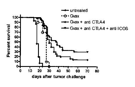
and/or stimulating agent encoding nucleic acid into the genome of a transduced
human cell.
[0083] In another embodiment, the viral vector provides for integration of
a blocking and/or
stimulating agent encoding nucleic acid into the genome of a transduced human
cell.
[0084] In one embodiment, the invention provides methods of transducing a
human cell in
vivo, comprising contacting a solid tumor in vivo with an viral vector of the
invention.
[0085] In another embodiment, the invention provides methods of transducing
a human cell
ex vivo, comprising contacting a human cell ex vivo with the blocking and/or
stimulating agent
viral vector of the invention. In one embodiment, the human cell is a tumor
cell. In one
embodiment, the human cell is allogeneic. In one embodiment, the tumor cell is
derived from the
patient. In one embodiment, the human cell is a non-tumor cell, such as, e.g.,
an antigen
presenting cell (APC), or a T cell.
[0086] Virus particle coats may be modified to alter specificity and
improve cell/tissue
targeting, as is well known in the art. Viral vectors may also be delivered in
other vehicles, for
example, liposomes. Liposomes may also have targeting moieties attached to
their surface to
improve cell/tissue targeting.
[0087] The present application is directed to human cells expressing the
blocking and/or
stimulating agent. In a preferred embodiment, the human cell expresses a
stimulating agent to
ICOS (e.g., ICOSL, which may be secreted or expressed as a cell surface
protein) that
specifically binds to the extracellular domain of ICOS and activates ICOS
mediated negative
-20-
CA 02775761 2012-03-27
WO 2011/041613 PCT/US2010/051008
signaling. In certain embodiments, the human cell expresses the ICOS ligand
proximal to a
tumor cell for example in a cancer patient. Thus, the human cell is capable of
localized
expression of the ligand at a tumor cell or tumor cell mass. The ICOS ligand
can activate ICOS
signaling in cells proximal to said tumor cell, and/or break immune tolerance
against a tumor-
associated self antigen and stimulate an autoreactive T cell response to said
tumor cell. In a
preferred embodiment, localized expression of the ICOS ligand reduces or
inhibits undesired
adverse immune responses.
[0088] It is not necessary for the practice of the invention that the
mechanism of action be
understood. The cells and methods described herein provide human cells
proximal to tumor cells
or tumor cell masses. Expression of stimulating agents to ICOS and optionally
blocking agents to
T cell inhibitory proteins or additional cytokines in proximity to the tumor
cells enhances anti-
tumor immune responses.
[0089] Methods of Treatment
[0090] Described herein is a method of treating a patient afflicted with a
cancer comprising
administering to the patient a pharmaceutical composition comprising a
pharmacologically
effective amount of a blocking agent to a T cell inhibitory receptor and
stimulating agent to
ICOS. The method described herein is directed toward the treatment of cancer,
e.g., leukemias
and solid tumors (e.g., melanomas, carcinomas, sarcomas, lymphomas, etc.).
More common
solid cancers include bladder cancer, bone cancer (osteosarcoma), colorectal
cancer, brain
cancer, breast cancer, cervical cancer, oesophageal cancer, Hodgkin's
lymphoma, kidney cancer,
liver cancer, lung cancer, mesothelioma, multiple myeloma, non-Hodgkin's
lymphoma, ovarian
cancer, pancreatic cancer, penile cancer, prostate cancer, skin cancer
(melanoma and non-
melanoma) soft tissue carcinoma, gastric cancer, testicular cancer, thyroid
cancer and
endometrial cancer.
[0091] The administered pharmaceutical compositions will often further
comprise one or
more buffers (e.g., neutral buffered saline or phosphate buffered saline),
carbohydrates (e.g.,
glucose, mannose, sucrose or dextrans), mannitol, proteins, polypeptides or
amino acids such as
glycine, antioxidants (e.g., ascorbic acid, sodium metabisulfite, butylated
hydroxytoluene,
butylated hydroxyanisole, etc.), bacteriostats, chelating agents such as EDTA
or glutathione,
solutes that render the formulation isotonic, hypotonic or weakly hypertonic
with the blood of a
-21-
CA 02775761 2012-03-27
WO 2011/041613 PCT/US2010/051008
recipient, suspending agents, thickening agents, preservatives, flavoring
agents, sweetening
agents, and coloring compounds as appropriate.
[0092] While any suitable carrier known to those of ordinary skill in the
art may be
employed in the compositions, the type of carrier will typically vary
depending on the mode of
administration. The therapeutic compositions may be formulated for any
appropriate manner of
administration, including for example, oral, nasal, mucosal, rectal, vaginal,
topical, intravenous,
intraperitoneal, intradermal, subcutaneous, and intramuscular administration.
[0093] For parenteral administration, the compositions can be administered
as injectable
dosages of a solution or suspension of the blocking agents of one or more T
cell inhibitory
receptors, the stimulating agents of ICOS, an expression vector expressing one
or more blocking
agents to a T cell inhibitory receptor and/or stimulating agent of ICOS, cells
transformed with
expression vectors expressing one or more blocking agents to a T cell
inhibitory receptor and/or
stimulating agent of ICOS, or a combination thereof, in a physiologically
acceptable diluent with
a pharmaceutical carrier that can be a sterile liquid such as sterile pyrogen
free water, oils, saline,
glycerol, polyethylene glycol or ethanol. Additionally, auxiliary substances,
such as wetting or
emulsifying agents, surfactants, pH buffering substances and the like can be
present in
compositions. Other components of pharmaceutical compositions are those of
petroleum, animal,
vegetable, or synthetic origin, for example, non-aqueous solutions of peanut
oil, soybean oil,
corn oil, cottonseed oil, ethyl oleate, and isopropyl myristate.
[0094] The blocking and/or stimulating agents described herein (including
expression
vectors and/or transformed cells expressing such blocking and/or stimulating
agents) may be
presented in unit-dose or multi-dose containers, such as sealed infusion bags,
ampoules or vials.
Such containers are typically sealed in such a way to preserve the sterility
and stability of the
formulation until use. In general, formulations may be preserved as
suspensions, solutions or
emulsions in oily or aqueous vehicles, as indicated above. Alternatively, a
pharmaceutical
composition may be preserved in a freeze-dried condition requiring only the
addition of a sterile
liquid carrier immediately prior to use.
[0095] The amount administered to the host will vary depending upon what is
being
administered, the purpose of the administration, such as prophylaxis or
therapy, the state of the
host, the manner of administration, the number of administrations, interval
between
-22 -
CA 02775761 2012-03-27
WO 2011/041613 PCT/US2010/051008
administrations, and the like. These can be determined empirically by those
skilled in the art and
may be adjusted for the extent of the therapeutic response. Factors to
consider in determining an
appropriate dose include, but is not limited to, size and weight of the
patient, the age and sex of
the patient, the severity of the symptom, the stage of the disease, method of
delivery of the agent,
half-life of the agents, and efficacy of the agents. Stage of the disease to
consider includes
whether the disease is acute or chronic, relapsing or remitting phase, and the
progressiveness of
the disease.
[0096] Determining the dosages and times of administration for a
therapeutically effective
amount are well within the skill of the ordinary person in the art. For
example, an initial effective
dose can be estimated from cell culture or other in vitro assays. A dose can
then be formulated in
animal models to generate a circulating concentration or tissue concentration,
including that of
the IC50 as determined by the cell culture assays.
[0097] In addition, toxicity and therapeutic efficacy are generally
determined by cell culture
assays and/or using experimental animals, typically by determining a LD50
(lethal dose to 50%
of the test population) and ED50 (therapeutically effectiveness in 50% of the
test population).
Guidance is found in standard reference works, for example, Goodman & Gilman's
The
Pharmacological Basis of Therapeutics, 10th Ed. (Hardman, J. G. et al., eds.)
McGraw-Hill, New
York, N.Y. (2001).
[0098] For the purposes of this invention, the methods of administration
are chosen
depending on the condition being treated and the pharmaceutical composition.
Administration of
the blocking and or stimulating agent(s) can be done in a variety of ways,
including, but not
limited to, subcutaneously, intravenously, intraperitoneally, intramuscularly,
and possibly direct
injection to specified organs or tumors, although systemic administration is
preferred.
Administration of the pharmaceutical compositions may be through a single
route or
concurrently by several routes.
[0099] The compositions may be administered once per day, a few or several
times per day,
or even multiple times per day, depending upon, among other things, the
indication being treated
and the judgment of the prescribing physician.
[00100] The amount of blocking and/or stimulating agent needed for achieving a
therapeutic
effect may be determined empirically in accordance with conventional
procedures for the
-23 -
CA 02775761 2012-03-27
WO 2011/041613 PCT/US2010/051008
particular purpose. Generally, for administering the cells for therapeutic
purposes, the cells are
given at a pharmacologically effective dose. "Pharmacologically effective
amount" or
"pharmacologically effective dose" refers to an amount sufficient to produce
the desired
physiological effect or amount capable of achieving the desired result,
particularly for treating
the disorder or disease condition, including reducing or eliminating one or
more symptoms or
manifestations of the disorder or disease. As an illustration, administration
of cells to a patient
suffering from cancer provides a therapeutic benefit not only when the
underlying condition is
eradicated or ameliorated, but also when the patient reports a decrease in the
severity or duration
of the symptoms associated with the disease, e.g., a decrease in tumor burden
including
disseminated tumor cells (DTC), a decrease in circulating tumor cells, an
increase in progression
free survival. Therapeutic benefit also includes halting or slowing the
progression of the
underlying disease or disorder, regardless of whether improvement is realized.
Pharmacologically effective dose, as defined above, will also apply to
therapeutic compounds
used in combination with the cells, as further described below.
[0100] Preferably, the effect will result in a quantifiable change of at
least about 10%,
preferably at least 20%, 30%, 50%, 70%, or even 90% or more. Therapeutic
benefit also includes
halting or slowing the progression of the underlying disease or disorder,
regardless of whether
improvement is realized. When the combination of a blocking agent of a T cell
inhibitory
receptor and a stimulating agent of ICOS is used in with other treatment
protocols, an effective
amount is in ratio to a combination of components and the effect is not
limited to individual
components alone.
[0101] A pharmacologically effective amount that will treat cancer will
modulate the
symptoms typically by at least about 10%; usually by at least about 20%;
preferably at least
about 30%; or more preferably at least about 50%. Such will result in, e.g.,
statistically
significant and quantifiable changes in the numbers of cells being affected.
This may be a
decrease in the numbers of micrometastases in distant organs, a decrease in
recurrent metastatic
disease, etc.
[0102] The blocking and stimulating agents described herein may be combined
with other
antitumor treatments, e.g., surgical resection, radiation therapy,
chemotherapy, immunotherapy,
and supportive therapy (e.g., painkillers, diuretics, antidiuretics,
antivirals, antibiotics, nutritional
-24 -
CA 02775761 2012-03-27
WO 2011/041613 PCT/US2010/051008
supplements, anemia therapeutics, blood clotting therapeutics, bone
therapeutics, and psychiatric
and psychological therapeutics). Such other antitumor treatments, including
treatment with one
or more blocking agents to one or more T cell inhibitory receptors, may be
provided sequentially
(e.g., before or after) or simultaneously with the administration of the
stimulating agent of
ICOS.
EXAMPLES
[0103] Example 1: Stimulating agents to ICOS enhance the anti-tumor effects
of
anti-CTLA-4 antibody and anti-PD-Li antibody
[0104] Example 1.1: Effect of a stimulating agent to ICOS antibody on CD4+
T cell
proliferation.
[0105] CD4+ T cells were prepared from C57BL/6 mice spleen by Dynal murine
CD4+ T
cell negative selection kit according to the manufacturer's instruction. Fifty
thousand CD4+ T
cells were stimulated in a 96 well plate pre-coated with or without anti-CD3
mAb (0.5 g/m1)
and 2 g/m1 of anti-CD28, 5 g/m1 of anti-ICOS mAb (clones C398.4A and
7E.17G9). Cells
were incubated at 37 C, in a 5% of CO2, for 72 hr and 1 !lei of 3H-thymidine
was added into
each well 8 hr before the end of the culture. Plate was harvested and analyzed
for 3H-thymidine
incorporation.
[0106] As shown in FIG. 1, anti-ICOS antibodies enhanced proliferation of
CD4+ T cells in
the presence of anti-CD3 antibody.
[0107] Example 1.2: Indirect Correlation between anti-CTLA-4 induced ICOS
expression
and tumor growth
[0108] Mice were challenged with 2 X 10⁴ B16/F10 tumor cells. Mice were
untreated or
treated. Treated animals received 200 g anti-CTLA-4 antibody on day 3 and 100
g anti-
CTLA-4 antibody on days 6, 9, 12, 15, 18, and 21 post tumor challenge. Tumor
growth and
levels of ICOS on CD4+FOXP3- effector T cells in the blood were monitored
every three days.
[0109] As shown in FIG. 2, CD4+FOXP3" cells isolated from treated animals
expressed
increased levels of ICOS. Additionally, the increased expression of ICOS
indirectly correlated
with tumor burden (FIG. 2).
- 25 -
CA 02775761 2012-03-27
WO 2011/041613 PCT/US2010/051008
[0110] Example 1.3: 'COS" or 'COSI; mice demonstrated a reduced anti-tumor
response
mediated by anti-CTLA-4 antibody.
[0111] Wild type C57BL/6, ICOS deficient C57BL/6, and ICOS-ligand (ICOSL)
deficient
C57BL/6 mice bearing B16/BL6 tumors were either left untreated or treated s.c.
(on day 3 post-
tumor implantation) with 1 x 10⁶ irradiated GM-CSF-producing B16 (GVAX) and
anti-CTLA-4
i.p. (9H10), at a dosing of 0.2, 0.1 and 0.1 mg on days 3, 5 and 7,
respectively. Tumor growth
was monitored and percent survival calculated on day 80.
[0112] Wild type, ICOS deficient, or ICOSL deficient mice bearing tumors
and left untreated
died between 25 and 41 days after tumor implantation (open circle, open
triangle and open
square respectively). Conversely, 90% survival was observed when wild type
mice were treated
with GVAX and anti-CTLA-4 combination therapy (closed circles). Remarkably,
ICOS
deficient (closed triangles) and ICOSL deficient mice (closed squares) showed
significantly
lower protection after being treated with GVAX and anti-CTLA-4 antibody,
demonstrating a key
role for this ligand/receptor pair interaction during GVAX/anti-CTLA-4
combination therapy.
[0113] Example 1.4: Enhanced anti-tumor effect using GVAX, anti-CTLA-4
antibody, and
anti-ICOS antibody.
[0114] Mice challenged with 5 X 10⁴ B16/BL6 tumor cells were (1) untreated,
(2) treated
with 1 X10⁶ irradiated GM-CSF-producing B16 only (GVAX; s.c. 3, 6, and 9 days
post-
implantation), (2) treated with 1 X 10⁶ irradiated GVAX (s.c. 3, 6, and 9 days
post-implantation),
200 ttg anti-CTLA-4 antibody (day 3 post-tumor implantation), and 100 ttg anti-
CTLA-4
antibody (days 6, 9, 13, and 17 post-tumor implantation), or (3) treated with
1 X 10⁶ irradiated
GVAX (3, 6, and 9 days post-implantation), 200 lig anti-CTLA-4 antibody (day 3
post-tumor
implantation), 100 ttg anti-CTLA-4 antibody (days 6, 9, 13, and 17 post-tumor
implantation) and
200 lig anti-ICOS antibody (days 3, 6, 9, 13, and 17 post-tumor implantation).
Tumor growth
was monitored and survival was calculated on day 80.
[0115] As shown in FIG. 4, treating animals with a combination of GVAX,
anti-CTLA-4
antibody and anti-ICOS antibody resulted in delayed tumor growth compared to
treating animals
with GVAX and anti-CTLA-4 antibody only. This finding is consistent with the
finding that
mice treated with GVAX with anti-CTLA-4 and anti-ICOS antibodies exhibit
higher survival
rates compared to mice treated with GVAX and anti-CTLA4 antibody only (FIG.
5).
-26 -
CA 02775761 2012-03-27
WO 2011/041613 PCT/US2010/051008
[0116] Example 1.5: Enhanced anti-tumor effect using anti-ICOS and anti-PD-
Li antibodies
[0117] Three-day B16/BL6-bearing mice were either untreated or treated s.c.
(on day 3 post-
tumor implantation) with 1 x 10⁶ irradiated GM-CSF-producing B16 (GVAX) and
i.p. anti-ICOS
antibody (7E.17G9), anti-PD-Li antibody(10F.9G2) or the combination at a
dosing of 0.2, 0.1
and 0.1 mg on days 3, 5 and 7 respectively. Tumor growth was monitored and
percent survival
calculated on day 80.
[0118] Mice treated with a combination of GVAX and anti-ICOS antibody, or
GVAX and
anti-PD-Li antibody demonstrated poor survival rates (FIG. 6). In contrast,
combination therapy
using anti-PD-Li antibody, anti- ICOS antibody and GVAX resulted in 50%
survival,
demonstrating a potent synergistic effect obtained with the combination of
anti-ICOS antibody
(7E.17G9) with anti-PD-Li antibody (10F.9G2) and GVAX.
[0119] Example 2: Use of ICOS ligand expressing tumor cells as an anti-
tumor vaccine
[0120] Example 2.1
[0121] Example 2.1.1: Antibodies.
[0122] Anti-CTLA4 (clone 9H10) was purchased from Bio X Cell.
[0123] Example 2.1.2: Cell lines
[0124] The highly tumorigenic and poorly immunogenic melanoma cell line
B16/BL6 was
used for tumor challenge. B16/BL6-expressing GM-CSF, here referred to as GVAX,
was used
for treatment of tumor-bearing mice. B16-Thy1.1 was generated through
retroviral transduction
of B16/BL6 cells with the vector MSCV-IRES-Thy1.1 which was a gift from Dr.
Leo lefrancois
at University of Connecticut. B16-mICOSL was generated through retroviral
transduction of
B16/BL6 cells with the vector MSCV-ICOSL expressing full length of mouse ICOSL
(gift from
Dr. William Sha, University of California, Berkeley). GVAX cells were also
transduced with the
MSCV-ICOSL vector to generate GVAX-mICOSL.
[0125] Example 2.1.3: Tumor challenge and treatment experiments.
[0126] Mice were injected in the right flank i.d. on day 0 with 50,000
B16/BL6 melanoma
cells and treated on days 3, 6, 9, and 12 with 7.5 x 10⁵ irradiated (150 Gy)
GVAX mixed with
7.5 x 10⁵ irradiated (150 Gy) B16/BL6-Thy1.1 (n = 10) or B16-mICOSL (n = 10)
on the left
- 27 -
CA 02775761 2016-07-06
flank, in combination with 10Ong anti-CTLA4 i.p. (200 ng on day 3). Tumor
growth and
rejection were monitored over time.
101271 Mice were injected in the right flank i.d. on day 0 with 20,000
1316431,6 melanoma
cells and treated or not on days 3, 6, 9, and 12 with 1 x 10⁶ irradiated (150
(iy) GVAX (n = 10)
or GVAX-mICOSL (n = 10) on the left flank, in combination with 100 g anti-
CTLA4 i.p. (200
jig on day 3). Tumor growth and rejection were monitored over time.
[01281 Mice were injected in the right flank i.d. on day 0 with 20,000
B16/131.6 melanoma
cells and treated or not on days 3, 6, 9, and 12 with I x 106 irradiated (150
Gy) B16/B1,6-Thy1.1
(n = 10) or B16-mICOSL (n = 10) on the left flank, with or without 10Ong anti-
C11,A4 i.p.
(200 g on day 3). Tumor growth and rejection were monitored over time.
101291 Mice were injected in the right flank i.d, on day 0 with 20,000
1316/F10 melanoma
cells and treated or not on days 3, 6, 9, and 12 with I x 106 irradiated (150
Gy) B16-mICOSI,
(n = 5) on the left flank, with or without I 00 jig anti-CTLA4 i.p. (200 g on
day 3). Tumor growth
and rejection were monitored over time.
101301 Example 2.2: Results
101311 The 1316 cellular vaccine expressing ICOSIõ in the absence or
presence of GVAX,
did not add to the tumor protection rate beyond the previous combination
therapy of GVAX and
CTLA-4 blockade (FIGs. 7 and 8).
10132) In the setting without GM-CSF, the 816 cellular vaccine expressing
ICOSI. has a
synergistic effect together with C'11,A-4 blockade to provide delay in tumor
growth and/or
overall advantage in tumor rejection (FIGs. 9-12).
101331
101341 certain modifications and improvements will occur to those skilled
in the art upon a
reading of the foregoing description. It should be understood that all such
modifications and
improvements have been deleted herein for the sake of conciseness and
readability but are
properly within the scope of the following claims.
- 28 -