Note: Descriptions are shown in the official language in which they were submitted.
CA 02776320 2016-05-13
Systems, Devices, and Method for Providing Insertable Robotic Sensory
and Manipulation Platforms for Single Port Surgery
Field of the Invention
[0001] The present invention relates to devices, systems and surgical
techniques
for minimally invasive surgery and more particularly to minimally invasive
devices,
systems and surgical techniques/methods associated with treatment, biopsy and
the
like of body cavities.
Background
[0002] Laparoscopic and other minimally invasive surgeries have
successfully
reduced patients' post operative pain, complications, hospitalization time and
improved cosmesis. See D. J. Deziel, K. W. Millikan, S. G. Economou, M. A.
Doolas,
S.-T. Ko, and M. C. Airan, "Complications of Laparoscopic Cholecystectomy: A
National Survey of 4,292 Hospitals and an Analysis of 77,604 Cases," The
American
Journal of Surgery, vol. 165, No.1, pp. 9-14. Jan 1993; and M. J. Mack,
"Minimally
Invasive and Robotic Surgery," The Journal of the American Medical
Association, vol.
285, No.5, pp. 568-572, Feb 7 2001 During most laparoscopic procedures, two or
more incisions are used for surgical instruments, visualization, and
insufflation. See E.
Berber, K. L. Engle, A. Garland, A. String, A. Foroutani, J. M. Pearl, and A.
E.
Siperstein, "A Critical Analysis of Intraoperative Time Utilization in
Laparoscopic
Cholecystectomy," Surgical Endoscopy, vol. 15, No.2, pp. 161-165, 2004. Before
Natural Orifice Transluminal Endoscopic Surgery (N.O.T.E.S), which eliminates
all
skin incisions, can be widely applied to broader procedures, population
researchers
and surgeons may focus on single port access (SPA) surgeries which reduce the
number of skin incisions to one and therefore generate a better outcome than
traditional laparoscopic procedures.
[0003] Most existing robotic surgical systems are designed for minimally
invasive
laparoscopic procedures. Although robotic assistance has greatly enhanced
surgeons'
capabilities in performing standardized laparoscopic techniques, these
existing robotic
systems are not suitable for SPA surgeries due to the large size of their
instruments
and lack of overarching and collision avoidance among its multiple arms.
Therefore,
SPA surgeries are currently limited to just a few academic centers using
specifically
1
US1DOCS 7316220v3
CA 02776320 2016-05-13
modified laparoseopic tools (such as RealHandTm (Novare Surgical Systems,
Inc.,
Cupertino, CA)).
Summary
[0004] The present disclosure relates to systems, devices, and methods for
providing foldable, insertable robotic sensory and manipulation platforms for
single
port surgery. The device is referred to herein as an Insertable Robotic
Effector
Platform (IREP). The IREP provides a self-deployable insertable device that
provides
stereo visual feedback upon insertion, implements a backbone structure having
a
primary backbone and four secondary backbones for each of the robotic arms,
and
implements a radial expansion mechanism that can separate the robotic arms.
All of
these elements together provide an anthropomorphic endoscopic device.
[0005] In one aspect, the IREP provides endoscopic imaging and distal
dexterity
enhancement. The IREP robot includes two five-degree of freedom snake-like
continuum robots, two two-degree of freedom parallelogram mechanisms, and one
three-degree of freedom stereo vision module. The IREP can be used in
abdominal
SPA procedures, such as cholecystectomy, appendectomy, liver resection, among
others. The IREP can fit through a small skin incision while providing vision
feedback to guide insertion and deployment of two dexterous arms with a
controllable
stereo vision module.
Brief Description of the Drawings
[0006] For a more complete understanding of various embodiments of the
present
disclosure, reference is now made to the following descriptions taken in
connection
with the accompanying drawings in which:
[0007] Figure IA depicts a system overview of the IREP Robot in a folded
configuration, according to one or more embodiments of the present disclosure;
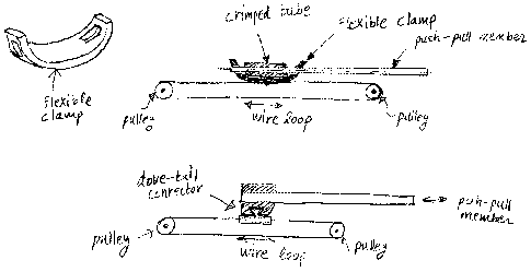
[0008] Figure 18 depicts methods of detachable actuation transmission using
wire
actuation and push-pull super-elastic NiTi backbones;
[0009] Figure 2 depicts a system overview of the IREP Robot in a working
configuration, according to one or more embodiments of the present disclosure;
[0010] Figures 3A-3F depict an image sequence showing the deployment of the
IRE? robot, according to one or more embodiments of the present disclosure;
2
us 1DOCS 7316220v3
CA 02776320 2016-05-13
[0011] Figure 4A is a depiction of the camera module of the IREP robot,
according to one or more embodiments of the present disclosure;
[0012] Figure 4B is an exploded view of the camera module shown in Figure
4,
according to one or more embodiments of the present disclosure;
[0013] Figures 5 is a depiction of a single dexterous arm of the IRE?,
according to
one or more embodiments of the present disclosure;
[0014] Figure 6 is a depiction of a backbone structure of the IREP Robot,
according to one or more embodiments of the present disclosure;
[0015] Figure 7A is a depiction of a parallelogram actuation unit of the
IRE?
Robot, according to one or more embodiments of the present disclosure;
[0016] Figure 7B is another depiction of the parallelogram actuation unit
of the
IREP Robot, according to one or more embodiments of the present disclosure;
[0017] Figure 8 is a depiction of the translational workspaces of the right
arm, left
arm and overlapping areas, according to one or more embodiments of the present
disclosure;
[0018] Figure 9 is a depiction of a gripper of the IRE? Robot, according to
one or
more embodiments of the present disclosure;
[0019] Figure 10 is a depiction of gripper teeth, showing different teeth
for
different suture sizes, according to one or more embodiments of the present
disclosure;
[0020] Figure 11 is a depiction of two connected slots for both high end
gripping
force and wide open angle of the IREP Robot, according to one or more
embodiments
of the present disclosure;
[0021] Figure 12 is a graph depicting actuation force with respect to jaw
angle of
the IRE? Robot, according to one or more embodiments of the present
disclosure;
[0022] Figure 13A is a depiction of a wrist of the IREP Robot, according to
one or
more embodiments of the present disclosure;
[0023] Figure 13B is an exploded view of the wrist shown in Figure 13A;
[0024] Figure 14 depicts a dual arm suturing capability of the IREP Robot,
according to one or more embodiments of the present disclosure;
[0025] Figures 15A-F depicts a suturing simulation using the IREP Robot,
according to one or more embodiments of the present disclosure; and
[0026] Figure 16 is a block diagram of the control system architecture for
the
IREP Robot, according to one or more embodiments of the present disclosure.
3
us I DOCS 7316220v3
CA 02776320 2016-05-13
Detailed Description
[00271 The present disclosure relates to a foldable, insertable robotic
surgical
device and its method of use. The IREP robot includes two five-degree of
freedom
snake-like continuum robots, two two-degree of freedom radial extension
mechanisms,
and one three-degree of freedom stereo vision module.
[00281 Robot-assisted SPA surgery desirably has the following capabilities:
i) the robot has a folded configuration for it to pass through a single small
skin incision,
ii) the robot is self deployable into a working configuration,
iii) the target organs and their related tissues (such as gallbladder, hepatic
tissues, pancreas, etc.) can be manipulated with enough precision and force,
iv) the translational workspace is bigger than 50mmx5Ommx5Omm (e.g.,
the size of the target organs),
v) the robot has a stereo vision unit for depth perception and tool tracking,
and
vi) the illumination device is integrated into the robot.
[0029] Figure 1 depicts a system overview of the IREP Robot 100 in a folded
configuration, according to one or more embodiments of the present disclosure.
The
IREP robot of Figure 1 demonstrates the features and capabilities for SPA
surgery.
When it is in its folded configuration (as illustrated in Figure 1), it can be
deployed
into the abdomen through a small, e.g., 015 mm skin incision, while using its
forward-looking stereo vision module 220 to guide surgeons through the
insertion
phase. The IREP Robot 100 includes an elongated lumen 110 that encloses the
various elements of the robot. The lumen 110 can be constructed from the
following
materials: stainless steel, anodized aluminum, titanium, or molded plastic. In
some
embodiments, the lumen has an outer diameter of 15 mm.
[0030] In some embodiments, the outer diameter of the IREP in folded
configuration is 15 mm. In some embodiments, the lumen 110 is rigid. This
dimension is currently limited by the 06.5 mm diameter of the CCD cameras
(Model
Number, CSH-l.4-V4-END-R1 from NET, Inc.) used in the stereo vision module
120.
The two cameras are placed next to one another in order to simulate the
positioning of
human eyes. Placing the cameras axially displaced along the axis of the IREP
will
make the 1REP's insertable portion too long to allow its deployment inside a
small
cavity. Placing the cameras in parallel will take a diameter of 13 mm, which
leaves
4
us1DOCS 7316220v3
CA 02776320 2016-05-13
space for protective covers. Since in a 020 mm incision is available for
transumbilical laparoscopic procedures, a diameter of 15 mm of the IREP is
acceptable. There are smaller cameras that suffer from image distortion and
sensitivity to lighting conditions that make 3D stereo-vision tool tracking
less accurate;
however, it is expected that improvements in cameras would permit
incorporation of
smaller cameras with resulting smaller outer diameter to the device. The other
limitation of the outer diameter can come from the required diameter for the
dexterous
snake arms (continuum robots) in order to support forces of interaction
typical to
abdominal applications.
[0031] In some embodiments, a passively flexible central lumen may be
constructed using wire actuated designs wherein the superstructure of the
lumen may
be made of a flexible structure that passively bends to accommodate the
anatomy and
provides passage for the actuation wires of the IREP. The flexible lumen may
be
made of polymer elastomers that are superelastic tube micro-machined to
provide
flexure hinges, or any other serial linkage design.
[0032] When using a passively flexible central lumen, the actuation of the
IREP
may still be achieved using a connection method between the push-pull
components of
the IREP and the actuation wires as shown in Figure 1B. The distal and
proximal ends
of the flexible lumen can be modified to include small pulleys used to tension
actuation wire loops. Through actuation of these wire loops, all the
components of the
IREP can be actuated through fast clamping attachments such as the flexible
clamp or
the dove-tail connector of Figure 1B.
[0033] Actively actuated central lumens may be designed using, for example,
wire-actuated articulated designs such as (Degani et al. 2006) and
(Gottumukkala et
al. 2004). These designs allow alternating relaxation and locking of a passive
lumen in
order to allow it to follow the shape of the anatomy. Regardless of the
technology
used to achieve a passively steerable lumen, the IREP may still be actuated
using the
same approach as in passively flexible central lumens.
[0034] The IREP can unfold itself into a working configuration to perform
SPA
procedures, as shown in Figure 2. Figure 2 depicts a system overview of the
IREP
Robot in a working configuration, according to one or more embodiments of the
present disclosure. The IREP robot 100 consists of two snakelike continuum
robots
200, 205, two radial extension mechanisms 210, 215, two flexible stems, 217,
219,
and one 3D stereo vision module 220, wherein the vision module 220 is
comprised of
1DOCS 7316220v3
CA 02776320 2016-05-13
two CCD cameras for stereo visual feedback. The two dexterous snake-like arms
are
equipped with distal wrists 510, 512 and grippers 505, 507.
[0035] When in a deployed configuration, as shown in Figure
2, the proximal
portion of the lumen 225 remains intact, while the distal portion of the lumen
230
separates into multiple segments. These segments can include a top semi-
circular
segment 235 that overlays the stereo vision module 220. The bottom semi-
circular
portion of the lumen can be divided in four segments. Two quarter-circular
segments
240, 245, for example, each half the length of the top segment 235, extend
from the
proximate portion of the lumen 225 along each of flexible stems 217, 219. The
other
= two quarter-circular segments 250, 255 are located at the joint between
the flexible
stems 217, 219 and the continuum robots 200, 205. The segmentation of the
lumen
provides a compact deployment mechanism. Instead of having to use an overtube
to
protect the robot, the thin segmented lumen reduces the set up time of the
procedure
and the size of the incision. The segmented sections also prevent the opened
lumen
segments from interfering with the procedure.
[0036] Figures 3A-3F depict an image sequence showing the
deployment of the
IREP robot, according to one or more embodiments of the present disclosure.
The
IREP robot can be inserted into patient's abdominal cavity in its folded
configuration
and then the device can unfold itself into a working or deployed
configuration. Figure
3A depicts the stereo vision module 220 separating from the lumen 110. Figures
3B
and 3C shows further separation from the vision module 220 and the lumen 110
and
exposes the continuum robot arms 200, 205. Figures 3D and 3E show the
continuum
robot arms 200, 205 extending along the longitudinal axis of the lumen. Figure
3F
shows the final deployed configuration where the radial extension devices 210,
215
(also referred to herein as parallelogram devices) have radially separated the
continuum robot arms 200, 205 from each other.
[0037] The IREP has a plurality of actuators, for example, 21
actuators, that drive
its two dexterous or continuum arms, vision module, and two five-bar (radial
extension) mechanisms that allow self deployment of the dexterous arms and
adjustment of the distance between the bases of the two arms. The IREP can
actively
change from insertion to working configuration while providing uninterrupted
3D
stereo vision feedback to the user. During insertion, the IREP is folded into
a
cylindrical configuration with a diameter of about 15 mm (Figure 1). Insertion
into
the patient abdomen can be carried out using a trocar at the umbilicus. After
insertion,
6
es 1DOCS 7316220v3
CA 02776320 2016-05-13
the IREP deploys two dexterous snake-like arms equipped with distal wrists
510, 512
and grippers 505, 507, A third arm is also deployed with a 3D vision module
comprised of two CCD cameras for stereo visual feedback. Each dexterous arm
includes a four degree of freedom two-segment continuum snake-like robot, a
single
degree of freedom wrist, and a gripper. When supported on a five-bar radial
extension
mechanism 215, 210, the robot arm can provide seven degrees of freedom of
motion
using its eight actuated joints and the additional actuated joint available
for its gripper.
[00381 Figure 4A is a depiction of the stereo vision camera module 220 of
the
IREP robot, according to one or more embodiments of the present disclosure.
Figure
4B is an exploded view of the camera module of Figure 4A. The stereo vision
module
220 has a pair of CCD cameras 401, 402 for depth perception as well as
surgical tool
tracking. The camera module has three degrees of freedoms for pan (using the
panning
mechanism 410), tilt (using the tilting mechanism 405), and zoom adjustments.
A
light source using optic fiber bundles 400 is also integrated into the camera
module.
The device can close to a 015 mm cross section. The camera housing encloses
two
camera units consisting of housing and two degree of freedom actuated joint
that
allows panning and tilting the housing in two directions as shown in Figure 4.
The
camera module 220 is supported on one side of the lumen and can be controlled
independently of the lumen opening. The control mechanism for the camera
module
uses a slider-crank mechanism for control of the tilt angle. Actuation of the
tilting
mechanism is achieved via a thin NiTi superelastic wire that is supported in a
dedicated channel in the central lumen 110 such that it can withstand
compressive and
tensile forces (push-pull actuation). The panning mechanism is used to control
the
panning angle of the camera module. This mechanism is also actuated by a NiTi
wire
in push pull actuation. The axial translation of the actuation wire translates
a pin in a
helical slot in the panning mechanism tube. This causes the panning mechanism
to
rotate about its longitudinal axis, which provides the panning degree of
freedom. The
electronic signals to the camera module are transmitted using a flexible
printed circuit
board (PCB). The angle of the outer shell carrying the camera module and its
actuation mechanisms is controlled via a slider-crank mechanism in which the
shell
actuating link acts as the pushrod and the shell acts as the crank. This shell
actuating
link is actuated by a link that translates prismatically inside the central
lumen.
[0039] The camera system is used as follows:
7
us I DOCS 7316220v3
CA 02776320 2016-05-13
1) it provides the surgeon with a means for monitoring and controlling the
movements of the robotic arms;
2) it provides a means for light-based imaging that the surgeon can use for
identifications of pathologies;
3) in the folded state of the robot of Figure 1 the cameras point forward in
the
direction of insertion and help the surgeon see the various stages of the
insertion of the
robot into the anatomy.
[0040] One advantage of the proposed design in Figure 4 is that it offers
an
anthropomorphic stereo-vision and manipulation setup that mimics the human
anatomy in which the field of focus of the eyes is located between the two
manipulation arms. The vision module has two integrated stereo vision CCD
cameras
with a baseline of 7.6 mm. These CCD cameras are attached to a controllable
shell
with adjustable pan and tilt for increased visual field. This camera-between-
hands
arrangement provides an anthropomorphic and intuitive image to surgeons who
are
used to operating on surgical sites located between their own arms. The pan
and tilt
angles of the stereo vision cameras are controllable by a pull-push mechanism
that
allows instrument tracking. During insertion, the robotic platform is folded
and its
stereo vision module points forward in order to provide vision feedback to the
surgeon.
[0041] To integrate a stereo vision module for tracking surgical tool tip's
movement, the baseline between the two CCD cameras can be maximized for
improved tracking precision.
[0042] The system configuration is shown Figure 1, where the two CCD
cameras
are packed together. A fixed baseline simplifies calibration. Initial
simulation showed
an accuracy of approximate 0.16 mm. In addition, the central stem has
available a
cross sectional area of 36 mm2 in for passing through optic fiber bundles for
illuminations.
[0043] Figures 5 is a depiction of a single dexterous arm of the [REP,
according to
one or more embodiments of the present disclosure. Each dexterous arm includes
at
least four components:
i) a gripper 500,
ii) a one-Degree of freedom wrist 505,
iii) a four-Degree of freedom continuum robot/snake arm 205,
iv) a radial extension mechanism 215 and
v) a flexible stem 217.
8
us I DOCS 7316220v3
CA 02776320 2016-05-13
[0044] Each single dexterous arm acts as a surgical telemanipulation slave
for dual
arm interventions and delivery of sensors (e.g. ultrasound probe) or energy
sources
(e.g. cautery). During SPA procedures, each of the arms of the 1REP robot can
be
independently pulled out and replaced with another arm equipped with different
surgical end effectors. As shown in Figure 5, the continuum robot can include
two
structures or segments: a first structure 520 and a second structure 525.
These
structures are referred to as backbones and are discussed in more detail
below.
[0045] One purpose of the dual arm device of Figure 5, for example, is to
provide
dexterous tool manipulation. Some embodiments of the design in Figure 5 can be
combined with, for example, U.S. Patent Application No. 10/850,821, filed May
21,
2004. The '821 application discloses devices, systems, and methods for
minimally
invasive surgery of the throat and other portions of the mammalian body. The
'821
discloses a dexterous arm having a primary backbone and three secondary
backbones.
[0046] Figure 6 is a depiction of a backbone structure 700 of the IREP
Robot,
according to one or more embodiments of the present disclosure. The present
design
uses one central super-elastic backbone 705 surrounded by four secondary
superelastie
tubular backbones 610, 615, 620 and 625. The backbones are connected through a
series of disks, including a base disk 630, an end disk 635 and one or more
spacer
disks 640. While one spacer disk is shown in Figure 6, a plurality of spacer
disks can
be used, depending on the size of the backbone structure. Four identical
secondary
backbones are equidistant from each other and from the primary backbone. The
secondary backbones are only attached to the end disk and can slide in
appropriately
toleranced holes in the base disk and in the spacer disks. The two degree of
freedom
bending motion of this continuum segment is achieved through simultaneous
differential actuation of the four secondary backbones. Each primary of
secondary
backbone can be composed of nickel titanium (NiTi) wires, cylinders or
concentric
cylinders. The backbones of the first and the second segments (shown in Figure
2) are
concentric NiTi super-elastic tubes with outer and inner diameters of
0.90x0.76 mm
and 0.64x0.51 mm. The disks each can have a diameter of about 6.4 mm and a
height
of about 3.2 mm. The disks can be made from stainless steel. The diameter can
be
between 4.0-6.4 mm and height between 3.2-1.6 mm.
[0047] In some embodiments, two or more backbone structures can be stacked
on
top of each other to form elongated backbone structures with a higher degree
of
freedom. In one embodiment, the continuum arm is composed of two backbone
9
t Ici nne-c 71101mA
CA 02776320 2016-05-13
structures to form the four-Degree of freedom continuum snake arm. Each
structure
consists of several super-elastic NiTi tubes as backbones and several disks.
For
example, in Figure 5, the continuum arm can include a first structure 520 and
a second
structure 525. Figure 6 shows one segment, where one primary backbone is
centrally
located and is attached to the base disk and the end disk.
[0048] The payload of the four degree of freedom continuum NiTi snake
continuum arms determines the payload of the entire IREP robot since it is the
weakest portion of the IREP robot. For this reason, the 06.4 mm diameter of
the four-
Degree of freedom continuum snake arm was maximized to use all available space
in
folded configuration. The diameters of the backbones were chosen to be 0 0.90
mm
for the first segments of the continuum snakes and 00.64 mm for the distal
segments.
All backbones are made from super-elastic NiTi tubes to provide channels for
actuation of the gripper and the wrist, suction, cautery, light, and delivery
of wiring
for sensors.
[0049] Previous works demonstrated that continuum snake-like robots as in
Figure
can serve as distal dexterity tools for enabling complicated surgical tasks
such as
suturing and knot tying in confined spaces. The proven dexterity plus the
scalability
and load-carrying capability of this type of continuum robots make it an ideal
choice
for the IREP robot's arms. Furthermore, its intrinsic force sensing capability
developed in allows equipping the IREP robot with force sensing capabilities.
For
details of the force sensing capabilities, please see related application no.
PCT/US09/032068, entitled, SYSTEMS AND METHODS FOR FORCE SENSING
IN A ROBOT.
[0050] The choice of continuum flexible robots using NiTi backbones was
motivated by the inherent safety of flexible robots in manipulating organs,
the
enhanced miniaturization of these arms.
[00511 All these controlled joints can be actuated by NiTi tubes or
stainless steel
rods in push-pull mode. The actuation unit will remain outside patient's body.
This
configuration simplifies the design of the actuation unit for the snakes
because
opposing secondary backbones have to be pushed and pulled on in the same
amount.
Two of the secondary backbones are used for delivering wire actuation for the
writs.
The central backbone is used for delivering actuation for the gripper by using
a
superelastic wire in pushing mode. The two remaining backbones may be used for
delivering other sources of energy or for sensory data.
US1DOCS 731622Dv3
CA 02776320 2016-05-13
[0052] The advantage of the five backbone design is in the simplicity of
actuation
since each backbone can be pulled on while the other radially-opposing
backbone can
be pushed by the same amount. This modification eliminates the need for
software
kinematic coupling between opposing backbones ¨ a feature that simplifies
deployment and homing of these robots. The wrist is a wire-driven joint that
allows
independent rotation of the gripper about its longitudinal axis, therefore
adding
dexterity critical to suturing tasks in confined spaces. While it is possible
to provide
rotation about the axis of the gripper by using the continuum robots as a
constant
velocity joint through careful coordination of actuation of all backbones, the
use of an
independent wrist simplifies the control and improves dexterity.
[0053] Since the two snake-like continuum robots are deployed through the
IREP's 015mm central stem, their direct implementation will not provide enough
overlapped translational workspace. For this reason, two radial extension
mechanisms,
also referred to as parallelogram mechanisms, are included to control the
position of
the bases of the snake-like continuum robots. Translational workspace of the
single
four-degree of freedom continuum snakelike robot used in the arms of the IREP
in
Figure 2.
[0054] Figure 7 is a depiction of a radial extension structure unit of the
IREP
Robot, according to one or more embodiments of the present disclosure. Each
radial
extension structure 215 has two degree of freedoms for a translational
placement of
the snake-like continuum robot 215, The flexible stem 217 will be
independently fed
in and out to comply with the radial extension structure's motion. The radial
extension structure serves at least two purposes:
i) retracting the snake arms into the shell in a closed configuration (Figure
1), and
ii) changing the distance between the base of each arm to allow for dual-
arm end effector triangulation (Figure 3E).
[0055] The radial extension structures also help in avoiding dexterity
deficiencies
due to "sword fighting" of the instruments. In some embodiments, the radial
extension structures can be a five bar parallelogram mechanism, as shown in
Figure
713. A shown in Figure 7B, the five bar mechanism includes a first bar 700
between
points P2 and P3, a second bar 705 between points P2 and P5, a third bar 710
between
points P3 and P6, a fourth bar 715 between points P5 and P6, and a fifth bar
720
between points P1 and P4. All of the bars in the parallelogram mechanism can
be
11
1110CS 711h7701.1
CA 02776320 2016-05-13
made of stainless steel. This embodiment of the radial extension mechanism is
called
a parallelogram mechanism because of the parallelogram formed by points P2,
P35 P6,
and P5. The first bar 700 and the fourth bar 715 remain at the same
orientation with
respect to each other while the parallelogram mechanism is moved. The
dimensions
of the bars can be as follow: the first bar 700 can be about 2.3 mm, the
second bar
705 can be about 35 mm, the third bar 710 can be about 35 mm, the fourth bar
715 can
be about 2.3 mm, and the fifth bar 720 can be about 20 mm. The five bar
mechanism
is actuated by two push-pull members located in the base of the flexible stem
217. The
push-pull members in the flexible stem 217 move the fifth bar 720 relative to
the first
bar 700, second bar 705, third bar 710 and fourth bar 715, which rotates the
parallelogram mechanism radially from the lumen 110. This structure provides
two
degrees of freedom. These two degrees of freedom yield planar motion of the
base of
the snake while restricting the orientation of the base disk to be parallel
with the end
of the flexible stem 217.
[0056] In an embodiment of the system of Figure 1 where the central lumen
is
rigid the actuation members of the parallelograms may be rigid strips actuated
in push-
pull mode. In an embodiment in which the central lumen of the system of Figure
1 is
flexible, the actuation of the five-bars may be achieved by wire actuation, or
through
flexible passively articulated linkage actuated by push-pull actuation. The
wire-
actuation mechanism for the case where the central lumen is flexible is as
shown in
Figure 1B. Referring to Figure 1B, it is shown that a closed-loop wire
actuation
mechanism is used to axially translate a flexible clamp or a dove-tail
connector that is
used to connect to the superelastic NiTi backbones of the continuum robots. In
another
embodiment, a passively articulated linkage is used to actuate the backbones
of the
continuum robots. The passively articulated mechanism is composed from
serially
connected linkage arms with passive joints connecting them. Axial transmission
of
load is possible as long as an outer external sheath is present to support
this linkage.
The function of the outer support sheath is provided by the outer flexible
lumen.
[0057] Combining the workspace of the snake-like continuum robot and that
of
the parallelogram mechanism, the translational workspace of the dual-arm IREP
robot
is plotted in Figure 8. The figure shows that the final design fulfills the
workspace
requirement. When the parallelogram mechanism is actuated, the flexible stem
will be
fed through the central stem by the external actuation system. Thus, through
the use
of the radial extension mechanism, the effective workspace of the IREP is
increased.
12
US] DOCS 7316220v3
CA 02776320 2016-05-13
[0058] Figure 9 is a depiction of a gripper 900 of the IREP Robot,
according to
one or more embodiments of the present disclosure. The gripper is attached to
the
wrist. The gripper includes a first opposable end piece 910 and a second
opposable
end piece 915. To stabilize a suture 905, the gripper is expected to provide
around
40N gripping force. The gripper design then has two requirements:
i) the gripper should guarantee 40N gripping force with minimal actuation
force; and
ii) it should open as wide as possible. Suitable materials for the gripper
include stainless steel and titanium. The gripper size can be smaller than the
diameter
of 6.5 mm in the support lumen 110 in order to allow insertion and extraction
of the
snake robot with the gripper assembled on it. The inner faces of the gripper
jaws must
be machined with carefully spaced grooves in order to provide stable 3-point
grasp for
needles with triangular cross sections.
[0059] Figure 10 is a depiction of gripper teeth of the first and second
opposable
end pieces 910, 915, showing different teeth for different suture sizes,
according to
one or more embodiments of the present disclosure. Since this gripper design
only
can provide enough gripping force when the jaws are almost closed, the teeth
heights
were assigned differently to accommodate different sutures sizes. The
gripper's teeth
also can be misaligned to ensure three-point contact to stabilize needles with
triangular and round cross sections.
[0060] Figure 11 is a depiction of two connected slots for both high end
gripping
force and wide open angle of the IREP Robot, according to one or more
embodiments
of the present disclosure. The first and second opposable end pieces can be
slidably
attached to one another. They can be connected through a first surface and a
second
surface of the second end piece 915 that form a slot 1100. The slot can have a
first
section 1105 with a first slope and a second section 1110 with a second slope.
When
the gripper is actuated by pushing or pulling a NiTi wire, the portion of the
slot 1100
with steep slope 1105 helps generate a large gripping force by a small
actuation force,
while the mild slope portion 1110 opens the gripper wide over a short
actuation length.
This provides a gripper that has wide opening angle and a very large gripping
force in
a closed configuration. Simulation was conducted using the ProEngineer
software
program to validate the design. The results are plotted in Figure 12. From the
results,
when a gripping force of 40N was maintained, the actuation force rapidly
declined to
around ION, which can be easily actuated by a 00.4mm NiTi Wire.
13
1DOCS 7316220v3
CA 02776320 2016-05-13
[0061] Figure 13A is a depiction of a wrist of the IREP Robot, according to
one or
more embodiments of the present disclosure. The wrist includes a channel for
the
gripper's actuation 1300, a shear pin 1305, a capstan assembly 1310, a wire
rope
1315, with a terminal 1317, a bearing assembly 1320, a pulley 1325, and a wire-
rope
1330 passing through the backbone of the continuum structure. The wire-rope
1330
can be 00.33 mm.
[0062] Figure 13B is an exploded view of the wrist assembly 1300. The
following
parts can be constructed of stainless steel, however, some biocompatible
materials
may be feasible for construction): snake end disk 1335, capstan lock nut 1340,
lower
bearing race 1345, wrist base 1350, wire routing pulleys 1355, and the capstan
1310.
All shear pins and the ball bearings are constructed of hardened tool steel.
The overall
outside dimension of the assembly is about 6.4 mm.
[0063] The wire 1315 actively drives the wrist mechanism. The wire 1315
passes
through two continuum backbones and over the capstan 1310. The terminal 1317
is
connected directly to the wire rope 1315 and interfaces with the capstan 1310
as a lock
mechanism such that the capstan 1310 does not slip with respect to the wire
1315.
The wrist is actuated through a wire loop that passes through the super-
elastic tubes of
the snake arms and wraps around the capstan 1310 hinged about the longitudinal
axis
of the gripper. Actuation of the wire loop back and forth causes the rotation
of the
gripper about its longitudinal axis. A contributor to the dexterity of the
IREP robot for
fine manipulation tasks (including blunt dissection, dual arm manipulation and
suturing) is the freedom to rotate an attached surgical end effector, such as
the
presented gripper, about its longitudinal axis. Previous works showed that the
four
degree of freedom continuum snake arm can transmit axial rotation provided
that
synchronous actuation of all secondary backbones is ensured by proper
compensation
for model imperfections. However, when the parallelogram mechanism opens and
deforms the flexible stem, interaction forces can affect the transmission of
the required
torque of 50 mNm for suturing.
[0064] To simplify the design and control of the IREP arms, an independent
single
degree of freedom wrist located at the distal end of each IREP arm was chosen
to meet
the functional requirements, including dexterity, actuation speed and payload
ability.
This wrist design presents a unique challenge for robotic mechanisms of this
size.
Critical factors constraining the wrist design included payload, a maximum
overall
outside diameter defined by the external superstructure and a requirement for
14
us I DOCS 7316220,3
CA 02776320 2016-05-13
robustness and smooth operation in the surgical environment. The disclosed
design
achieves axial rotation and delivers torque via a 00.33 mm wire-rope running
over
pulleys and around a capstan arranged axially in line with the gripper. This
design
achieves approximately 150 of axial rotation. The distal effector platform
employs a
novel axial wrist design actuated by a capstan and pulley system. This wrist
allows
direct control of the gripper orientation about the longitudinal axis of the
gripper. This
added degree of freedom supports knot tying and passing sutures in very
confined
spaces while minimizing the required motion of the snakes. Also, this wrist
allows for
avoiding the requirements for very precise actuation compensation for the
flexible
snakes if they were used for delivering rotation along their backbone.
[0065] The actuation unit of the IREP contains three modules: a base module
and
two identical actuation units for two dexterous arms of the IREP (Figure 2).
The base
module actuates all components of the IREP that are not interchangeable. These
components include the vision module and the two five-bar parallelogram
mechanisms. In addition, the base module carries all motors for the IREP and
it
provides gross axial motion along the axis of the IREP lumen. The actuation
unit of
each dexterous arm connects to the base module via a quick-connecting
interface
equipped with six Oldham couplings. All motors have been removed from this
actuation unit in order to reduce weight and to support interchangeability of
the
robotic arms of the IRE?. This actuation unit includes four twin lead screws
for
actuating the two-segment continuum robot, two lead screws to actuate the
distal wrist
and gripper. The distal wrist is wire-actuated and the gripper is actuated by
a NiTi
wire.
[0066] Figure 14 depicts a dual arm suturing capability of the IREP Robot,
according to one or more embodiments of the present disclosure. The dexterity
of
the IREP arms was verified for passing circular suturing needles at multiple
locations
along a sinusoidal path in the X-Y cross section of the desired workspace. The
path
had amplitude of 4 mm and a wave length of 40 mm. At each point along the
path, the
IREP inserted a 3/8 circular needle (diameter 16 mm) through 1000 while
holding the
axis normal to its plane was tangentially aligned with the curve, shown in the
inset of
Figure 14.
[0067] Figures 15A-F depicts a suturing simulation using the IREP Robot,
according to one or more embodiments of the present disclosure. Figures 15A-C
represent left hand suturing. 15D-F represent right hand suturing. The
suturing arm
usiDOCS 7316220v3
CA 02776320 2016-05-13
for each segment of the curve was selected for maximum dexterity. The needle
insertion motion is most easily achieved via rotated wrist joint and hence the
robot is
most dexterous when the wrist is aligned with given sinusoidal curve tangent.
Otherwise the continuum arm will be bended in "S" shape to align of the wrist
with
suturing curve tangent. Figures 15A-C show the robot's left arm passing a
circular
needle at 00, 45 , and 90 of rotation about the needle axis. Figures 15D-F
show the
right hand performing a similar task.
[0068] Though the IREP has a distal wrist, it is possible to perform the
same task
of passing circular sutures by using the continuum robot as a constant
velocity joint to
transmit rotation from its base to its gripper. This design alternative using
"rotation
about the central backbone" was previously explored for minimally invasive
surgery
of the throat. We carried out a simulation comparing the dexterity of two
alternative
designs of the IREP with a distal wrist or without a distal wrist. The design
alternative without a distal wrist was assumed to have one degree of freedom
of
rotation about the base disks of each arm of the IREP in order to perform
rotation
about the central backbone of each arm.
[0069] In some embodiments, the IREP provides channels for energy delivery
for
applications such as laser surgery, cautery, radio-frequency ablation,
cryosurgery,
ultrasonic dissection, and new forms of energy. The MEP provides channels for
sensor data and can carry sensory devices such as ultrasound probe, chemical
and
temperature sensors, spectral light imaging, fluorescence imaging,
radioisotope
imaging, or confocal microscopy. Future imaging technologies may also be
deployable using this platform. The control algorithm of the IREP is capable
of using
information from joint level and external sensory sources for estimating the
interaction
forces with the tissue. This can be done using tool tip tracking (either by
vision or
using magnetic tracking) and by monitoring the loads on the robot arm joints.
[0070] Figure 16 is a block diagram of the control system architecture for
the
IREP Robot, according to one or more embodiments of the present disclosure.
[0071] The control system of the IREP robot uses a host-target environment
powered by xE'C TargetTm from The MathWorks, Inc, which provides a rapid
prototyping approach for control system setup in an open hardware
architecture. Our
control hierarchy is presented in Figure 16. A GUI running on the host PC
takes
motion inputs from two master manipulators and then sends them down to the
target
PC via Ethernet connection after scaling and mapping. Target PC processes the
16
us 'flocs 73162200
CA 02776320 2016-05-13
desired motions xd of the 1REP robot by solving kinematics and redundancy
resolution in a 1 kHz servo control loop. A third PC running vision processing
module will output the stereo display for surgeons and feed tool tracking
results xv to
the host PC for future motion compensation of the IREP's dual snake-like arm.
[0072] Although exemplary embodiments of the present application are
described
herein, it should be understood that the scope of protection, as defined by
the
appended claims, should not be limited by the preferred embodiments set forth
in the
examples, but should be given the broadest interpretation consistent with the
specification as a whole.
[0073] What is claimed, is:
17
us irlocs 1316220v3