Note: Descriptions are shown in the official language in which they were submitted.
CA 02776613 2012-04-03
WO 2011/044329 PCT/US2010/051772
TITLE
DEVICES, SYSTEMS AND METHODS FOR CELL MODIFICATION
CROSS-REFERENCE TO RELATED APPLICATIONS
[01] This application claims benefit of U.S. Provisional Patent Application
Serial
No. 61/249,318, filed October 7, 2010, the disclosure of which is incorporated
herein by
reference.
GOVERNMENTAL INTEREST
[02] This invention was made with government support under grant no. RO1
HL080926 awarded by the National Institutes of Health. The government has
certain rights
in this invention.
BACKGROUND
[03] The following information is provided to assist the reader to understand
the
technology described below and certain environments in which such technology
can be used.
The terms used herein are not intended to be limited to any particular narrow
interpretation
unless clearly stated otherwise in this document. References set forth herein
may facilitate
understanding of the technology or the background thereof. The disclosure of
all references
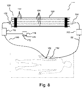
cited herein are incorporated by reference.
[04] Cell-cell interactions are central to both pathology and effective host
defense to a
myriad of diseases. Many cell functions are stimulated or dampened by binding
of various
agents, binding agents or ligands to their respective receptors on the cell
surface. Ligands
can, for example, include agonists, antagonists and inverse agonists. In
general, agonists are
able to activate a receptor. Antagonists bind to receptors but do not provoke
a biological
response upon binding. Binding of an antagonist disrupts interaction and
inhibits function of
an agonist. Inverse agonists reduce the activity of receptors by inhibiting
constitutive activity
of the receptor.
[05] Modulating or modifying the surface receptor profile of cells before
those cells
interact with other cells inside the body has the potential to modify or
program the action of
1
CA 02776613 2012-04-03
WO 2011/044329 PCT/US2010/051772
those cells towards a desired response while attenuating less desired
responses. Systemic
drug administration can, for example, be used to modulate surface receptor
profile but can
also result in undesirable side effects toward other cells or tissues.
SUMMARY
[06] In one aspect, a method of modifying cells includes removing fluid
including cells
from a patient, contacting the removed fluid from the patient with at least
one surface upon
which at least one agent to interact at least one cell receptor is immobilized
to modify cells in
the fluid, and returning the fluid to the patient. The agent can, for example,
be immobilized
via covalent bonding or ionic bonding to the at least one surface. The fluid
can, for example,
be blood or a blood fraction. The agent can, for example, be an agonist, an
antagonist or an
inverse agonist.
[07] In a number of embodiments, the agent includes a protein or a fragment of
a
protein. The agent can, for example, include a cytokine. The cytokine can, for
example, be a
chemokine. In a number of embodiments, the agent is an interleukin. The agent
can, for
example, be IL-8 (interleukin 8). In a number of embodiments, the agent is a
ligand selected
from the group of IL-1, IL-4, IL-6, IL-8, IL-10, IL-18, IL-33, TNF, FAS, MIF,
F1t3, a ligand
form the Bcl-2 family of ligands, an L-selectin, a P-selectin, ICAM-1 or an
antibody.
[08] The fluid (for example, blood or a blood fraction) can, for example, be
passed in a
continuous loop from a blood vessel of the patient to contact the at least one
surface and back
to a blood vessel of the patient. The fluid can, for example, be is passed
continuously for at
least a period of time from a blood vessel of the patient to contact the at
least one surface and
back to the blood vessel or another blood vessel. The fluid can, for example,
be passed
discontinuously from a blood vessel of the patient to contact the at least one
surface and back
to the blood vessel or another blood vessel.
[09] The fluid (for example, blood or a blood fraction) can, for example, be
contacted
with the at least one surface in an extracorporeal device including the at
least one surface.
The extracorporeal device can, for example, include a plurality of surfaces
upon which at
least one agent to interact with at least one cell receptor is immobilized.
The plurality of
surfaces can include a plurality of hollow fibers. The plurality of surfaces
can include a
plurality of beads.
2
CA 02776613 2012-04-03
WO 2011/044329 PCT/US2010/051772
[10] The period of contact for cells targeted for modification can, for
example, be
extended. The period of contact for cells targeted for modification can, for
example, be
extended by the immobilization of an adhesion agent on the at least one
surface, by at least
one physiological characteristic of the at least one surface, or by a geometry
of a volume
through which the fluid containing cells flows.
[11] Cells can, for example, be modified in treatment of sepsis, treatment of
inflammatory disease, treatment of cancer, immune system regulation, or
treatment of
cardiovascular disease.
[12] In another aspect, an extracorporeal device includes a vessel, an inlet
adapted to
pass fluid including cells removed from a patient into the vessel, at least
one surface within
the vessel upon which at least one agent to interact with at least one cell
receptor is
immobilized, and an outlet adapted to return the fluid from the vessel to the
patient. The
device can, for example, include a plurality of surfaces upon which at least
one agent to
interact with at least one cell receptor is immobilized. The plurality of
surfaces can, for
example, include a plurality of hollow fibers. The plurality of surfaces can,
for example,
include a plurality of beads. As described above, the agent can, for example,
be an agonist,
an antagonist or an inverse agonist. The agent can, for example, be
immobilized via covalent
bonding or ionic bonding to the at least one surface.
[13] The fluid can, for example, be passed in a continuous loop from a blood
vessel of
the patient to contact the at least one surface and back to a blood vessel of
the patient. The
fluid can, for example, be passed continuously for at least a period of time
from a blood
vessel of the patient to contact the at least one surface and back to the
blood vessel or to
another blood vessel. The fluid can, for example, be passed discontinuously
from a blood
vessel of the patient to contact the at least one surface and back to the
blood vessel or to
another blood vessel.
[14] The residence time for cells targeted for modification within the devices
can, for
example, be extended. The residence time for cells targeted for modification
can, for
example, be extended by the immobilization of an adhesion agent on the at
least one surface,
by at least one physiological characteristic of the at least one surface, or
by a geometry of a
volume through which the fluid containing cells flows.
3
CA 02776613 2012-04-03
WO 2011/044329 PCT/US2010/051772
[15] In a further aspect, a system for modifying cells includes a first
conduit adapted to
be placed in fluid connection with a patient; an extracorporeal device
including a vessel, an
inlet in fluid connection with the first conduit, at least one surface within
the vessel upon
which at least one agent to interact at least one cell receptor is
immobilized, and an outlet; a
second conduit in fluid connection with the outlet and adapted to be placed in
fluid
connection with the patient; and at least one pump system to circulate fluid
from the patient
through the system.
[16] The technology described herein, along with the attributes and attendant
advantages thereof, will best be appreciated and understood in view of the
following detailed
description taken in conjunction with the accompanying drawings.
BRIEF DESCRIPTION OF THE DRAWINGS
[17] Figure 1 illustrates a mechanism for G-protein-coupled receptor ligand
internalization.
[18] Figure 2A illustrates an embodiment of method to covalently immobilize
interleukin 8 or IL-8 upon a surface including hydroxyl groups.
[19] Figure 2B illustrates an idealized schematic representation of the
interaction of
neutrophils with immobilized IL-8 and/or other agents.
[20] Figure 3 illustrates a study of anti-IL-8 capture using cellulose fibers
modified with
immobilized IL-8 and unmodified cellulose fibers.
[21] Figure 4 illustrates IL-8 loss during immobilization (in g) as measured
using
enzyme-linked immunosorbent assay (ELISA) on wash eluent samples following the
immobilization procedure.
[22] Figure 5 illustrates a study of IL-8 leaching into buffer (in pg IL-8/ml
buffer)
during a 90 minute incubation in the buffer wherein less than 0.01% of
immobilized IL-8 was
lost during the incubation.
[23] Figure 6A illustrates CXCR1 expression over time during incubation with
IL-8-
modified/immobilized fibers and unmodified fibers wherein the dashed line
represents
CXCR1 expression in a sample of blood spiked with free IL-8.
4
CA 02776613 2012-04-03
WO 2011/044329 PCT/US2010/051772
[24] Figure 6B illustrates CXCR2 expression over time during incubation with
IL-8-
modified/immobilized fibers and unmodified fibers wherein the dashed line
represents
CXCR2 expression in a sample of blood spiked with free IL-8.
[25] Figure 7A illustrates the results of white blood cell counts for
heparinized blood
from healthy volunteers (n=5) recirculated at a flow rate of 0.5m1/min for 4
hours (using a
20m1 reservoir) through an empty device including no sorbent(Sham), through a
hemofiltration circuit (HF), through a device including standard size beads
(HA) and a
through a device including smaller sized beads (HAs).
[26] Figure 7B illustrates the results of white blood cell counts for
heparinized blood
from patients with sepsis (n=21) recirculated at a flow rate of 0.5m1/min for
4 hours (using a
20m1 reservoir) through an empty device including no sorbent(Sham), through a
hemofiltration circuit (HF), through a device including standard size beads
(HA) and a
through a device including smaller sized beads (HAs).
[27] Figure 8 illustrates an embodiment of a system including an
extracorporeal device
including a plurality of hollow fibers having a cell receptor ligand such as
IL-8 immobilized
thereon.
[28] Figure 9 illustrates an embodiment of an extracorporeal device including
a
plurality of beads having a cell receptor ligand immobilized on the surfaces
thereof.
DETAILED DESCRIPTION
[29] As used herein and in the appended claims, the singular forms "a," "an",
and "the"
include plural references unless the content clearly dictates otherwise. Thus,
for example,
reference to "an agent" includes a plurality of such agents and equivalents
thereof known to
those skilled in the art, and so forth, and reference to "the agent" is a
reference to one or more
such agents and equivalents thereof known to those skilled in the art, and so
forth.
[30] As opposed to systemic drug administration, devices, systems and/or
methods in
which blood is perfused through a system external to the body (for example, an
extracorporeal hemoperfusion system), wherein one or more internal surfaces of
the external
or extracorporeal system include immobilized agents to interact with one or
more cell
receptors, offers the opportunity to manipulate, modulate, modify or program
circulating cells
CA 02776613 2012-04-03
WO 2011/044329 PCT/US2010/051772
outside the human body in a well-defined environment. In this manner,
circulating cells can
be directly targeted while undesirable side-effects towards other cells or
tissues are limited.
[31] The extracorporeal devices, systems and methods hereof provide a platform
that
can be applied to numerous conditions and diseases involving circulating
cells, such as
atherosclerosis, cancer, HIV, sepsis and many others. By altering the behavior
of circulating
cells in a defined manner, it is possible to treat disease in a fundamentally
different manner
than previously.
[32] In a number of representative studies, the modification or reprogramming
of white
blood cells (neutrophils) was demonstrated. The incubation of isolated white
blood cells
(neutrophils) with immobilized cell activators (chemokine CXCL1) leads to
selective down-
regulation of the respective receptor on neutrophils over time. That process
effectively
renders the cells unresponsive to activation and thus fundamentally changes
their biology.
[33] This devices, systems and/or methods hereof can readily be adapted or
extended to
alter the responses of various cells and in a variety of different ways (for
example, increasing
or decreasing their responses to a variety of stimuli). Interaction of agents,
binding agents or
ligands with cell receptors or binding partners on a cell can, for example,
modify surface
receptor, modify cellular function, modify cellular activity, modify cellular
phenotype, etc.,
thereby modifying (modulating, increasing, decreasing, or otherwise changing)
an activity or
specificity of the cell. The devices, systems and/or methods hereof can be
applied to virtually
any condition in which circulating cells are involved in pathology or
mitigation of disease.
Although white blood cells such a neutrophils are modified in several
representative studies
hereof, many types of cells can be modified via the interaction with
immobilized agent with
cell receptors.
[34] Modulating or modifying cells via, for example, modulating or modifying
surface
receptor profile of cells before the cells interact with other cells inside
the body provide a
platform to program the action of these cells towards a desired response while
attenuating
less desired responses.
[35] In a number of representative studies, they chemokine interleukin-8 or IL-
8 was
immobilized on a surface to interact with its neutrophil surface receptors.
Cytokines are cell-
signaling molecules secreted by a number of cells and used extensively in
intercellular
communication. Chemokines are a type of cytokine which are named for their
ability to not
6
CA 02776613 2012-04-03
WO 2011/044329 PCT/US2010/051772
only perform the immunoregulatory functions characteristic of many cytokines
but also for
their ability to induce chemotaxis (that is, cellular movement or migration)
of leukocytes by
binding to specific receptors on their surface. Chemokines bind to G-protein-
coupled
receptors (GPCRs) on the leukocyte surface, causing internalization and
consequently
degradation or recycling of the receptor to occur. The activation of
leukocytes via chemokine
binding leads to cellular migration during times of both routine
immunomodulation and
inflammation. Often, surface GPCRs bind several different chemokines, such as
IL-8 binding
to the chemokine receptors CXCRI and CXCR2. Using chemokine naming
conventions, IL-
8 is also known as CXCL8, representing the ligand of a CXC chemokine which by
definition
has two amine-terminated cysteine residues separated by a single amino acid
residue. Of all
15 identified CXC chemokines, IL-8 displays the greatest ability to induce
migration of
neutrophils to sites of inflammation.
[36] Although small amounts have been identified on other cell types, both
CXCR1 and
CXCR2 are expressed almost exclusively on monocytes and neutrophils. It has
been showed
that IL-8 downregulated over 90% of its neutrophil surface receptor within
10min at 37 C.
That data suggests that IL-8 is a good candidate for GPCR antagonism.
Downregulation of
receptors after binding with chemokines is achieved through internalization,
which occurs by
a number of different mechanisms. For the case of IL-8 binding to CXCRI and
CXCR2, the
receptors undergo phosphorylation in their carboxyl-terminus and intracellular
loops by G
protein-coupled receptor kinases (GRKs). The G protein subunits then uncouple
from the
subunits and the phosphorlyated areas become associated with adaptor molecules
(3-arrestin
and adaptin 2 (AP-2). Clathrin is then recruited by the adapter molecules and
clathrin-coated
pits are formed. These pits become clathrin-coated vesicles through the
localization of
dynamin and its ability to cause the pits to encapsulate themselves and pinch
off from the
membrane. Internalization occurs when the vesicle becomes uncoated and is
taken up into the
early endosomal compartment. From here, the chemokine receptor can take one of
two
actions: it can enter the perinuclear compartment and be recycled to the
plasma membrane
where it will be reexposed to ligand, or it can move on to the late endosomal
compartment
where it will eventually be sorted and degraded. Most of the chemokine
receptor is recycled
to the plasma membrane.
[37] Figure 1 shows a diagram of proposed steps associated with GPCR
internalization
after chemokine binding. In the illustrated mechanism for GPCR
internalization, chemokine
7
CA 02776613 2012-04-03
WO 2011/044329 PCT/US2010/051772
binds to a cell receptor (1). A clathrin-coated pit is then formed and
association with various
cofactors occurs (2). A clathrin-coated vesicle is then formed (4).
Subsequently, either
recycling (4a) or degradation (4b) occurs.
[38] IL-8 receptor downregulation has been well-characterized but very little
is known
about the requirements for binding. Although both free and bound IL-8 are
found in vivo, one
study suggested that tethering to glycoasaminoglycans (GAGs) on the
extracellular matrix
and endothelial cell wall is necessary to maintain the in vivo activity of
chemokines. Prior to
the present studies, little was known about whether or not ex vivo binding of
IL-8 to its
receptors could be accomplished without GAG anchoring or presence in free
solution.
[39] Additionally, the question remained as to whether or not IL-8 or other
agents or
ligand are internalized with its cell-surface receptors after binding. Until
the present studies,
it had not been demonstrated that cell receptor interactive agents immobilized
upon a surface
via, for example, atomic bonds (covalent or ionic bonds) could interact with
cell receptors to
modify cells in the manner that free cell receptor interactive agents have
been shown to do.
[40] Representative studies hereof indicate that covalently immobilized IL-8
can
modify neutrophils in a manner to disable migratory action of the neutrophils
in response to a
chemotactic gradient as free IL-8 is known to do. The migratory action of
polymorphonuclear neutrophils (PMNs) is mediated by CXCR1 and CXCR2, both of
which
bind to IL-8. While low concentrations of IL-8 (10-50 ng/ml) trigger
activation and
migration of white blood cells, while high concentrations (1000 ng/ml) cause
migratory
activity to shut off. Downregulation of chemotaxis and subsequent expression
of more
inflammatory mediators may be a potential new treatment for sepsis.
[41] Activation of PMNs in response to inflammation causes the release of
cytokines
such as TNF and interleukin-12, and of chemokines such as macrophage
inflammatory
protein (MIP)-la, MIP-3a, and MIP-10. The increased expression of these
inflammatory
mediators contributes to the worsening immune response seen in septic
patients. Experiments
in which the gene encoding for CXCR2 (the only marine IL-8 receptor) in mice
was deleted
showed that the mutant mice did not develop sepsis in a peritonitis model,
whereas control
animals did. In has been shown that the receptors to IL-8 are globally
inactivated by agonist
concentrations above a certain threshold, which has been hypothesize to
correspond to
neutrophils reaching the site of inflammation in vivo. It has also been shown
that the CXCR1
8
CA 02776613 2012-04-03
WO 2011/044329 PCT/US2010/051772
and CXCR2-targeted chemokine receptor pepducins (lipid-conjugated peptides
which
selectively inhibit GPCR signaling) prevent IL-8 from binding and
significantly reduce
mortality in mice undergoing cecal ligation and puncture (CLP) as a model for
sepsis. It was
further shown a pepducin for CXCR4, a neutrophil surface receptor which has an
effect on
migration but does not bind IL-8, had no effect on survival up to 9 days after
CLP despite
showing a similar decrease in migratory activity.
[42] The effect of immobilized IL-8 on its neutrophil receptors was
investigated using
cellulose fibers as a substrate for immobilization. Cellulose contains exposed
hydroxyl
groups which can readily be modified for protein immobilization. Well-
characterized
cyanogen bromide (CNBr) activation chemistry was used. That procedure created
extremely
reactive cyanate ester groups on the fiber surface which could become inert
carbamate groups
or cyclic imidocarbamates which react with exposed amine groups on the ligand
IL-8. Fifty
cellulose triacetate fibers were removed from a hollow fiber dialyzer (Baxter
CT1lOG) and
cut to 4.5cm each, giving a total surface area of 14.3 cm2. The fibers were
rinsed with
deionized for 30min and swollen in 0.2N NaOH for lh on ice. The fibers were
next rinsed
with a 1:1 ratio of O.1M ice cold sodium bicarbonate buffer (O.1M NaHCO3, pH
8.5) and
0.5M ice cold NaCl at pH 8.3 for 15 min. CNBr was dissolved in 0.2 N NaOH
(0.5g in 5ml)
and incubated with the fibers for lh, using ION NaOH to maintain the pH above
11.0 and ice
to keep the temperature at 25 C. The fibers were then rinsed two times each
with deionized
water and sodium bicarbonate buffer. IL-8 was immobilized by incubating the
fibers with
25 g of recombinant human IL-8 (Invitrogen) in 0.1M sodium carbonate buffer at
pH 8.5 on
a shaker at 4 C overnight. The fibers were then washed with 200m1 each of 1.OM
NaCl and
DI water. To block any remaining active groups, the fibers were incubated with
100ml of
1.OM ethanolamine for one hour. Fibers were then washed once again with 200m1
each of
1.OM NaCl and DI water. Figure 2A illustrates the process of CNBr activation
of cellulose
wherein an intermediate imine is formed which reacts with secondary amine
groups on IL-8,
forming a covalent bond. Figure 2B illustrates an idealized schematic
representation of the
interaction of neutrophil receptors with immobilized IL-8 and/or other
immobilized ligands.
[43] To confirm the presence of IL-8 on the cellulose fiber pieces,
biotinylated anti-IL-8
(available from Invitrogen Corporation of Carlsbad, California) capture was
performed in a
batch experiment. These results were compared to results from batch capture of
biotinylated
anti-IL-8 antibodies using unmodified fibers. Fibers were mixed continuously
with a solution
9
CA 02776613 2012-04-03
WO 2011/044329 PCT/US2010/051772
of 20 g anti-IL-8 in 15m1 of PBS with 0.05% Tween 20 added to prevent protein
aggregation. 100ul samples were taken before starting the experiment and again
at 15, 30, 60,
90, 120, 180, and 240 min. Anti-IL-8 concentration was determined using a
modified ELISA
technique. A 96-well polystyrene microwell plate was incubated overnight at 4
C with 25 g
of IL-8 in 5ml of sodium carbonate coating buffer (l00 1 per well). Wells were
washed and
then blocked with 1% BSA in PBS for 2h at 37 C. Wells were washed again and
then
incubated with l00 1 of biotinylated anti-IL-8 standards or samples.
Streptavidin-linked
horseradish peroxidase was conjugated to the biotinylated antibodies and the
optical density
associated with the color change that takes place when chromagen was added was
read at
450nm. ELISA data for anti-IL-8 capture as set forth in Figure 3. The results
show that the
test fibers contained IL-8 and the control fibers did not. Thus, the
immobilized IL-8 retained
its ability to bind anti-IL-8 with high affinity following immobilization
while the control
fibers (containing no IL-8) showed no affinity for IL-8.
[44] Figure 4 illustrates IL-8 loss during immobilization (in g) as measured
using
ELISA on wash eluent samples following the immobilization procedure. A degree
of
immobilization as defined below was determined to be 96%.
Degree of li?4~ Maass of IL-8 In wash aaluent
Immobilization Starting uses of IL-0 I
[45] = l
[46] After immobilization, the IL-8-immobilized fibers were incubated with a
buffer
solution for 90min to determine if any significant amount of IL-8 would leach
off into blood.
No significant loss (that is, <0.01%) of IL-8 observed over the time course of
the experiment.
Data from that study are illustrated in Figure 5. It is desirable to minimize
leaching of
immobilized agent into the fluid (for example, blood). Therefore, relatively
high affinity
immobilizing techniques can be used including, for example, covalent bonding,
ionic
bonding, and adsorption. In many cases, covalent bonding and/or ionic bonding
can be used
to reduce or minimize leaching of immobilized agent.
[47] After the presence of IL-8 was confirmed on the cellulose fibers, a new
batch of
modified fibers was prepared and incubated with 15ml of healthy human blood
with sodium
heparin added as an anticoagulant. The blood was gently mixed throughout the
experiment
and 500 1 samples were taken at 5, 15, 30, 60, and 90 min and stored on ice
until assay with
flow cytometry. Blood incubated with unmodified cellulose fibers was used as
the negative
CA 02776613 2012-04-03
WO 2011/044329 PCT/US2010/051772
control, and the positive control was obtained by incubating blood with 5 g/ml
free IL-8.
Neutrophil expression of the receptors CXCR1 and CXCR2 was quantified using a
Beckman
Coulter Epics XL-MCL flow cytometer. Anti-CXCR1 PE-Cy5 conjugated antibodies
(available from BD Biosciences of San Jose, California under BD catalog number
551081)
and anti-CXCR2 FITC conjugated antibodies (BD catalog number 551126) were used
to
label the receptors. Cells were sorted into monocyte and then PMN fractions
and analyzed.
The results of this experiments are set forth in in Figures 6A and 6B. As
illustrated in
Figure 4A, expression of CXCR1 significantly decreased over time compared to
the control.
As illustrated in Figure 4B, both the test and control fibers resulted in a
decrease in CXCR2
expression. The reason for the decrease in CXCR2 expression using control
fibers is unclear,
but may be the result of external factors causing activation of neutrophils.
[48] As set forth above, GAG binding can be of importance for in vivo
chemokine
activity. A hypothesis is that in some chemokines, including IL-8, the active
site for
endothelial GAG linkers such as protamine sulfate or heparin sulfate is
spatially separated
from the active site for its cell surface GPCRs. The interaction of
immobilized IL-8 with
CXCR1 and CXCR2 may, for example, be enhanced by first immobilizing (with, for
example, 10mg/ml) the GAG heparin on the cellulose fibers, followed by IL-8
immobilization on heparin. GAG immobilization on cellulose membranes can be
effected
using the same CNBr chemistry as set forth above for IL-8 immobilization.
[49] It has been observed that IL-8 interactions with neutrophils in a flowing
environment can be enhanced by slowing of the neutrophils using adhesion
molecules. In a
number of embodiments, adhesion molecules such as p-selectin and intracellular
adhesion
molecule-1 (ICAM-1) can be immobilized at physiologically relevant
concentrations (for
example, concentrations of 0.3 and 0.1 g/ml, respectively) onto, for example,
cellulose
membranes.
[50] Receptor expression may, for example, be diminished both initially and
for a finite
time after contacting IL-8 while recycling takes place. Based on previous
studies, the time
required to achieve maximum internalization may, for example, be 30-60min
after contacting
IL-8 and the time for recycling may, for example, be approximately 90-180 min
after
contacting IL-8. Sufficient or optimal contact or residence time for a
cell/immobilized agent
system is readily via routine evaluation. As described above, the inclusion of
adhesion
molecules can slow down neutrophil rolling. Moreover, receptor expression can
be further
11
CA 02776613 2012-04-03
WO 2011/044329 PCT/US2010/051772
diminished if oriented binding can be achieved using GAG linkers. If, for
example, adhesion
molecules are not sufficient to slow down neutrophils enough for sustained
interaction with
immobilized IL-8, slower flow rates or a system where flow can be stopped and
restarted
periodically can be utilized in an extracorporeal device or system.
[51] Furthermore, slowing or sequestering of cells targeted for modification
via
immobilized agents or ligands (such a white blood cells or specifics white
blood cells) can
also or alternatively be accomplished using, for example, a packed bead device
with
relatively small interstitial spaces. In a number of studies, blood was
withdrawn from either
septic patients or healthy volunteers. Blood was circulated through
miniaturized
extracorporeal ex vivo circuits with either standard beads or small beads.
Blood samples were
obtained and white blood cell (WBC) counts (with differential measurements)
were obtained.
After 4 hours of circulation in these closed loop circuits, another blood
sample from each
circuit was obtained and WBC counts determined.
[52] Figures 7A and 7B the results of experiments conducted using a closed
loop with
miniature devices containing 1 gram of sorbent beads for blood from health
patients and
blood from septic patients, respectively. Heparinized blood from healthy
volunteers (n=5) or
patients with sepsis (n=21) was recirculated at a flow rate of 0.5m1/min for 4
hours using a
20m1 reservoir. As controls, a sham circuit was used with an empty device (no
sorbent) as
well as a hemofiltration circuit (HF). Figures 7A and 7B illustrate results
from the standard
size bead device (HA) and a smaller bead device (HAs). White blood cells were
removed
(70-90%) by these devices compared to much smaller changes for the sham device
and HF
device. Furthermore, lymphocytes were not substantially effected by the
devices while the
target cells, neutrophils (PMN) and monocytes (Mono) were captured. Electronic
microscopy photographs for both the smaller beads and the standard beads
confirmed the
presence of neutrophils and monocytes thereon.
[53] The above studies confirm that a device using a geometry of, for example,
spherical elements or beads (narrow channels between beads) can be used to
selectively
sequester neutrophils and monocytes (while excluding lymphocytes and red blood
cells).
Platelets are also removed. Without limitation to any mechanism, neutrophils
and monocytes
may tend to settle onto the surfaces of the beads because of the adhesion
molecules of these
cells whereas other cells tend to glide past. In nature, neutrophils and
monocytes tend to
enter tissue more than, for example, lymphocytes. Neutrophils and monocytes
are more
12
CA 02776613 2012-04-03
WO 2011/044329 PCT/US2010/051772
"programmed" to latch onto the surfaces. The sorbent beads used for these
experiments were
obtained from CytoSorbents, Inc. of Monmouth Junction, New Jersey and are
constructed of
a polystyrene divinyl benzene copolymer.
[54] Figure 8 illustrates an embodiment of an extracorporeal system 100
including an
extracorporeal device 120 in which a receptor interactive agent or ligand 122
(for example, a
cytokine such as IL-8) is immobilized onto the inner lumen of hollow fibers
124 within a
housing 130. Housing 130 includes an inlet 132 via which cell-containing fluid
(for example,
blood of a blood fraction) from a patient enters housing 130 and an outlet 134
via which fluid
is returned to the patient. Inlet 132 can, for example, be placed in fluid
connection with a
blood vessel of a patient via a conduit 140 (which can, for example, include a
catheter 142) to
deliver blood from the patient to extracorporeal device 120. Outlet 134 can,
for example, be
placed in fluid connection with a blood vessel of the patient via a conduit
150 (which can, for
example, include a catheter 152) to deliver blood including modified or
programmed cells to
the patient from extracorporeal device 120. One or more pump systems 160 such
as a
peristaltic pump can, for example, be placed in fluid connection with conduit
140. A control
system 170 (for example, comprising at least one processor 172 and at least
one memory
system 174 in communication therewith) can, for example, be in operative
communication
with pump system 160 and/or other flow control systems (for example, valves
(not shown);
air detection systems, temperature control systems etc. to control flow
through and operation
of system 100.
[55] Fibers 124 can, for example, be potted in a manifold manner at each end
of
housing 120 so that the inlets thereof are in fluid connection with inlet 132
and the outlets
thereof are in fluid connection with outlet 134. Fibers such a cellulose
fibers can, for
example, be potted into polymeric/plastic modules (for example, polycarbonate
modules)
using, for example, UV curing glue (available, for example, from Dymax
Corporation, USA
of Torington, Connecticut). The elements of housing 130 can, for example, be
formed from a
polymeric material such as polycarbonate. Immobilization can be achieved as
described
above by circulating the solutions through used in the immobilization device
120 using pump
system 160 (for example, a peristaltic pump).
[56] IL-8 and/or other agents can, for example, be immobilized within device
120 a part
of a therapy for sepsis. System 100 can, for example, be operated in the
manner of a
13
CA 02776613 2012-04-03
WO 2011/044329 PCT/US2010/051772
hemofiltration device in a relatively slow continuous, partially continuous
and/or batch
process.
[57] System 100 or device 120 can, for example, be used in connection with
other
devices and/or systems. In the treatment of, for example, sepsis, other
inflammation and
immune system responses and/or other conditions, system 100 can, for example,
be used in
connection with one or more hemoadsorption systems (represented schematically
as
system 200 in Figure 7) for removal of, for example, cytokines (for example,
using
CytoSorbTM beads available from MedaSorb Technologies Corporation of Monmouth
Junction, New Jersey). See, for example, U.S. Patent No. 7,556,768.
[58] In the embodiment of device 120 described above, binding agents 122 are
immobilized on the interior wall of the lumens of fibers 124. The cell-
containing fluid flows
through the lumens so that cells can interact with the immobilized agents.
Alternatively,
interactive agents can be immobilized on the exterior of fibers 124 and the
cell-containing
fluid can flow through the volume surrounding fibers 124. In the case of
agents adapted to
interact with white blood cell, it can be advantageous to immobilize such
agents on the
interior wall of a lumen or other flow channel or conduit as white blood cells
tend to flow
along the walls of blood vessels.
[59] The immobilized agents hereof can be immobilized on many types of surface
conformations including hollow fibers as described above, membranes or sheets,
beads etc.
Moreover, many types of surface compositions can be used (for example,
polymeric surfaces,
glass surfaces, etc.) In a number of surface immobilizations techniques,
actions taken to
effect immobilization onto the surface can include one or more of the
following: 1) chemical
modification of the surface, 2) activation of the functional groups that have
been exposed on
the surface, and 3) covalent or ionic coupling of the agent or ligand to the
surface via
interaction/reaction of one or more functional groups on the surface with one
or more
functional groups of the agent or ligand. Many different surface
immobilization chemistries
have been developed for various applications which can readily be used herein.
In addition to
the cyanogen bromide activation chemistry discussed above, which can be used
in connection
with a wide variety of surfaces and agents, many agents include reactive
hydrogen groups
(for example, hydroxyl groups, amine groups and/or thiol groups) which can for
example, be
reacted with isocyanate functionality to immobilize an agent upon a surface.
For example,
14
CA 02776613 2012-04-03
WO 2011/044329 PCT/US2010/051772
agents can be immobilized within a polyurethane composition via reaction with
isocyanate
groups during polymerization.
[60] Figure 9, for example, illustrates an embodiment of an extracorporeal
device 320
including a plurality of beads 324 upon which one or more binding agents are
immobilized
(for example, via covalent bonding). As described in connection with device
120, device 320
includes a housing 330 which includes an inlet 332 via which cell-containing
fluid from a
patient enters housing 330 (wherein the fluid flows through the interstitial
volume between
beads 324) and an outlet 334 via which fluid is returned to the patient.
[61] As described above, many types of agents can be immobilized in the
extracorporeal devices hereof for use in a variety of clinical applications.
For example, in the
treatment of sepsis and/or system inflammation, down regulating the response
to various
ligands decreases cell activation and chemotaxis with the result of decreased
organ injury.
Up regulation of the response to various molecules results in increased
bacterial killing and
change in leukocyte trafficking.
[62] With respect to cytokines, in addition to interleukins such as IL-8, a
variety of
cytokines/chemokines, including CXRC1-8 ligands can be immobilized for
interaction with
corresponding receptors. Interleukins such as IL-1, IL-4, IL-6, IL-8, IL-10,
IL-18, and IL-33
can, for example, be immobilized in connection with treatment of, for example,
sepsis and/or
inflammatory diseases.
[63] Further, ligands for interaction with the tumor necrosis factor receptor
families can
be immobilized (for example, TNF for TNFr1/r2 receptors, FAS ligand for FAS
receptors
(which also directly affect PMN apoptosis) in connection with treatment of,
for example,
sepsis and/or inflammatory diseases.
[64] Cytokine macrophage migration inhibitory factor (MIF) can also be
immobilized
in connection with treatment of, for example, sepsis and/or inflammatory
diseases.
[65] Various binding agents or ligands for toll-like receptors can be
immobilized. Toll-
like receptors are a class of proteins that play a role in the innate immune
system. These
receptors on the surface of various cells recognize molecules or agents from
bacterial cell
walls, viral DNA and other pathogen-associated molecular patterns as well as
damage-
CA 02776613 2012-04-03
WO 2011/044329 PCT/US2010/051772
associated molecular patterns. By signaling through these receptors via
immobilized binding
agents, cells can be made to be more activated or down regulated to a given
response.
[66] Immobilized binding agent in an extracorporeal device can also be used in
connection with clinical applications for cancer and transplantation. The
interaction between
the immune system and "foreign" tissues involves a series of events that begin
with
recognition of foreign antigens. When the tissues in question are from a
tumor, the goal is to
increase recognition by the immune system. In the case of transplantation, the
goal is to have
the immune system ignore these tissues. Immobilized binding agents can, for
example, be
used in activation of natural killer cells and in cell differentiation into
natural killer cells.
Examples of ligands include, but are not limited to, F1t3 ligand (which may
also be used in
connection with anti-viral therapy), IL-2 and ligands for CD4/CD25 positive T-
cells
(regulatory T-cells). In the case of modulation of auto-immunity, the Bc12
family of ligands
(including, for example, Bim) can be immobilized.
[67] Binding agents can also be immobilized for use in connection with
clinical
applications for cardiovascular disease. For example, myocardial infraction
and stroke
involve a complex interplay between circulating cells (leukocytes and
platelets) and
endothelial cells. Modulation of the interactions between these cells can be
important for the
prevention and treatment of many forms of cardiovascular disease. For example,
in the case
of atherosclerotic plaque formation/rupture, binding agents or ligands for
selectins such as L-
selectins and/or P-selectins can be immobilized (for example, P-selectin
glycoprotein ligand
or PSGL). Inter-cellular adhesion molecule 1 or ICAM-1, which is a ligand for
integrins, can
also be immobilized. For inhibition of post-ischemic inflammation, binding
agents for
selectins, FAS/FAS Ligand, and/or Bcl-2 ligand can be immobilized.
[68] With respect to vaccination and immune stimulation, the process of making
an
effective vaccine can be complex and can represent some risk since live
attenuated viruses
can sometimes cause disease particularly in immuno-compromised patients.
Antigen-cell
interactions involve antigen processing and complex cell-cell interactions. In
several
embodiments, antigen reactions can be produced by presenting immobilized
antigen to cells
in ways that resemble what macrophages and similar cells do in the normal host
response.
This methodology would improve the number of vaccines one could develop,
extending
application of the devices, systems and methods hereof to treatments of
retroviruses (for
example, HIV) and other difficult to manage diseases.
16
CA 02776613 2012-04-03
WO 2011/044329 PCT/US2010/051772
[69] The foregoing description and accompanying drawings set forth a number of
representative embodiments at the present time. Various modifications,
additions and
alternative designs will, of course, become apparent to those skilled in the
art in light of the
foregoing teachings without departing from the scope hereof, which is
indicated by the
following claims rather than by the foregoing description. All changes and
variations that fall
within the meaning and range of equivalency of the claims are to be embraced
within their
scope.
17