Note: Descriptions are shown in the official language in which they were submitted.
CA 02777412 2012-04-11
WO 2011/046586 PCT/US2010/002658
Neural Stimulator with Percutaneous Connectivity
RELATED APPLICATIONS
[0001] This application claims priority based on U.S. Provisional Application
61/250,974 filed 10/13/2009. This application is also related to U.S.
Provisional
Application Serial No. 61/224,211, entitled "Percutaneous Cochlear Implant
Systems
and Methods," filed 7/9/2009, which application is incorporated herein by
reference.
BACKGROUND
[0002] The invention relates generally to implantable medical devices or
systems, and more particularly to a neural stimulator having percutaneous
connectivity between implanted and external (non-implanted) components of the
device or system.
[0003] A neural stimulator is an electrical stimulator that selectively
applies
electrical stimulation to a target stimulation site, usually a nerve, muscle
or other
body tissue. Neurostimulation systems have been used to provide electrical
stimuli
to the heart, spinal cord system, peripheral nerves, lungs, inner ear, brain,
and many
other body organs and tissue.
[0004] A problem that has long plagued the use of implantable medical
devices is establishing reliable connectivity between implanted and external
(non-
implanted) portions or components of the system. Most, if not all, implantable
medical devices and systems include one or more external components used with
one or more implanted components. The external component(s) may be simple or
complex. For example, the external component may be as simple as a permanent
magnet that is placed over a magnetic reed switch located inside of the
implanted
device. When the magnet is placed over the magnetic reed switch, the state of
the
magnetic reed switch changes, which in turn may change the operating mode or
state of the implanted device. Alternatively, the external component may be as
complex as a programming/monitoring device that allows a user to program the
implanted device to operate in accordance with a very sophisticated operating
procedure.
1
CA 02777412 2012-04-11
WO 2011/046586 PCT/US2010/002658
[0005] Similarly, the implantable component(s) may be simple or complex.
For example, the implanted component may be as simple as a wire or lead having
an electrode at a distal end. The distal end is placed near tissue that is to
be
stimulated (referred to herein as "target tissue"), while the proximal end is
placed
near the surface of the skin, but still under the skin, where it can be
coupled more
efficiently to an external source of stimulation energy. Alternatively, the
implanted
component(s) may be as complex as a fully implantable medical device that
selectively generates and applies electrical stimulation to target tissue
through at
least one of a large number of electrodes as a function of sensed conditions
or
events, and that further regularly transmits status signals to an external
device to
provide a status report of its operating condition.
[0006] Regardless of the complexity or simplicity of the implanted or external
components of the system, there is a critical need for the implanted and
external
components to reliably communicate with each other at certain times during the
operation of the system.
[0007] Early in the development of implantable medical devices, connectivity
between the implanted and external components was achieved by simply passing a
wire through the skin, with a proximal end of the wire being connected to the
external device and a distal end being connected to the implanted device.
(Typically, rather than having a wire or lead dangling from an incision in the
skin, a
connector of some type was used near the skin surface to allow easy detachable
connectivity with the connector at a point near the skin surface so that only
a short
length of wire extended from the skin. However, the wire on the back side of
the
connector still passed through the skin.) Such wire provided good
connectivity, but
created other problems, most notably soreness and infection. As a result, a
wire
passing directly through the skin could never be left in place for very long
without
constant attention being given to keeping the hole or stoma through which the
wire
passed clean and disinfected.
[0008] For example, a cochlear implant system is described in U.S. Patent
No. 4,400,590 which uses wire(s) passing through the skin. However, in use,
such
system left an opening in the patient's skin through which infection could
easily
enter. Thus, because infection was a continual risk, use of a wire-through-the-
skin
2
CA 02777412 2012-04-11
WO 2011/046586 PCT/US2010/002658
to provide electrical connectivity between external and implanted components
of a
cochlear implant system of the type described in U.S. Patent No. 4,400,590 was
effectively abandoned over 25 years ago.
[0009] In order to ameliorate the disadvantages associated with a wire
passing directly through the skin, other types of signal coupling links have
been
employed that do not require a direct signal connection through an opening
made in
the skin. Such links pass a signal through the skin without wires, i.e., a
wireless
communication. Typically, such wireless signal transfer links have included
inductive coupling or radio-frequency (RF) coupling, but other types of
wireless
communication links are also known, e.g., optical coupling, magnetic coupling,
infrared coupling, and the like.
[0010] The problem with wireless communication links, however, is that they
require additional electronic circuitry on both the transmitting side and
receiving side
of the link. Such additional communication circuitry disadvantageously adds to
the
complexity, cost, size, power consumption, and efficiency of the system.
Moreover,
such additional communication circuitry reduces the overall reliability of the
system
because it inherently includes additional critical components which could
fail, and in
the event of such failure, shut down the system, or worse, cause the system to
operate in an unsafe manner. Hence, there remains a critical need to develop
smaller, simpler, more reliable, and more efficient communication links for
use
between the implanted and external components of an implantable medical device
system.
[0011] To address this need, some have recently proposed going back to the
wire-through-the-skin approach, while taking precautions to minimize the
undesirable
effects (soreness and infection) that normally occur when any foreign object
is
inserted in, or passes through, the skin. See, e.g., patent publication US
2008/0243216, published Oct. 2, 2008, entitled "System and Method For
Percutaneous Delivery of Electrical Stimulation To a Target Body Tissue",
hereafter
the '216 Publication, which publication is incorporated herein by reference in
its
entirety.
[0012] In accordance with the teachings of the '216 publication, an conductive
stub is embedded in the skin so as to provide an electrical pathway for
electrically
3
CA 02777412 2012-04-11
WO 2011/046586 PCT/US2010/002658
connecting an external component and an implanted component. At least one
embodiment suggests that this stub be electrically insulated except at its
distal and
proximal tips. The insulation around the stub is made from a biocompatible
material
that has a fibrous or porous layer on its outer surface. Thus, when the ' stub
is
inserted through an incision made in the skin, tissue ingrowth into the
fibrous or
porous layer will occur over time thereby promoting anchorage and sealing of
the
epidermas around the stub. That is, a fibrin clot forms around the outer
surface of
the insulation that, in theory, acts as a barrier to infection, and over time
becomes
new skin integral with the stub. Such tissue ingrowth further serves to hold
the stub
in place. See, paragraphs [0089] and [0090] of the `216 Publication.
[0013] While the "stub" approach described in the `216 publication may
provide a viable alternative for making a direct electrical connection through
the skin
when only a small number of percutaneous direct electrical connections are
needed,
e.g., one or two, many implantable systems used today require many more
percutaneous connections than just one or two. In such situations, the "stub"
approach is unsightly and unsatisfactory.
[0014] Thus, it is seen that there remains a critical need for improved
connectivity between external and implanted components of a neurostimulation
system. More particularly, there is a need for a percutaneous communication
link
that provides direct electrical connection through the skin while avoiding the
problems of infection and soreness that have plagued previous through-the-skin
approaches, and that also allows a sufficiently large number of independent,
direct
electrical connections through the skin in order to support the operation of
the most
sophisticated and complex medical device systems.
SUMMARY
[0015] The present invention addresses the above and other needs by
providing a "percutaneous port" that may be used wherever a reliable signal
communication and/or power link must be established between external and
implanted components of a medical system. While a preferred embodiment of the
invention to be described comprises an implanted neurostimulation system, such
as
a peripheral nerve stimulation system, a spinal cord stimulation system, a
cochlear
4
CA 02777412 2012-04-11
WO 2011/046586 PCT/US2010/002658
implant system, or any other electrical stimulation system where body tissue
benefits
from the selective application of electrical stimulation pulses thereto, it is
to be
understood that any medical system having both implanted and external
components may benefit from the invention.
[0016] The percutaneous port herein described advantageously allows a
large number, e.g., 3-20, or more, independent direct electrical connections
to be
made through the skin without creating the risk of infection that has
heretofore
plagued percutaneous connections. In a preferred embodiment, the percutaneous
port, sometimes referred to herein as a "percuport", resembles a shallow
thimble in
shape, with the open, or proximal, end of the port being accessible from the
outside
of the skin, but with the port being inserted into the skin so only a lip of
the port's
proximal end extends above i.e., exteriously of the skin. The percuport shape
and
structure thus creates a cavity that is positioned below the surface of the
skin, but
which is open or accessible from outside or above the skin. As explained more
fully
below, selected external components or elements of the system may be removably
inserted, as needed or desired, into this cavity, thus providing a great deal
of
flexibility in how such implantable medical system is configured and used. (As
used
herein, the term or phrase "removably inserted", or similar language, means
that an
item may be placed in a first position, such as inside of the cavity of the
percuport,
and then later removed therefrom, e.g., later being extracted or pulled from
the
cavity of the percuport. This process of "insertion" and subsequent "removal"
can
occur over and over, as many times as is needed or desired, without harm or
damage to the components being thus "removably inserted.")
[0017] Porous, e.g., mesh material is bonded to the exterior surface of the
percuport's cavity, where "exterior in this context means all or most all of
the
surfaces of the percuport except those on the inside of the cavity. This
porous or
mesh material may be made from a fine mesh material, e.g., a titanium mesh, as
described more fully hereinafter. Because titanium is compatible with body
tissue,
tissue ingrowth occurs in the mesh. This is the desired consequence because
such
ingrowth effectively anchors the percuport in place and seals the mesh with
new-
grown skin and vascularized tissue, resulting in a percutaneous seal around
the
percuport that blocks bacterial and/or viral infections from entering the body
adjacent
CA 02777412 2012-04-11
WO 2011/046586 PCT/US2010/002658
the percutaneous port. The percutaneous port thus becomes an integral part of
the
skin once this tissue ingrowth into the mesh occurs, with the open cavity of
the
percuport becoming, as it were, a dimple or indentation in the skin.
[0018] A bottom or distal end of the percuport, e.g., a bottom surface of the
cavity created by the percuport, is made, at least partially, from a non-
conductive
plate or sheet material. That is, this non-conductive plate or sheet is made
from a
material that acts as an electrical insulator, such as a ceramic or some types
of
polymers. Typically, a multiplicity (three or more) of feedthrough pins extend
through this insulative sheet or plate. Such feedthrough pins are not limited
to
extending through the bottom or distal end of the percuport, but can also
extend
through the walls of the percuport, as a particular design or application may
dictate.
These feedthrough pins (sometime referred to herein as "feedthrus"),
strategically
postioned in the percuport's side and/or bottom surfaces, allow direct
electrical
connection to be established between the implanted and non-implanted (i.e.,
external) components of the system.
[0019] The implantable medical systems utilizing the percutaneous
connectivity provided by the inventions described herein may take on a wide
variety
of configurations and applications. Some systems, for example, may include all
external (non-implanted) circuitry and components with only leads and
electrodes
being implanted. Other systems may include all, or mostly all, implanted
circuitry
and components, including a rechargeable power source, with only programming,
diagnostic and/or recharging components being external. Still other systems
may
include some implanted components, such as pulse generator circuitry, a
multiplexer
or switch, leads and electrodes, and some external components, such as a power
source, a control unit, and diagnostic and programming units.
[0020] One embodiment of a percuport system made in accordance with the
teachings presented herein comprises a peripheral nerve stimulation system
that
includes implanted leads and electrodes and an external (non-implanted)
stimulator
circuit. The stimulator circuit is connected to a selected set of leads and
electrodes
so as to provide a desired stimulation pulse to an electrode(s) using a
desired
stimulation mode (e.g., monopolar or bipolar stimulation) at a desired target
tissue
location. In one variation of this embodiment, the particular leads/electrodes
that
6
CA 02777412 2012-04-11
WO 2011/046586 PCT/US2010/002658
provide the stimulation may be manually selected by the user of the system
through
rotation or positioning of a plug or cartridge that is inserted into the
percuport cavity.
In another variation of this embodiment, the particular leads/electrodes that
provide
the stimulation may be electronically selected by including an implanted
multiplexer
circuit inserted in series with the distal end of the percuport feedthrus and
the
implanted leads/electrodes.
[0021] Another embodiment of a percuport system made in accordance with
the teachings provided herein comprises a fully implantable neurostimulation
system
that includes an implantable rechargeable battery and an hermetically-sealed
housing wherein electrical neurostimulator circuits reside. The implantable
battery
may reside in the same housing wherein the neurostimulator circuits reside, or
in a
separate housing flexibly connected to the neural stimulator circuits. A
percutaneous port advantageously provides direct through-the-skin connectivity
with
the battery and neurostimulation circuits. Hence, when recharging the battery,
or
reprogramming the neurostimulation circuits, external units that perform the
recharging or programming function may connect directly with the implanted
battery
and/or neurostimulation circuits through a cable having a plug at its distal
end
configured to be removably inserted into the percuport cavity.
[0022] In one variation of this fully implantable embodiment, during normal
operation (i.e., when not recharging or reprogramming), a cover plug (which
does
not have a cable or wires connected to it) is inserted into the percuport
cavity.
Rotation of the cover plug relative to the percuport cavity allows a user of
the
percuport system to manually control some basic functions associated with
operation of the neural stimulator system, such as on/off, electrode
selection,
stimulation magnitude, and the like.
[0023] In accordance with another variation of this fully implantable
embodiment, a plug having a wireless receiver embedded therein, such as a
Bluetooth receiver, receives control signals from a remote control unit and
sends
such signals through the percuport to the implanted stimulator circuits. This
allows
the user, through use of the remote control, to control some basic functions
of the
percuport system, such as on/off, electrode selection, stimulation magnitude,
and
the like.
7
CA 02777412 2012-04-11
WO 2011/046586 PCT/US2010/002658
[0024] Advantageously, the percutaneous connectivity provided as described
herein provides a high degree of flexibility in how a system using a
percutaneous
port (i.e., a "percuport system") may be configured and optimally used to best
meet
the needs and wants of a particular patient or a particular application. That
is,
numerous configurations or embodiments of a percuport system allow different
combinations of components of the system to be either permanently implanted or
not implanted, as needed, to suit the needs of a particular design or
application.
The non-implantable components can be readily replaced or removed, as needed,
and replaced with new, upgraded or recharged components.
[0025] In operation and use, implantable components of the percuport system
may attach or be connected to the implanted, or distal, side of the feedthrus,
while
non-implantable components of the percuport system, e.g., a battery (in some
embodiments), or test/programming cables, may connect to the non-implanted, or
proximal, side of the feedthrus. Some of the non-implantable components may be
sized to fit within the percuport cavity so as to make necessary contact with
the
proximal side of the feedthrus located on the inside surfaces of the percuport
cavity.
[0026] It is a feature of the systems herein described to provide a
neurostimulation system wherein some components of the system are implanted
and some components of the system are non-implanted, and wherein the required
electrical or signal links between the implanted components and non-implanted
components are made through a percutaneous port embedded in the skin of a user
of the system.
[0027] It is another feature of the systems herein described to provide a
neurostimulation system that is at least partially implanted and that does not
require
radio frequency telemetry nor inductive coupling to provide a communicative
link for
power and/or data signals that must be transferred between the implanted
portions
of the system and the non-implanted portions of the system.
[0028] If is still a further feature of the neurostimulation system described
herein to provide electrical connectivity between implanted and non-implanted
components through a percutaneous port, and wherein the percutaneous port is
configured to allow tissue ingrowth and vascularization, which tissue ingrowth
and
8
CA 02777412 2012-04-11
WO 2011/046586 PCT/US2010/002658
vascularization provides a percutaneous seal around the periphery of the
perctaneous port that functions as a very effective barrier to infection.
[0029] Yet another feature of the systems herein described is to provide a
modular-based implant system wherein different component groupings or modules
provide different embodiments suited for different applications or needs. In
one
embodiment or configuration, for example, most components of the system may be
implanted and only a few components of the system (such as a
programming/testing
module and recharging module) are non-implanted. In another embodiment or
configuration, most components of the system may be non-implanted and only a
few
components of the system (such as an electrode lead) are implanted. In this
manner, a full spectrum of possible embodiments and configurations of the
implant
system -- ranging from a system that is almost fully implanted to a system
that is
mostly non-implanted -- may be designed and fabricated in order to best meet
the
needs and demands of a particular patient group or application.
[0030] As an additional feature of the systems herein described, in
accordance with one aspect thereof, an implant system having implantable and
non-
implantable components electrically coupled together through a percutaneous
port
allows existing, approved and fully tested implantable components to be used
in
implantable modules or housings, and existing, approved and tested non-
implantable components to be used in non-implantable modules, housings or
configurations, to thereby shorten the time required to obtain regulatory
approval for
the implant system as a whole.
BRIEF DESCRIPTION OF THE DRAWINGS
[0031] The above and other aspects, features and advantages of the present
invention will be more apparent from the following more particular description
thereof, presented in conjunction with the accompanying drawings. These
drawings
illustrate various embodiments of the principles described herein and are a
part of
the specification. The illustrated embodiments are merely examples and do not
limit
the scope of the disclosure.
[0032] FIG. 1 schematically illustrates an implantable medical system having
both external (non-implanted) components and implanted components, and wherein
9
CA 02777412 2012-04-11
WO 2011/046586 PCT/US2010/002658
a communication link is established between the external and implanted
components in order to allow data, power or other signals to be passed between
the
external and implanted components.
[0033] FIG. 2A schematically illustrates a peripheral nerve stimulation system
wherein an external pulse generator provides monopolar stimulation to a target
tissue location by passing a wire through-the-skin to an electrode located at
or near
the target tissue location, and wherein a return path for the stimulation
signal is
provided through conductive tissue back to the skin surface where a return
electrode
is located.
[0034] FIG. 2B schematically illustrates a peripheral nerve stimulation system
as shown in FIG. 2A, but wherein the electrical stimulation of the target
tissue
location is achieved using bipolar stimulation achieved by passing two wires
through-
the-skin that are connected to respective electrodes at or near the desired
target
tissue location.
[0035] FIG. 3A schematically depicts a monopolar peripheral nerve
stimulation system as shown in FIG. 2A, but wherein the through-the-skin
connection to the implanted electrode at or near the target tissue location is
eliminated and replaced with a capacitive, or non-direct contact, type of
coupling
between a surface electrode and a subcutaneous electrode.
[0036] FIG. 3B schematically depicts a bipolar peripheral nerve stimulation
system as shown in FIG. 2B, but wherein the through-the-skin connections that
connect the pair of implanted electrodes at or near the target tissue are
eliminated
and replaced with capacitive, or non-direct contact, coupling between surface
electrodes and subcutaneous electrodes.
[0037] FIG. 4 schematically illustrates a bipolar peripheral nerve stimulation
system using two "stub" through-the-skin connectors that allow direct,
electrical
connection between the external pulse generator and the implanted electrodes.
[0038] FIG. 5 schematically illustrates a percutaneous port, or "percuport",
that allows multiple through-the-skin electrical connections to be established
while at
the same time minimizing the risk of infection and soreness.
CA 02777412 2012-04-11
WO 2011/046586 PCT/US2010/002658
[0039] FIG. 6 shows one embodiment of a percuport made in accordance with
the teachings presented herein, prior to embedding the percuport in the skin
of a
user.
[0040] FIG. 7 shows a cross-sectional view of one embodiment of a percuport
made in accordance with the teachings presented herein wherein a bottom edge
of
the percuport is adapted to be placed against and secured to a bone surface.
[0041] FIG. 8 schematically depicts the manner in which a percutaneous port
may be used with the systems and methods described herein to provide a link
between external and implanted components of an implanted neurostimulation
system.
[0042] FIG. 9 schematically depicts, in an exploded view, exemplary
components of an implanted neurostimulation system, or elements used with an
implanted neurostimulation system, that may reside external to the
percutaneous
port, any one of which may be selectively removably inserted into the
percutaneous
port in order to provide a desired function.
[0043] FIG. 10A schematically illustrates, in an exploded view, a peripheral
nerve stimulation system employing a percuport as described herein wherein a
rotatable plug (or cartridge or insert) may be removably inserted into the
cavity of the
percuport, and wherein the structure of the plug and percuport are such that
electrical connectivity may be selectively established between an external
pulse
generator and a selected one of multiple target tissue locations as a function
of the
rotated position of the plug within the cavity, whereby the rotational
position of the
plug acts as a manual stimulation router.
[0044] FIG. 10B illustrates in schematic fashion, as viewed from the top of
the
percuport, the system of FIG. 10A, and illustrates how the selection of a
desired
target tissue location is realized using the rotatable plug within the cavity
of the
percuport.
[0045] FIGS. 11A-11F are plan views showing a plurality of sensible members
moving relative to a pair of sensors contained within a bottom edge of a
percutaneous port, wherein being able to sense the location of the sensible
members provides a manual user interface that allows a user the ability to
generate
control signals for controlling at least some functions of an implantable
11
CA 02777412 2012-04-11
WO 2011/046586 PCT/US2010/002658
neurostimulation system through manual rotation of a plug or cartridge
inserted into
the percutaneous port.
[0046] FIG. 12 is a flow chart that illustrates, in accordance with the
embodiment of the invention illustrated in FIGS. 11A-11F, how rotational
direction
and magnitude may be detected using a rotatable cartridge (or selector plug)
inserted into a percutaneous port.
[0047] FIG. 13 schematically depicts a multi-channel implantable
neurostimulation system that uses a percutaneous port to selectively establish
power connectively between an external power source and an implanted power
source (for recharging or replenishing the implanted power source), and to
also
selectively establish signal/data connectivity between an external
programmer/diagnostic device and an implantable programmable pulse generator
and an implantable stimulation router control module.
[0048] Throughout the drawings, identical reference numbers used in different
drawings represent functionally equivalent elements, but not necessarily
identical
elements.
DETAILED DESCRIPTION
[0049] The following is a detailed description of the best presently known
modes of carrying out the inventions. This description is not to be taken in a
limiting
sense, but is made merely for the purpose of illustrating the general
principles of the
inventions.
[0050] The detailed description of the preferred embodiments is organized as
follows:
1. Introduction and Overview
II. Exemplary Percutaneous Port
III. Exemplary Neurostimulation Systems utilizing a Percutaneous Port
IV. Exemplary Manual Control Methodologies
V. Conclusion
The section titles and overall organization of the present detailed
description are for
the purpose of convenience only and are not intended to limit the present
inventions.
12
CA 02777412 2012-04-11
WO 2011/046586 PCT/US2010/002658
1. Introduction and Overview
[0051] FIG. 1 schematically illustrates an implantable medical system 100
having both external (non-implanted) components 200 and implanted components
300, and wherein a communication link 400 is established between the external
and
implanted components in order to allow data, power or other signals to be
passed
between the external and implanted components. As shown in FIG. 1, the
implanted components 300 are placed or "implanted" so as to reside beneath or
under the skin layer 500 of a patient. As such, the implanted components are
surrounded by living tissue 510.
[0052] As is known in the art, living tissue is made up of and includes many
ingredients and substances, all of which in combination provide a very harsh
environment in which to place anything that is to last or survive. Materials
that can
survive in living tissue, and which are compatible with the harsh environment
provided by living tissue, are said to be "biocompatible". Biocompatible
materials
are also not harmful to the living tissue, i.e., do not dissolve or infuse
harmful
substances into the living tissue that could cause the tissue, or living
organs that are
fluidly coupled to the living tissue, to become severely damaged, or to cause
a
cancer to develop, or to die. Thus, when foreign materials are placed or
implanted
in living tissue, it is critically important that the materials be
biocompatible, not only
to assure the survival of the materials thus implanted, but also to protect
the living
tissue, and its surrounding organs, from being damaged.
[0053] Living tissue has electrical properties very similar to a saline
solution.
For purposes of the present inventions, that means the tissue is conductive,
and
electrical current can readily flow therethrough as guided by different
voltage
potentials. As is well known in the art, electrical current always flows from
a point of
a first voltage potential to a point of a second voltage potential, where the
first and
second voltage potentials differ, i.e., are not the same. The amount of
current that
flows between the first and second points is a function of the difference
between the
first and second voltage potentials. The resistance that exists between the
two
points as current flows between them can be expressed as V=IR (an expression
known as Ohm's Law), where "V" represents the voltage potential difference
between the two points between which the current flows, "P' is the current,
and "R' is
13
CA 02777412 2012-04-11
WO 2011/046586 PCT/US2010/002658
the resistance. What this means for directing current flow in living tissue is
that a
current path must be provided to the target tissue location that offers a much
lower
resistance path to flowing current than does the surrounding tissue. As is
known in
the art, such current path(s) can be provided by implanting wires, or leads,
made
from biocompatible materials. A typical implantable lead includes a conductive
core,
made from a biocompatible conductive metal, surrounded by an insulative sheath
made from a biocompatible, non-conductive material, such as silicone or some
polymers.
[0054] Thus, still with reference to FIG. 1, it is seen that the implanted
components 300, as well as those portions of the communication link 400 that
reside
under the skin layer 500, must either be made from a biocompatible material,
or
placed in a housing made from a biocompatible material, e.g., such as titanium
or
stainless steel, in order to survive the harsh environment created by living
tissue.
[0055] Those portions of the communication link 400 that are implanted may
be made from an implantable lead, as described above. Alternatively, wireless
communication links can also be employed, as is known in the art.
[0056] Those portions of the implanted components 300 that comprise
electrical circuitry, on the other hand, are typically not made from
biocompatible
materials. Moreover, electrical circuits, made from, e.g., capacitors,
resistors,
transistors, integrated circuits, and the like, cannot function properly when
connected
as an electrical circuit when the electrical circuit is immersed in a saline
solution,
without some type of protective barrier that coats or surrounds them to shield
them
from the conductive and harmful properties of living tissue. Thus, when the
implanted components 300 include electrical circuitry, such circuitry must be
housed
in a suitable biocompatible housing. Further, such biocompatible housing must
be
hermetically sealed to prevent fluids associated with the surrounding living
tissue
from leaking inside the housing and causing the electrical circuitry to stop
working.
Any electrical contact with the circuitry inside of the hermetically sealed
implantable
housing must occur through an electrical feedthrough pin that passes through a
wall
of the hermetically sealed housing. Herein, such feedthrough pin may also be
referred to as just a "feedthrough" (and sometimes spelled simply as
"feedthru").
14
CA 02777412 2012-04-11
WO 2011/046586 PCT/US2010/002658
[0057] An electrical feedthrough is typically made by a conductive pin having
exposed distal and proximal ends to which electrical contact can be made, but
with
the body of the pin being embedded and sealed in a ceramic or other suitable
insulator. The insulator is also hermetically sealed around its periphery to
the wall(s)
of the housing in which the electrical circuitry is housed. Thus sealed, when
a
housing having feedthrough pins is implanted in living tissue, no fluid path
exists
through or around the feedthrough pin through which body fluids can flow or
enter
the inside of the housing. Thus, the circuitry inside the housing is protected
from
harmful body fluids so that it can perform its proper function. Moreover, the
living
tissue that surrounds the housing is likewise protected from the non-
biocompatible
materials found in the components of the electrical circuitry.
[0058] Referring next to FIG. 2A, a simple neurostimulation system 600 is
schematically illustrated. The system includes a pulse generator 602 that
selectively
generates and provides an electrical stimulation pulse that is delivered to a
desired
target tissue location 520 near or on a nerve 512. As shown, the pulse
generator
602 is an external (non-implanted) pulse generator, and the type of
stimulation
provided is "monopolar" stimulation. Monopolar stimulation occurs when the
stimulation pulse is delivered to the target tissue location 520 through a
single wire
or lead 604 connected to an electrode 610 located at or near the desired
target
tissue location 520. A return path for the current associated with the
stimulation
pulse occurs through the conductive body tissue 510 to a location on the skin
500
where a reference electrode 620 is located. The reference electrode 620, in
turn, is
connected to the pulse generator through a suitable external wire or lead 606.
Thus, monopolar stimulation occurs through a single electrode 610 located at
or
near the target tissue stimulation site 520, with a return path for the
stimulation
current being provided through the tissue. The electrode 610 is connected or
coupled to the stimulation source, the pulse generator 602, through a single
wire or
lead 604. The advantage of monopolar stimulation is that it only requires one
implantable electrode at the target simulation site, and hence only one
implantable
wire or lead that connects to the electrode.
[0059] As shown in FIG. 2A, the single wire or lead 604 that connects the
pulse generator 604 to the implanted stimulating electrode 610 passes through
the
CA 02777412 2012-04-11
WO 2011/046586 PCT/US2010/002658
skin 500 by way of a through-the-skin connection point 514. Such connection
point
514 has heretofore been simply a hole or tunnel made through the skin 500
through
which the wire or lead 604 passes. Such through-the-skin connection functions
suitably for only a short period of time, and must be continually monitored
for
cleanliness to prevent infection. Through-the-skin connections made by
tunneling a
passage way through the skin in order to allow a wire or lead 604 to connect
an
external component (e.g., the pulse generator 602) to an implanted component
(e.g.,
the electrode 610) are undesirable for most purposes.
[0060] FIG. 2B schematically illustrates a peripheral nerve stimulation system
as shown in FIG. 2A, but the electrical stimulation of the target tissue
location 520 is
achieved using "bipolar" stimulation. Bipolar stimulation is achieved by
passing two
wires or leads through-the-skin, each of which is connected to respective
electrodes
at or near the desired target tissue location 520. A first lead 604 connects
the pulse
generator 602 to a first electrode 610 located at or near the target
stimulation site
520. A second lead 606 connects the return path of the pulse generator 602 to
a
second electrode 616 located in close proximity to the first electrode 610.
Bipolar
stimulation offers the advantage of allowing stimulation to be more focused at
the
desired tissue target site 520, and can often achieve desired results using a
stimulation pulse of less energy or amplitude than is required for monopolar
stimulation. Bipolar stimulation has the disadvantage of requiring an
additional
implanted lead and electrode, which adds to the complexity of the implantation
process and to the system.
[0061] For the configuration shown in FIG. 2B, the bipolar stimulation system
requires two through-the-skin connection points 514 and 516. The disadvantages
and undesirability of through-the-skin connection points have already been
described.
[0062] Turning next to FIG. 3A, there is schematically illustrated a simple
monopolar peripheral nerve stimulation system, similar to that shown and
described
in connection with FIG. 2A, but wherein the undesirable through-the-skin
connection
514 (which connects to the implanted electrode at or near the target tissue
location)
is eliminated and replaced with a capacitive, or non-direct contact, type of
coupling
between a surface electrode 622 and a subcutaneous electrode 624. Such through-
16
CA 02777412 2012-04-11
WO 2011/046586 PCT/US2010/002658
the-skin coupling avoids the problems, described above, of having to maintain
a hole
or tunnel made in the skin through which a lead or wire can pass. Rather, the
skin
500 is left intact so that it can provide its intended function for protecting
the tissue
underneath it, yet a stimulation signal (generated by the pulse generator 602)
can be
coupled from the surface electrode 622 to the subcutaneous electrode 624, and
then be directed by a fully implanted lead 608 to the stimulation electrode
610
implanted at the stimulation target site 520. As is characteristic of
monopolar
stimulation, the return signal associated with a stimulation pulse directed to
the
electrode 624 as thus described passes through conductive tissue 510 and is
coupled with a return electrode 620. For the configuration shown in FIG. 3A,
the
return electrode 620 is located on the surface of the skin 500. The return
electrode
620 is connected to the pulse generator 602 via external lead 606.
[0063] FIG. 3B schematically depicts a bipolar peripheral nerve stimulation
system similar to that shown in FIG. 2B, described above. However, unlike the
configuration shown in FIG. 2B, where undesirable through-the-skin connections
514
and 516 provide a hole or tunnel through the skin to allow lead wires 604 and
606 to
pass therethrough, the system of FIG. 3B does not pass any wires through the
skin.
Rather, the system shown in FIG. 3B utilizes capacitive, or non-direct
contact,
electrode pairs for each current path, to couple the stimulation current pulse
through
the skin 500. That is, one surface electrode 622 couples with one subcutaneous
electrode 624. A stimulation pulse applied to surface electrode 622 is coupled
to
subcutaneous electrode 624. This coupling allows the stimulation pulse to pass
along implantable lead 608 to distal electrode 610, where the stimulation
pulse is
applied to the tissue 510 at or near the desired target site 520. A return
path for this
stimulation pulse is provided through nearby electrode 616, which conducts the
return stimulation current through implanted lead 607 to subcutaneous
electrode
626. Subcutaneous electrode 626 then couples this return stimulation current
through the skin 500 to surface electrode 620, and surface electrode 620
passes the
current back to the signal generator 602. In this manner, the desired target
stimulation site 520 may be stimulated in bipolar fashion with the paired
electrodes
610 and 616 without having to have a wire or lead passing through the skin.
The
non-invasive bipolar stimulation scheme shown in FIG. 3B thus advantageously
17
CA 02777412 2012-04-11
WO 2011/046586 PCT/US2010/002658
avoids the problems attendant with the use of open through-the-skin
passageways,
as are used with the stimulation scheme shown in FIG. 2B.
[0064] Representative peripheral nerve stimulation systems that utilize
configurations similar to those shown in FIGS. 3A and 3B, and which generally
avoid
direct through-the-skin connectivity links, as are used in the configurations
of FIGS.
2A and 2B, are described more fully in Gaunt et al., "Method of Routing
Electrical
Current to Bodily Tissues Via Implanted Passive Conductors", US Patent
7,502,652;
Glukhovsky et al., "System for Transmitting Electrical Current to a Bodily
Tissue", US
Patent Publication US 2009/0054952 Al; and Glukhovsky et al., "Improvements to
an Implant System and Method Using Implanted Passive Conductors for Routing
Electrical Current", WIPO Publication WO 2007/002741 Al. This patent (US
7,502,652) and these patent publications (US 2009/0054952 Al and WIPO WO
2007/002741 Al) are incorporated herein by reference.
[0065] The systems of the type shown in FIGS. 3A and 3B, where direct
electrical connectivity through the skin via a lead wire passing through a
hole formed
in the skin is avoided, function adequately for many applications. However,
for other
applications, it may still be advantageous to have an implantable stimulation
system
where the connectivity between the external components and the implanted
components can be realized through a direct electrical connection. The biggest
advantage of direct electrical connection through the skin is simplicity. With
simplicity comes reduced size and cost, fewer components, higher reliability,
and
lower power consumption. In short, but for the problems (e.g., infection,
discomfort)
associated with having to pass a wire or other conductor through an opening in
the
skin, the direct-electrical-connection approach of the systems described in
connection with FIGS. 2A and 2B would generally be preferable.
[0066] Recognizing this potential advantage, a way of conducting an electrical
signal directly through the skin without having to leave an open hole or wound
in the
skin through which a wire or lead can pass has recently been proposed. See,
e.g.,
Zilberman et al., US Patent Publication US 2008/0243216 Al, which publication
is
incorporated herein by reference. Zilberman et al. teach, among other things,
the
use of a "stub" type of terminal that can be embedded in the skin. The stub
terminal
includes a center conductive post, or element, surrounded, at least in some
18
CA 02777412 2012-04-11
WO 2011/046586 PCT/US2010/002658
embodiments, by a coating of insulative material. When embedded in the skin,
one
end of the conductive post extends above the skin, and the other end of the
conductive post extends below the skin. The insulative material insulates the
conductive post from the surrounding conductive tissue. Thus, a current
flowing
through the conductive post is confined to flowing through the conductive
post, and
does not flow through tissue surrounding the conductive post in the area
immediately around the stub terminal. Instead, the current can be directed to
wherever the lead or wire attached to the ends of the stub direct it. The
surrounding
insulative material is configured to encourage ingrowth of tissue. Such tissue
ingrowth, over time, heals the skin so that the stub terminal eventually
becomes like
it is part of the skin, and prevents infection from entering the skin at the
stub terminal
location.
[0067] FIG. 4 schematically illustrates a bipolar peripheral nerve stimulation
system of the type described previously in connection with FIG. 2B, but
wherein two
"stub" terminals, of the type described in the Zilberman publication, US
2008/0243216 Al, are used to provide the electrical connectivity through the
skin,
rather than having a lead wire(s) pass through the skin. Thus, as seen in FIG.
4,
connection between an external pulse generator 602 and a pair of implanted
electrodes 610 and 616, positioned at or near the desired target stimulation
site 520,
is achieved through the use of two stub terminals 634 and 636 that are
embedded in
the skin. Each stub terminal has a center conductive post 642 having a
proximal
end and a distal end. In this context, the "proximal" end is the end of the
post that
extends above the skin, and the "distal" end is the end of the post that
extends
below the skin. (In other contexts, e.g., when describing an implantable lead,
the
"proximal" end of the lead is that end closest to the signal source, and the
"distal"
end of the lead is the end of the lead farthest away form the signal source,
and is
usually the end where a terminating electrode is placed. The "distal" end of
the lead,
with its accompanying electrode, is thus often placed at or near the desired
target
stimulation site.)
[0068] As further seen in FIG. 4, an external lead or wire 635 connects one
side of an external pulse generator 602 with the proximal end of the stub
terminal
634. An implantable lead 630 connects the distal end of the stub terminal 634
to a
19
CA 02777412 2012-04-11
WO 2011/046586 PCT/US2010/002658
first stimulating electrode 610 located at or near the target tissue
stimulation
location. A second stimulating electrode 616, paired with the electrode 610
for
bipolar stimulation, is connected to the distal end of the second stub
terminal 636 via
implantable lead 632. The proximal end of the stub terminal 636 is connected
to the
other side of the pulse generator 602. With this configuration, bipolar
stimulation of
the target tissue location 620 can readily be achieved via direct electrical
connectively between the stimulating electrodes 610 and 616, yet without
having to
have an open hole in the skin through which the wires connecting the generator
602
to the electrodes 610 and 616 must pass.
[0069] The stimulation systems illustrated thus far in connection with FIGS.
2A, 2B, 3A, 3B and 4 have been greatly simplified. In reality, stimulation
systems
may utilize numerous configurations in order to be used for numerous
applications.
In order to better accommodate such various configurations and applications,
what
is needed is a way to provide direct electrical connectivity through the skin
for a
multiplicity of separate, independent electrical connections. In order to
address this
need, the inventions disclosed herein incorporate a percutaneous port, or
"percuport", as part of the stimulation system.
II. Exemplary Percutaneous Port
[0070] As used herein, the term "percutaneous port" (or "percuport", for
short,
or sometimes just "port") refers to a means for making electrical and/or
signal
connection through the skin of a patient, e.g., from an external component or
device
to an implanted device or component, or vice versa, without the need for
transmitting
an RF signal or using inductive coupling schemes. In its simplest form, one
could
argue that a "percutaneous port" is simply a wire that passes through the
skin.
However, a wire that just passes through the skin would not function for
purposes of
the present disclosed subject matter because infection would occur within a
short
time, and the wire would have to be removed. Hence, a "percutaneous port" of
the
type used with the systems described herein not only must provide the direct
electrical or signal connection that a wire, or wires, passing through the
skin would
provide, but it must do so in a way that greatly minimizes or eliminates the
risk of
infection. An exemplary percutaneous port of the type that may be used with
the
CA 02777412 2012-04-11
WO 2011/046586 PCT/US2010/002658
invention(s) described herein is more fully described in applicant's copending
U.S.
Patent Application Serial No. 12/390425, filed 02/21/2009, entitled "Partially
Implantable Medical Devices and Methods", which application is incorporated
herein
by reference.
[0071] As already mentioned, an exemplary percutaneous port made in
accordance with the teachings of the inventions described herein,
advantageously
provides direct electrical connectivity through the skin for numerous
connections,
typically 3-20 independent connections, or more. Moreover, such connectivity
is
achieved in a relatively small surface area of the skin and in a way that is
non-
obtrusive and aesthetically pleasing. For many applications, a wired cable
connects
through the percuport to implanted components only for programming, recharging
or
diagnostic purposes. Hence, during normal operation of the neurostimulation
system, i.e., after programming, recharging or testing, there are no wires or
cables at
all that need to be connected to the percuport, During these times (when not
programming, recharging or testing) the percuport can be hidden with a cover
that is
flush with the skin.
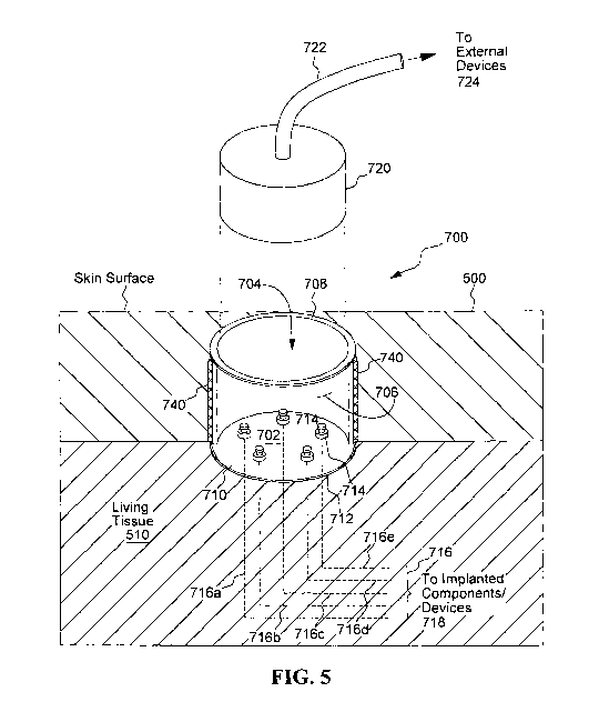
[0072] FIG. 5 schematically illustrates one embodiment of a percutaneous
port 700, or "percuport", made in accordance with one preferred embodiment of
the
inventions described herein. The percuport advantageously allows multiple
through-
the-skin electrical connections to be established while at the same time
minimizing
the risk of infection and soreness. The schematic illustration of the
percutaneous
port 700 in FIG. 5 shows five separate connections that can be made through
the
percuport in order to interconnect external devices or equipment with
implanted
components or devices. This number of connections is only exemplary. As few as
one connection, and as many as 20 or more connections may be provided
depending upon the particular type of neurostimulation system, or other
medical
system, that uses the percuport. In most instances, and for most applications,
at
least three or four independent connections will be provided through the
percuport.
[0073] The percutaneous port 700 shown in FIG. 5 includes an insulative
plate 702 located at the bottom of a cavity 704. The cavity 704 is formed by a
cylindrical or tubular side wall 706 in combination with the bottom insulative
plate
702. An upper edge 708 of the cylindrical side wall forms a rim. When the
21
CA 02777412 2012-04-11
WO 2011/046586 PCT/US2010/002658
percuport 700 is embedded on or in the skin 500 of living tissue 510, the rim
708.is
typically positioned so that the rim is flush with, or extends slightly above,
e.g., 1 to 3
mm above, the surface of the skin 500. Most of the volume of the cavity 704,
however, resides below the surface of the skin, thereby having the cavity 704
appear
as a dimple or indentation in the skin.
[0074] The insulative plate 702 located at the bottom of the cavity 704, for
the
embodiment shown in FIG. 5, has a first surface 710 that faces upward (as the
cavity is oriented in FIG. 5) towards the open end of the cavity 704 where the
skin
surface is located. A second surface 712 of the insulative plate 702 is
exposed to
tissue 510 below and around the cavity 704. Thus, when embedded in the skin,
the
wall(s) 706 and bottom insulative plate 702 of the Percuport 700 serve the
same
basic function as the skin 500 - they provide a protective barrier or layer
that
protects the living tissue 510 under the skin from exposure to the external
environment.
[0075] Still with reference to FIG. 5, it is seen that a plurality of
feedthrough
pins 714 extend through the insulative plate 702. Five such feedthrough pins
714
are shown in FIG. 5, but this number is only exemplary. Typically, as has been
previously indicated, for most applications with which the percuport is used,
at least
three or four feedthrough pins will be used, but as many as twenty, or more,
could
also be employed, depending on the particular application. A percuport could
be
fabricated with only one feedthrough pin 714, but if only one electrical
connection
through the skin was all that were needed, a plurality of feedthrough pins
would still
likely be employed so that the pins could be electrically connected in
parallel to
provide redundancy and thereby improve reliability.
[0076] Each feedthrough pin 714 is made from a biocompatible conductive
material, such as a biocompatible metal, that allows an electrical current to
flow
through it with little or no resistance, and thus allows an electrical
connection to be
established between a proximal end of the pin (the end extending out or
accessible
from the first surface 710 of the plate 702) and a distal end of the pin (the
end
extending out or accessible from the second surface 712 of the plate 702,
which
second surface is exposed to the body tissue 510 below the skin, and is the
surface
on the underneath side of the plate 702 as drawn in FIG. 5).
22
CA 02777412 2012-04-11
WO 2011/046586 PCT/US2010/002658
[0077] It should be noted that the insulative plate 702 need not necessarily
comprise the entire bottom surface of the cavity 704 as shown in FIG. 5, All
that is
required is that the insulative surface comprise a portion of the bottom
surface, or of
the wall surface, where the feedthrough pins are placed. Because the
insulative
plate 702 is typically made from some sort of ceramic material, or other
material that
has electrical insulative properties, how much of the bottom surface (or of a
wall
surface) that is made from the ceramic or other insulative material will be
determined
in large part by how the percuport is assembled during manufacture. Numerous
manufacturing techniques could be used to assemble the percuport, and to
include
therein an appropriate surface area through which the feedthrough pins could
be
placed. For purposes of this patent application, and the inventions described
herein,
any of these known, or yet to be developed, manufacturing techniques could be
used to manufacture and assemble the percutaneous port 700.
[0078] The distal end of each feedthrough pin 714 is connected to a
respective lead 716. Five such leads, 716a, 716b, 716c, 716d and 716e, are
shown
in FIG. 5 with a proximal end of each lead being attached to the distal end of
the one
of the five feedthrough pins 714. The number five is only exemplary, and any
number of leads may be used depending upon how many feedthrough pins 714 are
needed for a particular neurostimulator system application. A distal end of
each
lead 716a, 716b, 716c, 716d and 716e is then directed through tissue 510 to
appropriate or designated implanted components/devices 718 (not shown in FIG.
5).
Sometimes the devices or components to which the distal end of the implanted
leads are attached will be as simple as an electrode that is positioned near
target
tissue that is to be stimulated. Other times the devices may be complex
implantable
neurostimulator circuits or devices, or power sources for such devices, or
sensors
used with such devices, as dictated by the particular application with which
the
neurostimulator system is used.
[0079] An external plug or cartridge 720 is configured to be inserted into the
cavity 704 of the percuport 700 in order to facilitate electrical connection
with the
proximal ends of the feedthrough pins 714. For many applications, a cable 722
is
connected to this plug 720. The cable may have a plurality of wires or
conductors in
it, e.g., five wires or conductors, and each wire or conductor is terminated
inside of
23
CA 02777412 2012-04-11
WO 2011/046586 PCT/US2010/002658
the plug 720 at a respective terminal so that when the plug 720 is inserted
all the
way into the cavity 704, each terminal makes contact with the proximal end of
a
respective feedthrough pin 714. Thus, by removably inserting the plug 720 into
the
cavity 704 of the percuport 700, it is possible to have individual wires
within the cable
722 establish electrical connectivity with the respective implanted leads
716a, 716b,
716c, 716d and/or 716e, via the feedthrough pins 714. In this manner,
electrical
connectivity can be established through the percutaneous port 700 between
external
devices 722 (that are connected to a proximal end of the cable 722) and
implanted
devices 718 (that are connected to a distal end of the leads 716).
[0080] Still with reference to FIG. 5, a mesh material 740 is disposed around
a
periphery of the insulative plate 702 or tubular wall 706. This mesh material,
for the
configuration shown in FIG. 5, is attached to the cylindrical wall 706 that
engages
with the periphery of the insulative plate 702. This mesh material 740 is made
from
a biocompatible material and is configured to promote tissue ingrowth and
vascularization. More details concerning this mesh material are described
below
and/or in the references cited herein that are incorporated herein by
reference.
[0081] As thus described, it is seen that through use of the percutaneous port
700, an external part, e.g., the plug. 720, or an external device 724
connected
through a cable to the plug 720, is able to establish connectivity with a
proximal end
of at least one of the feedthrough pins 714 located in the cavity 704 of the
percutaneous port 700 when the plug is removably inserted into the
percutaneous
port 700. When this connectivity occurs between the external device 724 and
the
proximal end of a feedthrough pin 714 located in the cavity of the percuport
700,
connectivity is also established with the distal end of the feedthrough pin
714, which
also establishes direct connectivity with an implanted part 718 via a lead 716
attached to a distal end of the feedthrough pin 714. Hence, use of the
percuport
700 advantageously establishes electrical connectivity between the external
part and
the implanted part of the percutaneous implant system through direct
electrical
connection through the feedthrough pins passing through the insulative plate
of the
percutaneous port.
[0082] Turning next to FIGS. 6 and 7, there is shown another embodiment of
a percuport 700 made in accordance with the teachings presented herein. The
24
CA 02777412 2012-04-11
WO 2011/046586 PCT/US2010/002658
percuport 700 shown in FIG. 6 shows the percuport prior to being embedded in
the
skin of a user. The configuration shown in FIG. 6 is particularly well suited
for
situations where its lower surface rests against the surface of a bone, or
other hard
tissue, such as the skull. Placing the bottom surface of the percuport against
the
skull is something that is may be needed, e.g., when the percuport is used as
part of
a cochlear implant system, a middle ear implant system, or deep brain
stimulation
system.
[0083] FIG. 6 shows a perspective view of an exemplary percuport 700 which
is merely illustrative of the many different types of ports that may be used
in
connection with the systems and methods described herein. FIG. 7 shows a
sectional view of the port 700 when embedded in skin tissue so that its base
resides
against the skull of a patient.
[0084] An exemplary implant location of percutaneous port 700, when used,
e.g., in a cochlear implant system, or a deep brain stimulation system, is on
the head
of a user,.as described more fully, e.g., in Applicant's copending
application, Serial
No. 61/224211, filed 7/9/2009, entitled "Percutaneous Cochlear Implant Systems
and Methods", which application is also incorporated herein by reference.
Typically,
when used in a cochlear implant system, the port 700 will be located a certain
distance behind the ear (e.g., 2-3 cm) and behind the hair line. Such an
implant
location is advantageous for many reasons. For example, because port 404 is
located behind the hairline, it is generally not visible or noticeable to
others because
it is just a small circle near the skin surface, much like a mole or scab. In
some
examples, this circle may be colored or otherwise disguised.
[0085] The exemplary port 700 shown in FIGS. 6 and 7 is circular in cross-
section in order to accommodate one or more circular components. It should be
noted, however, that percutaneous port 700 may have cross-sectional shapes
other
than circular in order to, for example, accommodate components that are oval,
square, rectangular, or otherwise shaped.
[0086] Percutaneous port 700 may have any suitable length as may serve a
particular patient or application. In some examples, the length of port 700
may be
slightly more than the thickness of the skin. If mounted on the surface of a
bone,
e.g., on the skull, a pocket having a depth of a few millimeters may be made
in the
CA 02777412 2012-04-11
WO 2011/046586 PCT/US2010/002658
skull (or other bone surface), or a spacer can be added in order to
accommodate a
port 700 having a depth greater than the depth of the skin above the skull. In
some
examples, a proximal end of port 700 may extend beyond the skin when implanted
by up to 2 or 3 mm. Alternatively, the proximal end of port 700 may be
substantially
flush with the surface of the skin. Hence, an exemplary length of port 700 may
be
12 to 14 mm. In other patients (e.g., children) with skin that is less thick
(e.g., 5
mm), the length of port 700 may be reduced accordingly. For example, the
length of
port 700 may be 6 to 7 mm for such patients. Likewise, the diameter of port
404
may vary as may serve a particular patient. It will be recognized that these
measurements, and all others presented herein and in the drawings, are merely
illustrative and are not to be construed as limiting in any way.
[0087] As shown in FIGS. 6 and/or 7, port 700 may include a tubular or
cylindrical wall 706 with a rounded rim 708, a layer of porous material 740
surrounding wall 706, and a base flange 709. Rounded rim 504, which may be
located adjacent to the epidermal surface when port 700 is implanted into the
patient, strengthens tubular wall 706 and eliminates what might otherwise be a
sharp
edge that could be uncomfortable to the touch. Tubular wall 706 defines a
tubular
or cylindrically shaped lumen or cavity 704 in which one or more external
components of a percutaneous neorostimulation system 100 may be housed and/or
through which one or more components may be accessed and/or controlled. (As
previously mentioned, the cavity 704 may be made to have cross-sectional
shapes
other than tubular or cylindrical, e.g., oval, rectangular, square, or
triangular,
although any corners associated with polygonal shapes are typically rounded
sufficiently to avoid sharp or uncomfortable edges). Tubular wall 706 may be
made
out of any suitable biocompatible material (e.g., titanium, nitinol, stainless
steel, gold,
or platinum) as may serve a particular application.
[0088] In some embodiments, a center protrusion may extend up from the
bottom or floor of the port 700 to accommodate rotation or keyed-positioning
of
components that are inserted into the cavity 704 of the port 700.
[0089] The layer of porous material 740, which may at a minimum be located
just below the patient's epidermis and in contact with the dermis, is
configured to
encourage tissue ingrowth and vascularization so as to create an infection
resistant
26
CA 02777412 2012-04-11
WO 2011/046586 PCT/US2010/002658
barrier, or percutaneous seal, around tubular or cylindrical wall 706 after
implantation. The layer of porous material 740 extends around the entire
circumference of tubular wall 706 (as shown) and may extend from one
longitudinal
end of tubular wall 706 to the other, or over only a portion of tubular wall
706. In
certain exemplary implementations, the layer of porous material 740 may
include a
mesh of intersecting fibers of any suitable biocompatible material, such as a
biocompatible metal (e.g., titanium, nitinol, stainless steel, gold, or
platinum) or a
biocompatible polymeric material (e.g., polyolefins, Teflon, nylon, Dacron, or
silicone). The mesh is formed by cross-winding the fibers in multiple layers
to define
a porosity conducive to promoting tissue ingrowth (e.g., pore sizes within a
range of
50 to 200 microns and having a porosity of 60 to 95%). The infection resistant
barrier may be enhanced by incorporating antimicrobial and/or anti-
inflammatory
constituents into or beyond the layer of porous material 740. Additional
details
concerning such porous material layers may be found in U.S. Patent Pub. Nos.
2004/0204686, 2007/0112334 and 2007/0149949, each of which is incorporated
herein by reference.
[0090] Base flange 709 may be configured to facilitate fixation of port 700 to
the skull or other bone or hard tissue surface. To this end, one or more
screws 711,
or other affixation devices, may be used to affix base flange 709 of port 700
to the
skull or other hard tissue surface. In some alternative embodiments, port 700
is not
affixed to the skull and instead simply floats with the tissue ingrowth that
forms into
porous material 740 to secure port 700 within the tissue.
[0091] As shown in FIG. 7, a feedthrough plate 713 is disposed in a portion of
wall 706 near a distal end of cavity 704, but not at the distal end of cavity
704. For
the configuration shown in FIG. 7, where base flange 709 presupposes that the
distal end of port .700 will reside against a hard surface, such as the skull,
the
feedthrough pins 714 may extend out through the side wall 706, thereby
avoiding the
hard bone tissue of the skull or other hard surface . Thus, the feedthrough
plate 713
is positioned above the distal end of cavity 704 so that the distal end of the
feedthrough pins 714 reside above the surface of the skull, thereby
facilitating
attaching leads thereto without compromising the integrity of the skull.
27
CA 02777412 2012-04-11
WO 2011/046586 PCT/US2010/002658
[0092] Thus, it is seen that in combination the tubular wall 706, the distal
end
or bottom of port 700, and the feedthrough plate 604 (which comprises a
portion of
the wall 706) define a receiving region or cavity 704 into which one or more
components may be inserted. In some embodiments, as shown in FIG. 5,
feedthrough plate 710 comprises a bottom surface of port 700. In other
embodiments, as shown in FIG. 7, feedthrough plate 713 comprises a portion of
tubular wall 706. In yet other embodiments, as shown in Applicant's copending
patent application Serial No. 61/224211, the feedthrough plate may comprise a
wall
of an hermetic chamber built into the bottom of port 700.
[0093] Feedthrough plate 710 or 713 may assume various shapes and forms.
Whatever the shape or form, however, the function of the plate is essentially
the
same: to provide a surface through which feedthrough pins 714 may extend in
order
to provide electrical connectivity between one side of the plate with the
other. This is
necessary because one side of the plate defines a region or surface area that
is
appropriately sealed or protected from the surrounding environment, while the
other
side of the plate is not. Electrical circuitry that is implanted, for example,
must
typically reside in an hermetically sealed cavity or otherwise be sealed and
protected
from body fluids and tissue if it is to reliably perform its intended function
over a long
period of time.
III. Exemplary Neurostimulation Systems utilizing a Percutaneous Port
[0094] FIG. 8 schematically depicts the manner in which a percutaneous port
may be used with the systems and methods described herein to provide a link
between external and implanted components of an implanted neurostimulation
system. As FIG. 8 depicts, a percutaneous port 700 is found in all embodiments
of
the systems and methods described herein relating to a percutaneous
nerostimulation system. Thus, as seen in FIG. 8, every such system includes a
percuport 700 that is embedded in the skin 500 of a patient. Below the skin,
or
"implanted" in the patient, are implanted circuits 302 that carry out the
functions of
the system. These functions are the same as are carried out in any implant
system.
The circuits 302 may have implantable leads 312 and 316 extending therefrom
that
connect respectively to a suitable sensor or a lead with electrodes. The
circuits in
28
CA 02777412 2012-04-11
WO 2011/046586 PCT/US2010/002658
housing 302 are connected to the percuport 700 via a suitable connection 306,
which may be a flexible cable or other suitable implantable cable or lead.
Alternatively, in some embodiments, the percuport 700 may be affixed to the
top or
side of the circuitry housing 302, in which case feedthrough pins 314 (see
FIG. 5)
may extend all the way through a bottom insulative plate 702 of the port 700
into the
inside of the housing 302.
[0095] The particular electronic circuitry housed in the implanted circuits
302,
including any particular modules of a particular configuration, along with its
manner
of operation, programming codes, stimulation levels and/or stimulation
patterns, and
the like, will not be described in detail in this patent application, if at
all. This is
because such details are generally not the subject of the present application
and the
invention(s) described and claimed herein. Rather, the invention(s) described
and
claimed herein focus more on the manner in which the particular modules used
by or
within a particular configuration of a neurostimulation system can be
configured or
arranged relative to a percutaneous port 700. Thus, it is seen that a
percutaneous
port 700 is a common feature of all of these configurations.
[0096] The actual circuitry used within the various modules associated with
the configurations of the neurostimulation systems of the present
invention(s), as
well as the assembly and manufacturing techniques used to make the implantable
housings, leads, connectors and electrodes associated with these
configurations,
may be of any suitable design, whether presently existing or yet to be
developed. In
fact, that is one of the potential advantages of the present invention (in
some
configurations): by using circuits and components that already exist, and that
have
been tried and tested and approved for use in medical implantable devices, the
percutaneous neurostimulation implant system(s) described herein may be
brought
to market much quicker than could otherwise occur.
[0097] Of course, as with any new configuration, some changes or revisions in
existing designs and circuits need to be made in order to have all the
modules,
circuits and components of the invention interface and cooperate together for
the
system to function correctly and efficiently. Where such changes are more than
routine, and not readily discernable by a person of skill in the art given the
descriptions and explanations already provided herein, or provided in the
documents
29
CA 02777412 2012-04-11
WO 2011/046586 PCT/US2010/002658
that are incorporated herein by reference, such will be described, as
necessary, with
sufficient detail to allow a person of skill in the art to make and practice
those
revisions and changes.
[0098] As seen in FIG. 8, various elements or components of a stimulation
system are external to the patient, i.e., not implanted under the skin. Yet,
these
external components or elements must interface with the implanted circuits
302.
The purpose of the percutaneous port 700 is to allow this interface or
connectivity to
occur, regardless of the form the external components may take. FIG. 9, for
example, shows, in an exploded view, exemplary external devices and components
that may interface with the implanted circuits 302. The particular external
devices
and components which are used depend on the particular application and system
design that is used for the implanted circuits 312.
[0099] For example, one external component that may interface with the
implanted circuits 302 through the percuport 700 is a battery/circuit module
220.
Such module includes a battery, which provides operating power for both the
implanted and external components of the system. Such module may also include,
as needed, at least some additional circuitry, e.g., power management and
monitoring circuitry, used with the neurostimulation system. While FIG. 9
shows the
battery and circuit module 220 as one module, it is to be understood that
these
components could be realized in separate modules or components that are
placed,
e.g., piggy-back into the percuport 700. That is, a power management module
could
be inserted into the most distal end of the percuport 700, and then a battery,
e.g., a
disc battery, could be inserted in the proximal end of the percuport so as to
reside
on top of the power management module. The advantage of having both the
battery
and power management module circuits located or housed in the percuport 700 is
that they can be readily replaced and upgraded, or recharged, as needed.
[00100] Alternatively, if the implanted circuits 302 include all of the
circuitry
necessary to carry out the functions of the neurostimulation system, but do
not
include a battery, then a battery module 224, which in its simplest form is
just a
battery, e.g., a disc battery, may be all that is needed to be inserted into
the
percuport 704.
CA 02777412 2012-04-11
WO 2011/046586 PCT/US2010/002658
[00101] Other modules, represented by the generic box 228 in FIG. 9, may
also be fabricated for insertion into the percuport 700 in order to add
functionality to
the neurostimulation system. Module 228, for example, could be an FM receiver
adapted to receive a FM control or informational signal that, once received
within the
module 228, could be sent to the implanted circuits 302, thereby enabling the
user of
the system to remotely send, e.g., control or informational signals to the
implanted
circuits 302.
[00102] In a similar manner, module 228 could be a Bluetooth receiver that
enables reception of signals that are transmitted to or from a mobile phone or
other
device that utilizes Bluetooth technology. Alternatively, module 228 could
include
a flash memory that stores prerecorded signals, such as MP3 files, that when
inserted into the percuport 700 allows the user to use such prerecorded
signals in a
beneficial manner.
[00103] Another component that could be inserted into the percuport 700, in
accordance with some embodiments of the neuron stimulator systems described
herein, is a passive selector plug 222. In this context, the term "passive"
simply
means that in this embodiment, there is no electronic circuitry included
within the
selector plug 222. Rather, the passive plug functions as a stopper, like a
cork, that
is inserted into the cavity of the percuport 700. Unlike a cork, however, the
plug is
adapted for rotational movement within the cavity 704 of the percuport 700,
and
includes some sensible elements, e.g., conductive metal contacts or traces,
spaced
around its distal end or sides in a desired pattern. Because of this
rotational
movement, and the pattern of conductive traces or contacts included on at
least one
surface thereof, this passive selector plug may also be referred to herein as
a
"cartridge".
[00104] The passive selector plug 222 will be described in more detail
hereinafter. Essentially, however, the passive selector plug 222, when
inserted into
the port 700, allows the user, by manually rotating the plug in prescribed
directions
(clockwise, counterclockwise), and prescribed distances or magnitudes (1/4
turn, 1/3
turn, '/2 turn, etc.) to manually control some functions of the implanted
neurostimulation system, such as on/off, amplitude of a stimulus pulse,
selection of
an electrode(s) where a stimulus pulse is to be applied, and the like.
31
CA 02777412 2012-04-11
WO 2011/046586 PCT/US2010/002658
[00105] For many embodiments of an implanted neurostimulation system,
there is a recurring need to access the implanted circuits 302 for the purpose
of
charging the battery (if a rechargeable battery is included in the implanted
circuits
302) and for programming the circuits or performing diagnostic tests on the
circuits.
Battery charging is readily achieved by simply inserting a battery charger
plug 230
into the cavity of the percuport 700. Such battery charger plug 230 is
connected to a
cable 232 that in turn connects to an appropriate external battery charger
circuit.
Alternatively the battery charger can be a small module that includes a
battery that is
connected to a plug that fits into the percuport 700. Also, an auxiliary
battery can be
inserted into the percuport 700 to extend the operating time of a system with
an
implanted rechargeable batttery.
[00106] Programming is similarly achieved by inserting a programming plug
234 into the percutaneous port 700. A cable 236 attached to the plug 234
allows the
implanted circuits 302, via the connectivity provided by the percuport 700, to
be
connected directly to external programming or diagnostic equipment. Such
external
programming or diagnostic equipment is typically realized through using custom
software loaded on a laptop or other suitable computer, as is known in the
art.
[00107] Thus, it is seen that the neurostimulation system shown in FIGS. 8 and
9 allows a wide variety of configurations and embodiments to be realized. The
percutaneous port 700 is the common element that makes all of these
configurations
and embodiments possible.
[00108] Turning next to FIG. 13, there is shown a schematic representation of
a fully implantable, programmable, multi-channel neurostimulation system 303.
A
key component of the system 303 is the percutaneous port 700. Use of the port
700
advantageously allows a user of the system 303 to selectively establish direct
electrical connectivity between external components and implanted components.
The external components used with the system 303 typically include an external
power source 240 and an external programming/diagnostic device 250. The
implanted components used with the system typically include an implanted
replenishable power source 324, such as a rechargeable battery, a programmable
implantable pulse generator 322 and an implantable stimulation router circuit
326.
32
CA 02777412 2012-04-11
WO 2011/046586 PCT/US2010/002658
[00109] Control circuitry for controlling the operation of the implantable
circuits
may be included within the programmable pulse generator circuits 322, the
stimulation router circuits 326, or both. Such control circuitry includes the
necessary
processing circuitry, memory circuitry, switching circuitry, and the like,
used to cause
stimulation pulses to be generated at appropriate times and with desired pulse
amplitudes and pulse widths, and delivered to desired target tissue locations,
as
programmed by the external programming device 250. An appropriate signal/data
bus 327 connects the programmable pulse generator circuits 322 with the
stimulation router circuits 326
[00110] The external power source 240 may comprise a recharging circuit
connected to conventional source of power, such as a 110 VAC socket located in
a
user's residence (connected through a. power cord 243), which voltage is then
isolated, e.g., through a transformer, and converted to a lower AC voltage,
and then
rectified and converted to the desired dc voltage level. Alternatively, the
external
power source 240 may comprise a conventional battery, or some other suitable
source of power. The external power source 240 connects with the implanted
power
source via a power cable 242 that is coupled to a suitable plug 238 adapted to
be
removably inserted into the cavity 704 of the percuport 700. Such external
power
source is typically used only to recharge or replenish the implantable power
source
324. Thus, so long as the implanted power source 324 has sufficient energy
stored
therein to power the operation of the system 303, the external power source is
not
needed and need not be connected to the system 303 through the percutaneous
port 700.
[00111] The external programming device 250 may comprise a conventional
laptop or notebook computer loaded with appropriate programming software. It
may
connect with the implanted programmable pulse generator 322 and/or stimulation
router circuits 326 via a USB cable 252, or similar cable, that also connects
to the
plug 238, which plug 328 is adapted to be removably inserted into the cavity
704.
Once the implanted circuits have been programmed, there is no need to keep the
programming device 250 connected to the implanted circuits, unless testing or
monitoring of the implanted circuits is desired.
33
CA 02777412 2012-04-11
WO 2011/046586 PCT/US2010/002658
[00112] For the discussion that follows relative to FIG. 13, it is assumed
that all
of the implanted components reside in the same hermetically sealed implantable
housing 304, and that electrical connection with the circuits and components
housed
within the implantable housing is established through the use of feedthrough
pins
328 that are mounted in the housing walls at appropriate locations. (A
proximal end
of these feedthrough pins 328 is connected to the distal end of feedthrough
pins 714
[see FIG. 5] of percuport 700 via a suitable implantable wire 329. A proximal
end of
the perpuport feedthrough pins 714 makes electrical contact with respective
terminals of the plug 238 when such plug is removably inserted into the cavity
704 of
the percuport 700, as previously described.) Thus, as seen in FIG. 13, in
order to
establish electrical connectivity between the implanted circuits and the
external
circuits through the percuport 700, implanted wires 329 interconnect the
distal end of
the feedthrough pins 714 located in the bottom (or other location) of the
percuport
700 with a proximal end of feedthrough pins 328 mounted on one of the surfaces
of
the implantable housing 304. In some embodiments, one or more of the implanted
components 324, 322, and 326 may reside in a separate hermetically-sealed
implantable housing that is electrically coupled with the other implantable
components through appropriate implanted leads or insulated wires that
interconnect
the implanted housings through feedthrough pins mounted on the respective
housings.
[00113] Use of a percuport 700 with the system 303 shown in FIG. 13 allows
direct power connectively to be selectively established between an external
power
source 240 and an implanted power source 324. Such direct connectivity allows
the
implanted power source 324 to be recharged or replenished, when needed,
without
having to inductively couple power into the system. Hence, power transmission,
reception, rectification and regulating circuits that have traditionally been
used within
an implanted device to receive power from an external device through the use
of,
e.g., an implanted coil inductively coupled with an external coil, and all the
associated circuitry used therewith to generate and transmit a carrier signal,
rectify
the received signal, filter it, and then regulate power voltages are not
needed. Being
able to eliminate these types of power circuits from the implanted circuitry
greatly
simplifies the system 303 and reduces its cost, and improves its reliability.
34
CA 02777412 2012-04-11
WO 2011/046586 PCT/US2010/002658
[00114] Further, still with reference to FIG. 13, the percutaneous port 700
used
with the system 303 also allows direct signal connectivity to be selectively
established between the implanted circuit portions of the neurostimulation
system
303 and the external programmer/diagnostic device 250. Thus, the wireless
transmission schemes, with their modulated-data-signal-superimposed-on-a-
carrier
transmission signal, that have heretofore been used to provide such signal and
data
connectivity between external and implanted devices need not be employed. This
means that much, if not all, of the circuitry needed to support and carry out
such
signal transmission schemes is not needed, thereby again greatly simplifying
the
system 303, reducing its cost, and making it more reliable.
[00115] The implantable, programmable neurostimulation system 303 shown in
FIG. 13 may be used for a wide variety of applications where a stimulation
signal or
pulse needs to be routed to different tissue locations in a controlled manner.
Such
system 303 employs multiple electrodes El, E2, E3.... En, where n may be as
low
as two, and as high as 20 or more, depending on the application involved. Each
electrode El, E2, E3, ... En is connected to a stimulation router control
module 326
via respective implanted insulated wires 316a, 316b, 316c, ... 316n.
Feedthrough
pins 328 allow these leads to interconnect with the stimulation router control
circuitry
326 located in the hermetically sealed housing 304. Typically several, if not
all, of
these wires 316a, 316b, 316c, ... 316n may be included in the same lead wire
bundle or cable.
[00116] In operation, stimulation pulses may be directed to multiple
electrodes
El, E2, E3, ... En at the same time. Further, different groupings of the
electrodes
El, E2, E3 ... En may also operate as separate stimulation channels. Hence,
the
system 303 may be programmed to function as a multichannel neurostimualtion
system by programming different regimes of stimulation pulses to be generated
and
directed to different groups of electrodes at the same or different times.
[00117] The stimulation router control circuitry 326 receives the stimulation
signal or pulse from the programmable pulse generator 322. In operation, the
pulse
generator and stimulation router circuits, in combination, may be as simple as
a
single current source connected to a multiplexer circuit, or as complicated as
a bank
of programmable, bidirectional current sources connected to each electrode
wire,
CA 02777412 2012-04-11
WO 2011/046586 PCT/US2010/002658
wherein each current source can be selectively turned on or off so as to
provide
current stimuli of any desired amplitude, pulse width and polarity on any
selected
electrode at any selected time.
[00118] The programmable pulse generator 322 (or the stimulation router
control circuitry 326) may also have at least one sensor S1 connected to it
through
an insulated wire 312a via a feedthrough pin 328. For many applications, a
second
sensor S2, connected to the programmable pulse generator 322 (or to the router
control circuitry 326) by way of insulated wire 312b and a different
feedthrough pin
328, may also be employed to compliment sensor S1. The sensors S1 and/or S2
may simply be an electrode positioned to sense potentials or voltages at
selected
tissue locations. Alternatively, the sensors S1 and/or S2 may comprise
implantable
sensors adapted to sense, e.g., body temperature, blood SO2 levels, blood
sugar
levels, tissue movement, or the like. In some embodiments, one or more of the
sensors S1 or S2, or additional sensors, may be mounted inside of the
hermetically-
sealed housing 304, in order to sense events or other operating data that
occur
during the operation the system.
[00119] As has been indicated, the programmable pulse generator 322
generates stimulation pulses in accordance with a regime that has been pre-
programmed into its circuitry. Such regime may be automatically altered or
adjusted, as needed, as a function of parameters sensed through the sensors S1
and/or S2. Further, parameters sensed through sensors S1 and/or S2 may be sent
through direct signal connections established through the percuport 700 to the
external programmer/tester device 250. These parameters may be sent to the
programmer/tester 250 either in real time (if the programmer/tester is
connected
through the percuport 700 at the time of transmission), or they may be stored
in the
programmable pulse generator in a suitable memory device and uploaded to the
programmer/tester 250 at a later time when a connection is established through
the
percuport 250.
[00120] Operational data associated with the operation of the implanted
neurostimulation system 303 may likewise be transferred to the external
programmer/tester device 250. Such operational data may include the amount of
charge left in the implantable power source 324, impedances measured at each
36
CA 02777412 2012-04-11
WO 2011/046586 PCT/US2010/002658
electrode, internal temperature of the implantable pulse generator, data
stored in the
memory of the implantable pulse generator, and the like. By monitoring such
operational data, the programmer/tester 250 is able to monitor operation of
the
neuostimulation system 303, as well as the condition of the tissue in which
the
system is implanted.
IV. Exemplary Manual Control Methodologies
[00121] Next, with reference to FIGS. 10A and 10B, exemplary manual control
mechanisms will be described that may be used with a peripheral nerve
stimulation
system of the type described herein. Such control mechanisms allow electrical
connectivity to be selectively established between an external pulse generator
602
and a selected one of multiple target tissue locations A, B, C, D, E on nerves
512 or
513 as a function of the rotated position of a plug 721 that is removably
inserted into
the percuport cavity 704. FIG. 10B provides of top schematic view of that
which is
shown in the perspective schematic view of FIG. 10A.
[00122] As seen in FIGS. 10A and 10B, various or multiple tissue locations
may require a stimulus pulse to be delivered to them by an external pulse
generator
602. Such tissue locations are identified in FIGS. 10A and 10B by the letters
A, B,
C, D and E. Three of these tissue locations, "A", "B" and "C" are shown in
FIG. 10A
as being located along nerves 513 and 512. (The other two tissue target
locations,
"D" and "E", are not shown in FIG. 10A, but are schematically depicted in FIG.
10B.)
[00123] In order to stimulate a desired or target tissue location, a
percutaneous
port 700 is embedded in the skin 500 of a patient. Leads 652a, 652b, 652c,
652d
and 652e are implanted so that a distal electrode 654a, 654b, 654c, 654d and
654e
on each lead is respectively positioned at or near the target tissue location
"A", "B",
"C", "D" or "E". The proximal end of each of these leads is then connected to
the
distal end of a respective feedthrough pin 650 located on the tissue side of
the
percuport's bottom insulative plate 702. Thus, a proximal end of lead 652a
connects
to the distal end of feedthrough pin 650a, a proximal end of lead 652b
connects to
the distal end of feedthrough pin 650b, and so on.
37
CA 02777412 2012-04-11
WO 2011/046586 PCT/US2010/002658
[00124] In order to direct a stimulus pulse from the external pulse generator
602 to the desired target tissue location, "A", "B", "C", "D" or "E", a
conductive arm
725 is molded, or otherwise affixed, to a rotatable member within the plug
721. As
drawn in FIG. 10A, the entire plug 721 is rotatable when inserted into the
cavity 704
of the percutaneous port 700. Thus, in this instance, the conductive arm 725
may
be molded into a bottom surface of the plug 721. (Other configurations could
also
be fashioned where only the conductive arm 725 rotates relative to the plug
721 by,
e.g., twisting a knob on the top of the plug that is connected to an rotatable
axle that
passes through the center of the plug and is affixed to the proximal end of
the
conductive arm at the bottom surface of the plug.)
[00125] As drawn in FIG. 10B, a proximal end of the conductive arm 725 is
located near the center of a bottom surface of the plug 721. This proximal end
is
electrically connected by way of a conductive trace or wire 727 that extends
from the
bottom surface of the plug 721 to a top surface of the plug 721 where it
connects to
a plug terminal 723. One of the output signal wires 635 from the pulse
generator
connects to this top surface plug 723. For monopolar stimulation, the other
output
signal wire 606 from the pulse generator 602 is grounded, e.g., by connecting
it to a
surface electrode located on the surface of the skin of the patient. The other
end of
the conductive arm extends radially outward from the center of the plug and is
of a
sufficient length so as to engage a proximal end of a feedthrough pin 650 when
the
plug 721 is inserted into the percuport cavity 704 and is rotated therein so
as to
cause such engagement. To facilitate rotation of the plug 721 within the
cavity 704,
a center post 742 may extend upward from the bottom of the percuport cavity
704,
and a corresponding hole 743, adapted to receive the center post therein when
the
plug 721 is inserted into the cavity 704, extends upward from the bottom of
the plug
721 a sufficient distance to allow the plug 721 to be fully inserted into the
cavity 704.
[00126] The manner of operation of the selective stimulation mechanism
illustrated in FIGS. 10A and 10B is best illustrated from the top view
schematic
diagram of FIG. 10B. When a stimulation pulse is to be applied to target
tissue at a
selected location, the plug 721 is fully inserted into the cavity 704 of the
percutaneous port 700, the pulse generator is connected to the terminal 723
located
on the top of the plug 721, and the plug is rotated until the distal end of
the
38
CA 02777412 2012-04-11
WO 2011/046586 PCT/US2010/002658
conductive arm 725 engages the proximal end of the feedthrough pin 650 that is
connected through lead 652 to the desired target tissue location. For example,
if
target tissue location "A", located on nerve 513, is to be stimulated, the
plug 721
(after being fully inserted into the percuport cavity 704 and connecting the
output
signal line 635 from the pulse generator 602 to the terminal 723 located on
the top of
plug 721) is rotated within cavity 704 until the distal end of the conductive
arm 725
engages the proximal end of feedthrough pin 650a. When such engagement
occurs, electrical connectivity is established between the pulse generator and
target
tissue location A by way of electrode 654a, lead 652a, feedthrough pin 650a,
conductive arm 725, conductive trace 727, terminal 723, and wire 635. A new
stimulation target site is readily selected by simply rotating the plug 721 a
prescribed
amount, e.g., 1/5 of a turn. A 1/5 rotation clockwise, as viewed from the top
of the
plug 721, would deactivate stimulation target site "A" and activate
stimulation target
site "E". A 1/5 rotation counter-clockwise would deactivate stimulation target
site "A"
and activate stimulation target site "B". A less than 1/5 rotation of the plug
721 could
place the distal end of the conductive arm 725 in a location where it is not
engaged
with any of the feedthrough pins 650. Such positioning would effectively turn
the
stimulation pulses off. Thus it is seen that through selective rotation of the
plug 721,
while it is fully inserted into the cavity 704 of the percuport 700, any one
of the
stimulation sites "A", "B", "C", "D" or "E" may be selected as the site
receiving a
stimuli from pulse generator 602, or none of the stimulation sites may be
selected,
effectively disabling (turning off) the stimuli provided by the pulse
generator.
[00127] The technique to manually select a desired stimulation target site, as
described above (through selective rotation of the plug or cartridge inserted
into the
percuport's cavity 704) can also readily be achieved through electronic
switching
circuitry that is part of the implanted circuits 302 that form part of a
neurostimulation
system. The advantage of using a percuport 700 with such an implatable
neurostimulation system is that it greatly facilitates the functions of
powering,
programming and testing such system. External batteries, battery chargers,
programming circuits, and/or diagnostic equipment can be connected directly
with
the implanted circuits 302 of the system as needed, or desired, through the
connectivity provided by the percuport. When these external elements are not
39
CA 02777412 2012-04-11
WO 2011/046586 PCT/US2010/002658
needed, then nothing need be connected to the percuport. Rather, it can just
have a
passive plug or cover placed over it.
[00128] Next, with reference to FIGS. 11A-11F, a more robust mechanism is
disclosed that allows various controls or commands to be manually generated
through selective rotation of the plug 721 within the port's cavity 704. FIGS.
11A-
11 F are plan views showing a plurality of sensible members, e.g., conductive
pads,
moving relative to a pair of sensors contained within a bottom edge of a
percutaneous port 700. Advantageously, being able to sense the location of the
sensible members provides a manual user interface that allows a user the
ability to
generate control signals for controlling at least some functions of an
implantable
neurostimulation system through manual rotation of the plug or cartridge 721
inserted into the cavity 704 of the percutaneous port.
[00129] A neurostimulation system that utilizes a percutaneous port 700 in
accordance with the present inventions may be programmed and/or controlled in
any
suitable manner. For example, as described briefly previously in connection
with
FIG. 9, some implementations of the present system may include module 228
adapted for insertion into the percuport 700, wherein module 228 may include
an
antenna (e.g., in combination with an FM receiver and/or BlueTooth receiver)
and
receive instructions and/or programming information by way of a telemetric
programmer. Some implementations of the present system may include a data
connector (e.g. a micro-USB connector within the module 234 that allows
instructions and/or programming information to be received by way of a wired
connection to a programmer.
[00130] Alternatively, or in addition, the percutaneous port 700 and a passive
selector plug 222 or 721 may be configured to function as a user interface
that
allows attending medical personnel and/or the patient (user of the system) to
control
various aspects of the operation of the system and/or to input programming
commands while implanted. This is accomplished by rotating the plug 222 or 721
relative to the percuport 700 in a prescribed direction for a prescribed
amount in a
prescribed sequence. For such rotation to generate the needed control signals,
the
percuport 700 has a pattern of paired contacts, e.g., contacts 170a and 170b,
and
CA 02777412 2012-04-11
WO 2011/046586 PCT/US2010/002658
contacts 172a and 172b, placed in the bottom of the cavity 704 thereof. These
contacts are arranged in a pattern as illustrated, e.g., in FIGS. 11A-11F.
(00131] More specifically, in the exemplary implementation, the pair of
contacts
170a and 170b comprise a control sensor 124, and the pair of contacts 172a and
172b comprise a control sensor 126. Together, these two pairs of contacts
provide
a pair of circumferentially spaced control sensors 124 and 126 embedded in the
bottom or floor of the percuport 700. The passive selector plug 721 (also
shown as
222 in FIG. 9), functions, when inserted into the cavity 704 of the percuport
700, as
a rotatable cartridge. It has a pattern of spaced metal (or conductive)
surfaces, or
sensible members 250, spaced around its bottom surface as shown in FIGS. 11A-
11 F. When the selector plug, or cartridge, 721 is inserted into the percuport
404, the
conductive surface of the sensible members 250 makes electrical contact with
none
or both of the paired contacts of a given sensor 124 or 126. That is, the
exemplary
spaced sensible members 250 are electrically conductive pads. These
electrically
conductive pads either short together the paired contacts, or not, depending
upon
the rotational position of the cartridge on which the spaced sensible members
250
are placed. Thus, by monitoring the individual contacts associated with the
contacts
170a and 170b (for sensor 124), and the contacts 172a and 172b (for sensor
126)
with appropriate monitoring circuitry, it is possible to detect when the
paired contacts
170a and 170b, or 172a and 172b, are shorted together (which occurs when the
sensible member 250 is in contact with both contacts), or are not shorted
together
(which occurs when the sensible member 250 is not in contact with both
contacts).
[00132] Thus it is seen that a detectable short occurs between contact 170a
and contact 170b when these contacts are both aligned with one of the
electrically
conductive pads 250. Similarly, a detectable short occurs between contact 172a
and contact 172b when these contacts are both aligned with one of the
electrically
conductive pads 250.
[00133] Such sensing advantageously may be used by the circuitry within the
neural stimulator system to determine the direction and magnitude of the
rotational
movement of the cartridge (passive selector plug) 721 relative to the
percutaneous
port 700, as is discussed below with reference to FIGS. 11A-11F. The number of
times there is (and is not) a short across contacts 170a/170b and contacts
41
CA 02777412 2012-04-11
WO 2011/046586 PCT/US2010/002658
172a/172b, and the order in which the short or open changes occur, is
indicative of
the magnitude and direction of the rotational movement of the cartridge 721
relative
to the percutaneous port 700. The patient or other medical personnel may
simply
rotate the passive selector plug (cartridge) 721 in a predetermined manner to
input
commands and/or otherwise interface with the implanted circuits that form part
of the
neural stimulator system, as is discussed below with reference to FIG. 12.
[00134] In FIGS. 11A-11F, the exemplary sensible members 250 (which are
spaced around a bottom or distal surface of the passive selector plug (or
cartridge)
721 are superimposed over an end wall 702 of the cavity 704 of the percuport
700.
That is, the end wall contains control sensors 124 and 126. The relative
position of
the control sensors 124 and 126 with respect to the superimposed sensible
members 250 is shown in FIGS. 11A-11F in order to illustrate the changes in
the
relative rotational orientations of the sensible members and control sensors
that
occur when a cartridge 721 is located within the cavity 704 of the
percutaneous port
700 and rotated relative thereto.
[00135] FIG. 12 illustrates the manner in which the direction and magnitude of
the rotational movement of the passive selector plug 721 relative to the
percuport
700 may be determined. FIG. 11A represents one exemplary initial orientation
of
the sensible members 250 and cartridge 721 (not shown) relative to the
percutaneous port 700. No sensible member 250 is aligned with the contacts on
either of the control sensors 124 and 126 in the illustrated rotational
orientation and,
accordingly, no sensible member is sensed at either of the control sensors (a
"124-
no/126-no" state). Of course, and as will be clear from the discussion below,
the
initial rotational orientation of the sensible members 250 (and cartridge 422)
need
not be that shown in FIG. 1 1A.
[00136] In FIG. 11 B, the sensible members 250 (and cartridge 721) have been
rotated relative to the percutaneous port 700 in the direction of arrow A such
that the
sensible member 250a is aligned with the contacts 172a/172b of control sensor
126
and no sensible member is aligned with the contacts 170a/170b of control
sensor
124. A sensible member will, accordingly, not be sensed at control sensor 124
and
will be sensed at control sensor 126 (a "124-no/126-yes" state). The
transition from
42
CA 02777412 2012-04-11
WO 2011/046586 PCT/US2010/002658
the 124-no/126-no state to the 124-no/126-yes state indicates that the
sensible
members 250 (and cartridge 721) are moving in the counter-clockwise direction.
.
[00137] Turning to FIG. 11C, the sensible members 250 (and cartridge 721)
have been further rotated relative to the percutaneous port 700 in the
direction of
arrow A such that the sensible member 250a remains aligned with the contacts
172a/172b of control sensor 126 and the sensible member 250a is now also
aligned
with the contacts 170a/170b of control sensor 124. A sensible member will,
accordingly, be sensed at both control sensor 124 and control sensor 126 (a
"124-
yes/126-yes" state). The transition from the 124-no/126-yes state to the 124-
yes/126-yes state, without reversion to the prior 124-no/126-no state,
indicates that
the cartridge 721 is continuing to move in the counter-clockwise direction
without any
appreciable movement in the clockwise direction.
[00138] In FIG. 11 D, the sensible members 250 (and cartridge 721) have been
further rotated relative to the percutaneous port 700 in the direction of
arrow A such
that the sensible member 250a is no longer aligned with the contacts 172a/172b
of
control sensor 126 and the sensible member 250a remains aligned with the
contacts
170a/170b of control sensor 124. A sensible member 250 will, accordingly, be
sensed at control sensor 124 and not sensed at control sensor 126 (a "124-
yes/126-
no" state). The transition from the 124-yes/126-yes state to the 124-yes/126-
no
state, without reversion to the prior 124-no/126-yes state, indicates that the
cartridge
is continuing to move in a counter-clockwise direction without any appreciable
movement in the clockwise direction.
[00139] A subsequent transition from the 124-yes/126-no state to the 124-
no/126-no state (i.e. the initial state), without reversion to the prior
state, will indicate
that the movement has continued in the direction of arrow A and, in the
context of
the illustrated implementation, that there has been a single sensor cycle and
that the
cartridge has rotated a total of about 60 degrees from the initial location
(FIG. 11A).
Continued rotation in the direction of arrow A to the location illustrated in
FIG 11 E,
i.e. 180 degrees from the initial location (FIG 11A), will result in two more
sensor
cycles. Again, each sensor cycle is a transition from 124-no/126-no state to
another
124-no/126-no state in the manner described above, and each cycle represents a
43
CA 02777412 2012-04-11
WO 2011/046586 PCT/US2010/002658
rotation of 60 degrees (for the particular spaced orientation of the sensible
members
250 shown in FIGS. 11A-11 F).
[00140] It should be noted here that the 124-no/126-no state need not be the
initial state when monitoring rotational movement of the passive selector plug
721
(or "cartridge" 721, as it is termed here for purposes of this discussion)
relative to the
percutaneous port 700. The initial state is merely the state present when
rotational
movement begins after a predetermined period without rotational movement (e.g.
at
least 5-10 seconds). If, for example, a sensible member 250 is aligned with
the
contacts on both of the control sensors 124 and 126, then the initial state
will be the
124-yes/126-yes state, and a cycle will be a transition from a 124-yes/126-yes
state
to another 124-yes/126-yes state.
[00141] Rotational movement in the opposite direction is sensed in essentially
the same way, although the yes/no transitions will occur in a different order.
For
example, FIGS. 11 E and 11 F show the rotation of the sensible members 250
(and
cartridge 721) relative to the percutaneous port 700 in the direction of arrow
B. The
sensible member 250b will be sensed at control sensor 124 and not sensed at
control sensor 126 in FIG. 11F. The transition from the 124-no/126-no state
(FIG.
11 E) to the 124-yes/126-no state (FIG. 11 F) indicates that the cartridge is
moving in
a clockwise direction.
[00142] Regardless of the type of sensors and sensible members that are
employed, and the manner in which the sensors and sensible members are used to
identify rotational movement of the selector plug (or cartridge) 721 relative
to the
percutaneous port 700, the ability to identify and track such rotational
movement
facilitates the use of the percutaneous port and the cartridge as a user
interface. By
way of example, but not limitation, a variety of user-initiated implantable
medical
device operations may be pre-programmed into the partially implantable medical
device and such operations may be actuated by the port/cartridge user
interface.
Each user-initiated operation may be assigned a unique defined cartridge
rotational
movement or a unique defined combination of rotational movements (collectively
"defined cartridge rotational movement"). A time limit may be applied in at
least
some embodiments. For example, a defined cartridge rotational movement may be
44
CA 02777412 2012-04-11
WO 2011/046586 PCT/US2010/002658
deemed ineffective unless the combination is completed within a predetermined
time
period (e.g. about 15 seconds from the initial detection of rotation).
[00143] The general operation of the user interface and the associated aspects
of the control circuitry used to detect the relative magnitude and direction
of the
rotation of the selector plug 721 is graphically illustrated in the flow chart
of FIG. 12.
More specifically, with respect to user-initiated operation, the control
circuitry of the
implanted circuitry 302 will remain in a standby state (step S01) until
rotational
movement of the cartridge is sensed (step S02). A timer is initiated in
response to
the sensing of cartridge rotation (step S03). If one of the defined cartridge
rotational
movements is received prior to the expiration of the predetermined period
(steps
S04 and S05), then the user-initiated operation associated with the defined
cartridge
rotational movement will be initiated (step S06). If, on the other hand, one
of the
defined cartridge rotational movements is not received prior to the expiration
of the
predetermined period (steps S04 and S05), the control circuitry will return to
the
standby state with respect to the user interface aspects of its operation.
[00144] For example, an operation may be initiated in response to the
following
cartridge rotational movement: at least 360 degrees in one direction followed
by
rotation of at least 360 degrees in the opposite direction, with both
rotations
occurring within 15 seconds of the initiation of the first rotation. Another
exemplary
rotation combination is rotation of at least 180 degrees in a particular
direction that is
completed within 15 seconds of the initiation of the rotation. The control
circuitry may
also be configured to actuate an audible and/or vibratory alarm (not shown)
that is
located within the housing 302 (FIG. 9) in response to a successful input of a
defined cartridge rotational movement and/or an unsuccessful input attempt.
Different versions of the alarm (e.g. one beep vs. two beeps) may be used when
the
alarm is actuated in response to both successful and unsuccessful attempts.
[00145] With respect to the user-initiated operations themselves, one example
involves turning the system on or off. Turning the system on/off is somewhat
of a
misnomer because at least some circuits of the system are always on. What
typically occurs when a user decides to turn his or her neural stimulator
system "off
"is that most of the circuits of the system are put in a sleep state, or the
stimulation
CA 02777412 2012-04-11
WO 2011/046586 PCT/US2010/002658
circuits are shut down, so that the user does not receive any stimulation
until such
circuits are turned "on", or placed in an "awake" state.
[00146] Another exemplary user-initiated operation is stimulation magnitude or
intensity adjustment. (For a cochlear implant system, this could be termed
volume
adjustment.) To activate magnitude adjustment, for example, a user may rotate
the
cartridge 721 a prescribed amount, e.g., 60 degrees, in one direction followed
within
a few seconds by rotation in the other direction by the same amount. Then,
once
magnitude control has been activated, a clockwise rotation of the cartridge
would be
interpreted by appropriate control circuitry as a desire to increase the
magnitude of
the stimuli being applied at the target tissue location, whereas a counter-
clockwise
rotation of the cartridge would be interpreted as a desire to decrease the
magnitude
of the applied stimuli.
[00147] There are a variety of advantages associated with a user interface
that
is defined by the percutaneous port 700 and cartridge or plug 721 inserted
therein.
By way of example, and not by limitation, the present user interface obviates
the
need for the patient or user to possess a telemetric remote control and,
accordingly,
obviates the expense and potential inconvenience (if lost or otherwise
unavailable)
associated with a remote control. The present user interface may also
eliminate the
need for telemetric control for programming by the physician, or other medical
personnel, thereby eliminating the need for an antenna and associated
telemetric
circuitry in the implanted neurostimulator system.
V. Conclusion
[00148] As described above, it is thus seen that the inventions described
herein provide a neurostimulation system(s) wherein some components of the
system are implanted and some components of the system are non-implanted, and
wherein the required electrical or signal connectivity between the implanted
components and non-implanted components is readily established through use of
a
percutaneous port embedded in the skin of a user of the system.
[00149] It is further seen that percutaneous connectivity, when implemented as
described herein, provides a high degree of flexibility in how a system using
such
percutaneous connectivity may be configured and optimally used to best meet
the
46
CA 02777412 2012-04-11
WO 2011/046586 PCT/US2010/002658
needs and wants of a particular user or a particular application. That is,
numerous
configurations or embodiments of a percuport system allow different
combinations of
components of the system to be either permanently implanted or not implanted,
as
needed. The advantage of having some of the system components being external
or non-implanted is that the non-implanted components can be readily replaced,
removed upgraded, or recharged as needed.
[00150] Additionally, it is seen that the system(s) described herein, which
include both implanted and non-implanted components, advantageously avoid the
necessity of having to use radio frequency telemetry or inductive coupling to
establish a communicative link for power and/or data signals to pass between
the
implanted portions of the system and the non-implanted portions of the system.
[00151] Moreover, it is seen that a preferred percutaneous port as described
herein allows tissue ingrowth and vascularization. Such tissue ingrowth and
vascularization advantageously provides a percutaneous seal around the
periphery
of the perctaneous port that functions as a very effective barrier to prevent
infection.
[00152] It is also seen that the percuport-based systems described herein
advantageously provide a modular-based implantable neruostimulation system
wherein different component groupings or modules provide different embodiments
suited for different applications or needs. Thus, one embodiment or
configuration
provides a system wherein most components of the system are implanted and only
a
few components of the system (such as a programming/testing module and
recharging module) are non-implanted. On the other hand, another embodiment or
configuration provides a system where most components of the system are non-
implanted and only a few components of the system (such as an electrode lead)
are
implanted. Hence, the modularity of the systems described herein provide a
full
spectrum of possible embodiments -- ranging from a system that is almost fully
implanted to a system that is mostly non-implanted - any of which may be used
to
best meet the needs and demands of a particular patient group or application.
[00153] It is further seen that with the modularity provided by the percuport-
based system(s) described herein, existing, approved and fully tested
implantable
components may be used in implantable modules or housings, and existing,
approved and tested non-implantable components may similarly be used in non-
47
CA 02777412 2012-04-11
WO 2011/046586 PCT/US2010/002658
implantable modules, housings or configurations. Such modularity, and use of
modules containing circuits and designs that are already approved, can greatly
shorten the time required to obtain regulatory approval for the implant system
as a
whole.
[00154] The preceding description(s) has been presented only to illustrate and
describe embodiments of the invention. It is not intended to be exhaustive or
to limit
the invention to any precise form disclosed. Many modifications and variations
are
possible in light of the above teachings. Thus, while the invention(s) herein
disclosed has been described by means of specific embodiments and applications
thereof, numerous modifications and variations could be made thereto by those
skilled in the art without departing from the scope of the invention(s) set
forth in the
claims
48