Note: Descriptions are shown in the official language in which they were submitted.
CA 02778866 2012-04-24
WO 2011/058136 PCT/EP2010/067379
-1-
Calibration reagent and uses thereof
Reverse phase protein arrays (RPA) have been developed and established in the
recent years as a
convenient method to analyze focused sets of proteins representing key
analytes of different
signal transduction cascades in minute amounts of biological samples (e.g.
cell lysates, tissue
lysates, or body fluids). Relative differences of protein expression,
representing not only the
abundance of specific key proteins, but also activated, post-translationally
modified (e.g.
phosphorylated) forms of such key proteins can describe and classify e.g.
specific treatment
effects of pharmaceutical compounds given to cell cultures e.g. inhibitory
effects of drug
candidates on kinases, or describe and classify different disease states e.g.
sub-types of tumors in
their different progression states. RPA can perform comparative measurements
of many samples
in parallel, e.g. samples from differently treated cell cultures or samples
from different disease
populations. Significant changes of protein expression or protein activation
patterns to be found
in distinct sample cohorts will foster e.g. the identification of most
efficient drug candidates, the
elucidation of treatment induced mode-of-action schemes or the discovery of
new
diagnostic/prognostic disease markers.
Immunoaffinity assays such as used in Reverse Phase Protein Arrays (RPA) are
based on
specific interactions between an affinity reagent and a protein of interest.
The assay comprises
the immobilization of the biological samples on the array forming the sample
spots. The sampled
array is incubated with an affinity reagent, i.e. an antibody, and the
subsequently formed
complex of affinity reagent and protein of interest is measured by the
generated detection signal
e.g. a luminescence signal. Each array is stained with an analyte-specific
affinity reagent, which
can be labeled or is incubated with a secondary detection reagent. Formed
complexes are
detected by various means (colorimetric, fluorescence, chemiluminescence
etc.). Typically RPA
measure relative changes of expression or activation signals between different
samples.
The quantitative analysis of samples requires the use of calibration reagents.
Currently, for
protein analytes, the calibration reagents are recombinant proteins having the
same amino
sequence as the analyte. For example, patent application W02007/048436A1
describes
calibration curves for Reverse Phase Protein Microarrays whereby different
concentrations of
purified protein of interest (Akt) were added to spotting buffer comprising
BSA or rat serum.
However, the production of recombinant protein presenting the correct epitope
is time-
CA 02778866 2012-04-24
WO 2011/058136 PCT/EP2010/067379
-2-
consuming and often not successful. In particular for phosphorylated epitopes,
so far no reliable
calibration reagents are available.
Therefore, there is a need for a reagent designed to provide universal
applicability with
choosable specificity for the different analyte epitopes of interest. This
would allow to calibrate
results from experiments performed, e.g. at separated times, by different lab
personal, on
different devices or on arrays constructed in different print runs. Also the
linear range of the
protein-specific RPA signals to be generated by the respective affinity
reagent can be optimally
pre-defined.
Therefore, the present invention provides a calibration reagent comprising a
peptide which
is attached via a linker to a protein carrier, wherein said peptide comprises
an epitope of interest.
Preferably, said epitope of interest is phosphorylated.
With this calibration reagent reliable standard curves can be generated for
quantifying protein
with an RPA or another affinity assay. RPAs are constructed by the deposition
of small sample
volumes e.g. of cell or tissue lysates, onto highly binding substrate surfaces
using often a robotic
microarrayer. Each lysate spot on the substrate contains the full complement
of cellular proteins
and analytes. Hundreds of samples can be spotted in parallel into one
microarray allowing high
throughput cross-comparisons of samples in the same assay. Replicate arrays
containing the
same set of samples, can be easily produced from the same initial volume of
sample material,
since consumption of sample volume per spot is extremely low.
The calibration reagent of the current invention is particular useful for
quantifying proteins
which comprise a phosphorylated epitope of interest.
The term "epitope of interest" refers to a part of a polypeptide which is
recognized by the
affinity reagent of interest. The affinity reagent of interest is preferably
specific for the epitope of
interest.
The term "epitope peptide" as used herein refers to the peptide comprising the
epitope of
interest. The epitope peptide is preferably between 12 and 25 amino acids
long. More preferably,
the length of the peptide is 12 to 20, most preferably 14 to 17 amino acids
long.
The epitope of interest can be modified, e.g. phosphorylated. The term
"phosphorylated
epitope" as used herein refers to an epitope which comprises at least one
amino acid with a
phosphate group. Preferably, the epitope of interest comprises 1 to 5
phosphorylated amino acids.
Preferably, the position of the modified amino acid is approximately in the
middle of the epitope
CA 02778866 2012-04-24
WO 2011/058136 PCT/EP2010/067379
-3-
peptide. Fore example in a peptide of 15 amino acids length, the modified
amino acid is
preferably at position 7, 8 and/or 9 (see figure 2C). Methods for modifying an
amino acid (e.g. to
phosphorylate) are well known to the skilled person in the art.
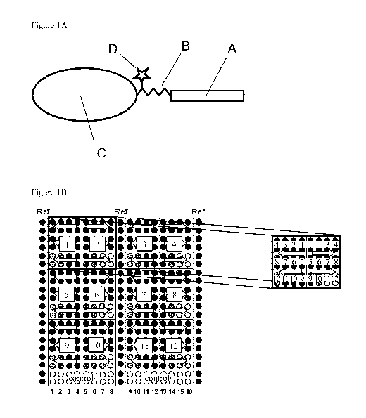
The epitope peptide is covalently bound to the protein carrier via a linker
(see Figure IA),
whereby the epitope peptide is covalently bound to the linker and the linker
is covalently bound
to the protein carrier. In a preferred embodiment, the linker is covalently
bound to the free N-
terminal Cysteine (Cys) group of the BSA, wherein a free Cys group is a
cysteine residue which
is not involved in a disulfide bridge.
The epitope peptide can be attached to the protein carrier in essentially two
steps:
Step 1) The linker is conjugated to the epitope peptide, wherein said linker
is preferably
labeled with a tag. The linker can be attached to the N- or C-terminus of the
peptide. Preferably,
the linker is attached to the N-terminus of the peptide.
Step 2) the free end of the linker is conjugated to the protein carrier.
The linker or spacer is a peptide comprising 2 to 10, preferably 2 to 5, more
preferably 3 to
4 natural or unnatural amino acids. Natural amino acids are naturally
occurring amino acids such
as in particular alanine, cysteine, lysine, histidine, arginine, aspartate,
glutamate, serine,
threonine, methionine, glycine, valine, leucine, isoleucine, asparagine,
glutamine, proline,
tryptophane, phenylalanine, tyrosine. Unnatural amino acids are amino acids
which do not
naturally occur. Examples for unnatural amino acids are 8-amino-3, 6 dioxa-
octanoic acid (Doa)
and aminooxy-acetic acid.
The linker is hydrophilic and can comprise only natural amino acids or only
unnatural
amino acids or a mixture of both, natural and unnatural amino acids.
Preferably, the linker
comprises one or more of the following natural amino acids: cysteine, lysine,
histidine, arginine,
aspartate, glutamate. Also preferably, the linker comprises one or more Doa.
More preferably,
the linker is Cysteine-Doa-Doa.
Preferably, the linker is labeled with Dabsyl.
Methods to produce peptides with a specific amino acid sequence are well known
to the
skilled person in the art. A suitable method is e.g. is the solid phase
synthesis described in
Merrifield, Science 1986, 232:341-347 (method) and Carpino et al., J. Am.
Chem. 1990, 112:
9651-52 (reagents).
CA 02778866 2012-04-24
WO 2011/058136 PCT/EP2010/067379
-4-
The protein carrier is a protein which unspecifically binds to surfaces.
Preferably, the
protein carrier is a protein of at least 20kDa and shows no or low cross
reactivity with the
affinity reagent used in an affinity assay. The protein carrier is preferably
an albumin, more
preferably a serum albumin, such as e.g. bovine serum albumin (BSA) or human
serum albumin.
The preferred serum albumin is BSA.
An "affinity reagent of interest" is a reagent which recognizes and binds the
epitope of
interest. Preferably, the affinity reagent of interest is specific and
selective for the epitope of
interest. The affinity reagent can be an antibody, an aptamer, or a designed
ankyrin repeat
protein (DARPin). Preferably, the affinity reagent is an antibody.
An "antibody of interest" can be any antibody. Preferably, said antibody is an
IgG antibody,
more preferably a monoclonal antibody. The antibody of interest includes but
is not limited to
humanized antibody and rodent antibody. A rodent antibody includes but is not
limited to a
mouse, rabbit and rat antibody. Preferably, the antibody is a rabbit
monoclonal antibody.
An "aptamer of interest" is a single-stranded RNA or DNA oligonucleotide 15 to
60 base
in length that bind with high affinity to the epitope of interest.
A "designed ankyrin repeat protein" or "DARPin" is a binding molecule
comprising at
least one ankyrin repeat. An ankyrin repeat is a motif in proteins consisting
of two alpha helices
separated by loops, which can be selected to recognize specifically a wide
variety of target
proteins. The typical length of an ankyrin repeat is 33 amino acids. Unlike
antibodies they do not
contain any disulfide bonds and are found in all cellular compartments.
Furthermore, the present invention provides the use of the calibration reagent
as described
above for the generation of a standard curve. The present invention provides a
method for
generating a standard curve comprising the steps of:
a) immobilizing the above described calibration reagent in two or more
concentrations on
an array,
b) incubating said array with a detectable affinity reagent of interest,
c) measuring the signal intensity of the bound affinity reagent for each of
the two or more
concentrations of the calibration reagent, and
d) correlating the signal intensity with amount of epitope of interest.
CA 02778866 2012-04-24
WO 2011/058136 PCT/EP2010/067379
-5-
A standard or calibration curve is a quantitative research tool, a method of
plotting assay
data that is used to determine the concentration of a substance, i.e. the
concentration of the
epitope of interest.
The term "bound affinity reagent" refers to the affinity reagent forming a
complex with a
protein or peptide comprising the epitope of interest. Formed complexes are
detected by various
means such as for example colorimetric, fluorescence, or chemiluminescence.
An affinity reagent can be detected, by a detectable label attached to the
affinity reagent.
Preferably, said label is a fluorophore, allowing thereby to determining the
amount of bound
antibody by the fluorescence intensity. Other suitable labels are e.g.
alkaline phosphatase (AP)
and horseradish peroxidase (HRP).
An affinity reagent can also be detected by a secondary detection reagent. A
secondary
detection reagent is a labeled molecule which selectively binds the affinity
reagent. The bound
affinity reagent on the microarray can be detected for example by using a
second antibody or a
Fab fragment, which is labeled and recognizes species-specific epitopes of the
affinity reagent.
Suitable labels include but are not limited to fluorophore, biotin,
horseradish peroxidase, and
isotopes. Preferably, the secondary detection reagent (e.g. a secondary
antibody) is labeled with a
fluorophore.
The amount of affinity reagent bound to the calibration reagent is preferably
detected by an
optical signal such as a fluorescence signal.
The amount of bound affinity reagent of interest is correlated with amount of
epitope of
interest by measuring the detectable signal of the affinity reagent and
attribute each signal a
concentration of the epitope of interest. These results are displayed in a
standard curve. A
standard curve can be drawn by plotting the determined amount of bound
affinity reagent of
interest for each concentration of the epitope of interest (on the Y axis)
versus the concentration
of the epitope of interest (on the X axis). The amount of bound affinity
reagent is usually
displayed as the strength of the detected signal (signal intensity).
Preferably said signal is an
optical signal, more preferably fluorescence intensity. Typically, for the
purpose of generating a
standard curve, the spots on the array comprise different concentrations of
the calibration reagent,
preferably as a serial dilution (e.g. a series of two-fold dilution).
CA 02778866 2012-04-24
WO 2011/058136 PCT/EP2010/067379
-6-
The concentration of the epitope of interest in a known concentration of
calibration reagent
is obtained by determining the peptide:carrier ratio, which is the number of
peptides conjugated
to one protein carrier.
Methods for determining the peptide:carrier ratio are well known to the
skilled person in
the art. A suitable method is e.g. a method comprising the following steps:
step 1: determining the conjugated peptide concentration by for example
photometric absorbance
measurement, whereby the peptides or the linker attached to the peptides are
preferably labeled
with a tag (e.g. Dabsyl); step 2: determining the total protein concentration
of the conjugated
product via Bradford test and step 3: calculating the peptide:protein ratio.
Preferably, the linker
is labeled with tag such as e.g. Dabsyl, which allows to determine the
peptide:protein carrier
ratio. Suitable ratios for use in the methods of the invention can be up to 10
and higher.
Preferably, the ratio is lower than 3, more preferably, the ratio is equal to
or lower than 1, most
preferably the ratio is between 0.3 and 1.
The affinity reagent of interest is incubated on the array for at least 30
minutes, preferably
for more than 1 hour, more preferably for 1 to 16 hours, most preferably about
12 hours (12
hours 30 minutes). The excess of affinity reagent is removed and preferably
the array is washed
before measuring the signal intensity.
An array is a solid support with has a hydrophobic surface, allowing the
binding of
proteins to the surface. Arrays for RPAs and other affinity assays are
commercially available and
well known to the skilled person in the art. The calibration reagent is
immobilized on the array
by interaction of the carrier protein with the surface of the array. To avoid
unspecific binding to
the hydrophobic surface the spotted array preferably is subsequently coated
with an unspecific
protein, such as e.g. BSA.
The calibration reagent is applied on the array in two or more concentrations.
Preferably,
the applied concentrations form a dilution series (e.g. a dilution series of
1:2, 1:5, or 1:10). The
calibration reagent is preferably applied in at least three different
concentrations. More
preferably, the calibration reagent is applied in 3 to 20 different
concentrations, even more
preferred are 5 to 15 different concentrations.
The calibration reagent can be applied at the desired position on the array as
a spot. The
calibration reagent is typically solved in a buffer. A buffer solution is an
aqueous solution
CA 02778866 2012-04-24
WO 2011/058136 PCT/EP2010/067379
-7-
consisting of a mixture of a weak acid and its conjugate base or a weak base
and its conjugate
acid. A suitable buffer is e.g. CSBL spotting buffer (Product number 9020,
Zeptosens,
Witterswil, Switzerland). In a preferred embodiment said buffer comprises
matrix proteins. A
preferred matrix protein is BSA, more preferably the matrix protein is
acetylated BSA. Typically,
the applied calibration reagent solution is allowed to dry before incubating
the array with the
affinity reagent of interest.
The spots of the calibration reagent on an array are typically arranged in
fields. Array
fields can form geometrical areas such as e.g. squares, rectangles, circles,
and triangles.
Examples of an array layout are shown in Figures lB and 12. Spots in two
fields can have e.g.
different dilution series (different ranges of concentrations). Positive and
negative controls are
typically arranged in a different field than the calibration reagent.
With the standard curve the concentration of a protein of interest in a sample
proceeded in
the same way as the calibration reagent can be back calculated.
Therefore, the present invention provides a method for quantifying a protein
comprising
the epitope of interest in a biological sample comprising
a) immobilizing on an array
i) the above described calibration reagent in two or more concentrations, and
ii) one or more biological samples,
b) incubating said array with a detectable affinity reagent of interest,
c) measuring the signal intensity of the bound affinity reagent for each of
the two or more
concentrations of the calibration reagent and for each of the one or more
biological
samples,
d) correlating the signal intensity with the amount of epitope of interest,
and
e) quantifying the protein comprising the epitope of interest in the one or
more biological
samples.
The biological sample is of biological origin and a complex mixture of
molecules. A
sample can be formed e.g. by lysates of cells, cell extracts, body fluids
(e.g. whole blood, serum,
plasma, urine, tissue fluid, synovial fluid, tears, urine, saliva, and lymph).
The samples may be
fractioned or non-fractioned.
CA 02778866 2012-04-24
WO 2011/058136 PCT/EP2010/067379
-8-
The biological sample, like the calibration reagent, is applied at the desired
position on the
array as a spot. The biological samples may be diluted or undiluted with a
buffer. In a preferred
embodiment said buffer comprises matrix proteins. A preferred matrix protein
is BSA, more
preferably the matrix protein is acetylated BSA. Typically, the applied
samples are allowed to
dry before incubating the array with the affinity reagent of interest.
The spots of the biological samples and the calibration reagent on an array
are typically
arranged in fields. Array fields can form geometrical areas such as e.g.
squares, rectangles,
circles, and triangles. Examples of an array layout are shown in Figures lB
and 12. Preferably,
the spots of the biological samples are arranged in another field than the
spots of the calibration
reagent.
Preferably, the applied concentrations of the calibration reagent form a
dilution series (e.g.
a dilution series of 1:2, 1:5, or 1:10). Also preferred is that the
calibration reagent is applied in at
least three different concentrations. More preferably, the calibration reagent
is applied in 3 to 20
different concentrations, even more preferred are 5 to 15 different
concentrations. It is well
known by the person skilled in the art how to choose the range of
concentrations of the
calibration reagent near the expected concentration of the peptide of interest
in the biological
samples and within the working range of the detection method.
The affinity reagent of interest is incubated at least for 30 minutes on the
array, preferably,
it is incubated on the array for more than 1 hour, more preferably for 1 to 16
hours, most
preferably about 12 hours (12 hours 30 minutes). The excess of affinity
reagent is removed and
preferably the array is washed before measuring the signal intensity.
Furthermore, the present invention provides the use of the above described
standard curve
for characterizing the affinity reagent by determining the lower limit of
detection, the sensitivity
and the dynamic range of the affinity reagent of interest.
The term "lower limit of detection (LOD)" refers to minimum amount of the
epitope of
interest that can be detected with the affinity reagent. The term "dynamic
range" of the affinity
reagent refers to the measurable range of concentration of the calibration
reagent. The dynamic
range is typically determined with a standard curve, whereby the dynamic range
is the range of
calibration reagent concentrations for which there is a linear or
substantially linear correlation to
the measured signal. The terms "lower limit of detection" and "dynamic range"
are well known
to the skilled person in the art.
CA 02778866 2012-04-24
WO 2011/058136 PCT/EP2010/067379
-9-
Therefore, the present invention provides a method for determining the lower
limit of
detection of an affinity reagent of interest comprising
a) immobilizing the above described calibration reagent in two or more
concentrations on
an array,
b) incubating said array with an detectable affinity reagent of interest,
c) measuring the signal intensity of the bound affinity reagent for each of
the two or more
concentrations of the calibration reagent,
d) correlating the signal intensity with amount of epitope of interest, and
e) determining the minimum amount of the epitope of interest that can be
detected with
the affinity reagent.
In a preferred embodiment, the minimum amount of the detectable epitope of
interest can be
determined with back-calculating the concentrations which correspond to the
signal measured at
the blank plus three times the standard deviation of the blank. The blank
level is the detected
signal of a sample which does not comprise the calibration reagent but is
otherwise identical with
the samples comprising the calibration reagent.
The calibration reagent is applied as described above. Preferably, the applied
two or more
concentrations form a dilution series (e.g. a dilution series of 1:2, 1:5, or
1:10). Also preferably,
the calibration reagent is applied in at least three different concentrations.
More preferably, the
calibration reagent is applied in 3 to 20 different concentrations, even more
preferred are 5 to 15
different concentrations.
The affinity reagent of interest is incubated at least for 30 minutes on the
array, preferably,
it is incubated on the array for more than 1 hour, more preferably for 1 to 16
hours, most
preferably about 12 hours (12 hours 30 minutes). The excess of affinity
reagent is removed and
preferably the array is washed before measuring the signal intensity.
The present invention provides a method for determining the sensitivity of the
affinity
reagent of interest comprising
a) immobilizing the above described calibration reagent in two or more
concentrations on
an array,
b) incubating said array with a detectable affinity reagent of interest,
wherein said affinity
reagent of interest,
CA 02778866 2012-04-24
WO 2011/058136 PCT/EP2010/067379
-10-
c) measuring the signal intensity of the bound affinity reagent for each of
the two or more
concentrations of the calibration reagent,
d) correlating the signal intensity with the amount of epitope of interest,
and thereby
generating a standard curve,
e) determining the linear part of the standard curve and
f) determining the slope of the linear part of the standard curve.
The calibration reagent is applied as described above. Preferably, the applied
two or more
concentrations form a dilution series (e.g. a dilution series of 1:2, 1:5, or
1:10). Also preferably,
the calibration reagent is applied in at least three different concentrations.
More preferably, the
calibration reagent is applied in 3 to 20 different concentrations, even more
preferred are 5 to 15
different concentrations.
The affinity reagent of interest is incubated at least for 30 minutes on the
array, preferably,
it is incubated on the array for more than 1 hour, more preferably for 1 to 16
hours, most
preferably about 12 hours (12 hours 30 minutes). The excess of affinity
reagent is removed and
preferably the array is washed before measuring the signal intensity.
The present invention provides a method for determining the dynamic range of
the affinity
reagent of interest comprising
a) immobilizing the above described calibration reagent in two or more
concentrations on
an array,
b) incubating said array with a detectable affinity reagent of interest,
wherein said affinity
reagent of interest,
c) measuring the signal intensity of the bound affinity reagent for each of
the two or more
concentrations of the calibration reagent,
d) correlating the signal intensity with the amount of epitope of interest,
and thereby
generating a standard curve,
e) determining the linear part of the standard curve and
f) determining the range of concentration of the calibration reagent of
interest of the linear
part of the standard curve.
The calibration reagent is applied as described above. Preferably, the applied
two or more
concentrations form a dilution series (e.g. a dilution series of 1:2, 1:5, or
1:10). Also preferably,
CA 02778866 2012-04-24
WO 2011/058136 PCT/EP2010/067379
-11-
the calibration reagent is applied in at least three different concentrations.
More preferably, the
calibration reagent is applied in 3 to 20 different concentrations, even more
preferred are 5 to 15
different concentrations.
The affinity reagent of interest is incubated at least for 30 minutes,
preferably it is
incubated on the array for more than 1 hour, more preferably for 1 to 16
hours, most preferably
about 12 hours (12 hours +30 minutes). The excess of affinity reagent is
removed and preferably
the array is washed before measuring the signal intensity.
In addition, the calibration reagent can be used for determining the
specificity of an affinity
reagent. The term "specificity" as used herein refers to the selectivity of
the affinity reagent for
the epitope of interest. An low specific affinity reagent binds also epitopes
other than the epitope
of interest.
Therefore, the present invention provides a method for determining the
specificity of an
affinity reagent comprising the following steps:
a) immobilizing on an array
i) the above described calibration reagent comprising the epitope of interest
and
ii) at least one sample comprising a control peptide conjugated to protein
carrier,
wherein the control peptide does not comprise the epitope of interest,
b) incubating the array with a detectable affinity reagent of interest,
c) measuring the signal intensity of the bound affinity reagent on the array,
and
d) comparing the signal intensity correlating with the epitope of interest of
the calibration
reagent with the signal intensity correlating with the control peptide.
Detection of a significant signal means that the antibody of interest has a
low specificity as
it recognizes also epitopes other than the epitope of interest. The term
"significant signal" as
used herein is a signal which is significant higher than the background
signal, wherein a
background signal is the signal detected in the absence of the a sample (e.g.
signal detected
between the spots). Significant higher means that the difference to the
background signal is
statistically relevant (p < 0.05, preferably, p < 0.01).
Preferably, the concentration of the control peptide applied per spot on the
array is close
(+/- 5%) to the concentration of the epitope peptide. Spots of the calibration
reagent and spots of
samples comprising the control peptide having a similar peptide concentration
are preferably
grouped on the array in fields, whereby the fields can form geometrical areas
like for example
CA 02778866 2012-04-24
WO 2011/058136 PCT/EP2010/067379
-12-
squares, rectangles, circles, and triangles. The total protein concentration
of the samples in two
fields can be different (e.g. a high epitope concentration in field 1 and a
low epitope
concentration in field 2).
The control epitope does not comprise the epitope of interest, but it
comprises an epitope
which is different from the epitope of interest. This epitope of the control
epitope (control
epitope) can for example be the modified equivalent of the epitope of interest
(e.g. the
unphosphorylated equivalent of the epitope of interest). Preferably, more than
one sample
comprising a control peptide conjugated to protein carrier is applied on the
array. The control
peptide in these samples can have different concentrations or they can
comprise different
epitopes. The control epitope in one sample can for example be the un-
phosphorylated
equivalent of the epitope of interest and in another sample the control
epitope has a different
amino acid sequence than the epitope of interest.
The affinity reagent of interest is for at least 30 minutes incubated on the
array, preferably
for at least 1 hour, more preferably for 1 to 16 hours, most preferably about
12 hours (12 hours
30 minutes). The excess of the mixture is removed and preferably the array is
washed before
measuring the signal intensity.
Alternatively, the detectable affinity reagent of interest is incubated with a
free epitope
peptide of interest prior to step a) and in step b) the array is incubated
with the mixture of the
affinity reagent and free peptide.
Therefore, the present invention also provides a method for determining the
specificity of
an affinity reagent comprising the following steps:
a) incubating a detectable affinity reagent of interest with free epitope
peptide of interest,
b) immobilizing on an array
i) the above described calibration reagent comprising the epitope of interest
and
ii) a sample comprising a control peptide conjugated to protein carrier,
wherein the
control peptide does not comprise the epitope of interest,
c) incubating the array with the mixture of the affinity reagent of interest
and the free
epitope peptide of step a),
d) measuring the signal intensity of the bound affinity reagent on the array
and
e) comparing the signal intensity correlating with the epitope of interest of
the calibration
reagent with the signal intensity correlating with the control peptide.
CA 02778866 2012-04-24
WO 2011/058136 PCT/EP2010/067379
-13-
A "free epitope peptide of interest" is an epitope peptide of interest which
is not attached to
another molecule. In particular, the free peptide is not attached to a protein
carrier.
The concentration of the free peptide is chosen so that the affinity reagent
of interest is
saturated with the free peptide. This concentration can be determined for
example by the
following method: a) incubating an detectable affinity reagent of interest
with at least to two
different concentrations of free epitope peptide, b) immobilizing the above
described calibration
reagent on at least two arrays, c) incubating the arrays with a mixture of the
affinity reagent of
interest and the free epitope peptide of step a) wherein each array with a
mixture comprising a
different concentration of free epitope peptide, d) measuring the signal
intensity of the bound
affinity reagent on the arrays and determining the concentration of free
peptide at which the
affinity reagent is saturated with it so that no affinity reagent binds to the
array.
The free peptide is preferably incubated with the affinity reagent of interest
for at least 30
minutes, preferably for at least 1 hour, more preferably for 1 to 16 hours,
most preferably about
12 hours (12 hours 30 minutes).
The mixture of affinity reagent of interest and free peptide is for at least
30 minutes,
preferably incubated on the array for at least 1 hour, more preferably for 1
to 16 hours, most
preferably about 12 hours (12 hours 30 minutes). The excess of the mixture is
removed and
preferably the array is washed before measuring the signal intensity.
Having now generally described this invention, the same will become better
understood by
reference to the specific examples, which are included herein for purpose of
illustration only and
are not intended to be limiting unless otherwise specified, in connection with
the following
figures.
CA 02778866 2012-04-24
WO 2011/058136 PCT/EP2010/067379
-14-
Fi ures
Figure IA shows the structure of a tagged calibration reagent. (A) Peptide
comprising the
epitope of interest, (B) Hydrophilic linker, (C) Carrier protein, (D) Label
for concentration
determination of the calibration reagent (e.g. Dabsyl). The peptide is
covalently bound to the
linker and the linker is covalently bound to the protein carrier.
Figure IB shows a schematic array layout. The array is divided into 12 Array
fields
(number 1-12 in squares) and a Control field (1-16). Each array field
comprises the 12 sample
positions (3 rows x 4 positions, each in duplicate spots => 24 spots) of a
complete standard
dilution series (position 1-12). Arrows indicate the direction of decreasing
concentration. Two
adjacent array fields are arranged in mirror position, to avoid that spots of
high standard
concentration meet spots of low standard concentration. The Control field was
used to co-array
lysate controls (16 sample positions in duplicate spots).
Figures 2A and 2B shows the peptide sequences of human Erkl (figure 2A) and
human
Erk2 (Figure 2B) proteins. Selected peptide sequence around the
phosphorylation sites in the
center of the two proteins is underlined (identical for both proteins).
Selected peptide sequence
(peptide comprising the epitope) for total Erkl (BioSource antibody) is marked
with framed.
Different amino acids in the corresponding sequence of Erk2 protein are
indicated by arrows.
Figure 2C shows the preferred positions of phosphorylated amino acids
(A...(p)) in a peptide
comprising the epitope of interest. Figure 2D shows a schematic representation
of a standard
curve whereby the dynamic range (dr) is indicated. C = concentration of the
calibration reagent,
S = signal intensity.
Figure 3 shows assay image sections of arrays containing printed standard
curves of
peptide-BSA reagents of different peptide-protein conjugate ratios and probed
with antibodies
against the epitope. Figure 3A: Histone H3-BSA, ratio: 0.7x (I), 2.7x (II),
13.4 (III), Antibody:
1:5000 Abeam ab1791. Figure 3B: pRb-BSA, ratio: 0.25x (I), Ix (II), lOx (III),
Antibody: 1:500
CST no 9308. Figure 3C: pErkl/2-BSA, ratio: 0.7x (I), 2.7x (II), 13.4x (III),
Antibody: 1:500
CST no 9101. Array layout (AL): The standard curves were printed as 12 serial
dilution curves
(2-fold dilutions), each dilution as duplicate spots. Start concentrations of
the different peptide-
BSA reagents were adjusted to a uniform epitope concentration of 50nM (spot
1).
Figure 4 shows a quantitative assay signals analyzed from standard curves of
printed
HistoneH3-BSA 2.7x reagent (Histone H3 assay, 1:5000) (Figure 4A-C) and
printed pErk-BSA
2.7x reagent (pErkl/2 assay, 1:500) (figure 4D-F). Solid circles represent the
measured standard
curve signals as mean of duplicate spots, the solid line represents the fit
curve of the 1-site
CA 02778866 2012-04-24
WO 2011/058136 PCT/EP2010/067379
-15-
binding model; squares indicate the signals of corresponding co-arrayed lysate
controls, total
protein concentration of the lysates: 0.25mg/ml (neg = negative and pos =
positive treated
control, number indicates the identity of the lysate (see table 1)). Figure 4A
and 4D: lin-log
curves; Figure 4C and Figure 4F: log-log curves. Figure 4B and Figure 4E show
assay images
with specific epitope binding responses. The standard curves were printed as
12 serial dilution
curves (2-fold dilutions), each dilution as duplicate spots. Start
concentrations of the different
peptide-BSA reagents were adjusted to a uniform epitope concentration of 50nM
(spot 1). Both
cases show a dynamic range of about 4 orders of magnitude in concentration and
signal range
within one image.
Figure 5A shows an effect of increasing additions of matrix protein (acBSA) to
print
solutions of standard curve reagents, shown for the case of pErkl/2 assay
(1:500). (Left side
a,b,c) printed pErk-BSA 2.7x reagent; (right side d,e,f,) printed recombinant
Erkl protein
(Invitrogen). (Top a,d) Dilution series printed in pure spotting buffer, (mid
b,d) in spotting buffer
plus 50 gg/ml acBSA, and (bottom) in spotting buffer plus 100 gg/ml acBSA.
acBSA =
acetylated BSA. The addition of acBSA led to more homogenous spot morphology.
Figure 5B shows the effect that the type of matrix protein (acBSA vs BSA) has
which is
added to print solutions, shown for the case of Histone H3 assay. (Left side
a,b) Dilution series
of Histone-BSA 2.7x reagent; (right side) dilution series of recombinant
Histone H3 protein
(Roche). No major signal difference was detected, but acBSA addition led to
more homogeneous
spot morphology.
Figure 6 shows array signal images of Histone H3 assay (1:10"000 dilution of
abl79lantibody) in absence (normal assay: A)) and presence (competition assay:
B to D) of
increasing concentrations (B: lOOnM, C: 1000nM; D: 10'OOOnM) of corresponding
free epitope
peptide in antibody solution. Specific antibody signals of standards / lysate
controls in the
normal assay were completely / completely suppressed by the competition
reaction at the
highest concentration of free peptide (10000 nM). Exposure time: 0.5s and
Display Range (DR)
of images 0...10000.
Figure 7 shows standard curves for Histone H3 assay (1:10000 Abeam ab1791)
from 12
point dilutions curves of Histone H3 peptide standard Histone H3-BSA 2.7x
(solid circles) and
Histone H3 recombinant protein from Upstate with most prominent signals (solid
triangles).
Signals of control lysates (250 g/ml) were added to the peptide standard
curves for comparison
(solid squares); concentrations were back-calculated from signals. Graphs show
mean signals of
printed standard dilutions (solid data points) and the fit curve of a one site
binding model (solid
CA 02778866 2012-04-24
WO 2011/058136 PCT/EP2010/067379
-16-
line, Hill fit). Data points correspond to mean signals of duplicate spots,
error bars indicate their
standard deviations. LOD values were back-calculated concentrations from the
fit curve at mean
blank signal level plus 3-fold standard deviation (see dotted lines in the
right graphs: - - - - for
Histone H3 peptide standard, ............ for Histone H3 recombinant protein
from Upstate). Figure 7A:
Assay 1, Lin-Log plot, LOD (Histone H3 protein) = 0.133nM, LOD (Histone H3-BSA
2.7x) _
0.104nM. Figure 7B: Assay 1, Log-Log plot. Figure 7C: Assay 2, Lin-Log plot,
LOD (Histone
H3 protein) = 0.178nM, LOD (Histone H3-BSA 2.7x) = 0.l4lnM. Figure 7D: Assay
2, Log-Log
plot. (correlation coefficients of r2 > 0.99).
Figure 8 shows standard curves for pRb assay (1:250 CST #9308) from 12 point
dilutions
curves of pRb peptide standard (solid circles) and pRb recombinant protein
from Active Motif
(solid triangles). Signals of control lysates (400 g/ml) were added to the
peptide standard curves
for comparison (solid squares, pos= positive, neg=negative, number indicates
identity of lysate,
see table 1); concentrations were back-calculated from signals. Graphs show
mean signals of
printed standard dilution (solid data points) and the fit curve of a one site
binding model (solid
line, Hill fit). Data points correspond to mean signals of duplicate spots;
error bars indicate their
standard deviations. LOD values were back-calculated concentrations from the
fit curve at mean
blank signal level plus 3-fold standard deviation (see dotted lines in the
right graphs: ---- for Rb
peptide standard, ............ for Rb recombinant protein from Active Motif).
Figure 8A: Lin-Log plot
of Assay 1, LOD (pRb protein) = 0.l l7nM, LOD (pRb-BSA lx) = 0.024nM. Figure
8B: Assay 1,
Log-Log plot. Figure 8C: Assay 2, Lin-Log plot, LOD (pRb protein) = 0.077nM,
LOD (pRb-
BSA lx) = 0.026nM. Figure 8D: Assay 2, Log-Log plot. Correlation coefficients
of r2 > 0.99
Figure 9 shows standard curves for pErkl/2 assay (1:500 CST #9101) from 12
point
dilutions curves of pErkl/2 peptide standard pErkl-BSA 2.7x (solid circles)
and pErkl
recombinant protein from Invitrogen with most prominent signals (solid
triangles). Signals of
control lysates (400 g/ml) were added to the peptide standard curves for
comparison (solid
squares, pos = positive, neg = negative, number indicates identity of lysate,
see table 1);
concentrations were back-calculated from signals. Graphs show mean signals of
printed standard
dilution (solid data points) and the fit curve of a one site binding model
(solid line, Hill fit). Data
points correspond to mean signals of duplicate spots; error bars indicate
their standard deviations.
LOD values were back-calculated concentrations from the fit curve at mean
blank signal level
plus 3-fold standard deviation (see dotted lines in the right graphs: ---- for
Erkl/2 peptide
standard, ............ for Erkl/2 recombinant protein from Invitrogen). Figure
9A: Assay 1 (Lin-Log
plot), LOD (pErkl protein) = 0.058nM, LOD (pErkl-BSA2.7x) = 0.028nM. Figure
9B: Assay 1
CA 02778866 2012-04-24
WO 2011/058136 PCT/EP2010/067379
-17-
(Log-Log-plot). Figure 9C: Assay 2 (Lin-Log plot), LOD (pErkl protein) =
0.052nM, LOD
(pErkl-BSA2.7x) = 0.032nV1. Figure 9D: Assay 2 (Log-Log-plot). Correlation
coefficients of r2
> 0.99
Figure 10 shows Standard curves for Erkl/2 assay (1:1000 Biosource 44-654G)
from 12
point dilutions curves of Erkl peptide standard Erkl-BSA 2.7x (solid circles)
and Erkl
recombinant protein from Invitrogen with most prominent signals (solid
triangles). Total Erk
signals of pErk control lysates (400 g/ml) were added to the peptide standard
curves for
comparison (solid squares, pos= positive, neg=negative, number indicates
identity of lysate, see
table 1); concentrations were back-calculated from the signals. Graphs show
mean signals of
printed standard dilution (solid data points) and the fit curve of a one site
binding model (solid
line, Hill fit). Data points correspond to mean signals of duplicate spots;
error bars indicate their
standard deviations. LOD values were back-calculated concentrations from the
fit curve at mean
blank signal level plus 3-fold standard deviation (see dotted lines in the
right graphs). Figure
10A: Lin-Log plots of Assay 1, LOD (Erkl protein) = 0.059nM, LOD (Erkl-
BSA2.7x) =
0.045nM. Figure 10B: Log-Log-plots of Assay 1. Figure 10C: Lin-Log plot of
Assay 2 (bottom):
LOD (Erkl protein) = 0.084nM, LOD (Erkl-BSA2.7x) = 0.046nM. Figure 10D: Assay
2, Log-
Log-plot. Correlation coefficients of r2 > 0.99
Figure 11 shows Standard curves for Erkl/2 assay (1:1000 Biosource 44-654G)
from 12
point dilutions curves of Erkl peptide standard Erk-BSA 2.7x (solid circles)
and pErkl
recombinant protein from Invitrogen with prominent signals comparable to total
Erkl protein
from Invitrogen (solid diamonds). Total Erk signals of pErk control lysates
were added to the
peptide standard curves for comparison (solid squares); concentrations were
back-calculated
from the signals. Graphs show mean signals of printed standard dilution (solid
data points) and
the fit curve of a one site binding model (solid line, Hill fit). Data points
correspond to mean
signals of duplicate spots; error bars indicate their standard deviations. LOD
values were back-
calculated concentrations from the fit curve at mean blank signal level plus 3-
fold standard
deviation (see dotted lines in the right graphs). Figure 1 IA: Assay 1 (Lin-
Log plot), LOD (pErkl
protein) = 0.04OnM, LOD (Erkl-BSA2.7x) = 0.045nM. Figure 11B: Assay l(Log-Log-
plot).
Figure 11C: Assay 2 (Lin-Log plot), LOD (pErkl protein) = 0.047nM, LOD (Erkl-
BSA2.7x) _
0.046nM. Figure 11D: Assay 2 (Log-Log-plot). Correlation coefficients of r2 >
0.99.
Figure 12 shows the Array layout for spiking experiments of Example 5.
Conditions of printed standard dilution series, applied reagents and spiked
lysates are given in
Table 7.
CA 02778866 2012-04-24
WO 2011/058136 PCT/EP2010/067379
-18-
Figure 13 shows Array signal images of Histone H3 assay (1:10'000 dilution of
abl 79 1 antibody). Duplicate assay 1 (Al) and assay 2 (A2) as well as blank
assay (B) are shown.
Exposure time: is and Display Range (DR) of images: 300... 15000. Assays were
performed
with Histone H3 antibody (Abcam, ab1791) at 1:10'000 dilution.
Figure 14 shows Standard curves for Histone H3 assay (1:10000 Abcam ab1791): 8
point
dilutions curves of Histone H3 peptide standard (Histone H3-BSA 2.7x) in
buffer (solid circles)
and 7 point dilution series of Histone H3 peptide standard spiked into control
HistoneH3(-)
lysate 6 (solid triangles). Shown signals of the spike-in curves were
corrected for the signal
contribution of the endogenous concentration of Histone H3 of the pure lysate
(values of offset
(blank) signals and of back-calculated endogenous protein concentrations are
indicated in the
graphs). Graphs show the measured data points (solid data points) and the fit
curves of a one site
binding model (solid line, Hill fit). Data points correspond to the mean
signals of N=5 replicate
spots per concentration, error bars indicate their standard deviations. Assay
1 (top) and Assay 2
(bottom). Lin-Log plots (left side) and Log-Log plots (right side).
Correlation coefficients is r2 >
0.99.
Figure 15 shows Array signal images of pRb assay (1:500 dilution of CST #9308
antibody). Duplicate assay 1 (Al) and assay 2 (A2) as well as blank assay (B)
are shown.
Exposure time: 16s and Display Range (DR) of images: 1500 ... 30000. Assays
were performed
with pRb antibody (CST #9308) at 1:250 dilution.
Figure 16 shows Standard curves for pRb assay (1:250 CST #9308): 8 point
dilutions
curves of pRb peptide standard (pRb-BSA lx) in buffer (solid circles) and 7
point dilution series
of pRb peptide standard spiked into pRB(-) lysate 12 (solid triangles). Shown
signals of the
spiked-in curves were corrected for the signal contribution of the endogenous
pRb concentration
of the pure lysate (values of offset (blank) signals and of back-calculated
endogenous pRb
concentrations are indicated in the graphs). Graphs show the measured data
points (solid data
points) and the fit curves of a one site binding model (solid line, Hill fit).
Data points correspond
to the mean signals of N=5 replicate spots per concentration, error bars
indicate their standard
deviations. Assay 1 (top) and Assay 2 (bottom). Lin-Log plots (left side) and
Log-Log plots
(right side). Correlation coefficients is r2 > 0.99.
Figure 17 shows array signal images of pErkl/2 assay (1:500 dilution of CST
#9101
antibody). Duplicate assay 1 (Al) and assay 2 (A2) as well as blank assay (B)
are shown.
Exposure time: 2s and Display Range (DR) of images: 500...30000. Assays were
performed
with pErkl/2 antibody (CST #9101) at 1:500 dilution.
CA 02778866 2012-04-24
WO 2011/058136 PCT/EP2010/067379
-19-
Figure 18 shows Standard curves for pErkl/2 assay (1:500 CST #9101): 8 point
dilutions
curves of pErkl/2 peptide standard (pErkl-BSA 2.7x) in buffer (solid circles)
and 7 point
dilution series of pErkl/2 peptide standard spiked into pErk(-) lysate 13
(solid triangles). Shown
signals of the spiked-in curves were corrected for the signal contribution of
the endogenous
pErkl/2 concentration of the pure lysate (values of offset (blank) signals and
back-calculated
endogenous protein concentrations are indicated in the graphs). Graphs show
the measured data
points (solid data points) and the fit curves of a one site binding model
(solid line, Hill fit). Data
points correspond to the mean signals of N=10 replicate spots per
concentration, error bars
indicate their standard deviations. Assay 1 (top) and Assay 2 (bottom). Lin-
Log plots (left side)
and Log-Log plots (right side).
Figure 19 shows Array images of Erkl/2 assay (1:1000 dilution of BioSource 44-
654G
antibody). Duplicate assay 1 (Al) and assay 2 (A2) as well as blank assay (B)
are shown.
Exposure time: 2s and Display Range (DR) of images: 500...30000.
Figure 20 shows Standard curves for Erkl/2 assay (1:1000 Biosource 44-654G): 8
point
dilutions curves of pErkl/2 peptide standard pErkl-BSA 2.7x in buffer (solid
circles) and 7 point
dilution series of pErkl/2 peptide standard spiked into pErk(-) lysate 13
(solid triangles). Graphs
show the measured data points (solid points) and the mean signals of all data
points (solid lines).
The Erkl/2 assay generated, as expected, almost zero signals for the pErkl/2
peptide standard
dilutions in buffer, and uniform prominent signals for all lysate spots spiked
with different
concentrations of pErkl/2. The mean signals represent the endogenous Erkl/2
levels of lysate 13
independent on spike-in concentration. Signal axes were scaled as for the
pErkl/2 assay (see
Figure 19). Data points correspond to the mean signals of N=10 replicate spots
for each spike-in
concentration, error bars indicate their standard deviations. Assay 1 (left):
mean signal (standard
curve) = 0.010 0.002 RFI, mean signal (spike curve) = 1.097 0.057 RFI and
Assay 2 (right):
mean signal (standard curve) = 0.008 0.002 RFI, mean signal (spike curve) =
0.969 0.016
RFI.
CA 02778866 2012-04-24
WO 2011/058136 PCT/EP2010/067379
-20-
Examples:
Commercially available reagents referred to in the examples were used
according to
manufacturer's instructions unless otherwise indicated.
Example 1:Materials and Methods
Lysate samples
Protein concentrations were determined in a modified Bradford test (Coomassie
Plus
Protein Assay Reagent, no. 23238, Pierce). The lysate samples were stored in
the freezer at -
70 C until use.
Lysate# Protein Q Protein detected: Cell line Protein conc.
yes/no [mg/ml]
5 Histone H3 yes HCT 116 2.5
6 Histone H3 no HCT 116 1.5
7new Erk 1/2p(Thr202/Tyr204) yes Hela 9.6
8new Erk 1/2p(Thr202/Tyr204) no Hela 11.6
9new Erk 1/2p(Thr202/Tyr204) no HCT116 3.7
l0new Rb p(Ser807/Ser8l l) yes Hela 11.5
12 Rb p(Ser807/Ser8l l) no Hela 10.2
13 Rb p(Ser807/Ser8l l) no HCT116 3.0
Table 1. Control lysate samples. Rb= Retinoblastoma tumor suppressor protein,
Erk=
Extracellular signal-regulated kinase, p = phosphorylated amino acid residue
For array printing in the different working packages, the lysate samples were
adjusted to a
given protein concentration in CLB1 (lysis buffer) (Zeptosens) and finally
diluted 1:10 in CSBL
spotting buffer (Zeptosens)). The final printed protein concentrations are
always indicated in the
respective sections.
Reverse Phase Protein Microarrays Array printing
The typical array layout is depicted in Figure lB. Each array contained 19 x
20 (380) spots.
320 spots were used for 160 sample positions, each to be printed in duplicate
spots. Spot
CA 02778866 2012-04-24
WO 2011/058136 PCT/EP2010/067379
-21-
diameters were about 150 m. The spot-to-spot distance was 280 m in
horizontal axis and 300
m in vertical axis.
The array was divided into 12 array fields. Each field comprised 12 sample
positions, each
position printed in duplicate spots. The 12 sample positions were arranged as
3 rows of 4
positions each. 12-point dilutions series (2-fold dilutions) were printed in
the order of position 1
(highest concentration = start concentration) to position 12 (lowest
concentration), see Figure lB.
The arrows indicate the direction of decreasing concentrations. Adjacent array
fields were
arranged in mirror position. This was done to avoid that spots of highest
standard concentrations
met spots of lowest standard concentrations. Lysates were co-arrayed as
controls in the Control
field (16 positions in duplicate spots).
Arrays for the different work packages were printed in series of replicates (6
arrays per
chip) in a number sufficient to perform all experiments. Print solutions for
each series were
prepared freshly in 384 well plates by means of a liquid handling robot (Tecan
Genesis RSP100).
For each standard curve, a stock solution of standard reagent at the start
concentration (e.g. 50
nM) was prepared. The different samples (12 x 2-fold dilutions) were prepared
as serial dilutions
in the plate wells. The volume per well was 25 l. For printing of control
lysates, samples were
adjusted to uniform starting concentration (e.g. 1.5 mg/ml) and diluted 1:10
in spotting buffer
CSBL (e.g. final concentration = 150 gg/ml).
Each spot was arrayed as a single droplet of about 400 picoliter volume, using
a
commercial piezo-electric arrayer (NanoPlotter NP2, GeSim GmbH, D-
GroBerkmannsdorf).
Together with the dilution series and lysate samples, a reference material
consisting of
fluorescence-labeled protein was co-arrayed into three separate rows of
landing marks (see
Figure 1B). These reference spots (Ref) were used to compensate for eventual
local
inhomogeneities of array illumination, array-to-array and chip-to-chip
variations. Arrays were
produced under clean-room conditions.
Samples for dilution series and lysate controls were always prepared freshly
from frozen
stocks.
After spotting, the microarrays were blocked with BSA, thoroughly washed with
ddH2O,
dried under a nitrogen stream and stored in the dark at +4 C until use. For
the measurements, a
CA 02778866 2012-04-24
WO 2011/058136 PCT/EP2010/067379
-22-
fluidic structure is attached to the chip to address each of the 6 identical
arrays of a chip
individually with analyte-specific antibody solution at the respective assay
condition (the
chamber volume per array was about 15 L).
Antibodies and assay reagents
Table 2 lists the proteins and corresponding antibodies used in this study.
# antigen activation antibody order conc.
site supplier no. species lot [mg/ml]
1 Histone H3 Abcam ab1791 rb n.a. n.a.
2 Rb phospho (pRb) Ser807/81 1 CST 9308 rb 8 0.050
3 p44/42 MAPK phospho (pERK1/2) Thr202/T r204 CST 9101 rb 21 0.131
4 p44/42 MAPK (Erkl/2) Biosource 44-654G rb 136 8666A 1.000
5 p44/42 MAPK (Erkl/2) CST 4695 rb monocl. 4 0.110
6 p44/42 MAPK (Erkl/2) CST 9102 rb 18 0.111
Table 2 List of protein analytes and antibodies.
NMI-TT provided all other reagents, e.g. labeled detection reagents, buffers,
needed to
perform the assays on Reverse Phase Protein Arrays (RPA).
Anti-species Fab fragments were used as detection reagents for assay signal
generation on
the micro arrays.
= Alexa Fluor 647 labeled anti-rabbit IgG Fab molecules (Z-25308, Molecular
Probes),
to detect rabbit polyclonal antibodies bound to the respective analyte
Assay buffer (antibody)
The assay buffer for RPA measurements (assay buffer) was 50 mM imidazole/HC1,
150
mM NaCl, 0.1% Tween20, 0.005% sodium azide, pH7.4 with addition of 5% (w/v)
BSA
Print buffers (calibration reagent)
The print buffer was CSBL (Zeptosens- a Division of Bayer Schweiz AG).
The following reagents were used as additions during the study: BSA (#T844.2,
Roth)
BSA acetylated (#05491, Fluka)
Reverse Phase Protein Arrays - Assay procedure and data analysis
The detection of the protein analyte on the array was performed in a direct
two-step
sequential immunoassay. The first step comprised the addition of analyte-
specific antibody in
CA 02778866 2012-04-24
WO 2011/058136 PCT/EP2010/067379
-23-
assay buffer onto the microarray and incubation for over night at 25 C. After
removal of excess
antibody by washing with assay buffer, the microarrays were incubated with
fluorescence-
labeled anti-species Fab fragment for 1 hour at 25 C in the dark. For the
detection of the rabbit
antibodies applied in this study, Fab fragments at a 500-fold dilution in
assay buffer were used.
Finally, the arrays were washed and imaged in solution (assay buffer) with the
ZeptoREADER
imager instrument (Zeptosens).
Additional competition experiments were performed to test the specificity of
antibody-
antigen binding in solution and on the array spot. For this, free synthesized
peptide product
(specific binding epitope sequences for the respectively applied antibodies)
was mixed together
with primary antibody in assay buffer solution and incubated for 30 min at
room temperature,
before the reaction mixture was incubated on the array. All other assay steps
were performed at
conditions comparable to the normal assay described above. The concentrations
of the free
peptide were chosen in molar excess of the applied antibody concentration (see
also Table 2).
The peptide concentrations for competition were typically chosen at 1000 nM,
100 nM and 10
nM, if not stated otherwise.
The ZeptoREADER is a bench top solution for automatic high throughput readout
of
microarrays. Shortly, up to 36 microarrays (6 chips) can be mounted into one
carrier (MTP
footprint format). An integrated stacker allows the unattended readout up to
360 microarrays (10
fully loaded carriers) in a single run. Microarrays can be excited at 532 nm
(green) and 635 nm
(red); fluorescence emission is detected with emission filters passing between
547-597 nm
(green) and 650-700 nm (red). For this study, a series of typically 9
fluorescence images for each
array was taken in the red detection channel at exposure times in the range of
0.5 -16 seconds
and stored in a 16 bit of format for further analysis with ZeptoVIEWTM PRO
software
(Zeptosens).
Microarray analysis
Microarray images were analyzed using the software ZeptoVIEWTM Pro 2.0
(Zeptosens).
The spot diameter of the array analysis grid, which was aligned to the
microarrays, was set
constant at 160 m.
The data analysis for each measurement was performed as follows:
= Selection of one analyte image of adequate exposure time (all spot signals
below saturation
of the image).
CA 02778866 2012-04-24
WO 2011/058136 PCT/EP2010/067379
-24-
= Calculation of background-corrected, referenced mean signal intensities for
each individual
spot (in RFI = Referenced Fluorescence Intensity units). The referenced signal
is
calculated as the ratio of local sample and reference spot signal.
= For all replicates spots of one print condition (in most cases duplicate
spots), the
background-corrected, referenced mean intensities (RFIs) of each duplicate
spot pair
were averaged. Signals in the diagrams and tables represent the respective
mean signals,
error bars correspond to the standard deviations. Numbers of applied replicate
spots (N)
for averaging are indicated.
In addition, Blank assay experiments in the absence of analyte-specific
primary antibody
were performed to control for possible non-specific binding contributions of
the secondary
detection reagents. The RFI signals of all blank images were negligible low
(for standards as
well as lysate samples) and therefore were not considered in the data analysis
process.
Data points of the dilution curves (mean signals of duplicate spots) were
fitted using the
Excel Add-in software package XLfit v4.3.0 (IDBS, Guildford UK). A one-site
binding site
model was chosen for the fitting (fit function #251: y =
D+((Vmax*(x^n))/((x^n)+(Km^n))) with
D = signal offset, Vmax = saturation signal, Km = affinity constant, n=1
binding site).
Limits-of-Detection (LODs) were as standard concentrations back-calculated
from the fit
at mean blank signal (4 lowest data points) plus 3-fold standard deviation.
CA 02778866 2012-04-24
WO 2011/058136 PCT/EP2010/067379
-25-
Example 2: Production of calibration reagents (peptide-protein conjugates)
Peptide sequences
Four antigens were selected to be investigated. These antigens were Histone
H3, Rb
phosphorylated, Erkl/2 phosphorylated and Erkl. The 4 peptide sequences
represent the linear
binding epitopes of the 4 selected antigens to respectively chosen antibodies.
Epitope sequence
information was obtained from the antibody vendors. Antigens, epitope amino
acid sequences,
lengths, epitope position of the antigens and respective antibody information
are summarized in
Table 3.
Antigen Vendor of
# Antigen Peptide Sequence Length
position Antibody / no
1 Histone H3 IQLARRIRGERA l2mer 124-135 Abeam/
(SEQ. ID NO: 1) ab1791
2 Rb phospho (Ser807/81 1) GNIYIS(p)PLKS(p)PYKIS l5mer 802-816 Cell Signaling
(SEQ. ID NO: 2) Tech./ 9308
Erkl/2
3 p44/p42 MAPK DHTGFLT(p)EY(p)VATRWY l5mer 196-210 Cell Signaling
(SEQ. ID NO: 3) Tech. / 9101
phospho (Thr202/Tyr204)
4 Erkl p42 MAPK KRITVEEALAHPYLEQYYDPT 23mer 317-339 BioSource /
DE (SEQ. ID NO: 4) 44-654G
Table 3. List of antigens, epitope peptide sequences and corresponding
antibody
information.
Antibodies against the phosphorylation sites of human Erkl (p44 MAPK) and Erk2
(p42
MAPK), positioned in the center of the protein, share the same epitope amino
acid sequence
around amino acid position Thr202 and Tyr204. The antibody against the total
form of Erkl
MAPK, here selected from BioSource, was raised against a different linear
sequence region
positioned at the C-terminal end of the protein. This sequence region is often
used also for
antibodies of other vendors. The complete peptide sequences of the two
proteins Erkl (SEQ. ID
NO: 5) and Erk2 (SEQ. ID NO: 6) are shown in Figure 2A and 2B. The selected
epitope
sequence used in this study for Erkl protein (position 317-339) is homologous
to the
corresponding C-terminal Erk2 sequence, except for the differences of amino
acids in three
positions.
CA 02778866 2012-04-24
WO 2011/058136 PCT/EP2010/067379
-26-
Peptide synthesis
For each of the four selected antigens, two peptides have been synthesized at
and by NMI-
TT. The two peptides comprised (i) a free peptide form to be used as
competition reagents in the
immunoassays and (ii) a functionalized form to be used for conjugation to BSA
proteins as
standard reagent molecules on RPA chips. The functionalized peptides were
synthesized with a
N-terminal Cys-spacer function for covalent coupling to serum albumin protein
using capping
cycles. Doa-Doa (Doa = 8-Amino-3,6-Dioxaoctanoic acid) was chosen as a C18
length
equivalent (PEG-like) hydrophilic spacer. Capping cycles were used in the
synthesis to achieve a
good specificity and final enrichment of the right target sequences for the
protein conjugation.
After synthesis, peptides were quality controlled by HPLC for good purity, and
mass
spectrometry (MS) for the correct molecular mass. Sequence information of the
synthesized
peptide products with corresponding mass information and achieved purity are
summarized in
Table 4.
Antigen Peptide sequence Form Mass Found HPLC
(Da) mass purity
(Da) (%)
Histone Ac-IQLARRIRGERA-COOH Free 1479.87 1480.10 >99
H3 Dabs-C-Doa-Doa-IQLARRIRGERA-COOH funct. 2118.09 2118.49 99.7
Rb H2N-GNIYIS(p)PLKS(p)PYKIS-CONH2 Free 1837.88 1837.90 95.4
phospho Dabs-C-Doa-Doa-GNIYIS(p)PLKS(p)PYKIS-CONH2 funct. 2518.11 2518.56 96.9
Erkl/2 Ac-DHTGFLT(p)EY(p)VATRWY-CONH2 Free 2058.83 2058.68 95.0
phospho Dabs-C-Doa-Doa-DHTGFLT(p)EY(p)VATRWY-CONH2 funct. 2697.05 2696.88 >99
Erkl Ac-KRITVEEALAHPYLEQYYDPTDE-CONH2 Free 2820.36 2820.15 >99
Dabs-C-Doa-Doa-KRITVEEALAHPYLEQYYDPTDE- funct. 3460.85 3460.35 99.0
CONH2
Table 4. List of 8 peptide products synthesized at and by NMI-
Technologietransfer GmbH
(Reutlingen, Germany). For each antigen, a free form of the peptide was
synthesized as
competition reagent, and a functionalized form for covalent conjugation to BSA
protein
(standard reagent). The functionalized form of the peptides were synthesized
with an appended
N-terminal spacer Dabs-Cys(C)-Doa-Doa. Dabs = Dabsyl (absorbance label). Doa =
8-Amino-
3,6-Dioxaoctanoic acid (hydrophilic spacer). Cys (C) was used as functional
group for covalent
coupling the peptide to BSA via thiol chemistry. Peptides were synthesized as
N-terminal free
(H2N) or acetylated forms (Ac), and C-terminal free (COOH) or amidated forms
(CONH2).
CA 02778866 2012-04-24
WO 2011/058136 PCT/EP2010/067379
-27-
Phosphorylated amino acids are marked by bold letters. (funct. =
functionalized, phospho, p =
phosphorylated)
All peptides reached a high purity of > 95% as specified (most of them >99%)
and correct
molecular mass. No major difficulties were encountered during the synthesis of
these peptides.
Peptide-Protein conjugation
Final standard reagents were produced as respective peptide-protein
conjugates. For this,
each functionalized form of the peptide was conjugated in molar excess at 3
different ratios
(rations are indicated in Table 5) to pre-activated (maleimide-activated)
bovine serum albumin
(BSA) via covalent coupling to its free N-terminal Cys group, 2 mg of protein
were used for
each coupling reaction. The peptide coupling was described earlier in Poetz el
al., Proteomics 5,
2402-2411 (2005). Shortly, the solid peptides were dissolved as concentrated
stocks in 100%
DMSO and were subsequently diluted to working concentrations in PBS pH 7.4
buffer
containing DMSO at a maximum of 20%. Peptide and activated BSA solutions were
mixed and
incubated in the dark for 2h at room temperature. Unconjugated peptide was
removed by means
of a spin column (size exclusion) and fractions of the conjugate proteins were
collected in PBS
pH7.4. Subsequently, the (coupled) peptide concentrations of the peptide-
protein fractions were
determined by spectrophotometric absorbance measurement at 466 nm (maximum of
spectral
Dabsyl absorbance, extinction coefficient 33'000 M-' cm', one label per
peptide). The
absorbance color of the peptide-protein fractions was clearly visible by eye
(see Figure2).
The protein concentrations of the conjugate fractions were determined
according to
Bradford. Total protein concentrations were around 1.5 mg/ml. Measured peptide
concentrations,
total protein concentrations and final calculated peptide:protein
(dye:protein) ratios of formed
products are summarized in Table 5.
CA 02778866 2012-04-24
WO 2011/058136 PCT/EP2010/067379
-28-
0
o
CO CO M co N O N
O O CD CD CO O O Cn - CD
N
a
c C/)
CO M N IC) CO N CO N M C) C)
N M N N N N N N M N
C
N
C
0
N CO r- N v C~ CO - C) N LO O
=~ C) CO - r- 00 rI.: O C) M
0 CV - - - - - - N
0 .^
O C) N 6) (V CO CC)
N M N
I- M (0 M LO CO I CO
C) CO Izl- O CO M C) (0 ti N 0)
.2 N O O O O O M O (D O O
C) O O O O O (D O C) C) C) C)
C
N
C
0 M
0 C) CO Izl- O CO M O CO CO M
N O O O O O M O O O
C7 O O O O O O O O O O (D O
a
Cl) Cl) Cl) Cl) Cl) Cl) Cl) Cl) Cl) Cl) Cl) Cl) U)
m m m m m m m m m m m m m
x x x x x x x x x x x x
M N O N C'1 N C~ M N O
r = ' r
M 0
= N -
c 0 O Y CU
O
0 d' W Q
v 2 0. 0. W
0. CO CO CO CO CO
a m m m m a m
Table 5. 12 standard reagents produced as peptide-protein conjugates. For each
antigen, 3
variants of peptide-proteins conjugates were prepared comprising different
peptide:protein molar
ratios. Conjugated peptide concentration was determined via photometric
absorbance
measurement of the peptide-integrated Dabsyl label using activated unlabeled
protein (BSA) as a
control. Total protein concentration of conjugated product was determined via
a Bradford test.
Peptide:Protein ratio was calculated as molar dye:protein ratio, corrected for
mass addition of the
conjugated peptides.
CA 02778866 2012-04-24
WO 2011/058136 PCT/EP2010/067379
-29-
Quality control of the peptide conjugates was performed via SDS-PAGE (4-12%
gel) using
the pure pre-activated BSA as a reference. Gels were Coomassie stained for 60
min. The gel
images showed pure product bands as expected, with mass shifts corresponding
to the different
calculated peptide:protein coupling ratios. Typically, finally determined
coupling ratios reached
17-40% of the initially prepared molar excess ratios of peptide:protein, which
was according to
our previous experience with other peptides. The variations may be due to
different solubilities at
the applied high starting concentrations and/or e.g. different peptide
conformational structures of
the peptides in the aqueous coupling buffer.
All four peptide-protein conjugate standard reagents were pure according to
PAGE.
Finally, all peptide reagents were lyophilized: min. 5 mg of each free peptide
(4 competitor
reagents), and min. 1 mg of each peptide-protein conjugate (4 standard
reagents, at selected
peptide:protein ratio).
Recombinant proteins and SDS-PAGE control (Histone, Erk)
8 recombinant proteins from different vendors were mutually selected as full
protein
alternatives to peptide standards (Table 6). 7 proteins (2x Histone H3, 5x
Erk) were quality
controlled by SDS-PAGE using BSA as a reference proteins.
correction
# SDS protein species supplier ordering lot conc. factor
PAGE no. [mg/rri] (SDS-PACE)
1 x Histone H3 calf Rodie 11 034 758 001 n.a. 3.00 0.73
2 x Histone H3 human Upstate 14-494 30 036 0.25 0.56
3 x ERK1 (p42) alive (phospho) human Active Motif 31 152 01 806 001 0.50 0.56
4 pRb human Active Motif 31 128 07904001 0.50 1.00
5 x ERK1 (p44) human CST 7416 1 0.10 0.50
6 x ERK1 (p44) inactive (GST tagged) human I nitrogen PV 3312 32 403 A 1.00
0.84
7 x ERK1 (p44) alive (GST tagged) human I nitrogen PV 3311 35 296 B 0.40 0.89
8 x ERK2 (p42) human Biosource RD 0104 P 092604 0.90 0.96
Table 6. List of recombinant proteins.
The purity of the proteins was good as evident from the single bands after the
gel
electrophoresis. However, signal instensities of the single bands showed large
differences
indicating different concentrations of the protein when compared to same
amounts of BSA
loaded as a reference. Obviously values of concentrations given in the data
sheets of the vendors
were not reliable. Therefore, the integral signal intensities of the protein
bands were analyzed to
CA 02778866 2012-04-24
WO 2011/058136 PCT/EP2010/067379
-30-
estimate at best the right concentrations relative to co-loaded BSA. Resulting
correction factors
(see Table 6) were therefore considered in the sample preparation of all
standard dilution series
printed in the following.
Optimization of assay and print conditions - First standard curves
In first experiments, assays were performed on different sets of arrays which
were printed
with standard curves of the different peptide-BSA reagents at different
compositions and
conditions, to examine their effects on subsequently tested immunoassay
performance. The
following conditions were examined:
= Standard curves of peptide-BSA reagents with the different peptide:protein
conjugate
ratios
(tested for HistoneH3-BSA, pRb-BSA and pErk-BSA)
= Standard curves of peptide-BSA reagents printed in the absence and presence
of
additional protein matrix addition (BSA)
Standard curves were printed as 12 serial dilutions curves (2-fold dilutions),
each dilution
as duplicate spots (as described in Example 1: Material and Methods). Start
concentrations of the
different reagents for printing were adjusted to a uniform epitope
concentration of 50 nM. In
addition, positive and negative control lysates were co-printed into same
arrays. The lysate
samples were arrayed at a total protein concentration of 0.25 mg/ml.
Immunoassays were
performed at the antibody conditions indicated. Observed differences on assay
performance were
evaluated qualitatively and, based on these results and previous experience
with these types of
reagents, best print and assay conditions were selected.
Standard curves of peptide reagents containing different peptide:protein
conjugate
ratios
Generally, standard curves of printed peptide standard reagents at different
peptide:protein
ratios provided almost comparable signals in the assays. Assay images are
depicted in Figure 3.
However, peptide standard reagents of the lowest peptide-protein conjugate
ratios (much below 1)
tended to show more donut-like spot morphologies, whereas reagents of the
highest conjugate
ratios tended to provide lower assay signal response (see especially for
pErkl/2 assay). The latter
trend may be interpreted as a lower (less than linear) binding accessibility
of assay antibodies to
immobilized peptide-BSA molecules containing more than one epitope sequence
per BSA
CA 02778866 2012-04-24
WO 2011/058136 PCT/EP2010/067379
-31-
molecule (here typically 3-6 for the highest ratios). For the further
experiments, we therefore
selected the reagents of intermediate conjugation ratios: HistoneH3-BSA 2.7x
(0.41
peptide:protein), pRb-BSA lx (0.41 peptide:protein) and pErk-BSA 2.7x (0.89
peptide:protein).
Dynamic range of signals and concentrations
The assays on the printed standard curves demonstrated very prominent signals
and a high
dynamic range of signals which can be extracted from one and the same
measurement in one
image. Figure 4 shows the analyzed quantitative signals representatively for
the case of
HistoneH3-BSA 2.7x and pErk-BSA 2.7x. The signals fitted perfectly to the low-
end curve of a
1-site binding model (r2 > 0.99) with good linearity. The dynamic range of
signals covered 4
orders of magnitude over 4 orders of concentration within one image (one
exposure time). The
dynamic range of the assay may be even further expanded by 1-2 orders (to our
experience),
since the reader allows image recordings at different exposure times and the
use of different gray
filters in addition.
The start concentrations of these first standard curves printed was chosen at
50 nM. In a
signal comparison to the co-printed control lysate samples, it turned out that
assays signals of the
standard curves and hence the highest start concentration of standards were
much higher than the
intrinsic values (levels) of the respective control lysates, especially for
the phophorylated protein
analytes. Therefore start concentrations of standard curves had to be adjusted
respectively. Also
the signal differences of negative and positive control lysates, respectively
expression levels,
were very low, especially for the phosphorylated analytes pErk and pRb
(obviously positive
treatment of the prepared cell lines had been suboptimal). Therefore new
control lysates were
prepared and provided (see Table 1).
Standard curves of peptide-BSA reagents in absence/presence of matrix protein
additions
In another set of arrays, standard curves of peptide standard reagents and
first recombinant
proteins were printed at three different buffer conditions: (i) in the absence
of any additional
protein addition, and in the presence of (ii) 50 gg/ml an (iii) 100 gg/ml
matrix protein (acetylated
BSA = acBSA), as depicted in Figure 5A. This was done to test the effect of
matrix protein
additions on spot morphology. Standard curve signals, generated in
subsequently performed
assays, showed that matrix protein addition generally led to a better and more
homogeneous
signal distribution as compared to no addition of matrix protein (as expected
from other
CA 02778866 2012-04-24
WO 2011/058136 PCT/EP2010/067379
-32-
applications). This effect was observed for standard spots of peptide reagents
as well as of
recombinant proteins. At the same time, mean spot signals remained almost
unchanged,
indicating that protein additions mainly led to a reorganization of a constant
number of standard
molecules within the spot. Added matrix protein typically generated larger
spot diameters which
were better comparable to spot diameters of the lysate samples. This also made
subsequent data
analysis of standard and lysate sample spots more consistent. Additions of
higher concentrations
of acBSA matrix protein (100 gg/ml) led do donut shaped signal spot
morphologies, for the
peptide reagents. Another experiment was performed to examine the effect of
the type of matrix
protein: additions of non-modified and acetylated forms of BSA were directly
compared in
standard curves. The results are depicted in Figure 5B (shown for Histone H3)
and revealed that
acBSA had the higher potency to generate homogeneous spot signals, especially
for standard
spots of recombinant proteins. Mean signals of spots were comparable. In a
conclusion from our
examinations so far, we have selected the best uniform condition for the
printing of standard
curves of peptide standard reagent as well as recombinant proteins (CSBL
buffer plus 50 gg/ml
acBSA).
Summary of best selected print and assay conditions for this study:
Uniform print condition selected for all standard curves:
Spotting buffer CSBL plus addition of 50 gg/ml acetylated BSA (acBSA)
Start concentrations selected for dilution series (peptide standards):
10 nM Histone H3
1 nM pRb
2.5 nM pERkl/2
5 nM ERkl/2
Assay conditions (antibody dilutions) selected:
1:10000 for Histone H3 assay
1:250 for pRb assay
1:500 for pErkl/2 assay
1:1000 for Erkl/2 assays (3 antibodies)
Signals of printed reference spots were adjusted to typically 15000 gray
levels at 4 s image
exposure time.
CA 02778866 2012-04-24
WO 2011/058136 PCT/EP2010/067379
-33-
Example 3: Specificity of calibration reagents as derived from competition
experiments with free peptide in solution
Arrays were printed with standard curves of the 4 peptide standard reagents
(HistoneH3-
BSA 2.7x, pRb-BSA lx, pErk-BSA 2.7x and Erkl-BSA 2.7x) as well as all
available
recombinant proteins for comparison (12 standard curves with 12 point dilution
curves). Control
lysate sample controls (negative and positive controls, new delivery samples)
were co-printed at
a total protein concentration of 400 gg/ml and 250 gg/ml. Standards and
control lysates were
prepared in spotting buffer CSBL, standards with additions of 50 gg/ml acBSA.
Start
concentrations of the standard curve samples were adjusted to reach in minimum
the assay
signals of the positive control lysates. Array layout and conditions are
summarized in Table 7.
Array Type Standard reagent Supplier Start
field conc.
1 Standard curve (1 2x / 2x) Histone H3-BSA (2.7x) NMI peptide conjugate 10 nM
2 Standard curve (1 2x / 2x) Histone H3 Roche 10 nM
3 Standard curve (1 2x / 2x) Histone H3 Upstate 28 nM
4 Standard curve (12x / 2x) Erk1 Invitrogen 7.5 nM
5 Standard curve (12x / 2x) pErkl/2-BSA (2.7x) NMI peptide conjugate 2.5 nM
6 Standard curve (1 2x / 2x) Erk1-BSA (2.7x) NMI peptide conjugate 5 nM
7 Standard curve (1 2x / 2x) Erk1 phosphorylated Active Motif 1.5 nM
8 Standard curve (12x / 2x) Erk1 phosphorylated Invitrogen 6 nM
9 Standard curve (12x / 2x) pRb-BSA (1.0x) NMI peptide conjugate 1 nM
10 Standard curve (12x / 2x) pRb (Rb phosphorylated?) Active Motif 1 nM
11 Standard curve (1 2x / 2x) Erk2 Biosource 5.4 nM
12 Standard curve (12x / 2x) Erk1 CST 2.2 nM
Controls total protein conc.
1 Lysate#7new pErk(+) 400 pg/mI
2 Lysate#7new pErk(+) 250 pg/mI
3 Lysate#8new pErk(-) 400 pg/mI
4 Lysate#8new pErk(-) 250 pg/mI
5 Lysate#9new pRb(+) 400 pg/mI
6 Lysate#9new pRb(+) Roche 250 pg/mI
7 Lysate#10new pRb(-) 400 pg/mI
8 Lysate#10new pRb(-) 250 pg/mI
9 Lysate #5 Histone H3 (+) 250 pg/mI
10 Lysate #5 Histone H3 (+) 150 pg/mI
11 Lysate #6 Histone H3 (-) 150 pg/mI
12 Lysate #6 Histone H3 (-) 90 pg/mI
13 Spotting Buffer CSBL 0
14 Spotting Buffer CSBL 0
Lysate buffer CLB1:CSBL(1:10) 0
16 Lysate buffer CLB1:CSBL(1:10) 0
Table 7. Array layout for competition experiments: Conditions of printed
standard curves,
applied reagents and lysate controls. The numbers in the first column refer to
the array fields or
15 the position in the control fields shown in Figure lB.
CA 02778866 2012-04-24
WO 2011/058136 PCT/EP2010/067379
-34-
Assay were performed on the arrays for each of the four protein analytes in
the absence
(normal assay) and presence of increasing concentrations of corresponding free
peptide, which
was pre-mixed with the respective antibody solution before incubation on the
arrays
(competition assays). Typically three different concentrations of free peptide
(1000 nM, 100 nM
and 10 nM, if not otherwise indicated) were tested for their efficiency to
complex with respective
antibody to suppress the formation of specific antibody-protein analyte
complexes on the array
spots. In addition, competition assay were performed with antibody solutions
which were pre-
incubated with corresponding recombinant proteins, to compare their
competition efficiency and
specificity with that of the free peptide reagents. Blank assays (in absence
of primary antibody)
were performed as additional controls but their signals were negligibly low
and therefore were
not considered in the quantitative data analysis.
Tables 8 to 11 summarize the quantitative results in terms of maximum standard
curve
signals.
The results of the Erkl/2 assays nicely demonstrated that not only the Erkl/2
antibody
form Biosource, but also the two additionally chosen CST antibodies (#4695 rb
monoclonal and
#9102 rb polyclonal) specifically recognized only the Erkl-BSA standard spots
(and at
comparable signal intensities), but not the pErk-BSA standard spots. This
implies that all three
antibodies from the three different vendors used in this project obviously
were raised against a
very similar peptide motif at the C-terminal end of the protein. This was
further corroborated by
an additional earlier competition experiment (add-on experiment), which was
performed with the
Erkl/2 antibody form CST (#9102) in the presence of a free epitope peptide
which represented
the amino acid sequence of the Erkl/2 phosphorylation site (as used in this
project) but was not
phosphorylated (de-phospho peptide, available at NMI). In the competition
assay, performed
under otherwise comparable conditions as was shown before, this de-phospho
peptide was not
able to suppress the specific signals of the standard and lysate spots
observed in the respective
Erkl/2 normal assay (data not shown). It is therefore shown that the Erk
antibodies purchased
from CST use different protein epitope sequences to differentiate between the
total and
phosphorylated forms of the Erkl/2 protein.
CA 02778866 2012-04-24
WO 2011/058136 PCT/EP2010/067379
-35-
IN C) 6) 6) CD ti CO CD Ln
c0 N- c0 Lfl (fl N- N O CO N
N N O N M O N CO O O O
O O O CD CD CD CD CD CD CD CD CD
=; M O c0 ti CD CD CD CD 6)
O C ) LC) - Lfl Lfl 00 N LO I-- M
O M N M N N N N N N N N N
Q
NI I cCO1 00
M - CO N
y N N O U? O O L!)
O O
O O O CD CD CD CD CD CD CD
=.d+ 00 co O CO CO CO O
Q C coI u') OI N M a0 N CO M N Lfl CO
N N - - N N
CD C,4 CD C,4 C,4 CO
00 N - lzt 0 0 CD CD CD CD CD
O O O O O O O O O O O
OI OI O CD CD CD CD CD CD CD CD CD
G)
CO
U r- N M O O O O O O O O O O CD --t O
O O O O O O O O O O
Q V V V V V V V V V
y +
R
2 V 00I OI LC) I CD O CO O CD O CD CO O
=y O O O O O O O O O O O
G) O O O CD CD CD CD CD CD CD CD CD
G)
MI OI LC)I N
V M N O O O O O O O O O
=~ O M O CD CD CD CD CD CD CD CD CD
R Q V V V V V V V
+
U) V NI OI MI CO CD CD CD CD O CD O CD CO
O
E U) O O O O O O O O O O O
O O O CD CD CD CD CD CD CD CD CD
E c
X V M
R O ~I OI O O O O O O O O O
=~ O O O O O O O O O O O
M O V V V V V V V V
+ r
O CD
R V - OI N- OI O CO CD N CD O O CD CD O O CD
to
O O O O O O O O O O O
to O
R O O O CD CD CD CD CD CD CD CD CD
C
O
O O o 0 0 0 0 0 0 0 0
.Q =~ C OI OI OI O O O O O O O O O
E 6-5 V V V V V V V V
O + 0O
U
LC) LC) O O N N N N N O
N O CD CD CD CD CD CD O CD CD
M O O O O O O O O O O
O O O CD CD CD CD CD CD CD CD CD
R
to
R R MI ~ ~I O O O O O O O O O
LC) O O O O O O O O O O
E V V V V V V V
0 Z
N a)
N a)
R = N I~ ti M O U
i M M M p N N 0
2 2 2 >. O O
R d d a) > W W !U O C/)
o 0 0 m CO Q CO Q m U
R N
H y S S W Q W Q Q Q Q W W
i N M Lc') CO 1 00 C) O CM
Table 8. Table of signal values (maxima) of standard curve signals of all
array fields for
Histone H3 assay (1:10'000). Signals of specific standard curves are
underlined.
CA 02778866 2012-04-24
WO 2011/058136 PCT/EP2010/067379
-36-
O O CO O C) -r-- M CD
70 0 0 0 0 0 0 0 C) 0 0 C) C)
C) C) C) C) C) 0 0 0 0 0 0 0
0)
C) C) C) C) 0 0 0 0 0 0 0 0
C) C) C) C) C) C) C) C) 0 0 C) C)
Q V V V V V V V V V
LL zl- O O O- O O O M O C) C)
70 0 0 0 0 0 0 0 0 0 0 C) C)
., y C) C) C) C) C) C) 0 0 0 0 0 0
CD C) C) C) C) C) C) C) 0 0 C) C)
ca
CD CD CD CD CD CD CD CD (D (D C) C)
O O O O O O O O O O O O O
0.5 V V V V V V V V V V V
+
V
CU T-
N 0 0 0 0 0 0 0 0
O
N O O O O O O O O O O O
70 Cb O O O O O O O O O O O O
CU r O O O O O O O O O O O O
N O
= a) +~+ r- C) C) C) C) 0 0 0 0 0 0 0 0
E Q
E a) Q O O O O O O O O O O O O O
X O Q CD V V V V V V V V V V V
r C) C) --- C) C) C) N O O
O O O O O O O O O O O O
O O O O O O O O O O O O
CC
N
N CO
- I`
Ca 0 0 0 0 0 C) C) C) Iq* 0 C) C)
cC r- O O O O O O O O O O O O
E . V V V V V V V
%- Cb
0
Z
X
r -
a)
N N
d)
U) X X 0 a) O Q 0
CC D N ~ ~ 0 U
L
L C") CO CO 0 N N U) r
) > U) U) U V) ""' 0 U)
O C E m m Q m Q m U
0) 00
O Y Y Y N
5. rte.. U) U) (0 L W W W LL LL L L
H N 2 2 2 W Q W Q Q Q Q W W
N M t() CO I, 00 C) O N
Table 9. Table of signal values (maxima) of standard curve signals of all
array fields for pRb
assay (1:250). Maximum signals of specific standard curves are indicated in
bold.
CA 02778866 2012-04-24
WO 2011/058136 PCT/EP2010/067379
-37-
C4 N r 1~ LO
.0 O O O O O O O O O O O O
to O O O O O O O O O O O O
O O O O O O O O O O O O
a)
O r r r L() r r L() r r r
Q O O O O O O O O O O O
O O O O r O O O O O O O
Q O
LL CO O O N le f` N le O CO
O CD O CD N O O O C) O C) O
~ to O O O O O O O O O O O O
N O O O O O O O O O O O O
O
O v O LO N r r
W 7 0 C) C) le C) 0 0 C) C) C) C)
a) C O O O O CD O CD O O CD CD CD CD
Q O V V
+ r
V
72 >1
CO CO 00 CO CO O O
N O O O O T- O O O CD CD CD CD
O O O O O O O O O O O O
CD CD CD CD O O O O O O O O
4-4 a
U) O
.. a)
E. O O N r r
O O O O O O O O O O
E Q Q
O O O O O O O O O O O O
x E Qo V
C) f` N C) O O C)
O O O C) C) L() O O O O
O O O O O O O O O O O O
O O O O O O O O O O O O
N
CC O O N 4e O 0 0 0 O O
O O O O M O O r O O O O
.
O
Z
X
ti
4-4
a~
(If D
m co O N
H
2 2 2 >
Q Q 0
V a) a) a) mma WQmW
M c c c r m
0 Q - 0 0 L L i
M U) U)
H y 2 2 2 W Q W Q Q Q Q W W
> M
8 r N M-e L() CO 1- 00 C) r r
Table 10. Table of signal values (maxima) of standard curve signals of all
array fields for
pErkl/2 assay (1:500). Maximum signals of specific standard curves are
indicated in bold.
CA 02778866 2012-04-24
WO 2011/058136 PCT/EP2010/067379
-38-
Biosource 44-654G rb polyclon Maximum standard curve signals (RFI)
Normal assay Competition assa
Array Type +peptide +protein +protein
field standard reagent signal std 1000 nM std 100 nM std 10 nM std
1 Histone H3-BSA 2.7x 0.04 0.000 0.02 0.001 0.22 0.019 0.13 0.013
2 Histone H3 Roche 0.02 0.003 0.01 0.001 0.15 0.015 0.07 0.004
3 Histone H3 Upstate < 0.01 0.006 0.01 0.003 0.16 0.014 0.09 0.003
4 Erkl Invitrogen 10.91 0.409 0.02 0.001 0.38 0.014 5.17 0.056
pErk-BSA 2.7x < 0.01 0.037 0.01 0.000 0.14 0.022 0.08 0.031
6 Erkl -BSA 2.7x 8.24 0.356 0.14 0.018 5.69 0.327 7.88 0.210
7 pErk Active Motif 1.17 0.129 0.01 0.000 0.15 0.003 0.31 0.007
8 pErkl Invitrogen 10.43 0.372 0.02 0.003 0.32 0.026 3.88 0.010
9 pRb-BSA 1x 0.03 0.003 0.01 0.003 0.16 0.001 0.10 0.010
pRb Active Motif 0.02 0.004 0.01 0.002 0.17 0.001 0.11 0.006
11 Erk2 Biosource 3.57 0.085 0.01 0.001 0.18 0.005 0.90 0.095
12 Erkl CST 0.28 0.017 < 0.01 0.001 0.16 0.004 0.15 0.002
CST #4695 rb monoclonal ab Maximum standard curve signals (RFI)
Normal assay Competition assa
Array Type +peptide +protein +protein
field standard reagent signal std 1000 nM std 100 nM std 10 nM std
1 Histone H3-BSA 2.7x 0.01 0.001 0.01 0.001 0.12 0.006 no assay -
2 Histone H3 Roche 0.01 0.001 < 0.01 0.001 0.08 0.001 no assay -
3 Histone H3 Upstate 0.01 0.002 < 0.01 0.001 0.10 0.007 no assay -
4 Erkl Invitrogen 7.12 0.152 0.04 0.001 0.59 0.006 no assay -
5 pErk-BSA 2.7x < 0.01 0.007 0.01 0.002 0.09 0.016 no assay -
6 Erkl -BSA 2.7x 2.77 0.159 0.01 0.002 0.29 0.015 no assay -
7 pErk Active Motif 0.22 0.052 < 0.01 0.000 0.11 0.001 no assay -
8 pErk1 Invitrogen 5.17 0.373 0.02 0.000 0.39 0.003 no assay -
9 pRb-BSA 1x 0.01 0.002 0.01 0.000 0.10 0.002 no assay -
10 pRb Active Motif 0.01 0.001 < 0.01 0.002 0.09 0.004 no assay -
11 Erk2 Biosource 1.51 0.072 0.01 0.001 0.16 0.002 no assay -
12 Erkl CST 0.04 0.003 < 0.01 0.002 0.10 0.001 no assay -
CST #9102 rb polyclonal ab Maximum standard curve signals (RFI)
Normal assay Competition assa
Array Type +peptide +protein +protein
field standard reagent signal std 1000 nM std 100 nM std 10 nM std
1 Histone H3-BSA 2.7x 0.08 0.007 0.07 0.006 0.10 0.008 no assay -
2 Histone H3 Roche < 0.01 0.000 < 0.01 0.001 0.03 0.005 no assay -
3 Histone H3 Upstate < 0.01 0.008 < 0.01 0.002 0.03 0.001 no assay -
4 Erkl Invitrogen 8.96 0.047 7.28 0.221 0.19 0.000 no assay -
5 pErk-BSA 2.7x < 0.01 0.006 0.01 0.002 0.04 0.004 no assay -
6 Erkl -BSA 2.7x 2.44 0.002 0.06 0.000 0.12 0.004 no assay -
7 pErk Active Motif 0.36 0.041 0.23 0.007 0.03 0.004 no assay -
8 pErk1 Invitrogen 7.20 0.261 4.65 0.049 0.11 0.001 no assay -
9 pRb-BSA 1x 0.01 0.001 0.01 0.001 0.04 0.007 no assay -
10 pRb Active Motif < 0.01 0.002 < 0.01 0.000 0.04 0.004 no assay -
11 Erk2 Biosource 1.51 0.222 0.91 0.034 0.07 0.003 no assay -
12 Erkl CST 0.05 0.000 0.03 0.003 0.03 0.001 no assay -
Table It. Table of signal values (maxima) of standard curve signals of all
array fields for Erkl/2
5 assay conducted with 3 different antibodies at 1:1000 dilutions: (top)
Biosource 44-
654G, (middle) CST #4695 rb monoclonal, and (bottom) CST #9102 rb polyclonal
antibody. Maximum signals of specific standard curves are indicated in bold.
CA 02778866 2012-04-24
WO 2011/058136 PCT/EP2010/067379
-39-
Example 4: Sensitivity of calibration reagent - Limits-of-Detection (LOD)
All assays were performed on arrays of the same layout as shown in Figure lB
and table 7.
Arrays comprised the standard curves of all 4 peptide standard reagents
(HistoneH3-BSA 2.7x,
pRb-BSA lx, pErk-BSA 2.7x and Erkl-BSA 2.7x) as well as all available
recombinant proteins
for comparison (12 standard curves with 12 point dilution curves). Highest
concentrations of the
peptide standard curves were adjusted to 10 nM for Histone H3 standards, 1 nM
for pRb
standards, 2.5 nM for pErk standards and 5 nM for Erk standards. These start
concentrations
were chosen according to the highest endogenous signals generated by the
positive control
lysates. Concentrations of protein standards were prepared accordingly and
adjusted applying the
SDS-PAGE correction factors. Control lysate samples (negative and positive
controls, including
new delivery) were co-printed at a total protein concentration of 400 gg/ml
(for stocks available
with >_ 4 mg/ml protein concentration) and 250 gg/ml. Standards and control
lysates were
prepared in spotting buffer CSBL, standards with additions of 50 gg/ml acBSA.
Assays were performed for each of the four protein analytes in the absence
(normal assay)
and presence of free peptide at the highest concentration effective for
complete competition
(competition assays). Each condition (normal assay, competition assay) was
measured in
duplicate assays (two arrays per condition). Blank assays (in absence of
primary antibody) were
additionally measured as a control. All array images were analyzed
quantitatively. For each
assay, standard signal curves for each of the 12 array fields of each array
were generated by
fitting a one-site binding model to the data points extracted from each of the
printed 12-point
dilution series. Limits-of-detection (LOD) were determined from the fit curve
as back-calculated
concentrations which corresponded to the mean signals at blank levels (four
lowest data points)
plus 3-fold respective standard deviations.
Generated standard curves of the normal and competition assays (data points
and fitting
curves, as well as back-calculated LOD values) for the duplicate assays are
shown in the figures
7 to 11. LODs are given in the graphs for each standard curve. Good quality of
fit curves were
achieved with correlation coefficients of r2 > 0.99.
Abcam antibody specifically bound to HistoneH3-BSA peptide standard and the
Abcam
antibody specifically bound also to Histone H3 recombinant proteins, most
prominently to the
human protein from Upstate. Signal intensities of standard curves of peptide
standard and
recombinant protein (Upstate) were well comparable. Reproducibilities of the
two assays were
CA 02778866 2012-04-24
WO 2011/058136 PCT/EP2010/067379
-40-
very good. Signal CVs were typically about 12% for the peptide standards and
about 13% for
Histone H3 protein (Upsate). The mean LODs were 0.123 0.019 nM for peptide
standard, and
0.156 0.023 nM for recombinant protein (Upstate). LOD values were well
reproducible for the
duplicate assays and comparable for peptide standard and protein (Figure 7).
The CST antibody specifically bbound to pRb-BSA peptide standard and the CST
antibody
specifically bound also to pRb recombinant protein from Active Motif.
However the signal intensities of the protein standard curves were clearly
lower and reach only
about 10%.of the peptide standard curves. We presume that the protein is not
or only partly
phosphorylated (note: pRb and Rb annotation in public data banks is obviously
used in parallel
for the same protein and it was not clear to us whether pRb used here
indicated the
phosphorylated protein). Reproducibilities of the two assays were very good.
Signal CVs were
typically about 7% for the peptide standards and slightly higher at about 12%
for pRb protein.
LOD values were well reproducible for the duplicate assays. The mean LODs were
0.025
0.001 nM for peptide standard, and 0.097 0.020 nM for recombinant protein
(Figure 8.
The CST antibody specifically bound only to pErkl/2-BSA peptide standard, and
not to
Erkl-BSA standard. The CST antibody specifically bound also prominently to the
pErkl
recombinant protein (Invitrogen) and reaches signal intensities of about 25%
of the respective
pErkl/2-BSA peptide standard signals. The CST antibody bound to a minor degree
also to
pErk from Active Motif (about 12%) >_ Erkl from CST (about 11%) > Erkl protein
from
Invitrogen (about 3%). Signals are given relative to the signal of pErkl
protein (Invitrogen) in %.
Reproducibilities of the two assays were very good. Signal CVs were typically
about 2% for the
peptide standard, and slightly higher at about 6% for the pErkl protein
(Invitrogen). LOD values
were well reproducible for the duplicate assays. The mean LODs were 0.030
0.002 nM for
peptide standard, and 0.055 0.003 nM for pErkl protein (Invitrogen) (Figure
9).
Good quality of fit curves achieved with correlation coefficients of r2 > 0.99
Biosource antibody specifically bound only to Erkl-BSA peptide standard, not
to pErkl-
BSA standard. Biosource antibody specifically bound to Erkl and pErkl
recombinant proteins
(most prominently among the different proteins available), and generated well
comparable signal
intensities for these proteins and Erkl-BSA peptide standards. Biosource
antibody bound to a
lower degree also to Erk2 protein (Biosource) > pErk (Active Motif) > Erkl
(CST).
CA 02778866 2012-04-24
WO 2011/058136 PCT/EP2010/067379
-41-
Reproducibilities of the two assays were very good. Signal CVs were typically
about 3% for the
peptide standard, and slightly higher about 8% for Erkl protein and about
5%for pErkl protein.
LOD values were well reproducible for the duplicate assays. The mean LODs were
0.046
0.001 nM for peptide standard, and 0.072 0.013 nM for recombinant Erkl
protein (Invitrogen),
and 0.044 0.004 nM for recombinant pErkIprotein (Invitrogen) (Figure 10 and
11).
Good quality of fit curves achieved with correlation coefficients of r2 > 0.99
Example 5: Standard curves spiked into lysates (5 and 10 replicate spots)
Determination of absolute protein analyte concentrations
Assays were performed on arrays of the layout shown in the following Figure
12. Arrays
comprised the dilution series of the 3 peptide standard reagents HistoneH3-BSA
2.7x, pRb-BSA
lx and pErk-BSA 2.7x. Dilution series were printed as 8 point series with 2-
fold dilutions. Two
types of dilution series were printed for each peptide reagent: one series was
printed in spotting
buffer (CSBL plus 50 gg/ml acBSA) similar to example 4, applying the same
start
concentrations as used for example 4. The other series was printed as a 7-
point dilution series
spiked into lysates which were negative for the respective protein. The
applied total protein
concentration of the lysates in the spiked dilution series was kept constant
at 150 gg/ml. The
highest start concentration spiked into the lysate was chosen as half of the
start concentration of
the respective series in buffer. As last sample of each spike-in series, the
pure negative lysate (in
absence of any spike concentration) was printed as a blank control.
Array Type of curve Standard reagent Start
field (#points/#replicate spots) conc.
1 Standard dilution series (8x/10x) pErk-BSA (2.7x) 2.5 nM
in buffer
2 Dilution series (8x/1 Ox) pErk-BSA (2.7x) 1.25 nM
spiked into pErk(-) lysate 13
3 Standard dilution series (8x/5x) HistoneH3-BSA (2.7x) 10 M
in buffer
4 Dilution series (8x/5x) HistoneH3-BSA (2.7x) 5 nM
spiked into HisH3(-) lysate 6
5 Standard dilution series (8x/5x) pRb-BSA (1.0x) 1 nM
in buffer
6 Dilution series (8x/5x) pRb-BSA (1.0x) 0.5 nM
spiked into pRb(-) lysate 12
Table 11: Conditions of printed standard dilution series, applied reagents and
spiked
lysates are given in Table 1. Assay layout is shown in Figure 12.
CA 02778866 2012-04-24
WO 2011/058136 PCT/EP2010/067379
-42-
Duplicate assays (on 2 arrays) were performed for each of the four protein
analytes. Blank
assays (in absence of primary antibody) were additionally measured as a
control. All array
images were analyzed quantitatively. For each assay, standard signal curves
for each of the 6
array fields of each array were generated by fitting a one-site binding model
to the data points
extracted from each of the printed 8-point dilution series. Data points were
averaged for the
maximum number of replicate spots available in each series (N=5 or N=10). For
a comparison,
mean signals were also calculated for duplicate spots (center rows of each
field). Limits-of-
detection (LODs) were determined from the fit curves as described before. In
addition, signals of
spike-in series were corrected for the endogenous (blank) signals and
corrected signal were
projected into the standard curves in buffer.
Results are shown in Figures 13 to 20.
Histone H3 peptide: The assays revealed the specific signal response for
dilutions curves of the
Histone H3 peptide standard. Blank assay showed zero response. Signals of
lysate spiked with
Histone H3 followed the signals of the standard curve in buffer at comparable,
but slightly lower
offset intensities (after subtraction of endogenous Histone H3 signal level of
the pure lysate).
Reproducibilities of the two assays were very good. Endogenous concentration
of Histone H3
protein in pure lysate was determined by back-calculation of the blank signals
of lysate from
standard curve fits. The mean concentration was 0.063 0.005 nM. Other lysate
spots showed
marginally low signals (Figure 13 and 14). Good quality of fit curves achieved
with correlation
coefficients of r2 > 0.99
pRB assay: The assays revealed the specific signal response for dilution
curves of the pRb
peptide standard. Blank assay showed zero response. Signals of lysate spiked
with pRb followed
the signals of the standard curve in buffer at comparable, but slightly lower
offset intensities
(after subtraction of endogenous pRb signal level of the pure lysate).
Reproducibilities of the two
assays were very good. Endogenous concentration of pRb protein in pure lysate
was determined
by back-calculation of the blank signals of lysate from the standard curve
fits. The mean
concentration of endogenous protein was 0.066 0.010 nM. Other spots
containing lysate 6
(negative for Histone H3) and lysate 13 (negative for pErkl/2) showed constant
signals at
different intensity which obviously represent their endogenous levels of pRb
protein in these
CA 02778866 2012-04-24
WO 2011/058136 PCT/EP2010/067379
-43-
lysates. Good quality of fit curves achieved with correlation coefficients of
r2 > 0.99 (Figures 15
and 16)
pERK1/2 assay (CST #9101): The assays revealed the specific signal response
for dilution
curves of the pErkl/2 peptide standard. Blank assay showed zero response.
Signals of lysate
spiked with pErkl/2 followed the signals of the standard curve in buffer at
comparable, but
slightly higher offset intensities (after subtraction of endogenous pErk
signal level of the pure
lysate). Reproducibilities of the two assays were very good. Endogenous
concentration of
pErkl/2 protein in pure lysate was determined by back-calculation of the blank
signals of lysate
from the standard curve fits. The mean concentration of endogenous protein was
0.149 0.005
nM. Other spots containing lysate 6 (negative for Histone H3) and lysate 12
(negative for pRb)
showed constant signals at different intensity which obviously represent their
endogenous levels
of pErkl/2 protein in these lysates. Good quality of fit curves achieved with
correlation
coefficients of r2 > 0.99 (Figures 17 and 18)
pERK1/2 assay (BioSource 44-654G): The assays revealed no signal response for
dilution
curves of all applied peptide standards, as expected. Blank assay showed zero
response. All spots
containing lysates showed constant signals at different intensity which
obviously represent their
endogenous levels of Erkl/2 protein in these lysates.
General remark to effect of increased number of replicate spots:
For all 4 assays, analyzed data were compared for the effect of number of
replicate spots
on coefficients of variations (CVs). Mean signals of all analyte-specific
signals were formed
from all available number of replicate spot signals (N=5 or N=10 per
condition) and from N=2
replicate spot signals (chosen form the center rows in each array field). In
almost all cases, the
CVs of the duplicate spot analysis were comparable or slightly smaller than
for the N=5 or N=10
replicate spot analysis (to mention that this was observed at a persistently
low level of CVs
throughout all experiments).
CA 02778866 2012-04-24
WO 2011/058136 PCT/EP2010/067379
-44-
Example 6: General remarks
Advantages of peptide standard reagents:
composition of the molecules (peptide to protein ratios) can be well prepared
in a reproducible
manner- concentration/number of epitope sequences per molecules could be well
determined by
the introduction of a small absorbance label (Dabsyl) in each peptide
sequence. No adverse
effects of the label were observed in the RPA assays
degree of phsophorylation of synthetic peptide standards is well determined
and 100%
In contrary, the degree of phosphorylation of commercial recombinant protein
preparations is
probably largely variable and not easy to determine (see our results with
several protein
candidates of different vendors, for the case of Erk/pErk)
peptide-protein conjugates use BSA as uniform carrier protein (BSA is a well
characterized
molecule for protein array applications). We expect signal responses of
different epitope peptide
standards not largely impacted by the (same) carrier protein properties.
In contrary, we have measured prominent differences in assay signal response
from recombinant
proteins of the different vendors (e.g for the case of Erk), which might be
also due to the
different protein preparations and characteristics (different expression
systems, GST-tag, His-
tag etc.)
Competition
- All 4 free forms of synthesized peptides achieved complete competition of
peptide standard
signals. There was a trend that phospho-peptides reached the full competition
at lower
concentrations which may indicative for higher affinities of the applied
antibodies to phospho-
epitopes. We observed no major impact or adverse effects of the competitor
peptide on assay
response or array quality.
- In contrary, recombinant proteins used as competitors generated additional
signal background
on the arrays (partly by factors higher than the specific spot signals e.g.
for the case of Histone
H3 proteins) which made the array analysis difficult or even impossible.
Nevertheless, also the
recombinant proteins seemed to suppress the signals of the original standard
curves. However,
the recombinant proteins could not suppress the signals of the control
lysates, but even generated
additional signals on the lysate spots which might be due to non-specific
binding to other
proteins in the lysates. This implies that peptides are clearly preferable as
competitor reagents.
- On the lysate spots, competition with peptides could lead to complete
suppression of lysate
signals (e.g. for Histone H3 lysates), but also to partial (not complete)
suppression leaving a
CA 02778866 2012-04-24
WO 2011/058136 PCT/EP2010/067379
-45-
basal signal even at the highest concentrations of competitor applied (e.g for
pRb, pErk lysates)
which might be due to a certain non-specific binding contribution of the
applied antibodies.
Therefore using competition and normal assays in parallel might be proposed as
a universal
concept to measure all future analytes-of-interest.
Quality of standard curves and assays
- Standard curves generated form the dilution series printed on the RPA were
generally of high
quality which manifested in low CVs of replicate spots signals and good fits
to data points with
correlation coefficients of r2 >0.99 in all cases. Standard curves of peptide
reagents showed the
trend for better fit correlations (smaller r2 values) than standard curves of
recombinant proteins.
- Signal CVs of duplicate spots (N=2) and of increased number of replicate
spots (N=5, N=10)
were comparable indicating that standard curves from printed duplicate spots
already provided
optimum results.
- Reproducibility of duplicate assay were also very good as manifested in low
CVs of mean
standard signals (array-to-array), which were in the range of a few to 10
percent. Standard curves
of peptide reagents showed the trend for lower CVs (mean CV= 6%) than for
recombinant
proteins (mean CV = 9%)
- Signal intensities of standard curves of the 2 total protein analytes
(Histone H3 and Erkl)
matched very good. Standard curves of the 2 phosphorylated protein analytes
(pRb and pErk)
showed lower signals for the recombinant proteins, probably due to a lower and
less defined
degree of phopshorylation
The figures depict the array images of the duplicate assays and the graphs of
the peptide
standard curves in buffer, curves of the peptide standards spiked into
respective lysate and
combined curves of peptide standards in buffer and spiked into lysate after
correction of
endogenous protein concentrations of the pure lysates.