Note: Descriptions are shown in the official language in which they were submitted.
CA 02780089 2012-06-15
TITLE: METHODS AND APPARATUS FOR
MANUFACTURING AN INTRAVASCULAR STENT
CROSS-REFERENCE TO RELATED APPLICATIONS
[0001] This nonprovisional application claims the benefit of provisional
application USSN
60/206,060, filed May 19, 2000, now abandoned.
BACKGROUND OF THE INVENTION
[0002] 1. FIELD OF THE INVENTION
The invention relates to methods and apparatus for manufacturing intravascular
stents,
wherein the intravascular stent has its inner surface treated to promote the
migration of
endothelial cells onto the inner surface of the intravascular stent.
2. DESCRIPTION OF RELATED ART
[0003] Various types of intravascular stents have been used in recent years.
An intravascular
stent generally refers to a device used for the support of living tissue
during the healing phase,
including the support of internal structures. Intravascular stents, or stents,
placed intraluminally,
as by use of a catheter device, have been demonstrated to be highly
efficacious in initially
restoring patency to sites of vascular occlusion. Intravascular stents, or
stents, may be of the
balloon-expandable type, such as those of U.S. Patent Nos. 4,733,665;
5,102,417; or 5,195,984,
which are distributed by Johnson & Johnson Interventional Systems, of Warren,
New Jersey, as
the PalmazTM and the Palmaz-SchatzTM balloon-expandable stents or balloon
expandable stents
of other manufacturers, as are known in the art. Other types of intravascular
stents are known as
self-expanding stents, such as Nitinol coil stents or self-expanding stents
made of stainless steel
wire formed into a zigzag tubular configuration.
[0004] Intravascular stents are used, in general, as a mechanical means to
solve the most common
problems ofpercutaneous balloon angioplasty, such as elastic recoil and
intimal dissection. One
problem intraluminal stent placement shares with other revascularization
procedures, including
CA 02780089 2012-06-15
2
bypass surgery and balloon angioplasty, is restenosis of the artery. An
important factor
contributing to this possible reocclusion at the site of stent placement is
injury to, and loss of, the
natural nonthrombogenic lining ofthe arterial lumen, the endothelium. Loss of
the endothelium,
exposing the thrombogenic arterial wall matrix proteins, along with the
generally thrombogenic
nature of prosthetic materials, initiates platelet deposition and activation
of the coagulation
cascade. Depending on a multitude of factors, such as activity of the
fibrinolytic system, the use
of anticoagulants, and the nature ofthe lesion substrate, the result of this
process may range from
a small mural to an occlusive thrombus. Secondly, loss of the endothelium at.
the interventional
site may be critical to the development and extent of eventual intimal
hyperplasia at the site.
Previous studies have demonstrated that the presence of an intact endothelial
layer at an injured
arterial site can significantly inhibit the extent of smooth muscle cell-
related intimal hyperplasia.
Rapid re-endothelialization of the arterial wall, as well as
endothelialization of the prosthetic
surface, or inner surface of the stent, are therefore critical for the
prevention of low-flow
thrombosis and for continued patency. Unless endothelial cells from another
source are somehow
introduced and seeded at the site, coverage of an injured area of endothelium
is achieved
primarily, at least initially, by migration of endothelial cells from adjacent
arterial areas of intact
endothelium.
[0005] Although an in vitro biological coating to a stent in the form of
seeded endothelial cells
on metal stents has been previously proposed, there are believed to be serious
logistic problems
related to live-cell seeding, which may prove to be insurmountable. Thus, it
would be
advantageous to increase the rate at which endothelial cells from adjacent
arterial areas of intact
endothelium migrate upon the inner surface of the stent exposed to the flow of
blood through the
artery. At present, most intravascular stents are manufactured of stainless
steel and such stents
become embedded in the arterial wall by tissue growth weeks to months after
placement. This
favorable outcome occurs consistently with any stent design, provided it has a
reasonably low
metal surface and does not obstruct-the fluid, or blood, flow through the
artery. Furthermore,
because of the fluid dynamics along the inner arterial walls caused by blood
pumping through the
arteries, along with the blood/endothelium interface itself, it has been
desired that the stents have
a very smooth surface to facilitate migration of endothelial cells onto the
surface of the stent. In
fact, it has been reported that smoothness of the stent surface after
expansion is crucial to the
CA 02780089 2012-06-15
3
biocompatibility of a stent, and thus, any surface topography other than
smooth is not desired.
Christoph Hehriein, et. al., Influence of Surface Texture and Charge On the
Biocompatibility of
Endovascular Stents, CoronaryArtery Disease, Vol. 6, pages 581-586 (1995).
After the stent has
been coated with serum proteins, the endothelium grows over the fibrin-coated
metal surface on
the inner surface of the stent until a continuous endothelial layer covers the
stent surface, in days
to weeks. Endothelium renders the thrombogenic metal surface protected from
thrombus
deposition, which is likely to form with slow or turbulent flow. At present,
all intravascular
stents made of stainless steel, or other alloys or metals, are provided with
an extremely smooth
surface finish, such as is usually obtained by electropolishing the metallic
stent surfaces.
Although presently known intravascular stents, specific including the PalmazTM
and Palmaz-
SchatzTM balloon-expandable stents have been demonstrated to be successful in
the treatment of
coronary. disease, as an adjunct to balloon angioplasty, intravascular stents
could be even more
successful and efficacious, if the rate and/or speed of endothelial cell
migration onto the inner
surface of the stent could be increased. It is believed that providing at
least one groove disposed
in the inner surface of a stent increases the rate of migration of endothelial
cells upon the inner
surface of the stent after it has been implanted. Accordingly, the art has
sought methods and
apparatus for manufacturing an intravascular stent with at least one groove
disposed in the inner
surface of the stent.
SUMMARY OF THE INVENTION
[0006] In accordance with the invention, the foregoing advantage has been
achieved through the
present methods and apparatus for manufacturing an intravascular stent with at
least one groove
disposed in the inner surface of the stent.
[0007] In one embodiment ofthe present invention, there is provided a method
of manufacturing
a metallic intravascular stent by first forming a stent having an inner
surface and an outer surface;
and then forming at least one groove in the inner surface of the stent by
etching the inner surface
with a mechanical process.
[0008] Various mechanical etching processes can be used. In one preferred
embodiment, a
mandrel is placed inside the stent, and then a mechanical force is provided to
impart at least one
groove formed on the outer surface of the mandrel to the inner surface of the
stent. Such
CA 02780089 2012-06-15
4
mechanical force may be provided by one or more calendaring rollers rotating
against the outer
surface of the stent, or by one or more stamping devices disposed about the
outer surface of the
stent. The mandrel may have an outer diameter equal to the inner diameter of
the stent when the
stent is expanded.
[00091 In another preferred embodiment, the mechanical etching process may
comprise the steps
ofplacing an impression roller inside the stent, and rotating the impression
roller within the stent
to impart at least one groove formed on the exterior of the impression roller
into the inner surface
of the stent.
[00101 In still another preferred embodiment, the mechanical etching process
may comprise the
steps of disposing the stent upon an expanding mandrel in the unexpanded
configuration of the
mandrel, and then expanding the mandrel outwardly to impart at least one
groove on the outer
surface of the mandrel to the inner surface of the stent. Particularly, the
expanding mandrel may
be formed of a plurality of mating and tapered segments having at least one
groove on the outer
surface.
[00111 In another preferred embodiment, the mechanical etching process may
comprise the step
of moving a tapered mandrel into and along the inner surface of the stent.
During the movement,
the tapered mandrel provides a cutting force, which cuts at least one groove
onto the inner surface
of the stent. Particularly, the stent is in an expanded configuration, and the
tapered mandrel either
has a plurality of cutting teeth on its outer surface, or has an outer surface
with a metal cutting
profile. More particularly, the cutting teeth may be abrasive particles
including diamond chips
and tungsten carbide chips.
[0012] In another embodiment of the present invention, there is provided a
method of
manufacturing a metallic intravascular stent by first forming a stent having
an inner surface and
an outer surface; and then forming at least one groove on the inner surface of
the stent by etching
the inner surface with a chemical process. Preferably, the chemical process
may comprise the
steps of coating the inner surface of the stent with a photosensitive
material; inserting a mask into
the stent; irradiating the inner surface of the stent by a light source;
removing the mask from the
stent; and etching light exposed areas to produce at least one groove In the
inner surface of the
stent. The mask may be disposed upon a deflated balloon before its insertion,
and the balloon
becomes expanded after the insertion. The light source may be a coaxial light
source with
CA 02780089 2012-06-15
multiple beams of light in a single plane, and maybe displaced along the
longitudinal axis of the
stent. During the etching process, either the light source may be driven by a
stepper motor for
rotational movements, or the mask maybe driven for rotational movements with
the light source
fixed.
[0013] In still another embodiment of the present invention, there is provided
a method of
manufacturing a metallic intravascular stent by first forming a stent having
an inner surface and
an outer surface; and then forming at least one groove on the inner surface of
the stent by etching
the inner surface with a laser.
[0014] In yet another embodiment of the present invention, there is provided a
method of
manufacturing a metallic intravascular stent by first forming a stent having
an inner surface and
an outer surface; and then forming at least one groove in the inner surface of
the stent by etching
the inner surface with an electric discharge machining process. The electric
discharge
machining process may include the steps of inserting an electric discharge
machining electrode
into the stent; rotating the electrode within the stent; and providing current
to the electrode to cut
at least one groove into the inner surface of the stent.
[0015] It is believed that the improvements in methods and apparatus for
manufacturing
intravascular stents of the present invention, when compared with presently
known methods for
manufacturing such stents, has the advantage of increasing the rate of
migration of endothelial
cells upon the inner surface of the intravascular stent.
BRIEF DESCRIPTION OF THE DRAWING
[0016] In the drawing:
[0017] FIG. 1 is a partial cross sectional perspective view of a portion of a
intravascular stent
embedded within an arterial wall of a patient;
[0018] FIG. 2 is an exploded view of the outlined portion of FIG. 1 denoted as
FIG.2;
[0019] FIG. 3 is a partial cross-sectional, perspective view corresponding to
FIG. 1 after the
passage of time;
[0020] FIG. 4 is an exploded view of the outlined portion of FIG. 3 denoted as
FIG. 4;
[0021] FIG. 5 is a partial cross-sectional view of the stent and artery of
FIGS. 1 and 3 after a
further passage of time;
CA 02780089 2012-06-15
6
[0022] FIG. 6 is an exploded view of the outlined portion of FIG. 5 denoted as
FIG. 6;
[0023] FIG. 7 is a partial cross-sectional view of the stent and artery of
FIG. 5, taken along lines
7-7 ofFIG. 5, and illustrates rapid endothelialization resulting in a thin
neointimal layer covering
the stent;
[0024] FIG. 8 is a plan view of an interior portion of an unexpanded
intravascular stent in
accordance with the present invention;
[0025] FIGS. 9-16 are various embodiments of an exploded view of a groove
taken along line
9-9 of FIG. 8, illustrating various cross-sectional configurations and
characteristics of various
embodiments of grooves in accordance with the present invention;
[0026] FIG. 17 is an exploded perspective view of a calendaring apparatus for
manufacturing
stents in accordance with the present invention;
[0027] FIG. 18 is a partial cross-sectional view of a stamping apparatus for
manufacturing stents
in accordance with the present invention, looking down the longitudal axis of
a mandrel;
[0028] FIG. 19 is an exploded perspective view of an apparatus utilizing an
impression roller to
manufacturer stents in accordance with the present invention;
[0029] FIG. 20 is an exploded perspective view of an expanding mandrel
apparatus for
manufacturing stents in accordance with the present invention;
[0030] FIG. 21 is a partial cross-sectional view of the mandrel of FIG. 20,
taken along lines 21-21
of FIG. 20;
[0031] FIG. 22 is an exploded perspective view of an apparatus utilizing a
tapered mandrel to
manufacture stents in accordance with the present invention;
[0032] FIG. 23 is an exploded perspective view of an apparatus utilizing a
chemical removal
method to manufacture stents in accordance with the present invention;
[0033] FIG. 23A is a partial cross-sectional exploded view of a portion of
FIG. 23;
[0034] FIG. 23B is a partial cross-sectional exploded view of a portion of
FIG. 23; and
[0035] FIG. 24A is an exploded perspective view of an apparatus utilizing a
rotating coaxial light
source to inscribe microgrooves inside an intact tubular stent in accordance
with the present
invention;
[0036] FIG. 24B is an exploded perspective view of an apparatus utilizing a
rotating mask and
fixed light source to inscribe microgrooves inside an intact tubular stent in
accordance with the
CA 02780089 2012-06-15
7
present invention; and
[0037] FIG. 25 is an exploded perspective view of an electric discharge
machining apparatus for
manufacturing stents in accordance with the present invention.
[0038] While the invention will
bedescribedinconnectionwiththepreferredembodim.ent,itwill
be understood that it is not intended to limit the invention of that
embodiment. On the contrary,
it is intended to cover all alternatives, modifications, and equivalents, as
may be included within
the spirit and scope of the invention as defined by the appended claims.
DETAILED DESCRIPTION OF THE INVENTION
[0039] With reference to FIGS. 1 and 2, an intravascular stent 200 is
illustrated being disposed
within an artery 290 in engagement with arterial wall 210. For illustrative
purposes only,
intravascular stent 200, shown in FIGS. 1-6 is a PalmazT"' balloon-expandable
stent, as is known
in the art, stent 200 having an inner surface 201 and an outer surface 202.
FIGS.1 and 2 illustrate
stent 200 shortly after it has been placed within artery 290, and after stent
200 has been embedded
into arterial wall 210, as is known in the art. FIGS. 1 and 2 illustrate what
may be generally
characterized as correct placement of an intravascular stent. Stent 200
preferably includes a
plurality of metal members, or struts, 203, which may be manufactured of
stainless steel, or other
metal materials, as is known in the art. As illustrated in FIGS. 1 and 2,
correct placement of stent
200 results in tissue mounds 211 protruding between the struts 203, after
struts 203 have been
embedded in the arterial wall 210. Struts 203 also form troughs, or linear
depressions, 204 in
arterial wall 210. Dependent upon the degree ofblockage of artery 290, and the
type and amount
of instrumentation utilized prior to placement of stent 200, the mounds of
tissue 211 may retain
endothelial cells (not shown).
[0040] With reference to FIGS. 3 and 4, after the passage of time, a thin
layer of thrombus 215
rapidly fills the depressions 204, and covers the inner surfaces 201 of stent
200. As seen in FIG.
4, the edges 216 of thrombus 215 feather toward the tissue mounds 211
protruding between the
struts 203. The endothelial cells which were retained on tissue mounds 211 can
provide for
reendothelialization of arterial wall 210.
[0041] With reference to FIGS. 5 and 6, endothelial regeneration of artery
wall 210 proceeds in
a multicentric fashion, as illustrated by arrows 217, with the endothelial
cells migrating to, and
CA 02780089 2012-06-15
8
over, the struts 203 of stent 200 covered by thrombus 215. Assuming that the.
stent 200 has been
properly implanted, or placed, as illustrated in FIGS. 1 and 2, the
satisfactory, rapid
endothelialization results in a thin tissue layer 218, as shown in FIG. 7. As
is known in the art,
to attain proper placement, or embedding, of stent 200, stent 200 must be
slightly overexpanded.
In the case of stent 200, which is a balloon-expandable stent, the balloon
diameter chosen for the
final expansion of stent 200 must be 10% to 15% larger than the matched
diameter of the artery,
or vessel, adjacent the site of implantation. As shown in FIG. 7, the diameter
Di of the lumen
219 of artery 290 is satisfactory. If the reendothelialization of artery wall
210 is impaired by
underexpansion of the stent or by excessive denudation of the arterial wall
prior to, or during,
stent placement, slower reendothelialization occurs. This results in increased
thrombus
deposition, proliferation of muscle cells, and a decreased luminal diameter
Di, due to the
formation of a thicker neointimal layer.
[0042] With reference to FIG. 8, an intravascular stent_ 300 in accordance
with the present
invention is illustrated. For illustrative purposes only, the structure of
intravascular stent 300 is
illustrated as being a PalmazTM balloon-expandable stent, as is known in the
art, illustrated in its
initial, unexpanded configuration. It should be understood that the
improvement of the present
invention is believed to be suitable for use with any intravascular stent
having any construction
or made of any material as will be hereinafter described. Similarly, the
improvement of the
present invention in methods for manufacturing intravascular stents, is also
believed to be
applicable to the manufacturing of any type of intravascular stent as will
also be hereinafter
described.
[0043] As illustrated in FIG. 8, intravascular stent, or stent, 300 has an
inner surface 301, and an
outer surface 302, outer surface 302 normally being embedded into arterial
wall 210 in an
abutting relationship. In accordance with the present invention, the inner
surface 301 of stent 300
is provided with at least one groove 400. If desired, as will be hereinafter
described in greater
detail, a plurality of grooves 400 could be provided on, or in, inner surface
301 of stent 300. The
use of the term "groove" throughout this specification and in the claims is
intended to be
construed as: a channel or depression; a notch or a V-shaped or rounded
indentation; or a scratch,
or a mark, having been made with something sharp or jagged. The at least one
groove 400, or
grooves, of the present invention may be provided in, or on, the inner surface
301 of stent 300
CA 02780089 2012-06-15
9
in any suitable manner, such as by: abrading the inner surface 301 of stent
300 to provide the at
least one groove 400; a chemical or mechanical etching process; use of a laser
or laser etching
process; use of a diamond-tipped tool; use of any suitable abrasive material;
or use of any tool
or process, which can provide the desired groove, or grooves, 400 in, or on,
the inner surface 301
of stent 300, as will be hereinafter described in greater detail.
[0044] As shown in FIG. 8, the at least one groove, or grooves, 400 may be
disposed with its
longitudinal axis 410 being disposed substantially parallel with the
longitudinal axis 305 of stent
300. Alternatively, the longitudinal axis 410 of the at least one groove 400
may be disposed
substantially perpendicular to the longitudinal axis 305 of stent 300, as
illustrated by groove
400""; or the longitudinal axis 410 of the groove may be disposed at an
obtuse, or acute, angle
with respect to the longitudinal axis 305 of stent 300, as illustratedby
groove 400'. The angle that
groove 400' makes with respect to longitudinal axis 305 is either an acute or
an obtuse angle
dependent upon from which direction the angle is measured with respect to the
longitudinal axis
305 of stent 300. For example, if the angle between the longitudinal axis of
groove 400' and
longitudinal axis 305 is measured as indicated by arrows A, the angle is an
acute angle. If the
angle is measured, as at arrows B, the angle is an obtuse angle.
[0045] Still with reference to FIG. 8, a plurality of grooves 400 may be
provided on the inner
surface 301 of stent 300, two grooves 400 being shown for illustrative
purposes only. Instead of
a plurality of individual grooves, such as grooves 400, a single groove 400"
could be provided
in a serpentine fashion, so as to cover as much of the inner surface 301 of
stent 300 as desired.
Similarly, the grooves could be provided in a cross-hatched manner, or
pattern, as shown by
grooves 400"'. Grooves 400, 400', 400", 400"', and 400"" could be provided
alone or in
combination with each other, as desired, to provide whatever pattern of
grooves is desired,
including a symmetrical, or an asymmetrical, pattern of grooves. It should be
noted that the
angular disposition and location of the various grooves 400-400"" will vary
and be altered upon
the expansion of stent 300 within artery 201 (FIG. 1), stent 300 being
illustrated in its
unexpanded configuration in FIG. 8. Similarly, if stent 300 were a stent made
of wire or lengths
of wire, the disposition and angular orientation of the grooves formed on such
wire, or wire
members, would similarlybe altered upon the expansion and implantation of such
stent. It should
be further noted, as previously discussed, that the groove, or grooves, may be
provided in, or on,
CA 02780089 2012-06-15
the inner surface of any intravascular stent, so as to increase the rate of
migration of endothelial
cells on, and over, the inner surface of the intravascular stent.
[0046] With reference to FIGS. 9-16, various embodiments of groove 400 will be
described in
greater detail. In general, as seen in FIG. 9, groove 400 has a width W, a
depth D, and a length
L (FIG. 8). The width W and depth D may be the same, and not vary, along the
length L of the
groove 400. Alternatively, the width W of the groove may vary along the length
L of the groove
400. Alternatively, the depth D of the groove may vary along the length L of
the at least one
groove. Alternatively, both the width W and the depth D of the groove 400 may
vary along the
length of the at least one groove. Similarly, as with the location and angular
disposition of
groove, or grooves, 400 as described in connection with FIG. 8, the width W,
depth D, and length
L of the groove, or grooves, 400 can vary as desired, and different types and
patterns of grooves
400 could be disposed on the inner surface 301 of stent 300.
[0047] As shown in FIGS. 9-16, groove 400 may have a variety of different
cross-sectional
configurations. As desired, the cross-sectional configuration of the groove,
or grooves, 400 may
vary along the length L of the groove; or the cross-sectional configuration of
the groove may not
vary along the length of the at least one groove 400. Similarly, combinations
of such cross-
sectional configurations for the grooves could be utilized. The cross-
sectional configuration of
the groove, or grooves, 400 maybe substantially symmetrical about the
longitudinal axis 410 of
groove 400 as illustrated in FIGS. 8 and 9; or the cross-sectional
configuration of the at least one
groove maybe substantially asymmetrical about the longitudinal axis 410 ofthe
least one groove,
as illustrated in FIGS. 14 and 16. The cross-sectional configurations of
groove 400 can assume
a variety of shapes, some ofwhich are illustrated in FIGS. 9-16, and include
those cross-sectional
configurations which are substantially: square shaped (FIG. 9); U shaped (FIG.
10); triangular,
or V shaped (FIG. 11); rectangular shaped (FIG. 12); and triangular, or keyway
shaped (FIG. 13).
The wall surface 303 of each groove 400 may be substantially smooth, such as
illustrated in
FIGS. 9-13, or wall surface 303 maybe jagged, or roughened, as illustrated in
FIGS. 14 and 16.
As illustrated in FIG. 15, wall surface 3 03 could also be provided with at
least one protrusion 3 04
and at least one indentation 305 if desired, and additional protrusions and
indentations 304, 305
could be provided as desired.
[0048] The depth D of groove, or grooves, 400 may fall within a range of
approximately one-half
CA 02780089 2012-06-15
11
to approximately ten microns. The width W of groove, or grooves, 400, may fall
within a range
of approximately two to approximately forty microns. Of course, the width Wand
depth D could
be varied from the foregoing ranges, provided the rate of migration of
endothelial cells onto stent
300 is not impaired. The length L of groove 400 may extend the entire length
of stent 300, such
as groove 400 of FIG. 8; or the length L' of a groove may be less than the
entire length of stent
300, such as groove 400""' in FIG. 8. The groove, or grooves, of the present
invention may be
continuous, or discontinuous, along inner surface 301 of stent 300.
[0049] The portion of the inner surface 301 of stent 300 which has not been
provided with a
groove, or grooves, 400 in accordance with the present invention, may have any
suitable, or
desired, surface finish, such as an electropolished surface, as is known in
the art, or may be
provided with whatever surface finish or coating is desired. It is believed
that when at least one
groove in accordance with the present invention is disposed, or provided, on,
or in, the inner
surface 3 01 of an intravascular stent 300, after the implantation of stent
300, the rate of migration
of endothelial cells upon the inner surface 301 of stent 300 will be increased
over that rate of
migration which would be obtained if the inner surface 301 were not provided
with at least one
groove in accordance with the present invention.
[0050] To manufacture intravascular stents with at least one groove disposed
in the inner surface
of the stent, the current best technology for inscribing microgrooves on
metals seems to be
photoetching. The present invention provides improved methods of inscribing
the grooved
pattern inside an intact tubular stent.
[0051] With reference to FIG. 17, a calendaring apparatus 450,is illustrated
forming at least one
groove 400 (not shown) on, or in, the inner surface 301 of stent blank 300.
Calendaring apparatus
450 includes at least one calendaring roller 451 and an inner mandrel 452.
Calendaring roller 451
is provided with a bearing shaft 453 and a pinion gear 454, which is driven by
a gear drive 455
and gear drive apparatus 456. Bearing shaft 453 is received in a bearing block
457, which has
a groove 458 for receipt ofbearing shaft 453. Bearing block 457 also includes
abottom plate 459
and bearing block 457 is movable therein, in the direction shown by arrows
460, as by slidably
mating with slots 461 formed in bottom plate 459. Bearing block 457 is further
provided with
an opening, or bearing journal, 465 for rotatably receiving mounting hub 466
disposed upon the
end of mandrel 452. Calendaring roller is rotated in the direction shown by
arrow 467 and bears
CA 02780089 2012-06-15
12
against the outer surface 302 of stent blank 300, with a force sufficient to
impart the groove
pattern 468 formed on the outer surface of mandrel 452 to the inner surface
301 of stent blank
300. Mandrel 452 will have a raised groove pattern 468 on the outer surface of
mandrel 452,
corresponding to the desired groove, or grooves, 400 to be formed on, or in,
the inner surface 301
of stent 300. The raised groove pattern 468 of mandrel 452 must be hardened
sufficiently to
enable the formation of many stents 300 without dulling the groove pattern 468
of mandrel 452.
Mandrel 452 may have a working length corresponding to the length of the stent
300 and an
overall length longer than its working length, to permit the receipt of
mandrel mounting hub 466
within bearing block 457 and mounting hub 466 within gear drive apparatus 456.
[0052] Still with reference to FIG. 17, the outer diameter of mandrel 452 is
preferably equal to
the inner diameter of the stent 300 in its collapsed state. The groove pattern
468 may correspond
to the desired groove pattern of groove, or grooves, 400 to be formed on the
inner surface 301 of
stent 300 after stent 300 has been fully expanded. If the desired groove
pattern upon expansion
of stent 300 is to have the groove, or grooves 400 become parallel to each
other upon expansion
of the stent 300, along the longitudal axis of the expanded stent 300, groove
pattern 468, or the
pre-expanded groove pattern, must have an orientati on to obtain the desired
post expansion
groove pattern, after radial expansion of stent 300. Stent 300 maybe pre-
expanded slightly to
facilitate its placement on the mandrel 452 in order to prevent scratching of
the stent 300.
Mandrel 452 may include an orientation mechanism, or pin 469 which mates with
a
corresponding notch 469' on stent blank 300, in order to insure proper
orientation of stent blank
300 with respect to mandrel 452. Stent 300 maybe crimped circumferentially
around mandrel
452 after it has been properly oriented. The force to impart the desired
groove pattern 468 upon,
or in, the inner surface 301 of stent 300 is provided by calendaring roller
451.
[0053] With reference to FIG. 18, an alternative structure is provided to
impart the desired groove
pattern in, or upon, the inner surface 301 of stent blank 300. In lieu of
calendaring roller 451, a
punch press, or stamping apparatus, 470 may be utilized to force the inner
surface 301 of stent
300 upon the groove pattern 468 of mandrel 452. Stamping apparatus 470 may
include a
hydraulic cylinder 471 and hydraulic piston 472, attached to a stamping
segment 473. The inner
surface 474 of stamping segment 473 has a radius of curvature which matches
the outer radius
of curvature 475 of stent 300, when it is disposed upon mandrel 452. If
desired, a plurality of
CA 02780089 2012-06-15
13
stamping devices 470' may be disposed about the outer surface 302 of stent
300, or alternatively
a single stamping device 470 may be utilized, and stent 300 and mandrel 452
may be rotated to
orient the stent 300 beneath the stamping segment 473.
[0054] With reference to FIG. 19, the desired grooves 400 may be formed on the
inner surface
301 of stent blank 300 by an impression roller 480 which serves as the inner
mandrel. Impression
roller 480 is supported at its ends by roller bearing block 481, similar in
construction to
previously described bearing block 457. Similarly, a gear drive, or drive gear
mechanism, 482
may be provided, which is also similar in construction to gear drive 455.
Impression roller 480
has a bearing shaft 483 at one end of impression roller 480, bearing shaft 483
being received by
an opening, or journal bearing, 484 in bearing block 481. The other end of
impression roller 480
may have a pinion gear 485 which is received within rotating ring gear 486 in
gear drive
mechanism 482. A backup housing, such as a two-part backup housing 487, 487'
may be
provided for fixedly securing stent blank 300 while impression roller 480 is
rotated within stent
blank 300 to impart groove pattern 468 formed on the exterior of impression
roller 480 to the
inner surface 301 of stent blank 300.
[0055] With reference to FIGS. 20 and 21, an expanding mandrel apparatus 500
for forming the
desired at least one groove 400 on, or in, the inner surface 301 of stent
blank 300 is illustrated.
Expanding mandrel 501 is preferably formed of a plurality of mating and
tapered segments 502
having the desired groove pattern 468 formed on the outer surface 503 of each
segment 502.
Stent blank 300 is disposed upon expanding mandrel 501 in the unexpanded
configuration of
expanding mandrel 501, stent blank 300 being oriented with respect to mandrel
501, as by the
previously described notch 469' and pin 469. A backup housing 487 and 487', as
previously
described in connection with FIG. 19, may be utilized to retain stent blank
300 while expanding
mandrel 501 is expanded outwardly to impart the desired groove pattern 468
upon, or in, the inner
surface 301 of stent blank 300. In this regard, expanding mandrel 501 is
provided with a tapered
interior piston 505, which upon movement in the direction of arrow 506 forces
mandrel segments
502 outwardly to assume their desired expanded configuration, which forces
groove pattern 468
on mandrel 501 against the inner surface 301 of stent blank 300. O-rings 507
may be utilized to
secure stent 300 upon mandrel 501.
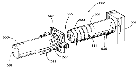
[0056] With reference to FIG. 22, a tapered mandrel groove forming apparatus
530 is illustrated.
CA 02780089 2012-06-15
14
Tapered mandrel 531 is supported by a mandrel support bracket, or other
suitable structure, 532
to fixedly secure tapered mandrel 531 as shown in FIG. 22. The end 533
oftapered mandrel 531,
has a plurality of cutting teeth 534 disposed thereon. The cutting teeth 534
may be abrasive
particles, such as diamond chips, or tungsten carbide particles or chips,
which are secured to
tapered mandrel 531 in any suitable manner, and the cutting teeth 534 form the
desired groove,
or grooves, 400 on, or in, the inner surface 301 of stent blank 300.
Alternatively, instead of
cutting teeth 534, the outer surface 535 of tapered mandrel 531 could be
provided with a surface
comparable to that formed on a metal cutting file or rasp, and the file, or
rasp, profile would form
the desired grooves 400. A stent holding fixture 537 is provided to support
stent blank 300 in any
desired manner, and the stent holding fixture 367 may be provided with a
piston cylinder
mechanism, 368, 369 to provide relative movement of stent 300 with respect to
tapered mandrel
531. Alternatively, stent 300 can be fixed, and a suitable mechanism can be
provided to move
tapered mandrel 531 into and along the inner surface 301 of stent 300.
Preferably, stent 300 is
in its expanded configuration.
[0057] With reference to FIGS. 23, 23A and 23B, a chemical removal technique
and apparatus
600 for forming the desired groove, or grooves, 400 on, or in, the interior
surface 301 of stent
blank 300 is illustrated. A stent holding fixture 601 is provided, and holding
fixture 601 may be
similar in construction to that of stent holding fixture 367 of FIG. 22.
Again, stent blank 300 is
provided with an orientation notch, or locator slot, 469. A photo mask 602 is
formed from a
material such as Mylar film. The dimensions of the mask, 602 correspond to the
inner surface
area of the inner surface 301 of stent 300. The mask 602 is formed into a
cylindrical orientation
to form a mask sleeve 603, which is wrapped onto a deflated balloon 605, such
as a balloon of
a conventional balloon angioplasty catheter. A conventional photoresist
material is spin coated
onto the inner surface 301 of stent blank 300. The mask sleeve 603, disposed
upon balloon 605
is inserted into stent 300, and balloon 605 is expanded to force the mask
sleeve 603 into an
abutting relationship with the photoresist coated inner surface 301 of stent
300. Balloon 605 may
be provided with an orientation pin 606 which corresponds with an orientation
notch 607 on mask
sleeve 603, which in turn is also aligned with locator slot 469' on stent
blank 300. The expansion
of balloon 605 is sufficient to sandwich mask sleeve 603 into abutting contact
with the
photoresist coated inner surface 301 ofstent 300; however, the balloon 605 is
not inflated enough
CA 02780089 2012-06-15
to squeeze the photoresist material off the stent 300. The interior surface
301 of stent 300 is then
irradiated through the inside of the balloon 605 through the balloon wall, as
by a suitable light
source 610. Balloon 605 is then deflated and mask sleeve 603 is removed from
the interior of
stent 300. The non-polymerized photoresist material is rinsed off and the
polymerized resist
material is hard baked upon the interior of stent 300. The groove, or grooves
400 are then
chemically etched into the non-protected metal surface on the interior surface
301 of stent 300.
The baked photoresist material is then removed by either conventional chemical
or mechanical
techniques.
[0058] Alternatively, instead of using a Mylar sheet as a mask 602 to form
mask sleeve 603,
mask 602 maybe formed directly upon the outer surface of balloon 605, as shown
in FIG. 23A.
The production of mask 602 directly upon the balloon outer surface can be
accomplished by
physically adhering the mask 602 onto the outer surface of balloon 605, or by
forming the mask
602 onto the surface of balloon 605 by deposition of the desired groove
pattern 468 by deposition
of UV absorbing material by thin film methods. In the case of utilizing mask
sleeve 603 as
shown in FIG. 23B, the balloon material must be compliant enough so as to
prevent creases from
the balloon wall which may shadow the resulting mask 602. In the case of mask
602 being
formed on balloon 605 as shown in FIG. 23A, a non-compliant balloon 605 should
be used, so
as not to distort the resulting image by the stretching of the compliant
balloon wall. If on the
other hand, the mask 602 is physically adhered to the outer wall of balloon
605, a compliant
balloon 605 may be used provided the mask 602 is adhered to the balloon 605
when the balloon
605 is in its fully expanded diameter.
[0059] With reference to FIGS. 24A and 24B, a method is shown for creating
grooves inside an
intact tubular stent 300, which involves casting patterned light inside a
stent 300 previously
coated with photosensitive material as discussed, for example, in connection
with FIG. 23 (PSM).
The light exposed areas are subjected to chemical etching to produce the
grooved pattern. This
method involves using a coaxial light source 800 with multiple small beams 801
of light in a
single plane. The light source 800 could be displaced along the longitudinal
axis of the tube, or
stent 300, at a rate consistent with adequate exposure of the photosensitive
material. Computer
driven stepper motors could be utilized to drive the light source in the x and
y planes, which
would allow for interlacing grooves (see FIG. 24A). One pass could create 1 mm
spacing, while
CA 02780089 2012-06-15
16
the next pass creates 500 m, and so on.
[0060] Rotational movements could introduce variability in the groove
direction for zig-zag,
spiral or undulating patterns. Alternatively, the light source 800 could be
fixed as shown in FIG.
24B, and the beams would be as narrow and long as the grooves needed on the
inner surface of
the mask 602. Stepping of the mask 602 would allow narrow spacing of the
grooves.
[00611 With reference to FIG. 25, an EDM process and apparatus 700 provide the
desired groove,
or grooves, 400 upon the interior 301 of stent 3 00. A non-conductive stent
alignment and holding
fixture 701, 701', similar in construction to backup housings 487, 487,
previously described, are
provided for holding stent like blank 300. A bearing block assembly 702,
similar to bearing
block assembly 481 of FIG. 19, is provided along with an indexing and current
transfer disk 703
provided within a drive gear mechanism 704, which is similar in construction
to drive gear
mechanisms 482 and 455, previously described in connection with FIGS. 19 and
17. An electric
discharge machining ("EDM") electrode 710 having bearing shafts 711, 712,
disposed at its ends,
for cooperation with bearing block assembly 702 and disk 703, respectively, is
rotated within
stent blank 300. Current is provided to the raised surfaces, or groove
pattern, 468, of electrode
710 to cut the desired groove, or grooves 400 into the inner surface 301 of
stent 300.
[0062] It is to be understood that the invention is not limited to the exact
details of construction,
operation, exact materials, or embodiments shown and described, as obvious
modifications and
equivalents will be apparent to one skilled in the art`. Accordingly, the
invention is therefore to
be limited only by the scope of the appended claims.