Note: Descriptions are shown in the official language in which they were submitted.
CA 02782330 2012-05-29
WO 2011/068758 PCT/US2010/058206
Improved Methods and Compositions for Detecting and Treating CEA-Expressing
Cancers
Related Applications
This application claims priority to application serial number 61/265,580,
filed December
1, 2009. The specification of the foregoing application is hereby incorporated
by reference in its
entirety.
Cross Reference to Sequence Listing
The instant application contains a Sequence Listing which has been submitted
in ASCII
format via EFS-Web and is hereby incorporated by reference in its entirety.
Said ASCII copy,
created on November 24, 2010, is named MED0526P.txt and is 71,511 bytes in
size.
Background of the Disclosure
Carcinoembryonic antigen (CEA) is a glycosylated human oncofetal antigen that
belongs
to the CEA-related cell adhesion molecule (CEACAM) family of the
immunoglobulin gene
superfamily. CEA has been suggested to mediate cell-cell adhesion, facilitate
bacterial
colonization of the intestine, and protect the colon from microbial infection
by binding and
trapping infectious microorganisms. Carcinoembryonic antigen (CEA) is a well-
characterized
tumor-associated antigen that is frequently over-expressed in human carcinomas
and melanomas.
Summary of the Disclosure
The disclosure provides improved methods and compositions for detecting,
monitoring
and/or treating CEA expressing cancers.
In one aspect, the disclosure provides a method of detecting recurrence of a
carcinoembryonic antigen (CEA) expressing cancer. The method involves
obtaining a sample
from a subject previously diagnosed with and treated for a carcinoembryonic
antigen (CEA)
expressing cancer. For example, the method involves obtaining a sample from a
human subject
(e.g., a human patient) previously diagnosed with and treated for cancer that
expresses human
carcinoembryonic antigen (CEA). The method may comprise a diagnostic step of
detecting in
said sample a concentration of full-length CEA protein using an antibody, an
antigen binding
fragment or an immunoglobulin-like molecule (e.g., a diagnostic reagent) that
-1-
CA 02782330 2012-05-29
WO 2011/068758 PCT/US2010/058206
immunospecifically binds to full-length CEA protein but does not
immunospecifically bind to
short form CEA protein, thereby detecting the concentration of full-length CEA
protein without
detecting the concentration of short form CEA protein in said sample.
Detecting a concentration
of full-length CEA protein in said sample above a range observed after
treatment indicates
recurrence of said CEA expressing cancer.
In certain embodiments, the diagnostic reagent (e.g., the antibody, antigen
binding
fragment or immunoglobulin-like molecule) immunospecifically binds to full-
length CEA
protein but does not immunospecifically bind to short form CEA protein. In
some embodiments,
the diagnostic reagent also does not immunospecifically bind to other related
CEACAM protein
family members (such as CEACAM 1, 3, 4, 6, 7, and 8). Thus, in certain
embodiments, the
diagnostic reagent is not a pan-CEA family member antibody, but is specific
for full-length CEA
protein.
In certain embodiments, the diagnostic reagent is an antibody, but with the
proviso that
the antibody is not A5B7 (the mouse monoclonal antibody known as A5B7). In
certain
embodiments, the diagnostic reagent is an antibody, but with the proviso that
the antibody is not
MEDI-565. In certain embodiments, the diagnostic reagent is an antibody, but
with the proviso
that the antibody is not the mouse monoclonal antibody known as IMMU-4 or
arcitumomab. In
certain embodiments, the diagnostic reagent is not a bispecific single chain
antibody that
includes an anti-CD3 binding portion. In certain embodiments, the diagnostic
reagent is an
antibody, but with the proviso that the antibody is not CEA.66, T84.66, or
T84.12, as identified
in Hefta et al. (1998) Immunotechnology 4: 49-57. In certain embodiments, the
diagnostic
reagent is an antibody, but with the proviso that the antibody is not PR1A3,
as identified in
Durbin (1994) PNAS 91: 4313. In certain embodiments, the diagnostic reagent is
an antibody,
but with the proviso that the antibody is not labetuzumab, as identified in
Liersch (2005) JCO 23:
6763. In certain embodiments, the diagnostic reagent is an antibody, but with
the proviso that
the antibody is not the antibody provided with the CanAg CEA EIA kit, as
distributed by
Immuno Biological Laboratories Inc. using Vendor cat # 401-85
(http://www.f(ii.comhvorld
hone/products/rnanual_kits/eia_kits/canag_cea.htrnl).
In certain embodiments, the method may comprise a diagnostic step of detecting
in said
sample a concentration of an RNA encoding full-length and/or short form CEA
protein. Said
method may employ quantitative RT-PCR, primers and/or probes specific for full-
length and/or
-2-
CA 02782330 2012-05-29
WO 2011/068758 PCT/US2010/058206
short form CEA RNA, such as in a TaqMan assay, and may employ SAGE, MPSS,
array-based
methods, or direct sequencing.
In certain embodiments, obtaining a sample comprises obtaining a blood sample,
a urine
sample, a fecal sample, or a sputum sample. In certain embodiments, obtaining
a sample
comprises obtaining a tumor biopsy or other tumor tissue sample. In certain
embodiments, the
sample is selected from one or more of whole blood, serum, plasma, saliva,
urine, feces, seminal
plasma, sweat, amniotic fluid, sputum, breast milk, breast nipple aspirates or
other fluid
including cells from the breast, bile, tissue homogenate, pleural effusion
fluids, and ascites.
In another aspect, the disclosure provides a method of detecting recurrence of
a
carcinoembryonic antigen (CEA) expressing cancer. The method includes
obtaining a first
sample from a subject having a carcinoembryonic antigen (CEA) expressing
cancer, wherein
said first sample is obtained prior to treatment. For example, the method
involves obtaining a
sample from a human subject (e.g., a human patient) diagnosed with a cancer
that expresses
human carcinoembryonic antigen (CEA) prior to the beginning of treatment. The
method may
comprise a diagnostic step of detecting in said first sample a pre-treatment
concentration of full-
length CEA protein using an antibody, an antigen binding fragment or an
immunoglobulin-like
molecule (e.g., a diagnostic reagent) that immunospecifically binds to full-
length CEA protein
but does not immunospecifically bind to short form CEA protein, thereby
detecting the
concentration of full-length CEA protein without detecting the concentration
of short form CEA
protein in said first sample. The method may include obtaining a second sample
from said
subject, and detecting in said second sample a concentration of full-length
CEA protein using
said antibody, antigen binding fragment or immunoglobulin-like molecule (e.g.,
using said
diagnostic reagent), thereby detecting the concentration of full-length CEA
protein without
detecting the concentration of short form CEA protein in said second sample.
For example, the
second sample may be collected at some period of time (or multiple time
points) after initiation
of treatment, so that decrease in CEA as a result of treatment can be
measured. The method may
include obtaining one or more further samples from said subject at a time
later than that for
obtaining said second sample, and detecting in said one or more further
samples a concentration
of full-length CEA protein using said antibody, antigen binding fragment or
immunoglobulin-
like molecule (e.g., said diagnostic reagent), thereby detecting the
concentration of full-length
CEA protein without detecting the concentration of short form CEA protein in
said one or more
-3-
CA 02782330 2012-05-29
WO 2011/068758 PCT/US2010/058206
further samples. Detecting a concentration of full-length CEA protein in said
one or more
further samples above the concentration of full-length CEA protein observed in
said second
sample indicates recurrence of said CEA expressing cancer. By way of example,
after successful
treatment, CEA levels will decrease. Following that decrease, the patient can
be monitored, and
an increase in CEA levels following this decrease may be indicative of
recurrence of the cancer.
In certain embodiments, an increase in CEA levels to approximately the same or
greater
concentration than that observed in the pre-treatment sample is indicative of
recurrence of the
CEA expressing cancer.
In certain embodiments, the initial treatment comprises surgery, and the first
sample is
taken prior to surgical resection of all or a portion of the CEA-expressing
tumor. In certain
embodiments, the initial treatment comprises one or more of surgery,
chemotherapy, radiation
therapy, immunotherapy, or a biological therapy, such as a monoclonal antibody
therapy, gene
therapy, oncolytic therapy, or viral therapy. In certain embodiments,
treatment is ongoing, such
that the first sample is taken prior to the commencement of any treatment, but
the second and/or
further samples are taken during a cycle of treatment (e.g., during a cycle of
chemotherapy or
radiation treatment).
In certain embodiments, the diagnostic reagent (e.g., the antibody, antigen
binding
fragment or immunoglobulin-like molecule) immunospecifically binds to full-
length CEA
protein but does not immunospecifically bind to short form CEA protein. In
some embodiments,
the diagnostic reagent also does not immunospecifically bind to other related
CEACAM protein
family members (such as CEACAM 1, 3, 4, 6, 7, and 8). Thus, in certain
embodiments, the
diagnostic reagent is not a pan-CEA family member antibody, but is specific
for full-length CEA
protein.
In certain embodiments, the diagnostic reagent is an antibody, but with the
proviso that
the antibody is not A5B7 (the mouse monoclonal antibody known as A5B7). In
certain
embodiments, the diagnostic reagent is an antibody, but with the proviso that
the antibody is not
MEDI-565. In certain embodiments, the diagnostic reagents is an antibody, but
with the proviso
that the antibody is not the mouse monoclonal antibody known as IMMU-4 or
arcitumomab. In
certain embodiments, the diagnostic reagent is not a bispecific single chain
antibody that
includes an anti-CD3 binding portion. In certain embodiments, the diagnostic
reagent is an
antibody, but with the proviso that the antibody is not CEA.66, T84.66, or
T84.12, as identified
-4-
CA 02782330 2012-05-29
WO 2011/068758 PCT/US2010/058206
in Hefta et al. (1998) Immunotechnology 4: 49-57. In certain embodiments, the
diagnostic
reagent is an antibody, but with the proviso that the antibody is not PR1A3,
as identified in
Durbin (1994) PNAS 91: 4313. In certain embodiments, the diagnostic reagent is
an antibody,
but with the proviso that the antibody is not labetuzumab, as identified in
Liersch (2005) JCO 23:
6763. In certain embodiments, the diagnostic reagent is an antibody, but with
the proviso that
the antibody is not the antibody provided with the CanAg CEA EIA kit, as
distributed by
Immuno Biological Laboratories Inc. using Vendor cat # 401-85
(http://www.fdi.com/world home/products/manual-kits/eiakits/canag_cea.html).
In certain embodiments, the method may comprise a diagnostic step of detecting
in said sample a
concentration of an RNA encoding full-length and/or short form CEA protein.
Said method may
employ quantitative RT-PCR, primers and/or probes specific for full-length
and/or short form
CEA RNA, such as in a TaqMan assay, and may employ SAGE, MPSS, array-based
methods, or
direct sequencing.
In certain embodiments, obtaining a sample comprises obtaining a blood sample,
a urine
sample, a fecal sample, or a sputum sample. In certain embodiments, obtaining
a sample
comprises obtaining a tumor biopsy or other tumor tissue sample. In certain
embodiments, the
sample is selected from one or more of whole blood, serum, plasma, saliva,
urine, feces, seminal
plasma, sweat, amniotic fluid, sputum, breast milk, breast nipple aspirates or
other fluid
including cells from the breast, bile, tissue homogenate, pleural effusion
fluids, and ascites.
In another aspect, the disclosure provides a method of determining
susceptibility to anti-
carcinoembryonic antigen (CEA) cancer therapy. The method includes detecting a
concentration
of full-length CEA protein in a sample from a subject using an antibody, an
antigen binding
fragment or an immunoglobulin-like molecule (e.g., a diagnostic reagent) that
immunospecifically binds to full-length CEA protein but does not
immunospecifically bind to
short form CEA protein, thereby detecting the concentration of full-length CEA
protein without
detecting the concentration of short form CEA protein in said sample. The
method may include
comparing said concentration of full-length CEA protein to a standard range
reflecting full-
length CEA protein concentration in samples from healthy subjects. Detecting a
concentration of
full-length CEA protein above said standard range indicates susceptibility to
anti-CEA cancer
therapy.
-5-
CA 02782330 2012-05-29
WO 2011/068758 PCT/US2010/058206
In certain of the foregoing and following embodiments, the standard range
reflecting full-
length CEA protein concentration in samples from healthy subject is less than
or equal to 3 ug/L
(3 ng/mL) in serum of non-smokers and less than or equal to 5 gg/L (5 ng/mL)
in serum of
smokers. In certain embodiments, the standard range is less than or equal to 5
gg/L (5 ng/mL) in
serum, regardless of smoking status.
In certain embodiments, the diagnostic reagent (e.g., the antibody, antigen
binding
fragment or immunoglobulin-like molecule) immunospecifically binds to full-
length CEA
protein but does not immunospecifically bind to short form CEA protein. In
some embodiments,
the diagnostic reagent also does not immunospecifically bind to other related
CEACAM protein
family members (such as CEACAM 1, 3, 4, 6, 7, and 8). Thus, in certain
embodiments, the
diagnostic reagent is not a pan-CEA family member antibody, but is specific
for full-length CEA
protein.
In certain embodiments, the diagnostic reagent is an antibody, but with the
proviso that
the antibody is not A5B7 (the mouse monoclonal antibody known as A5B7). In
certain
embodiments, the diagnostic reagent is an antibody, but with the proviso that
the antibody is not
MEDI-565. In certain embodiments, the diagnostic reagents is an antibody, but
with the proviso
that the antibody is not the mouse monoclonal antibody known as IMMU-4 or
arcitumomab. In
certain embodiments, the diagnostic reagent is not a bispecific single chain
antibody that
includes an anti-CD3 binding portion. In certain embodiments, the diagnostic
reagent is an
antibody, but with the proviso that the antibody is not CEA.66, T84.66, or
T84.12, as identified
in Hefta et al. (1998) Immunotechnology 4: 49-57. In certain embodiments, the
diagnostic
reagent is an antibody, but with the proviso that the antibody is not PR1A3,
as identified in
Durbin (1994) PNAS 91: 4313. In certain embodiments, the diagnostic reagent is
an antibody,
but with the proviso that the antibody is not labetuzumab, as identified in
Liersch (2005) JCO 23:
6763. In certain embodiments, the diagnostic reagent is an antibody, but with
the proviso that
the antibody is not the antibody provided with the CanAg CEA EIA kit, as
distributed by
Immuno Biological Laboratories Inc. using Vendor cat # 401-85
(http://www.fdi.com/world home/products/manual-kits/eiakits/canag_cea.html).
In certain embodiments, the method may comprise a diagnostic step of detecting
in said
sample a concentration of an RNA encoding full-length and/or short form CEA
protein. Said
method may employ quantitative RT-PCR, primers and/or probes specific for full-
length and/or
-6-
CA 02782330 2012-05-29
WO 2011/068758 PCT/US2010/058206
short form CEA RNA, such as in a TaqMan assay, and may employ SAGE, MPSS,
array-based
methods, or direct sequencing.
In certain embodiments, obtaining a sample comprises obtaining a blood sample,
a urine
sample, a fecal sample, or a sputum sample. In certain embodiments, obtaining
a sample
comprises obtaining a tumor biopsy or other tumor tissue sample. In certain
embodiments, the
sample is selected from one or more of whole blood, serum, plasma, saliva,
urine, feces, seminal
plasma, sweat, amniotic fluid, sputum, breast milk, breast nipple aspirates or
other fluid
including cells from the breast, bile, tissue homogenate, pleural effusion
fluids, and ascites.
In another aspect, the disclosure provides a method of monitoring anti-
carcinoembryonic
antigen (CEA) cancer therapy. The method includes detecting a concentration of
full-length
CEA protein in a sample from a subject undergoing treatment for a CEA
expressing cancer using
an antibody, an antigen binding fragment or an immunoglobulin-like molecule
(e.g., a diagnostic
reagent) that immunospecifically binds to full-length CEA protein but does not
immunospecifically bind to short form CEA protein, thereby detecting the
concentration of full-
length CEA protein without detecting the concentration of short form CEA
protein in said
sample. The method may include comparing said concentration of full-length CEA
protein to a
concentration of full-length CEA protein in a sample from said same subject,
which sample was
obtained prior to said treatment or at an earlier time point during said
treatment. A decrease in
full-length CEA concentration in a sample obtained at a later point during
treatment or after
conclusion of treatment versus that obtained prior to treatment or at an
earlier time point during
said treatment indicates effectiveness of said treatment, thereby monitoring
said anti-CEA cancer
therapy.
In certain embodiments, the initial treatment comprises surgery, and the first
sample is
taken prior to surgical resection of all or a portion of the CEA-expressing
tumor. In certain
embodiments, the initial treatment comprises one or more of surgery,
chemotherapy, radiation
therapy, immunotherapy, or a biological therapy, such as a monoclonal antibody
therapy, gene
therapy, oncolytic therapy, or viral therapy. In certain embodiments,
treatment is ongoing, such
that the first sample is taken prior to the commencement of any treatment, but
the second and/or
further samples are taken during a cycle of treatment (e.g., during a cycle of
chemotherapy or
radiation treatment).
-7-
CA 02782330 2012-05-29
WO 2011/068758 PCT/US2010/058206
In certain embodiments, the diagnostic reagent (e.g., the antibody, antigen
binding
fragment or immunoglobulin-like molecule) immunospecifically binds to full-
length CEA
protein but does not immunospecifically bind to short form CEA protein. In
some embodiments,
the diagnostic reagent also does not immunospecifically bind to other related
CEACAM protein
family members (such as CEACAM 1, 3, 4, 6, 7, and 8). Thus, in certain
embodiments, the
diagnostic reagent is not a pan-CEA family member antibody, but is specific
for full-length CEA
protein.
In certain embodiments, the diagnostic reagent is an antibody, but with the
proviso that
the antibody is not A5B7 (the mouse monoclonal antibody known as A5B7). In
certain
embodiments, the diagnostic reagent is an antibody, but with the proviso that
the antibody is not
MEDI-565. In certain embodiments, the diagnostic reagents is an antibody, but
with the proviso
that the antibody is not the mouse monoclonal antibody known as IMMU-4 or
arcitumomab. In
certain embodiments, the diagnostic reagent is not a bispecific single chain
antibody that
includes an anti-CD3 binding portion. In certain embodiments, the diagnostic
reagent is an
antibody, but with the proviso that the antibody is not CEA.66, T84.66, or
T84.12, as identified
in Hefta et al. (1998) Immunotechnology 4: 49-57. In certain embodiments, the
diagnostic
reagent is an antibody, but with the proviso that the antibody is not PR1A3,
as identified in
Durbin (1994) PNAS 91: 4313. In certain embodiments, the diagnostic reagent is
an antibody,
but with the proviso that the antibody is not labetuzumab, as identified in
Liersch (2005) JCO 23:
6763. In certain embodiments, the diagnostic reagent is an antibody, but with
the proviso that
the antibody is not the antibody provided with the CanAg CEA EIA kit, as
distributed by
Immuno Biological Laboratories Inc. using Vendor cat # 401-85
(http://www.f(ii.comhvorld
hone/products/rnanual_kits/eia_kits/canag_cea.htrnl).
In certain embodiments, the method may comprise a diagnostic step of detecting
in said
sample a concentration of an RNA encoding full-length and/or short form CEA
protein. Said
method may employ quantitative RT-PCR, primers and/or probes specific for full-
length and/or
short form CEA RNA, such as in a TaqMan assay, and may employ SAGE, MPSS,
array-based
methods, or direct sequencing.
In certain embodiments, obtaining a sample comprises obtaining a blood sample,
a urine
sample, a fecal sample, or a sputum sample. In certain embodiments, obtaining
a sample
comprises obtaining a tumor biopsy or other tumor tissue sample. In certain
embodiments, the
-8-
CA 02782330 2012-05-29
WO 2011/068758 PCT/US2010/058206
sample is selected from one or more of whole blood, serum, plasma, saliva,
urine, feces, seminal
plasma, sweat, amniotic fluid, sputum, breast milk, breast nipple aspirates or
other fluid
including cells from the breast, bile, tissue homogenate, pleural effusion
fluids, and ascites.
In another aspect, the disclosure provides a method of treating a subject
having a
carcinoembryonic antigen (CEA) expressing cancer. The method involves
obtaining a sample
from a subject prior to treatment for a carcinoembryonic antigen (CEA)
expressing cancer, such
as prior to initiation of treatment. The method may include detecting in said
sample (the initial
sample) a concentration of full-length CEA protein using an antibody, an
antigen binding
fragment or an immunoglobulin-like molecule (e.g., a diagnostic reagent) that
immunospecifically binds to full-length CEA protein but does not
immunospecifically bind to
short form CEA protein, thereby detecting the concentration of full-length CEA
protein without
detecting the concentration of short form CEA protein in said sample. The
method may include
comparing said concentration of full-length CEA protein to a standard range
reflecting full-
length CEA protein concentration in samples from healthy subjects, wherein
detecting the
concentration of full-length CEA protein above said standard range indicates
susceptibility to
anti-CEA cancer therapy. The method may include treating said subject who,
based on the initial
diagnostic testing is determined to be susceptible to anti-CEA cancer therapy,
with an anti-CEA
cancer therapeutic. The method may include detecting a concentration of full-
length CEA
protein in a post-treatment sample from said subject using an antibody, an
antigen binding
fragment or an immunoglobulin-like molecule that immunospecifically (e.g., a
diagnostic
reagent) binds to full-length CEA protein but does not immunospecifically bind
to a short form
CEA protein, thereby detecting the concentration of full-length CEA protein
without detecting
the concentration of short form CEA protein in said post-treatment sample. The
method may
include comparing said concentration of full-length CEA protein in said post-
treatment sample to
said concentration in the sample obtained prior to treatment. A decrease in
full-length CEA
protein concentration in said post-treatment sample relative to said pre-
treatment sample
indicates the effectiveness of said anti-CEA cancer therapeutic in said method
of treating said
subject.
In certain embodiments, treatment includes an anti-CEA cancer therapeutic
along with
one or more additional treatment modalities. Exemplary treatment modalities
include, but are
not limited to surgery, chemotherapy, radiation therapy, immunotherapy,
biological therapies
-9-
CA 02782330 2012-05-29
WO 2011/068758 PCT/US2010/058206
such as monoclonal antibodies and gene therapy, herbal therapy, acupuncture,
or dietary therapy.
In certain embodiments, treatment is ongoing, such that the first sample is
taken prior to the
commencement of any treatment, but the second and/or further samples are taken
during a cycle
of treatment (e.g., during a cycle of chemotherapy or radiation treatment).
In certain embodiments, the diagnostic reagent (e.g., the antibody, antigen
binding
fragment or immunoglobulin-like molecule) immunospecifically binds to full-
length CEA
protein but does not immunospecifically bind to short form CEA protein. In
some embodiments,
the diagnostic reagent also does not immunospecifically bind to other related
CEACAM protein
family members (such as CEACAM 1, 3, 4, 6, 7, and 8). Thus, in certain
embodiments, the
diagnostic reagent is not a pan-CEA family member antibody, but is specific
for full-length CEA
protein.
In certain embodiments, the diagnostic reagent and the anti-CEA therapeutic
bind to the
same or substantially the same epitope of CEA.
In certain embodiments, the diagnostic reagent is an antibody, but with the
proviso that
the antibody is not A5B7 (the mouse monoclonal antibody known as A5B7). In
certain
embodiments, the diagnostic reagent is an antibody, but with the proviso that
the antibody is not
MEDI-565. In certain embodiments, the diagnostic reagent is an antibody, but
with the proviso
that the antibody is not the mouse monoclonal antibody known as IMMU-4 or
arcitumomab. In
certain embodiments, the diagnostic reagent is not a bispecific single chain
antibody that
includes an anti-CD3 binding portion. In certain embodiments, the diagnostic
reagent is an
antibody, but with the proviso that the antibody is not CEA.66, T84.66, or
T84.12, as identified
in Hefta et al. (1998) Immunotechnology 4: 49-57. In certain embodiments, the
diagnostic
reagent is an antibody, but with the proviso that the antibody is not PR1A3,
as identified in
Durbin (1994) PNAS 91: 4313. In certain embodiments, the diagnostic reagent is
an antibody,
but with the proviso that the antibody is not labetuzumab, as identified in
Liersch (2005) JCO 23:
6763. In certain embodiments, the diagnostic reagent is an antibody, but with
the proviso that
the antibody is not the antibody provided with the CanAg CEA EIA kit, as
distributed by
Immuno Biological Laboratories Inc. using Vendor cat # 401-85
(http://wvv.fdi.com%'wvorld_homc/products/manualkits/eia_kits/canag_cea.html).
In certain embodiments, the method may comprise a diagnostic step of detecting
in said
sample a concentration of an RNA encoding full-length and/or short form CEA
protein. Said
-10-
CA 02782330 2012-05-29
WO 2011/068758 PCT/US2010/058206
method may employ quantitative RT-PCR, primers and/or probes specific for full-
length and/or
short form CEA RNA, such as in a TaqMan assay, and may employ SAGE, MPSS,
array-based
methods, or direct sequencing.
In certain embodiments, obtaining a sample comprises obtaining a blood sample,
a urine
sample, a fecal sample, or a sputum sample. In certain embodiments, obtaining
a sample
comprises obtaining a tumor biopsy or other tumor tissue sample. In certain
embodiments, the
sample is selected from one or more of whole blood, serum, plasma, saliva,
urine, feces, seminal
plasma, sweat, amniotic fluid, sputum, breast milk, breast nipple aspirates or
other fluid
including cells from the breast, bile, tissue homogenate, pleural effusion
fluids, and ascites.
In another aspect, the disclosure provides a method of determining
susceptibility to a
cancer therapeutic that immunospecifically binds to carcinoembryonic antigen
(CEA) protein.
The method comprises selecting a cancer therapeutic that will be used in the
treatment of a
subject with a CEA-expressing cancer, which cancer therapeutic
immunospecifically binds to
one form of CEA protein but does not immunospecifically bind to a second form
of CEA
protein, which one form of CEA protein is referred to as target CEA protein.
The method may
include detecting a concentration of said target CEA protein in a sample from
said subject using
an antibody, an antigen binding fragment or an immunoglobulin-like molecule
(e.g., a diagnostic
reagent) that immunospecifically binds to an epitope on said target CEA
protein that is the same
or substantially the same as the epitope that said cancer therapeutic
immunospecifically binds,
thereby detecting the concentration of the target CEA protein without
detecting the concentration
of other non-target forms of CEA protein in said sample. The method may
include comparing
said concentration of said target CEA protein to a standard range reflecting
target CEA protein
concentration in samples from healthy subjects. Detecting a concentration of
said target CEA
protein in said sample above said standard range indicates susceptibility to
said cancer
therapeutic.
In certain embodiments, the target CEA protein is full-length CEA protein. In
certain
embodiments, the target CEA protein is short form CEA protein. In either case,
it is envisioned
that a diagnostic reagent that immunospecifically binds a target CEA protein
will
immunospecifically bind both soluble and cell associated target CEA protein,
both of which are
mature CEA protein. Such a diagnostic reagent may also bind to the pro-form of
target CEA
protein. However, given that the diagnostically and therapeutically relevant
CEA is the mature
-11-
CA 02782330 2012-05-29
WO 2011/068758 PCT/US2010/058206
CEA protein expressed on tumors and present in bodily fluids, the relevant
reagents are those
that immunospecifically bind to mature target CEA.
In certain embodiments, the cancer therapeutic and the diagnostic reagent are
the same
protein. In certain embodiments, the cancer therapeutic and the diagnostic
reagent share at least
one antigen binding fragment.
In certain embodiments, the diagnostic reagent is an antibody, but with the
proviso that
the antibody is not A5B7 (the mouse monoclonal antibody known as A5B7). In
certain
embodiments, the diagnostic reagent is an antibody, but with the proviso that
the antibody is not
MEDI-565. In certain embodiments, the diagnostic reagent is an antibody, but
with the proviso
that the antibody is not the mouse monoclonal antibody known as IMMU-4 or
arcitumomab. In
certain embodiments, the diagnostic reagent is not a bispecific single chain
antibody that
includes an anti-CD3 binding portion. In certain embodiments, the diagnostic
reagent is an
antibody, but with the proviso that the antibody is not CEA.66, T84.66, or
T84.12, as identified
in Hefta et al. (1998) Immunotechnology 4: 49-57. In certain embodiments, the
diagnostic
reagent is an antibody, but with the proviso that the antibody is not PR1A3,
as identified in
Durbin (1994) PNAS 91: 4313. In certain embodiments, the diagnostic reagent is
an antibody,
but with the proviso that the antibody is not labetuzumab, as identified in
Liersch (2005) JCO 23:
6763. In certain embodiments, the diagnostic reagent is an antibody, but with
the proviso that
the antibody is not the antibody provided with the CanAg CEA EIA kit, as
distributed by
Immuno Biological Laboratories Inc. using Vendor cat # 401-85
(http://www.fdi.conm/world
home/pro(iucts/rnatnual_kits/eia_kits/canag_cea.htnml).
In certain embodiments, the method may comprise a diagnostic step of detecting
in said
sample a concentration of an RNA encoding full-length and/or short form CEA
protein. Said
method may employ quantitative RT-PCR, primers and/or probes specific for full-
length and/or
short form CEA RNA, such as in a TaqMan assay, and may employ SAGE, MPSS,
array-based
methods, or direct sequencing.
In certain embodiments, obtaining a sample comprises obtaining a blood sample,
a urine
sample, a fecal sample, or a sputum sample. In certain embodiments, obtaining
a sample
comprises obtaining a tumor biopsy or other tumor tissue sample. In certain
embodiments, the
sample is selected from one or more of whole blood, serum, plasma, saliva,
urine, feces, seminal
-12-
CA 02782330 2012-05-29
WO 2011/068758 PCT/US2010/058206
plasma, sweat, amniotic fluid, sputum, breast milk, breast nipple aspirates or
other fluid
including cells from the breast, bile, tissue homogenate, pleural effusion
fluids, and ascites.
In another aspect, the disclosure provides a method of monitoring treatment.
The method
comprises selecting a cancer therapeutic that will be used in the treatment of
a subject with a
CEA-expressing cancer, which cancer therapeutic immunospecifically binds to
one form of CEA
protein but does not immunospecifically bind to a second form of CEA protein,
which one form
of CEA protein is referred to as target CEA protein. The method may comprise
detecting a
concentration of said target CEA protein in a sample from said subject, which
subject is
undergoing treatment for a CEA expressing cancer, using an antibody, an
antigen binding
fragment or an immunoglobulin-like molecule (e.g., a diagnostic reagent) that
immunospecifically binds to an epitope on said target CEA protein that is the
same or
substantially the same as the epitope that said cancer therapeutic
immunospecifically binds,
thereby detecting the concentration of a target CEA protein without detecting
the concentration
of other non-target forms of CEA protein in said sample. The method may
include comparing
said concentration of target CEA protein to a concentration of target CEA
protein in an earlier
sample from said same subject, which earlier sample was obtained prior to
treatment with said
cancer therapeutic or at an earlier time point during treatment with said
cancer therapeutic. A
decrease in target CEA concentration in a sample obtained at a later point
during treatment with
said cancer therapeutic versus that obtained prior to treatment or at an
earlier time point during
treatment with said cancer therapeutic indicates effectiveness of said cancer
therapeutic, thereby
monitoring said treatment.
In certain embodiments, the target CEA protein is full-length CEA protein. In
certain
embodiments, the target CEA protein is short form CEA protein. In either case,
it is envisioned
that a diagnostic reagent that immunospecifically binds a target CEA protein
will
immunospecifically bind both soluble and cell associated target CEA protein.
Such a diagnostic
reagent may also bind to the pro-form of target CEA protein. However, given
that the
diagnostically and therapeutically relevant CEA is the mature CEA protein
expressed on tumors
and present in bodily fluids, the relevant reagents are those that
immunospecifically bind to
mature target CEA.
-13-
CA 02782330 2012-05-29
WO 2011/068758 PCT/US2010/058206
In certain embodiments, the cancer therapeutic and the diagnostic reagent are
the same
protein. In certain embodiments, the cancer therapeutic and the diagnostic
reagent share at least
one antigen binding fragment.
In certain embodiments, the diagnostic reagent is an antibody, but with the
proviso that the
antibody is not A5B7 (the mouse monoclonal antibody known as A5B7). In certain
embodiments, the diagnostic reagent is an antibody, but with the proviso that
the antibody is not
MEDI-565. In certain embodiments, the diagnostic reagent is an antibody, but
with the proviso
that the antibody is not the mouse monoclonal antibody known as IMMU-4 or
arcitumomab. In
certain embodiments, the diagnostic reagent is not a bispecific single chain
antibody that
includes an anti-CD3 binding portion. In certain embodiments, the diagnostic
reagent is an
antibody, but with the proviso that the antibody is not CEA.66, T84.66, or
T84.12, as identified
in Hefta et al. (1998) Immunotechnology 4: 49-57. In certain embodiments, the
diagnostic
reagent is an antibody, but with the proviso that the antibody is not PR1A3,
as identified in
Durbin (1994) PNAS 91: 4313. In certain embodiments, the diagnostic reagent is
an antibody,
but with the proviso that the antibody is not labetuzumab, as identified in
Liersch (2005) JCO 23:
6763. In certain embodiments, the diagnostic reagent is an antibody, but with
the proviso that
the antibody is not the antibody provided with the CanAg CEA EIA kit, as
distributed by
Immuno Biological Laboratories Inc. using Vendor cat # 401-85
(http:i/~vww.fdi.com/v%orld home/products/manual-
kits/eia_kits/canag_cea.html).
In certain embodiments, the method may comprise a diagnostic step of detecting
in said
sample a concentration of an RNA encoding full-length and/or short form CEA
protein. Said
method may employ quantitative RT-PCR, primers and/or probes specific for full-
length and/or
short form CEA RNA, such as in a TaqMan assay, and may employ SAGE, MPSS,
array-based
methods, or direct sequencing.
In certain embodiments, obtaining a sample comprises obtaining a blood sample,
a urine
sample, a fecal sample, or a sputum sample. In certain embodiments, obtaining
a sample
comprises obtaining a tumor biopsy or other tumor tissue sample. In certain
embodiments, the
sample is selected from one or more of whole blood, serum, plasma, saliva,
urine, feces, seminal
plasma, sweat, amniotic fluid, sputum, breast milk, breast nipple aspirates or
other fluid
including cells from the breast, bile, tissue homogenate, pleural effusion
fluids, and ascites.
-14-
CA 02782330 2012-05-29
WO 2011/068758 PCT/US2010/058206
In another aspect, the disclosure provides a method of treating a subject
having
carcinoembryonic antigen (CEA) expressing cancer. The method comprises
selecting a cancer
therapeutic that will be used in the treatment of a subject with a CEA-
expressing cancer, which
cancer therapeutic immunospecifically binds to one form of CEA protein but
does not
immunospecifically bind to a second form of CEA protein, which one form of CEA
protein is
referred to as target CEA protein. The method may include detecting a
concentration of said
target CEA protein in a sample from said subject using an antibody, an antigen
binding fragment
or an immunoglobulin-like molecule (e.g., a diagnostic reagent) that
immunospecifically binds to
an epitope on said target CEA protein that is the same or substantially the
same as the epitope
that said therapeutic immunospecifically binds, thereby detecting the
concentration of said target
CEA protein without detecting the concentration of other non-target forms of
CEA protein in
said sample. The method may include comparing said concentration of said
target CEA protein
to a standard range reflecting target CEA protein concentration in samples
from healthy subjects.
Detecting a concentration of said target CEA protein above said standard range
indicates
susceptibility to a cancer therapeutic that immunospecifically binds to target
CEA protein. The
method may include treating the subject with the cancer therapeutic that
immunospecifically
binds to target CEA protein if said subject is determined to be susceptible to
said cancer
therapeutic. The method may include detecting, in a post-treatment sample from
said subject
undergoing treatment with said cancer therapeutic, a concentration of target
CEA protein using
an antibody, an antigen binding fragment or an immunoglobulin-like molecule
that
immunospecifically binds to an epitope on target CEA that is the same or
substantially the same
as the epitope that said cancer therapeutic immunospecifically binds, thereby
detecting the
concentration of said target CEA protein without detecting the concentration
of other non-target
forms of CEA protein in said sample. The method may include comparing said
concentration of
target CEA protein to a concentration of target CEA protein in a sample from
said same subject,
which sample was obtained prior to treatment with said cancer therapeutic or
at an earlier time
point during said treatment, wherein a decrease in target CEA concentration in
a sample obtained
at a later point during treatment with said cancer therapeutic versus that
obtained prior to or at an
earlier time point during said treatment indicates effectiveness of said
treatment of said subject.
In certain embodiments, the target CEA protein is full-length CEA protein. In
certain
embodiments, the target CEA protein is short form CEA protein. In either case,
it is envisioned
-15-
CA 02782330 2012-05-29
WO 2011/068758 PCT/US2010/058206
that a diagnostic reagent that immunospecifically binds a target CEA protein
will
immunospecifically bind both soluble and cell associated target CEA protein.
Such a diagnostic
reagent may also bind to the pro-form of target CEA protein. However, given
that the
diagnostically and therapeutically relevant CEA is the mature CEA protein
expressed on tumors
and present in bodily fluids, the relevant reagents are those that
immunospecifically bind to
mature target CEA.
In certain embodiments, the target CEA is short form CEA and the diagnostic
reagent is
immunospecific for a short form CEA polymorphism comprising NIIQNELSVD (SEQ ID
NO:
11), but is not immunospecific for a short form CEA polymorphism comprising
NIIQNKLSVD
(SEQ ID NO: 12). In other embodiments, the diagnostic reagent is
immunospecific for a short
form CEA polymorphism comprising NIIQNKLSVD (SEQ ID NO: 12), and is not
immunospecific for a short form CEA polymorphism comprising NIIQNELSVD (SEQ ID
NO:
11). In other embodiments, the diagnostic reagent is immunospecific for both
of the foregoing
short form CEA polymorphisms.
In certain embodiments, the cancer therapeutic and the diagnostic reagent are
the same
protein. In certain embodiments, the cancer therapeutic and the diagnostic
reagent share at least
one antigen binding fragment.
In certain embodiments, the diagnostic reagent is an antibody, but with the
proviso that
the antibody is not A5B7 (the mouse monoclonal antibody known as A5B7). In
certain
embodiments, the diagnostic reagent is an antibody, but with the proviso that
the antibody is not
MEDI-565. In certain embodiments, the diagnostic reagent is an antibody, but
with the proviso
that the antibody is not the mouse monoclonal antibody known as IMMU-4 or
arcitumomab. In
certain embodiments, the diagnostic reagent is not a bispecific single chain
antibody that
includes an anti-CD3 binding portion. In certain embodiments, the diagnostic
reagent is an
antibody, but with the proviso that the antibody is not CEA.66, T84.66, or
T84.12, as identified
in Hefta et al. (1998) Immunotechnology 4: 49-57. In certain embodiments, the
diagnostic
reagent is an antibody, but with the proviso that the antibody is not PR1A3,
as identified in
Durbin (1994) PNAS 91: 4313. In certain embodiments, the diagnostic reagent is
an antibody,
but with the proviso that the antibody is not labetuzumab, as identified in
Liersch (2005) JCO 23:
6763. In certain embodiments, the diagnostic reagent is an antibody, but with
the proviso that
the antibody is not the antibody provided with the CanAg CEA EIA kit, as
distributed by
-16-
CA 02782330 2012-05-29
WO 2011/068758 PCT/US2010/058206
Immuno Biological Laboratories Inc. using Vendor cat # 401-85
(http://www.fdi.conm,/world home/products/manualkits/eia_kits/canag_cea.html).
In certain embodiments, the method may comprise a diagnostic step of detecting
in said
sample a concentration of an RNA encoding full-length and/or short form CEA
protein. Said
method may employ quantitative RT-PCR, primers and/or probes specific for full-
length and/or
short form CEA RNA, such as in a TaqMan assay, and may employ SAGE, MPSS,
array-based
methods, or direct sequencing.
In certain embodiments, obtaining a sample comprises obtaining a blood sample,
a urine
sample, a fecal sample, or a sputum sample. In certain embodiments, obtaining
a sample
comprises obtaining a tumor biopsy or other tumor tissue sample. In certain
embodiments, the
sample is selected from one or more of whole blood, serum, plasma, saliva,
urine, feces, seminal
plasma, sweat, amniotic fluid, sputum, breast milk, breast nipple aspirates or
other fluid
including cells from the breast, bile, tissue homogenate, pleural effusion
fluids, and ascites.
In another aspect, the disclosure provides a method comprising detecting a
concentration
of full-length CEA protein or RNA and a concentration of short form CEA
protein or RNA in a
sample from a subject and determining a ratio of full-length CEA protein or
RNA concentration
to short form CEA protein or RNA concentration.
In certain embodiments, the method comprises comparing said ratio to a
standard
reflecting the standard ratio of full-length CEA protein or RNA concentration
to short form CEA
protein or RNA concentration in samples from healthy subjects. A ratio that
varies significantly
from the standard ratio is indicative of presence of a CEA-expressing cancer.
In certain embodiments, detecting a concentration of full-length CEA protein
comprises
contacting a sample with an antibody, an antigen binding fragment or an
immunoglobulin-like
molecule (e.g., a diagnostic reagent) that immunospecifically binds to full-
length CEA protein
but does not immunospecifically bind to short form CEA protein.
In certain embodiments, detecting a concentration of full-length CEA protein
comprises
contacting a sample with an antibody, an antigen binding fragment or an
immunoglobulin-like
molecule (e.g., a first diagnostic reagent) that immunospecifically binds to
full-length CEA
protein but does not immunospecifically bind to short form CEA protein, and
detecting a
concentration of short form CEA protein comprises contacting a sample with an
antibody, an
antigen binding fragment or an immunoglobulin-like molecule (e.g., a second
diagnostic reagent)
-17-
CA 02782330 2012-05-29
WO 2011/068758 PCT/US2010/058206
that immunospecifically binds to short form CEA protein but does not
immunospecifically bind
to full-length CEA protein.
In certain embodiments, the diagnostic reagent that immunospecifically binds
to full-
length CEA protein does not immunospecifically bind to other CEACAM family
members
and/or the diagnostic reagent that immunospecifically binds to short form CEA
protein does not
immunospecifically bind to other CEACAM family members.
In certain embodiments, the diagnostic reagent is an antibody, but with the
proviso that the
antibody is not A5B7 (the mouse monoclonal antibody known as A5B7). In certain
embodiments, the diagnostic reagent is an antibody, but with the proviso that
the antibody is not
MEDI-565. In certain embodiments, the diagnostic reagent is an antibody, but
with the proviso
that the antibody is not the mouse monoclonal antibody known as IMMU-4 or
arcitumomab. In
certain embodiments, the diagnostic reagent is not a bispecific single chain
antibody that
includes an anti-CD3 binding portion. In certain embodiments, the diagnostic
reagent is an
antibody, but with the proviso that the antibody is not CEA.66, T84.66, or
T84.12, as identified
in Hefta et al. (1998) Immunotechnology 4: 49-57. In certain embodiments, the
diagnostic
reagent is an antibody, but with the proviso that the antibody is not PR1A3,
as identified in
Durbin (1994) PNAS 91: 4313. In certain embodiments, the diagnostic reagent is
an antibody,
but with the proviso that the antibody is not labetuzumab, as identified in
Liersch (2005) JCO 23:
6763. In certain embodiments, the diagnostic reagent is an antibody, but with
the proviso that
the antibody is not the antibody provided with the CanAg CEA EIA kit, as
distributed by
Immuno Biological Laboratories Inc. using Vendor cat # 401-85
(http :/,,'www. fdi.
coin/world_home/products/manual_kits/eia_kits/canag_cea.htral).
In certain embodiments, the method may comprise a diagnostic step of detecting
in said
sample a concentration of an RNA encoding full-length and/or short form CEA
protein. Said
method may employ quantitative RT-PCR, primers and/or probes specific for full-
length and/or
short form CEA RNA, such as in a TaqMan assay, and may employ SAGE, MPSS,
array-based
methods, or direct sequencing.
In certain embodiments, obtaining a sample comprises obtaining a blood sample,
a urine
sample, a fecal sample, or a sputum sample. In certain embodiments, obtaining
a sample
comprises obtaining a tumor biopsy or other tumor tissue sample. In certain
embodiments, the
sample is selected from one or more of whole blood, serum, plasma, saliva,
urine, feces, seminal
-18-
CA 02782330 2012-05-29
WO 2011/068758 PCT/US2010/058206
plasma, sweat, amniotic fluid, sputum, breast milk, breast nipple aspirates or
other fluid
including cells from the breast, bile, tissue homogenate, pleural effusion
fluids, and ascites.
In another aspect, the disclosure provides a method of detecting expression of
short form
carcinoembryonic antigen (CEA) RNA in a biological sample. The method
comprises providing
one or both of a nucleic acid probe or nucleic acid primers that hybridize to
a CEA nucleotide
sequence, and which probes and/or primers specifically identify expression of
short form CEA
by (i) hybridizing specifically to a short form CEA nucleotide sequence but
not to a full-length
CEA nucleotide sequence or (ii) hybridizing specifically to both short form
CEA nucleotide
sequence and full-length CEA nucleotide sequence in a manner that
distinguishes expression of
short form CEA from expression of full-length CEA. The method may include
providing RNA
from a biological sample. The method includes detecting expression of short
form CEA RNA in
said biological sample using said nucleic acid probe or nucleic acid primers.
In certain embodiments, the biological sample is a tumor tissue sample.
In certain embodiments, detecting expression comprises quantitative PCR or RT-
PCR
analysis. In certain embodiments, detecting expression comprises in situ
hybridization analysis.
In certain embodiments, the in situ hybridization analysis comprises FISH
(fluorescent in situ
hybridization). In certain embodiments, detecting expression comprises RNase
protection
analysis or Northern blot analysis. In certain embodiments, detecting
expression comprises
detecting expression with a microarray, SAGE, or MPSS.
In another aspect, the disclosure provides a method of detecting expression of
short form
carcinoembryonic antigen (CEA) protein in a biological sample. The method
comprises
providing an antibody, antigen binding fragment or immunoglobulin-like
molecule (e.g., a
diagnostic reagent) that immunospecifically binds to short form CEA protein
but does not
immunospecifically bind to full-length CEA protein. The method may include
providing a
biological sample. The method includes detecting expression of short form CEA
protein in said
biological sample using said antibody.
In certain embodiments, obtaining a sample comprises obtaining a blood sample,
a urine
sample, a fecal sample, or a sputum sample. In certain embodiments, obtaining
a sample
comprises obtaining a tumor biopsy or other tumor tissue sample. In certain
embodiments, the
sample is selected from one or more of whole blood, serum, plasma, saliva,
urine, feces, seminal
plasma, sweat, amniotic fluid, sputum, breast milk, breast nipple aspirates or
other fluid
-19-
CA 02782330 2012-05-29
WO 2011/068758 PCT/US2010/058206
including cells from the breast, bile, tissue homogenate, pleural effusion
fluids, and ascites.
In certain embodiments, the antibody is a monoclonal antibody. In certain
embodiments,
the monoclonal antibody is a chimeric antibody, a humanized antibody, or a
fully human
antibody. In certain embodiments, the antibody is a polyclonal antibody. In
certain
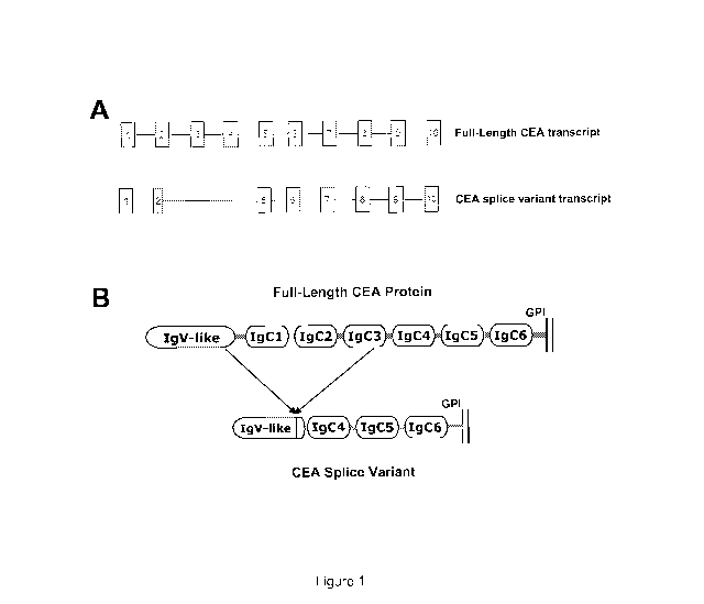
embodiments, the diagnostic reagent binds to a protein comprising the amino
acid sequence of
SEQ ID NO:1 .
In certain embodiments, detecting expression comprises immunohistochemistry or
immunocytochemistry analysis. In certain embodiments, detecting expression
comprises ELISA
analysis.
In another aspect, the disclosure provides a method of identifying patients
that may be
susceptible to a cancer therapeutic that immunospecifically binds to a target
carcinoembryonic
antigen (CEA) protein. In certain embodiments, the method comprises obtaining
a sample from
a patient, such as blood or tumor sample, for example a tumor biopsy, and
detecting in the tumor
sample expression of a target CEA RNA. In one example, the target CEA RNA is
detected using
a method such as Serial Analysis of Gene Expression (SAGE). In another
example, the target
CEA RNA is detected using Massively Parallel Signature Sequencing (MPSS). In
another
example, the target CEA RNA is detected using microarray that can specifically
detect long and
short form of CEA, such as an oligonucleotide array or an Affymetrix array. In
another example,
the target CEA RNA is detected using a probe and/or primers that distinguish
RNA expression of
full-length CEA from RNA expression of short form CEA. In another example, the
CEA RNA
may be reverse transcribed and sequenced, either in the form of a cDNA, or
after cloning the
cDNA in a suitable vector.
If the tumor sample from the patient expresses said target CEA RNA, the
patient may be
susceptible to treatment with a cancer therapeutic that immunospecifically
binds to that target
CEA protein. However, if the tumor sample from the patient does not express
said target CEA
RNA, the patient will not be susceptible to treatment with a cancer
therapeutic that
immunospecifically binds to that target CEA protein.
In certain embodiments, detecting target CEA RNA expression comprises
contacting the
sample with a probe and/or primers to evaluate expression of full-length CEA
RNA. In certain
embodiments, detecting target CEA RNA expression comprises contacting the
sample with a
probe and/or primers to evaluate expression of short form CEA RNA. In certain
embodiments,
-20-
CA 02782330 2012-05-29
WO 2011/068758 PCT/US2010/058206
detecting target CEA RNA expression comprises contacting the sample with one
or more sets of
probes and/or primers to evaluate expression of both full-length CEA RNA and
short form CEA
RNA.
In certain embodiments, the method further comprises obtaining from the
patient one or
more additional biological samples, and assaying the one or more biological
samples for
expression of the target CEA protein. For example, these additional one or
more biological
samples may be contacted with an antibody that immunospecifically binds to
full-length CEA
protein.
In certain embodiments, the method further comprises treating said subject
with a cancer
therapeutic.
In another aspect, the disclosure provides a method of generating antibodies
immunospecific for full-length carcinoembryonic antigen (CEA) protein. The
method comprises
providing a portion of full-length CEA protein that is not present in short
form CEA protein and
using said portion of full-length CEA protein as an antigen for generating
said antibodies.
In certain embodiments, the antibodies immunospecifically bind to full-length
CEA
protein but do not immunospecifically bind to short form CEA protein, and also
do not
immunospecifically bind to other related CEACAM protein family members (such
as CEACAM
1, 3, 4, 6, 7, and 8).
In certain embodiments, said antibodies are monoclonal antibodies.
In certain embodiments, said antibodies are polyclonal antibodies.
In certain embodiments, the method includes generating an antigen binding
fragment
from said antibodies.
In certain embodiments, the method involves immunizing a non-human animal with
a
portion of full-length CEA protein that is not present in short form CEA
protein.
In another aspect, the disclosure provides a method of generating antibodies
immunospecific for short form CEA protein. In certain embodiments, the method
comprises
providing a fragment comprising a portion of consecutive amino acid residues
present in short
form CEA protein that are not present in full-length CEA protein and using
said fragment as an
antigen for generating said antibodies. An exemplary fragment includes
consecutive amino acid
residues that bridge the splice junction unique to short form CEA protein.
-21-
CA 02782330 2012-05-29
WO 2011/068758 PCT/US2010/058206
In certain embodiments, the fragment comprises the following consecutive amino
acid
residues: NIIQNELSVD (SEQ ID NO: 11). In certain embodiments, the fragment
comprises the
following consecutive amino acid residues: NIIQNKLSVD (SEQ ID NO: 12). In
either case, the
fragment may also include additional amino acid sequences such that the total
size of the
fragment is at least 10, 12, 14, 15, 16, 18, 20, 25, 30, 35, 40, 45, 50, 55,
60, 65, 70, 75, 80, 85,
90, 95, or 100 consecutive amino acids.
In certain embodiments, the fragment comprises substantially the same epitope
as the
foregoing fragments. A fragment comprising substantially the same epitope
includes fragments
of the foregoing with a small number (e.g., 1, 2, 3) of conservative amino
acid substitutions.
In certain embodiments, the antibodies immunospecifically bind to short form
CEA
protein but do not immunospecifically bind to full-length CEA protein, and
also do not
immunospecifically bind to other related CEACAM protein family members (such
as CEACAM
1, 3, 4, 6, 7, and 8).
In certain embodiments, the antibodies are immunospecific for a short form CEA
polymorphism comprising NIIQNELSVD (SEQ ID NO: 11), and are not immunospecific
for a
short form CEA polymorphism comprising NIIQNKLSVD (SEQ ID NO: 12). In other
embodiments, the antibodies are immunospecific for a short form CEA
polymorphism
comprising NIIQNKLSVD (SEQ ID NO: 12), and are not immunospecific for a short
form CEA
polymorphism comprising NIIQNELSVD (SEQ ID NO: 11). In other embodiments, the
antibodies are immunospecific for both of the foregoing short form CEA
polymorphisms.
In certain embodiments, said antibodies are monoclonal antibodies.
In certain embodiments, the method includes generating an antigen binding
fragment
from said antibodies.
In certain embodiments, the method involves immunizing a non-human animal with
a
portion of short form CEA protein that is not present in full-length CEA
protein.
In another aspect, the disclosure provides a purified polypeptide comprising
the amino
acid sequence represented in SEQ ID NO: 1 (in the presence or absence of pro-
sequences), or a
fragment thereof comprising the following consecutive amino acid residues:
NIIQNELSVD
(SEQ ID NO: 11). In certain embodiments, the disclosure provides a purified
polypeptide
comprising the amino acid sequence represented in SEQ ID NO: 1 (in the
presence or absence of
all or a portion of N and/or C terminal pro-sequences), or a fragment thereof
comprising the
-22-
CA 02782330 2012-05-29
WO 2011/068758 PCT/US2010/058206
following consecutive amino acid residues: NIIQNKLSVD (SEQ ID NO: 12). The N-
terminal
pro sequence is an approximately 34 amino acid residue signal peptide and the
C-terminal pro
sequence is an approximately 17 amino acid peptide.
In certain embodiments, the purified polypeptide comprises the amino acid
sequence
represented in SEQ ID NO: 1 (in the presence or absence of pro-sequences
absent from mature
CEA). In certain embodiments, the purified polypeptide comprises a fragment of
SEQ ID NO: 1
comprising the following consecutive amino acid residues: NIIQNELSVD (SEQ ID
NO: 11). In
certain embodiments, the purified polypeptide comprises a fragment of SEQ ID
NO: 1
comprising the following consecutive amino acid residues: NIIQNKLSVD (SEQ ID
NO: 12). In
certain embodiments, said fragment is approximately 10 consecutive amino acid
residues. In
certain embodiments, said fragment is approximately 12, 14, 15, 16, 18, or 20
consecutive amino
acids. In certain embodiments, said fragment is approximately 25, 30, 33, 35,
40, 45, 48, 50, 55,
60, 65, 70, 75, 80, 85, 90, 95, or 100 consecutive amino acid residues. In
certain embodiments,
said fragment is at least 20, at least 25, at least 30, at least 35, at least
40, at least 45, or at least
50 consecutive amino acid residues. In certain embodiments, said fragment is
less than 250, less
than 200, less than 175, less than 150, less than 125, less than 100, less
than 90, less than 85, less
than 80, less than 75, less than 70, less than 65, less than 60, less than 55,
or less than 50
consecutive amino acid residues. In certain embodiments, said fragment is at
least 100, at least
150, at least 200, at least 250, at least 300, at least 350, or at least 400
consecutive amino acid
residues.
In certain embodiments, the purified polypeptide is conjugated to an adjuvant.
In certain embodiments, the purified polypeptide is used to generate
immunospecific
antibodies.
In certain embodiments, a nucleotide sequence encoding the purified
polypeptide is used
to generate immunospecific antibodies.
In another aspect, the disclosure provides diagnostic methods having any of
the
properties described herein, but wherein the diagnostic reagent is
administered to a patient.
Following administration to a patient, CEA expression can be visualized using
in vivo imaging
techniques. Alternatively, a sample can be taken from the patient and CEA
concentration can be
assessed ex vivo (e.g., concentration is assayed ex vivo but the contact
between the diagnostic
reagent and CEA protein occurs in vivo). This aspect of the disclosure can be
applied to and
-23-
CA 02782330 2012-05-29
WO 2011/068758 PCT/US2010/058206
combined with any one or more of the aspects and embodiments of the disclosure
described in
detail herein.
In certain embodiments of any of the foregoing or following aspects or
embodiments, the
method may comprise treating the subject, such as the human patient, with an
anti-CEA cancer
therapeutic. In certain embodiments, such anti-CEA cancer therapeutic
immunospecifically
binds to the same or substantially the same epitope as that immunospecifically
bound by the
diagnostic reagent used in the detecting steps. In certain embodiments, the
anti-CEA cancer
therapeutic is a bispecific antibody (including a bispecific single chain
antibody) including an
anti-CEA portion and an anti-CD3 portion. In certain embodiments, the anti-CEA
cancer
therapeutic is the bispecific antibody MEDI-565. In certain embodiments, the
anti-CEA cancer
therapeutic is a bispecific antibody having an anti-CEA portion that is the
same as MEDI-565 or
an anti-CEA portion that binds the same or substantially the same epitope as
MEDI-565. In
certain embodiments, the anti-CEA cancer therapeutic includes an anti-CEA
portion that is the
same as MEDI-565 or an anti-CEA portion that binds the same or substantially
the same epitope
as MEDI-565, but which anti-CEA therapeutic is not a bispecific antibody. In
certain
embodiments, the therapeutic regimen comprises treatment with a bispecific
antibody (including
a bispecific single chain antibody) that includes both an anti-CEA portion and
an anti-CD3
portion.
In certain embodiments, the therapeutic to be used includes, at least, a CEA
binding
portion comprising the amino acid sequence represented in any of SEQ ID NOs:
28-44 and 46-
51. In certain embodiments, the therapeutic to be used is a bispecific
antibody comprising the
amino acid sequence represented in any of SEQ ID NOs: 28-44 and 47. In certain
embodiments,
the therapeutic to be used is a bispecific antibody comprising the amino acid
sequence
represented in any of SEQ ID NOs: 34, 36, 41, 42, 43, and 47. In certain
embodiments, the
therapeutic to be used is a bispecific antibody comprising the amino acid
sequence represented in
any of SEQ ID NOs: 37-40. In certain embodiments, the therapeutic to be used
is a bispecific
antibody comprising the amino acid sequence represented in SEQ ID NO: 48. In
certain
embodiments, the therapeutic to be used is a bispecific antibody comprising
the amino acid
sequence represented in SEQ ID NO: 49. In certain embodiments, the therapeutic
to be used is a
bispecific antibody comprising the amino acid sequence represented in SEQ ID
NOs: 48 and 49.
In certain embodiments, the therapeutic to be used is a bispecific antibody
comprising the amino
-24-
CA 02782330 2012-05-29
WO 2011/068758 PCT/US2010/058206
acid sequence represented in SEQ ID NO: 46. In any of the forgoing or previous
embodiments,
SEQ ID NO: 46 may father comprise six histidines on the C-terminus.
In certain embodiments of any of the foregoing or following aspects or
embodiments, the
cancer therapeutic immunospecifically binds to an epitope on CEA protein that
is the same or
substantially the same as that immunospecifically bound by said antibody,
antigen binding
fragment or immunoglobulin-like molecule used in said detecting steps (e.g.,
the diagnostic
reagent).
In certain embodiments of any of the foregoing or following aspects or
embodiments, the
method comprises more than one diagnostic step, and the same diagnostic
reagent is used at each
diagnostic step. In certain embodiments, the method comprises more than one
diagnostic step,
and although the same diagnostic reagent is not used at all steps, each of the
diagnostic reagents
bind the same or substantially the same epitope.
In certain embodiments of any of the foregoing or following aspects or
embodiments, the
subject is a human.
In certain embodiments of any of the foregoing or following aspects or
embodiments, the
diagnostic and/or therapeutic reagent immunospecifically binds to human CEA.
In certain embodiments of any of the foregoing or following aspects or
embodiments, the
diagnostic step or steps are performed ex vivo (e.g., outside of the patient's
body).
In certain embodiments of any of the foregoing or following aspects or
embodiments, the
method includes one or more treatment steps. Exemplary treatments include one
or more of
surgery, chemotherapy, radiation therapy, immunotherapy, biological therapy,
herbal therapy,
acupuncture, or an anti-CEA cancer therapeutic. A suitable treatment regimen
includes any one
or more of these and other treatment modalities delivered according to a
dosage and time course
prescribed by a suitable medical professional.
In certain embodiments of any of the foregoing or following aspects or
embodiments, the
cancer therapeutic comprises a protein therapeutic. In certain embodiments,
the protein
therapeutic is an antibody or antigen binding fragment. In certain
embodiments, the antibody or
antigen binding fragment is from a monoclonal antibody. In certain
embodiments, the
monoclonal antibody is a chimeric antibody, a humanized antibody, or a fully
human antibody.
In certain embodiments, the protein therapeutic immunospecifically binds to a
protein
comprising the amino acid sequence of SEQ ID NO: 2 (mature full-length human
CEA). In
-25-
CA 02782330 2012-05-29
WO 2011/068758 PCT/US2010/058206
certain embodiments, the protein therapeutic immunospecifically binds to a
protein comprising
the amino acid sequence of SEQ ID NO:2, but which protein therapeutic does not
immunospecifically bind to a protein comprising the amino acid sequence of SEQ
ID NO: 1 in
the presence and/or absence of the pro-sequence. In certain embodiments, the
protein
therapeutic immunospecifically binds to a protein comprising the amino acid
sequence of SEQ
ID NO:2, but which protein therapeutic does not immunospecifically bind to a
protein
comprising the amino acid sequence of SEQ ID NO: 1 (in the presence or absence
of pro-
sequences) and does not immunospecifically bind to other CEACAM family
members. In
certain embodiments, the protein therapeutic comprises an antigen binding
domain of antibody
A5B7. In certain embodiments, the protein therapeutic is a bispecific
antibody. In certain
embodiments, the bispecific antibody is MEDI-565.
In certain embodiments of any of the foregoing or following aspects or
embodiments, the
sample (e.g., the biological sample on which diagnostic testing is performed)
is selected from
one or more of. whole blood, serum, plasma, saliva, urine, feces, seminal
plasma, sweat,
amniotic fluid, sputum, breast milk, breast nipple aspirates or other fluid
including cells from the
breast, bile, tissue homogenate, pleural effusion fluids, and ascites. Note
that such biological
samples may contain both cellular and non-cellular elements. In certain
embodiments, the
sample is a tumor tissue sample. For embodiments in which multiple samples are
taken, the
disclosure contemplates that each of those samples may be from the same tissue
source (e.g.,
serum or feces), or the samples may be from different tissue sources.
Furthermore, the
disclosure contemplates that a diagnostic step may include detecting CEA
expression or
concentration in a sample from a single source or may include detecting CEA
expression or
concentration in samples from more than one tissue source.
In certain embodiments of any of the foregoing or following aspects or
embodiments, the
CEA-expressing cancer may be any one or more of the following: colon cancer,
rectal cancer,
pancreatic cancer, esophageal cancer, gastroesophageal cancer, stomach cancer,
lung cancer and
breast cancer. In certain embodiments, the CEA-expressing cancer is colon
cancer. In certain
embodiments, the classification of the type of cancer (e.g., pancreatic or
colon) refers to the
classification of the initial tumor - although it is recognized that
metastases may appear in other
tissues. In such cases, the cancer will still be categorized as, for example,
a CEA-expressing
-26-
CA 02782330 2012-05-29
WO 2011/068758 PCT/US2010/058206
colon cancer, even though the cancer may have metastasized to non-colon
tissue. Any of the
cancers discussed herein may be primary or metastatic (e.g., metastatic
colorectal cancer).
In certain embodiments of any of the foregoing or following aspects or
embodiments, the
detecting step comprises contacting the sample with said antibody, antigen
binding fragment or
immunoglobulin-like molecule and detecting the concentration of full-length
CEA protein by
immunohistochemistry or immunocytochemistry. In certain embodiments of any of
the
foregoing or following aspects or embodiments, the detecting step comprises
contacting the
sample with said antibody, antigen binding fragment or immunoglobulin-like
molecule and
detecting the concentration of full-length CEA protein by ELISA.
In certain embodiments of any of the foregoing or following aspects or
embodiments, the
diagnostic reagent is a monoclonal antibody. In certain embodiments, the
monoclonal antibody
is a chimeric antibody, a humanized antibody, or a fully human antibody. In
some embodiments,
the antibody is a polyclonal antibody.
In certain embodiments of any of the foregoing or following aspects or
embodiments, the
detecting step comprises contacting the sample with the diagnostic reagent
(said antibody,
antigen binding fragment or immunoglobulin-like molecule), which antibody,
antigen binding
fragment or immunoglobulin-like molecule immunospecifically binds to a protein
comprising the
amino acid sequence of SEQ ID NO:2 and detecting the concentration of full-
length CEA protein
by immunohistochemistry or ELISA.
In certain embodiments of any of the foregoing or following aspects or
embodiments, the
detecting step comprises contacting the sample with an antibody or antigen
binding fragment
comprising the antigen binding domain of antibody A5B7, with the proviso that
the antibody is
not A5B7.
In certain embodiments of any of the foregoing or following aspects or
embodiments, the
detecting step comprises contacting said sample with an antibody or antigen
binding fragment
comprising an antigen binding domain that binds the same or substantially the
same epitope as
A5B7, with the proviso that the antibody is not A5B7.
In certain embodiments of any of the foregoing or following aspects or
embodiments, the
detecting step comprises contacting said sample with an antibody or antigen
binding fragment
comprising an antigen binding domain that binds the same or substantially the
same epitope as
A5B7, with the proviso that the antibody is not A5137 or MEDI-565.
-27-
CA 02782330 2012-05-29
WO 2011/068758 PCT/US2010/058206
In certain embodiments of any of the foregoing or following aspects or
embodiments, the
cancer therapeutic comprises a bispecific antibody (including a bispecific
single chain antibody)
that includes an anti-CEA portion and an anti-CD3 portion. In certain
embodiments of any of
the foregoing or following aspects or embodiments, the cancer therapeutic
includes MEDI-565.
In certain embodiments of any of the foregoing or following aspects or
embodiments, the
diagnostic reagents comprises a bispecific antibody (including a bispecific
single chain antibody)
that includes an anti-CEA portion and an anti-CD3 portion. In certain
embodiments of any of
the foregoing or following aspects or embodiments, the diagnostic reagent is
MEDI-565. In
certain embodiments of any of the foregoing or following aspects or
embodiments, both the
cancer therapeutic and the diagnostic reagent comprise a bispecific antibody
(including a
bispecific single chain antibody) that includes an anti-CEA portion and an
anti-CD3 portion. In
certain embodiments of any of the foregoing or following aspects or
embodiments, both the
cancer therapeutic and the diagnostic reagent include MEDI-565.
In certain embodiments of any of the foregoing or following aspects or
embodiments, the
diagnostic reagent is selected from A5B7 (the mouse monoclonal antibody known
as A5B7),
arcitumomab, CEA.66, T84.66, T84.12, PR1A3, or labetuzumab.
In certain embodiments of any of the foregoing or following aspects or
embodiments, the
cancer therapeutic includes MEDI-565 or an antibody comprising an anti-CEA
portion that is the
same or that binds the same or substantially the same epitope as MEDI-565. In
certain
embodiments of any of the foregoing or following aspects or embodiments, the
diagnostic
reagent is MEDI-565 or an antibody comprising an anti-CEA portion that is the
same or that
binds the same or substantially the same epitope as MEDI-565. In certain
embodiments of any
of the foregoing or following aspects or embodiments, both the cancer
therapeutic and the
diagnostic reagent include MEDI-565, or comprise an anti-CEA portion that is
the same or that
binds the same or substantially the same epitope as MEDI-565.
In certain embodiments, the diagnostic reagent (e.g., the antibody, antigen
binding
fragment or immunoglobulin-like molecule) immunospecifically binds to full-
length CEA
protein but does not immunospecifically bind to short form CEA protein. In
some embodiments,
the diagnostic reagent also does not immunospecifically bind to other related
CEACAM protein
family members (such as CEACAM 1, 3, 4, 6, 7, and 8). Thus, in certain
embodiments, the
-28-
CA 02782330 2012-05-29
WO 2011/068758 PCT/US2010/058206
diagnostic reagent is not a pan-CEA family member antibody, but is specific
for full-length CEA
protein.
Specificity with respect to a diagnostic or therapeutic reagent that
immunospecifically
binds to full-length (or short form) CEA is intended to refer to
immunospecificity for mature
CEA (without regard for the presence or absence of pro-sequences). However, it
is readily
appreciated that an antibody immunospecific for mature full-length CEA protein
may also
immunospecifically bind to full-length CEA in the presence of pro-sequences.
Similarly, an
antibody immunospecific for short form CEA protein may also immunospecifically
bind to short
form CEA in the presence (or absence of pro-sequences). An antibody will be
considered
immunospecific for a target CEA protein as long as it immunospecifically binds
to the mature
target CEA protein (and does not immunospecifically bind to non-target CEA
proteins).
However, the pro-protein corresponding to that mature target CEA will not be
considered
another form or a non-target form of CEA. Similarly, binding to both soluble
and membrane
anchored mature target CEA will not be considered binding to a non-target form
of CEA.
In certain embodiments of any of the foregoing or following, CEA concentration
is
compared to a standard range reflecting full-length CEA protein concentration
in samples from
healthy subject. In certain embodiments, the standard range is less than or
equal to 3 ug/L (3
ng/mL) in serum of non-smokers and less than or equal to 5 ug/L (5 ng/mL) in
serum of
smokers. In certain embodiments, the standard range is less than or equal to 5
ug/L (5 ng/mL) in
serum, regardless of smoking status.
The disclosure contemplates all combinations of any of the foregoing aspects
and
embodiments, as well as combinations with any of the embodiments set forth in
the detailed
description and examples.
Brief Description of the Drawings
Figure IA provides a schematic representation of the exon structure of full-
length CEA
and short form CEA. The boxes represent exons, and the exons are numbered. The
splice
variant short form CEA is missing exons 3 and 4, and part of exon 2, relative
to full-length CEA.
Figure I B provides a representation of the domain structure of the full-
length and short form
CEA. IgV-like refers to Immunoglobulin Variable like domain; Ig refers to the
various
Immunoglobulin-like domains. The lines in these schematics indicate GPI
linkage of the
-29-
CA 02782330 2012-05-29
WO 2011/068758 PCT/US2010/058206
proteins to the plasma membrane. The N-terminus of each protein begins on the
left side of each
schematic.
Figure 2 provides an alignment of the amino acid sequence for full-length CEA
and short
form CEA (CEA splice variant). In Figure 2, the CEA proteins are shown with
the N- and C-
terminal pro-sequences indicated in black boxes. These pro-sequences are not
typically present
in the mature CEA protein. The amino acid sequence for short form CEA (CEA
splice variant)
is provided as SEQ ID NO: 1. The amino acid sequence for mature full-length
CEA, without N-
and C-terminal pro-sequences, is provided as SEQ ID NO: 2. Note that the
regions of the protein
identified in the examples as important for antibody binding are also boxed
and labeled as
"epitope residues 326-349" and "epitope residues 388-410". In Figure 2, these
regions are
indicated using numbering that includes the N-terminal signal sequence. Thus,
these regions can
also be referred to using numbering relative to the mature CEA protein as
"epitope residues 292-
315" and "epitope residues 354-376."
Figure 3 provides a comparison of the domain structure of full-length CEA
versus that of
short form CEA. In figure 3, Ig refers to immunoglobulin-like domains, V-set
refers to Ig
variable region-like N-terminal domain, and numbers represent sequential V-set
and Ig domains
of mature proteins. Note that these Ig domains are also referred to as IgC
domains, and are
numbered sequentially from 1 through 6. The schematic provided in Figure 3 is
an alternative
illustration of the schematic provided in Figure 1B.
Figure 4 provides the results of a screen of single cell clones of CHO cells
for expression
of short form CEA. CHO cells infected with lentivirus carrying the sequence
for short form
CEA were screened for cell surface expression of short form CEA. Experiments
were performed
with IgG B9 and with anti-CEACAM5 monoclonal antibody using a FACS-based
approach.
<FITC-A> denotes the relative fluorescence intensity of bound antibody. The 72
series numbers
along the right hand side of the figure refer to cell line clone numbers.
Parental CHO cells
lacking expression of CEA are referred to as CHO dhfr-. CHO cells expressing
full-length
human CEA are referred to as CHOhuCEA.
Figure 5 depicts the ability of various anti-CEA antibodies to bind to single
cell CHO cell
clones 72-4 and 72-14, which express short form CEA. FITC-A denotes the
relative
fluorescence intensity of bound antibody. 2 only refers to anti-mouse IgG
Alexa Fluor 488
conjugated secondary antibody bound in the absence of primary antibody. Panel
A shows the
-30-
CA 02782330 2012-05-29
WO 2011/068758 PCT/US2010/058206
lack of expression of CEA (full-length or short form) observed in parental
dhfr- CHO cells
lacking expression of CEA. Panel B shows that the B9 IgG and CEACAM5 antibody,
as well as
the pan-antibodies CEACAM 1, 3, 4, 5, 6 and CEACAM 1, 3, 5, 6 bind CHO cells
expressing
full-length CEA. Panel C shows that the CEACAM5 antibody also binds to CHO
cell clone 72-
4, which expresses short form CEA. However, B9 IgG, and the pan-antibodies
CEACAM 1, 3,
4, 5, 6 and CEACAM 1, 3, 5, 6 do not appear to significantly bind to clone 72-
4. Panel D shows
that the CEACAM5 antibody also binds to CHO cell clone 72-14, which expresses
short form
CEA. However, B9 IgG, and the pan-antibodies CEACAM 1, 3, 4, 5, 6 and CEACAM
1, 3, 5, 6
do not appear to significantly bind to clone 72-14.
Figure 6 shows the results of experiments examining T cell mediated killing by
MEDI-
565 of CHO cells expressing short form CEA. CHO cells expressing full-length
CEA
(CHOhuCEA; circle), parental CHO cells lacking CEA expression (CHO dhfr-;
square), and
CHO cell short form CEA expressing clones 72-4 and 72-14 (CHO-CEA SV 72-4;
triangle and
CHO-CEA SV 72-14; upside down triangle) were tested for susceptibility to
killing by CD3+
enriched T cells from two individuals (donors 1 and 2) in the presence of MEDI-
565. Panel A
depicts the results from experiments using material from donor 1 and shows
that only cells
expressing full-length CEA are susceptible to T cell mediated killing in the
presence of MEDI-
565. Panel B depicts similar results obtained using material from donor 2.
Figure 7 shows the results of experiments examining activation of human T
cells by
MEDI-565 in the presence of CHO cell clones that express short form CEA.
Activation of
CD8+ or CD4+ T cells is assessed by increased surface CD25 levels. CD25-PE-MFI
refers to
mean fluoresence intensity of bound PE-labeled mouse anti-human CD25 antibody.
CD4+ and
CD8+ T cells from donor 1 (panel A) or donor 2 (panel B) were incubated with
various doses of
MEDI-565 and either (i) CHO cells expressing full-length CEA (CHOhuCEA;
circle), (ii)
parental CHO cells lacking CEA expression (CHO dhfr-; square), (iii) CHO cell
short form CEA
expressing clone 72-4 (CHO-CEA SV 72-4; triangle), or (iv) CHO cell short form
CEA
expressing clone 72-14 (CHO-CEA SV 72-14; upside down triangle). Panel A
depicts the
results of experiments using material from donor 1 and shows that MEDI-565
activates CD8+ T
cells and CD4+ T cells only in the presence of CHO cells expressing full-
length CEA. Panel B
depicts similar results obtained using material from donor 2.
-31-
CA 02782330 2012-05-29
WO 2011/068758 PCT/US2010/058206
Figure 8 shows the results of experiments examining deletion mutants of human
CEA.
Deletion mutants were constructed by deleting indicated IgC-like domains
(panel A). Expression
of deletion mutants and wild type CEA was monitored using an anti-CEA
polyclonal antibody
("poly"; upper row, panel B). Transfectants were incubated with 10 gg/mL of
MEDI-565,
followed by anti-penta-His Alexa Fluor 488 ("penta-His" disclosed as SEQ ID
NO: 27), and
detected by flow cytometry (lower row, panel B).
Figure 9 shows the results of experiments examining swap mutants between IgC3
and
IgC5 domains of CEA. Sequence homology analysis revealed 21 residues different
between
IgC3 and IgC5 domains of CEA (panel A). Three segments were defined as A, B,
and C in IgC3
and IgC5 domains (panel A) to generate swap mutants. Swap mutants were
constructed by
exchanging segments A, B, or C between IgC3 and IgC5 using truncated protein
IgC3_GPI or
IgC5_GPI as templates, which encode the N-domain, IgC3 or IgC5 domain, and GPI
region
(panel B). Expression of swap mutants was monitored using an anti-CEA
polyclonal antibody
("poly"; upper row, panel Q. Transfectants were incubated with 10 gg/mL of
MEDI-565,
followed by penta-His Alexa Fluor 488 ("penta-His" disclosed as SEQ ID NO:
27), and detected
by flow cytometry (lower row, panel Q. Substitution of segments A (KO A) or C
(KO_C) of
IgC3 with the counterpart of IgC5 abolished the binding of MEDI-565. Grafting
both segments
A and C of IgC3 into IgC5 led to a gain in binding to MEDI-565.
Figure 10 shows the results of experiments examining critical residues of CEA
involved
in the binding of MEDI-565. The residues that differ between IgC3 and IgC5 in
segment A and
C of IgC3 were replaced with the corresponding residues of IgC5:
F292T294N299(KO_FTN),
N299E304(KO NE), E304L309I315 (KOELI), 7354 , G355P356E358(KOVGPE),
E364L365V367D368H369(KO_ELVDH), and 1374N376(KO_IN) (panel A). Mutants KO_FTN
and
KO NE showed no binding to MEDI-565 (panel A). A modeled structure of the IgC3
domain of
CEA was constructed using the crystal structure of murine CEACAMIA (33.7%
sequence
homology) as a template with SWISS-MODEL workspace (panel B). The modeled
structure
showed that two clusters of residues (V354G355P356E358 and 1374N376) in
segment C were spatially
close to the critical residue N299 of segment A. Based on the results in panel
A and the modeled
structure, three additional mutants were constructed, replacing N299 of IgC3
with the
corresponding residue of IgC5 (KO N), substituting V354G355P356E358 and
I374N376 of IgC3 with
the counterparts of IgC5 (KOVGPE+IN), and grafting
F292T294N299,V354G355P356E358, and
-32-
CA 02782330 2012-05-29
WO 2011/068758 PCT/US2010/058206
1374N376 of IgC3 into IgC5 (KI_FTN+VGPE+IN) (panel Q. Knocking-out the residue
N299
abolished MEDI-565 binding. Grafting F292T294N299,V354G355P356E358, and
1374N376 into IgC5 led
to gain in binding of MED1565. Amino acid numbering is based on the mature CEA
protein.
Figure 11 shows the results of experiments examining the specificity of MEDI-
565 for
full-length CEA over the short-form splice variant. Panel A depicts expression
of full-length
CEA and CEA splice variant proteins in CHO cells as determined by flow
cytometry. MEDI-
565 (10 g/mL) and the CEACAM5-specific mAb (clone 26/3/13; 10 g/mL) were
used to
detect by flow cytometry expression levels of full-length CEA and co-
expression of full-length
CEA and CEA splice variant expression in CHO cells, respectively. Mouse IgGI,
mouse IgGI
control antibody. <FITC-A>, fluorescence in FITC channel. MEDI-565 induced
human T-cell
killing is depicted in panel B. Activation of human CD8+ T cells and CD4+ T
cells are depicted
in panels C and D, respectively. For panel B, CHO DHFR-, CHO FL, CHO SV and
CHO FL +
SV cells were tested for their susceptibility to be killed by CD3+ T cells
from 3 individual
donors in the presence of MEDI-565 at the indicated concentrations.
Experiments were
performed in duplicate. EC50 values listed indicate the mean value among 3
donors SEM. p =
0.79 comparing cytotoxicity EC50 values between CHO FL CEA and CHO FL + SV CEA
cells.
The activation (increased surface CD25 levels) of CD8+ (panel C) and CD4+
(panel D) T cells
isolated from each of the 3 healthy donors was investigated concurrently with
the cytotoxicity
assays shown in panel B. p = 0.60 comparing CD8+ T cell activation EC50 values
between CHO
FL CEA and CHO FL + SV CEA; p = 0.15 comparing CD4+ T cell activation EC50
values
between CHO FL CEA and CHO FL + SV CEA. MFI CD25-APC = median fluorescence
intensity of bound APC labeled, anti-human CD25 mAb. Experiment was repeated
once with
similar results.
Detailed Description of the Disclosure
(i) Introduction
Carcinoembryonic antigen (CEA; CEACAM5; CD66e) is a glycosylated human
oncofetal antigen that belongs to the CEA-related cell adhesion molecule
(CEACAM) family of
the immunoglobulin gene superfamily. CEACAM5 is closely related to CEACAM1,
CEACAM3, CEACAM4, CEACAM6, CEACAM7 and CEACAM8. CEA has been suggested
to mediate cell-cell adhesion, facilitate bacterial colonization of the
intestine, and protect the
-33-
CA 02782330 2012-05-29
WO 2011/068758 PCT/US2010/058206
colon from microbial infection by binding and trapping infectious
microorganisms. As used
herein, CEA refers to CEACAM5, particularly human CEACAM5.
CEA is expressed at low levels in normal tissues of epithelial origin
(Hammarstrom,
1999) in a polarized manner, and such expression is only observed at the
luminal portion of the
cell. In contrast, expression of CEA is high in carcinomas (including colon,
pancreatic, gastric,
esophageal, lung, breast, uterine, ovarian, and endometrial) and in a subset
of melanomas
(Hammarstrom, 1999; Sanders et al, 1994). Cancer cells not only lose polarized
(luminal)
expression of CEA, but actively cleave CEA from their surface by
phospholipases, an action that
results in high serum levels of CEA (Hammarstrom, 1999).
Serum levels of CEA serve as a useful prognostic indicator in patients with
gastrointestinal cancers (Duffy, 2001, Locker et al, 2006; Rother, 2007);
elevated levels indicate
a poor prognosis and correlate with reduced overall survival. Additionally,
there are therapies
being developed to treat CEA-expressing cancers. Despite the development of
diagnostic tools
and putative therapeutics, much remains unknown about CEA.
Prior to the experiments described herein, it was assumed that CEA expressed
on certain
cancer cells and shed into serum, as well as other bodily fluids, was a single
protein, albeit a
protein observed bound to cell surfaces and in serum and other fluids. Thus,
it was assumed that
reagents that detected CEA expression were detecting expression of a single
protein expressed
on cell surfaces and found in certain fluids, and that compounds that targeted
CEA to provide a
therapeutic effect were binding to this protein. However, as detailed herein,
CEA appears to
exist in at least two forms: a full-length form, corresponding to an
approximately 702 amino acid
protein (prior to removal of an approximately 34 amino acid N-terminal signal
peptide pro-
sequence and an approximately 17 amino acid C-terminal pro-sequence not
present in the mature
protein) typically referred to as CEA, and a splice variant, referred to
herein as short form CEA.
The existence of two forms of CEA raises questions about the specificity and
relevance of
diagnostic tests based on detecting CEA in cells or fluids. For example, if a
diagnostic test
unwittingly relies on a reagent that can recognize both the full-length and
the short form CEA, it
is unclear whether the test will provide consistent results of biological
relevance. As such,
identification of short form CEA, and the appreciation that human CEA exists
in both a full-
length and short form (both of which may be detected on cell surfaces or in
fluids), provides
-34-
CA 02782330 2012-05-29
WO 2011/068758 PCT/US2010/058206
critical information that allows the making and testing of improved diagnostic
and therapeutic
reagents.
Additionally, as detailed herein, identification of short form CEA allows
analysis of
expression of short form CEA in samples from healthy patients and in CEA-
expressing cancers
and biological fluids. Further, identification of short form CEA allows, for
example, analysis of-
concordance/discordance of expression of full-length CEA versus short form
CEA; prognostic
value of short form CEA expression; prognostic value of the ratio of full-
length CEA to short
form CEA; and generation and/or optimization of specific diagnostic reagents
that target either
full-length or short form CEA, but not both. Moreover, appreciation of the
existence of two
forms of CEA, as well as the potential value of generating diagnostic and
therapeutic reagents
that are specific for only one such form, allows the use of matched reagents.
In other words,
health care providers can select for diagnostic purposes a reagent that
recognizes the same or
substantially the same epitope of CEA as that recognized by the therapeutic
reagent that targets
CEA as part of the patient's treatment plan. This type of matching ensures
that the diagnostic
and therapeutic reagents have the same specificity for a CEA form. Without
being bound by
theory, such matching likely improves the correlation between therapeutic
efficacy and
diagnostic data.
In certain aspects and embodiments, the methods of the disclosure utilize
diagnostic
reagents that bind to the same or substantially the same epitope as a
therapeutic reagent (e.g., a
cancer therapeutic). In this context, two reagents bind to substantially the
same epitope if they
can compete with one another, at relevant concentrations, for binding to that
epitope. For
example, reagents that bind to overlapping epitopes would be referred to as
reagents that bind to
substantially the same epitope. Such antibodies may said to be competitive
inhibitors.
(ii) Terminology
Before continuing to describe the present disclosure in further detail, it is
to be
understood that this disclosure is not limited to specific compositions or
process steps, as such
may vary. It must be noted that, as used in this specification and the
appended claims, the
singular form "a", "an" and "the" include plural referents unless the context
clearly dictates
otherwise.
Amino acids may be referred to herein by either their commonly known three
letter
symbols or by the one-letter symbols recommended by the IUPAC-IUB Biochemical
-35-
CA 02782330 2012-05-29
WO 2011/068758 PCT/US2010/058206
Nomenclature Commission. Nucleotides, likewise, may be referred to by their
commonly
accepted single-letter codes.
The numbering of amino acids in the variable domain, complementarity
determining
region (CDRs) and framework regions (FR), of an antibody follow, unless
otherwise indicated,
the Kabat definition as set forth in Kabat et al. Sequences of Proteins of
Immunological Interest,
5th Ed. Public Health Service, National Institutes of Health, Bethesda, MD.
(1991). Using this
numbering system, the actual linear amino acid sequence may contain fewer or
additional amino
acids corresponding to a shortening of, or insertion into, a FR or CDR of the
variable domain.
For example, a heavy chain variable domain may include a single amino acid
insertion (residue
52a according to Kabat) after residue 52 of H2 and inserted residues (e.g.
residues 82a, 82b, and
82c, etc according to Kabat) after heavy chain FR residue 82. The Kabat
numbering of residues
may be determined for a given antibody by alignment at regions of homology of
the sequence of
the antibody with a "standard" Kabat numbered sequence. Maximal alignment of
framework
residues frequently requires the insertion of "spacer" residues in the
numbering system, to be
used for the Fv region. In addition, the identity of certain individual
residues at any given Kabat
site number may vary from antibody chain to antibody chain due to interspecies
or allelic
divergence.
As used herein, the terms "antibody" and "antibodies", also known as
immunoglobulins,
encompass monoclonal antibodies (including full-length monoclonal antibodies),
polyclonal
antibodies, multispecific antibodies formed from at least two different
epitope binding fragments
(e.g., bispecific antibodies), human antibodies, humanized antibodies,
camelised antibodies,
chimeric antibodies, single-chain Fvs (scFv), single-chain antibodies, single
domain antibodies,
domain antibodies, Fab fragments, F(ab')2 fragments, antibody fragments that
exhibit the desired
biological activity (e.g. the antigen binding portion), disulfide-linked Fvs
(dsFv), and anti-
idiotypic (anti-Id) antibodies (including, e.g., anti-Id antibodies to
antibodies of the disclosure),
intrabodies, and epitope-binding fragments of any of the above. In particular,
antibodies include
immunoglobulin molecules and immunologically active fragments of
immunoglobulin
molecules, i.e., molecules that contain at least one antigen-binding site.
Immunoglobulin
molecules can be of any isotype (e.g., IgG, IgE, IgM, IgD, IgA and IgY),
subisotype (e.g., IgGi,
IgG2, IgG3, IgG4, IgAl and IgA2) or allotype (e.g., Gm, e.g., Glm(f, z, a or
x), G2m(n),
G3m(g, b, or c), Am, Em, and Km(1, 2 or 3)). Antibodies may be derived from
any mammal,
-36-
CA 02782330 2012-05-29
WO 2011/068758 PCT/US2010/058206
including, but not limited to, humans, monkeys, pigs, horses, rabbits, dogs,
cats, mice, etc., or
other animals such as birds (e.g. chickens).
As used herein, the term "immunoglobulin-like molecule" refers to an antibody
mimic or
antibody-like scaffold. In certain embodiments, immunoglobulin-like molecules
may be any
polypeptide comprising a non-immunoglobulin antigen binding scaffold,
including, single chain
antibodies, diabodies, minibodies, etc. Immunoglobulin-like molecules may
contain an
immunoglobulin-like fold. In certain aspects, the immunoglobulin-like
molecules may be
derived from a reference protein by having a mutated amino acid sequence. In
certain
embodiments, the immunoglobulin-like molecule may be derived from an antibody
substructure,
minibody, adnectin, anticalin, affibody, knottin, glubody, C-type lectin-like
domain protein,
tetranectin, kunitz domain protein, thioredoxin, cytochrome b562, zinc finger
scaffold,
Staphylococcal nuclease scaffold, fibronectin or fibronectin dimer, tenascin,
N-cadherin, E-
cadherin, ICAM, titin, GCSF-receptor, cytokine receptor, glycosidase
inhibitor, antibiotic
chromoprotein, myelin membrane adhesion molecule P0, CD8, CD4, CD2, class I
MHC, T-cell
antigen receptor, CD 1, C2 and I-set domains of VCAM-1,1-set immunoglobulin
domain of
myosin-binding protein C, 1-set immunoglobulin domain of myosin-binding
protein H, I-set
immunoglobulin domain of telokin, NCAM, twitchin, neuroglian, growth hormone
receptor,
erythropoietin receptor, prolactin receptor, interferon-gamma receptor, f3-
galactosidase/glucuronidase, (3-glucuronidase, transglutaminase, T-cell
antigen receptor,
superoxide dismutase, tissue factor domain, cytochrome F, green fluorescent
protein, GroEL, or
thaumatin.
As used herein, the term "full-length CEA" refers to the full-length CEACAM5
protein
coded by 10 exons or to a nucleotide sequence that encodes such a protein. The
full-length CEA
protein is approximately 702 amino acids (prior to removal of N- and C-
terminal pro-sequences;
N-terminal pro-sequence is approximately 34 amino acid signal sequence and C-
terminal pro-
sequence is approximately 17 amino acid sequence). The amino acid sequence for
full-length
CEA protein, including the pro-sequences, is set forth in Figure 1 and can
also be found in
GenBank at NCBI RefSeq NP_004354.2. Mature full-length CEA protein is set
forth in SEQ ID
NO: 2. Note that the sequence set forth in SEQ ID NO: 2 references a
particular polymorphism
(shown bolded and underlined). Other CEA polymorphisms have been identified in
the art.
However, the indicated polymorphism occurs at the splice junction unique to
short form CEA,
-37-
CA 02782330 2012-05-29
WO 2011/068758 PCT/US2010/058206
and thus has potential relevance to the instant disclosure. For example, it is
possible that
polymorphisms at this splice junction may influence binding specificity of
particular antibodies.
In certain embodiments, full-length CEA refers to the mature full-length form
of CEA (without
pro-sequences). Specificity with respect to a diagnostic or therapeutic
reagent that
immunospecifically binds to full-length (or short form) CEA is intended to
refer to
immunospecificity for mature CEA (without regard for the presence or absence
of pro-
sequences). However, it is readily appreciated that, for example, an antibody
immunospecific
for mature full-length CEA protein may also immunospecifically bind to full-
length CEA in the
presence of pro-sequences. A reagent will be considered immunospecific for a
target CEA
protein as long as it immunospecifically binds to the mature target CEA
protein (and does not
immunospecifically bind to non-target CEA proteins). However, the pro-protein
corresponding
to that mature target CEA will not be considered another form or a non-target
form of CEA.
Similarly, binding to both soluble and membrane anchored mature target CEA
will not be
considered binding to a non-target form of CEA, nor will it be considered as
evidence of lack of
immunospecificity. Further, although it is appreciated that certain reagents
may be
immunospecific for a particular CEA polymorphism, immunospecificity of a
reagent for more
than one polymorphism of a target CEA will not be considered binding to a non-
target form of
CEA, nor will it be considered as evidence of lack of immunospecificity.
As used herein, the term "short form CEA" refers to a splice variant of
CEACAM5 of
approximately 420 amino acids or a nucleotide sequence that encodes such a
protein. The short
form CEA protein is a splice variant of full-length CEA missing exons 3 and 4.
In other words,
the short form CEA includes an in-frame truncation from residues 116 to 396
(when numbered to
include the N-terminal pro-sequence) of the full-length CEA. The amino acid
sequence for short
form CEA protein, including pro-sequences, is set forth in SEQ ID NO: 1. Note
that the
sequence set forth in SEQ ID NO: 1 references a particular polymorphism (shown
bolded and
underlined). Other CEA polymorphisms have been identified in the art. However,
the indicated
polymorphism occurs at the splice junction unique to short form CEA, and thus
has potential
relevance to the instant disclosure. In certain embodiments, short form CEA
refers to the mature
short form of CEA without pro-sequences not present in the mature protein.
Specificity with
respect to a diagnostic or therapeutic reagent that immunospecifically binds
to short form CEA is
intended to refer to immunospecificity for mature CEA (without regard for the
presence or
-38-
CA 02782330 2012-05-29
WO 2011/068758 PCT/US2010/058206
absence of pro-sequences). However, it is readily appreciated that, for
example, an antibody
immunospecific for mature short form CEA protein may also immunospecifically
bind to short
form CEA in the presence of pro-sequences. A reagent will be considered
immunospecific for a
target CEA protein as long as it immunospecifically binds to the mature target
CEA protein (and
does not immunospecifically bind to non-target CEA proteins). However, the pro-
protein
corresponding to that mature target CEA will not be considered another form or
a non-target
form of CEA. Similarly, binding to both soluble and membrane anchored mature
target CEA
will not be considered binding to a non-target form of CEA, nor will it be
considered as evidence
of lack of immunospecificity. Further, although it is appreciated that certain
reagents may be
immunospecific for a particular CEA polymorphism, immunospecificity of a
reagent for more
than one polymorphism of a target CEA will not be considered binding to a non-
target form of
CEA, nor will it be considered as evidence of lack of immunospecificity.
As used herein, the term "immunospecifically binds" refers to the situation in
which one
member of a specific binding pair, such as an antibody, does not significantly
bind to molecules
other than its specific binding partner(s) (i.e., cross-reactivity of less
than about 25%, 20%, 15%,
10%, or 5%) as measured by a technique in the art, at a diagnostically or
therapeutically relevant
concentration e.g., by competition ELISA or by measurement of KD with BIACORE
or
KINEXA assay. The term is also applicable where e.g., an antigen-binding
domain of an
antibody of the disclosure is specific for a particular epitope that is
carried by a number of
antigens, in which case the specific antibody carrying the antigen-binding
domain will be able to
specifically bind to the various antigens carrying the epitope. In certain
embodiments, an
antibody that immunospecifically binds to CEA does not bind to
carcinoembryonic antigen-
related cell adhesion proteins. In certain embodiments, an antibody that
immunospecifically
binds to CEA does not bind to CEACAM1, CEACAM3, CEACAM4, CEACAM6, CEACAM7
and CEACAM8. In certain embodiments, an antibody that immunospecifically binds
to CEA
does not bind to at least one of the proteins chosen from: CEACAMI, CEACAM3,
CEACAM4,
CEACAM6, CEACAM7 and CEACAM8. Note that the ability of a diagnostic or
therapeutic
reagent to bind to both soluble and membrane bound target CEA is not evidence
of lack of
immunospecificity. In fact, it is often preferable that an immunospecific
reagent
immunospecifically bind to both soluble and membrane bound target CEA.
Moreover, the
-39-
CA 02782330 2012-05-29
WO 2011/068758 PCT/US2010/058206
ability of a diagnostic or therapeutic reagent to bind to both a mature target
CEA and the pro-
protein corresponding to that mature target CEA is not considered lack of
immunospecificity.
As used herein, the term "A5B7" refers to a mouse monoclonal antibody
immunospecific
for CEA and described in, for example, W007/071426, Int J Cancer (1988) 3: 34-
37; British J
Cancer (1994) 69: 307-314; Clin Cancer Res (2008) 14: 2639-2646; British J
Cancer (1986) 54:
75-82; Cancer Res (1992) 52: 2329-2339.
As used herein, the term "MEDI-565" refers to a bispecific single chain
antibody of the
BiTE class that includes an anti-CEA binding portion and an anti-CD3 binding
portion. The
anti-CEA binding portion is a humanized scFv derived from mouse monoclonal
antibody A5B7.
MEDI-565 is described and disclosed in W007/071426, Lutterbuese et al., 2009,
Journal of
Immunother 32: 341-352, and Osada et al. 2010, British Journal of Cancer, 102:
124-33. The
term "BITE", when referring to a class of antibody or antibody-like molecules
refers to bi-
specific T-cell engagers. Such molecules have a portion that is immunospecific
for an antigen
associated with a diseased state (e.g., an antigen expressed on cancerous
cells) and a portion that
links such a diseased cell to T cells. W007/071426 provides additional
exemplary description of
BiTE type molecules.
(iii) Diagnostic and Prognostic Methods of Use
Antibodies, antigen binding fragments or immunoglobulin-like molecules
disclosed
herein are useful in diagnostic and prognostic evaluation of diseases and
disorders, particularly
cancers associated with CEA expression. At each stage of disease, antibodies
may be used to
improve diagnostic accuracy and facilitate treatment decisions. Unlike
standard diagnostic
methods for tumors and cancer, such as computed topographic (CT) scans, which
depend on a
change in size or architecture of organs or lymph nodes, labeled antibodies
can detect abnormal
cells at an early stage, because of their expression of tumor antigens, such
as CEA or the
shedding of such antigens into bodily fluids. Once cancer is diagnosed,
accurate staging is
important in deciding on the most appropriate therapy. Later, during follow-up
post surgery or
during or following other treatment, rising levels of tumor antigens in bodily
fluids, such as
serum and feces, may indicate recurrence before it can be detected by
conventional methods.
These methods may be used with any of the antibodies, antigen binding
fragments or
immunoglobulin-like molecules described herein such as antibodies that
immunospecifically
-40-
CA 02782330 2012-05-29
WO 2011/068758 PCT/US2010/058206
bind to a specific target form of CEA and do not immunospecifically bind to
other forms of
CEA.
In certain aspects, the disclosure provides methods of detecting recurrence of
a CEA
expressing cancer. Patients who have previously been diagnosed with and
treated for a CEA
expressing cancer can be tested for recurrence of the cancer by detecting a
concentration of full-
length CEA protein. This method can be repeated over time comparing the
concentration of full-
length CEA protein found to the previous results for that patient. These
methods may be used
with any of the antibodies, antigen binding fragments or immunoglobulin-like
molecules (e.g.,
diagnostic reagents) described herein that immunospecifically bind to full-
length CEA and do
not immunospecifically bind to short form CEA. When the diagnostic step is
performed multiple
times, it is contemplated that detecting can be with the same diagnostic
reagent or with a
diagnostic reagent that binds the same or substantially the same epitope.
Similarly, it is
contemplated that CEA concentrations can be detected in a single type of
biological sample (e.g.,
serum or feces) or that CEA concentrations can be measured in more than one
type of biological
sample.
In certain aspects, the disclosure provides methods of determining
susceptibility to anti-
CEA cancer therapy. Patients who have previously been diagnosed with a CEA
expressing
cancer can be tested for susceptibility to anti-CEA cancer therapy by
detecting the concentration
of full-length CEA protein. In certain embodiments, the method further
provides comparing the
concentration of full-length CEA protein to a standard reflecting the standard
concentration of
full-length CEA protein in samples from healthy subjects. These methods may be
used with any
of the antibodies, antigen binding fragments or immunoglobulin-like molecules
described herein
(e.g., diagnostic reagents), such as antibodies that immunospecifically bind
to a specific target
form of CEA and do not immunospecifically bind to other forms of CEA. When the
diagnostic
step is performed multiple times, it is contemplated that detecting can be
with the same
diagnostic reagent or with a diagnostic reagent that binds the same or
substantially the same
epitope. Similarly, it is contemplated that CEA concentrations can be detected
in a single type of
biological sample (e.g., serum or feces) or that CEA concentrations can be
measured in more
than one type of biological sample.
In certain aspects, the disclosure provides methods of monitoring anti-CEA
cancer
therapy. Patients who have previously been diagnosed with a CEA expressing
cancer and are
-41-
CA 02782330 2012-05-29
WO 2011/068758 PCT/US2010/058206
undergoing treatment can be monitored by detecting the concentration of full-
length CEA
protein. In certain embodiments, the method further provides comparing the
concentration of
full-length CEA protein to previous concentrations determined for the same
patient either before
or at an earlier point in treatment. These methods may be used with any of the
antibodies,
antigen binding fragments or immunoglobulin-like molecules described herein,
such as
antibodies that immunospecifically bind to a specific target form of CEA and
do not
immunospecifically bind to other forms of CEA. When the diagnostic step is
performed multiple
times, it is contemplated that detecting can be with the same diagnostic
reagent or with a
diagnostic reagent that binds the same or substantially the same epitope.
Similarly, it is
contemplated that CEA concentrations can be detected in a single type of
biological sample (e.g.,
serum or feces) or that CEA concentrations can be measured in more than one
type of biological
sample.
In certain aspects, the disclosure provides methods of monitoring an anti-CEA
treatment
or determining susceptibility to an anti-CEA treatment by using an antibody
that binds
immunospecifically to the same or substantially the same epitope of CEA as the
cancer
therapeutic used or being considered for that patient. In certain embodiments,
the anti-CEA
therapeutic binds immunospecifically to a target form of CEA and does not bind
immunospecifically to other forms of CEA.
In certain aspects, the disclosure provides methods for determining a ratio of
full-length
CEA protein concentration to short form CEA protein concentration by detecting
a concentration
of full-length CEA protein and a concentration of short form CEA protein in a
sample from a
subject. In certain embodiments, the method further provides comparing the
ratio to a standard
reflecting the standard ratio of full-length CEA protein concentration to
short form CEA protein
concentration in samples from healthy subjects. In certain embodiments, a
ratio that varies
significantly from the standard ratio is indicative of presence of a CEA-
expressing cancer. In
certain embodiments, a ratio that varies significantly from the standard ratio
is indicative of
susceptibility to an anti-CEA therapeutic. In certain embodiments, a change in
the ratio after
treatment indicates the effectiveness of the therapy. In certain embodiments,
the anti-CEA
therapeutic binds immunospecifically to a target form of CEA and does not bind
immunospecifically to other forms of CEA. In certain embodiments, detecting a
concentration
of full-length CEA protein is done by contacting a sample with an antibody, an
antigen binding
-42-
CA 02782330 2012-05-29
WO 2011/068758 PCT/US2010/058206
fragment or an immunoglobulin-like molecule that immunospecifically binds to
full-length CEA
protein but does not immunospecifically bind to short form CEA protein. In
certain
embodiments, detecting a concentration of short form CEA protein is done by
contacting a
sample with an antibody, an antigen binding fragment or an immunoglobulin-like
molecule that
immunospecifically binds to short form CEA protein but does not
immunospecifically bind to
full-length CEA protein.
In certain aspects, the disclosure provides methods for determining a ratio of
RNA
encoding full-length CEA protein concentration to RNA encoding short form CEA
protein
concentration by detecting a concentration of RNA encoding full-length CEA
protein and a
concentration of RNA encoding short form CEA protein in a sample from a
subject. In certain
embodiments, the method further provides comparing the ratio to a standard
reflecting the
standard ratio of RNA encoding full-length CEA protein concentration to RNA
encoding short
form CEA protein concentration in samples from healthy subjects. In certain
embodiments, a
ratio that varies significantly from the standard ratio is indicative of
presence of a CEA-
expressing cancer. In certain embodiments, a ratio that varies significantly
from the standard
ratio is indicative of susceptibility to an anti-CEA therapeutic. In certain
embodiments, a change
in the ratio after treatment indicates the effectiveness of the therapy. In
certain embodiments, the
anti-CEA therapeutic binds immunospecifically to a target form of CEA and does
not bind
immunospecifically to other forms of CEA. In certain embodiments, detecting a
concentration
of full-length CEA protein is done using primers and or probes specific for
RNAs encoding full-
length and short from CEA proteins.
Methods of diagnosis can be performed in vitro (ex vivo) using a biological
sample (e.g.,
blood sample, lymph node biopsy or tissue) from a patient or can be performed
by in vivo
imaging. In certain embodiments, the biological sample (also referred to as
a'sample') is chosen
from: whole blood, serum, plasma, saliva, urine, feces, seminal plasma, sweat,
amniotic fluid,
sputum, breast milk, bile, tissue homogenate, and ascites. In certain
embodiments, the biological
sample is a tumor sample. In certain embodiments, the biological sample is
chosen based on the
particular CEA-expressing cancer that a patient has or is suspected of having.
For example, a
sputum sample may, in certain embodiments, be selected if the patient has lung
cancer. By way
of further example, a feces sample may, in certain embodiments, be selected if
the patient has
-43-
CA 02782330 2012-05-29
WO 2011/068758 PCT/US2010/058206
colon or rectal cancer. However, in other embodiments, a whole blood, serum,
or plasma sample
is examined, regardless of the type of cancer.
In particular embodiments, the present application provides an antibody
conjugate
wherein the antibodies, or other immunospecific diagnostic reagents of the
present disclosure,
are conjugated to a diagnostic imaging agent. Compositions comprising the
immunospecific
diagnostic reagents of the present application can be used to detect target
CEA, for example, by
radioimmunoassay, ELISA, flow cytometry analysis (including, but not limited
to, FACS),
immunocytochemistry, immunohistochemistry, etc. One or more detectable labels
can be
attached to the diagnostic reagent antibodies or to a secondary antibody used
to detect the
primary diagnostic reagents. Exemplary labeling moieties include radiopaque
dyes,
radiocontrast agents, fluorescent molecules, spin-labeled molecules, enzymes,
or other labeling
moieties of diagnostic value, particularly in radiologic or magnetic resonance
imaging
techniques. In certain embodiments, the labeling moiety is a fluorescent
molecule.
A radiolabeled antibody in accordance with this disclosure can be used for in
vitro
diagnostic tests. The specific activity of an antibody, binding portion
thereof, probe, or ligand,
depends upon the half-life, the isotopic purity of the radioactive label, and
how the label is
incorporated into the biological agent. In immunoassay tests, the higher the
specific activity, in
general, the better the sensitivity. Radioisotopes useful as labels, e.g., for
use in diagnostics,
include iodine (131I or 1211), indium ("In), technetium (99Tc), phosphorus
(32P), carbon (14C), and
tritium (3H), or one of the therapeutic isotopes listed above.
The radiolabeled antibody can be administered to a patient where it is
localized to cancer
cells bearing the antigen with which the antibody reacts, and is detected or
"imaged" in vivo
using known techniques such as radionuclear scanning using e.g., a gamma
detector or emission
tomography. See e.g., Bradwell et al., "Developments in Antibody Imaging",
Monoclonal
Antibodies for Cancer Detection and Therapy, Baldwin et al., (eds.), pp. 65-85
(Academic Press
1985); Wu and Olafsen (2008) Cancer J 14: 191-197, which are hereby
incorporated by
reference. Alternatively, a positron emission transaxial tomography scanner,
such as designated
Pet VI located at Brookhaven National Laboratory, can be used where the
radiolabel emits
positrons (e.g., 11C, 18F, 1s0, and 13N). Additional exemplary information on
positron scanning
technology can be found, for example, in Gambhir (2002) Nature Reviews 2: 683-
693.
-44-
CA 02782330 2012-05-29
WO 2011/068758 PCT/US2010/058206
Fluorophore and chromophore labeled biological agents can be prepared from
standard
moieties known in the art. Since antibodies and other proteins absorb light
having wavelengths
up to about 310 nm, the fluorescent moieties may be selected to have
substantial absorption at
wavelengths above 310 nm, such as for example, above 400 nm. A variety of
suitable
fluorescers and chromophores are described by Stryer, Science, 162:526 (1968)
and Brand et al.,
Annual Review of Biochemistry, 41:843-868 (1972), which are hereby
incorporated by
reference. The antibodies can be labeled with fluorescent chromophore groups
by conventional
procedures such as those disclosed in U.S. Patent Nos. 3,940,475, 4,289,747,
and 4,376,110,
which are hereby incorporated by reference.
A label conjugated to a diagnostic and/or therapeutic reagent, and used in the
present
methods and compositions described herein, is any chemical moiety, organic or
inorganic, that
exhibits an absorption maximum at wavelengths greater than 280 nm, and retains
its spectral
properties when covalently attached to an antibody. Labels include, without
limitation, a
chromophore, a fluorophore, a fluorescent protein, a phosphorescent dye, a
tandem dye, a
particle, a hapten, an enzyme and a radioisotope.
In certain embodiments, the anti-target CEA antibodies (diagnostic reagents
and/or
therapeutic reagents) are conjugated to a fluorophore. As such, fluorophores
used to label
antibodies of the disclosure include, without limitation; a pyrene (including
any of the
corresponding derivative compounds disclosed in US Patent 5,132,432), an
anthracene, a
naphthalene, an acridine, a stilbene, an indole or benzindole, an oxazole or
benzoxazole, a
thiazole or benzothiazole, a 4-amino-7-nitrobenz-2-oxa-1, 3-diazole (NBD), a
cyanine (including
any corresponding compounds in US Patent Nos.6,977,305 and 6,974,873), a
carbocyanine
(including any corresponding compounds in US Serial Nos. 09/557,275; U.S.;
Patents Nos.
4,981,977; 5,268,486; 5,569,587; 5,569,766; 5,486,616; 5,627,027; 5,808,044;
5,877,310;
6,002,003; 6,004,536; 6,008,373; 6,043,025; 6,127,134; 6,130,094; 6,133,445;
and publications
WO 02/26891, WO 97/40104, WO 99/51702, WO 01/21624; EP 1 065 250 Al), a
carbostyryl, a
porphyrin, a salicylate, an anthranilate, an azulene, a perylene, a pyridine,
a quinoline, a
borapolyazaindacene (including any corresponding compounds disclosed in US
Patent Nos.
4,774,339; 5,187,288; 5,248,782; 5,274,113; and 5,433,896), a xanthene
(including any
corresponding compounds disclosed in U.S. Patent No. 6,162,931; 6,130,101;
6,229,055;
6,339,392; 5,451,343; 5,227,487; 5,442,045; 5,798,276; 5,846,737; 4,945,171;
US serial Nos.
-45-
CA 02782330 2012-05-29
WO 2011/068758 PCT/US2010/058206
09/129,015 and 09/922,333), an oxazine (including any corresponding compounds
disclosed in
US Patent No. 4,714,763) or a benzoxazine, a carbazine (including any
corresponding
compounds disclosed in US Patent No. 4,810,636), a phenalenone, a coumarin
(including an
corresponding compounds disclosed in US Patent Nos. 5,696,157; 5,459,276;
5,501,980 and
5,830,912), a benzofuran (including an corresponding compounds disclosed in US
Patent Nos.
4,603,209 and 4,849,362) and benzphenalenone (including any corresponding
compounds
disclosed in US Patent No. 4,812,409) and derivatives thereof. As used herein,
oxazines include
resorufins (including any corresponding compounds disclosed in 5,242,805),
aminooxazinones,
diaminooxazines, and their benzo-substituted analogs.
In a specific embodiment, the fluorophores conjugated to the diagnostic or
therapeutic
reagents described herein include xanthene (rhodol, rhodamine, fluorescein and
derivatives
thereof) coumarin, cyanine, pyrene, oxazine and borapolyazaindacene. In other
embodiments,
such fluorophores are sulfonated xanthenes, fluorinated xanthenes, sulfonated
coumarins,
fluorinated coumarins and sulfonated cyanines. Also included are dyes sold
under the
tradenames, and generally known as, Alexa Fluor, DyLight, Cy Dyes, BODIPY,
Oregon Green,
Pacific Blue, IRDyes, FAM, FITC, and ROX.
The choice of the fluorophore attached to the anti-target CEA diagnostic
and/or
therapeutic reagent will determine the absorption and fluorescence emission
properties of the
conjugated reagent. Physical properties of a fluorophore label that can be
used for antibody and
antibody bound ligands include, but are not limited to, spectral
characteristics (absorption,
emission and stokes shift), fluorescence intensity, lifetime, polarization and
photo-bleaching rate,
or combination thereof. All of these physical properties can be used to
distinguish one
fluorophore from another, and thereby allow for multiplexed analysis. In
certain embodiments,
the fluorophore has an absorption maximum at wavelengths greater than 480 nm.
In other
embodiments, the fluorophore absorbs at or near 488 nm to 514 nm (particularly
suitable for
excitation by the output of the argon-ion laser excitation source) or near 546
nm (particularly
suitable for excitation by a mercury arc lamp). In other embodiments, a
fluorophore can emit in
the NIR (near infra red region) for tissue or whole organism applications.
Other desirable
properties of the fluorescent label may include cell permeability and low
toxicity, for example if
labeling is to be performed in a cell or an organism (e.g., a living animal).
-46-
CA 02782330 2012-05-29
WO 2011/068758 PCT/US2010/058206
In certain embodiments, an enzyme is a label and is conjugated to an anti-
target CEA
diagnostic and/or therapeutic reagents. Enzymes are desirable labels because
amplification of
the detectable signal can be obtained resulting in increased assay
sensitivity. The enzyme itself
does not produce a detectable response but functions to break down a substrate
when it is
contacted by an appropriate substrate such that the converted substrate
produces a fluorescent,
colorimetric or luminescent signal. Enzymes amplify the detectable signal
because one enzyme
on a labeling reagent can result in multiple substrates being converted to a
detectable signal. The
enzyme substrate is selected to yield the preferred measurable product, e.g.
colorimetric,
fluorescent or chemiluminescence. Such substrates are extensively used in the
art and are well
known by one skilled in the art.
In one embodiment, colorimetric or fluorogenic substrate and enzyme
combination uses
oxidoreductases such as horseradish peroxidase and a substrate such as 3,3'-
diaminobenzidine
(DAB) and 3-amino-9-ethylcarbazole (AEC), which yield a distinguishing color
(brown and red,
respectively). Other colorimetric oxidoreductase substrates that yield
detectable products
include, but are not limited to: 2,2-azino-bis(3-ethylbenzothiazoline-6-
sulfonic acid) (ABTS), o-
phenylenediamine (OPD), 3,3',5,5'-tetramethylbenzidine (TMB), o-dianisidine, 5-
aminosalicylic
acid, 4-chloro-l-naphthol. Fluorogenic substrates include, but are not limited
to, homovanillic
acid or 4-hydroxy-3-methoxyphenylacetic acid, reduced phenoxazines and reduced
benzothiazines, including Amplex Red reagent and its variants (U.S. Pat. No.
4,384,042) and
reduced dihydroxanthenes, including dihydrofluoresceins (U.S. Pat. No.
6,162,931) and
dihydrorhodamines including dihydrorhodamine 123. Peroxidase substrates that
are tyramides
(U.S. Pat. Nos. 5,196,306; 5,583,001 and 5,731,158) represent a unique class
of peroxidase
substrates in that they can be intrinsically detectable before action of the
enzyme but are "fixed
in place" by the action of a peroxidase in the process described as tyramide
signal amplification
(TSA). These substrates are extensively utilized to label targets in samples
that are cells, tissues
or arrays for their subsequent detection by microscopy, flow cytometry,
optical scanning and
fluorometry.
In another embodiment, a colorimetric (and in some cases fluorogenic)
substrate and
enzyme combination uses a phosphatase enzyme such as an acid phosphatase, an
alkaline
phosphatase or a recombinant version of such a phosphatase in combination with
a colorimetric
substrate such as 5-bromo-6-chloro-3-indolyl phosphate (BCIP), 6-chloro-3-
indolyl phosphate,
-47-
CA 02782330 2012-05-29
WO 2011/068758 PCT/US2010/058206
5-bromo-6-chloro-3-indolyl phosphate, p-nitrophenyl phosphate, or o-
nitrophenyl phosphate or
with a fluorogenic substrate such as 4-methylumbelliferyl phosphate, 6,8-
difluoro-7-hydroxy-4-
methylcoumarinyl phosphate (DiFMUP, U.S. Pat. No. 5,830,912) fluorescein
diphosphate, 3-0-
methylfluorescein phosphate, resorufin phosphate, 9H-(1,3-dichloro-9,9-
dimethylacridin-2-one-
7-yl) phosphate (DDAO phosphate), or ELF 97, ELF 39 or related phosphates
(U.S. Pat. Nos.
5,316,906 and 5,443,986).
Glycosidases, in particular beta-galactosidase, beta-glucuronidase and beta-
glucosidase,
are additional suitable enzymes. Appropriate colorimetric substrates include,
but are not limited
to, 5-bromo-4-chloro-3-indolyl beta-D-galactopyranoside (X-gal) and similar
indolyl
galactosides, glucosides, and glucuronides, o-nitrophenyl beta-D-
galactopyranoside (ONPG) and
p-nitrophenyl beta-D-galactopyranoside. In one embodiment, fluorogenic
substrates include
resorufin beta-D-galactopyranoside, fluorescein digalactoside (FDG),
fluorescein diglucuronide
and their structural variants (U.S. Pat. Nos. 5,208,148; 5,242,805; 5,362,628;
5,576,424 and
5,773,236), 4-methylumbelliferyl beta-D-galactopyranoside, carboxyumbelliferyl
beta-D-
galactopyranoside and fluorinated coumarin beta-D-galactopyranosides (U.S.
Pat. No.
5,830,912).
Additional enzymes include, but are not limited to, hydrolases such as
cholinesterases
and peptidases, oxidases such as glucose oxidase and cytochrome oxidases, and
reductases for
which suitable substrates are known.
Enzymes and their appropriate substrates that produce chemiluminescence are
preferred
for some assays. These include, but are not limited to, natural and
recombinant forms of
luciferases and aequorins. Chemiluminescence-producing substrates for
phosphatases,
glycosidases and oxidases such as those containing stable dioxetanes, luminol,
isoluminol and
acridinium esters are additionally useful.
In another embodiment, haptens such as biotin, are also utilized as labels.
Biotin is useful
because it can function in an enzyme system to further amplify the detectable
signal, and it can
function as a tag to be used in affinity chromatography for isolation
purposes. For detection
purposes, an enzyme conjugate that has affinity for biotin is used, such as
avidin-HRP.
Subsequently, a peroxidase substrate is added to produce a detectable signal.
-48-
CA 02782330 2012-05-29
WO 2011/068758 PCT/US2010/058206
Haptens also include hormones, naturally occurring and synthetic drugs,
pollutants,
allergens, effector molecules, growth factors, chemokines, cytokines,
lymphokines, amino acids,
peptides, chemical intermediates, nucleotides and the like.
In certain embodiments, fluorescent proteins may be conjugated to the
antibodies as a
label. Examples of fluorescent proteins include green fluorescent protein
(GFP) and the
phycobiliproteins and derivatives thereof. The fluorescent proteins,
especially phycobiliprotein,
are particularly useful for creating tandem dye labeled labeling reagents.
These tandem dyes
comprise a fluorescent protein and a fluorophore for the purposes of obtaining
a larger stokes
shift wherein the emission spectra is farther shifted from the wavelength of
the fluorescent
protein's absorption spectra. This is particularly advantageous for detecting
a low quantity of a
target in a sample wherein the emitted fluorescent light is maximally
optimized, in other words
little to none of the emitted light is reabsorbed by the fluorescent protein.
For this to work, the
fluorescent protein and fluorophore function as an energy transfer pair
wherein the fluorescent
protein emits at the wavelength that the fluorophore absorbs at and the
fluorphore then emits at a
wavelength farther from the fluorescent proteins than could have been obtained
with only the
fluorescent protein. A particularly useful combination is the
phycobiliproteins disclosed in US
Patent Nos. 4,520,110; 4,859,582; 5,055,556 and the sulforhodamine
fluorophores disclosed in
US Patent No. 5,798,276, or the sulfonated cyanine fluorophores disclosed in
US Patent Nos.
6,977,305 and 6,974,873; or the sulfonated xanthene derivatives disclosed in
US Patent No.
6,130,101 and those combinations disclosed in US Patent No. 4,542,104.
Alternatively, the
fluorophore functions as the energy donor and the fluorescent protein is the
energy acceptor.
The present application provides for a method of detecting a cancer comprising
detecting
the differential expression of mRNA or protein of CEA in said cancer cells in
a subject in need
of such detection. In one exemplary embodiment, the method of detecting cancer
comprising: a)
isolating a sample from a patient; b) contacting cells of said sample with the
immunospecific
diagnostic reagents of the present application; c) contacting non-cancerous
cells of the same type
of said sample cells with the immunospecific diagnostic reagents of the
present application; and
d) detecting and comparing the difference of expression of CEA in said sample
cells with the
non-cancerous cells.
In certain embodiments, antibody conjugates for diagnostic use in the present
application
are intended for use in vitro, where the antibody is linked to a secondary
binding ligand or to an
-49-
CA 02782330 2012-05-29
WO 2011/068758 PCT/US2010/058206
enzyme (an enzyme tag) that will generate a colored product upon contact with
a chromogenic
substrate. Examples of suitable enzymes include urease, alkaline phosphatase,
(horseradish)
hydrogen peroxidase and glucose oxidase. In certain embodiments, secondary
binding ligands
are biotin and avidin or streptavidin compounds. Moreover, it is readily
appreciated that any of
the fluorophores, radioactive moieties and the like can similarly be used in
the context of in vitro
diagnostic assays.
In certain embodiments the diagnostic methods of the application may be used
in
combination with other cancer diagnostic tests.
The present application also provides for a diagnostic kit comprising anti-CEA
immunospecific diagnostic reagents. Such a diagnostic kit may further comprise
a packaged
combination of reagents in predetermined amounts with instructions for
performing the
diagnostic assay. Where the immunospecific diagnostic reagent is labeled with
an enzyme, the
kit will include substrates and co-factors required by the enzyme. In
addition, other additives
may be included such as stabilizers, buffers and the like. The relative
amounts of the various
reagents may be varied widely to provide for concentrations in solution of the
reagents that
substantially optimize the sensitivity of the assay. Particularly, the
reagents may be provided as
dry powders, usually lyophilized, including excipients that, on dissolution,
will provide a reagent
solution having the appropriate concentration.
In another aspect, the present disclosure concerns immunoassays for binding,
purifying,
quantifying and otherwise generally detecting CEA protein components. As
detailed below,
immunoassays, in their most simple and direct sense, are binding assays. In
certain
embodiments, immunoassays are the various types of enzyme linked
immunoadsorbent assays
(ELISAs) and radioimmunoassays (RIA) known in the art. Immunohistochemical
detection
using tissue sections is also particularly useful. However, it will be readily
appreciated that
detection is not limited to such techniques, and Western blotting, dot and
slot blotting, flow
cytometry analyses, and the like may also be used.
The steps of various useful immunoassays have been described in the scientific
literature,
such as, e.g., Nakamura et al., in Enzyme Immunoassays: Heterogeneous and
Homogeneous
Systems, Chapter 27 (1987), incorporated herein by reference.
In general, the immunobinding methods include obtaining a sample suspected of
containing a protein or peptide, in this case, CEA and contacting the sample
with a first antibody
-50-
CA 02782330 2012-05-29
WO 2011/068758 PCT/US2010/058206
immunoreactive with CEA under conditions effective to allow the formation of
immunocomplexes.
Immunobinding methods include methods for purifying CEA proteins, as may be
employed in purifying protein from patients' samples or for purifying
recombinantly expressed
protein. They also include methods for detecting or quantifying the amount of
CEA in a tissue
sample or other biological sample, which requires the detection or
quantification of any immune
complexes formed during the binding process.
The biological sample analyzed may be any sample that is suspected of
containing CEA
such as a homogenized neoplastic tissue sample. Contacting the chosen
biological sample with
the immunospecific diagnostic reagent (under conditions effective and for a
period of time
sufficient to allow the formation of primary immune complexes) is generally a
matter of adding
the immunospecific diagnostic reagent to the sample and incubating the mixture
for a period of
time long enough for the immunospecific diagnostic reagents to form immune
complexes with,
i.e., to bind to, any target CEA present. The sample-antibody composition is
washed extensively
to remove any non-specifically bound species, allowing only those
immunospecific diagnostic
reagents specifically bound within the primary immune complexes to be
detected.
In general, the detection of immunocomplex formation is well known in the art
and may
be achieved through the application of numerous approaches. These methods are
based upon the
detection of radioactive, fluorescent, biological or enzymatic tags. Of
course, one may find
additional advantages through the use of a secondary binding ligand such as a
second antibody or
a biotin/avidin ligand binding arrangement, as is known in the art.
The anti-CEA immunospecific diagnostic reagents used in the detection may
itself be
conjugated to a detectable label, wherein one would then simply detect this
label. The amount of
the primary immune complexes in the composition would, thereby, be determined.
Alternatively, the first immunospecific diagnostic reagent that becomes bound
within the
primary immune complexes may be detected by means of a second binding ligand
that has
binding affinity for the antibody. In these cases, the second binding ligand
may be linked to a
detectable label. The second binding ligand is itself often an antibody, which
may thus be
termed a "secondary" antibody. The primary immune complexes are contacted with
the labeled,
secondary binding ligand, or antibody, under conditions effective and for a
period of time
sufficient to allow the formation of secondary immune complexes. The secondary
immune
-51-
CA 02782330 2012-05-29
WO 2011/068758 PCT/US2010/058206
complexes are washed extensively to remove any non-specifically bound labeled
secondary
antibodies or ligands, and the remaining label in the secondary immune complex
is detected.
Particularly for embodiments for which detection of both full-length and short
form are
desirable (e.g., when evaluating the ratio of full-length to short form), it
is contemplated to
measure concentration of both proteins in the same assay by, for example,
using different
detectable labels to detect the concentration of full-length CEA protein and
short form CEA
protein. However, it is also contemplated that the two forms of CEA protein
can be evaluated
separately, such as in two aliquots of the same patient sample.
An enzyme linked immunoadsorbent assay (ELISA) is a type of binding assay. In
one
type of ELISA, anti-CEA immunospecific diagnostic reagents used in the
diagnostic method of
this application are immobilized onto a selected surface exhibiting protein
affinity, such as a well
in a polystyrene microtiter plate. Then, a suspected neoplastic tissue sample
is added to the
wells. After binding and washing to remove non-specifically bound immune
complexes, the
bound target CEA may be detected. Detection is generally achieved by the
addition of another
anti-CEA antibody, which need not be immunospecific exclusively for target
CEA, that is linked
to a detectable label. This type of ELISA is a simple "sandwich ELISA."
Detection may also be
achieved by the addition of a second anti-CEA antibody, followed by the
addition of a third
antibody that has binding affinity for the second antibody, with the third
antibody being linked to
a detectable label.
In another type of ELISA, the neoplastic tissue samples are immobilized onto
the well
surface and then contacted with the anti-CEA immunospecific diagnostic
reagents used in this
application. After binding and washing to remove non-specifically bound immune
complexes,
the bound anti-CEA immunospecific diagnostic reagents are detected. Where the
initial anti-
CEA immunospecific diagnostic reagents are linked to a detectable label, the
immune complexes
may be detected directly. Alternatively, the immune complexes may be detected
using a second
antibody that has binding affinity for the first anti-CEA immunospecific
diagnostic reagents,
with the second antibody being linked to a detectable label.
Irrespective of the format employed, ELISAs have certain features in common,
such as
coating, incubating or binding, washing to remove non-specifically bound
species, and detecting
the bound immune complexes.
The radioimmunoassay (RIA) is an analytical technique which depends on the
-52-
CA 02782330 2012-05-29
WO 2011/068758 PCT/US2010/058206
competition (affinity) of an antigen for antigen-binding sites on antibody
molecules. Standard
curves are constructed from data gathered from a series of samples each
containing the same
known concentration of labeled antigen, and various, but known, concentrations
of unlabeled
antigen. Antigens are labeled with a radioactive isotope tracer. The mixture
is incubated in
contact with an immunospecific diagnostic reagent. Then the free antigen is
separated from the
immunospecific diagnostic reagents and the antigen bound thereto. Then, by use
of a suitable
detector, such as a gamma or beta radiation detector, the percent of either
the bound or free
labeled antigen or both is determined. This procedure is repeated for a number
of samples
containing various known concentrations of unlabeled antigens and the results
are plotted as a
standard graph. The percent of bound tracer antigens is plotted as a function
of the antigen
concentration. Typically, as the total antigen concentration increases the
relative amount of the
tracer antigen bound to the antibody decreases. After the standard graph is
prepared, it is
thereafter used to determine the concentration of antigen in samples
undergoing analysis.
In an analysis, the sample in which the concentration of antigen is to be
determined is
mixed with a known amount of tracer antigen. Tracer antigen is the same
antigen known to be in
the sample but which has been labeled with a suitable radioactive isotope. The
sample with
tracer is then incubated in contact with the antibody. Then it can be counted
in a suitable
detector which counts the free antigen remaining in the sample. The antigen
bound to the
antibody or immunoadsorbent may also be similarly counted. Then, from the
standard curve, the
concentration of antigen in the original sample is determined.
The foregoing are merely exemplary of particular diagnostic techniques that
can be used
to detect concentration of target CEA in a biological sample. The
immunospecific diagnostic
reagents of the present disclosure immunospecifically bind to target CEA but
do not
immunospecifically bind to non-target forms of CEA.
(iv) Therapeutic Methods of Uses
In certain embodiments, the anti-CEA therapeutics and compositions thereof of
the
disclosure may be administered for prevention and/or treatment of cancer. The
disclosure
encompasses methods of preventing, treating, maintaining, ameliorating, or
inhibiting a CEA-
mediated disease or disorder, wherein the methods comprise administering anti-
CEA
therapeutics. In certain embodiments, the anti-CEA therapeutic binds
immunospecifically to a
target form of CEA and does not bind immunospecifically to other forms of CEA.
In certain
-53-
CA 02782330 2012-05-29
WO 2011/068758 PCT/US2010/058206
embodiments, the therapeutic is an antibody. In certain embodiments, the anti-
CEA antibodies
bind immunospecifically to a target form of CEA and do not bind
immunospecifically to other
forms of CEA.
As used herein, an "effective amount" or "therapeutically effective amount" of
a
pharmaceutical composition of the disclosure in the context of epithelial or
other tumors refers to
that amount of the therapeutic agent sufficient to destroy, modify, control or
remove primary,
regional or metastatic tumor tissue. A therapeutically effective amount may
refer to the amount
of therapeutic agent sufficient to delay or minimize the spread of the
epithelial tumor(s). A
therapeutically effective amount may also refer to the amount of the
therapeutic agent or
pharmaceutical agent that provides a therapeutic benefit in the treatment or
management of the
epithelial tumor(s). Further, a therapeutically effective amount with respect
to a therapeutic
agent or pharmaceutical agent of the disclosure means that amount of
therapeutic agent or
pharmaceutical agent alone, or in combination with other therapies, that
provides a therapeutic
benefit in the treatment or management of an epithelial tumor. Used in
connection with an
amount of an antibody of the disclosure, the term can encompass an amount that
improves
overall therapy, reduces or avoids unwanted effects, or enhances the
therapeutic efficacy of or
synergizes (as defined herein) with another therapeutic agent. In certain
embodiments, a
therapeutically effective amount of a therapeutic improves overall therapy,
reduces or avoids
unwanted effects, or enhances the therapeutic efficacy of or synergizes with
another therapeutic
agent in the treatment of (an) epithelial tumor(s). For example, an anti-CEA
antibody may cause
a shrinkage of the diameter of an epithelial tumor of 20% if administered to a
patient as a mono-
therapy. In contrast, a second therapeutic e.g. an anti-cancer agent as
defined below, may cause
a tumor shrinkage of 10%. However, if both the anti-CEA antibody and said
second therapeutic
are administered in combination in form of a co-therapy, a tumor shrinkage of
50% may be
observed. Such an effect is understood as a synergistic effect as used herein.
As referred to
herein, the term "therapy" refers to any administration scheme, method and/or
agent that can be
used in the prevention, treatment or amelioration of an epithelial tumor. The
term "prevention,
treatment or amelioration of an epithelial tumor" is set forth in more detail
below. The terms
"therapies" and "therapy" may refer to a biological therapy, supportive
therapy, chemotherapy,
radiation therapy and/or other therapies useful in treatment, prevention, or
amelioration of an
epithelial tumor, or one or more symptoms thereof.
-54-
CA 02782330 2012-05-29
WO 2011/068758 PCT/US2010/058206
As used herein, the terms "treat", "treatment" and "treating" in the context
of
administering a therapy or therapies to a patient refer to the reduction or
amelioration of the
progression, severity, and/or duration of an epithelial tumor. Said epithelial
tumor(s) may be
associated with aberrant expression e.g., overexpression or activity of CEA,
and/or the
amelioration of one or more symptoms thereof resulting from the administration
of one or more
therapies (including the administration of one or more pharmaceutical or
therapeutic agents).
Treatment can encompass administering therapeutic agents of the present
disclosure via
oral administration, topical administration, via injection, intranasally,
rectally, transdermally, via
an impregnated or coated device such as an ocular insert, catheter, wire or
implant, or
iontophoretically, amongst other routes of administration.
Note that, where context indicates, treatment is also used interchangeably
with
therapeutic regimen or therapy. In other words, the term treatment can refer
to all or a portion of
a patient's therapeutic regimen. In certain embodiments, such therapeutic
regimen includes
administration of an anti-CEA cancer therapeutic that immunospecifically binds
a target CEA.
Such treatment may also include one or more additional therapeutic modalities
alone or in
addition to an anti-CEA cancer therapeutic. Such one or more additional
therapeutic modalities
include, but are not limited to, surgery, chemotherapy, radiation therapy,
acupuncture, nutritional
therapy, herbal therapy and the like. In certain embodiments, treatment
includes administration
of an anti-CEA cancer therapeutic that immunospecifically binds a target CEA,
and the patient is
followed diagnostically with a diagnostic reagent that immunospecifically
binds the same or
substantially the same epitope as the anti-CEA cancer therapeutic. In certain
embodiments, the
diagnostic reagent and the anti-CEA cancer therapeutic are the same antibody
or share at least
one antigen binding domain. In certain embodiments, the diagnostic reagent is
not MEDI-565
and/or a variant thereof, but the therapeutic reagent is MEDI-565 and/or a
variant thereof.
For administration via injection, the anti-CEA therapeutic can be injected
intraocularly,
periocularly, intramuscularly, intra-arterially, subcutaneously, or
intravenously. A pump
mechanism may be employed to administer the anti-CEA therapeutic over a
preselected period.
For some embodiments of the disclosure it is desirable to deliver drug
locally, thus injections
may be made periocularly, intraocularly, intravitreally, subconjunctively,
retrobulbarly, into the
sclera, or intercamerally. For some embodiments of the disclosure, systemic
delivery is
-55-
CA 02782330 2012-05-29
WO 2011/068758 PCT/US2010/058206
preferred. In certain embodiments, administration is an intravenous
administration over a given
time/time period.
For systemic administration, the anti-CEA therapeutic can be formulated for
and
administered orally. For administration that may result in either regional or
systemic distribution
of the therapeutic agents, the composition of the disclosure may be
administered intranasally,
transdermally, by inhalation or via some forms of oral administration, e.g.
with use of a
mouthwash or lozenge incorporating a compound of the disclosure that is poorly
absorbed from
the G.I. tract. For administration that may result in regional or local
delivery of the composition
of the disclosure, iontophoretic or topical administration may be used.
While the anti-CEA therapy may be administered alone, in certain embodiments
administration is in a pharmaceutically acceptable carrier. Examples of
suitable pharmaceutical
carriers are well known in the art and include phosphate buffered saline
solutions, water,
liposomes, various types of wetting agents, sterile solutions, etc.
Compositions comprising such
carriers can be formulated by well known conventional methods. These
pharmaceutical
compositions can be administered to the subject at a suitable dose. The dosage
regimen will be
determined by the attending physician and clinical factors. As is well known
in the medical arts,
dosages for any one patient depends upon many factors, including the patient's
size, body surface
area, age, the particular compound to be administered, sex, time and route of
administration,
general health, and other drugs being administered concurrently. Preparations
for parenteral
administration include sterile aqueous or non-aqueous solutions, and
suspensions. Examples of
non-aqueous solvents are propylene glycol, polyethylene glycol, and injectable
organic esters
such as ethyl oleate. Aqueous carriers include water, aqueous solutions, or
suspensions,
including saline and buffered media. Parenteral vehicles include sodium
chloride solution,
Ringer's dextrose, dextrose and sodium chloride, lactated Ringer's, or fixed
oils. Intravenous
vehicles include fluid and nutrient replenishes, electrolyte replenishers
(such as those based on
Ringer's dextrose), and the like. Preservatives and other additives may also
be present such as,
for example, antimicrobials, anti-oxidants, chelating agents, and inert gases
and the like. In
addition, the composition might comprise proteinaceous carriers, like, e.g.,
serum albumin or
immunoglobulin, in certain embodiments of human origin. It is envisaged that
the co-therapy
might comprise, in addition to the anti-CEA therapy further biologically
active agents, depending
on the intended use of the pharmaceutical composition. Such agents might be
agents acting on
-56-
CA 02782330 2012-05-29
WO 2011/068758 PCT/US2010/058206
the gastrointestinal system, agents acting as cytostatica, agents preventing
hyperurikemia, agents
inhibiting immune reactions (e.g. corticosteroids, FK506), drugs acting on the
circulatory system
and/or agents such as T-cell co-stimulatory molecules or cytokines known in
the art. In certain
embodiments, the anti-CEA therapy is formulated in a buffer, a stabilizer and
a surfactant. The
buffer may be a phosphate, citrate, succinate or acetate buffer. The
stabilizer may be (an) amino
acid(s) and/or a sugar. The surfactants may be detergents, PEGs, or the like.
In certain
embodiments, the anti-CEA therapy is formulated in citrate, lysine, trehalose
and Tween 80. As
a diluent for said pharmaceutical composition, isotonic saline and Tween 80 is
preferred.
The term "amelioration" as used herein refers to an improvement or a
moderation in the
severity of a disease, i.e. an epithelial tumor. For example, such an
amelioration may be the
achievement of a stable disease - or even more preferred - a shrinkage of the
epithelial tumor(s),
i.e. a minimal, partial response or complete response, due to the
administration of the
pharmaceutical compositions of the disclosure. "Stable disease" refers to a
disease state in which
no or no significant tumor progression/growth can be observed or detected by
clinical and/or
histological diagnostic methods. For example, a shrinkage of the tumor greater
than 50%
shrinkage of the sum of cross-sectional areas of index lesions may be
considered as a "partial
response". A "complete response" denotes a state in which no lesion(s) can be
detected any
more after treatment. A response with a tumor shrinkage between stable disease
and partial
response may be considered as a minimal response. For instance, a 20%, 25% or
30% shrinkage
of the sum of cross-sectional areas of index lesions may be referred to as a
minimal response.
The term "amelioration" as used herein encompasses also a reduction of the
number of epithelial
tumors. It furthermore denotes the prevention/slowdown of tumor progression.
Moreover, an
improvement of the overall survival of treated tumor patients in comparison to
non-treated tumor
patients may be considered as an "amelioration" as used herein. This applies
mutatis mutandis to
an improvement of the progression-free survival or the relapse-free survival
of treated tumor
patients as compared to non-treated tumor patients. In addition, the term
"amelioration" can also
refer to a reduction of the intensity of the symptoms of an epithelial tumor,
resulting e.g. in an
improvement of the quality of life of the treated tumor patients.
The term "prevention of an epithelial tumor" as used herein is to be
understood as
follows: After surgical removal of the primary epithelial tumor(s) from a
human patient and/or
after chemotherapeutic or radiological treatment of the primary epithelial
tumor(s), it may be the
-57-
CA 02782330 2012-05-29
WO 2011/068758 PCT/US2010/058206
case that not all tumor cells could be eliminated from the body. However,
these remaining tumor
cells may give rise to recurrent cancer, i.e. local recurrence and/or
metastases in the patient.
Metastasis is a frequent complication of cancer, yet the process through which
cancer cells
disseminate from the primary tumor(s) to form distant colonies is poorly
understood. Metastatic
cancers are almost without exception uncurable raising the necessity for new
therapeutic
modalities. The pharmaceutical composition of the disclosure can be used to
kill these
disseminated tumor cells in order to prevent the formation of secondary tumors
(originating from
the tumor cells remaining in the body after primary therapy). In this way, the
pharmaceutical
composition helps to prevent the formation of local recurrence and/or
metastases in tumor
patients.
The success of the anti-tumor therapy may be monitored by established standard
methods
for the respective disease entities, e.g. by computer-aided tomography, X-
ray, nuclear magnetic
resonance tomography (e.g. for National Cancer Institute- criteria based
response assessment
[Cheson (1999), J. Clin. Oncol.; 17(4):1244]), positron-emission tomography
scanning,
endoscopy, Fluorescence Activated Cell Sorting, aspiration of bone marrow,
pleural or peritoneal
fluid, tissue /histologies, and various epithelial tumor specific clinical
chemistry parameters (e.g.
soluble CEA concentration in serum) and other established standard methods may
be used. In
addition, assays determining T cell activation may be used; see e.g.
W099/054440. Statistics for
the determination of overall survival, progression-free survival or relapse-
free survival of treated
tumor patients in comparison to non-treated tumor patients may also be used.
In certain embodiments, said epithelial tumor is a gastrointestinal
adenocarcinoma, a
breast adenocarcinoma or a lung adenocarcinoma. In certain embodiments, said
gastrointestinal
adenocarcinoma is a colorectal, pancreatic, an oesophageal or a gastric
adenocarcinoma.
In other embodiments, said pharmaceutical composition of the disclosure is for
the
treatment of progressive tumors, late stage tumors, tumor patients with high
tumor load/burden,
metastatic tumors, or tumor patients with a CEA serum concentration higher
than 100 ng/ml (as
determined e.g. by ELISA).
In another embodiment of the uses or methods of the disclosure, said
pharmaceutical
composition as defined hereinabove is suitable to be administered in
combination with an
additional drug, i.e. as part of a co-therapy.
In certain embodiments, the anti-CEA antibody or therapeutic pharmaceutical
-58-
CA 02782330 2012-05-29
WO 2011/068758 PCT/US2010/058206
composition is administered in combination with one or more other therapies.
In certain
embodiments, the anti-CEA antibody or therapeutic pharmaceutical composition
is administered
to a patient concurrently with one or more other therapies. In certain
embodiments, such
therapies are useful for the treatment of epithelial tumors. The term
"concurrently" is not limited
to the administration of pharmaceutical compositions or therapeutic agents at
exactly the same
time, but rather it is meant that the anti-CEA antibody or therapeutic
pharmaceutical composition
and the other agent(s) are administered to a patient in a sequence and within
a time interval such
that the anti-CEA antibody or therapeutic pharmaceutical composition can act
together with the
other agent to provide an increased benefit than if they were administered
otherwise. For
example, each therapeutic agent may be administered at the same time or
sequentially in any
order at different points in time; however, if not administered at the same
time, they should be
administered sufficiently close in time so as to provide the desired
therapeutic effect. Each
therapeutic agent can be administered separately, in any appropriate form and
by any suitable
route. In other embodiments, the anti-CEA antibody or therapeutic
pharmaceutical composition
are administered before, concurrently or after surgery. In certain embodiments
the surgery
completely removes localized epithelial tumors or reduces the size of large
epithelial tumors.
Surgery can also be done as a preventive measure or to relieve pain. The
dosage amounts and
frequencies of administration provided herein are encompassed by the term
"therapeutically
effective" as defined above. The dosage and frequency further will typically
vary according to
factors specific for each patient depending on the specific therapeutic or
prophylactic agents
administered, the severity and type of epithelial tumor, the route of
administration, as well as
age, body weight, response, and the past medical history of the patient.
Suitable regimens can be
selected by one skilled in the art by considering such factors and by
following, for example,
dosages reported in the literature and recommended in the Physicians' Desk
Reference (5 9th ed.,
2005).
In some embodiments, therapy by administration of the anti-CEA antibody or
therapeutic
pharmaceutical composition is combined with the administration of one or more
therapies such
as chemotherapies, radiation therapies, hormonal therapies, and/or biological
therapies/immunotherapies. Therapeutic agents include, but are not limited to,
proteinaceous
molecules, including, but not limited to, peptides, polypeptides, proteins,
including post-
translationally modified proteins, antibodies etc.; or small molecules (less
than 1000 daltons),
-59-
CA 02782330 2012-05-29
WO 2011/068758 PCT/US2010/058206
inorganic or organic compounds; or nucleic acid molecules including double-
stranded or single-
stranded DNA, or double-stranded or single-stranded RNA, as well as triple
helix nucleic acid
molecules. Therapeutic agents can be derived from any known organism
(including, but not
limited to, animals, plants, bacteria, fungi, and protista, or viruses) or
from a library of synthetic
molecules.
In a specific embodiment, the methods and uses of the disclosure encompass
administration of the anti-CEA antibody or therapeutic pharmaceutical
composition combination
with the administration of one or more therapeutic agents that are inhibitors
of kinases such as
Gefitinib (Iressa), Erlotinib (Tarceva), anti-EGFR-antibodies (e.g. Cetuximab;
Erbitux), or anti-
Her2/neu-antibodies (e.g. Trastuzumab; Herceptin) described in the art; see
e.g., Hardie and
Hanks (1995) The Protein Kinase Facts Book, I and II, Academic Press, San
Diego, California.
In another specific embodiment, the methods and uses of the disclosure
encompass
administration of the anti-CEA antibody or therapeutic pharmaceutical
composition in
combination with the administration of one or more therapeutic agents that are
angiogenesis
inhibitors such as anti-VEGF-antibodies (e.g. Bevacizumab; Avastin), small
molecular
compounds (e.g. Vatalanib or Sorafenib) or COX-inhibitors described in the
art.
In another specific embodiment, the methods and uses of the disclosure
encompass
administration of the anti-CEA antibody or therapeutic pharmaceutical
composition in
combination with the administration of one or more therapeutic agents that are
anti-cancer agents
such as 5-Fluorouracil, Leucovorin, Capecitabine, Oxaliplatin, Irinotecan,
Gemcitabine,
Doxorubicin, Epirubicin, Etoposide, Cisplatin, Carboplatin, Taxanes (e.g.
Docetaxel, Paclitaxel)
described in the art.
In certain embodiments, a co-therapy of a patient with an epithelial tumor
using an anti-
CEA antibody or therapeutic pharmaceutical composition in combination with (a)
further
therapeutic agent(s) results in a synergistic effect. As used herein, the term
"synergistic" refers to
a combination of therapies (e.g., a combination of an anti-CEA antibody or
therapeutic and (a)
further therapeutic agent(s) as set forth above) which is more effective than
the additive effects
of any two or more single therapies (e.g., one or more therapeutic agents).
For example, an anti-
CEA antibody or therapeutic may cause a shrinkage of the diameter of an
epithelial tumor of
20% if administered to a patient as a mono-therapy. In contrast, a second
therapeutic e.g. an anti-
cancer agent as defined below, may cause a tumor shrinkage of 10%. However, if
both the anti-
-60-
CA 02782330 2012-05-29
WO 2011/068758 PCT/US2010/058206
CEA antibody or therapeutic and said second therapeutic are administered in
combination in
form of a co-therapy, a tumor shrinkage of 50% may be observed.
A synergistic effect of a combination of therapies (e.g., a combination of an
anti-CEA
antibody or therapeutic and (a) further therapeutic agent(s) as set forth
above) permits the use of
lower dosages of one or more of therapies (e.g., one or more therapeutic
agents) and/or less
frequent administration of said therapies to a patient with a disease, e.g. an
epithelial tumor. The
ability to utilize lower dosages of therapies (e.g., therapeutic agents)
and/or to administer said
therapies less frequently reduces the toxicity associated with the
administration of said therapies
to a subject without reducing the efficacy of said therapies in the prevention
or treatment of a
disease, e.g. an epithelial tumor. In addition, a synergistic effect can
result in improved efficacy
of therapies (e.g., therapeutic agents) in the prevention, management,
treatment and/or
amelioration of an epithelial tumor (which may be associated with aberrant
expression (e.g.,
overexpression) or activity of CEA). Finally, synergistic effect of a
combination of therapies
(e.g., therapeutic agents) may avoid or reduce adverse or unwanted side
effects associated with
the use of any single therapy.
In said co-therapy, an active agent may be optionally included in the same
pharmaceutical composition as the anti-CEA antibody or therapeutic, or may be
included in a
separate pharmaceutical composition. In this latter case, said separate
pharmaceutical
composition is suitable for administration prior to, simultaneously as or
following administration
of said pharmaceutical composition comprising the anti-CEA antibody or
therapeutic. The
additional drug or pharmaceutical composition may be a non-proteinaceous
compound or a
proteinaceous compound. In the case that the additional drug is a
proteinaceous compound, it is
advantageous that the proteinaceous compound be capable of providing an
activation signal for
immune effector cells.
In certain embodiments, said proteinaceous compound or non-proteinaceous
compound
may be administered simultaneously or non-simultaneously with an anti-CEA
antibody or
therapeutic. In certain embodiments, said subject to be treated is a human.
Cancer therapies and their dosages, routes of administration and recommended
usage are
known in the art and have been described in such literature as the Physician
's Desk Reference
(56th ed., 2002).
-61-
CA 02782330 2012-05-29
WO 2011/068758 PCT/US2010/058206
In certain embodiments, the therapeutic regimen comprises treatment with a
bispecific
antibody (including a bispecific single chain antibody) that includes both an
anti-CEA portion
and an anti-CD3 portion. In certain embodiments, the therapeutic to be used
with the methods of
the disclosure is MEDI-565. Specific methods for treating with such bispecific
antibodies,
including MEDI-565, are found in PCT publication W02007/071426, incorporated
herein by
reference in its entirety. See also, Lutterbuese et al., 2009, Journal of
Immunotherapy 32: 341-
352, Osada et al. 2010, British Journal of Cancer, 102: 124-33, and Medical
News Today
(http://www.medicalnewstoday.com/articles/145690.php), each of which describe
MEDI-565
and are incorporated by reference in their entirety. In certain embodiments,
the therapeutic to be
used includes, at least, a CEA binding portion that binds to the same or
substantially the same
epitope as MEDI-565.
In certain embodiments, the therapeutic to be used includes, at least, a CEA
binding
portion comprising the amino acid sequence represented in any of SEQ ID NOs:
28-44 and 46-
51. In certain embodiments, the therapeutic to be used is a bispecific
antibody comprising the
amino acid sequence represented in any of SEQ ID NOs: 28-44 and 47. In certain
embodiments,
the therapeutic to be used is a bispecific antibody comprising the amino acid
sequence
represented in any of SEQ ID NOs: 34, 36, 41, 42, 43, and 47. In certain
embodiments, the
therapeutic to be used is a bispecific antibody comprising the amino acid
sequence represented in
any of SEQ ID NOs: 37-40. In certain embodiments, the therapeutic to be used
is a bispecific
antibody comprising the amino acid sequence represented in SEQ ID NO: 48. In
certain
embodiments, the therapeutic to be used is a bispecific antibody comprising
the amino acid
sequence represented in SEQ ID NO: 49. In certain embodiments, the therapeutic
to be used is a
bispecific antibody comprising the amino acid sequence represented in SEQ ID
NOs: 48 and 49.
In certain embodiments, the therapeutic to be used is a bispecific antibody
comprising the amino
acid sequence represented in SEQ ID NO: 46.
In certain embodiments, the therapeutic to be used is a bispecific antibody,
such as a
bispecific single chain antibody. The order of arrangement of the first and
second binding
domains, such as within the bispecific antibody or bispecific single chain
antibody, is relevant. It
is envisaged that the arrangement of the binding domains may be VHCEA-VLCEA-
VHCD3-VLCD3,
VLCEA-VHCEA-VHCD3-VLCD3, VHCD3-VLCD3-VHCEA-VLCEA or VHCD3-VLCD3-VLCEA-VHCEA=
In
some examples, the first binding domain specifically binding to human CD3 is
arranged in the
-62-
CA 02782330 2012-05-29
WO 2011/068758 PCT/US2010/058206
VH-VL orientation. For example, the binding domains of the bispecific single
chain antibodies
defined herein may be arranged in the order VHCEA-VLCEA-VHCD3-VLCD3 or VLCEA-
VHCEA-
VHCD3-VLCD3. As used in this context, "N-terminally to" or "C-terminally to"
and grammatical
variants thereof denote relative location within the primary amino acid
sequence rather than
placement at the absolute N- or C-terminus of a molecule. Hence, as a non-
limiting example, a
first binding domain which is "located C-terminally to the second binding
domain" simply
denotes that the first binding domain is located to the carboxyl side of the
second binding domain
within the bispecific antibody, and does not exclude the possibility that an
additional sequence,
for example a tag as set forth above, or another proteinaceous or non-
proteinaceous compound
such as a radioisotope, is located at the ultimate C-terminus of the
bispecific antibody.
In certain embodiments, the therapeutic is a bispecific antibody or a single
chain
bispecific antibody with binding domains arranged in the order VHCEA-VLCEA-
VHCD3-VLCD3 or
VLCEA-VHCEA-VHCD3-VLCD3. In certain embodiments, the arrangement is VLCEA-
VHCEA-
VHCD3-VLCD3. In certain embodiments, the therapeutic is a bispecific single
chain antibody
construct A240 VL-B9 VH x SEQ ID NO. 50 VHVL as defined in SEQ ID NO. 46.
In some examples, the binding domain specifically binding to human CEA of the
bispecific
antibody or bispecific single chain antibody comprises at least one CDR, such
as a CDR-H3,
such as a part of the CDR-H3 of murine monoclonal antibody A5B7 with the amino
acid
sequence "FYFDY" (SEQ ID NO. 28) corresponding to Kabat positions 100, 100a,
100b, 101,
and 102, respectively, of CDR-H3 of murine monoclonal antibody A5B7. In some
examples, the
CDH-H3 has the amino acid sequence "DX1X2X3X4FYFDY" (SEQ ID NO. 29), wherein
"Xi",
"X2", "X3" or "X4" represents any amino acid residue, and the amino acid
residue "D"
corresponds to Kabat position 95 of CDR-H3 of murine monoclonal antibody A5B7
and the
amino acid residues "FYFDY" correspond to Kabat positions 100, 100a, 100b,
101, and 102,
respectively, of CDR-H3 of murine monoclonal antibody A5B7. Herein, "Xi",
"X2", "X3" and
"X4" correspond to Kabat positions 96 ("Xi"), 97 ("X2"), 98 ("X3") and 99
("X4"), respectively,
of CDR-H3 of murine monoclonal antibody A5B7. It is envisaged that "Xi", "X2",
"X3" or "X4"
represent amino acid residue "R" (Arginine), "G" (Glycine), "L" (Leucine), "Y"
(Tyrosine), "A"
(Alanine), "D" (Aspartic acid), "S" (Serine), "W" (Tryptophan), "F"
(Phenylalanine) or "T"
(Threonine). In certain embodiments, it is excluded from the scope of the
disclosure that "Xi",
"X2", "X3" and "X4" represent the same amino acid, e.g. that "Xi", "X2", "X3"
and "X4" are all
-63-
CA 02782330 2012-05-29
WO 2011/068758 PCT/US2010/058206
"F" (Phenylalanine). In certain embodiments, "Xi" represents "R" (Arginine),
"F"
(Phenylalanine), "M" (Methionine), "E" (Glutamic acid), or "T" (Threonine);
"X2" represents
"G" (Glycine), "Y" (Tyrosine), "A" (Alanine), "D" (Aspartic acid), or "S"
(Serine); "X3"
represents "L" (Leucine), "F" (Phenylalanine), "M" (Methionine), "E" (Glutamic
acid), or "T"
(Threonine); and "X4" represents "R" (Arginine), "Y" (Tyrosine), "A"
(Alanine), "D" (Aspartic
acid), or "S" (Serine).
In some examples, the second binding domain specific for human CEA comprises
at least
the amino acid sequence "RFYFDY" (SEQ ID NO. 30), "LRFYFDY" (SEQ ID NO. 31),
"GLRFYFDY" (SEQ ID NO. 32), or "RGLRFYFDY" (SEQ ID NO. 33) of CDR-H3 of
monoclonal antibody A5B7. In some examples, the second binding domain
comprises the
complete CDR-H3 of A5B7 with the amino acid sequence "DRGLRFYFDY" (SEQ ID NO.
34)
corresponding to Kabat positions 95 ("D", Aspartic acid), 96 ("R"; Arginine),
97 ("G"; Glycine),
98 ("L"; Leucine), 99 ("R"; Arginine), 100 ("F"; Phenylalanine), 100a ("Y";
Tyrosine), 100b
("F"; Phenylalanine), 101 ("D"; Aspartic acid), and 102 ("Y"; Tyrosine),
respectively.
Numbering according to the Kabat system is set forth e.g. in Kabat, E. A., T.
T. Wu, H. M.
Perry, K. S. Gottesman, and C. Foeller. 1991. Sequences of Proteins of
Immunological Interest,
5th ed. Bethesda, Md.: National Center for Biotechnology Information, National
Library of
Medicine.
In certain embodiments, it may be desirable to further modify this A5137-
derived
"DRGLRFYFDY"(SEQ ID NO: 34) CDR-H3 amino acid sequence e.g. in order to
improve
affinity for the CEA target antigen (on the epithelial tumor cells) and/or to
optimize "fine
specificity" of the bispecific single chain antibody as defined herein. To
this end, in the amino
acid sequence "DX1X2X3X4FYFDY" (SEQ ID NO. 29)", various amino acid residues
may be
tested at positions "Xl", "X2", "X3" and/or "X4" (corresponding to Kabat
positions 96 ("Xi"), 97
("X2"), 98 ("X3") and 99 ("X4"), respectively, of CDR-H3 of murine monoclonal
antibody A5B7)
in order to identify a modified CDR-H3 with improved affinity and/or fine
specificity. For
instance, "Xi", "X2", "X3" or "X4" may represent amino acid residue "R"
(Arginine), "G"
(Glycine), "L" (Leucine), "Y" (Tyrosine), "A" (Alanine), "D" (Aspartic acid),
"S" (Serine), "W"
(Tryptophan), "F" (Phenylalanine) or "T" (Threonine). Herein, one, two, three
or all four of the
indicated "X" positions may be exchanged in comparison to the original "RGLR"
amino acid
sequence at Kabat positions 96 to 99 in the CDR-H3 "DRGLRFYFDY" (SEQ ID NO.
34) amino
-64-
CA 02782330 2012-05-29
WO 2011/068758 PCT/US2010/058206
acid sequence. In certain embodiments, it is excluded that "Xi", "X2", "X3"
and "X4" represent
the same amino acid, e.g. that "Xi", "X2", "X3" and "X4" are all "F"
(Phenylalanine). The above-
mentioned modification of the A5137-derived "DRGLRFYFDY" CDR-H3 amino acid
sequence
can be achieved by methods known in the art, such as PCR using randomized
primers, which
allows the generation of bispecific single chain antibodies with such modified
CDR-H3 regions
in the CEA-binding domain. Affinity or fine specificity of these modified
bispecific single chain
antibodies can be tested by methods described in the art, e.g. by ELISA,
Biacore or FACS
analysis.
In some embodiments, the binding domain specific for human CEA of the
therapeutic
agent, such as a bispecific single chain antibody, comprises a CDR-H1 having
the amino acid
sequence "SYWMH" (SEQ ID NO. 36) and/or a CDR-H2 having the amino acid
sequence
"FIRNKANGGTTEYMSVKG" (SEQ ID NO. 37) or "FILNKANGGTTEYMSVKG" (SEQ ID
NO. 38).
In some embodiments, the binding domain specific for human CEA of the
therapeutic
agent, such as a bispecific single chain antibody, comprises a CDR-H1 having
the amino acid
sequence "SYWMH" (SEQ ID NO. 36) and/or a CDR-H2 having the amino acid
sequence
"FIRNKANGGTTEYMSVKG" (SEQ ID NO. 37) or " FIRNKANGGTTEYAASVKG" (SEQ ID
NO. 47).
Alternatively, said second binding domain specific for human CEA of the
bispecific
single chain antibodies defined herein comprises a CDR-H1 having the amino
acid sequence
"TYAMH" (SEQ ID NO. 39) and/or a CDR-H2 having the amino acid sequence
"LISNDGSNKYYADSVKG" (SEQ ID NO. 40).
In certain embodiments, the amino acid sequence of the VH region of the
binding domain
specific for human CEA, such as of a bispecific antibody or a bispecific
single chain antibody
comprises "DRGLRFYFDY" (SEQ ID NO. 34) corresponding to Kabat positions 95-102
of the
CDR-H3 of murine monoclonal antibody A5B7 and a CDR-H1 having the amino acid
sequence
"SYWMH" (SEQ ID NO.36) and a CDR-H2 having the amino acid sequence
"FIRNKANGGTTEYMSVKG" (SEQ ID NO. 37).
In certain embodiments, the amino acid sequence of the VH region of the
binding domain
specific for human CEA, such as in a bispecific format or a bispecific single
chain format, is
SEQ ID NO. 146 comprising "DRGLRFYFDY" (SEQ ID NO. 34) corresponding to Kabat
-65-
CA 02782330 2012-05-29
WO 2011/068758 PCT/US2010/058206
positions 95-102 of the CDR-H3 of murine monoclonal antibody A5B7 and a CDR-H1
having
the amino acid sequence "SYWMH" (SEQ ID NO. 36) and a CDR-H2 having the amino
acid
sequence "FILNKANGGTTEYAASVKG" (SEQ ID NO.44).
In certain embodiments, the amino acid sequence of the VH region of the
binding domain
specific for human CEA, such as in a bispecific format or a bispecific single
chain format,
comprises "DRGLRFYFDY" (SEQ ID NO. 34) corresponding to Kabat positions 95-102
of the
CDR-H3 of murine monoclonal antibody A5B7 and a CDR-H1 having the amino acid
sequence
"SYWMH" (SEQ ID NO. 36) and a CDR-H2 having the amino acid sequence "
FIRNKANGGTTEYAASVKG" (SEQ ID NO.47).
In certain embodiments, the amino acid sequence of the VH region of the
binding domain
specific for human CEA, such as in a bispecific single chain formant,
comprises
"DRGLRFYFDY" (SEQ ID NO. 34) corresponding to Kabat positions 95-102 of the
CDR-H3 of
murine monoclonal antibody A5B7 and a CDR-H1 having the amino acid sequence
"TYAMH"
(SEQ ID NO. 39) and a CDR-H2 having the amino acid sequence
"LISNDGSNKYYADSVKG"
(SEQ ID NO. 40).
In certain embodiments, the amino acid sequence of the VH region of the
binding domain
specific for human CEA comprises an amino acid sequence having the sequence of
SEQ ID NO:
59.
Thus, said binding domain specific for human CEA of, for example a bispecific
single
chain antibody, may comprise one, two or three CDR-H regions as defined above.
In certain embodiments, the amino acid sequence of the VL region of the
binding domain
specific for human CEA, such as in a bispecific format or a bispecific single
chain format,
comprises CDR-L1 having the amino acid sequence "TLRRGINVGAYSIY" (SEQ ID NO.
41)
and a CDR-L2 having the amino acid sequence "YKSDSDKQQGS" (SEQ ID NO. 42 and a
CDR-L3 having the amino acid sequence "MIWHSGASAV" (SEQ ID NO. 43).
In certain embodiments, the amino acid sequence of the VH region of the
binding domain
specific for human CEA comprises an amino acid sequence having the sequence of
SEQ ID NO:
48.
As noted above, the order or arrangement of the variable regions of the second
binding
domain specifically binding to CEA may be VH-VL or VL-VH.
-66-
CA 02782330 2012-05-29
WO 2011/068758 PCT/US2010/058206
In certain embodiments, the V regions of the CEA binding portion of a
therapeutic agent,
such as a therapeutic bispecific antibody, or a bispecific single chain
antibody is chosen from:
(a) the VH region consists of the amino acid sequence shown in SEQ ID NO. 49
and the VL
region consists of the amino acid sequence shown in SEQ ID NO. 48;
(b) the VH region consists of the amino acid sequence shown in SEQ ID NO. 51
and the VL
region consists of the amino acid sequence shown in SEQ ID NO. 48;
In certain embodiments, the therapeutic is a bispecific single chain antibody
comprising
an amino acid sequence chosen from:
(a) an amino acid sequence as depicted in any of SEQ ID NOs. 28-51
(b) an amino acid sequence encoding any of SEQ ID NOs. 28-51
(c) an amino acid sequence encoded by a nucleic acid sequence hybridizing
under stringent
conditions to the complementary nucleic acid sequence of (b);
(d) an amino acid sequence encoded by a nucleic acid sequence which is
degenerate as a result of
the genetic code to a nucleotide sequence of (b); and
(e) an amino acid sequence at least 85% identical, at least 90% identical, or
at least 95% identical
to the amino acid sequence of (a) or (b).
In certain embodiments, the therapeutic is a bispecific single chain antibody
comprising
the amino acid sequence of SEQ ID NO: 46.
(v) Pharmaceutical Formulations
Formulations particularly useful for antibody-based therapeutic agents are
also described
in U.S. Patent App. Publication Nos. 20030202972, 20040091490 and 20050158316.
In certain
embodiments, the liquid formulations of the application are substantially free
of surfactant and/or
inorganic salts. In another specific embodiment, the liquid formulations have
a pH ranging from
about 5.0 to about 7Ø In yet another specific embodiment, the liquid
formulations comprise
histidine at a concentration ranging from about 1 mM to about 100 mM. In still
another specific
embodiment, the liquid formulations comprise histidine at a concentration
ranging from 1 mM to
100 mM. It is also contemplated that the liquid formulations may further
comprise one or more
excipients such as a saccharide, an amino acid (e.g., arginine, lysine, and
methionine) and a
polyol. Additional descriptions and methods of preparing and analyzing liquid
formulations can
be found, for example, in PCT publications WO 03/106644, WO 04/066957, and WO
04/091658.
-67-
CA 02782330 2012-05-29
WO 2011/068758 PCT/US2010/058206
Wetting agents, emulsifiers and lubricants, such as sodium lauryl sulfate and
magnesium
stearate, as well as coloring agents, release agents, coating agents,
sweetening, flavoring and
perfuming agents, preservatives and antioxidants can also be present in the
pharmaceutical
compositions of the application.
In certain embodiments, formulations of the subject antibodies are pyrogen-
free
formulations which are substantially free of endotoxins and/or related
pyrogenic substances.
Endotoxins include toxins that are confined inside microorganisms and are
released when the
microorganisms are broken down or die. Pyrogenic substances also include fever-
inducing,
thermostable substances (glycoproteins) from the outer membrane of bacteria
and other
microorganisms. Both of these substances can cause fever, hypotension and
shock if
administered to humans. Due to the potential harmful effects, it is
advantageous to remove even
low amounts of endotoxins from intravenously administered pharmaceutical drug
solutions. The
Food & Drug Administration ("FDA") has set an upper limit of 5 endotoxin units
(EU) per dose
per kilogram body weight in a single one hour period for intravenous drug
applications (The
United States Pharmacopeial Convention, Pharmacopeial Forum 26 (1):223
(2000)). When
therapeutic proteins are administered in amounts of several hundred or
thousand milligrams per
kilogram body weight, as can be the case with monoclonal antibodies, it is
advantageous to
remove even trace amounts of endotoxin.
Formulations of the subject therapeutic reagents include those suitable for
oral, dietary,
topical, parenteral (e.g., intravenous, intraarterial, intramuscular,
subcutaneous injection),
ophthalmologic (e.g., topical or intraocular), inhalation (e.g.,
intrabronchial, intranasal or oral
inhalation, intranasal drops), rectal, and/or intravaginal administration.
Other suitable methods
of administration can also include rechargeable or biodegradable devices and
controlled release
polymeric devices. Stents, in particular, may be coated with a controlled
release polymer mixed
with an agent of the application. The pharmaceutical compositions of this
disclosure can also be
administered as part of a combinatorial therapy with other agents (either in
the same formulation
or in a separate formulation). Formulations for diagnostic reagents are within
the level of skill in
the art and include suitable excipients for in vitro (ex vivo) or in vivo use.
The amount of the formulation which will be therapeutically effective can be
determined
by standard clinical techniques. In addition, in vitro assays may optionally
be employed to help
identify optimal dosage ranges. The precise dose to be employed in the
formulation will also
-68-
CA 02782330 2012-05-29
WO 2011/068758 PCT/US2010/058206
depend on the route of administration, and the seriousness of the disease or
disorder, and should
be decided according to the judgment of the practitioner and each patient's
circumstances.
Effective doses may be extrapolated from dose-response curves derived from in
vitro or animal
model test systems. The dosage of the compositions to be administered can be
determined by the
skilled artisan without undue experimentation in conjunction with standard
dose-response
studies. Relevant circumstances to be considered in making those
determinations include the
condition or conditions to be treated, the choice of composition to be
administered, the age,
weight, and response of the individual patient, and the severity of the
patient's symptoms. For
example, the actual patient body weight may be used to calculate the dose of
the formulations in
milliliters (mL) to be administered. There may be no downward adjustment to
"ideal" weight.
In such a situation, an appropriate dose may be calculated by the following
formula:
Dose (mL) = [patient weight (kg) x dose level (mg/kg)/ drug concentration
(mg/mL)]
To achieve the desired reductions of body fluid parameters, such anti-CEA
antibodies can
be administered in a variety of unit dosage forms. The dose will vary
according to the particular
antibody. For example, different antibodies may have different masses and/or
affinities, and thus
require different dosage levels. Antibodies prepared as Fab' fragments or
single chain antibodies
will also require differing dosages than the equivalent native
immunoglobulins, as they are of
considerably smaller mass than native immunoglobulins, and thus require lower
dosages to reach
the same molar levels in the patient's blood.
Other therapeutics of the disclosure can also be administered in a variety of
unit dosage
forms and their dosages will also vary with the size, potency, and in vivo
half-life of the
particular therapeutic being administered.
For the purpose of treatment of disease, the appropriate dosage of the
compounds (for
example, antibodies) will depend on the severity and course of disease, the
patient's clinical
history and response, the toxicity of the antibodies, and the discretion of
the attending physician.
The initial candidate dosage may be administered to a patient. The proper
dosage and treatment
regimen can be established by monitoring the progress of therapy using
conventional techniques
known to those of skill in the art.
The formulations of the application can be distributed as articles of
manufacture
comprising packaging material and a pharmaceutical agent which comprises,
e.g., the antibody
and a pharmaceutically acceptable carrier as appropriate to the mode of
administration. The
-69-
CA 02782330 2012-05-29
WO 2011/068758 PCT/US2010/058206
packaging material will include a label which indicates that the formulation
is for use in the
treatment of cancer.
The efficient dosages and the dosage regimens for the diagnostic reagents of
the
disclosure depend on the disease or condition to be treated and can be
determined by the persons
skilled in the art.
(vi) Articles of Manufacture and Kits
The disclosure provides a pharmaceutical pack or kit comprising one or more
containers
filled with a liquid formulation or lyophilized formulation of the disclosure.
In a specific
embodiment, the formulations of the disclosure comprise anti-CEA diagnostic or
therapeutic
reagents recombinantly fused or chemically conjugated to another moiety,
including but not
limited to, a heterologous protein, a heterologous polypeptide, a heterologous
peptide, a large
molecule, a small molecule, a marker sequence, a diagnostic or detectable
agent, a therapeutic
moiety, a drug moiety, a radioactive metal ion, a second antibody, and a solid
support. In a
specific embodiment, the formulations of the disclosure are formulated in
single dose vials as a
sterile liquid. The formulations of the disclosure may be supplied in 3 cc USP
Type I
borosilicate amber vials (West Pharmaceutical Services - Part No. 6800-0675)
with a target
volume of 1.2 mL. Optionally associated with such container(s) can be a notice
in the form
prescribed by a governmental agency regulating the manufacture, use or sale of
pharmaceuticals
or biological products, which notice reflects approval by the agency of
manufacture, use or sale
for human diagnosis and/or administration.
In certain embodiments, kits comprising anti-CEA diagnostic reagents are also
provided
that are useful for various purposes, e.g., research and diagnostic including
for purification or
immunoprecipitation of CEA from cells, detection of target CEA, etc. For
isolation and
purification of CEA, the kit may contain an anti-CEA diagnostic reagent
coupled to beads (e.g.,
sepharose beads). Kits may be provided which contain the antibodies for
detection and
quantitation of target CEA in vitro, e.g. in an ELISA or a Western blot. As
with the article of
manufacture, the kit comprises a container and a label or package insert on or
associated with the
container. The container holds a composition comprising at least one anti-CEA
diagnostic
reagent of the disclosure. Additional containers may be included that contain,
e.g., diluents and
buffers, control diagnostic reagents. The label or package insert may provide
a description of the
composition as well as instructions for the intended in vitro or diagnostic
use.
-70-
CA 02782330 2012-05-29
WO 2011/068758 PCT/US2010/058206
In certain embodiments, the disclosure provides kits that include both an anti-
CEA
therapeutic reagent and a suitable diagnostic reagent (so called dual reagent
kits). Such kits may
include any of the foregoing embodiments, such as instructions for use,
suitable buffers, etc. In
certain embodiments, the dual reagents kits are matched so that the
therapeutic reagent and the
diagnostic reagent bind the same or substantially the same epitope. For
example, the therapeutic
reagent and the diagnostic reagent immunospecifically bind a target CEA (full-
length or short
form), and the diagnostic and therapeutic reagents immunospecifically bind the
same or
substantially the same epitope. In certain embodiments, the diagnostic reagent
and the
therapeutic reagent are the same. In certain embodiments, the two reagents
share at least one
antigen binding portion.
(vii) RNA Detection Methods
The present application provides for a method of detecting expression of full-
length an/or
short form carcinoembryonic antigen (CEA) RNA in a biological sample. One or
both of a
nucleic acid probe or nucleic acid primers that hybridize to a CEA nucleotide
sequence to
specifically identify expression of full-length and/or short form CEA by (i)
hybridizing
specifically to a sequence present in full-length CEA nucleotide sequence
(e.g. the region spliced
out of short form) or to short form CEA nucleotide sequence but not to a full-
length CEA
nucleotide sequence (e.g., a splice junction in short form CEA nucleotide that
is absent from full-
length) (ii) hybridizing specifically to both short form CEA nucleotide
sequence and full-length
CEA nucleotide sequence in a manner that distinguishes expression of short
form CEA from
expression of full-length CEA are provided. RNA from a biological sample is
further provided.
Finally, the expression of short form CEA RNA in the biological sample is
detected by using the
nucleic acid probe and/or nucleic acid primers.
Detection of a nucleic acid of interest in a biological sample may optionally
be effected
by hybridization-based assays using an oligonucleotide probe (non-limiting
examples of probes
according to the present disclosure were previously described).
Traditional hybridization assays include PCR, RT-PCR, Real-time PCR, RNase
protection, in-situ hybridization, primer extension, Southern blots (DNA
detection), dot or slot
blots (DNA, RNA), and Northern blots (RNA detection). More recently, PNAs have
been
described (Nielsen et al. 1999, Current Opin. Biotechnol. 10:71-75). Other
detection methods
include kits containing probes on a dipstick setup and the like.
-71-
CA 02782330 2012-05-29
WO 2011/068758 PCT/US2010/058206
Hybridization based assays which allow the detection of a target of interest
(i.e., DNA or
RNA) in a biological sample rely on the use of oligonucleotides which can be,
for example, 10,
15, 20, or 30 to 100 nucleotides long, from 10 to 50 or from 40 to 50
nucleotides long.
In certain embodiments, the isolated polynucleotides (oligonucleotides) of the
present
disclosure are hybridizable with any of the herein described nucleic acid
sequences under
moderate to stringent hybridization conditions.
Detection of a nucleic acid of interest in a biological sample may also
optionally be
effected by NAT-based assays, which involve nucleic acid amplification
technology, such as
PCR (or variations thereof such as real-time PCR, for example).
Examples of detecting RNA expression using different techniques are provided
in the Examples
section of the instant application. Such examples include the use of primers
to amplify RNA via
a traditional PCR reaction, as well as the use of a combination of a probe and
primers employed
in an approach referred to as TaqMan. Further examples of methodology for
detecting RNA
include quantitative RT-PCR, SAGE, MPSS, array-based methods, and direct
sequencing.
As used herein, a "primer" defines an oligonucleotide which is capable of
annealing to (hybridizing with) a target sequence, thereby creating a double
stranded region
which can serve as an initiation point for DNA synthesis under suitable
conditions.
Amplification of a selected, or target, nucleic acid sequence may be carried
out by a
number of suitable methods. See generally Kwoh et al., 1990, Am. Biotechnol.
Lab. 8:14.
Numerous amplification techniques have been described and can be readily
adapted to suit
particular needs of a person of ordinary skill. Non-limiting examples of
amplification techniques
include polymerase chain reaction (PCR), ligase chain reaction (LCR), strand
displacement
amplification (SDA), transcription-based amplification, the q3 replicase
system and NASBA
(Kwoh et al., 1989, Proc. Natl. Acad. Sci. USA 86, 1173-1177; Lizardi et al.,
1988,
BioTechnology 6:1197-1202; Malek et al., 1994, Methods Mol. Biol., 28:253-260;
and
Sambrook et al., 1989, supra).
The terminology "amplification pair" (or "primer pair") refers herein to a
pair of
oligonucleotides (oligos) of the present disclosure, which are selected to be
used together in
amplifying a selected nucleic acid sequence by one of a number of types of
amplification
processes, such as a polymerase chain reaction. Other types of amplification
processes include
ligase chain reaction, strand displacement amplification, or nucleic acid
sequence-based
-72-
CA 02782330 2012-05-29
WO 2011/068758 PCT/US2010/058206
amplification, as explained in greater detail below. As commonly known in the
art, the oligos
are designed to bind to a complementary sequence under selected conditions.
In one particular embodiment, amplification of a nucleic acid sample from a
patient is
amplified under conditions which favor the amplification of the most abundant
differentially
expressed nucleic acid. In one preferred embodiment, RT-PCR is carried out on
an mRNA
sample from a patient under conditions which favor the amplification of the
most abundant
mRNA. In another preferred embodiment, the amplification of the differentially
expressed
nucleic acids is carried out simultaneously. It will be realized by a person
skilled in the art that
such methods could be adapted for the detection of differentially expressed
proteins instead of
differentially expressed nucleic acid sequences.
Oligonucleotide primers of the present disclosure may be of any suitable
length,
depending on the particular assay format and the particular needs and targeted
genomes
employed. Optionally, the oligonucleotide primers are at least 12 nucleotides
in length, in
certain embodiments between 15 and 24 molecules, and they may be adapted to be
especially
suited to a chosen nucleic acid amplification system. As commonly known in the
art, the
oligonucleotide primers can be designed by taking into consideration the
melting point of
hybridization thereof with its targeted sequence (Sambrook et al., 1989,
Molecular Cloning--A
Laboratory Manual, 2nd Edition, CSH Laboratories; Ausubel et al., 1989, in
Current Protocols in
Molecular Biology, John Wiley & Sons Inc., N.Y.).
In an alternative embodiment, expression can be detected by direct sequencing
of cDNA.
Such direct sequencing provides an additional method for detecting and
quantitating RNA.
(viii) Diagnostic Methods Using Detection of RNA
In another aspect, the disclosure provides methods of identifying patients
that may be
susceptible to a cancer therapeutic that immunospecifically binds to a target
carcinoembryonic
antigen (CEA) protein by evaluating a tumor sample to assess whether the tumor
expresses one
or both of full-length CEA RNA and/or short form CEA RNA. In certain
embodiments, the
method entails obtaining a tumor sample from a patient, and detecting
expression in the tumor
sample of a target CEA RNA. For example, the sample may be contacted with a
probe and/or
primers that distinguish RNA expression of full-length CEA from RNA expression
of short form
CEA. In certain embodiments, contacting the sample with a probe and/or primers
(or a
combination of multiple probes and primers) permits identification of both
full-length and short
-73-
CA 02782330 2012-05-29
WO 2011/068758 PCT/US2010/058206
form CEA RNA. In other embodiments, contacting the sample with a probe and/or
primers
uniquely identifies either full-length or short form CEA RNA. Regardless of
the specific way in
which the assay is conducted, it provides information regarding whether (or
not) a tumor sample
expresses full-length CEA RNA and/or short form CEA RNA. This information may
be useful
in determining whether a patient is potentially susceptible to treatment with
a cancer therapeutic
that immunospecifically binds to a particular target CEA protein (either long
form or short form).
Without being bound by theory, it is unlikely that tumors that do not express
a particular
target form of CEA RNA, or which express low levels of that RNA, would be
responsive to a
treatment regimen based on administering a therapeutic agent that
immunospecifically binds to
that the CEA protein encoded by that RNA. Thus, for example, if a tumor sample
from a patient
does not express full-length CEA RNA, or expresses low levels of that RNA, the
patient is not a
good candidate for treatment with a therapeutic that immunospecifically binds
to full-length
CEA protein. Similarly, if a tumor sample from a patient does not express
short form CEA
RNA, or expresses a low level of that RNA, the patient is not a good candidate
for treatment with
a therapeutic that immunospecifically binds to short form CEA protein.
However, if a tumor sample from a patient does express one or both of full-
length and/or
short form CEA, the patient may be susceptible to treatment with a therapeutic
that
immunospecifically binds that target form of CEA protein. Thus, if the tumor
sample expresses
full-length CEA RNA, the patient may be susceptible to treatment with a
therapeutic that
immunospecifically binds to full-length CEA. If the tumor sample expresses
short form CEA
RNA but not full-length CEA RNA, the patient may be susceptible to treatment
with a
therapeutic that immunospecifically binds to short form CEA. Thus, in certain
embodiments,
following analysis of RNA expression in a tumor sample taken from a patient,
the patient can be
treated with the appropriate therapeutic, such as an anti-CEA therapeutic.
Moreover, if the
tumor sample does not express CEA RNA, the patient's therapeutic regimen can
be crafted along
other lines without unnecessary exposure to anti-CEA therapies.
In certain embodiments, if a tumor sample tests positive for RNA expression of
a
particular target CEA, one or more additional biological samples (e.g., blood,
serum, sputum,
feces, urine, etc.) may be obtained from the patient to evaluate protein
expression for that
particular target CEA. For example, these one or more additional biological
samples may be
obtained and contacted with an antibody that immunospecifically binds to full-
length CEA or
-74-
CA 02782330 2012-05-29
WO 2011/068758 PCT/US2010/058206
short form CEA to evaluate whether samples from patients whose tumors tested
positive for
RNA expression also test positive for protein expression. In certain
embodiments, depending on
the results of tests for protein expression in one or more biological samples,
the subject may be
treated with a cancer therapeutic that immunospecifically binds to the target
CEA protein (e.g.,
immunospecifically binds to full-length CEA protein if the patient is positive
for full-length CEA
protein and/or short form CEA protein, and immunospecifically binds to short
form CEA protein
if the patient is positive for only short form CEA protein.)
Diagnostic methods for detecting RNA in a sample, such as a tumor sample, are
well
known in the art. By way of non-limiting example, RNA expression may be
detected using RT-
PCR analysis, SAGE, MPSS, microarray, direct sequencing or TaqMan quantitative
analysis.
Methods for evaluating protein expression in one or more biological samples
are as provided
elsewhere herein. Moreover, if the patient is identified as suitable for
treatment with an agent
that, for example, immunospecifically binds to full-length CEA, exemplary
cancer therapeutics
are provided herein.
(ix) Methods of Producing Proteins
The disclosure provides purified polypeptides comprising the amino acid
sequence
represented in SEQ ID NO: 1 (in the presence or absence of pro-sequences), or
a fragment
thereof comprising the following consecutive amino acid residues: NIIQNELSVD
(SEQ ID NO:
11). The disclosure provides purified polypeptides comprising the amino acid
sequence
represented in SEQ ID NO: 1, or a fragment thereof comprising the following
consecutive amino
acid residues: QNIIQNELSVDH (SEQ ID NO: 13). The disclosure provides purified
polypeptides comprising the amino acid sequence represented in SEQ ID NO: 1,
or a fragment
thereof comprising the following consecutive amino acid residues:
IQNIIQNELSVDHS (SEQ
ID NO: 14). In certain embodiments, the fragment comprises about 10, 15, 20,
25 or 30, 35, 40,
45, 50, 55, 60, 65, 70, 75, 80, 85, 90, 95, 100, 150, 200, 250, 300, 350, or
400 amino acids.
The disclosure provides purified polypeptides comprising the amino acid
sequence
represented in SEQ ID NO: 1 (in the presence or absence of pro-sequences), or
a fragment
thereof comprising the following consecutive amino acid residues: NIIQNKLSVD
(SEQ ID NO:
12). The disclosure provides purified polypeptides comprising the amino acid
sequence
represented in SEQ ID NO: 1, or a fragment thereof comprising the following
consecutive amino
acid residues: QNIIQNKLSVDH (SEQ ID NO: 15). The disclosure provides purified
-75-
CA 02782330 2012-05-29
WO 2011/068758 PCT/US2010/058206
polypeptides comprising the amino acid sequence represented in SEQ ID NO: 1,
or a fragment
thereof comprising the following consecutive amino acid residues:
IQNIIQNKLSVDHS (SEQ
ID NO: 16). In certain embodiments, the fragment comprises about 10, 15, 20,
25 or 30, 35, 40,
45, 50, 55, 60, 65, 70, 75, 80, 85, 90, 95, 100, 150, 200, 250, 300, 350, or
400 amino acids.
Target CEA protein or fragments can be produced in any of a number of ways. By
way
of example, such proteins and fragments can be produced synthetically. By way
of further
example, such proteins and fragments can be produced recombinantly.
Recombinant expression of a target CEA protein can be performed by
constructing an
expression vector containing a polynucleotide that encodes all or a portion of
the desired target
CEA protein. Once a polynucleotide encoding such a target CEA protein has been
obtained, the
vector for the production of the epitope binding molecule may be produced by
recombinant DNA
technology using techniques well-known in the art. See, e.g., U.S. Pat. No.
6,331,415, which is
incorporated herein by reference in its entirety. Thus, methods for preparing
a protein by
expressing a polynucleotide containing an encoding nucleotide sequence are
described herein.
The target CEA proteins can be produced in many different expression systems.
In one
embodiment, the target CEA proteins are produced and secreted by mammalian
cells. In another
embodiment, the target CEA proteins are produced and secreted in human cells.
In a specific
embodiment, the target CEA proteins of the disclosure are produced in cells of
the 293F, CHO,
or NSO cell line. In other embodiments, the target CEA proteins are produced
in yeast or
bacterial cells. In another embodiment, the target CEA proteins are produced
using baculovirus
mediated expression in, for example, SF9 cells.
Methods which are known to those skilled in the art can be used to construct
expression
vectors containing protein coding sequences and appropriate transcriptional
and translational
control signals. These methods include, for example, in vitro recombinant DNA
techniques,
synthetic techniques, and in vivo genetic recombination. The disclosure, thus,
provides replicable
vectors comprising a nucleotide sequence encoding a CEA protein molecule
operably linked to a
promoter.
Once the expression vector is transferred to a host cell by conventional
techniques, the
transfected cells are then cultured by conventional techniques to produce a
CEA protein. Thus,
the disclosure includes host cells containing a polynucleotide encoding a
target CEA protein
operably linked to a heterologous promoter.
-76-
CA 02782330 2012-05-29
WO 2011/068758 PCT/US2010/058206
A variety of host-expression vector systems may be utilized to express a CEA
protein or
portions thereof as described in U.S. Pat. No. 5,807,715. For example,
mammalian cells such as
Chinese hamster ovary cells (CHO), in conjunction with a vector such as the
major intermediate
early gene promoter element from human cytomegalovirus is an effective
expression system for
CEA proteins (Foecking et al., Gene, 45:101 (1986); and Cockett et al.,
Bio/Technology, 8:2
(1990)). In addition, a host cell strain may be chosen which modulates the
expression of inserted
sequences, or modifies and processes the gene product in the specific fashion
desired. Such
modifications (e.g., glycosylation) and processing (e.g., cleavage) of protein
products may be
important for the function of the protein. Different host cells have
characteristic and specific
mechanisms for the post-translational processing and modification of proteins
and gene products.
Appropriate cell lines or host systems can be chosen to ensure the correct
modification and
processing of the protein of the disclosure. To this end, eukaryotic host
cells which possess the
cellular machinery for proper processing of the primary transcript,
glycosylation, and
phosphorylation of the gene product may be used. Such mammalian host cells
include but are
not limited to CHO, VERY, BHK, Hela, COS, MDCK, 293, 3T3, W138, BT483, Hs578T,
HTB2, BT2O and T47D, NSO, CRL7O3O and HsS78Bst cells.
In bacterial systems, a number of expression vectors may be advantageously
selected
depending upon the use intended for the protein molecule being expressed. For
example, when a
large quantity of such a CEA protein is to be produced, for the generation of
pharmaceutical or
diagnostic compositions comprising a CEA protein, vectors which direct the
expression of high
levels of fusion protein products that are readily purified may be desirable.
Such vectors include,
but are not limited to, the E. coli expression vector pUR278 (Ruther et al.,
EMBO, 12:1791
(1983)), in which the coding sequence may be ligated individually into the
vector in frame with
the lac Z coding region so that a fusion protein is produced; pIN vectors
(Inouye & Inouye, 1985,
Nucleic Acids Res. 13:3101-3109 (1985); Van Heeke & Schuster, 1989, J. Biol.
Chem.,
24:5503-5509 (1989)); and the like. pGEX vectors may also be used to express
foreign
polypeptides as fusion proteins with glutathione-S-transferase (GST). In
general, such fusion
proteins are soluble and can easily be purified from lysed cells by adsorption
and binding to
glutathione-agarose affinity matrix followed by elution in the presence of
free glutathione. The
pGEX vectors are designed to introduce a thrombin and/or factor Xa protease
cleavage sites into
the expressed polypeptide so that the cloned target gene product can be
released from the GST
-77-
CA 02782330 2012-05-29
WO 2011/068758 PCT/US2010/058206
moiety.
In an insect system, Autographa californica nuclear polyhedrosis virus (AcNPV)
is used
as a vector to express foreign genes. The virus grows in Spodoptera frugiperda
cells. The
protein coding sequence may be cloned individually into non-essential regions
(for example, the
polyhedrin gene) of the virus and placed under control of an AcNPV promoter
(for example, the
polyhedrin promoter).
In mammalian host cells, a number of virus based expression systems may be
utilized. In
cases where an adenovirus is used as an expression vector, the coding sequence
of interest may
be ligated to an adenovirus transcription/translation control complex, e.g.,
the late promoter and
tripartite leader sequence. This chimeric gene may then be inserted in the
adenovirus genome by
in vitro or in vivo recombination. Insertion into a non-essential region of
the viral genome (e.g.,
region El or E3) will result in a recombinant virus that is viable and capable
of expressing the
antibody molecule in infected hosts (e.g., see, Logan & Shenk, Proc. Natl.
Acad. Sci. USA,
81:355-359 (1984)). Specific initiation signals may also be required for
efficient translation.
These signals include the ATG initiation codon and adjacent sequences.
Furthermore, the
initiation codon should generally be in frame with the reading frame of the
desired coding
sequence to ensure translation of the entire insert. These exogenous
translational control signals
and initiation codons can be of a variety of origins, both natural and
synthetic. The efficiency of
expression may be enhanced by the inclusion of appropriate transcription
enhancer elements,
transcription terminators, etc. (see, e.g., Bittner et al., Methods in
Enzymol., 153:51-544 (1987)).
(x) Methods of Making Antibodies
This disclosure also provides monoclonal anti-CEA antibodies. A monoclonal
antibody
can be obtained from a population of substantially homogeneous antibodies,
i.e., the individual
antibodies comprising the population are identical except for possible
naturally-occurring
mutations that may be present in minor amounts. Monoclonal antibodies are
highly specific,
being directed against a single antigenic site. Furthermore, in contrast to
conventional
(polyclonal) antibody preparations that typically include different antibodies
directed against
different determinants (epitopes), each monoclonal antibody is directed
against a single
determinant on the antigen. In addition to their specificity, the monoclonal
antibodies are
advantageous in that they are often synthesized by the hybridoma culture,
uncontaminated by
other immunoglobulins. Monoclonal antibodies may also be produced in
transfected cells, such
-78-
CA 02782330 2012-05-29
WO 2011/068758 PCT/US2010/058206
as CHO cells and NSO cells. The modifier "monoclonal" indicates the character
of the antibody
as being obtained from a substantially homogeneous population of antibodies
and does not
require production of the antibody by any particular method. For example, the
monoclonal
antibodies to be used in accordance with the present disclosure may be made by
the hybridoma
method first described by Kohler et al., Nature 1975; 256:495, or may be made
by recombinant
DNA methods (see, e.g., U.S. Patent Nos. 4,816,567 and 6,331,415). The
"monoclonal
antibodies" may also be isolated from phage antibody libraries using the
techniques described in
Clackson et al., Nature 1991; 352:624-628 and Marks et al., J. Mol. Biol.1991;
222:581-597, for
example.
General methods for the immunization of animals (in this case with CEA),
isolation of
antibody producing cells, fusion of such cells with immortal cells (e.g.,
myeloma cells) to
generate hybridomas secreting monoclonal antibodies, screening of hybridoma
supernatants for
reactivity of secreted monoclonal antibodies with a desired antigen (in this
case the immunogen
or a molecule containing the immunogen), the preparation of quantities of such
antibodies in
hybridoma supernatants or ascites fluids, and for the purification and storage
of such monoclonal
antibodies, can be found in numerous publications. These include: Coligan et
al., eds. Current
Protocols In Immunology, John Wiley & Sons, New York, 1992; Harlow and Lane,
Antibodies,
A Laboratory Manual, Cold Spring Harbor Laboratory, New York, 1988; Liddell
and Cryer, A
Practical Guide To Monoclonal Antibodies, John Wiley & Sons, Chichester, West
Sussex,
England, 1991; Montz et al., Cellular Immunol. 127:337-351, 1990; Wurzner et
al., Complement
Inflamm. 8:328-340, 1991; and Mollnes et al., Scand. J. Immunol. 28:307-312,
1988.
Other antibodies specifically contemplated are oligoclonal antibodies. As used
herein,
the phrase "oligoclonal antibodies" refers to a predetermined mixture of
distinct monoclonal
antibodies. See, e.g., PCT publication WO 95/20401; U.S. Patent Nos. 5,789,208
and 6,335,163.
In one embodiment, oligoclonal antibodies consisting of a predetermined
mixture of antibodies
against one or more epitopes are generated in a single cell. In other
embodiments, oligoclonal
antibodies comprise a plurality of heavy chains capable of pairing with a
common light chain to
generate antibodies with multiple specificities (e.g., PCT publication WO
04/009618).
Oligoclonal antibodies are particularly useful when it is desired to target
multiple epitopes on a
single target molecule. In view of the assays and epitopes disclosed herein,
those skilled in the
-79-
CA 02782330 2012-05-29
WO 2011/068758 PCT/US2010/058206
art can generate or select antibodies or mixtures of antibodies that are
applicable for an intended
purpose and desired need.
Alternatively, when the above-described methods are used for producing
polyclonal
antibodies, then following immunization, the polyclonal antibodies which
secreted into the
bloodstream can be recovered using known techniques. Purified forms of these
antibodies can, of
course, be readily prepared by standard purification techniques, such as for
example, affinity
chromatography with Protein A, anti-immunoglobulin, or the antigen itself. In
any case, in order
to monitor the success of immunization, the antibody levels with respect to
the antigen in serum
will be monitored using standard techniques such as ELISA, RIA and the like.
Furthermore, the anti-CEA antibodies can also be produced via CEA immunization
of a
transgenic mouse lacking the genes encoding mouse CEA in their genomes (a CEA
knock-out
mouse).
Methods for making bispecific antibodies are within the purview of those
skilled in the
art. Traditionally, the recombinant production of bispecific antibodies is
based on the co-
expression of two immunoglobulin heavy-chain/light-chain pairs, where the two
heavy chains
have different specificities (Milstein and Cuello, Nature, 305:537-539
(1983)). Antibody variable
domains with the desired binding specificities (antibody-antigen combining
sites) can be fused to
immunoglobulin constant domain sequences. In certain embodiments, the fusion
is with an
immunoglobulin heavy-chain constant domain, including at least part of the
hinge, CH2, and
CH3 regions. DNAs encoding the immunoglobulin heavy-chain fusions and, if
desired, the
immunoglobulin light chain, are inserted into separate expression vectors, and
are co-transfected
into a suitable host organism. For further details of illustrative currently
known methods for
generating bispecific antibodies see, for example, Suresh et al., Methods in
Enzymology,
121:210 (1986); WO 96/27011; Brennan et al., Science 229:81 (1985); Shalaby et
al., J. Exp.
Med. 175:217-225 (1992); Kostelny et al., J. Immunol. 148(5):1547-1553 (1992);
Hollinger et
al., Proc. Natl. Acad. Sci. USA 90:6444-6448 (1993); Gruber et al., J.
Immunol. 152:5368
(1994); and Tutt et al., J. Immunol. 147:60 (1991). Bispecific antibodies also
include cross-
linked or heteroconjugate antibodies. Heteroconjugate antibodies may be made
using any
convenient cross-linking methods. Suitable cross-linking agents are well known
in the art, and
are disclosed in U.S. Pat. No. 4,676,980, along with a number of cross-linking
techniques.
-80-
CA 02782330 2012-05-29
WO 2011/068758 PCT/US2010/058206
Various techniques for making and isolating bispecific antibody fragments
directly from
recombinant cell culture have also been described. For example, bispecific
antibodies have been
produced using leucine zippers. Kostelny et al., J. Immunol., 148(5):1547-1553
(1992). The
leucine zipper peptides from the Fos and Jun proteins may be linked to the
Fab' portions of two
different antibodies by gene fusion. The antibody homodimers may be reduced at
the hinge
region to form monomers and then re-oxidized to form the antibody
heterodimers. This method
can also be utilized for the production of antibody homodimers. A strategy for
making bispecific
antibody fragments by the use of single-chain Fv (scFv) dimers has also been
reported. See
Gruber et al., J. Immunol., 152:5368 (1994). Alternatively, the antibodies can
be "linear
antibodies" as described in Zapata et al. Protein Eng. 8(10):1057-1062 (1995).
Briefly, these
antibodies comprise a pair of tandem Fd segments (VH -CH1-VH -CH1) which form
a pair of
antigen binding regions. Linear antibodies can be bispecific or monospecific.
In order to produce the chimeric antibodies, the portions derived from two
different
species (e.g., human constant region and murine variable or binding region)
can be joined
together chemically by conventional techniques or can be prepared as single
contiguous proteins
using genetic engineering techniques. The DNA molecules encoding the proteins
of both the
light chain and heavy chain portions of the chimeric antibody can be expressed
as contiguous
proteins. The method of making chimeric antibodies is disclosed in U.S. Pat.
No. 5,677,427; U.S.
Pat. No. 6,120,767; and U.S. Pat. No. 6,329,508, each of which is incorporated
by reference in
its entirety.
Fully human antibodies against CEA may be produced by a variety of techniques.
One
example is trioma methodology. The basic approach and an exemplary cell fusion
partner,
SPAZ-4, for use in this approach have been described by Oestberg et al.,
Hybridoma 2:361-367
(1983); Oestberg, U.S. Pat. No. 4,634,664; and Engleman et al., U.S. Pat. No.
4,634,666 (each of
which is incorporated by reference in its entirety).
Human antibodies against CEA can also be produced from non-human transgenic
animals having transgenes encoding at least a segment of the human
immunoglobulin locus. The
production and properties of animals having these properties are described in
detail by, see, e.g.,
Lonberg et al., W093/12227; U.S. Pat. No. 5,545,806; and Kucherlapati, et al.,
W091/10741;
U.S. Pat. No. 6,150,584, which are herein incorporated by reference in their
entirety.
-81-
CA 02782330 2012-05-29
WO 2011/068758 PCT/US2010/058206
Various recombinant antibody library technologies may also be utilized to
produce fully
human antibodies. For example, one approach is to screen a DNA library from
human B cells
according to the general protocol outlined by Huse et al., Science 246:1275-
1281 (1989).
Antibodies binding CEA or a fragment thereof are selected. Sequences encoding
such antibodies
(or binding fragments) are then cloned and amplified. The protocol described
by Huse is
rendered more efficient in combination with phage-display technology. See,
e.g., Dower et al.,
WO 91/17271 and McCafferty et al., WO 92/01047; U.S. Pat. No. 5,969,108, (each
of which is
incorporated by reference in its entirety). In these methods, libraries of
phage are produced in
which members display different antibodies on their outer surfaces. Antibodies
are usually
displayed as Fv or Fab fragments. Phage displaying antibodies with a desired
specificity are
selected by affinity enrichment to CEA or a fragment thereof. Additional
approaches may be
used with this application (U.S. Patent Application Nos. 20040072164 and
20040175736, each
of which is incorporated by reference in its entirety).
Eukaryotic ribosome can also be used as means to display a library of
antibodies and
isolate the binding human antibodies by screening against the target antigen,
such as CEA, as
described in Coia G, et al., J. Immunol. Methods 1: 254 (1-2):191-7 (2001);
Hanes J. et al., Nat.
Biotechnol. 18(12):1287-92 (2000); Proc. Natl. Acad. Sci. U. S. A.
95(24):14130-5 (1998); Proc.
Natl. Acad. Sci. U. S. A. 94(10):4937-42 (1997), each which is incorporated by
reference in its
entirety.
The yeast system is also suitable for screening mammalian cell-surface or
secreted
proteins, such as antibodies. Antibody libraries may be displayed on the
surface of yeast cells for
the purpose of obtaining the human antibodies against a target antigen. This
approach is
described by Yeung, et al., Biotechnol. Prog. 18(2):212-20 (2002); Boeder, E.
T., et al., Nat.
Biotechnol. 15(6):553-7 (1997), each of which is herein incorporated by
reference in its entirety.
Alternatively, human antibody libraries may be expressed intracellularly and
screened via the
yeast two-hybrid system (WO0200729A2, which is incorporated by reference in
its entirety).
Recombinant DNA techniques can be used to produce the recombinant anti-CEA
antibodies, as well as the chimeric or humanized anti-CEA antibodies or any
other anti-CEA
genetically-altered antibodies and the fragments or conjugate thereof in any
expression systems
including both prokaryotic and eukaryotic expression systems, such as
bacteria, yeast, insect
cells, plant cells, mammalian cells (for example, NSO cells).
-82-
CA 02782330 2012-05-29
WO 2011/068758 PCT/US2010/058206
Once produced, the whole antibodies, their dimers, individual light and heavy
chains, or
other immunoglobulin forms of the present application can be purified
according to standard
procedures of the art, including ammonium sulfate precipitation, affinity
columns, column
chromatography, gel electrophoresis and the like (see, generally, Scopes, R.,
Protein Purification
(Springer-Verlag, N.Y., 1982)). In certain embodiments, substantially pure
immunoglobulins,
such as for example, at least about 90 to 95% homogeneity, in certain
embodiments 98 to 99% or
more homogeneity, may be used for pharmaceutical purposes. Once purified,
partially or to
homogeneity as desired, the polypeptides may then be used therapeutically
(including
extracorporeally) or in developing and performing assay procedures,
immunofluorescent
stainings, and the like. (See, generally, Immunological Methods, Vols. I and
II (Lefkovits and
Perris, eds., Academic Press, NY, 1979 and 1981).
Exemplary Embodiments
1. A method of detecting recurrence of a carcinoembryonic antigen (CEA)
expressing
cancer comprising
obtaining a sample from a subject previously diagnosed with and treated for a
carcinoembryonic antigen (CEA) expressing cancer;
detecting in said sample a concentration of full-length CEA protein using an
antibody, an
antigen binding fragment or an immunoglobulin-like molecule that
immunospecifically binds to
full-length CEA protein but does not immunospecifically bind to short form CEA
protein,
thereby detecting the concentration of full-length CEA protein without
detecting the
concentration of short form CEA protein in said sample,
wherein detecting a concentration of full-length CEA protein in said sample
above a
concentration observed after treatment indicates recurrence of said CEA
expressing cancer.
2. A method of detecting recurrence of a carcinoembryonic antigen (CEA)
expressing
cancer comprising
obtaining a first sample from a subject having a carcinoembryonic antigen
(CEA)
expressing cancer, wherein said first sample is obtained prior to treatment;
detecting in said first sample a pre-treatment concentration of full-length
CEA protein
using an antibody, an antigen binding fragment or an immunoglobulin-like
molecule that
-83-
CA 02782330 2012-05-29
WO 2011/068758 PCT/US2010/058206
immunospecifically binds to full-length CEA protein but does not
immunospecifically bind to
short form CEA protein, thereby detecting the concentration of full-length CEA
protein without
detecting the concentration of short form CEA protein in said first sample;
obtaining a second sample from said subject, and detecting in said second
sample a
concentration of full-length CEA protein using said antibody, antigen binding
fragment or
immunoglobulin-like molecule, thereby detecting the concentration of full-
length CEA protein
without detecting the concentration of short form CEA protein in said second
sample;
obtaining one or more further samples from said subject at a time later than
that for
obtaining said second sample, and detecting in said one or more further
samples a concentration
of full-length CEA protein using said antibody, antigen binding fragment or
immunoglobulin-
like molecule, thereby detecting the concentration of full-length CEA protein
without detecting
the concentration of short form CEA protein in said one or more further
samples,
wherein detecting a concentration of full-length CEA protein in said one or
more further samples
above the concentration of full-length CEA protein observed in said second
sample indicates
recurrence of said CEA expressing cancer.
3. A method of determining susceptibility to anti-carcinoembryonic antigen
(CEA) cancer
therapeutic comprising
detecting a concentration of full-length CEA protein in a sample from a
subject using an
antibody, an antigen binding fragment or an immunoglobulin-like molecule that
immunospecifically binds to full-length CEA protein but does not
immunospecifically bind to
short form CEA protein, thereby detecting the concentration of full-length CEA
protein without
detecting the concentration of short form CEA protein in said sample and
comparing said concentration of full-length CEA protein to a standard range
reflecting
full-length CEA protein concentration in samples from healthy subjects;
wherein detecting a concentration of full-length CEA protein above said
standard range indicates
susceptibility to anti-CEA cancer therapy.
4. A method of monitoring anti-carcinoembryonic antigen (CEA) cancer therapy
comprising
-84-
CA 02782330 2012-05-29
WO 2011/068758 PCT/US2010/058206
detecting a concentration of full-length CEA protein in a sample from a
subject
undergoing treatment for a CEA expressing cancer using an antibody, an antigen
binding
fragment or an immunoglobulin-like molecule that immunospecifically binds to
full-length CEA
protein but does not immunospecifically bind to short form CEA protein,
thereby detecting the
concentration of full-length CEA protein without detecting the concentration
of short form CEA
protein in said sample and
comparing said concentration of full-length CEA protein to a concentration of
full-length
CEA protein in a sample from said same subject, which sample was obtained
prior to said
treatment or at an earlier time point during said treatment;
wherein a decrease in full-length CEA concentration in a sample obtained at a
later point during
treatment versus that obtained prior to treatment or at an earlier time point
during said treatment
indicates effectiveness of said treatment, thereby monitoring said anti-CEA
cancer therapy.
5. A method of treating a subject having a carcinoembryonic antigen (CEA)
expressing
cancer comprising
obtaining a sample from a subject prior to treatment for a carcinoembryonic
antigen
(CEA) expressing cancer;
detecting in said sample a concentration of full-length CEA protein using an
antibody, an
antigen binding fragment or an immunoglobulin-like molecule that
immunospecifically binds to
full-length CEA protein but does not immunospecifically bind to short form CEA
protein,
thereby detecting the concentration of full-length CEA protein without
detecting the
concentration of short form CEA protein in said sample;
comparing said concentration of full-length CEA protein to a standard range
reflecting
full-length CEA protein concentration in samples from healthy subjects,
wherein detecting the concentration of full-length CEA protein above said
standard range
indicates susceptibility to anti-CEA cancer therapy;
treating said subject with an anti-CEA cancer therapeutic if said subject is
determined to
be susceptible to anti-CEA cancer therapy;
optionally detecting a concentration of full-length CEA protein in a post-
treatment
sample from said subject using an antibody, an antigen binding fragment or an
immunoglobulin-
like molecule that immunospecifically binds to full-length CEA protein but
does not
-85-
CA 02782330 2012-05-29
WO 2011/068758 PCT/US2010/058206
immunospecifically bind to a short form CEA protein, thereby detecting the
concentration of
full-length CEA protein without detecting the concentration of short form CEA
protein in said
post-treatment sample; and
comparing said concentration of full-length CEA protein in said post-treatment
sample to
said concentration in the sample obtained prior to treatment,
wherein a decrease in full-length CEA protein concentration in said post-
treatment sample
relative to said pre-treatment sample indicates the effectiveness of said anti-
CEA cancer
therapeutic in said method of treating said subject.
6. A method of determining susceptibility to a cancer therapeutic that
immunospecifically
binds to carcinoembryonic antigen (CEA) protein comprising
selecting a cancer therapeutic that will be used in the treatment of a subject
with a CEA-
expressing cancer, which cancer therapeutic immunospecifically binds to one
form of CEA
protein but does not immunospecifically bind to a second form of CEA protein,
which one form
of CEA protein is referred to as target CEA protein;
detecting a concentration of said target CEA protein in a sample from said
subject using
an antibody, an antigen binding fragment or an immunoglobulin-like molecule
that
immunospecifically binds to an epitope on said target CEA protein that is the
same or
substantially the same as the epitope that said cancer therapeutic
immunospecifically binds,
thereby detecting the concentration of the target CEA protein without
detecting the concentration
of non-target forms of CEA protein in said sample; and
comparing said concentration of said target CEA protein to a standard range
reflecting
target CEA protein concentration in samples from healthy subjects;
wherein detecting a concentration of said target CEA protein in said sample
above said standard
range indicates susceptibility to said cancer therapeutic.
7. A method of monitoring treatment comprising
selecting a cancer therapeutic that will be used in the treatment of a subject
with a CEA-
expressing cancer, which cancer therapeutic immunospecifically binds to one
form of CEA
protein but does not immunospecifically bind to a second form of CEA protein,
which one form
of CEA protein is referred to as target CEA protein;
-86-
CA 02782330 2012-05-29
WO 2011/068758 PCT/US2010/058206
detecting a concentration of a said target CEA protein in a sample from said
subject,
which subject is undergoing treatment for a CEA expressing cancer, using an
antibody, an
antigen binding fragment or an immunoglobulin-like molecule that
immunospecifically binds to
an epitope on said target CEA protein that is the same or substantially the
same as the epitope
that said cancer therapeutic immunospecifically binds, thereby detecting the
concentration of a
target CEA protein without detecting the concentration of other non-target
forms of CEA protein
in said sample; and
comparing said concentration of target CEA protein to a concentration of
target CEA
protein in an earlier sample from said same subject, which earlier sample was
obtained prior to
treatment with said cancer therapeutic or at an earlier time point during
treatment with said
cancer therapeutic;
wherein a decrease in target CEA concentration in a sample obtained at a later
point during
treatment with said cancer therapeutic versus that obtained prior to treatment
or at an earlier time
point during treatment with said cancer therapeutic indicates effectiveness of
said cancer
therapeutic, thereby monitoring said treatment.
8. A method of treating a subject having a carcinoembryonic antigen (CEA)
expressing
cancer comprising
selecting a cancer therapeutic that will be used in the treatment of a subject
with a CEA-
expressing cancer, which cancer therapeutic immunospecifically binds to one
form of CEA
protein but does not immunospecifically bind to a second form of CEA protein,
which one form
of CEA protein is referred to as target CEA protein;
detecting a concentration of said target CEA protein in a sample from said
subject using
an antibody, an antigen binding fragment or an immunoglobulin-like molecule
that
immunospecifically binds to an epitope on said target CEA protein that is the
same or
substantially the same as the epitope that said therapeutic immunospecifically
binds, thereby
detecting the concentration of said target CEA protein without detecting the
concentration of
other non-target forms of CEA protein in said sample;
comparing said concentration of said target CEA protein to a standard range
reflecting
target CEA protein concentration in samples from healthy subjects;
-87-
CA 02782330 2012-05-29
WO 2011/068758 PCT/US2010/058206
wherein detecting a concentration of said target CEA protein above said
standard range indicates
susceptibility to a cancer therapeutic that immunospecifically binds to target
CEA protein;
treating said subject with said cancer therapeutic that immunospecifically
binds to target
CEA protein if said subject is determined to be susceptible to said cancer
therapeutic;
optionally detecting, in a post-treatment sample from said subject undergoing
treatment
with said cancer therapeutic, a concentration of target CEA protein using an
antibody, an antigen
binding fragment or an immunoglobulin-like molecule that immunospecifically
binds to an
epitope on target CEA that is the same or substantially the same as the
epitope that said cancer
therapeutic immunospecifically binds, thereby detecting the concentration of
said target CEA
protein without detecting the concentration of other non-target forms of CEA
protein in said
sample; and
comparing said concentration of target CEA protein to a concentration of
target CEA
protein in a sample from said same subject, which sample was obtained prior to
treatment with
said cancer therapeutic or at an earlier time point during said treatment;
wherein a decrease in target CEA concentration in a sample obtained at a later
point during
treatment with said cancer therapeutic versus that obtained prior to or at an
earlier time point
during said treatment indicates effectiveness of said treatment of said
subject.
9. A method comprising
detecting a concentration of full-length CEA protein and a concentration of
short form
CEA protein in a sample from a subject and
determining a ratio of full-length CEA protein concentration to short form CEA
protein
concentration.
10. The method of embodiment 9, further comprising
comparing said ratio to a standard reflecting the standard ratio of full-
length CEA protein
concentration to short form CEA protein concentration in samples from healthy
subjects;
wherein a ratio higher or lower than the standard ratio is indicative of
presence of a CEA-
expressing cancer.
-88-
CA 02782330 2012-05-29
WO 2011/068758 PCT/US2010/058206
11. The method of embodiment 9 or 10, wherein detecting a concentration of
full-length
CEA protein comprises contacting a sample with an antibody, an antigen binding
fragment or an
immunoglobulin-like molecule that immunospecifically binds to full-length CEA
protein but
does not immunospecifically bind to short form CEA protein.
12. The method of any of embodiments 9-11, wherein detecting a concentration
of full-length
CEA protein comprises contacting a sample with an antibody, an antigen binding
fragment or an
immunoglobulin-like molecule that immunospecifically binds to full-length CEA
protein but
does not immunospecifically bind to short form CEA protein, and detecting a
concentration of
short form CEA protein comprises contacting a sample with an antibody, an
antigen binding
fragment or an immunoglobulin-like molecule that immunospecifically binds to
short form CEA
protein but does not immunospecifically bind to full-length CEA protein.
13. The method of any of embodiments 1-4 or 6-7, further comprising treating
said subject
with an anti-CEA cancer therapeutic.
14. The method of any of embodiments 5, 8, or 13, wherein said cancer
therapeutic
immunospecifically binds to an epitope on CEA protein that is the same or
substantially the same
as that immunospecifically bound by said antibody, antigen binding fragment or
immunoglobulin-like molecule used in said detecting steps.
15. The method of any of embodiments 1-14, wherein said subject is a human.
16. The method of any of embodiments 3-8, wherein said cancer therapeutic
comprises a
protein therapeutic.
17. The method of embodiment 16, wherein said protein therapeutic is an
antibody or antigen
binding fragment.
18. The method of embodiment 17, wherein said protein therapeutic is a
monoclonal
antibody.
-89-
CA 02782330 2012-05-29
WO 2011/068758 PCT/US2010/058206
19. The method of embodiment 18, wherein said monoclonal antibody is a
chimeric
antibody, a humanized antibody, or a fully human antibody.
20. The method of any of embodiment 17-19, wherein said protein therapeutic
immunospecifically binds to a protein comprising the amino acid sequence of
SEQ ID NO: 2.
21. The method of embodiment 20, wherein said protein therapeutic
immunospecifically
binds to a protein comprising the amino acid sequence of SEQ ID NO:2, but
which protein
therapeutic does not immunospecifically bind to a protein comprising the amino
acid sequence of
SEQ ID NO: 1.
22. The method of embodiment 17, wherein said protein therapeutic comprises an
antigen
binding domain of antibody A5B7.
23. The method of embodiment 17, wherein said protein therapeutic is a
bispecific antibody.
24. The method of embodiment 23, wherein said bispecific antibody comprises a
CEA
binding portion and a CD3 binding portion.
25. The method of any of embodiments 1-24, wherein said sample is chosen from
whole
blood, serum, plasma, saliva, urine, feces, seminal plasma, sweat, amniotic
fluid, sputum, breast
milk, bile, tissue homogenate, and ascites.
26. The method of any of embodiments 1-24, wherein said sample is a tumor
tissue sample.
27. The method of any of embodiments 1-26, wherein said CEA-expressing cancer
is chosen
from colon cancer, rectal cancer, pancreatic cancer, esophageal cancer,
gastroesophageal cancer,
stomach cancer, lung cancer and breast cancer.
28. The method of any of embodiments 1-8, wherein said detecting step
comprises
-90-
CA 02782330 2012-05-29
WO 2011/068758 PCT/US2010/058206
contacting said sample with said antibody, antigen binding fragment or
immunoglobulin-
like molecule and
detecting the concentration of full-length CEA protein by
immunohistochemistry.
29. The method of embodiment 28, wherein said sample is contacted with said
antibody, and
wherein said antibody is a monoclonal antibody.
30. The method of embodiment 29, wherein said monoclonal antibody is a
chimeric
antibody, a humanized antibody, or a fully human antibody.
31. The method of any of embodiments 1-8, wherein said detecting step
comprises
contacting said sample with said antibody, antigen binding fragment or
immunoglobulin-
like molecule, which antibody, antigen binding fragment or immunoglobulin-like
molecule binds
to a protein comprising the amino acid sequence of SEQ ID NO:2 and
detecting the concentration of full-length CEA protein by
immunohistochemistry.
32. The method of any of embodiments 1-8 or 28-31, wherein said detecting step
comprises
contacting said sample with an antibody or antigen binding fragment comprising
the
antigen binding domain of antibody A5B7, with the proviso that the antibody is
not A5B7.
33. The method of any of embodiments 1-8 or 28-31, wherein said detecting step
comprises
contacting said sample with an antibody or antigen binding fragment comprising
an
antigen binding domain that binds the same or substantially the same epitope
as A5B7, with the
proviso that the antibody is not A5B7.
34. The method of any of embodiments 1-8 or 28-31, wherein said detecting step
comprises
contacting said sample with an antibody or antigen binding fragment comprising
an
antigen binding domain that binds the same or substantially the same epitope
as A5B7, with the
proviso that the antibody is not A5B7 or a bispecific antibody.
-91-
CA 02782330 2012-05-29
WO 2011/068758 PCT/US2010/058206
35. A method of detecting expression of short form carcinoembryonic antigen
(CEA) RNA in
a biological sample comprising
providing one or both of a nucleic acid probe or nucleic acid primers that
hybridize to a
CEA nucleotide sequence, and which specifically identify expression of short
form CEA by (i)
hybridizing specifically to a short form CEA nucleotide sequence but not to a
full-length CEA
nucleotide sequence or (ii) hybridizing specifically to both short form CEA
nucleotide sequence
and full-length CEA nucleotide sequence in a manner that distinguishes
expression of short form
CEA from expression of full-length CEA;
providing RNA from a biological sample; and
detecting expression of short form CEA RNA in said biological sample using
said nucleic
acid probe or nucleic acid primers.
36. A method of detecting expression of short form carcinoembryonic antigen
(CEA) protein
in a biological sample comprising
providing an antibody, antigen binding fragment or immunoglobulin-like
molecule that
immunospecifically binds to short form CEA protein but does not
immunospecifically bind to
full-length CEA protein;
providing a biological sample; and
detecting expression of short form CEA protein in said biological sample using
said
antibody.
37. The method of embodiment 35 or 36, wherein said biological sample is a
tumor tissue
sample.
38. The method of embodiment 36, wherein said biological sample is chosen from
whole
blood, serum, plasma, saliva, urine, feces, seminal plasma, sweat, amniotic
fluid, tissue
homogenate, sputum, breast milk, bile, and ascites.
39. The method of embodiment 36, comprising providing an antibody, wherein
said antibody
is a monoclonal antibody.
-92-
CA 02782330 2012-05-29
WO 2011/068758 PCT/US2010/058206
40. The method of embodiment 39, wherein said monoclonal antibody is a
chimeric
antibody, a humanized antibody, or a fully human antibody.
41. The method of embodiment 36, wherein said antibody binds to a protein
comprising the
amino acid sequence of SEQ ID NO:1.
42. A method of generating antibodies immunospecific for full-length
carcinoembryonic
antigen (CEA) protein comprising
providing a portion of full-length CEA protein that is not present in short
form CEA
protein and
using said portion of full-length CEA protein as an antigen for generating
said antibodies.
43. The method of embodiment 42, wherein said antibodies are monoclonal
antibodies.
44. A purified polypeptide comprising the amino acid sequence represented in
SEQ ID NO:
1, or a fragment thereof comprising the following consecutive amino acid
residues:
NIIQNELSVD (SEQ ID NO: 11) or NIIQNKLSVD (SEQ ID NO: 12).
45. A method of identifying patients that may be susceptible to a cancer
therapeutic that
immunospecifically binds to a target carcinoembryonic antigen (CEA) protein
comprising
obtaining a sample from a patient;
detecting in the sample expression of a target CEA RNA to distinguish RNA
expression
of full-length CEA from RNA expression of short form CEA;
wherein, if the tumor sample from the patient expresses said target CEA RNA,
the patient may
be susceptible to treatment with a cancer therapeutic that immunospecifically
binds to that target
CEA protein, and wherein, if the tumor sample from the patient does not
express said target CEA
RNA, the patient will not be susceptible to treatment with a cancer
therapeutic that
immunospecifically binds to that target CEA protein.
46. The method of embodiment 45, wherein detecting target CEA RNA expression
comprises
contacting the sample with probe and/or primers to evaluate expression of full-
length CEA RNA.
-93-
CA 02782330 2012-05-29
WO 2011/068758 PCT/US2010/058206
47. The method of embodiment 45, wherein detecting target CEA RNA expression
comprises
contacting the sample with probe and/or primers to evaluate expression of
short form CEA RNA.
48. The method of embodiment 45, wherein detecting target CEA RNA expression
comprises
contacting the sample with probe and/or primers to evaluate expression of both
full-length CEA
RNA and short form CEA RNA.
49. The method of any of embodiments 45-48, wherein the method further
comprises taking
one or more additional biological samples from said patient, and assaying the
one or more
biological samples for expression of the target CEA protein.
50. The method of any of embodiments 45-49, wherein the method further
comprises treating
said subject with a cancer therapeutic.
51. The method of any of embodiments 1-8, 11-34 and 36-41, wherein the
antibody, an
antigen binding fragment or an immunoglobulin-like molecule comprises an amino
acid
sequence chosen from the amino acid sequences of SEQ ID NOs: 28-44 and 46-51.
52. The method of any of embodiments 3, 5-8, 13-34, and 45-50 wherein the anti-
CEA
therapeutic comprises an amino acid sequence chosen from the amino acid
sequences of SEQ ID
NOs: 28-44 and 46-5 1.
53. The method of embodiment 51, wherein the antibody, an antigen binding
fragment or an
immunoglobulin-like molecule comprises the amino acid sequence of SEQ ID NO:
48.
54. The method of embodiment 51, wherein the antibody, an antigen binding
fragment or an
immunoglobulin-like molecule comprises the amino acid sequence of SEQ ID NO:
49.
55. The method of embodiment 51, wherein the antibody, an antigen binding
fragment or an
immunoglobulin-like molecule comprises the amino acid sequence of SEQ ID NO:
46.
-94-
CA 02782330 2012-05-29
WO 2011/068758 PCT/US2010/058206
56. The method of embodiment 51, wherein the antibody, an antigen binding
fragment or an
immunoglobulin-like molecule comprises the amino acid sequence of SEQ ID NO:
51.
57. The method of embodiment 52, wherein the anti-CEA therapeutic comprises
the amino
acid sequence of SEQ ID NO: 48.
58. The method of embodiment 52, wherein anti-CEA therapeutic comprises the
amino acid
sequence of SEQ ID NO: 49.
59. The method of embodiment 52, wherein the anti-CEA therapeutic comprises
the amino
acid sequence of SEQ ID NO: 46.
59. The method of embodiment 60, wherein the anti-CEA therapeutic comprises
the amino
acid sequence of SEQ ID NO: 51.
60. A method of detecting recurrence of a carcinoembryonic antigen (CEA)
expressing
cancer comprising
obtaining a sample from a subject previously diagnosed with and treated for a
carcinoembryonic antigen (CEA) expressing cancer;
detecting in said sample a concentration of RNA encoding full-length CEA
protein,
wherein detecting a concentration of full-length CEA RNA in said sample above
a concentration
observed after treatment indicates recurrence of said CEA expressing cancer.
61. A method of detecting recurrence of a carcinoembryonic antigen (CEA)
expressing
cancer comprising
obtaining a first sample from a subject having a carcinoembryonic antigen
(CEA)
expressing cancer, wherein said first sample is obtained prior to treatment;
detecting in said first sample a pre-treatment concentration of RNA encoding
full-length
CEA;
-95-
CA 02782330 2012-05-29
WO 2011/068758 PCT/US2010/058206
obtaining a second sample from said subject, and detecting in said second
sample a
concentration of RNA encoding full-length CEA protein
obtaining one or more further samples from said subject at a time later than
that for
obtaining said second sample, and detecting in said one or more further
samples a concentration
of RNA encoding full-length CEA protein,
wherein detecting a concentration of RNA encoding full-length CEA protein in
said one or more
further samples above the concentration of RNA encoding full-length CEA
protein observed in
said second sample indicates recurrence of said CEA expressing cancer.
62. A method of determining susceptibility to anti-carcinoembryonic antigen
(CEA) cancer
therapeutic comprising
detecting a concentration of RNA encoding full-length CEA protein in a sample
from a
subject and
comparing said concentration of RNA encoding full-length CEA protein to a
standard
range reflecting the concentration of RNA encoding full-length CEA protein in
samples from
healthy subjects;
wherein detecting a concentration of RNA encoding full-length CEA protein
above said standard
range indicates susceptibility to anti-CEA cancer therapy.
63. A method of monitoring anti-carcinoembryonic antigen (CEA) cancer therapy
comprising
detecting a concentration of RNA encoding full-length CEA protein in a sample
from a
subject undergoing treatment for a CEA expressing and
comparing said concentration of RNA encoding full-length CEA protein to a
concentration of RNA encoding full-length CEA protein in a sample from said
same subject,
which sample was obtained prior to said treatment or at an earlier time point
during said
treatment;
wherein a decrease in RNA encoding full-length CEA concentration in a sample
obtained at a
later point during treatment versus that obtained prior to treatment or at an
earlier time point
during said treatment indicates effectiveness of said treatment, thereby
monitoring said anti-CEA
cancer therapy.
-96-
CA 02782330 2012-05-29
WO 2011/068758 PCT/US2010/058206
64. A method of treating a subject having a carcinoembryonic antigen (CEA)
expressing
cancer comprising
obtaining a sample from a subject prior to treatment for a carcinoembryonic
antigen
(CEA) expressing cancer;
detecting in said sample a concentration of RNA encoding full-length CEA;
comparing said concentration of RNA encoding full-length CEA protein to a
standard
range reflecting the concentration of RNA encoding full-length CEA protein
concentration in
samples from healthy subjects,
wherein detecting the concentration of RNA encoding full-length CEA protein
above said
standard range indicates susceptibility to anti-CEA cancer therapy;
treating said subject with an anti-CEA cancer therapeutic if said subject is
determined to
be susceptible to anti-CEA cancer therapy;
optionally detecting a concentration of RNA encoding full-length CEA protein
in a post-
treatment sample from said subject; and
comparing said concentration of RNA encoding full-length CEA protein in said
post-
treatment sample to said concentration in the sample obtained prior to
treatment,
wherein a decrease in RNA encoding full-length CEA protein concentration in
said post-
treatment sample relative to said pre-treatment sample indicates the
effectiveness of said anti-
CEA cancer therapeutic in said method of treating said subject.
65. A method of determining susceptibility to a cancer therapeutic that
immunospecifically
binds to carcinoembryonic antigen (CEA) protein comprising
selecting a cancer therapeutic that will be used in the treatment of a subject
with a CEA-
expressing cancer, which cancer therapeutic immunospecifically binds to one
form of CEA
protein but does not immunospecifically bind to a second form of CEA protein,
which one form
of CEA protein is referred to as target CEA protein;
detecting a concentration of RNA encoding said target CEA protein in a sample
from said
subject without detecting the concentration of RNA encoding non-target forms
of CEA protein;
and
-97-
CA 02782330 2012-05-29
WO 2011/068758 PCT/US2010/058206
comparing said concentration of said RNA encoding said target CEA protein to a
standard range reflecting the concentration of RNA encoding said target CEA
protein in samples
from healthy subjects;
wherein detecting a concentration of said RNA encoding said target CEA protein
in said sample
above said standard range indicates susceptibility to said cancer therapeutic.
66. A method of monitoring treatment comprising
selecting a cancer therapeutic that will be used in the treatment of a subject
with a CEA-
expressing cancer, which cancer therapeutic immunospecifically binds to one
form of CEA
protein but does not immunospecifically bind to a second form of CEA protein,
which one form
of CEA protein is referred to as target CEA protein;
detecting a concentration of a RNA encoding said target CEA protein in a
sample from
said subject, which subject is undergoing treatment for a CEA expressing
cancer, without
detecting the concentration of non-target forms of CEA protein in said sample;
and
comparing said concentration of RNA encoding said target CEA protein to a
concentration of RNA encoding said target CEA protein in an earlier sample
from said same
subject, which earlier sample was obtained prior to treatment with said cancer
therapeutic or at
an earlier time point during treatment with said cancer therapeutic;
wherein a decrease in RNA encoding said target CEA concentration in a sample
obtained at a
later point during treatment with said cancer therapeutic versus that obtained
prior to treatment or
at an earlier time point during treatment with said cancer therapeutic
indicates effectiveness of
said cancer therapeutic, thereby monitoring said treatment.
67. A method of treating a subject having a carcinoembryonic antigen (CEA)
expressing
cancer comprising
selecting a cancer therapeutic that will be used in the treatment of a subject
with a CEA-
expressing cancer, which cancer therapeutic immunospecifically binds to one
form of CEA
protein but does not immunospecifically bind to a second form of CEA protein,
which one form
of CEA protein is referred to as target CEA protein;
detecting a concentration of RNA encoding said target CEA protein in a sample
from said
subject using primers and/or probe to detect RNA encoding an epitope on said
target CEA
-98-
CA 02782330 2012-05-29
WO 2011/068758 PCT/US2010/058206
protein that is the same or substantially the same as the epitope that said
therapeutic
immunospecifically binds, thereby detecting the concentration of RNA encoding
said RNA
target CEA protein without detecting the concentration of RNA encoding non-
target forms of
CEA protein in said sample;
comparing said concentration of RNA encoding said target CEA protein to a
standard
range reflecting target CEA protein concentration in samples from healthy
subjects;
wherein detecting a concentration of said target CEA protein above said
standard range indicates
susceptibility to a cancer therapeutic that immunospecifically binds to target
CEA protein;
treating said subject with said cancer therapeutic that immunospecifically
binds to target
CEA protein if said subject is determined to be susceptible to said cancer
therapeutic;
optionally detecting, in a post-treatment sample from said subject undergoing
treatment
with said cancer therapeutic, a concentration of RNA encoding target CEA
protein, thereby
detecting the concentration of said target CEA protein without detecting the
concentration of
RNA encoding non-target forms of CEA protein in said sample; and
comparing said concentration of RNA encoding said target CEA protein to a
concentration of RNA encoding said target CEA protein in a sample from said
same subject,
which sample was obtained prior to treatment with said cancer therapeutic or
at an earlier time
point during said treatment;
wherein a decrease in RNA encoding said target CEA concentration in a sample
obtained at a
later point during treatment with said cancer therapeutic versus that obtained
prior to or at an
earlier time point during said treatment indicates effectiveness of said
treatment of said subject.
68. A method comprising
detecting a concentration of RNA encoding full-length CEA protein and a
concentration
of RNA encoding short form CEA protein in a sample from a subject and
determining a ratio of concentration of RNA encoding full-length CEA protein
to
concentration of RNA encoding short form CEA protein concentration.
69. The method of embodiment 68, further comprising
-99-
CA 02782330 2012-05-29
WO 2011/068758 PCT/US2010/058206
comparing said ratio to a standard reflecting the standard ratio of
concentration of RNA
encoding full-length CEA protein to concentration of RNA encoding short form
CEA protein in
samples from healthy subjects;
wherein a ratio higher or lower than the standard ratio is indicative of
presence of a CEA-
expressing cancer.
11. The method of embodiment 68 or 69, wherein detecting a concentration of
RNA
encoding full-length CEA protein comprises contacting a sample with primers
and or probes that
hybridize to RNA encoding full-length CEA protein but do not hybridize to
short form CEA
protein.
71. The method of any of embodiments 68-70, wherein detecting a concentration
of RNA
encoding full-length CEA protein comprises contacting a sample with primers
and or probes that
hybridize to RNA encoding full-length CEA protein but do not hybridize to
short form CEA
protein,, and detecting a concentration of RNA encoding short form CEA protein
comprises
contacting a sample with primers and or probes that hybridize to RNA encoding
short form CEA
protein but do not hybridize to full-length CEA protein.
72. The method of any of embodiments 60-63 or 65-66, further comprising
treating said
subject with an anti-CEA cancer therapeutic.
73. The method of any of embodiments 64, 67, or 72, wherein said cancer
therapeutic
immunospecifically binds to an epitope on CEA protein that is the same or
substantially the same
as that immunospecifically bound by said antibody, antigen binding fragment or
immunoglobulin-like molecule used in said detecting steps.
74. The method of any of embodiments 60-73, wherein said subject is a human.
75. The method of any of embodiments 63-67, wherein said cancer therapeutic
comprises a
protein therapeutic.
-100-
CA 02782330 2012-05-29
WO 2011/068758 PCT/US2010/058206
76. The method of embodiment 75, wherein said protein therapeutic is an
antibody or antigen
binding fragment.
77. The method of embodiment 76, wherein said protein therapeutic is a
monoclonal
antibody.
78. The method of embodiment 77, wherein said monoclonal antibody is a
chimeric
antibody, a humanized antibody, or a fully human antibody.
79. The method of any of embodiment 76-78, wherein said protein therapeutic
immunospecifically binds to a protein comprising the amino acid sequence of
SEQ ID NO: 2.
80. The method of embodiment 79, wherein said protein therapeutic
immunospecifically
binds to a protein comprising the amino acid sequence of SEQ ID NO:2, but
which protein
therapeutic does not immunospecifically bind to a protein comprising the amino
acid sequence of
SEQ ID NO: 1.
81. The method of embodiment 76, wherein said protein therapeutic comprises an
antigen
binding domain of antibody A5B7.
82. The method of embodiment 76, wherein said protein therapeutic is a
bispecific antibody.
83. The method of embodiment 82, wherein said bispecific antibody comprises a
CEA
binding portion and a CD3 binding portion.
84. The method of any of embodiments 60-83, wherein said sample is chosen from
whole
blood, serum, plasma, saliva, urine, feces, seminal plasma, sweat, amniotic
fluid, sputum, breast
milk, bile, tissue homogenate, and ascites.
85. The method of any of embodiments 60-83, wherein said sample is a tumor
tissue sample.
-101-
CA 02782330 2012-05-29
WO 2011/068758 PCT/US2010/058206
86. The method of any of embodiments 60-85, wherein said CEA-expressing cancer
is
chosen from colon cancer, rectal cancer, pancreatic cancer, esophageal cancer,
gastroesophageal
cancer, stomach cancer, lung cancer and breast cancer.
87. The method of any of embodiments 60-67, wherein said detecting step
comprises the
method of embodiment 35 or 45.
88. The method of embodiment 87, wherein said sample is contacted with the
probe/and or
primers of embodiment 46.
89. The method of embodiment 87, wherein said sample is contacted with the
probe/and or
primers of embodiment 47 and optionally, the probe and/or primers of
embodiment 46.
90. The method of any of embodiments 60-67, wherein said detecting step
comprises
contacting said sample with primers and/or probes that hybridize to an RNA
encoding a
protein comprising the amino acid sequence of SEQ ID NO:2.
91. The method of any of embodiments 60-67 or 87-90, wherein said detecting
step
comprises
contacting said sample with contacting said sample with primers and/or probes
that
hybridize to an RNA encoding a protein comprising an epitope bound by the
antigen binding
domain of antibody A5B7.
92. The method of any of embodiments 60-67 or 87-90, wherein said detecting
step
comprises
contacting said sample with primers and/or probes that hybridize to an RNA
encoding a
protein comprising the same or substantially the same epitope as bound by
A5B7.
93. The method of any of embodiments 60-67 or 87-90, wherein said detecting
step
comprises
-102-
CA 02782330 2012-05-29
WO 2011/068758 PCT/US2010/058206
contacting said sample with primers and/or probes that hybridize to an RNA
encoding a
protein that does not comprise the same or substantially the same epitope as
bound by A5B7.
94. A method of detecting expression of short form carcinoembryonic antigen
(CEA) protein
in a biological sample comprising
providing primer and or probes that hybridize to RNA encoding short form CEA
protein
but does to RNA encoding full-length CEA protein;
providing a biological sample; and
detecting expression of RNA encoding short form CEA protein in said biological
sample
using said primers and or probes.
95. The method of embodiment 94, wherein said biological sample is a tumor
tissue sample.
96. The method of embodiment 94, wherein said biological sample is chosen from
whole
blood, serum, plasma, saliva, urine, feces, seminal plasma, sweat, amniotic
fluid, tissue
homogenate, sputum, breast milk, bile, and ascites.
97. The method of embodiment 94, comprising providing primers.
98. The method of embodiment 94, comprising providing primers and probe.
99. The method of embodiment 94, wherein said primers and/or probe bind to an
RNA
encoding a protein comprising the amino acid sequence of SEQ ID NO: 1.
100. The method of any of embodiments 60-67, 70-93 and 94-99, wherein the
antibody, an
antigen binding fragment or an immunoglobulin-like molecule comprises an amino
acid
sequence chosen from the amino acid sequences of SEQ ID NOs: 28-44 and 46-51.
101. The method of any of embodiments 62, 64-67 and 72-93, wherein the anti-
CEA
therapeutic comprises an amino acid sequence chosen from the amino acid
sequences of SEQ ID
NOs: 28-44 and 46-5 1.
-103-
CA 02782330 2012-05-29
WO 2011/068758 PCT/US2010/058206
102. The method of embodiment 100, wherein the antibody, an antigen binding
fragment or an
immunoglobulin-like molecule comprises the amino acid sequence of SEQ ID NO:
48.
103. The method of embodiment 100, wherein the antibody, an antigen binding
fragment or an
immunoglobulin-like molecule comprises the amino acid sequence of SEQ ID NO:
49.
104. The method of embodiment 100, wherein the antibody, an antigen binding
fragment or an
immunoglobulin-like molecule comprises the amino acid sequence of SEQ ID NO:
46.
105. The method of embodiment 100, wherein the antibody, an antigen binding
fragment or an
immunoglobulin-like molecule comprises the amino acid sequence of SEQ ID NO:
51.
106. The method of embodiment 101, wherein the anti-CEA therapeutic comprises
the amino
acid sequence of SEQ ID NO: 48.
107. The method of embodiment 101, wherein anti-CEA therapeutic comprises the
amino acid
sequence of SEQ ID NO: 49.
108. The method of embodiment 101, wherein the anti-CEA therapeutic comprises
the amino
acid sequence of SEQ ID NO: 46.
109. The method of embodiment 101, wherein the anti-CEA therapeutic comprises
the amino
acid sequence of SEQ ID NO: 51.
110. The method of any of embodiments 6, 7, 8, 13-34, 45-48, 65-67, and 72-93,
wherein said
target is full-length CEA protein.
111. The method of any of embodiments 6, 7, 8, 13-34, and 45-48, 65-67, and 72-
93, wherein
said target is short form CEA protein.
-104-
CA 02782330 2012-05-29
WO 2011/068758 PCT/US2010/058206
112. A method of detecting CEA protein of any of embodiments 1-34, 36-41, and
51-60
further comprising detecting RNA encoding CEA protein of any of embodiments 61-
111.
-105-
CA 02782330 2012-05-29
WO 2011/068758 PCT/US2010/058206
Sequence Listing
<210> 1
<211> 420
<212> PRT
<213> Homo sapiens
<220>
<221> MOD RES
<222> (116) .. (116)
<223> Glu or Lys
<400> 1
Met Glu Ser Pro Ser Ala Pro Pro His Arg Trp Cys Ile Pro Trp Gln
1 5 10 15
Arg Leu Leu Leu Thr Ala Ser Leu Leu Thr Phe Trp Asn Pro Pro Thr
20 25 30
Thr Ala Lys Leu Thr Ile Glu Ser Thr Pro Phe Asn Val Ala Glu Gly
35 40 45
Lys Glu Val Leu Leu Leu Val His Asn Leu Pro Gln His Leu Phe Gly
50 55 60
Tyr Ser Trp Tyr Lys Gly Glu Arg Val Asp Gly Asn Arg Gln Ile Ile
65 70 75 80
Gly Tyr Val Ile Gly Thr Gln Gln Ala Thr Pro Gly Pro Ala Tyr Ser
85 90 95
Gly Arg Glu Ile Ile Tyr Pro Asn Ala Ser Leu Leu Ile Gln Asn Ile
100 105 110
Ile Gln Asn Xaa Leu Ser Val Asp His Ser Asp Pro Val Ile Leu Asn
115 120 125
Val Leu Tyr Gly Pro Asp Asp Pro Thr Ile Ser Pro Ser Tyr Thr Tyr
130 135 140
Tyr Arg Pro Gly Val Asn Leu Ser Leu Ser Cys His Ala Ala Ser Asn
145 150 155 160
Pro Pro Ala Gln Tyr Ser Trp Leu Ile Asp Gly Asn Ile Gln Gln His
-106-
CA 02782330 2012-05-29
WO 2011/068758 PCT/US2010/058206
165 170 175
Thr Gln Glu Leu Phe Ile Ser Asn Ile Thr Glu Lys Asn Ser Gly Leu
180 185 190
Tyr Thr Cys Gln Ala Asn Asn Ser Ala Ser Gly His Ser Arg Thr Thr
195 200 205
Val Lys Thr Ile Thr Val Ser Ala Glu Leu Pro Lys Pro Ser Ile Ser
210 215 220
Ser Asn Asn Ser Lys Pro Val Glu Asp Lys Asp Ala Val Ala Phe Thr
225 230 235 240
Cys Glu Pro Glu Ala Gln Asn Thr Thr Tyr Leu Trp Trp Val Asn Gly
245 250 255
Gln Ser Leu Pro Val Ser Pro Arg Leu Gln Leu Ser Asn Gly Asn Arg
260 265 270
Thr Leu Thr Leu Phe Asn Val Thr Arg Asn Asp Ala Arg Ala Tyr Val
275 280 285
Cys Gly Ile Gln Asn Ser Val Ser Ala Asn Arg Ser Asp Pro Val Thr
290 295 300
Leu Asp Val Leu Tyr Gly Pro Asp Thr Pro Ile Ile Ser Pro Pro Asp
305 310 315 320
Ser Ser Tyr Leu Ser Gly Ala Asn Leu Asn Leu Ser Cys His Ser Ala
325 330 335
Ser Asn Pro Ser Pro Gln Tyr Ser Trp Arg Ile Asn Gly Ile Pro Gln
340 345 350
Gln His Thr Gln Val Leu Phe Ile Ala Lys Ile Thr Pro Asn Asn Asn
355 360 365
Gly Thr Tyr Ala Cys Phe Val Ser Asn Leu Ala Thr Gly Arg Asn Asn
370 375 380
Ser Ile Val Lys Ser Ile Thr Val Ser Ala Ser Gly Thr Ser Pro Gly
385 390 395 400
Leu Ser Ala Gly Ala Thr Val Gly Ile Met Ile Gly Val Leu Val Gly
405 410 415
-107-
CA 02782330 2012-05-29
WO 2011/068758 PCT/US2010/058206
Val Ala Leu Ile
420
<210> 2
<211> 651
<212> PRT
<213> Homo sapiens
<220>
<221> MOD RES
<222> (364) .. (364)
<223> Glu or Lys
<400> 2
Lys Leu Thr Ile Glu Ser Thr Pro Phe Asn Val Ala Glu Gly Lys Glu
1 5 10 15
Val Leu Leu Leu Val His Asn Leu Pro Gln His Leu Phe Gly Tyr Ser
20 25 30
Trp Tyr Lys Gly Glu Arg Val Asp Gly Asn Arg Gln Ile Ile Gly Tyr
35 40 45
Val Ile Gly Thr Gln Gln Ala Thr Pro Gly Pro Ala Tyr Ser Gly Arg
50 55 60
Glu Ile Ile Tyr Pro Asn Ala Ser Leu Leu Ile Gln Asn Ile Ile Gln
65 70 75 80
Asn Asp Thr Gly Phe Tyr Thr Leu His Val Ile Lys Ser Asp Leu Val
85 90 95
Asn Glu Glu Ala Thr Gly Gln Phe Arg Val Tyr Pro Glu Leu Pro Lys
100 105 110
Pro Ser Ile Ser Ser Asn Asn Ser Lys Pro Val Glu Asp Lys Asp Ala
115 120 125
Val Ala Phe Thr Cys Glu Pro Glu Thr Gln Asp Ala Thr Tyr Leu Trp
130 135 140
Trp Val Asn Asn Gln Ser Leu Pro Val Ser Pro Arg Leu Gln Leu Ser
145 150 155 160
-108-
CA 02782330 2012-05-29
WO 2011/068758 PCT/US2010/058206
Asn Gly Asn Arg Thr Leu Thr Leu Phe Asn Val Thr Arg Asn Asp Thr
165 170 175
Ala Ser Tyr Lys Cys Glu Thr Gln Asn Pro Val Ser Ala Arg Arg Ser
180 185 190
Asp Ser Val Ile Leu Asn Val Leu Tyr Gly Pro Asp Ala Pro Thr Ile
195 200 205
Ser Pro Leu Asn Thr Ser Tyr Arg Ser Gly Glu Asn Leu Asn Leu Ser
210 215 220
Cys His Ala Ala Ser Asn Pro Pro Ala Gln Tyr Ser Trp Phe Val Asn
225 230 235 240
Gly Thr Phe Gln Gln Ser Thr Gln Glu Leu Phe Ile Pro Asn Ile Thr
245 250 255
Val Asn Asn Ser Gly Ser Tyr Thr Cys Gln Ala His Asn Ser Asp Thr
260 265 270
Gly Leu Asn Arg Thr Thr Val Thr Thr Ile Thr Val Tyr Ala Glu Pro
275 280 285
Pro Lys Pro Phe Ile Thr Ser Asn Asn Ser Asn Pro Val Glu Asp Glu
290 295 300
Asp Ala Val Ala Leu Thr Cys Glu Pro Glu Ile Gln Asn Thr Thr Tyr
305 310 315 320
Leu Trp Trp Val Asn Asn Gln Ser Leu Pro Val Ser Pro Arg Leu Gln
325 330 335
Leu Ser Asn Asp Asn Arg Thr Leu Thr Leu Leu Ser Val Thr Arg Asn
340 345 350
Asp Val Gly Pro Tyr Glu Cys Gly Ile Gln Asn Xaa Leu Ser Val Asp
355 360 365
His Ser Asp Pro Val Ile Leu Asn Val Leu Tyr Gly Pro Asp Asp Pro
370 375 380
Thr Ile Ser Pro Ser Tyr Thr Tyr Tyr Arg Pro Gly Val Asn Leu Ser
385 390 395 400
Leu Ser Cys His Ala Ala Ser Asn Pro Pro Ala Gln Tyr Ser Trp Leu
-109-
CA 02782330 2012-05-29
WO 2011/068758 PCT/US2010/058206
405 410 415
Ile Asp Gly Asn Ile Gln Gln His Thr Gln Glu Leu Phe Ile Ser Asn
420 425 430
Ile Thr Glu Lys Asn Ser Gly Leu Tyr Thr Cys Gln Ala Asn Asn Ser
435 440 445
Ala Ser Gly His Ser Arg Thr Thr Val Lys Thr Ile Thr Val Ser Ala
450 455 460
Glu Leu Pro Lys Pro Ser Ile Ser Ser Asn Asn Ser Lys Pro Val Glu
465 470 475 480
Asp Lys Asp Ala Val Ala Phe Thr Cys Glu Pro Glu Ala Gln Asn Thr
485 490 495
Thr Tyr Leu Trp Trp Val Asn Gly Gln Ser Leu Pro Val Ser Pro Arg
500 505 510
Leu Gln Leu Ser Asn Gly Asn Arg Thr Leu Thr Leu Phe Asn Val Thr
515 520 525
Arg Asn Asp Ala Arg Ala Tyr Val Cys Gly Ile Gln Asn Ser Val Ser
530 535 540
Ala Asn Arg Ser Asp Pro Val Thr Leu Asp Val Leu Tyr Gly Pro Asp
545 550 555 560
Thr Pro Ile Ile Ser Pro Pro Asp Ser Ser Tyr Leu Ser Gly Ala Asn
565 570 575
Leu Asn Leu Ser Cys His Ser Ala Ser Asn Pro Ser Pro Gln Tyr Ser
580 585 590
Trp Arg Ile Asn Gly Ile Pro Gln Gln His Thr Gln Val Leu Phe Ile
595 600 605
Ala Lys Ile Thr Pro Asn Asn Asn Gly Thr Tyr Ala Cys Phe Val Ser
610 615 620
Asn Leu Ala Thr Gly Arg Asn Asn Ser Ile Val Lys Ser Ile Thr Val
625 630 635 640
Ser Ala Ser Gly Thr Ser Pro Gly Leu Ser Ala
645 650
-110-
CA 02782330 2012-05-29
WO 2011/068758 PCT/US2010/058206
<210> 3
<211> 356
<212> PRT
<213> Artificial Sequence
<220>
<223> Description of Artificial Sequence: Synthetic
polypeptide
<400> 3
Lys Leu Thr Ile Glu Ser Thr Pro Phe Asn Val Ala Glu Gly Lys Glu
1 5 10 15
Val Leu Leu Leu Val His Asn Leu Pro Gln His Leu Phe Gly Tyr Ser
20 25 30
Trp Tyr Lys Gly Glu Arg Val Asp Gly Asn Arg Gln Ile Ile Gly Tyr
35 40 45
Val Ile Gly Thr Gln Gln Ala Thr Pro Gly Pro Ala Tyr Ser Gly Arg
50 55 60
Glu Ile Ile Tyr Pro Asn Ala Ser Leu Leu Ile Gln Asn Ile Ile Gln
65 70 75 80
Asn Asp Leu Tyr Gly Pro Asp Asp Pro Thr Ile Ser Pro Ser Tyr Thr
85 90 95
Tyr Tyr Arg Pro Gly Val Asn Leu Ser Leu Ser Cys His Ala Ala Ser
100 105 110
Asn Pro Pro Ala Gln Tyr Ser Trp Leu Ile Asp Gly Asn Ile Gln Gln
115 120 125
His Thr Gln Glu Leu Phe Ile Ser Asn Ile Thr Glu Lys Asn Ser Gly
130 135 140
Leu Tyr Thr Cys Gln Ala Asn Asn Ser Ala Ser Gly His Ser Arg Thr
145 150 155 160
Thr Val Lys Thr Ile Thr Val Ser Ala Glu Leu Pro Lys Pro Ser Ile
165 170 175
Ser Ser Asn Asn Ser Lys Pro Val Glu Asp Lys Asp Ala Val Ala Phe
-111-
CA 02782330 2012-05-29
WO 2011/068758 PCT/US2010/058206
180 185 190
Thr Cys Glu Pro Glu Ala Gln Asn Thr Thr Tyr Leu Trp Trp Val Asn
195 200 205
Gly Gln Ser Leu Pro Val Ser Pro Arg Leu Gln Leu Ser Asn Gly Asn
210 215 220
Arg Thr Leu Thr Leu Phe Asn Val Thr Arg Asn Asp Ala Arg Ala Tyr
225 230 235 240
Val Cys Gly Ile Gln Asn Ser Val Ser Ala Asn Arg Ser Asp Pro Val
245 250 255
Thr Leu Asp Val Leu Tyr Gly Pro Asp Thr Pro Ile Ile Ser Pro Pro
260 265 270
Asp Ser Ser Tyr Leu Ser Gly Ala Asn Leu Asn Leu Ser Cys His Ser
275 280 285
Ala Ser Asn Pro Ser Pro Gln Tyr Ser Trp Arg Ile Asn Gly Ile Pro
290 295 300
Gln Gln His Thr Gln Val Leu Phe Ile Ala Lys Ile Thr Pro Asn Asn
305 310 315 320
Asn Gly Thr Tyr Ala Cys Phe Val Ser Asn Leu Ala Thr Gly Arg Asn
325 330 335
Asn Ser Ile Val Lys Ser Ile Thr Val Ser Ala Ser Gly Thr Ser Pro
340 345 350
Gly Leu Ser Ala
355
<210> 4
<211> 563
<212> PRT
<213> Artificial Sequence
<220>
<223> Description of Artificial Sequence: Synthetic
polypeptide
<400> 4
-112-
CA 02782330 2012-05-29
WO 2011/068758 PCT/US2010/058206
Lys Leu Thr Ile Glu Ser Thr Pro Phe Asn Val Ala Glu Gly Lys Glu
1 5 10 15
Val Leu Leu Leu Val His Asn Leu Pro Gln His Leu Phe Gly Tyr Ser
20 25 30
Trp Tyr Lys Gly Glu Arg Val Asp Gly Asn Arg Gln Ile Ile Gly Tyr
35 40 45
Val Ile Gly Thr Gln Gln Ala Thr Pro Gly Pro Ala Tyr Ser Gly Arg
50 55 60
Glu Ile Ile Tyr Pro Asn Ala Ser Leu Leu Ile Gln Asn Ile Ile Gln
65 70 75 80
Asn Asp Thr Gly Phe Tyr Thr Leu His Val Ile Lys Ser Asp Leu Val
85 90 95
Asn Glu Glu Ala Thr Gly Gln Phe Arg Val Tyr Pro Glu Leu Val Leu
100 105 110
Tyr Gly Pro Asp Ala Pro Thr Ile Ser Pro Leu Asn Thr Ser Tyr Arg
115 120 125
Ser Gly Glu Asn Leu Asn Leu Ser Cys His Ala Ala Ser Asn Pro Pro
130 135 140
Ala Gln Tyr Ser Trp Phe Val Asn Gly Thr Phe Gln Gln Ser Thr Gln
145 150 155 160
Glu Leu Phe Ile Pro Asn Ile Thr Val Asn Asn Ser Gly Ser Tyr Thr
165 170 175
Cys Gln Ala His Asn Ser Asp Thr Gly Leu Asn Arg Thr Thr Val Thr
180 185 190
Thr Ile Thr Val Tyr Ala Glu Pro Pro Lys Pro Phe Ile Thr Ser Asn
195 200 205
Asn Ser Asn Pro Val Glu Asp Glu Asp Ala Val Ala Leu Thr Cys Glu
210 215 220
Pro Glu Ile Gln Asn Thr Thr Tyr Leu Trp Trp Val Asn Asn Gln Ser
225 230 235 240
Leu Pro Val Ser Pro Arg Leu Gln Leu Ser Asn Asp Asn Arg Thr Leu
-113-
CA 02782330 2012-05-29
WO 2011/068758 PCT/US2010/058206
245 250 255
Thr Leu Leu Ser Val Thr Arg Asn Asp Val Gly Pro Tyr Glu Cys Gly
260 265 270
Ile Gln Asn Glu Leu Ser Val Asp His Ser Asp Pro Val Ile Leu Asn
275 280 285
Val Leu Tyr Gly Pro Asp Asp Pro Thr Ile Ser Pro Ser Tyr Thr Tyr
290 295 300
Tyr Arg Pro Gly Val Asn Leu Ser Leu Ser Cys His Ala Ala Ser Asn
305 310 315 320
Pro Pro Ala Gln Tyr Ser Trp Leu Ile Asp Gly Asn Ile Gln Gln His
325 330 335
Thr Gln Glu Leu Phe Ile Ser Asn Ile Thr Glu Lys Asn Ser Gly Leu
340 345 350
Tyr Thr Cys Gln Ala Asn Asn Ser Ala Ser Gly His Ser Arg Thr Thr
355 360 365
Val Lys Thr Ile Thr Val Ser Ala Glu Leu Pro Lys Pro Ser Ile Ser
370 375 380
Ser Asn Asn Ser Lys Pro Val Glu Asp Lys Asp Ala Val Ala Phe Thr
385 390 395 400
Cys Glu Pro Glu Ala Gln Asn Thr Thr Tyr Leu Trp Trp Val Asn Gly
405 410 415
Gln Ser Leu Pro Val Ser Pro Arg Leu Gln Leu Ser Asn Gly Asn Arg
420 425 430
Thr Leu Thr Leu Phe Asn Val Thr Arg Asn Asp Ala Arg Ala Tyr Val
435 440 445
Cys Gly Ile Gln Asn Ser Val Ser Ala Asn Arg Ser Asp Pro Val Thr
450 455 460
Leu Asp Val Leu Tyr Gly Pro Asp Thr Pro Ile Ile Ser Pro Pro Asp
465 470 475 480
Ser Ser Tyr Leu Ser Gly Ala Asn Leu Asn Leu Ser Cys His Ser Ala
485 490 495
-114-
CA 02782330 2012-05-29
WO 2011/068758 PCT/US2010/058206
Ser Asn Pro Ser Pro Gln Tyr Ser Trp Arg Ile Asn Gly Ile Pro Gln
500 505 510
Gln His Thr Gln Val Leu Phe Ile Ala Lys Ile Thr Pro Asn Asn Asn
515 520 525
Gly Thr Tyr Ala Cys Phe Val Ser Asn Leu Ala Thr Gly Arg Asn Asn
530 535 540
Ser Ile Val Lys Ser Ile Thr Val Ser Ala Ser Gly Thr Ser Pro Gly
545 550 555 560
Leu Ser Ala
<210> 5
<211> 568
<212> PRT
<213> Artificial Sequence
<220>
<223> Description of Artificial Sequence: Synthetic
polypeptide
<400> 5
Lys Leu Thr Ile Glu Ser Thr Pro Phe Asn Val Ala Glu Gly Lys Glu
1 5 10 15
Val Leu Leu Leu Val His Asn Leu Pro Gln His Leu Phe Gly Tyr Ser
20 25 30
Trp Tyr Lys Gly Glu Arg Val Asp Gly Asn Arg Gln Ile Ile Gly Tyr
35 40 45
Val Ile Gly Thr Gln Gln Ala Thr Pro Gly Pro Ala Tyr Ser Gly Arg
50 55 60
Glu Ile Ile Tyr Pro Asn Ala Ser Leu Leu Ile Gln Asn Ile Ile Gln
65 70 75 80
Asn Asp Thr Gly Phe Tyr Thr Leu His Val Ile Lys Ser Asp Leu Val
85 90 95
Asn Glu Glu Ala Thr Gly Gln Phe Arg Val Tyr Pro Glu Leu Pro Lys
-115-
CA 02782330 2012-05-29
WO 2011/068758 PCT/US2010/058206
100 105 110
Pro Ser Ile Ser Ser Asn Asn Ser Lys Pro Val Glu Asp Lys Asp Ala
115 120 125
Val Ala Phe Thr Cys Glu Pro Glu Thr Gln Asp Ala Thr Tyr Leu Trp
130 135 140
Trp Val Asn Asn Gln Ser Leu Pro Val Ser Pro Arg Leu Gln Leu Ser
145 150 155 160
Asn Gly Asn Arg Thr Leu Thr Leu Phe Asn Val Thr Arg Asn Asp Thr
165 170 175
Ala Ser Tyr Lys Cys Glu Thr Gln Asn Pro Val Ser Ala Arg Arg Ser
180 185 190
Asp Ser Val Ile Leu Asn Ile Thr Val Tyr Ala Glu Pro Pro Lys Pro
195 200 205
Phe Ile Thr Ser Asn Asn Ser Asn Pro Val Glu Asp Glu Asp Ala Val
210 215 220
Ala Leu Thr Cys Glu Pro Glu Ile Gln Asn Thr Thr Tyr Leu Trp Trp
225 230 235 240
Val Asn Asn Gln Ser Leu Pro Val Ser Pro Arg Leu Gln Leu Ser Asn
245 250 255
Asp Asn Arg Thr Leu Thr Leu Leu Ser Val Thr Arg Asn Asp Val Gly
260 265 270
Pro Tyr Glu Cys Gly Ile Gln Asn Glu Leu Ser Val Asp His Ser Asp
275 280 285
Pro Val Ile Leu Asn Val Leu Tyr Gly Pro Asp Asp Pro Thr Ile Ser
290 295 300
Pro Ser Tyr Thr Tyr Tyr Arg Pro Gly Val Asn Leu Ser Leu Ser Cys
305 310 315 320
His Ala Ala Ser Asn Pro Pro Ala Gln Tyr Ser Trp Leu Ile Asp Gly
325 330 335
Asn Ile Gln Gln His Thr Gln Glu Leu Phe Ile Ser Asn Ile Thr Glu
340 345 350
-116-
CA 02782330 2012-05-29
WO 2011/068758 PCT/US2010/058206
Lys Asn Ser Gly Leu Tyr Thr Cys Gln Ala Asn Asn Ser Ala Ser Gly
355 360 365
His Ser Arg Thr Thr Val Lys Thr Ile Thr Val Ser Ala Glu Leu Pro
370 375 380
Lys Pro Ser Ile Ser Ser Asn Asn Ser Lys Pro Val Glu Asp Lys Asp
385 390 395 400
Ala Val Ala Phe Thr Cys Glu Pro Glu Ala Gln Asn Thr Thr Tyr Leu
405 410 415
Trp Trp Val Asn Gly Gln Ser Leu Pro Val Ser Pro Arg Leu Gln Leu
420 425 430
Ser Asn Gly Asn Arg Thr Leu Thr Leu Phe Asn Val Thr Arg Asn Asp
435 440 445
Ala Arg Ala Tyr Val Cys Gly Ile Gln Asn Ser Val Ser Ala Asn Arg
450 455 460
Ser Asp Pro Val Thr Leu Asp Val Leu Tyr Gly Pro Asp Thr Pro Ile
465 470 475 480
Ile Ser Pro Pro Asp Ser Ser Tyr Leu Ser Gly Ala Asn Leu Asn Leu
485 490 495
Ser Cys His Ser Ala Ser Asn Pro Ser Pro Gln Tyr Ser Trp Arg Ile
500 505 510
Asn Gly Ile Pro Gln Gln His Thr Gln Val Leu Phe Ile Ala Lys Ile
515 520 525
Thr Pro Asn Asn Asn Gly Thr Tyr Ala Cys Phe Val Ser Asn Leu Ala
530 535 540
Thr Gly Arg Asn Asn Ser Ile Val Lys Ser Ile Thr Val Ser Ala Ser
545 550 555 560
Gly Thr Ser Pro Gly Leu Ser Ala
565
<210> 6
<211> 556
<212> PRT
-117-
CA 02782330 2012-05-29
WO 2011/068758 PCT/US2010/058206
<213> Artificial Sequence
<220>
<223> Description of Artificial Sequence: Synthetic
polypeptide
<400> 6
Lys Leu Thr Ile Glu Ser Thr Pro Phe Asn Val Ala Glu Gly Lys Glu
1 5 10 15
Val Leu Leu Leu Val His Asn Leu Pro Gln His Leu Phe Gly Tyr Ser
20 25 30
Trp Tyr Lys Gly Glu Arg Val Asp Gly Asn Arg Gln Ile Ile Gly Tyr
35 40 45
Val Ile Gly Thr Gln Gln Ala Thr Pro Gly Pro Ala Tyr Ser Gly Arg
50 55 60
Glu Ile Ile Tyr Pro Asn Ala Ser Leu Leu Ile Gln Asn Ile Ile Gln
65 70 75 80
Asn Asp Thr Gly Phe Tyr Thr Leu His Val Ile Lys Ser Asp Leu Val
85 90 95
Asn Glu Glu Ala Thr Gly Gln Phe Arg Val Tyr Pro Glu Leu Pro Lys
100 105 110
Pro Ser Ile Ser Ser Asn Asn Ser Lys Pro Val Glu Asp Lys Asp Ala
115 120 125
Val Ala Phe Thr Cys Glu Pro Glu Thr Gln Asp Ala Thr Tyr Leu Trp
130 135 140
Trp Val Asn Asn Gln Ser Leu Pro Val Ser Pro Arg Leu Gln Leu Ser
145 150 155 160
Asn Gly Asn Arg Thr Leu Thr Leu Phe Asn Val Thr Arg Asn Asp Thr
165 170 175
Ala Ser Tyr Lys Cys Glu Thr Gln Asn Pro Val Ser Ala Arg Arg Ser
180 185 190
Asp Ser Val Ile Leu Asn Val Leu Tyr Gly Pro Asp Ala Pro Thr Ile
195 200 205
-118-
CA 02782330 2012-05-29
WO 2011/068758 PCT/US2010/058206
Ser Pro Leu Asn Thr Ser Tyr Arg Ser Gly Glu Asn Leu Asn Leu Ser
210 215 220
Cys His Ala Ala Ser Asn Pro Pro Ala Gln Tyr Ser Trp Phe Val Asn
225 230 235 240
Gly Thr Phe Gln Gln Ser Thr Gln Glu Leu Phe Ile Pro Asn Ile Thr
245 250 255
Val Asn Asn Ser Gly Ser Tyr Thr Cys Gln Ala His Asn Ser Asp Thr
260 265 270
Gly Leu Asn Arg Thr Thr Val Thr Thr Ile Thr Val Tyr Ala Glu Pro
275 280 285
Pro Thr Ile Ser Pro Ser Tyr Thr Tyr Tyr Arg Pro Gly Val Asn Leu
290 295 300
Ser Leu Ser Cys His Ala Ala Ser Asn Pro Pro Ala Gln Tyr Ser Trp
305 310 315 320
Leu Ile Asp Gly Asn Ile Gln Gln His Thr Gln Glu Leu Phe Ile Ser
325 330 335
Asn Ile Thr Glu Lys Asn Ser Gly Leu Tyr Thr Cys Gln Ala Asn Asn
340 345 350
Ser Ala Ser Gly His Ser Arg Thr Thr Val Lys Thr Ile Thr Val Ser
355 360 365
Ala Glu Leu Pro Lys Pro Ser Ile Ser Ser Asn Asn Ser Lys Pro Val
370 375 380
Glu Asp Lys Asp Ala Val Ala Phe Thr Cys Glu Pro Glu Ala Gln Asn
385 390 395 400
Thr Thr Tyr Leu Trp Trp Val Asn Gly Gln Ser Leu Pro Val Ser Pro
405 410 415
Arg Leu Gln Leu Ser Asn Gly Asn Arg Thr Leu Thr Leu Phe Asn Val
420 425 430
Thr Arg Asn Asp Ala Arg Ala Tyr Val Cys Gly Ile Gln Asn Ser Val
435 440 445
Ser Ala Asn Arg Ser Asp Pro Val Thr Leu Asp Val Leu Tyr Gly Pro
-119-
CA 02782330 2012-05-29
WO 2011/068758 PCT/US2010/058206
450 455 460
Asp Thr Pro Ile Ile Ser Pro Pro Asp Ser Ser Tyr Leu Ser Gly Ala
465 470 475 480
Asn Leu Asn Leu Ser Cys His Ser Ala Ser Asn Pro Ser Pro Gln Tyr
485 490 495
Ser Trp Arg Ile Asn Gly Ile Pro Gln Gln His Thr Gln Val Leu Phe
500 505 510
Ile Ala Lys Ile Thr Pro Asn Asn Asn Gly Thr Tyr Ala Cys Phe Val
515 520 525
Ser Asn Leu Ala Thr Gly Arg Asn Asn Ser Ile Val Lys Ser Ile Thr
530 535 540
Val Ser Ala Ser Gly Thr Ser Pro Gly Leu Ser Ala
545 550 555
<210> 7
<211> 473
<212> PRT
<213> Artificial Sequence
<220>
<223> Description of Artificial Sequence: Synthetic
polypeptide
<400> 7
Lys Leu Thr Ile Glu Ser Thr Pro Phe Asn Val Ala Glu Gly Lys Glu
1 5 10 15
Val Leu Leu Leu Val His Asn Leu Pro Gln His Leu Phe Gly Tyr Ser
20 25 30
Trp Tyr Lys Gly Glu Arg Val Asp Gly Asn Arg Gln Ile Ile Gly Tyr
35 40 45
Val Ile Gly Thr Gln Gln Ala Thr Pro Gly Pro Ala Tyr Ser Gly Arg
50 55 60
Glu Ile Ile Tyr Pro Asn Ala Ser Leu Leu Ile Gln Asn Ile Ile Gln
65 70 75 80
-120-
CA 02782330 2012-05-29
WO 2011/068758 PCT/US2010/058206
Asn Asp Thr Gly Phe Tyr Thr Leu His Val Ile Lys Ser Asp Leu Val
85 90 95
Asn Glu Glu Ala Thr Gly Gln Phe Arg Val Tyr Pro Glu Leu Pro Lys
100 105 110
Pro Ser Ile Ser Ser Asn Asn Ser Lys Pro Val Glu Asp Lys Asp Ala
115 120 125
Val Ala Phe Thr Cys Glu Pro Glu Thr Gln Asp Ala Thr Tyr Leu Trp
130 135 140
Trp Val Asn Asn Gln Ser Leu Pro Val Ser Pro Arg Leu Gln Leu Ser
145 150 155 160
Asn Gly Asn Arg Thr Leu Thr Leu Phe Asn Val Thr Arg Asn Asp Thr
165 170 175
Ala Ser Tyr Lys Cys Glu Thr Gln Asn Pro Val Ser Ala Arg Arg Ser
180 185 190
Asp Ser Val Ile Leu Asn Val Leu Tyr Gly Pro Asp Ala Pro Thr Ile
195 200 205
Ser Pro Leu Asn Thr Ser Tyr Arg Ser Gly Glu Asn Leu Asn Leu Ser
210 215 220
Cys His Ala Ala Ser Asn Pro Pro Ala Gln Tyr Ser Trp Phe Val Asn
225 230 235 240
Gly Thr Phe Gln Gln Ser Thr Gln Glu Leu Phe Ile Pro Asn Ile Thr
245 250 255
Val Asn Asn Ser Gly Ser Tyr Thr Cys Gln Ala His Asn Ser Asp Thr
260 265 270
Gly Leu Asn Arg Thr Thr Val Thr Thr Ile Thr Val Tyr Ala Glu Pro
275 280 285
Pro Lys Pro Phe Ile Thr Ser Asn Asn Ser Asn Pro Val Glu Asp Glu
290 295 300
Asp Ala Val Ala Leu Thr Cys Glu Pro Glu Ile Gln Asn Thr Thr Tyr
305 310 315 320
Leu Trp Trp Val Asn Asn Gln Ser Leu Pro Val Ser Pro Arg Leu Gln
-121-
CA 02782330 2012-05-29
WO 2011/068758 PCT/US2010/058206
325 330 335
Leu Ser Asn Asp Asn Arg Thr Leu Thr Leu Leu Ser Val Thr Arg Asn
340 345 350
Asp Val Gly Pro Tyr Glu Cys Gly Ile Gln Asn Glu Leu Ser Val Asp
355 360 365
His Ser Asp Pro Val Ile Leu Asn Val Leu Tyr Gly Pro Asp Thr Pro
370 375 380
Ile Ile Ser Pro Pro Asp Ser Ser Tyr Leu Ser Gly Ala Asn Leu Asn
385 390 395 400
Leu Ser Cys His Ser Ala Ser Asn Pro Ser Pro Gln Tyr Ser Trp Arg
405 410 415
Ile Asn Gly Ile Pro Gln Gln His Thr Gln Val Leu Phe Ile Ala Lys
420 425 430
Ile Thr Pro Asn Asn Asn Gly Thr Tyr Ala Cys Phe Val Ser Asn Leu
435 440 445
Ala Thr Gly Arg Asn Asn Ser Ile Val Lys Ser Ile Thr Val Ser Ala
450 455 460
Ser Gly Thr Ser Pro Gly Leu Ser Ala
465 470
<210> 8
<211> 214
<212> PRT
<213> Artificial Sequence
<220>
<223> Description of Artificial Sequence: Synthetic
polypeptide
<400> 8
Lys Leu Thr Ile Glu Ser Thr Pro Phe Asn Val Ala Glu Gly Lys Glu
1 5 10 15
Val Leu Leu Leu Val His Asn Leu Pro Gln His Leu Phe Gly Tyr Ser
20 25 30
-122-
CA 02782330 2012-05-29
WO 2011/068758 PCT/US2010/058206
Trp Tyr Lys Gly Glu Arg Val Asp Gly Asn Arg Gln Ile Ile Gly Tyr
35 40 45
Val Ile Gly Thr Gln Gln Ala Thr Pro Gly Pro Ala Tyr Ser Gly Arg
50 55 60
Glu Ile Ile Tyr Pro Asn Ala Ser Leu Leu Ile Gln Asn Ile Ile Gln
65 70 75 80
Asn Asp Thr Gly Phe Tyr Thr Leu His Val Ile Lys Ser Asp Leu Val
85 90 95
Asn Glu Glu Ala Thr Gly Gln Phe Arg Val Tyr Pro Glu Leu Lys Pro
100 105 110
Phe Ile Thr Ser Asn Asn Ser Asn Pro Val Glu Asp Glu Asp Ala Val
115 120 125
Ala Leu Thr Cys Glu Pro Glu Ile Gln Asn Thr Thr Tyr Leu Trp Trp
130 135 140
Val Asn Asn Gln Ser Leu Pro Val Ser Pro Arg Leu Gln Leu Ser Asn
145 150 155 160
Asp Asn Arg Thr Leu Thr Leu Leu Ser Val Thr Arg Asn Asp Val Gly
165 170 175
Pro Tyr Glu Cys Gly Ile Gln Asn Glu Leu Ser Val Asp His Ser Asp
180 185 190
Pro Val Ile Leu Asn Val Leu Tyr Gly Pro Asp Asp Ala Ser Gly Thr
195 200 205
Ser Pro Gly Leu Ser Ala
210
<210> 9
<211> 212
<212> PRT
<213> Artificial Sequence
<220>
<223> Description of Artificial Sequence: Synthetic
polypeptide
-123-
CA 02782330 2012-05-29
WO 2011/068758 PCT/US2010/058206
<400> 9
Lys Leu Thr Ile Glu Ser Thr Pro Phe Asn Val Ala Glu Gly Lys Glu
1 5 10 15
Val Leu Leu Leu Val His Asn Leu Pro Gln His Leu Phe Gly Tyr Ser
20 25 30
Trp Tyr Lys Gly Glu Arg Val Asp Gly Asn Arg Gln Ile Ile Gly Tyr
35 40 45
Val Ile Gly Thr Gln Gln Ala Thr Pro Gly Pro Ala Tyr Ser Gly Arg
50 55 60
Glu Ile Ile Tyr Pro Asn Ala Ser Leu Leu Ile Gln Asn Ile Ile Gln
65 70 75 80
Asn Asp Thr Gly Phe Tyr Thr Leu His Val Ile Lys Ser Asp Leu Val
85 90 95
Asn Glu Glu Ala Thr Gly Gln Phe Arg Val Tyr Pro Glu Leu Lys Pro
100 105 110
Ser Ile Ser Ser Asn Asn Ser Lys Pro Val Glu Asp Lys Asp Ala Val
115 120 125
Ala Phe Thr Cys Glu Pro Glu Ala Gln Asn Thr Thr Tyr Leu Trp Trp
130 135 140
Val Asn Gly Gln Ser Leu Pro Val Ser Pro Arg Leu Gln Leu Ser Asn
145 150 155 160
Gly Asn Arg Thr Leu Thr Leu Phe Asn Val Thr Arg Asn Asp Ala Arg
165 170 175
Ala Tyr Val Cys Gly Ile Gln Asn Ser Val Ser Ala Asn Arg Ser Asp
180 185 190
Pro Val Thr Leu Asp Val Leu Tyr Gly Pro Ala Ser Gly Thr Ser Pro
195 200 205
Gly Leu Ser Ala
210
<210> 10
<211> 702
-124-
CA 02782330 2012-05-29
WO 2011/068758 PCT/US2010/058206
<212> PRT
<213> Homo sapiens
<220>
<221> MOD RES
<222> (398) .. (398)
<223> Glu or Lys
<400> 10
Met Glu Ser Pro Ser Ala Pro Pro His Arg Trp Cys Ile Pro Trp Gln
1 5 10 15
Arg Leu Leu Leu Thr Ala Ser Leu Leu Thr Phe Trp Asn Pro Pro Thr
20 25 30
Thr Ala Lys Leu Thr Ile Glu Ser Thr Pro Phe Asn Val Ala Glu Gly
35 40 45
Lys Glu Val Leu Leu Leu Val His Asn Leu Pro Gln His Leu Phe Gly
50 55 60
Tyr Ser Trp Tyr Lys Gly Glu Arg Val Asp Gly Asn Arg Gln Ile Ile
65 70 75 80
Gly Tyr Val Ile Gly Thr Gln Gln Ala Thr Pro Gly Pro Ala Tyr Ser
85 90 95
Gly Arg Glu Ile Ile Tyr Pro Asn Ala Ser Leu Leu Ile Gln Asn Ile
100 105 110
Ile Gln Asn Asp Thr Gly Phe Tyr Thr Leu His Val Ile Lys Ser Asp
115 120 125
Leu Val Asn Glu Glu Ala Thr Gly Gln Phe Arg Val Tyr Pro Glu Leu
130 135 140
Pro Lys Pro Ser Ile Ser Ser Asn Asn Ser Lys Pro Val Glu Asp Lys
145 150 155 160
Asp Ala Val Ala Phe Thr Cys Glu Pro Glu Thr Gln Asp Ala Thr Tyr
165 170 175
Leu Trp Trp Val Asn Asn Gln Ser Leu Pro Val Ser Pro Arg Leu Gln
-125-
CA 02782330 2012-05-29
WO 2011/068758 PCT/US2010/058206
180 185 190
Leu Ser Asn Gly Asn Arg Thr Leu Thr Leu Phe Asn Val Thr Arg Asn
195 200 205
Asp Thr Ala Ser Tyr Lys Cys Glu Thr Gln Asn Pro Val Ser Ala Arg
210 215 220
Arg Ser Asp Ser Val Ile Leu Asn Val Leu Tyr Gly Pro Asp Ala Pro
225 230 235 240
Thr Ile Ser Pro Leu Asn Thr Ser Tyr Arg Ser Gly Glu Asn Leu Asn
245 250 255
Leu Ser Cys His Ala Ala Ser Asn Pro Pro Ala Gln Tyr Ser Trp Phe
260 265 270
Val Asn Gly Thr Phe Gln Gln Ser Thr Gln Glu Leu Phe Ile Pro Asn
275 280 285
Ile Thr Val Asn Asn Ser Gly Ser Tyr Thr Cys Gln Ala His Asn Ser
290 295 300
Asp Thr Gly Leu Asn Arg Thr Thr Val Thr Thr Ile Thr Val Tyr Ala
305 310 315 320
Glu Pro Pro Lys Pro Phe Ile Thr Ser Asn Asn Ser Asn Pro Val Glu
325 330 335
Asp Glu Asp Ala Val Ala Leu Thr Cys Glu Pro Glu Ile Gln Asn Thr
340 345 350
Thr Tyr Leu Trp Trp Val Asn Asn Gln Ser Leu Pro Val Ser Pro Arg
355 360 365
Leu Gln Leu Ser Asn Asp Asn Arg Thr Leu Thr Leu Leu Ser Val Thr
370 375 380
Arg Asn Asp Val Gly Pro Tyr Glu Cys Gly Ile Gln Asn Xaa Leu Ser
385 390 395 400
Val Asp His Ser Asp Pro Val Ile Leu Asn Val Leu Tyr Gly Pro Asp
405 410 415
Asp Pro Thr Ile Ser Pro Ser Tyr Thr Tyr Tyr Arg Pro Gly Val Asn
420 425 430
-126-
CA 02782330 2012-05-29
WO 2011/068758 PCT/US2010/058206
Leu Ser Leu Ser Cys His Ala Ala Ser Asn Pro Pro Ala Gln Tyr Ser
435 440 445
Trp Leu Ile Asp Gly Asn Ile Gln Gln His Thr Gln Glu Leu Phe Ile
450 455 460
Ser Asn Ile Thr Glu Lys Asn Ser Gly Leu Tyr Thr Cys Gln Ala Asn
465 470 475 480
Asn Ser Ala Ser Gly His Ser Arg Thr Thr Val Lys Thr Ile Thr Val
485 490 495
Ser Ala Glu Leu Pro Lys Pro Ser Ile Ser Ser Asn Asn Ser Lys Pro
500 505 510
Val Glu Asp Lys Asp Ala Val Ala Phe Thr Cys Glu Pro Glu Ala Gln
515 520 525
Asn Thr Thr Tyr Leu Trp Trp Val Asn Gly Gln Ser Leu Pro Val Ser
530 535 540
Pro Arg Leu Gln Leu Ser Asn Gly Asn Arg Thr Leu Thr Leu Phe Asn
545 550 555 560
Val Thr Arg Asn Asp Ala Arg Ala Tyr Val Cys Gly Ile Gln Asn Ser
565 570 575
Val Ser Ala Asn Arg Ser Asp Pro Val Thr Leu Asp Val Leu Tyr Gly
580 585 590
Pro Asp Thr Pro Ile Ile Ser Pro Pro Asp Ser Ser Tyr Leu Ser Gly
595 600 605
Ala Asn Leu Asn Leu Ser Cys His Ser Ala Ser Asn Pro Ser Pro Gln
610 615 620
Tyr Ser Trp Arg Ile Asn Gly Ile Pro Gln Gln His Thr Gln Val Leu
625 630 635 640
Phe Ile Ala Lys Ile Thr Pro Asn Asn Asn Gly Thr Tyr Ala Cys Phe
645 650 655
Val Ser Asn Leu Ala Thr Gly Arg Asn Asn Ser Ile Val Lys Ser Ile
660 665 670
Thr Val Ser Ala Ser Gly Thr Ser Pro Gly Leu Ser Ala Gly Ala Thr
-127-
CA 02782330 2012-05-29
WO 2011/068758 PCT/US2010/058206
675 680 685
Val Gly Ile Met Ile Gly Val Leu Val Gly Val Ala Leu Ile
690 695 700
<210> 11
<211> 10
<212> PRT
<213> Homo sapiens
<400> 11
Asn Ile Ile Gln Asn Glu Leu Ser Val Asp
1 5 10
<210> 12
<211> 10
<212> PRT
<213> Homo sapiens
<400> 12
Asn Ile Ile Gln Asn Lys Leu Ser Val Asp
1 5 10
<210> 13
<211> 12
<212> PRT
<213> Homo sapiens
<400> 13
Gln Asn Ile Ile Gln Asn Glu Leu Ser Val Asp His
1 5 10
<210> 14
<211> 14
<212> PRT
<213> Homo sapiens
<400> 14
Ile Gln Asn Ile Ile Gln Asn Glu Leu Ser Val Asp His Ser
1 5 10
-128-
CA 02782330 2012-05-29
WO 2011/068758 PCT/US2010/058206
<210> 15
<211> 12
<212> PRT
<213> Homo sapiens
<400> 15
Gln Asn Ile Ile Gln Asn Lys Leu Ser Val Asp His
1 5 10
<210> 16
<211> 14
<212> PRT
<213> Homo sapiens
<400> 16
Ile Gln Asn Ile Ile Gln Asn Lys Leu Ser Val Asp His Ser
1 5 10
<210> 17
<211> 25
<212> DNA
<213> Artificial Sequence
<220>
<223> Description of Artificial Sequence: Synthetic
primer
<400> 17
cgcatacagt ggtcgagaga taata
<210> 18
<211> 24
<212> DNA
<213> Artificial Sequence
<220>
<223> Description of Artificial Sequence: Synthetic
primer
-129-
CA 02782330 2012-05-29
WO 2011/068758 PCT/US2010/058206
<400> 18
cgctgtggtc aacacttaat ttgt
24
<210> 19
<211> 18
<212> DNA
<213> Artificial Sequence
<220>
<223> Description of Artificial Sequence: Synthetic
probe
<400> 19
atgcatccct gctgatcc
18
<210> 20
<211> 21
<212> DNA
<213> Artificial Sequence
<220>
<223> Description of Artificial Sequence: Synthetic
primer
<400> 20
gaaacccaga acccagtgag t
21
<210> 21
<211> 24
<212> DNA
<213> Artificial Sequence
<220>
<223> Description of Artificial Sequence: Synthetic
primer
<400> 21
-130-
CA 02782330 2012-05-29
WO 2011/068758 PCT/US2010/058206
gccatagagg acattcagga tgac
24
<210> 22
<211> 16
<212> DNA
<213> Artificial Sequence
<220>
<223> Description of Artificial Sequence: Synthetic
probe
<400> 22
caggcgcagt gattca
16
<210> 23
<211> 40
<212> DNA
<213> Artificial Sequence
<220>
<223> Description of Artificial Sequence: Synthetic
primer
<400> 23
taccgctagc gccaccatgg agtctccctc ggcccctccc
<210> 24
<211> 33
<212> DNA
<213> Artificial Sequence
<220>
<223> Description of Artificial Sequence: Synthetic
primer
<400> 24
-131-
CA 02782330 2012-05-29
WO 2011/068758 PCT/US2010/058206
gctcgaattc tcatatcaga gcaaccaacc agc
33
<210> 25
<211> 702
<212> PRT
<213> Homo sapiens
<400> 25
Met Glu Ser Pro Ser Ala Pro Pro His Arg Trp Cys Ile Pro Trp Gln
1 5 10 15
Arg Leu Leu Leu Thr Ala Ser Leu Leu Thr Phe Trp Asn Pro Pro Thr
20 25 30
Thr Ala Lys Leu Thr Ile Glu Ser Thr Pro Phe Asn Val Ala Glu Gly
35 40 45
Lys Glu Val Leu Leu Leu Val His Asn Leu Pro Gln His Leu Phe Gly
50 55 60
Tyr Ser Trp Tyr Lys Gly Glu Arg Val Asp Gly Asn Arg Gln Ile Ile
65 70 75 80
Gly Tyr Val Ile Gly Thr Gln Gln Ala Thr Pro Gly Pro Ala Tyr Ser
85 90 95
Gly Arg Glu Ile Ile Tyr Pro Asn Ala Ser Leu Leu Ile Gln Asn Ile
100 105 110
Ile Gln Asn Asp Thr Gly Phe Tyr Thr Leu His Val Ile Lys Ser Asp
115 120 125
Leu Val Asn Glu Glu Ala Thr Gly Gln Phe Arg Val Tyr Pro Glu Leu
130 135 140
Pro Lys Pro Ser Ile Ser Ser Asn Asn Ser Lys Pro Val Glu Asp Lys
145 150 155 160
Asp Ala Val Ala Phe Thr Cys Glu Pro Glu Thr Gln Asp Ala Thr Tyr
165 170 175
Leu Trp Trp Val Asn Asn Gln Ser Leu Pro Val Ser Pro Arg Leu Gln
180 185 190
-132-
CA 02782330 2012-05-29
WO 2011/068758 PCT/US2010/058206
Leu Ser Asn Gly Asn Arg Thr Leu Thr Leu Phe Asn Val Thr Arg Asn
195 200 205
Asp Thr Ala Ser Tyr Lys Cys Glu Thr Gln Asn Pro Val Ser Ala Arg
210 215 220
Arg Ser Asp Ser Val Ile Leu Asn Val Leu Tyr Gly Pro Asp Ala Pro
225 230 235 240
Thr Ile Ser Pro Leu Asn Thr Ser Tyr Arg Ser Gly Glu Asn Leu Asn
245 250 255
Leu Ser Cys His Ala Ala Ser Asn Pro Pro Ala Gln Tyr Ser Trp Phe
260 265 270
Val Asn Gly Thr Phe Gln Gln Ser Thr Gln Glu Leu Phe Ile Pro Asn
275 280 285
Ile Thr Val Asn Asn Ser Gly Ser Tyr Thr Cys Gln Ala His Asn Ser
290 295 300
Asp Thr Gly Leu Asn Arg Thr Thr Val Thr Thr Ile Thr Val Tyr Ala
305 310 315 320
Glu Pro Pro Lys Pro Phe Ile Thr Ser Asn Asn Ser Asn Pro Val Glu
325 330 335
Asp Glu Asp Ala Val Ala Leu Thr Cys Glu Pro Glu Ile Gln Asn Thr
340 345 350
Thr Tyr Leu Trp Trp Val Asn Asn Gln Ser Leu Pro Val Ser Pro Arg
355 360 365
Leu Gln Leu Ser Asn Asp Asn Arg Thr Leu Thr Leu Leu Ser Val Thr
370 375 380
Arg Asn Asp Val Gly Pro Tyr Glu Cys Gly Ile Gln Asn Glu Leu Ser
385 390 395 400
Val Asp His Ser Asp Pro Val Ile Leu Asn Val Leu Tyr Gly Pro Asp
405 410 415
Asp Pro Thr Ile Ser Pro Ser Tyr Thr Tyr Tyr Arg Pro Gly Val Asn
420 425 430
Leu Ser Leu Ser Cys His Ala Ala Ser Asn Pro Pro Ala Gln Tyr Ser
-133-
CA 02782330 2012-05-29
WO 2011/068758 PCT/US2010/058206
435 440 445
Trp Leu Ile Asp Gly Asn Ile Gln Gln His Thr Gln Glu Leu Phe Ile
450 455 460
Ser Asn Ile Thr Glu Lys Asn Ser Gly Leu Tyr Thr Cys Gln Ala Asn
465 470 475 480
Asn Ser Ala Ser Gly His Ser Arg Thr Thr Val Lys Thr Ile Thr Val
485 490 495
Ser Ala Glu Leu Pro Lys Pro Ser Ile Ser Ser Asn Asn Ser Lys Pro
500 505 510
Val Glu Asp Lys Asp Ala Val Ala Phe Thr Cys Glu Pro Glu Ala Gln
515 520 525
Asn Thr Thr Tyr Leu Trp Trp Val Asn Gly Gln Ser Leu Pro Val Ser
530 535 540
Pro Arg Leu Gln Leu Ser Asn Gly Asn Arg Thr Leu Thr Leu Phe Asn
545 550 555 560
Val Thr Arg Asn Asp Ala Arg Ala Tyr Val Cys Gly Ile Gln Asn Ser
565 570 575
Val Ser Ala Asn Arg Ser Asp Pro Val Thr Leu Asp Val Leu Tyr Gly
580 585 590
Pro Asp Thr Pro Ile Ile Ser Pro Pro Asp Ser Ser Tyr Leu Ser Gly
595 600 605
Ala Asn Leu Asn Leu Ser Cys His Ser Ala Ser Asn Pro Ser Pro Gln
610 615 620
Tyr Ser Trp Arg Ile Asn Gly Ile Pro Gln Gln His Thr Gln Val Leu
625 630 635 640
Phe Ile Ala Lys Ile Thr Pro Asn Asn Asn Gly Thr Tyr Ala Cys Phe
645 650 655
Val Ser Asn Leu Ala Thr Gly Arg Asn Asn Ser Ile Val Lys Ser Ile
660 665 670
Thr Val Ser Ala Ser Gly Thr Ser Pro Gly Leu Ser Ala Gly Ala Thr
675 680 685
-134-
CA 02782330 2012-05-29
WO 2011/068758 PCT/US2010/058206
Val Gly Ile Met Ile Gly Val Leu Val Gly Val Ala Leu Ile
690 695 700
<210> 26
<211> 420
<212> PRT
<213> Homo sapiens
<400> 26
Met Glu Ser Pro Ser Ala Pro Pro His Arg Trp Cys Ile Pro Trp Gln
1 5 10 15
Arg Leu Leu Leu Thr Ala Ser Leu Leu Thr Phe Trp Asn Pro Pro Thr
20 25 30
Thr Ala Lys Leu Thr Ile Glu Ser Thr Pro Phe Asn Val Ala Glu Gly
35 40 45
Lys Glu Val Leu Leu Leu Val His Asn Leu Pro Gln His Leu Phe Gly
50 55 60
Tyr Ser Trp Tyr Lys Gly Glu Arg Val Asp Gly Asn Arg Gln Ile Ile
65 70 75 80
Gly Tyr Val Ile Gly Thr Gln Gln Ala Thr Pro Gly Pro Ala Tyr Ser
85 90 95
Gly Arg Glu Ile Ile Tyr Pro Asn Ala Ser Leu Leu Ile Gln Asn Ile
100 105 110
Ile Gln Asn Glu Leu Ser Val Asp His Ser Asp Pro Val Ile Leu Asn
115 120 125
Val Leu Tyr Gly Pro Asp Asp Pro Thr Ile Ser Pro Ser Tyr Thr Tyr
130 135 140
Tyr Arg Pro Gly Val Asn Leu Ser Leu Ser Cys His Ala Ala Ser Asn
145 150 155 160
Pro Pro Ala Gln Tyr Ser Trp Leu Ile Asp Gly Asn Ile Gln Gln His
165 170 175
Thr Gln Glu Leu Phe Ile Ser Asn Ile Thr Glu Lys Asn Ser Gly Leu
180 185 190
-135-
CA 02782330 2012-05-29
WO 2011/068758 PCT/US2010/058206
Tyr Thr Cys Gln Ala Asn Asn Ser Ala Ser Gly His Ser Arg Thr Thr
195 200 205
Val Lys Thr Ile Thr Val Ser Ala Glu Leu Pro Lys Pro Ser Ile Ser
210 215 220
Ser Asn Asn Ser Lys Pro Val Glu Asp Lys Asp Ala Val Ala Phe Thr
225 230 235 240
Cys Glu Pro Glu Ala Gln Asn Thr Thr Tyr Leu Trp Trp Val Asn Gly
245 250 255
Gln Ser Leu Pro Val Ser Pro Arg Leu Gln Leu Ser Asn Gly Asn Arg
260 265 270
Thr Leu Thr Leu Phe Asn Val Thr Arg Asn Asp Ala Arg Ala Tyr Val
275 280 285
Cys Gly Ile Gln Asn Ser Val Ser Ala Asn Arg Ser Asp Pro Val Thr
290 295 300
Leu Asp Val Leu Tyr Gly Pro Asp Thr Pro Ile Ile Ser Pro Pro Asp
305 310 315 320
Ser Ser Tyr Leu Ser Gly Ala Asn Leu Asn Leu Ser Cys His Ser Ala
325 330 335
Ser Asn Pro Ser Pro Gln Tyr Ser Trp Arg Ile Asn Gly Ile Pro Gln
340 345 350
Gln His Thr Gln Val Leu Phe Ile Ala Lys Ile Thr Pro Asn Asn Asn
355 360 365
Gly Thr Tyr Ala Cys Phe Val Ser Asn Leu Ala Thr Gly Arg Asn Asn
370 375 380
Ser Ile Val Lys Ser Ile Thr Val Ser Ala Ser Gly Thr Ser Pro Gly
385 390 395 400
Leu Ser Ala Gly Ala Thr Val Gly Ile Met Ile Gly Val Leu Val Gly
405 410 415
Val Ala Leu Ile
420
<210> 27
-136-
CA 02782330 2012-05-29
WO 2011/068758 PCT/US2010/058206
<211> 5
<212> PRT
<213> Artificial Sequence
<220>
<223> Description of Artificial Sequence: Synthetic
penta-His tag
<400> 27
His His His His His
1 5
<210> 28
<211> 5
<212> PRT
<213> Artificial Sequence
<220>
<223> Description of Artificial Sequence: Synthetic
peptide
<400> 28
Phe Tyr Phe Asp Tyr
1 5
<210> 29
<211> 10
<212> PRT
<213> Artificial Sequence
<220>
<223> Description of Artificial Sequence: Synthetic
peptide
<220>
<221> MOD RES
<222> (2) .. (5)
<223> Any amino acid
<220>
-137-
CA 02782330 2012-05-29
WO 2011/068758 PCT/US2010/058206
<223> see specification as filed for detailed description of
substitutions and preferred embodiments
<400> 29
Asp Xaa Xaa Xaa Xaa Phe Tyr Phe Asp Tyr
1 5 10
<210> 30
<211> 6
<212> PRT
<213> Artificial Sequence
<220>
<223> Description of Artificial Sequence: Synthetic
peptide
<400> 30
Arg Phe Tyr Phe Asp Tyr
1 5
<210> 31
<211> 7
<212> PRT
<213> Artificial Sequence
<220>
<223> Description of Artificial Sequence: Synthetic
peptide
<400> 31
Leu Arg Phe Tyr Phe Asp Tyr
1 5
<210> 32
<211> 8
<212> PRT
<213> Artificial Sequence
<220>
<223> Description of Artificial Sequence: Synthetic
-138-
CA 02782330 2012-05-29
WO 2011/068758 PCT/US2010/058206
peptide
<400> 32
Gly Leu Arg Phe Tyr Phe Asp Tyr
1 5
<210> 33
<211> 9
<212> PRT
<213> Artificial Sequence
<220>
<223> Description of Artificial Sequence: Synthetic
peptide
<400> 33
Arg Gly Leu Arg Phe Tyr Phe Asp Tyr
1 5
<210> 34
<211> 10
<212> PRT
<213> Artificial Sequence
<220>
<223> Description of Artificial Sequence: Synthetic
peptide
<400> 34
Asp Arg Gly Leu Arg Phe Tyr Phe Asp Tyr
1 5 10
<210> 35
<211> 4
<212> PRT
<213> Artificial Sequence
<220>
<223> Description of Artificial Sequence: Synthetic
peptide
-139-
CA 02782330 2012-05-29
WO 2011/068758 PCT/US2010/058206
<400> 35
Arg Gly Leu Arg
1
<210> 36
<211> 5
<212> PRT
<213> Artificial Sequence
<220>
<223> Description of Artificial Sequence: Synthetic
peptide
<400> 36
Ser Tyr Trp Met His
1 5
<210> 37
<211> 18
<212> PRT
<213> Artificial Sequence
<220>
<223> Description of Artificial Sequence: Synthetic
peptide
<400> 37
Phe Ile Arg Asn Lys Ala Asn Gly Gly Thr Thr Glu Tyr Met Ser Val
1 5 10 15
Lys Gly
<210> 38
<211> 18
<212> PRT
<213> Artificial Sequence
<220>
<223> Description of Artificial Sequence: Synthetic
-140-
CA 02782330 2012-05-29
WO 2011/068758 PCT/US2010/058206
peptide
<400> 38
Phe Ile Leu Asn Lys Ala Asn Gly Gly Thr Thr Glu Tyr Met Ser Val
1 5 10 15
Lys Gly
<210> 39
<211> 5
<212> PRT
<213> Artificial Sequence
<220>
<223> Description of Artificial Sequence: Synthetic
peptide
<400> 39
Thr Tyr Ala Met His
1 5
<210> 40
<211> 17
<212> PRT
<213> Artificial Sequence
<220>
<223> Description of Artificial Sequence: Synthetic
peptide
<400> 40
Leu Ile Ser Asn Asp Gly Ser Asn Lys Tyr Tyr Ala Asp Ser Val Lys
1 5 10 15
Gly
<210> 41
<211> 14
<212> PRT
-141-
CA 02782330 2012-05-29
WO 2011/068758 PCT/US2010/058206
<213> Artificial Sequence
<220>
<223> Description of Artificial Sequence: Synthetic
peptide
<400> 41
Thr Leu Arg Arg Gly Ile Asn Val Gly Ala Tyr Ser Ile Tyr
1 5 10
<210> 42
<211> 11
<212> PRT
<213> Artificial Sequence
<220>
<223> Description of Artificial Sequence: Synthetic
peptide
<400> 42
Tyr Lys Ser Asp Ser Asp Lys Gln Gln Gly Ser
1 5 10
<210> 43
<211> 10
<212> PRT
<213> Artificial Sequence
<220>
<223> Description of Artificial Sequence: Synthetic
peptide
<400> 43
Met Ile Trp His Ser Gly Ala Ser Ala Val
1 5 10
<210> 44
<211> 19
<212> PRT
<213> Artificial Sequence
-142-
CA 02782330 2012-05-29
WO 2011/068758 PCT/US2010/058206
<220>
<223> Description of Artificial Sequence: Synthetic
peptide
<400> 44
Phe Ile Leu Asn Lys Ala Asn Gly Gly Thr Thr Glu Tyr Ala Ala Ser
1 5 10 15
Val Lys Gly
<210> 45
<211> 1506
<212> DNA
<213> Artificial Sequence
<220>
<223> Description of Artificial Sequence: Synthetic A240 VL
bispecific single chain antibody polynucleotide
<400> 45
caggccgtgc tgactcagcc ggcttccctc tctgcatctc ctggagcatc
agccagtctc 60
acctgcacct tgcgcagggg catcaatgtt ggtgcctaca gtatatactg
gtaccagcag 120
aagccaggga gtcctcccca gtatctcctg aggtacaaat cagactcaga
taagcagcag 180
ggctctggag tctccagccg cttctctgca tccaaagatg cttcggccaa
tgcagggatt 240
ttactcatct ctgggctcca gtctgaggat gaggctgact attactgtat
gatttggcac 300
agcggcgctt ctgcggtgtt cggcggaggg accaagttga ccgtcctagg
tggtggtggt 360
tctggcggcg gcggctccgg tggtggtggt tctgaggtgc agctggtcga
gtctggggga 420
-143-
CA 02782330 2012-05-29
WO 2011/068758 PCT/US2010/058206
ggcttggtcc agcctgggag gtccctgaga ctctcctgtg cagcgtctgg
attcaccgtc 480
agtagctact ggatgcactg ggtccgccaa gctccaggga aggggctgga
atgggtaggt 540
ttcattagaa acaaagctaa tggtgggaca acagaatacg ccgcgtctgt
gaaaggcaga 600
ttcaccatct caagagatga ttccaagaac acgctgtatc ttcaaatgaa
cagcctgaga 660
gccgaggaca cggccgtgta ttactgtgca agagataggg ggctacggtt
ctactttgac 720
tactggggcc aagggaccac ggtcaccgtc tcctcatccg gaggtggtgg
atccgacgtc 780
caactggtgc agtcaggggc tgaagtgaaa aaacctgggg cctcagtgaa
ggtgtcctgc 840
aaggcttctg gctacacctt tactaggtac acgatgcact gggtaaggca
ggcacctgga 900
cagggtctgg aatggattgg atacattaat cctagccgtg gttatactaa
ttacgcagac 960
agcgtcaagg gccgcttcac aatcactaca gacaaatcca ccagcacagc
ctacatggaa 1020
ctgagcagcc tgcgttctga ggacactgca acctattact gtgcaagata
ttatgatgat 1080
cattactgcc ttgactactg gggccaaggc accacggtca ccgtctcctc
aggcgaaggt 1140
actagtactg gttctggtgg aagtggaggt tcaggtggag cagacgacat
tgtactgacc 1200
cagtctccag caactctgtc tctgtctcca ggggagcgtg ccaccctgag
ctgcagagcc 1260
agtcaaagtg taagttacat gaactggtac cagcagaagc cgggcaaggc
acccaaaaga 1320
-144-
CA 02782330 2012-05-29
WO 2011/068758 PCT/US2010/058206
tggatttatg acacatccaa agtggcttct ggagtccctg ctcgcttcag
tggcagtggg 1380
tctgggaccg actactctct cacaatcaac agcttggagg ctgaagatgc
tgccacttat 1440
tactgccaac agtggagtag taacccgctc acgttcggtg gcgggaccaa
ggtggagatc 1500
aaatag
1506
<210> 46
<211> 501
<212> PRT
<213> Artificial Sequence
<220>
<223> Description of Artificial Sequence: Synthetic A240 VL
bispecific single chain antibody polypeptide
<400> 46
Gln Ala Val Leu Thr Gln Pro Ala Ser Leu Ser Ala Ser Pro Gly Ala
1 5 10 15
Ser Ala Ser Leu Thr Cys Thr Leu Arg Arg Gly Ile Asn Val Gly Ala
20 25 30
Tyr Ser Ile Tyr Trp Tyr Gln Gln Lys Pro Gly Ser Pro Pro Gln Tyr
35 40 45
Leu Leu Arg Tyr Lys Ser Asp Ser Asp Lys Gln Gln Gly Ser Gly Val
50 55 60
Ser Ser Arg Phe Ser Ala Ser Lys Asp Ala Ser Ala Asn Ala Gly Ile
65 70 75 80
Leu Leu Ile Ser Gly Leu Gln Ser Glu Asp Glu Ala Asp Tyr Tyr Cys
85 90 95
Met Ile Trp His Ser Gly Ala Ser Ala Val Phe Gly Gly Gly Thr Lys
100 105 110
Leu Thr Val Leu Gly Gly Gly Gly Ser Gly Gly Gly Gly Ser Gly Gly
-145-
CA 02782330 2012-05-29
WO 2011/068758 PCT/US2010/058206
115 120 125
Gly Gly Ser Glu Val Gln Leu Val Glu Ser Gly Gly Gly Leu Val Gln
130 135 140
Pro Gly Arg Ser Leu Arg Leu Ser Cys Ala Ala Ser Gly Phe Thr Val
145 150 155 160
Ser Ser Tyr Trp Met His Trp Val Arg Gln Ala Pro Gly Lys Gly Leu
165 170 175
Glu Trp Val Gly Phe Ile Arg Asn Lys Ala Asn Gly Gly Thr Thr Glu
180 185 190
Tyr Ala Ala Ser Val Lys Gly Arg Phe Thr Ile Ser Arg Asp Asp Ser
195 200 205
Lys Asn Thr Leu Tyr Leu Gln Met Asn Ser Leu Arg Ala Glu Asp Thr
210 215 220
Ala Val Tyr Tyr Cys Ala Arg Asp Arg Gly Leu Arg Phe Tyr Phe Asp
225 230 235 240
Tyr Trp Gly Gln Gly Thr Thr Val Thr Val Ser Ser Ser Gly Gly Gly
245 250 255
Gly Ser Asp Val Gln Leu Val Gln Ser Gly Ala Glu Val Lys Lys Pro
260 265 270
Gly Ala Ser Val Lys Val Ser Cys Lys Ala Ser Gly Tyr Thr Phe Thr
275 280 285
Arg Tyr Thr Met His Trp Val Arg Gln Ala Pro Gly Gln Gly Leu Glu
290 295 300
Trp Ile Gly Tyr Ile Asn Pro Ser Arg Gly Tyr Thr Asn Tyr Ala Asp
305 310 315 320
Ser Val Lys Gly Arg Phe Thr Ile Thr Thr Asp Lys Ser Thr Ser Thr
325 330 335
Ala Tyr Met Glu Leu Ser Ser Leu Arg Ser Glu Asp Thr Ala Thr Tyr
340 345 350
Tyr Cys Ala Arg Tyr Tyr Asp Asp His Tyr Cys Leu Asp Tyr Trp Gly
355 360 365
-146-
CA 02782330 2012-05-29
WO 2011/068758 PCT/US2010/058206
Gln Gly Thr Thr Val Thr Val Ser Ser Gly Glu Gly Thr Ser Thr Gly
370 375 380
Ser Gly Gly Ser Gly Gly Ser Gly Gly Ala Asp Asp Ile Val Leu Thr
385 390 395 400
Gln Ser Pro Ala Thr Leu Ser Leu Ser Pro Gly Glu Arg Ala Thr Leu
405 410 415
Ser Cys Arg Ala Ser Gln Ser Val Ser Tyr Met Asn Trp Tyr Gln Gln
420 425 430
Lys Pro Gly Lys Ala Pro Lys Arg Trp Ile Tyr Asp Thr Ser Lys Val
435 440 445
Ala Ser Gly Val Pro Ala Arg Phe Ser Gly Ser Gly Ser Gly Thr Asp
450 455 460
Tyr Ser Leu Thr Ile Asn Ser Leu Glu Ala Glu Asp Ala Ala Thr Tyr
465 470 475 480
Tyr Cys Gln Gln Trp Ser Ser Asn Pro Leu Thr Phe Gly Gly Gly Thr
485 490 495
Lys Val Glu Ile Lys
500
<210> 47
<211> 19
<212> PRT
<213> Artificial Sequence
<220>
<223> Description of Artificial Sequence: Synthetic
peptide
<400> 47
Phe Ile Arg Asn Lys Ala Asn Gly Gly Thr Thr Glu Tyr Ala Ala Ser
1 5 10 15
Val Lys Gly
<210> 48
-147-
CA 02782330 2012-05-29
WO 2011/068758 PCT/US2010/058206
<211> 116
<212> PRT
<213> Artificial Sequence
<220>
<223> Description of Artificial Sequence: Synthetic
polypeptide
<400> 48
Gln Ala Val Leu Thr Gln Pro Ala Ser Leu Ser Ala Ser Pro Gly Ala
1 5 10 15
Ser Ala Ser Leu Thr Cys Thr Leu Arg Arg Gly Ile Asn Val Gly Ala
20 25 30
Tyr Ser Ile Tyr Trp Tyr Gln Gln Lys Pro Gly Ser Pro Pro Gln Tyr
35 40 45
Leu Leu Arg Tyr Lys Ser Asp Ser Asp Lys Gln Gln Gly Ser Gly Val
50 55 60
Ser Ser Arg Phe Ser Ala Ser Lys Asp Ala Ser Ala Asn Ala Gly Ile
65 70 75 80
Leu Leu Ile Ser Gly Leu Gln Ser Glu Asp Glu Ala Asp Tyr Tyr Cys
85 90 95
Met Ile Trp His Ser Gly Ala Ser Ala Val Phe Gly Gly Gly Thr Lys
100 105 110
Leu Thr Val Leu
115
<210> 49
<211> 121
<212> PRT
<213> Artificial Sequence
<220>
<223> Description of Artificial Sequence: Synthetic
polypeptide
<400> 49
-148-
CA 02782330 2012-05-29
WO 2011/068758 PCT/US2010/058206
Glu Val Gln Leu Val Glu Ser Gly Gly Gly Leu Val Gln Pro Gly Arg
1 5 10 15
Ser Leu Arg Leu Ser Cys Ala Ala Ser Gly Phe Thr Val Ser Ser Tyr
20 25 30
Trp Met His Trp Val Arg Gln Ala Pro Gly Lys Gly Leu Glu Trp Val
35 40 45
Gly Phe Ile Arg Asn Lys Ala Asn Gly Gly Thr Thr Glu Tyr Ala Ala
50 55 60
Ser Val Lys Gly Arg Phe Thr Ile Ser Arg Asp Asp Ser Lys Asn Thr
65 70 75 80
Leu Tyr Leu Gln Met Asn Ser Leu Arg Ala Glu Asp Thr Ala Val Tyr
85 90 95
Tyr Cys Ala Arg Asp Arg Gly Leu Arg Phe Tyr Phe Asp Tyr Trp Gly
100 105 110
Gln Gly Thr Thr Val Thr Val Ser Ser
115 120
<210> 50
<211> 243
<212> PRT
<213> Artificial Sequence
<220>
<223> Description of Artificial Sequence: Synthetic
polypeptide
<400> 50
Asp Val Gln Leu Val Gln Ser Gly Ala Glu Val Lys Lys Pro Gly Ala
1 5 10 15
Ser Val Lys Val Ser Cys Lys Ala Ser Gly Tyr Thr Phe Thr Arg Tyr
20 25 30
Thr Met His Trp Val Arg Gln Ala Pro Gly Gln Gly Leu Glu Trp Ile
35 40 45
Gly Tyr Ile Asn Pro Ser Arg Gly Tyr Thr Asn Tyr Ala Asp Ser Val
-149-
CA 02782330 2012-05-29
WO 2011/068758 PCT/US2010/058206
50 55 60
Lys Gly Arg Phe Thr Ile Thr Thr Asp Lys Ser Thr Ser Thr Ala Tyr
65 70 75 80
Met Glu Leu Ser Ser Leu Arg Ser Glu Asp Thr Ala Thr Tyr Tyr Cys
85 90 95
Ala Arg Tyr Tyr Asp Asp His Tyr Cys Leu Asp Tyr Trp Gly Gln Gly
100 105 110
Thr Thr Val Thr Val Ser Ser Gly Glu Gly Thr Ser Thr Gly Ser Gly
115 120 125
Gly Ser Gly Gly Ser Gly Gly Ala Asp Asp Ile Val Leu Thr Gln Ser
130 135 140
Pro Ala Thr Leu Ser Leu Ser Pro Gly Glu Arg Ala Thr Leu Ser Cys
145 150 155 160
Arg Ala Ser Gln Ser Val Ser Tyr Met Asn Trp Tyr Gln Gln Lys Pro
165 170 175
Gly Lys Ala Pro Lys Arg Trp Ile Tyr Asp Thr Ser Lys Val Ala Ser
180 185 190
Gly Val Pro Ala Arg Phe Ser Gly Ser Gly Ser Gly Thr Asp Tyr Ser
195 200 205
Leu Thr Ile Asn Ser Leu Glu Ala Glu Asp Ala Ala Thr Tyr Tyr Cys
210 215 220
Gln Gln Trp Ser Ser Asn Pro Leu Thr Phe Gly Gly Gly Thr Lys Val
225 230 235 240
Glu Ile Lys
<210> 51
<211> 121
<212> PRT
<213> Artificial Sequence
<220>
-150-
CA 02782330 2012-05-29
WO 2011/068758 PCT/US2010/058206
<223> Description of Artificial Sequence: Synthetic
polypeptide
<400> 51
Glu Val Gln Leu Val Glu Ser Gly Gly Gly Leu Val Gln Pro Gly Arg
1 5 10 15
Ser Leu Arg Leu Ser Cys Ala Ala Ser Gly Phe Thr Val Ser Ser Tyr
20 25 30
Trp Met His Trp Val Arg Gln Ala Pro Gly Lys Gly Leu Glu Trp Val
35 40 45
Gly Phe Ile Leu Asn Lys Ala Asn Gly Gly Thr Thr Glu Tyr Ala Ala
50 55 60
Ser Val Lys Gly Arg Phe Thr Ile Ser Arg Asp Asp Ser Lys Asn Thr
65 70 75 80
Leu Tyr Leu Gln Met Asn Ser Leu Arg Ala Glu Asp Thr Ala Val Tyr
85 90 95
Tyr Cys Ala Arg Asp Arg Gly Leu Arg Phe Tyr Phe Asp Tyr Trp Gly
100 105 110
Gln Gly Thr Thr Val Thr Val Ser Ser
115 120
-151-
CA 02782330 2012-05-29
WO 2011/068758 PCT/US2010/058206
Exemplification
The disclosure now being generally described, it will be more readily
understood by
reference to the following examples, which are included merely for purposes of
illustration of
certain aspects and embodiments of the present disclosure, and are not
intended to limit the
disclosure. For example, the particular constructs and experimental design
disclosed herein
represent exemplary tools and methods for validating proper function. As such,
it will be readily
apparent that any of the disclosed specific constructs and experimental plan
can be substituted
within the scope of the present disclosure.
Example 1: Identification of Short Form CEA
The full-length transcript of CEA (NCBI RefSeq NM_002483; CEACAM5; CD66e)
contains 10 exons; it translates into a protein precursor of 702 amino acids
(aa) (NCBI RefSeq
NP004354.2; Figures 1 and 2). The N-terminal 34 amino acid signal peptide and
a 17 amino
acid polypeptide in the C-terminus of CEA are removed during protein
maturation (Bachman,
1987). A cDNA sequence of a shorter CEA splice variant (NCBI RefSeq CR749337;
short
CEA) from colonic cancer tissue was identified in the Genbank genetic sequence
database. This
transcript uses an alternative splicing donor site in exon 2 and skips exons 3
and 4; thus, the
transcript results in a 420 as protein with an in-frame truncation from as 116
to 396 (as counted
when including the N-terminal pro-sequence) of the full-length CEA (Figure 2).
The truncation
deletes a small part of the V-set domain and three Ig C2-like domains (Figure
3).
Example 2: Quantitative Analysis of Full-length CEA and Short Form CEA
TissueScanTM Disease Tissue quantitative polymerase chain reaction (qPCR)
arrays were
employed to determine the expression of full-length and short form CEA
transcripts in normal
and cancerous tissues of various stages and grades. Arrays included the
TissueScanTM Colon
Cancer Tissue qPCR Array I, TissueScanTM Pancreatic Cancer Tissue qPCR Array
I,
TissueScanTM Gastroesophageal Cancer Tissue qPCR Array I, TissueScanTM Lung
Cancer Tissue
qPCR Array I, and the TissueScanTM Breast Cancer Tissue qPCR Array I. For each
tissue cDNA
array, lyophilized cDNA was resuspended in 2.5 L of ribonuclease- (RNase)
free water. Plates
were sealed, vortexed, and centrifuged to ensure resuspension of the full DNA
sample. Primers
and probes that specifically targeted the splice junctions of exons 2 and 5
(for the short CEA)
-152-
CA 02782330 2012-05-29
WO 2011/068758 PCT/US2010/058206
and the splice junctions of exons 3 and 4 (for the full-length CEA) were used
in analyses (Table
1).
Table 1 Primers and Probes Used in Taqman Gene Expression
Assays
Primers and Probes Sequence SEQ ID
NO:
CEA splice variant forward primer CGCATACAGTGGTCGAGAGAT 17
AATA
CEA splice variant reverse primer CGCTGTGGTCAACACTTAATTT 18
GT
CEA splice variant probe 6FAM- 19
ATGCATCCCTGCTGATCC-
MGBNFQ
CEA full length forward primer GAAACCCAGAACCCAGTGAGT 20
CEA full length reverse primer GCCATAGAGGACATTCAGGAT 21
GAC
CEA full length probe 6FAM-CAGGCGCAGTGATTCA- 22
MGBNFQ
18S Tagman Gene Expression Assay Refer to Applied Biosystems Assay
ID No. Hs99999901 sl
A pool of TaqMan assays was then created according to protocols supplied by
the manufacturer
for preamplification and subsequent amplification of the specific target
transcripts. Specifically,
L of 20X Custom TaqMan gene expression assay mix specific for the short CEA,
full-length
CEA, or ribosomal subunit 18S cDNA were combined with 990 L of water to bring
the final
concentration of the assays to 0.2X. A preamplification reaction was prepared
in the supplied 96
well plates by adding 2.5 L of the pooled assay mix and 5 L of TaqMan PreAmp
Master Mix
to the resuspended cDNA. The preamplification reactions were run on a Tetrad
Thermalcycler
under the conditions shown in Table 2. Following preamplification, samples
were diluted to 100
L by adding 90 L of DNA resuspension buffer at pH 8Ø Quantitative PCRs were
prepared
with the preamplified cDNA samples and the TaqMan assay components for each of
the target
genes (Table 3).
-153-
CA 02782330 2012-05-29
WO 2011/068758 PCT/US2010/058206
Table 2 Preamplification Reaction Conditions for Quantitative Polymerase
Chain Reactions
Hold Cycling (Cycle Number:
14)
Temperature/Time 95 C/l0 s 95 C/15 s; 60 C/4 min
Table 3 Components Used in Quantitative Polymerase Chain Reaction
Amplification Reactions
Component Volume ( L)
Taqman Gene Expression Assay (20X) 1
Pre-amplified cDNA product 5
Taqman Fast Universal PCR master mix 10
(2X) without AmpErase UNG
RNAse/DNase free water 4
Total volume 20
Following addition of all of the necessary reaction components, each plate was
sealed with a
MicroAmp Optical Adhesive Film, vortexed, briefly centrifuged, and then
processed for qPCR in
an ABI 7900HT Fast Real Time PCR Instrument in Fast Mode using the cycling
conditions
shown in Table 4.
Table 4 Amplification Reaction Conditions for Quantitative Polymerase
Chain Reactions
Hold Cycling (Cycle Number:
40)
Temperature/Time 95 C/20 s 95 C/1 s; 60 C/20 s
Upon completion of qPCR, SDS 2.2 Software was used to generate threshold cycle
(Ct)
values. In a typical reaction, PCR products are produced exponentially.
Because it takes several
cycles for the product to become detectable, the plot of fluorescence versus
cycle number
-154-
CA 02782330 2012-05-29
WO 2011/068758 PCT/US2010/058206
exhibits a sigmoidal appearance. At later cycles, the reaction substrates
become depleted, PCR
product no longer doubles, and the curve begins to flatten. The point on the
curve at which the
amount of fluorescence begins to increase exponentially, usually a few
standard deviations above
the baseline, is termed the Ct value. Relative quantification calculations
were carried out after
all the Ct values were exported. A Ct cutoff value of 30, below which the
samples were
considered to be positive and above which the samples were considered to be
negative for gene
expression, was arbitrarily selected.
As positive controls, cDNAs from Chinese hamster ovary (CHO) cells expressing
either
full-length (CHO/huCEA) or short form CEA (CHO/shortCEA clone 72-4) were used.
Complementary DNAs were prepared by isolation of RNA with the RNeasy Mini Kit
followed
by cDNA synthesis utilizing the Superscript one-cycle cDNA kit. The resulting
cDNA was
cleaned by RNA clean beads and was resuspended in 25 L of RNase-free water.
Negative
control cDNA from parental CHO cells lacking the expression of dehydrofolate
reductase (dhfr-
CHO) that express neither full-length nor short form CEA was prepared in an
identical fashion.
Positive and negative control cDNAs (5 L) were diluted 50-fold in RNase-free
water and used
as preamplified array cDNA in the qPCR amplification protocol described above.
Positive
control cDNA demonstrated Ct values < 30 with their relevant gene-specific
primers and probe
sets, and Ct values > 30 with non-relevant sets. As expected, Ct values > 30
were observed for
negative control cDNA with primers and probe sets for both full-length and
short CEA. For the
normalization of Ct values for calculation of relative expression levels of
full-length and short
CEA in a given sample, Ct values of ribosomal 18S RNA cDNA were used.
Using this methodology, TissueScan Disease Tissue qPCR arrays were used to
determine
the expression of full-length and short form CEA transcripts in normal and
cancerous colonic,
pancreatic, gastroesophageal, lung, and breast tissues (Table 5).
-155-
CA 02782330 2012-05-29
WO 2011/068758 PCT/US2010/058206
Table 5 Expression of Full-length and Short Form Carcinoembryonic
Antigen Transcripts in Normal and Cancerous Tissues
Tissue Type Full-length CEA Short Form CEA
Expression a Expression b
Colon, normal 5/5 (100%) 5/5 (100%)
Colon, adenocarcinoma 41/42(98%) 41/42(98%)
Pancreas, normal 3/4 (75%) 0/4 (0%)
Pancreas, adenocarcinoma 4/5 (80%) 1/5 (20%)
Pancreas, islet cell cancer 1/4 (25%) 0/4 (0%)
Pancreas, neuroendocrine 1/9 (11%) 0/9 0%)
cancer
Pancreas, acinar cellcancer 0/1 (0%) 0/1 (0%)
Gastroesophageal, normal 6/6 (100%) 5/6 (83%)
Gastroesophageal, cancer 38/42 (90%) 21/42 (50%)
Lung, normal 8/8 (100%) 0/8 (0%)
Lung, cancer 39/40 (98%) 12/40 (30%)
Breast, normal 5/7(71%) 0/7 (0%)
Breast, adenocarcinoma 38/41 (93%) 5/41 (12%)
a Expression considered positive if Ct value < 30.
b Expression considered positive if Ct value < 30.
Full-length CEA transcripts were commonly detected (Ct < 30) in pancreatic
adenocarcinomas (4 of 5), colonic (41 of 42), gastroesophageal (38 of 42),
lung (39 of 40), and
breast cancers (38 of 41) of various grades and stages. Full-length CEA
transcripts were
infrequently detected in neuroendocrine (1 of 9), islet cell (1 of 4), and
acinar cell (0 of 1)
pancreatic cancers. In normal colonic, lung, and gastroesophageal tissues, the
full-length CEA
transcripts were expressed at relatively high numbers (Ct < 25). In contrast,
in normal breast
tissue, full-length CEA transcripts were expressed at lower numbers (25 < Ct <
30). Low
expression (Ct < 30) of full-length CEA transcripts was found in normal
pancreatic tissues from
TissueScan Arrays.
Expression (Ct < 30) of short form CEA transcripts was generally found in
colon (41 of
42) and in a half of gastroesophageal cancers (21 of 42). Expression was also
seen in a
-156-
CA 02782330 2012-05-29
WO 2011/068758 PCT/US2010/058206
proportion of lung (12 of 40) and breast cancers (5 of 4l) of various grades
and stages.
Expression of short form CEA was infrequently found in pancreatic
adenocarcinomas (1 of 5)
and was not found in neuroendocrine (0 of 9), islet cell (0 of 4) or acinar
cell pancreatic cancers
(0 of 1). Expression of short form CEA transcripts was always concordant with
expression of
full-length CEA transcripts. These results show that expression of short form
CEA splice
transcripts varies in different cancers, but is always coexpressed with the
full-length CEA
transcripts.
Example 3: Cloning and Expression of Short Form CEA In Chinese Hamster Ovary
Cells
The plasmid for cDNA clone DKFZp781M2392Q containing the sequence for short
form
CEA was purchased from ImaGenes GmbH in association with B Bridge
International. Plasmid
DNA was prepared from bacterial culture by plasmid DNA purification using a
QlAprep spin
miniprep kit. Purified DNA was subsequently used as a template in a PCR
reaction (Tables 6
and 7) to amplify the short form CEA using the Herculase II Fusion DNA
polymerase kit and
gene-specific primers 376 and 377 (Table 8).
Table 6 Polymerase Chain Reaction Cycling Conditions for Amplification
of Short Carcinoembryonic Antigen
Hold Cycling Cycle Hold Hold
Number: 30
Temperature/Ti 95 C/4 min 95 C/30 s; 72 C/7 min 4 C
me 60 C/30 s;
72 C /2 min
Table 7 Gene Specific Primers Used in Subcloning Polymerase Chain
Reactions of Short Carcinoembryonic Antigen
Primer Sequence (5' to 3')
376 TACCGCTAGCGCCACCATGGAGTCTCCCTCGGCCCCT
CCC (SEQ ID NO: 23)
377 GCTCGAATTCTCATATCAGAGCAACCAACCAGC (SEQ
ID NO: 24)
-157-
CA 02782330 2012-05-29
WO 2011/068758 PCT/US2010/058206
Table 8 Polymerase Chain Reaction Components for Amplification of
Short Carcinoembryonic Antigen
Component Volume ( L)
5X Reaction Buffer 10
(100 mM Tris HC1, pH 8.8; 10 mM MgSO4;
50 mM KC1; 50 mM (NH4)2SO4; 0.5%
Triton X-100;
0.5 mg/mL bovine serum albumin)
nM dNTP 1
0.1 mg/mL CEA splice variant DNA 1
10 gM primer 376 1
10 gM primer 377 1
Herculase II polymerase, 2.5 U/ L 1
H2O 35
Taq polymerase, 5 U/ L (added after cycle 1
27)
Total volume 51
The reaction product was detected as 1.2 kilo base DNA band after I% TAE (Tris
acetate
ethylenediaminetetraacetic acid) gel electrophoresis and was purified from the
gel using a
QlAquick PCR purification kit. The product was cloned by TOPO9 TA cloning R
into the
pCR2.1 vector and transformed into TOP 10 bacteria that were subsequently
screened by
blue/white selection and diagnostic PCR using CEA gene specific primers 376
and 377. The
DNA from positive colonies was sequenced by a Prism Big Dye Terminator to
confirm the
correct short CEA sequence. The verified short form CEA was subcloned into
pCDH1-HCS1-
EF1-Puro vector by ligation of NheI/EcoRI digested CEA splice variant into
NheI/EcoRI-
digested virus using a Quick Ligation KitTM. Such subcloning positioned the
short CEA
downstream of the cytomegaloviral promoter that directs constitutive
expression of the gene.
The lentiviral vector carrying the short form CEA was transformed into TOP 10
bacteria, and
bacterial colonies were screened by PCR using primers specific for lentiviral
and short form
CEA sequence. A positive colony containing the short form CEA in the correct
orientation was
used to prepare lentiviral DNA with the Rapid Plasmid Maxiprep System.
Purified lentiviral
DNA was transfected together with a packaging plasmid into 293T cells by
LipofectamineTM
-158-
CA 02782330 2012-05-29
WO 2011/068758 PCT/US2010/058206
2000 transfection reagent to produce infectious lentivirus. Cell culture media
containing the
lentivirus was used for infection of dhfr- CHO cells. The cells were
subsequently treated with 5
g/mL of puromycin in complete growth media for either 72 hours or 2 weeks for
the selection
of lentiviral-infected CHO cells expressing the short form CEA. Pooled clones
of cells selected
by puromycin were cloned as single cells by the limiting dilution technique.
Single-cell derived
clones expressing various levels of the short form CEA protein were identified
by an antibody
binding assay as defined in Example 4.
Example 4: Antibody Binding
For binding of IgG B9 (IgG B9 is a chimeric antibody with (a) humanized CDRs
of the
mouse A5B7 and (b) a mouse IgGi Fc domain) and anti-CEA antibodies (anti-
CEACAM 5
monoclonal antibody, FITC conjugated anti-human CD66 a, c, d, e, [CEACAM1, 3,
5, 6], and
anti-CEACAM1, 3, 4, 5, 6), the antibodies were incubated with cells at a
concentration of 10
g/mL diluted in fluorescence activated cell sorting (FACS) buffer (phosphate
buffered saline
[PBS] + 2% fetal bovine serum [FBS]) for 20 minutes at 4 C. For the detection
of bound IgG
B9 and unlabeled anti-CEA monoclonal antibodies, the cells were washed twice
with FACS
buffer and incubated with 10 g/mL of goat anti-mouse AlexaFluor 488 secondary
antibody in
FACS buffer for 15 minutes at 4 C in the dark. Before the determination of
fluorescence
intensities, the cells were washed one time with FACS buffer. Binding of
antibodies to T cells
was performed in FACS buffer using antibodies against CD4, CD8, and CD25 at
concentrations
recommended by the manufacturer. For the binding assays, the cells were
resuspended in FACS
buffer and incubated with antibodies and 10 g/mL of propidium iodide (PI) for
20 minutes at 4
C in the dark. The fluorescence intensities of bound secondary or directly
conjugated antibodies
were determined by flow cytometry using an LSRII flow cytometer; and the data
were analyzed
using FlowJo Software. Viable cells were examined for antibody binding
following fluorescence
compensation by single-stained control cells to establish compensation
matrices. Fluorescence-
minus-one controls were included for the gating of negative and positive
staining events.
Generation of CHO cells expressing the short form CEA is described in Example
3.
Single cell clones were screened for the cell surface expression of short form
CEA protein by
binding of IgG B9 or CEACAM5-specific MAb (Figure 4). Anti-CEACAM5 MAb bound
to
numerous clones at various levels. In contrast, IgG B9 did not substantially
bind to any clones.
Two clonal lines, 72-4 and 72-14, that lost the epitope recognized by IgG B9
and also lost
-159-
CA 02782330 2012-05-29
WO 2011/068758 PCT/US2010/058206
binding to two other anti-CEA monoclonal antibodies (Figure 5), were chosen
for further study.
These clones also showed the highest levels of anti-CEACAM5 specific
monoclonal antibody
binding (Figure 4).
Example 5: Cellular Cytotoxicity
Effector T cell-mediated killing of CHO/huCEA cells was determined using a
flow
cytometry-based assay that measures the viability of dye-labeled target cells.
Effector human
CD3+ T cells were isolated from heparinized whole blood of healthy donors by
the RosetteSep
human T cell enrichment cocktail and RosetteSep T cell density medium.
Briefly, 20 mL of
human whole blood was added to 1 mL of human T cell enrichment cocktail,
mixed, and
incubated at 25 C for 20 minutes. The blood was then diluted 1:1 with FACS
buffer and
centrifuged on RosetteSep T cell density medium. Enriched human T cells were
removed at the
serum/density medium interface and washed two times with FACS buffer. Finally,
CD3+
enriched T cells were resuspended in Roswell Park Memorial Institute (RPMI)-
1640 cell culture
medium containing 10% FBS and 4.5 g/L glucose at a density of 2.8 X 10⁶
cells/mL.
CHO/huCEA cells (expressing full-length CEA protein) or CHO/shortCEA clones 72-
4
and 72-14 (each of which express short form CEA protein) were used as target
cells. Cells were
harvested, washed, and finally resuspended in FACS buffer at a density of 1 X
106 cells/mL.
Cells were quickly added to 5 L of 3,3'-dioctadecyloxacarbocyanine (DiO) cell
labelling
solution per mL and incubated for 1 minute at 37 C. Cells were washed and
resuspended in
RPMI-1640 cell culture medium at a density of 2.8 x 10⁵ cells/mL.
Serial dilutions of BiTE antibodies (20 L) were added to 90 L each of target
and
effector cells in 96 well non-tissue culture treated plates at an effector-to-
target ratio of 10 to 1.
MEC14 BiTE was used as a control BiTE for MEDI-565. MEC14 BiTE is composed of
a
murine anti-Mecoprop (an herbicide) single-chain antibody linked to the same
anti-CD3
single-chain antibody used to construct MEDI-565. The BiTE-mediated cytotoxic
reactions
were carried out at 37 C in an atmosphere with 5% CO2 for 72 hours.
Cytotoxicity, defined as a loss of cell membrane integrity, was monitored by
cellular
intake of PI. Viable cells exclude PI, whereas PI stains nucleic acids in
nonviable cells. The
percentage of nonviable propidium iodide-positive, DiO-labeled target cells
was determined by
flow cytometry using an LSRII flow cytometer, and the data were analyzed using
FlowJo
software.
-160-
CA 02782330 2012-05-29
WO 2011/068758 PCT/US2010/058206
MEDI-565 mediated dose-dependent killing of CHO/huCEA cells (cells expressing
full-
length CEA protein). In contrast, MEDI-565 did not mediate killing of
CHO/shortCEA cell
clones 72-4 and 72-14 (cells expressing short form CEA protein) (Figure 6). As
anticipated,
MEDI-565 did not mediate killing of parental dhfr- CHO cells lacking
expression of CEA.
MEDI-565 induced expression of the surface antigen CD25 on human CD4+ and CD8+
T cells in a dose-dependent manner following coculture with CHO/huCEA cells
(Figure 7). This
effect on CD4+ and CD8+ T cells was absent in the presence of CHO/shortCEA
cell clones 72-4
and 72-14. In addition, MEDI-565 did not activate human CD4+ and/or CD8+ T
cells cultured
with parental dhfr- CHO cells lacking expression of CEA. These results are
consistent with the
inability of MEDI-565 to bind to or to meditate T cell killing of the cells
expressing only short
form CEA protein.
The following conclusions can be drawn from Examples 1-5. Full-length CEA
transcripts were widely expressed in colonic, pancreatic, gastroesophageal,
lung, and breast
cancers. Short form CEA transcripts were widely expressed in colonic cancers,
sparsely in
gastroesophageal, lung, and breast cancers, and seldom in pancreatic cancers.
However,
although expression levels and frequency differed, expression of short form
CEA transcripts in
all tissue samples was concordant with expression of the full-length CEA
transcripts.
Transduction of parental dhfr- CHO cell line lacking expression of CEA with
short form
CEA yielded single cell clones expressing short form CEA protein and that
bound a MAb
specific for CEACAM5. However, the short form CEA protein was not bound by IgG
B9, a
murine monoclonal antibody that shares the same CEA-binding moiety with MEDI-
565, or by
two separate monoclonal pan-CEA antibodies (CD66a,c,d,e/CEACAM1,3,5,6 and
CEACAM1,3,4,5,6). This result indicates that the short form CEA protein does
not retain the
specific epitope that is present in the full-length CEA protein and recognized
by IgG B9. As
anticipated, MEDI-565 did not mediate the T cell-directed killing of
CHO/shortCEA cells (CHO
cells expressing only short form CEA protein), and T cells were not activated
in these
cytotoxicity assays. This stands in contrast to MEDI-565 mediated killing of
CHO/huCEA cells
with concomitant T cell activation.
Collectively, the results presented herein suggest that the expression of
short CEA will
not interfere with the binding of MEDI-565 to full-length CEA on the cell
surface. Furthermore,
-161-
CA 02782330 2012-05-29
WO 2011/068758 PCT/US2010/058206
the presence of short form CEA will not impede MEDI-5 65 -mediated T cell
killing of cancer
cells expressing CEA.
Example 6: Epitope MappinThrough Deletion Mutants
Full-length human CEA precursor protein encoded with 702 amino acids (aa) is
comprised of a 34-amino acid N-terminal signal peptide (removed in mature
protein), one IgV-
like N-domain, six IgC-like domains, and a 17 amino acid C-terminal peptide
(removed in
mature protein). The amino acid sequence of the mature protein of full-length
human CEA is
SEQ ID NO:2. To identify the domain of CEA recognized by MEDI-565, CEA
deletion mutants
were engineered by truncating IgC-like domains. Five deletion mutants were
constructed as
shown in Figure 8: deletion of IgC-like domain 1-3 (Dell-3, SEQ ID NO:3),
deletion of IgC-like
domain 1 (Dell, SEQ ID NO:4), deletion of IgC-like domain 2 (De12, SEQ ID
NO:5), deletion of
IgC-like domain 3 (De13, SEQ ID NO:6), and deletion of IgC-like domain 4-5
(De14-5, SEQ ID
NO:7).
The deletion mutants were amplified and assembled through overlapping
extension PCR
using an in-house full-length CEA clone as template. Both full-length and
deletion mutants were
cloned into a mammalian expression vector with a human cytomegalovirus major
immediate
early (hCMVie) enhancer, promoter and 5'-untranslated region.
The deletion mutants were expressed as membrane bound GPI-anchored proteins by
transient transfection into HEK293 F cells using 293fectin. Transfectants were
incubated with
MEDI-565 followed by an anti-penta-His antibody labeled with Alexa Fluor 488,
then screened
for binding using the LSRII flow cytometer. Protein expression of all
constructs was monitored
with an anti-CEA polyclonal antibody.
The epitope of MEDI-565 was mapped to the IgC3 domain of CEA using the domain
deletion mutants. Deletion of the IgC3 or IgCl-3 domains disrupted the binding
of MEDI-565 to
CEA (Figure 8). The other deletion mutants retained MEDI-565 binding (e.g.,
MEDI-565
binding was not disrupted when the IgCl, 2, or 4-5 domains were deleted).
Furthermore, a
truncated CEA protein (IgC3_GPI) encoding the IgC3 domain, a flanking N-domain
and the GPI
region retained MEDI-565 binding equal to the full-length CEA protein (Figure
9).
Example 7: Epitope Mapping Using _ Swap wap Mutants
Swap mutants were constructed exchanging short segments of the third IgC-like
domain
(IgC3) of CEA with the fifth IgC-like domain (IgC5), and such swap mutants
were evaluated for
-162-
CA 02782330 2012-05-29
WO 2011/068758 PCT/US2010/058206
MEDI-565 binding. Because IgC5 is not involved in MEDI-565 binding but shares
high
homology with IgC3, it was selected for use in knocking-out each segment of
IgC3 (Figure 9).
Two truncated CEA proteins were constructed as templates for swap mutants,
IgC3_GPI (SEQ
ID NO:8) and IgC5_GPI (SEQ ID NO:9; Figure 9). Each construct is composed of
the N-
domain, the IgC3 or the IgC5 domain, and the GPI region, but these constructs
do not encode the
other five IgC-like domains (Figure 9).
The IgC3 and IgC5 domains were divided into three segments of approximately 30-
amino acids, and each segment was denoted as segments A, B, and C (Figure 9).
For "knock-
out" mutants (KO), IgC3_GPI was used as a template in which segments of IgC3
were
substituted with the corresponding segments of IgC5 to engineer KO-A, KO-B,
and KOC
(Figure 9). For "knock-in" mutants (KI), IgC5_GPI was used as a structural
template in which
IgC3 segments were grafted in place of IgC5 counterparts, constructing KIA,
KIB, KIC, and
KIA+C (Figure 9).
Swap mutants were assembled using overlapping extension PCR and cloned into a
mammalian expression vector encoding a human cytomegalovirus major immediate
early
(hCMVie) enhancer, promoter and 5'-untranslated region for transient mammalian
expression.
The swap mutants were expressed as membrane bound GPI-anchored proteins by
transient transfection into HEK293F cells. Transfectants were incubated with
MEDI-565
followed by an anti-penta-His monoclonal antibody labeled with Alexa Fluor
488, then screened
for binding using the LSRII flow cytometer. Protein expression of all
constructs was monitored
with an anti-CEA polyclonal antibody.
Two segments of the IgC3 domain were found to be important for MEDI-565
binding.
Substitution of segments A (292-315) or C (354-376) of IgC3_GPI with the
corresponding IgC5
segments abolished the binding of MEDI-565 (numbering of segments is with
respect to mature,
full-length CEA protein). Consistently, grafting both segments A and C of IgC3
into IgC5_GPI
resulted in MEDI-565 binding. Segment B had no impact on binding. Therefore,
the studies
using swap mutants revealed that MEDI-565 binds to a nonlinear epitope in the
IgC3 domain of
CEA comprised of segments A (residues 292-315) and residues C (354-376). As
evident from
the above analysis of short form CEA, this epitope is not present in short
form CEA protein.
Example 8: Epitope Mappin_ through Site-Directed Mutagenesis and Computational
Homology
Modeling
-163-
CA 02782330 2012-05-29
WO 2011/068758 PCT/US2010/058206
Site-directed mutagenesis and computational homology modeling were implemented
to
identify residues of CEA critical for MEDI-565 binding. Because the following
residues are
localized in the IgC3 epitope-containing regions of segment A and segment C,
and because these
residues differ between IgC3 and IgC5, they were mutated as clusters to encode
the
corresponding IgC5 amino acids: F292T294N299, N299E304, E304L3091315,
V354G355P356E358,
E364L365V367D368H369, and 1374N376 (numbering of mature protein).
A modeled structure of IgC3 domain of CEA was constructed using the crystal
structure
of murine CEACAMIA (33.7% sequence homology) as a template using the SWISS-
MODEL
workspace (Figure 10). Based on the results from the site-directed mutagenesis
and the modeled
structure, three additional mutants were constructed, replacing N299 of IgC3
with the
corresponding residue of IgC5 (KO N), substituting V354G355P356E358 and
I374N376 of IgC3 with
the counterparts of IgC5 (KOVGPE+IN), and grafting
F292T294N299,V354G355P356E358, and
1374N376 of IgC3 into IgC5 (KI_FTN+VGPE+IN).
All mutants were assembled using overlapping extension PCR using the truncated
CEA
proteins IgC3_GPI or IgC5_GPI as templates. The various mutant constructs were
cloned into a
mammalian expression vector encoding a human cytomegalovirus major immediate
early
(hCMVie) enhancer, promoter and 5'-untranslated region for transient mammalian
expression.
The mutant constructs were expressed as GPI-anchored proteins by transient
transfection
into HEK293 F cells. Transfectants were incubated with MEDI-565 followed by an
Alexa Fluor
488 labeled anti-penta-His Mab, then screened for binding using the LSRII flow
cytometer.
Protein expression of all constructs was monitored with an anti-CEA polyclonal
antibody.
When residues F292T294N299 in IgC3 segment A were replaced with the
corresponding
residues of IgC5, the binding of MEDI-565 was disrupted (Figure 10) (numbering
of mature
protein). Particularly, residue N299 was identified as a critical hot spot
residue since knocking-
out this single amino acid in IgC3 abolished MEDI-565 binding.
The residues V354, G355, P356, E358, 1374, and N376 in segment C of IgC3 are
also involved
in MEDI-565 binding. The modeled structure of IgC3 reveals two clusters of
residues
(V354G355P356E358 and I374N376) in segment C spatially close to the critical
residue N299 of
segment A (Figure 10) (numbering of mature protein). The model suggested that
these residues
in segment C could contribute to MEDI-565 binding. In fact, knocking-out these
residues
together significantly affected (but did not completely abolish) MEDI-565
binding. Finally,
-164-
CA 02782330 2012-05-29
WO 2011/068758 PCT/US2010/058206
grafting of amino acids F292T294N299 of segment A in combination with amino
acid
V354G355P356E358 and 1374N376 of segment C into IgC5 resulted in MEDI-565
binding; the level of
this binding was comparable to that of the knock-in mutant of both segments A
and C together (a
total of 60 amino acid residues knocked-in) (Figure 10).
The following conclusions can be drawn from Examples 6-8. With a combination
of
deletion and swap mutant analysis, site directed mutagenesis, and
computational homology
modeling, we mapped a nonlinear, conformational epitope of MEDI-565 in the
third
immunoglobulin C-like domain of CEA, comprised of two regions of residues 292-
315 and 354-
376, with the hot spot critical residue of N299 (numbering of mature protein).
Residues F292, T294,
V354, G3555 P3565 E358, 1374, and N376 are also important for the binding of
MEDI-565 (numbering
of mature protein). This epitope is present in full-length CEA protein, but
not in short form CEA
protein (the CEA splice variant described herein). Note that the above
numbering of residues in
CEA is shown with respect to the mature protein (e.g., the counting of
residues does not include
N-terminal pro-sequence). However, these residues can also be identified with
respect to the
pro-protein (e.g., the counting of residues does include the N-terminal pro-
sequence) by adding
34 to the residue numbers shown in, for example, Example 8.
Example 9: Review of CEA Pol orphisms
To identify other possible CEA variants, amino acid polymorphisms were
surveyed using
the NCBI single-nucleotide polymorphisms (SNP) database
(http://w~vw.ncbi.nlm.nih.gov/projects/SNP/). Ten individual, non-synonymous
coding
polymorphisms were identified in full-length CEA protein, as shown in the
table below.
Table 9
SNP ID Nucleotide Change Amino Acid Change Amino Acid Position in
Mature, Full-Length
CEA (corresponding
position if the N-
terminal pro-sequence is
included)
rs12971352 A/G I/V 46 (80)
rs28683503 T/C V/A 49 (83)
-165-
CA 02782330 2012-05-29
WO 2011/068758 PCT/US2010/058206
rs34155934 A/G I/V 78 (112)
rs35091611 T/C I/T 79 (113)
rs3815780 A/C Q/P 103 (137)
rs17853315 A/T T/S 176 (210)
rs10407503 C/A A/D 306 (340)
rs7249230 A/G K/E 364 (398)
rs10423171 C/A R/S 630 (664)
rs11545767 G/A G/R 644 (678)
Two non-synonymous coding SNPs (rs10407503, rs7249230) were identified in the
MEDI-565
binding epitope regions. The single-nucleotide change from C to A in
rs10407503 resulted in an
amino acid change from alanine (Ala or A) to aspartic acid (Asp or D) at amino
acid position 306
of mature, full-length CEA. The single-nucleotide change from A to G in
rs7249230 resulted in
an amino acid change from lysine (Lys or K) to glutamic acid (Glu or E) at
amino acid position
364 of mature, full-length CEA.
According to the SNP database, the average heterozygosity rates in the
population are
0.141 and 0.168 for rs10407503 and rs7249230, respectively. However, the minor
allele
homozygotes rate for rs10407503 is close to 0 in both European and Asian and
is 0.08 in Sub-
Saharan African populations. The minor allele homozygotes rate for rs7249230
was from 0 to
0.03 in different populations. Since the homozygotes rates of both SNPs are
very low and they
do not affect the critical residues involved in MEDI-565 binding, we
anticipate little to no impact
of these SNPs on the binding of MEDI-565 to CEA.
Example 10: Determining Susceptibility to Anti-CEA Cancer Therapy
A patient with CEA expressing cancer is eligible for testing of susceptibility
to anti-CEA
cancer treatment. A cancer therapeutic is selected for testing. The cancer
therapeutic
immunospecifically binds to full-length form of CEA protein (cell surface and
secreted full-
length CEA protein), the target CEA, but does not immunospecifically bind to
the short form of
CEA protein (in this example, a non-target CEA). A sample, such as a blood
sample, is taken
from the patient and analyzed with an ELISA assay specific to the target CEA
protein to
determine the concentration of the target CEA protein without detecting the
concentration of
other non-target forms of CEA protein. The concentration of the target CEA
protein is compared
-166-
CA 02782330 2012-05-29
WO 2011/068758 PCT/US2010/058206
to a standard range reflecting target CEA protein concentration in samples
from healthy subjects.
If the target CEA protein concentration is above the standard range, the
patient is susceptible to a
cancer therapeutic that immunospecifically binds to the target CEA protein.
Optionally, other
parameters are also evaluated to determine if the cancer treatment protocol is
favorable for the
patient.
Alternatively, or in addition, a sample, such as a blood sample, is taken from
the patient
and analyzed with a method to detect RNA encoding full-length and/or short
form CEA protein.
The concentration(s) of the target CEA RNA is compared to a standard range
reflecting target
CEA RNA concentration in samples from healthy subjects. If the target CEA
RNAconcentration
is above the standard range, the patient is susceptible to a cancer
therapeutic that
immunospecifically binds to the target CEA protein. Optionally, other
parameters are also
evaluated to determine if the cancer treatment protocol is favorable for the
patient.
It is expected that subjects with cancer will be susceptible to certain
therapies targeted to
specific forms of CEA described in this application.
Diagnostic monitoring of a patient determined to be susceptible to a cancer
therapeutic
that immunospecifically binds to a target CEA protein is with diagnostic
methods based on
detection of target CEA concentration and/or CEA RNA concentration at various
time periods
after treatment (either after specific treatments or after conclusion of the
entire regimen). For
example, diagnostic monitoring is with an antibody, antigen binding fragment
or
immunoglobulin-like molecule that immunospecifically binds the target CEA. In
certain
embodiments, the diagnostic antibody, antigen binding fragment or
immunoglobulin-like
molecule immunospecifically binds to the same or substantially the same
epitope on target CEA
as the cancer therapeutic.
Depending on the particular type of CEA-expressing cancer, target CEA
concentration
can be monitored by taking samples of other fluids. For example, particularly
for colon cancer
and rectal cancer, blood or fecal samples can be taken and used to monitor CEA
concentration
prior to, during, and/or following treatment. By way of further example, for
lung cancer, sputum
samples can be similarly used.
Example 11: Specificity for Full-Length CEA
The amino acids in full-length CEA important for MEDI-565 binding were found
to be
largely absent in the CEA splice variant, except amino acids 1408 and N410 in
segment C (Figure
-167-
CA 02782330 2012-05-29
WO 2011/068758 PCT/US2010/058206
2, shown for proteins that include the signal sequence; note these residues
correspond to 1374 and
N376 when counting only with respect to the mature protein.). In fact, the
knock-out (KO A) and
knock-in (KI_C) mutants constructed for epitope mapping, which have segment C
(containing
amino acids 1408 and N410), but which lack of A (containing the residue N333)
(numbering of full
length CEA protein), were not recognized by MEDI-565 (Figure 9). This
observation suggested
that binding of MEDI-565 to the CEA splice variant was unlikely to occur. To
test this
hypothesis, CHO cells were infected with lentivirus constructs directing the
expression of full-
length CEA (CHO FL CEA) or the CEA splice variant (CHO SV CEA), or both
sequentially
(CHO FL + SV CEA); CEA protein expression was verified by western blotting
(data not
shown). As anticipated, MEDI-565 bound to cells expressing full-length CEA but
not to cells
expressing the CEA splice variant (Figure 1 IA). Zhou et al. showed that the
IgV-like domain
and IgC5 mediate homophilic CEA interactions on apposed cell surfaces; both
domains are
present within the CEA splice variant. Thus, we wanted to test the hypothesis
that co-expression
of the CEA splice variant (short-form) and full-length CEA proteins may,
through homophilic
interactions, result in the MEDI-565 binding epitope being masked in the full-
length form and
subsequently prevent MEDI-565 binding to full-length CEA. Co-expression of the
CEA splice
variant (short-form) with full-length CEA on the same cells did not
significantly affect the
binding affinity of MEDI-565 to full-length CEA (CHO FL CEA, KD = 5.0 0.15
nM; CHO FL
+ SV CEA, KD = 5.6 3.0 nM; p = 0.86). Homophilic interactions do not appear
to prevent
MEDI-565 binding.
Consistent with the MEDI-565 binding data, CHO SV CEA did not mediate the
activation of T cells from healthy donors, nor were un-stimulated human T
cells induced to lyse
CHO SV CEA cells when cultured with MEDI-565 (Figure 11). In contrast, MEDI-
565
activated T cells and induced killing of CHO FL CEA or CHO FL + SV CEA cells
with similar
levels of activity (EC50 values); CHO FL CEA, EC50 = 75 35 ng/mL; CHO FL +
SV CEA,
EC50 = 59 43 ng/mL; p = 0.79. Since MEDI-565 does not bind the CEA splice
variant, the co-
expression of CEA splice variant and full-length CEA is not anticipated to
interfere with MEDI-
565 binding to full-length CEA, nor inhibit MEDI-5 65 -mediated T-cell killing
of cancer cells
expressing full-length CEA. However, discrimination of the full-length CEA
from the CEA
splice variant may be important while monitoring the status of CEA positive
tumor cells via
changes in serum CEA levels in patients that receive MEDI-565 therapy.
-168-
CA 02782330 2012-05-29
WO 2011/068758 PCT/US2010/058206
Example 12: Treatment of CEA Expressing Cancer
A patient with CEA expressing cancer is eligible for treatment. Optionally,
the patient
has been shown to be susceptible to a cancer therapeutic that
immunospecifically binds to a
target CEA protein as shown in Example 10. A sample, such as a blood sample,
is taken from
the patient and analyzed with an ELISA assay specific to the target CEA
protein, full-length
CEA, to determine the concentration of the target CEA protein without
detecting the
concentration of other non-target forms of CEA protein. Alternatively, a
sample, such as a blood
sample, is taken from a patent and analyzed for the concentration of RNA
encoding full-length
and/or short form CEA.
The patient is treated with the cancer therapeutic, MEDI-565, that
immunospecifically
binds to the target CEA protein. Optionally, a combination of therapeutics,
including
therapeutics like surgery, chemotherapy, and radiation that are not specific
to CEA expression, is
used in the treatment. A post-treatment blood sample is taken from the patient
and tested for the
concentration of the target CEA protein without detecting the concentration of
other non-target
forms of CEA protein. The concentration of the target CEA protein is compared
to the pre-
treatment target CEA protein concentration from the same patient.
It is expected that the change in target CEA protein concentration will
indicate the
effectiveness of the therapy.
Diagnostic monitoring of a patient determined to be susceptible to a cancer
therapeutic
that immunospecifically binds to a target CEA protein is with diagnostic
methods based on
detection of target CEA concentration at various time periods after treatment
(either after
specific treatments or after conclusion of the entire regimen). For example,
diagnostic
monitoring is with an antibody, antigen binding fragment or immunoglobulin-
like molecule that
immunospecifically binds the target CEA. In certain embodiments, the
diagnostic antibody,
antigen binding fragment or immunoglobulin-like molecule immunospecifically
binds to the
same or substantially the same epitope on target CEA as the cancer
therapeutic.
Depending on the particular type of CEA-expressing cancer, target CEA
concentration
can be monitored by taking samples of other fluids. For example, particularly
for colon cancer
and rectal cancer, blood fecal samples can be taken and used to monitor CEA
concentration prior
to, during, and/or following treatment. By way of further example, for lung
cancer, sputum
samples can be similarly used.
-169-
CA 02782330 2012-05-29
WO 2011/068758 PCT/US2010/058206
References
Behr TM, Liersch T, Greiner-Bechert L, et al. Radioimmunotherapy of small-
volume disease of
metastatic colorectal cancer. Cancer. 2002;94:1373-81.
Duffy, MJ. Carcinoembryonic antigen as a marker for colorectal cancer: Is it
clinically useful?
Clinical Chemistry. 2001;47:624-30.
Francis RJ, Sharma SK, Springer C, et al. A phase I trial of antibody directed
enzyme prodrug
therapy (ADEPT) in patients with advanced colorectal carcinoma or other CEA
producing tumours. Br J Cancer. 2002;87:600-7.
Granowska M, Britton KE, Mather SJ, et al. Radioimmunoscintigraphy with
technetium-99m
labelled monoclonal antibody, 1A3, in colorectal cancer. Eur J Nucl Med.
1993;20:690-8.
Hammarstrom S. The carcinoembryonic antigen (CEA) family: structures,
suggested functions
and expression in normal and malignant tissues. Semin Cancer Biol. 1999;9:67-
81.
Hance KW, Zeytin HE, Greiner JW. Mouse models expressing human
carcinoembryonic antigen
(CEA) as a transgene: evaluation of CEA-based cancer vaccines. Mutat Res.
2005;576:132-54.
Locker GY, Hamilton S, Harris J, et al. ASCO 2006 update of recommendations
for the use of
tumor markers in gastrointestinal cancer. J Clin Oncol. 2006;24:5313-27.
Mayer A, Tsiompanou E, O'Malley D, et al. Radioimmunoguided surgery in
colorectal cancer
using a genetically engineered anti-CEA single-chain Fv antibody. Clin Cancer
Res.
2000;6:1711-9.
Rother, M. Carcinoembryonic antigen monitoring for early detection of
asymptomatic incurable
metastatic colorectal cancer. J Clin Oncol. 2007;25:1293-4. Author reply on
p1294.
Sanders DS, Evans AT, Allen CA, et al. Classification of CEA-related
positivity in primary and
metastatic malignant melanoma. J Pathol. 1994; 172:343-8.
Wilkins D, A Mayer, S Sharma, et al. Evidence of efficacy of antibody directed
enzyme prodrug
therapy (ADEPT) in a Phase I trial in patients with advanced carcinoma. AACR
Meeting
Abstracts, Apr 2008: LB-200.
Zhou H, Fuks A, Alcaraz G, Bolling TJ, Stanners CP. Homophilic adhesion
between Ig
superfamily carcinoembryonic antigen molecules involves double reciprocal
bonds. J
Cell Biol 1993;122:951-60.
-170-
CA 02782330 2012-05-29
WO 2011/068758 PCT/US2010/058206
Sequences
SEQ ID NO:1 - Short form human CEA (including pro-sequences)
MESPSAPPHRWCIPWQRLLLTASLLTFWNPPTTAKLTIESTPFNVAEGKEVLLLVHNLPQ
HLFGYS WYKGERVDGNRQIIGYVIGTQQATPGPAYSGREIIYPNASLLIQNIIQNE/KLSV
DHSDPVILNVLYGPDDPTISPSYTYYRPGVNLSLSCHAASNPPAQYSWLIDGNIQQHTQE
LFISNITEKNSGLYTCQANNSASGHSRTTVKTITVSAELPKPSISSNNSKPVEDKDAVAFT
CEPEAQNTTYLWWVNGQSLPVSPRLQLSNGNRTLTLFNVTRNDARAYVCGIQNSVSAN
RSDPVTLDVLYGPDTPIISPPDSSYLSGANLNLSCHSASNPSPQYSWRINGIPQQHTQVLFI
AKITPNNNGTYACFVSNLATGRNNSIVKSITVSASGTSPGLSAGATVGIMIGVLVGVALI
Note that the bolded and underlined position denotes a polymorphism - such
that the
residue at that position may be either an E or a K (but not both).
SEQ ID NO:2 - Mature full-length CEA
KLTIESTPFNVAEGKEVLLLVHNLPQHLFGYS WYKGERVDGNRQIIGYVIGTQQATPGPA
YSGREIIYPNASLLIQNIIQNDTGFYTLHVIKSDLVNEEATGQFRVYPELPKPSISSNNSKP
VEDKDAVAFTCEPETQDATYLWWVNNQSLPVSPRLQLSNGNRTLTLFNVTRNDTASYK
CETQNPVSARRSDSVILNVLYGPDAPTISPLNTSYRSGENLNLSCHAASNPPAQYSWFVN
GTFQQSTQELFIPNITVNNSGSYTCQAHNSDTGLNRTTVTTITVYAEPPKPFITSNNSNPV
EDEDAVALTCEPEIQNTTYLWWVNNQSLPVSPRLQLSNDNRTLTLLSVTRNDVGPYECG
IQNE/KLSVDHSDPVILNVLYGPDDPTISPSYTYYRPGVNLSLSCHAASNPPAQYSWLID
GNIQQHTQELFISNITEKNSGLYTCQANNSASGHSRTTVKTITVSAELPKPSISSNNSKPVE
DKDAVAFTCEPEAQNTTYLWWVNGQSLPVSPRLQLSNGNRTLTLFNVTRNDARAYVC
GIQNSVSANRSDPVTLDVLYGPDTPIISPPDSSYLSGANLNLSCHSASNPSPQYSWRINGIP
QQHTQVLFIAKITPNNNGTYACFVSNLATGRNNSIVKSITVSASGTSPGLSA
Note that the bolded and underlined position denotes a polymorphism - such
that the
residue at that position may be either an E or a K (but not both).
SEQ ID NO:3 - CEA deletion mutant Dell-3
-171-
CA 02782330 2012-05-29
WO 2011/068758 PCT/US2010/058206
KLTIESTPFNVAEGKEVLLLVHNLPQHLFGYS WYKGERVDGNRQIIGYVIGTQQATPGPA
YSGREIIYPNASLLIQNIIQNDLYGPDDPTISPSYTYYRPGVNLSLSCHAASNPPAQYSWLI
DGNIQQHTQELFISNITEKNSGLYTCQANNSASGHSRTTVKTITVSAELPKPSISSNNSKPV
EDKDAVAFTCEPEAQNTTYLWWVNGQSLPVSPRLQLSNGNRTLTLFNVTRNDARAYVC
GIQNSVSANRSDPVTLDVLYGPDTPIISPPDSSYLSGANLNLSCHSASNPSPQYSWRINGIP
QQHTQVLFIAKITPNNNGTYACFVSNLATGRNNSIVKSITVSASGTSPGLSA
SEQ ID NO:4 - CEA deletion mutant Dell
KLTIESTPFNVAEGKEVLLLVHNLPQHLFGYS WYKGERVDGNRQIIGYVIGTQQATPGPA
YSGREIIYPNASLLIQNIIQNDTGFYTLHVIKSDLVNEEATGQFRVYPELVLYGPDAPTISP
LNTSYRSGENLNLSCHAASNPPAQYSWFVNGTFQQSTQELFIPNITVNNSGSYTCQAHNS
DTGLNRTTVTTITVYAEPPKPFITSNNSNPVEDEDAVALTCEPEIQNTTYLWWVNNQSLP
VSPRLQLSNDNRTLTLLSVTRNDVGPYECGIQNELSVDHSDPVILNVLYGPDDPTISPSYT
YYRPGVNLSLSCHAASNPPAQYSWLIDGNIQQHTQELFISNITEKNSGLYTCQANNSASG
HSRTTVKTITVSAELPKPSISSNNSKPVEDKDAVAFTCEPEAQNTTYLWWVNGQSLPV SP
RLQLSNGNRTLTLFNVTRNDARAYVCGIQNSVSANRSDPVTLDVLYGPDTPIISPPDSSY
LSGANLNLSCHSASNPSPQYS WRINGIPQQHTQVLFIAKITPNNNGTYACFV SNLATGRN
NSIVKSITVSASGTSPGLSA
SEQ ID NO:5 - CEA deletion mutant De12
KLTIESTPFNVAEGKEVLLLVHNLPQHLFGYS WYKGERVDGNRQIIGYVIGTQQATPGPA
YSGREIIYPNASLLIQNIIQNDTGFYTLHVIKSDLVNEEATGQFRVYPELPKPSISSNNSKP
VEDKDAVAFTCEPETQDATYLWWVNNQSLPVSPRLQLSNGNRTLTLFNVTRNDTASYK
CETQNPVSARRSDSVILNITVYAEPPKPFITSNNSNPVEDEDAVALTCEPEIQNTTYLWW
VNNQSLPVSPRLQLSNDNRTLTLLSVTRNDVGPYECGIQNELSVDHSDPVILNVLYGPDD
PTISPSYTYYRPGVNLSLSCHAASNPPAQYSWLIDGNIQQHTQELFISNITEKNSGLYTCQ
ANNSASGHSRTTVKTITVSAELPKPSISSNNSKPVEDKDAVAFTCEPEAQNTTYLWWVN
GQSLPVSPRLQLSNGNRTLTLFNVTRNDARAYVCGIQNSVSANRSDPVTLDVLYGPDTPI
ISPPDSSYLSGANLNLSCHSASNPSPQYSWRINGIPQQHTQVLFIAKITPNNNGTYACFVS
NLATGRNNSIVKSITVSASGTSPGLSA
-172-
CA 02782330 2012-05-29
WO 2011/068758 PCT/US2010/058206
SEQ ID NO:6 - CEA deletion mutant De13
KLTIESTPFNVAEGKEVLLLVHNLPQHLFGYS WYKGERVDGNRQIIGYVIGTQQATPGPA
YSGREIIYPNASLLIQNIIQNDTGFYTLHVIKSDLVNEEATGQFRVYPELPKPSISSNNSKP
VEDKDAVAFTCEPETQDATYLWWVNNQSLPVSPRLQLSNGNRTLTLFNVTRNDTASYK
CETQNPVSARRSDSVILNVLYGPDAPTISPLNTSYRSGENLNLSCHAASNPPAQYSWFVN
GTFQQSTQELFIPNITVNNSGSYTCQAHNSDTGLNRTTVTTITVYAEPPTISPSYTYYRPG
VNLSLSCHAASNPPAQYSWLIDGNIQQHTQELFISNITEKNSGLYTCQANNSASGHSRTT
VKTITVSAELPKPSISSNNSKPVEDKDAVAFTCEPEAQNTTYLWWVNGQSLPVSPRLQLS
NGNRTLTLFNVTRNDARAYVCGIQNSVSANRSDPVTLDVLYGPDTPIISPPDSSYLSGAN
LNLSCHSASNPSPQYSWRINGIPQQHTQVLFIAKITPNNNGTYACFVSNLATGRNNSIVKS
ITVSASGTSPGLSA
SEQ ID NO:7 - CEA deletion mutant De14-5
KLTIESTPFNVAEGKEVLLLVHNLPQHLFGYS WYKGERVDGNRQIIGYVIGTQQATPGPA
YSGREIIYPNASLLIQNIIQNDTGFYTLHVIKSDLVNEEATGQFRVYPELPKPSISSNNSKP
VEDKDAVAFTCEPETQDATYLWWVNNQSLPVSPRLQLSNGNRTLTLFNVTRNDTASYK
CETQNPVSARRSDSVILNVLYGPDAPTISPLNTSYRSGENLNLSCHAASNPPAQYSWFVN
GTFQQSTQELFIPNITVNNSGSYTCQAHNSDTGLNRTTVTTITVYAEPPKPFITSNNSNPV
EDEDAVALTCEPEIQNTTYLWWVNNQSLPVSPRLQLSNDNRTLTLLSVTRNDVGPYECG
IQNELSVDHSDPVILNVLYGPDTPIISPPDSSYLSGANLNLSCHSASNPSPQYSWRINGIPQ
QHTQVLFIAKITPNNNGTYACFVSNLATGRNNSIVKSITVSASGTSPGLSA
SEQ ID NO:8 - CEA truncated mutant IgC3-GPI
KLTIESTPFNVAEGKEVLLLVHNLPQHLFGYS WYKGERVDGNRQIIGYVIGTQQATPGPA
YSGREIIYPNASLLIQNIIQNDTGFYTLHVIKSDLVNEEATGQFRVYPELKPFITSNNSNPV
EDEDAVALTCEPEIQNTTYLWWVNNQSLPVSPRLQLSNDNRTLTLLSVTRNDVGPYECG
IQNELSVDHSDPVILNVLYGPDDASGTSPGLSA
SEQ ID NO:9 - CEA truncated mutant IgC5-GPI
KLTIESTPFNVAEGKEVLLLVHNLPQHLFGYS WYKGERVDGNRQIIGYVIGTQQATPGPA
YSGREIIYPNASLLIQNIIQNDTGFYTLHVIKSDLVNEEATGQFRVYPELKPSISSNNSKPV
-173-
CA 02782330 2012-05-29
WO 2011/068758 PCT/US2010/058206
EDKDAVAFTCEPEAQNTTYLWWVNGQSLPVSPRLQLSNGNRTLTLFNVTRNDARAYVC
GIQNSVSANRSDPVTLDVLYGPASGTSPGLSA
SEQ ID NO: 10 - Full-length human CEA (including pro-sequences)
MESPSAPPHRWCIPWQRLLLTASLLTFWNPPTTAKLTIESTPFNVAEGKEVLLLVHNLPQ
HLFGY S WYKGERVDGNRQIIGYVIGTQQATPGPAYS GREIIYPNASLLIQNIIQNDTGFYT
LHVIKSDLVNEEATGQFRVYPELPKPSISSNNSKPVEDKDAVAFTCEPETQDATYLWWV
NNQSLPVSPRLQLSNGNRTLTLFNVTRNDTASYKCETQNPVSARRSDSVILNVLYGPDA
PTISPLNTSYRSGENLNLSCHAASNPPAQYSWFVNGTFQQSTQELFIPNITVNNSGSYTCQ
AHNSDTGLNRTTVTTITVYAEPPKPFITSNNSNPVEDEDAVALTCEPEIQNTTYLWWVNN
QSLPVSPRLQLSNDNRTLTLLSVTRNDVGPYECGIQNE/KLSVDHSDPVILNVLYGPDD
PTISPSYTYYRPGVNLSLSCHAASNPPAQYSWLIDGNIQQHTQELFISNITEKNSGLYTCQ
ANNSASGHSRTTVKTITVSAELPKPSISSNNSKPVEDKDAVAFTCEPEAQNTTYLWWVN
GQSLPVSPRLQLSNGNRTLTLFNVTRNDARAYVCGIQNSVSANRSDPVTLDVLYGPDTPI
ISPPDSSYLSGANLNLSCHSASNPSPQYSWRINGIPQQHTQVLFIAKITPNNNGTYACFVS
NLATGRNNSIVKSITVSASGTSPGLSAGATVGIMIGVLVGVALI
Note that the bolded and underlined position denotes a polymorphism - such
that the
residue at that position may be either an E or a K (but not both).
Incorporation by Reference
All publications and patents mentioned herein are hereby incorporated by
reference in
their entirety as if each individual publication or patent was specifically
and individually
indicated to be incorporated by reference.
While specific embodiments of the subject disclosure have been discussed, the
above
specification is illustrative and not restrictive. Many variations of the
disclosure will become
apparent to those skilled in the art upon review of this specification and the
claims below. The
full scope of the disclosure should be determined by reference to the claims,
along with their full
scope of equivalents, and the specification, along with such variations.
-174-