Note: Descriptions are shown in the official language in which they were submitted.
CA 2784485 2017-05-12
WO 2011/073794 PCT/1111010/003304
METHOD OF PROCESSING IONS
CROSS-REFERENCE TO A RELATED SPECIFICATION
5- pow] This application claims priority from U.S. Provisional Patent
Application No.
61/288,045 filed December 18, 2009.
FIELD
100021 The embodiments described herein relate to methods of processing ions
and mass
spectrometers incorporating an ion containment device and more specifically to
the processing
of ions within such mass spectrometers.
INTRODUCTION
[00031 Mass spectrometers are often used to analyze the molecular and
elemental composition
of a sample. The sample is often ionized prior to being mass analyzed. The
ions may be
declustered prior to mass analysis. In addition, the ions may be fragmented.
SUMMARY
100041 The following summary is intended to introduce the reader to this
specification but not
to define any invention, One or more inventions may reside in a combination or
sub-
combination of the apparatus elements or method steps described below or in
other parts of this
document. The inventors do not waive OF disclaim their rights to any invention
or inventions
disclosed in this specification merely by not describing such other invention
or inventions in the
claims.
j0005) Some embodiments relate to a method of fragmenting ions, the method
comprising: a)
providing a selected RF field to an ion optical element upstream of an ion
containment field; b)
transmitting ions through the ion optical element and into the ion containment
field such that the
selected RF field determines, at least in part, a selected kinetic energy
profile of the ions within
the ion containment field, wherein the selected kinetic energy profile is
selected to fragment the
ions to concurrently provide a plurality of groups of product ions; and, e)
detecting each group
of product ions in the plurality of groups of product ions.
1
CA 02784485 2012-06-14
WO 2011/073794 PCT/IB2010/003304
[0006] In some embodiments, the selected kinetic energy profile comprises a
plurality of
kinetic energy levels for the ions including a highest kinetic energy level
and a lowest kinetic
energy level, the highest kinetic energy level being at least three times the
lowest kinetic energy
level; and, each group of product ions in the plurality of groups of product
ions comprises only
ions of the same mass to charge ratio and is generated by a precursor kinetic
energy level in the
plurality of kinetic energy levels.
[0007] In various embodiments, the plurality of kinetic energy levels
comprises at least three
kinetic energy levels, and the plurality of groups of product ions includes at
least four groups of
product ions. In some embodiments, each group of ions comprise fewer than half
the ions in the
plurality of groups of ions detected in c). In some embodiments, the highest
kinetic energy level
exceeds 50 eV. In some embodiments, the highest kinetic energy level exceeds
100 eV.
[0008] In some embodiments, the method further comprises after c), selecting a
second
selected RF field, then transmitting the ions through the ion optical element
and into the ion
containment field such that the second selected RF field determines, at least
in part, a second
selected kinetic energy profile of the ions within the ion containment field;
fragmenting the ions
to concurrently provide a second plurality of groups of product ions; and,
detecting each group
of product ions in the second plurality of groups of product ions; wherein the
second selected RF
field is different from the selected RF field, the second selected kinetic
energy profile is
different from the selected kinetic energy profile, and second plurality of
groups of product ions
is different from the plurality of groups of product ions.
[0009] In some embodiments, the ion optical element comprises an aperture
lens. In some
embodiments, the ion optical element comprises an element selected from the
group consisting
of: an interquad lens, a two wire element mounted transverse to the ion flow,
a conical orifice, a
skimmer plate, and a flat plate orifice.
[0010] In some embodiments, the method further comprises providing a force to
at least a
portion of ions upstream of the ion optical element wherein the force is
substantially directed
towards the ion optical element.
[0011] In some embodiments, the method further comprises providing a force to
at least a
portion of ions upstream of the ion optical element wherein the force is
substantially directed
away from the ion optical element.
2
CA 02784485 2012-06-14
WO 2011/073794
PCT/IB2010/003304
[0012] In some embodiments, the selected kinetic energy profile comprises a
continuous band
of kinetic energies.
[0013] In various embodiments, the method further comprises: providing an ion
source for
producing the ions from neutrals; and providing a continuous path for the ions
between the ion
source and the ion containment field.
[0014] In some embodiments, the ion optical element is an aperture lens. In
some
embodiments, the ion optical element is an interquad lens. In some
embodiments, the ion optical
element is an ion optical lens having a skimmer-type lens geometry. In some
embodiments, the
ion optical element is a flat plate orifice. In some embodiments, the ion
optical element is a
conical orifice. In some embodiments, the ion optical element is a wire grid,
such as for example
but not limited to a mesh. In some embodiments, the ion optical element is a
two-wire element
mounted transverse to the ion flow.
[0015] In some embodiments, the ion optical element comprises a plate with a
hole. In some
embodiments, the ion optical element is an aperture lens. In some embodiments,
the ion optical
element is an orifice plate. In some embodiments, the ion optical element is a
skimmer. In some
embodiments, the ion optical element is an interquad lens. In some
embodiments, the ion optical
element is an ion optical lens having a skimmer-type lens geometry. In some
embodiments, the
ion optical element is a conical orifice. In some embodiments, the ion optical
element is a wire
grid (i.e. a mesh). In some embodiments, the ion optical element is a two-wire
element mounted
transverse to the ion flow.
[0016] Some embodiments relate to a method of declustering ions, the method
comprising: a)
providing a selected RF field to an ion optical element upstream of an ion
containment field; and
b) transmitting analyte ions and solvent ions through the ion optical element
and into the ion
containment field, wherein the solvent ions are non-covalently bonded to the
analyte ions, such
that the selected RF field determines, at least in part, a selected kinetic
energy profile of the
analyte ions and the solvent ions within the ion containment field; wherein
the selected kinetic
energy profile is selected to decluster most of the analyte ions and the
solvent ions by breaking
non-covalent bonds between the analyte ions and the solvent ions without
breaking covalent
bonds within most of the analyte ions to fragment the analyte ions.
[0017] In some embodiments, the ion optical element comprises an element
selected from the
group consisting of: an interquad lens, a two wire element mounted transverse
to the ion flow, a
conical orifice, a skimmer plate, and a flat plate orifice.
3
CA 02784485 2012-06-14
WO 2011/073794 PCT/IB2010/003304
[0018] In some embodiments, a DC voltage is applied to the ion optical
element. Thus, in
some embodiments both a DC voltage and an RF field are applied to the ion
optical elements. In
some embodiments, no DC voltage is applied to the ion optical element.
[0019] In some embodiments, the ion optical element is an aperture lens. In
some
embodiments, the ion optical element is an interquad lens. In some
embodiments, the ion optical
element is an ion optical lens having a skimmer-type lens geometry. in some
embodiments, the
ion optical element is a flat plate orifice. In some embodiments, the ion
optical element is a
conical orifice. In some embodiments, the ion optical element is a wire grid,
such as for example
but not limited to a mesh. In some embodiments, the ion optical element is a
two-wire element
mounted transverse to the ion flow.
[0020] In some embodiments, the ion optical element comprises a plate with a
hole. In some
embodiments, the ion optical element is an aperture lens. In some embodiments,
the ion optical
element is an orifice plate. In some embodiments, the ion optical element is a
skimmer. In some
embodiments, the ion optical element is an interquad lens. In some
embodiments, the ion optical
element is an ion optical lens having a skimmer-type lens geometry. In some
embodiments, the
ion optical element is a conical orifice. In some embodiments, the ion optical
element is a wire
grid (i.e. a mesh). In some embodiments, the ion optical element is a two-wire
element mounted
transverse to the ion flow.
[0021] In some embodiments, the amplitude and frequency are selected to cause
declustering
without substantially causing fragmentation of analyte ions, so that an
intensity of cluster ions is
reduced. In some embodiments, the amplitude and frequency are selected to
cause fragmentation
of analyte ions.
[0022] Some embodiments relate to a method of encoding frequency information
into ions, the
method comprising: a) determining a first selected frequency; b) providing a
first selected RF
field of the selected frequency to an ion optical element upstream of an ion
containment field; c)
transmitting a first group of ions through the ion optical element and into
the ion containment
field such that a selected kinetic energy profile of the ions within the ion
containment field has
the selected frequency; d) measuring a frequency of ions within the ion
containment field to
determine if the frequency measured is the selected frequency.
[0023] In some embodiments, the method of encoding frequency information into
ions as
defined in claim 1 further comprises: a) determining a second selected
frequency; b) providing a
second selected RF field of the second selected frequency upstream of the ion
containment field;
4
CA 02784485 2012-06-14
WO 2011/073794
PCT/IB2010/003304
c) transmitting a second group of ions through the second selected RF field
and into the ion
containment field such that the first group of ions and second group of ions
are contained
together within the ion containment field, and the second group of ions within
the ion
containment field has a second selected kinetic energy profile of the second
selected frequency;
d) measuring a frequency of a kinetic energy profile of each ion in a
plurality of ions within the
ion containment field, to determine whether the frequency is the first
frequency or the second
frequency to determine whether each ion in the plurality of ions is in the
first group or the
second group of ions.
DRAWINGS
[0024] For a better understanding of the embodiments described herein and to
show more
clearly how they may be carried into effect, reference will now be made, by
way of example
only, to the accompanying drawings which show at least one example embodiment,
and in
which:
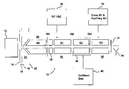
[0025] FIG. 1 is a schematic view of a conventional QTRAP hybrid quadrupole-
linear ion
trap mass spectrometer;
[0026] FIG. 2 is a schematic view of an alternative conventional QTRAP hybrid
quadrupole-
linear ion trap mass spectrometer;
[0027] FIGS. 3A to 3C are graphs illustrating axial energy of ions before and
after passing
through an ion optical element operated in accordance with Applicants'
teachings;
[0028] FIGS. 4A to 4C are graphs illustrating the normalized intensities of
fragments for
various methods of fragmenting epinephrine;
[0029] FIGS. 5A to 5C are graphs illustrating the normalized intensities of
fragments for
various methods of fragmenting clenbuterol;
[0030] FIGS. 6A to 6C are graphs illustrating the normalized intensities of
fragments for
various methods of fragmenting erythromycin;
[0031] FIG. 7 is a graph illustrating the intensity of an ion beam after
passing through exit
lens 32;
5
CA 02784485 2012-06-14
WO 2011/073794 PCT/1B2010/003304
[0032] FIGS. 8A and 8B are graphs illustrating normalized intensities of a
precursor ion signal
and a fragment ion signal, respectively, for various RF fields applied to an
ion optical element;
and
[0033] FIGS. 9A and 9B are graphs illustrating normalized intensities of a
precursor ion signal
and a fragment ion signal with and without, respectively, the application of
an RF field to an ion
optical element.
DESCRIPTION OF VARIOUS EMBODIMENTS
[0034] Referring first to FIGS. 1 and 2, there are shown two conventional
triple quadruple
mass spectrometer apparatus generally designated by references 10 and 10'
respectively. The
two embodiments are similar and will be described together except for the
parts that differ
between embodiments, which will be separately described. An ion source 12, for
example an
electrospray ion source, generates ions directed towards a curtain plate 14.
Behind the curtain
plate 14, there is an orifice plate 16, defining an orifice, in known manner.
[0035] A curtain chamber 18 is formed between the curtain plate 14 and the
orifice plate 16,
and a flow of curtain gas reduces the flow of unwanted neutrals into the
analyzing sections of
the mass spectrometer. The two embodiments illustrated in FIGS. 1 and 2 differ
in their structure
between the orifice plate and the interquad barrier IQ1 and these portions of
the mass
spectrometers will be discussed separately for each embodiment.
[0036] In mass spectrometer 10 of FIG. 1, following the orifice plate 16,
there is a skimmer
plate 20. An intermediate pressure chamber 22 is defined between the orifice
plate 16 and the
skimmer plate 20. The pressure in chamber 22 is typically of the order of 2
Torr. Ions pass
through the skimmer plate 20 into the first chamber of the mass spectrometer,
indicated at 24. A
quadruple rod set QO is provided in this chamber 24, for collecting and
focusing ions. This
chamber 24 serves to extract further remains of the solvent from the ion
stream, and typically
operates under a pressure of 7 mTorr. It provides an interface into the
analyzing sections of the
mass spectrometer.
[0037] Referring now to FIG. 2, in mass spectrometer 10', following the
orifice plate 16 there
is an ion guide 21. The ion guide 21 focuses the ions passing through it. In
some embodiments,
ion guide 21 has a length of approximately 55 mm and a diameter of
approximately 4 mm. In
addition, in various embodiments, an AC voltage with a frequency of
approximately 1.1 MHz
and an amplitude in the range of 0-300 V is applied to ion guide 21. An
interquad lens IQO
6
CA 02784485 2012-06-14
WO 2011/073794 PCT/1B2010/003304
separates the ion guide 21 and chamber 24. Ions pass through the interquad
lens IQO into the
first chamber of the mass spectrometer, indicated at 24'. A quadruple rod set
QO' is provided in
this chamber 24', for collecting and focusing ions. This chamber 24' serves to
extract further
remains of the solvent from the ion stream, and typically operates under a
pressure of 7 mTorr. It
provides an interface into the analyzing sections of the mass spectrometer.
[0038] In some embodiments of mass spectrometer 10', Quadruple rod set QO' and
chamber 24'
are shorter than quadruple rod set QO and chamber 24 respectively of mass
spectrometer 10. In
particular, as mentioned above, one function of QO and QO' is to collect and
focus the ions.
However, the ion guide 21 also serves to collect and focus the ions prior to
their entry into Q0'.
[0039] Referring now to both FIGS. 1 and 2, an interquad barrier or lens IQ1
separates the
chambers 24 and 24' respectively from the main mass spectrometer chamber 26
and has an
aperture for ions. Adjacent the interquad lens 1Q1, there is a short
"stubbies" rod set, or
Brubaker lens 28. A first mass resolving quadruple rod set Q1 is provided in
the chamber 26 for
mass selection of a precursor ion. Following the rod set Q1, there is a
collision cell 30
containing a second quadruple rod set Q2, and following the collision cell 30,
there is a third
quadruple rod set Q3 for effecting a second mass analysis step.
[0040] The final or third quadruple rod set Q3 is located in the main
quadruple chamber 26
and subjected to the pressure therein typically 1x1 0-5 Ton. As indicated, the
second quadruple
rod set Q2 is contained within an enclosure forming the collision cell 30, so
that it can be
maintained at a higher pressure; in known manner, this pressure is analyte
dependent and could
be 5 mTorr. Interquad lenses IQ2 and IQ3 are provided at either end of the
enclosure of the
collision cell of 30.
[0041] Ions leaving Q3 pass through an exit lens 32 to a detector 34. It will
be understood by
those skilled in the art that the representation of FIGS. 1 and 2 are
schematic, and in various
embodiments various additional elements would be provided to complete the
apparatus. For
example, in various embodiments, a variety of power supplies are utilized to
deliver AC and DC
voltages to different elements of the apparatus. In addition, in some
embodiments a pumping
arrangement or scheme is utilized to maintain the pressures at the desired
levels mentioned.
[0042] As indicated, a power supply 36 is provided for supplying RF and DC
resolving
voltages to the first quadruple rod set Ql. Similarly, a second power supply
38 is provided for
supplying drive RF and auxiliary AC voltages to the third quadruple rod set
Q3, for scanning
ions axially out of the rod set Q3. A collision gas is supplied, as indicated
at 40, to the collision
7
CA 02784485 2012-06-14
WO 2011/073794 PCT/1B2010/003304
cell 30, for maintaining the desired pressure therein, and an RF supply would
also be connected
to Q2 within the collision cell 30. As will be explained in greater detail
below, AC and/or DC
voltages may be applied to various ion optical elements such as the interquad
lenses.
[0043] Although two specific embodiments of mass spectrometers have been
discussed above,
it should be understood that various embodiments of the methods of processing
ions described
herein can be applied to any appropriate mass spectrometer including but not
limited to a
quadrupole, such as ion traps or time-of-flight mass spectrometers. In
addition other ion
containment devices, such as hexapoles, octupoles, and ring guides, may be
used. In particular,
various embodiments of the methods described herein can be applied to any
appropriate
arrangement that contains the ions radially and operates at an elevated
pressure.
[0044] In various embodiments, the methods of processing ions described herein
can be
applied to various applications including, but not limited to, declustering
and fragmenting ions.
Declustering can also be referred to as desolvating and is the process by
which analyte ions are
separated from other particles in the gas phase, such as solvent particles or
buffer particles,
where buffers can consist of acids or bases or salts that are added to the
solvent. Specifically, the
analyte may be in a solution prior to being mass analyzed and as discussed
above, in such cases,
it may be necessary to remove residual solvent molecules or other neutrals
from the ions prior to
analyzing them. In contrast, fragmentation involves breaking analyte ions into
their constituent
parts. Thus, a major difference between fragmentation and declustering is the
amount of kinetic
energy required to break apart the bonds of the particles. For the same type
of analyte,
fragmentation usually requires a greater amount of energy than declustering
given that
fragmentation generally involves breaking apart molecules that are made of
atoms that are
covalently bonded while declustering generally involves breaking apart species
that are not
covalently bonded. Declustering generally results in reducing the intensity of
cluster ion peaks
in the mass spectrum. Cluster ions can consist of solvent ions or buffer ions
clustered with
solvent or buffer molecules, or of analyte ions clustered with solvent or
buffer molecules.
100451 In various embodiments, the method includes the step of determining or
selecting a
kinetic energy profile for the ions within an ion containment field. As will
be explained below,
this is not necessarily the first step and in some embodiments, the kinetic
energy is selected
indirectly. The kinetic energy profile refers to the distribution of kinetic
energies of the ions that
are within the ion containment field. In various embodiments, the selected
kinetic energy profile
is selected to fragment the ions to concurrently provide a plurality of groups
of product ions.
8
CA 02784485 2012-06-14
WO 2011/073794 PCT/IB2010/003304
[0046] In various embodiments, the kinetic energy profile is a continuous
function. In
addition, in some embodiments, the kinetic energy profile is a continuous
function that includes
a wide band of kinetic energies. This is in contrast to known methods in which
a discrete kinetic
energy value is used to fragment ions. In some embodiments of the method of
fragmenting ions,
the highest kinetic energy level in the kinetic energy profile is at least
three times the lowest
kinetic energy in the kinetic energy profile. In some embodiments of the
method of fragmenting
ions, the highest kinetic energy level exceeds 50 eV. In various embodiments,
the highest kinetic
energy level exceeds 100 eV.
[0047] In various embodiments of the method of processing ions where the
method is applied
to fragmentation, the kinetic energy profile can be selected such that a
desired fragmentation
spectrum is achieved when the ions are fragmented. In various embodiments, the
ions are
fragmented in a collision cell such as collision cell 30. Accordingly, in some
such embodiments,
the ion containment field within which the ions are processed or fragmented is
the ion
containment field produced by Q2. The particular kinetic energy profile that
is selected can be
determined based on a variety of factors including but not limited to the
particular type of ions
that are to be fragmented and the desired fragmentation spectrum. The term
"fragmentation
spectrum" as used herein refers to the spectrum of ions produced from
fragmenting the analyte
precursor ions.
[0048] In some embodiments, the method further includes the step of
determining at least one
characteristic of a RF field based on the kinetic energy profile that has been
selected. The at
least one characteristic can include, but is not limited to, the amplitude and
frequency of the RF
field. As will be explained in greater detail below, the RF field determines,
at least in part, the
kinetic energy profile that is achieved.
[0049] In various embodiments, the selected RF field is applied to an ion
optical element that
is upstream of the ion containment field. Prior to entering the ion
containment field, the ions
pass through an ion optical element and interact with the RF field that is
applied to the ion
optical element. The ion optical element can be any appropriate ion optical
element. Thus, for
example, the ion optical element can be but is not limited to, any appropriate
aperture lens, such
as an interquad lens, an ion optical lens having a skimmer-type lens geometry,
a flat plate
orifice, a conical orifice, a wire grid (i.e. a mesh), or a two-wire element
mounted transverse to
the ion flow. Thus, for example, in various embodiments where the ion
containment field is that
of Q2 and the ion optical element is an interquad lens, the selected RF field
can be applied to
IQ2, IQ1, or IQ0.
9
CA 02784485 2012-06-14
WO 2011/073794
PCT/1B2010/003304
10050] In some embodiments, in addition to the RF field, a DC offset voltage
is also applied to
the ion optical element. The kinetic energy profile of the beam of ions
transmitted through the
ion optical element is determined primarily by the RF and DC voltages applied
to the element.
In certain instances it is desirable to add attractive or repulsive DC
voltages to the ion optical
element to control the resulting kinetic energy profile of the transmitted ion
beam. An attractive
DC voltage will add an offset energy to the ions transmitted through the ion
optical element. A
repulsive DC voltage will reduce the average ion energy of the ions
transmitted through the ion
optical element and, in some embodiments, may cause some ions not to be
transmitted at all.
[0051] In general, the ion optical element can be any appropriate ion optical
element,
including but not limited to any of the ion optical elements described above.
However, in some
embodiments, only ion optical elements that are not upstream of a mass
analyzer are selected for
application of the RF field. For example, in some embodiments, Q1 is operated
as a mass
analyzer. Accordingly, in some such embodiments, IQ1 is typically not used as
the ion optical
element to which the RF field is applied in the manner described herein. The
reason for this is
that it can be desirable to have a well-defined analyte ion energy entering a
mass analyzer.
Applying an RF field can cause the ion energy of the beam to change as
discussed below.
However, in some embodiments, Q1 is not operated as a mass analyzer and in
some such
embodiments the selected RF field is applied to IQ1 for example.
100521 The beam of ions produced at source 12 is transmitted through the ion
optical element
to which the selected RF field has been applied and travels into the ion
containment field. As the
ions are transmitted through the ion optical element, they interact with the
RF field that has been
applied to the ion optical element. Specifically, the selected RF field
affects the kinetic energy of
the ions that are transmitted through the ion optical element and move into
the ion containment
field. Accordingly, the selected RF field determines, at least in part, the
kinetic energy profile of
the ions within the ion containment field.
[00531 In various embodiments, the ions are processed in the ion containment
field by
introducing a neutral gas stream into the ion containment field. This can be
done as described
above with respect to collision cell 30 and collision gas 40. The ions collide
with the neutral gas
stream in the ion containment field with collision energies that are
determined by their kinetic
energy profile. Depending on the selected kinetic energy profile and the type
of ions, these
collisions can be used to fragment or decluster the ions.
CA 02784485 2012-06-14
WO 2011/073794
PCT/IB2010/003304
[0054] As mentioned above, in various embodiments, the selected kinetic energy
profile is
selected to fragment the ions to concurrently provide a plurality of groups of
product ions. For
example, in some embodiments, the kinetic energy profile is selected to
produce a given number
of groups of product ions. In some embodiments the kinetic energy profile is
selected so that
there are three energy levels in the kinetic energy profile that cause three
separate groups of
fragment ions to be formed. Each of these three energy levels can be referred
to as precursor
kinetic energy levels. In various embodiments, the kinetic energy profile is
selected such that the
product ions include at least four groups, where there are at least three
groups of fragment ions
and a group of precursor ions. It should be understood that this is an example
only and is not
intended to be limiting. For example, some embodiments have greater than three
groups of
fragment ions.
[0055] In various embodiments, each of the groups of product ions comprise
only ions of the
same mass to charge ratio. In other words, in various embodiments, each group
of product ions
refers to a particular generation of fragment ions or to precursor ions. In
addition, in some
embodiments, each of these groups of product ions comprise less than half of
the total ions that
are produced in the ion containment field.
[0056] Although some embodiments of the method have been described as
comprising the step
of determining a selected kinetic energy profile for the ions and then
selecting a RF field based
on the selected kinetic energy profile, in some embodiments, it is generally
the case that this is
not done in a series of independent discrete steps but rather is done in an
iterative manner.
Specifically, in some embodiments a RF field can be selected and applied to
the ion optical
element and the resulting fragmentation can be observed. From the resulting
fragmentation, one
can deduce the kinetic energy profile of the ions prior to fragmentation.
Based on the observed
level of fragmentation, the RF field can be adjusted until a desired
fragmentation spectrum is
achieved. The term "fragmentation spectrum" as used herein refers to the
spectrum of ions
produced from fragmenting the analyte precursor ions.
[0057] In other words, a second RF field can be selected and applied to the
ion optical
= element. The ions can then be transmitted through the ion optical element
and into the RF
containment field where, in some embodiments, the ions are fragmented and a
second plurality
of groups of product ions are produced concurrently. In various embodiments,
the second
plurality of groups of product ions can be different than the first. In some
embodiments, the
second plurality of groups can include all of the first plurality of groups or
vice-versa.
Accordingly, in some embodiments, the second plurality of groups may include a
greater or
11
CA 02784485 2012-06-14
WO 2011/073794
PCT/IB2010/003304
lesser number of generations of fragment ions. In some embodiments, the second
plurality of
groups of ions and the first plurality groups of ions are non-overlapping. In
various
embodiments, the product ions can be detected by detector 34.
100581 Alternatively, a RF field can be selected based on the ions that are
being processed. For
example, in some embodiments, it may be known that a given RF field will
produce a given
fragmentation spectrum and this RF field can be selected.,
[0059] Similarly, for the method of declustering an RF field can be selected
and applied to an
ion optical element. The analyte ions, which are non-covalently bonded, are
transmitted through
the ion optical element and into the ion containment field. The RF field
determiners, at least in
part, the kinetic energy profile of the analyte ions. In various embodiments,
the ions are
declustured in the ion containment field. The RF field is selected such that
when declustering the
analyte ions and solvent ions the non-covalent bonds between most of the
analyte ions and the
solvent ions are broken without breaking most of the covalent bonds of the
analyte ions
themselves. In other words, in various embodiments the RF field is selected
such that the
declustering occurs without any significant fragmentation of the analyte ions
occurring.
[0060] The kinetic energy profile of the analyte ions can be adjusted and
affected in various
ways. For example, various characteristics of the RF field applied to the ion
optical element can
be altered, including, but not limited to, the amplitude of the RF field and
the frequency of the
RF field. In addition, if a DC voltage is also applied then the DC voltage can
also be adjusted to
affect the kinetic energy profile. Altering one or more of the above-listed
variables can, for
example, adjust such things as the average energy in the kinetic energy
profile and the range of
kinetic energies in the kinetic energy profile.
[0061] Reference is now made to figure 3A to 3C, which illustrate axial energy
as a function
of axial position for different RF fields applied to the ion optical element
using computer
simulations for 50 ions. The ion optical element in this case is an interquad
lens IQ2. The dot-
dash vertical lines delimit the axial range of IQ2. In each of the three
figures the lens is
positioned at 20 mm. With one exception, all the ions pass through the lens.
The single
exception occurs in FIG. 3B where one of the ions is reflected from IQ2. In
FIG. 3A, the RF
field applied to the lens has a frequency of 50 kHz and an amplitude of 200
Vpp. In figure 3B,
the RF field applied to the lens has a frequency of 200 kHz and an amplitude
of 200 Vpp. In
FIG. 3C, no RF field is applied to the lens. In addition, in each of FIGS. 3A
to 3C, an attractive
V DC offset is applied to the lens.
12
CA 02784485 2012-06-14
WO 2011/073794
PCT/IB2010/003304
[0062] As can be seen from comparing the figures, the ions that are
transmitted through the
lens have a much higher average energy in the case of FIGS. 3A and 3B, than
they do in the case
of FIG. 3C. More specifically, looking at a distance of 5 mm in either
direction from the lens,
the axial energy of the ions is increased significantly after passing though
the lens. In addition,
these same ions have a greater or wider distribution of axial energy than the
case where no RF
field is applied to the lens. Specifically, in FIG. 3C, the ion axial energies
are clustered together;
while, in FIGS. 3A and 3B, the axial energies are spread out over a range of
roughly 100 eV or
more.
[0063] In various embodiments, the method described herein can produce a wide
fragmentation spectrum with the precursor ion and a plurality of generations
of fragments
observed simultaneously. Part of the reason for this is, as described above,
that the ions have a
wide kinetic energy profile and therefore a wide range of collision energies
can be achieved
simultaneously. Furthermore, a rather large average kinetic energy can also be
achieved and
therefore the range of energies can be useful for fragmentation.
100641 The RF field applied to the lens can be any appropriate voltage. In
some embodiments,
the voltage applied to the lens is in a range from 10 Vpp to 200 Vpp. In
addition, any
appropriate frequency can be used for the RF field. For example, in some
embodiments, a
frequency range of 1 kHz to 500 kHz is used. In some other embodiments, the
range of
frequencies used is 10 kHz to 200 kHz. These are example amplitude and
frequency ranges only
and are not intended to be limiting. Some other embodiments operate with RF
fields having
amplitudes and frequencies outside of these ranges. In various embodiments, an
appropriate RF
field can be selected based in part on the desired kinetic energy profile of
the ions and one or
more characteristics, such as the mass to charge ratio (m/z), of the
particular ions being
processed.
[0065] In some embodiments, the ion beam produced by ion source 12 is a
continuous or
uninterrupted beam of ions that extends from ion source 12, through the lens
to which the RF
field is applied, through the ion containment field (e.g. in the collision
cell) and into the detector.
In other words, in various embodiments, during operation, the beam is not
interrupted between
any of the above-mentioned sections of the mass spectrometer but rather there
is a continuous
path through each of those components starting from source 12 and extending to
detector 34 and
the beam of ions is simultaneously or concurrently present at each of those
components of the
mass spectrometer.
13
CA 02784485 2012-06-14
WO 2011/073794 PCT/1B2010/003304
[0066] In various other embodiments, the ion beam produced by ion source 12 is
a continuous
or uninterrupted beam of ions that extends from ion source 12, through the
lens to which the RF
field is applied, an into the ion containment field (e.g. in the collision
cell). In other words, in
various embodiments, during operation, the beam is not interrupted between any
of the above-
mentioned sections of the mass spectrometer but rather there is a continuous
path through each
of those components starting from source 12 and extending to ion containment
field and the
beam of ions is simultaneously or concurrently present at each of those
components of the mass
spectrometer.
[0067] The following data was obtained using a 4000QTRAP instrument. The RF/DC
quadrupole Q1 was used to select the m/z of the precursor ion. The selected
precursor ions were
passed through an aperture lens (IQ2) located in front of a quadrupole
collision cell and finally
into the Q3 linear ion trap. After an appropriate cooling time, the contents
of the linear ion trap
were scanned out using mass selective axial ejection toward the ion detector.
Reference is now
made to FIGS 4A to 4C, which are graphs illustrating the normalized
intensities of product ions
for various methods of fragmentation for epinephrine. In FIGS. 4A and 4B no RF
was applied to
the IQ2 aperture lens. In FIG. 4C, a 200 kHz RF field was applied to IQ2. More
specifically,
FIG. 4A illustrates a graph of normalized intensity of product ions against
collision energy in eV
for the case where conventional beam type collision induced dissociation (CID)
is used to
fragment epinephrine prior to the final mass analysis step. As can be seen
from the graph, there
is only a narrow region 420 in which the precursor and the low mass fragments
are
simultaneously observable. As can be seen from the figure, region 420 is less
than 5 eV wide. In
addition, there is no region in the graph where the precursor and the lowest
fragment can be
observed simultaneously.
[0068] FIG. 4B illustrates a graph of normalized intensity of product ions
versus excitation
energy in mV for the case where in-trap fragmentation within the Q3 linear ion
trap is used to
fragment epinephrine. As can be seen from the figure, only 1 fragment is
observed in the first
fragmentation stage. All other fragments, indicated at 430, have a normalized
intensity value of
0. In order to observe the remaining fragments multiple fragmentation stages
(MSn) are
required. Accordingly, as is the case with CID described in relation to FIG.
4A, it is not possible
to view the precursor and low mass fragments in the same fragmentation stage.
[0069] FIG. 4C illustrates a graph illustrating the intensity of product ions
that result from the
application of embodiments of the method described herein. Specifically, FIG.
4C illustrates the
normalized intensity of product ions versus the amplitude of the 200 kHz RF
field applied to the
14
CA 02784485 2012-06-14
WO 2011/073794
PCT/1B2010/003304
ion optical lens. More specifically, the voltage indicated on the x-axis is
the voltage that was
applied to interquad lens IQ2. In FIG. 4C, the frequency is held constant at
200 kHz and the DC
offset voltage that is applied to IQ2 is 46 V attractive.
[0070] FIGS. 5A to 5C and 6A to 6C are analogous to FIGS. 4A to 40 except that
they are for
clenbuterol and erythromycin respectively. They illustrate results that are
similar to those
discussed in relation to FIGS. 4A to 4C. Specifically, FIGS. 5A and 6A
illustrate that the use of
conventional CID results in only narrow regions 520 and 620 of collision
energies where
precursor and low mass fragments are simultaneously observed for
clenbuteroland and
erythromycin. As can be seen from the figures, region 520 is approximately 5
eV wide; while,
region 620 is approximately 20 eV wide. FIG. 5B illustrates that when in-trap
fragmentation
within the Q3 linear ion trap is used to fragment clenbuterol, the low mass
fragments, indicated
at 530, are not observed in the first fragmentation stage and therefore
multiple stages of
fragmentation are required. Similarly, FIG. 6B, illustrates that when in-trap
fragmentation is
used to fragment erythromycin, the low mass fragments are never observed due
to the low mass
cut-off of the linear ion trap.
[0071] Finally, FIGS. 5C and 6C illustrate that when embodiments of the method
described
herein are applied to fragmenting clenbuterol and erythromycin respectively,
then there are wide
regions 530 and 630 respectively where precursor ions and low mass fragments
are
simultaneously observed.
[0072] As discussed above, in various embodiments, the methods described
herein includes
the steps of applying a RF field to an ion optical element and transmitting
ions through the ion
optical element and then into an ion containment field. The RF field applied
to the ion optical
element determines, at least in part, the kinetic energy of the ions within
the containment field
and therefore the RF field can be adjusted to achieve a particular kinetic
energy profile. For
example, various parameters of the RF field can be adjusted including but not
limited to the
amplitude and frequency to adjust such things as the average energy and the
range of energies in
the kinetic energy profile. In addition, the selected kinetic energy profile
of the ions in the ion
containment field can have an axial energy profile that is modulated at the
frequency of the RF
applied to the ion optical element. If the containment device is pressurized
this modulation is
sometimes lost due to the large number of collisions with the background gas
molecules. The
modulation of the axial kinetic energy can be observed in the absence of
collisions.
CA 02784485 2012-06-14
WO 2011/073794
PCT/IB2010/003304
[0073] Reference is now made to FIG. 7, which illustrates two graphs of
intensity of the ion
beam after passing through exit lens 32. Specifically, a RF field with a
frequency of 50 kHz is
applied to IQ2 between 2 ms and 20 ms. In addition, a repulsive 20 V DC
voltage is applied to
exit lens 32. The DC repulsive barrier discriminates based on the kinetic
energy of the ions and
allows only ions with kinetic energies that are above a threshold energy level
to pass through
exit lens 32. The ions are detected at detector 34, which can detect the
energy level of the ions.
The plot on the right is a blown up version of the intensity between 8 ms and
9 ms. As can be
seen from FIG. 7, the intensity of the ion beam is a continuous function. The
frequency of the
intensity is 50 kHz which matches the frequency of the RF field applied to
IQ2. Thus, the ions
pick up energy as they pass through the lens and the amount of energy pickup
follows the phase
of the RF field applied to the IQ2 aperture lens. Accordingly, through the use
of the method
described herein, it is possible to encode the ion beam with frequency
information of the RF
field applied to the IQ2 aperture lens.
[0074] In various embodiments, the RF field applied to the ion optical element
can be varied
in any appropriate manner to encode any appropriate desired information in the
ions. For
example, although the use of a single discrete RF field frequency and
amplitude are illustrated in
FIG. 7, any appropriate RF field characteristics, including but not limited
to, frequency and
amplitude can be used. In addition, any of one or more of the RF field
characteristics can be
varied in any appropriate manner including, but not limited to, continuous and
discrete
variations.
[0075] In some embodiments of encoding ions, the method can include the step
of determining
or selecting a first frequency. Then an RF field having the selected frequency
can be applied to
an ion optical element. The ions can then be transmitted into an ion
containment field. The ions
can then be detected by a detector such as detector 34 and the frequency of
the ion kinetic
energy profile can be determined.
[0076] In some embodiments, multiple frequencies can be selected at different
times and the
frequency of the ion kinetic energy profile can be determined once detected.
In various
embodiments, identifying the frequency can be used to identify the particular
group of ions that
are detected. For example, different groups of ions can be transmitted through
the ion optical
element with RF fields having different frequencies applied to it.
[0077] In some embodiments, the Applicants have observed that the higher the
pressure in
which the optical element is situated, the larger the amplitude of the RF
voltage required to
16
CA 02784485 2012-06-14
WO 2011/073794 PCT/1B2010/003304
achieve the same result. Specifically, in some embodiments, if all other
variables are held
constant and the pressure is increased, then in order to maintain a given
level of fragmentation or
declustering, the amplitude of the RF field applied to the ion optical element
is increased.
[0078] The above discussion illustrated examples of various embodiments of the
method that
are carried out through the application of a RF field to an interquad lens.
However, as mentioned
above, the method can be implemented with any appropriate ion optical element
including but
not limited to curtain plate 14, the orifice plate 16, or IQ0. Accordingly,
the method can be
applied to virtually any ion optical element that is anywhere in the stream of
ions, including at
the front end of the mass spectrometer near the ion inlet.
[0079] The following data was obtained using a 4000 QTRAP instrument. The
precursor ions
were passed through orifice plate 16 located in front of QO. A RF field is
applied to orifice plate
16. A collision gas is introduced into chamber 24 such that QO can be used for
declustering the
precursor ions. After declustering, the ions were passed through the rest of
the 4000 QTRAP
instrument and finally into the Q3 linear ion trap. After an appropriate
cooling time, the contents
of the linear ion trap were scanned out using mass selective axial ejection
toward the ion
detector. Reference is now made to FIGS. 8A and 8B, which are graphs
illustrating the
normalized intensities of precursor ion signals and fragment ion signals
respectively.
[0080] More specifically, FIG. 8A illustrates the normalized intensity of the
clenbuterol
precursor ion (m/z 277), for various frequencies of RF field applied to
orifice plate 16, against
the declustering potential (DP). All the RF fields have a peak-to-peak
amplitude of 300 V (or
300 Vpp). The DP voltage is a DC potential difference between the orifice
plate 16 and skimmer
pate 20. In various embodiments, skimmer plate 20 is grounded.
[0081] Also illustrated in FIG. 8A is the plot of the normalized intensity of
the clenbuterol
precursor ion for the case where no RF field is applied to the orifice plate
16. As can be see from
FIG. 8A, when no RF field is applied, the intensity of the precursor ion is
maximized at a DP
voltage of approximately 110 V.
[0082] As can be seen from FIG. 8A, the application of a RF field to orifice
plate 16 causes
the maximum intensity of the ion signal to occur at a lower voltage as
compared to the case
where no RF field is applied to orifice plate 16. The Applicants postulate
that this indicates that
the presence of the auxiliary RF field is also a method for adding kinetic
energy to the ions as
they pass through the orifice plate.
17
CA 02784485 2012-06-14
WO 2011/073794 PCT/1B2010/003304
[0083] Reference is now made to FIG. 8B, which illustrates normalized
intensity of a
clenbuterol fragment ion, for various frequencies of RF field applied to
orifice plate, against the
declustering potential (DP). All the RF fields have peak-to-peak amplitudes of
300 V (or 300
Vpp).
[0084] Also illustrated in FIG. 8B is the plot of normalized intensity of the
clenbuterol
fragment ion (m/z 203) for the case where no RF field is applied to orifice
plate 16. As can be
see from FIG 8B, when no RF field is applied, the intensity of the precursor
ion is maximized at
a DP voltage above 200 V. As can be seen, the intensity of the fragment ion
signal maximizes at
a DP value that is higher than the maximum of the precursor ion. This is in
part due to the fact
that the fragment signal originates from the fragmentation of the precursor
ion, which requires a
higher energy than declustering.
[0085] In addition, as was the case with the precursor ion, the application of
an RF field to
orifice plate 16 causes the maximum intensity of the fragment ion signal to
occur at a lower
voltage as compared to the case where no RF field is applied to orifice plate
16. As stated above,
the Applicants postulate that this indicates that the presence of the
auxiliary RF field is also a
method for adding kinetic energy to the ions as they pass through the orifice
plate contributing
to the fragmentation process.
[0086] Reference is now made to FIGS. 9A and 9B which illustrate normalized
intensities of a
precursor ion signal and a fragment ion signal for the case where a 200 kHz
Auxiliary RF field
is applied to orifice plate 16 and the case where no auxiliary RF field is
applied to orifice
plate 16 against the DP voltage. Specifically, FIG. 9A illustrates the
clenburterol precursor ion
and clenbuterol fragment ion signals for the case where a 200 kHz auxilary RF
signal is applied
to orifice plate 16. FIG. 9B illustrates clenburterol precursor ion and
clenbuterol fragment ion
signals for the case where no RF field is applied to orifice plate 16. As can
be seen from
comparing FIGS. 9A and 9B, when an auxiliary RF is present on the orifice
pate, there is a much
better overlap between the DP curves for the precursor and fragment ions.
Specifically, in
FIG. 9A, there is a range of DP voltage values where both the fragment ion
intensity and the
precursor ion intensity are both relatively high and near their respective
maxima. In contrast, in
FIG. 9B, the overlap occurs at a lower intensities and the range of overlap is
smaller. The use of
the method as described herein, which for example creates a condition similar
to that illustrated
in FIG. 9A, allows the instrument to operate under orifice voltage conditions
that generate mass
spectra containing significant contributions of both precursor ions and
fragment ions.
18
CA 02784485 2012-06-14
WO 2011/073794
PCT/IB2010/003304
100871 While the above description provides example embodiments, it will be
appreciated that
the present invention is susceptible to modification and change without
departing from the fair
meaning and scope of the accompanying claims. Accordingly, what has been
described is
merely illustrative of the application of aspects of embodiments of the
invention and numerous
modifications and variations of the present invention are possible in light of
the above teachings.
19