Note: Descriptions are shown in the official language in which they were submitted.
CA 02785226 2012-06-20
WO 2011/084748 PCT/US2010/061335
PHOTO-INACTIVATED VIRUSES AND
SYSTEMS AND METHODS OF USING THE SAME
CROSS-REFERENCE TO RELATED APPLICATIONS
This application claims, under 35 U.S.C. 119(e), the benefit of U.S.
Provisional
Application Serial No. 61/288,756, filed 21 December 2009, the entire contents
and substance
of which are hereby incorporated by reference as if fully set forth below.
BACKGROUND OF THE INVENTION
1. TECHNICAL FIELD
The various embodiments of the present disclosure relate generally to systems
and
methods for the photo-inactivation of microorganisms. More specifically, the
various
embodiment of the present invention are directed towards the photo-
inactivation of
microorganisms, such as viruses, using at least one furanocoumarin and broad
spectrum pulsed
light.
2. DESCRIPTION OF RELATED ART
Herpes B virus (Herpesvirus simiae or Cercopithecine herpesvirus 1), a member
of the
Alphaherpesvirinae subfamily and the Simplexvirus group, is known to occur
naturally in
macaques (Macaca spp). Infection of macaques may be asymptomatic or may cause
a mild
disease. Infection of other species, such as humans, is rare but results in
severe, and if untreated,
lethal disease.
Past infections are determined by detection of anti B virus antibodies using
serological
assays. Serological diagnosis of B virus infections in humans, however, is
complicated by the
relatively high prevalence of the immunologically cross-reacting herpes
simplex virus
infections (e.g., HSV-1 and/or HSV-2). Past infections in macaques can be
established without
these complications because the only simplexvirus known to infect macaques is
B virus.
Identifying B virus infected macaques is important for managing macaques in
captivity, for
developing specific pathogen free colonies and for the prevention of the
potential exposure and
infection of humans who handle macaques.
Thus, what are needed are compositions, systems, and methods for the
identification of
individuals infected with a microorganism. The focus of the current
application is to such novel
1
CA 02785226 2012-06-20
WO 2011/084748 PCT/US2010/061335
composition, systems, and methods for the identification of individuals
infected with a
microorganism, such as B virus.
BRIEF SUMMARY OF THE INVENTION
The various embodiments of the present disclosure relate generally to systems
and
methods for the photo-inactivation of microorganisms, and more particularly,
to the photo-
inactivation of viruses using at least one furanocoumarin and broad spectrum
pulsed. For
example, an aspect of the present invention comprises a method for
inactivating a
microorganism, comprising: providing at least one furanocoumarin to a
microorganism; and
exposing the microorganism to at least one pulse of a broad spectrum pulsed
light, thereby
inactivating the microorganism. The microorganism can be selected from the
group consisting
of viruses, bacteria, and fungi, and preferably comprises a virus. An
exemplary virus
comprises a herpesvirus, such as herpes B virus or herpes virus papio 2. The
furanocoumarin
can comprise a psoralen, and the psoralen can be used at a concentration
ranging from about
0.1 g/ml to about 60 g/ml. In an exemplary embodiment, the psoralen is
present in a
concentration of at least about 5 tg/ml. Exposing the microorganism to at
least one pulse of a
broad spectrum pulsed light can comprise exposing the microorganism to about
0.45 Joule/cm2
to about 13.5 Joules/cm2 of broad spectrum light. In another embodiment,
exposing the
microorganism to at least one pulse of a broad spectrum pulsed light can
comprise exposing the
microorganism to at least about 4.05 Joules/cm2 of broad spectrum light to
about 13.5
Joules/cm2 of broad spectrum light.
Another aspect of the present invention comprises an inactivated microorganism
comprising a photo-chemically inactivated nucleic acid, wherein the photo-
chemically
inactivated nucleic acid is photo-chemically inactivated by at least one
furanocoumarin and at
least one pulse of a broad spectrum pulsed light. The microorganism can be
selected from the
group consisting of viruses, bacteria, and fungi, and is preferably a virus.
An exemplary virus
comprises a herpesvirus, such as herpes B virus or herpes virus papio 2. The
furanocoumarin
can comprise a psoralen, and the psoralen can be used at a concentration
ranging from about 0.1
g/ml to about 60 g/ml. In an exemplary embodiment, the psoralen is present in
a
concentration of at least about 5 tg/ml. Photo-chemical inactivation of the
virus can involve
exposing the microorganism to at least one pulse of a broad spectrum pulsed
light, which can
utilize about 0.45 Joule/cm2 to about 13.5 Joules/cm2 of broad spectrum light.
In an exemplary
2
CA 02785226 2012-06-20
WO 2011/084748 PCT/US2010/061335
embodiment, photo-chemical inactivation of the virus can involve exposing the
microorganism
to at least about 4.05 Joules/cm2 of broad spectrum light to about 13.5
Joules/cm2 of broad
spectrum light. For example, an inactivated microorganism can be inactivated
by exposure to
psoralen at a concentration of at least about 5 g/ml and at least one pulse
of a broad spectrum
pulsed light that comprises at least about 4.05 Joules/cm2 of broad spectrum
light.
Yet another aspect of the present invention comprises a system for detecting
an antibody
in a subject, comprising: an antigen component, wherein the antigen is exposed
to a
furanocoumarin and at least one pulse of a broad spectrum pulsed light; and a
reporter
component that is capable of detecting a binding of an antibody of a subject
to at least a portion
of the antigen. The antigen can be selected from the group consisting of a
virus, a bacterium,
and a fungus, and preferably comprises a virus. In an exemplary embodiment,
the viral antigen
is a herpesvirus antigen, which can include, but is not limited to an antigen
from herpes B virus
or herpes virus papio 2. The furanocoumarin is a psoralen, and the broad
spectrum pulsed light
can comprise about 0.45 Joule/cm2 to about 13.5 Joules/cm2 of broad spectrum
light. In one
embodiment, the antigen component can further comprise an antigen disposed on
a substrate.
The reporter component can comprise, for example, a reporter antibody capable
of binding at
least a portion of the antibody capable of binding at least a portion of the
antigen.
Still another aspect of the present invention comprises a method for
immunizing a
subject, comprising: inactivating an immunogenic microorganism comprising
exposing to the
immunogenic microorganism to a furanocoumarin and to at least one pulse of a
broad spectrum
pulsed light; and administering an effective amount of the immunogenic
microorganism to a
subject to produce an immune response. Such a method contemplates use of an
inactivated
immunogenic microorganism to immunize a subject. The immunogenic microorganism
can
include a virus, a bacterium, a fungus, or combinations thereof. In an
exemplary embodiment,
the immunogenic microorganism comprises a virus, preferably a herpesvirus, and
more
preferably a herpes B virus or herpes virus papio 2. The furanocoumarin can
comprise psoralen,
which can be present in a concentration of about 0.1 g/ml to about 60 g/ml.
In an exemplary
embodiment, psoralen is present in a concentration of at least about 5 g/ml.
Exposing the
immunogen to a furanocoumarin and to at least one pulse of a broad spectrum
pulsed light can
comprise exposing the immunogen to about 0.45 Joule/cm2 to about 13.5
Joules/cm2 of broad
spectrum light, and more specifically exposing the immunogen to at least about
4.05 Joules/cm2
of broad spectrum light.
3
CA 02785226 2012-06-20
WO 2011/084748 PCT/US2010/061335
Another aspect of the present invention comprises an antibody having specific
affinity
for at least a portion of an antigen, wherein the antigen is derived from a
microorganism that
has been exposed to at least one furanocoumarin and at least one pulse of a
broad spectrum
pulsed light. The antigen can be derived from a microorganism, such as a
virus, a bacterium, or
a fungus. In exemplary embodiment, the microorganism is a virus, more
specifically a
herpesvirus, and even more specifically a herpes B virus or a herpes virus
papio 2. The
furanocoumarin can be a psoralen that is present in a concentration of about
0.1 g/ml to about
20 g/ml. In an exemplary embodiment, the psoralen is present in a
concentration of at least
about 5 g/ml. The at least one pulse of a broad spectrum pulsed light can
comprises about
4.05 Joules/cm2 to about 13.5 Joules/cm2 of broad spectrum light. In an
exemplary embodiment,
the at least one pulse of a broad spectrum pulsed light comprises about at
least about 4.05
Joules/cm2 of broad spectrum light. The antibody can be a polyclonal antibody
or a fragment
thereof or monoclonal antibody or a fragment thereof.
Yet another aspect of the present invention comprises an inactivated
microorganism
comprising an inactivated nucleic acid, wherein the inactivated microorganism
retains its
antigenicity. The microorganism can include viruses, bacteria, or fungi. In an
exemplary
embodiment, the inactivated microorganism is a virus, such as herpesvirus. In
an exemplary
embodiment, the inactivated microorganism comprises herpes B virus or herpes
virus papio 2.
Te inactivated nucleic acid of inactivated microorganism can include a
crosslinked nucleic acid.
The inactivated microorganism is capable of producing an immune response in a
subject that is
substantially similar to an immune response produced by a non-inactivated
microorganism.
BRIEF DESCRIPTION OF DRAWINGS
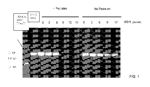
Fig. 1 illustrates PCR results for the different herpes virus papio 2 (HVP2)
samples that
were exposed to broad spectrum pulsed light (BSPL) in the presence (+
psoralen) and absence
(no psoralen) of psoralen.
Fig. 2 graphically depicts the antigenicity of HVP2 samples that were treated
with BSPL
as compared to the live HVP2 preparation (HVP-2 Prep). In the legend of the
graph "P" stands
for BSPL pulses and the number indicates the number of pulses.
Fig. 3 graphically depicts the antigenicity of HVP2 samples that were treated
with BSPL
plus psoralen and compared to the live HVP2 preparation to which psoralen was
added but not
4
CA 02785226 2012-06-20
WO 2011/084748 PCT/US2010/061335
exposed to BSPL (HVP-2+Psor). In the legend of the graph "P" stands for BSPL
pulses and the
number indicates the number of pulses.
Fig. 4 illustrates the PCR inhibition results for the different HVP2 samples
that were
exposed to BSPL in the presence (+ psoralen) and absence (no psoralen) of
psoralen.
Fig. 5 provides a dose response curve of psoralen versus the number of HVP2
plaques
from the data in Table 3.
Fig. 6 demonstrates PCR inhibition of HVP2 DNA by different concentrations of
psoralen and 9 pulses of BSPL.
Fig. 7 shows PCR inhibition results for B virus samples that were exposed to
BSPL in
the presence of psoralen.
Fig. 8 demonstrates the antigenicity of B virus samples that were photo-
inactivated
using psoralen plus BSPL. A standard rhesus anti-B virus serum was titrated on
both the photo-
inactivated antigens and on a standard "Tween/DOC" antigen (BV Ag). In the
legend of the
graph "P" stands for BSPL pulses and the number indicates the number of
pulses, UN =
uninfected, control antigen.
Fig. 9 illustrates amplification of extracted DNA using B virus specific gB
primers.
Fig. 10 demonstrates the antigenicity of the inactivated B virus immunogen as
tested by
tELISA.
Fig. 11 graphically depicts titers of mouse sera from three mice that were
immunized
with B virus (BV) grown in 3T3 cells in microtiter wells that were coated with
the original
immunogen and an uninfected (UN) control prepared from 3T3 cells.
Fig. 12 graphically depicts titers of the same three mouse sera as in Fig. 11
in microtiter
plate wells that were coated with B virus antigen grown in Vero cells and
uninfected (UN) Vero
cell controls.
Fig. 13 illustrates an embodiment of a design of a BV-Immuno Dip Strip.
Fig. 14 is a schematic representation of the well location numbers in the 96
deep well
box for placing and incubating the dip-strips that are labeled with the
corresponding numbers.
Figs. 15A-B illustrates expected negative (A) and positive (B) reactions with
the BV-
Immuno Dip Strips. Note the band at the third reaction site (UN) should always
be colorless.
Fig. 16 is a schematic of nitrocellulose preparation.
Fig. 17 is a schematic of nitrocellulose-card preparation
Fig. 18 is a schematic of strip preparation from the nitrocellulose card.
Fig. 19 is an embodiment of the a BV-Immuno Dip Strip.
5
CA 02785226 2012-06-20
WO 2011/084748 PCT/US2010/061335
DETAILED DESCRIPTION OF THE INVENTION
Various embodiments of the present invention are directed to photo-inactivated
microorganisms and systems and methods of using the same. For example, one
embodiment of
the present invention includes a method for inactivating a microorganism,
comprising:
providing at least one furanocoumarin to a microorganism; and exposing the
microorganism to
at least one pulse of a broad spectrum pulsed light, thereby inactivating the
microorganism.
As used herein, the term "microorganism" refers to many bacteria, viruses,
fungi, and
parasites. In an exemplary embodiment of the present invention, the
microorganism is a virus,
which can include, but is not limited to, adenoviridae, arenaviridae,
filoviridae, bornaviridae,
bunyaviridae, herpesviridae, orthomyxoviridae, polyomaviridae,
papillomaviridae,
paramyxoviridae, parvoviridae, picornaviridae, poxviridae, reoviridae,
retroviridae,
rhabdoviridae, togaviridae, hepadnaviridae, and bacteriophages. More
specifically, a virus can
include adenovirus 2, canine adenovirues, Pinchinde virus, Lassa virus,
Turlock virus,
California encephalitis virus, herpes simplex virus 1, herpes simplex virus 2,
cytomegalovirus,
pseudorabies virus, Epstein-Barr virus, varicella zoster virus, B virus
(Macacine herpesvirus 1),
herpesvirus papio 2 (Papiine herpesvirus 2), influenza virus, simian virus 40,
human papilloma
virus, measles virus, mumps virus, parainfluenza virus, poliovirus,
coxsackievirus, echovirus,
vaccinia virus, fowlpox virus, blue tongue virus, Colorado tick fever virus,
rotavirus, human
immuno-deficiency virus, Rous sarcoma virus, murine sarcoma virus, human T-
cell leukemia
virus, rhabies virus, vesticular stomatitis virus, Western equine encephalitis
virus, West Nile
virus, dengue virus, St. Louis encephalitis virus, hepatitis B virus,
hepatitis C virus,
lambdaphage, and Rickettsia, among others. In an exemplary embodiment of the
present
invention, the virus is Macacine herpesvirus 1 (also referred to as
Cercopithecine herpes virus 1,
herpesvirus simiae, herpes B virus, or B virus) or Papiine herpesvirus 2 (also
referred to as
Cercopithecine herpes virus 16, or herpes virus papio 2).
Inactivation of the microorganism refers to inhibition, interference,
prevention,
reduction, or alteration of replication or synthesis of nucleic acids, such as
DNA, RNA, or
combinations thereof. As used herein, the terms "preventing," "interfering,"
"reducing,"
"altering," or "inhibiting" refer to a difference in degree from a first
state, such as an untreated
state in a microorganism, to a second state, such as a treated state in a
microorganisms. For
example, in the absence of treatment with the methods or compositions of the
present invention,
nucleic acid replication or synthesis occurs at a first rate. If a
microorganism is exposed to
treatment with the methods or compositions of the present invention, nucleic
acid replication or
6
CA 02785226 2012-06-20
WO 2011/084748 PCT/US2010/061335
synthesis occurs at a second rate that is altered, lessened, or reduced from
the first rate. The
terms "preventing," "interfering," "inactivating," "reducing," "altering," or
"inhibiting" may be
used interchangeably through this application and may refer to a partial
reduction, substantial
reduction, near-complete reduction, complete reduction, or absence of nucleic
acid replication
or synthesis. As used herein, the term "nucleic acid" can refer to a
nucleotide, a nucleoside, a
polynucleotide or portion thereof, a genome or portion thereof, a gene or
portion thereof, an
oligonucleotide, an aptamer, a transcript, DNA, RNA, or a DNA/RNA chimera,
among others.
As used herein, the term "furanocoumarin" refers to a chemical substance
containing a
furan ring fused to a benzopyrone. Exemplary furanocoumarins comprise
naturally-occurring
psoralens or derivatives thereof, synthetic psoralens and derivatives thereof,
as well as
combinations thereof. For example, a psoralen can be a methoxypsoralen (e.g.,
8-MOP, 5-
MOP), a trimethylpsoralen (TMP), a 4-aminomethyl-trioxsalen (AMT), or
combinations thereof.
Providing at least one furanocoumarin to a microorganism comprises
administering an
effective amount of at least one furanocoumarin to intercalate a nucleic acid
of the
microorganism. The precise effective amount is an amount of the furanocoumarin
composition
that will yield effective results in terms of inactivation of a microorganism.
This amount (i.e.,
dosage) may vary depending upon a number of factors, including, but not
limited to, the
characteristics of the furanocoumarin or derivative thereof, the
microorganism, and the amount
of broad spectrum pulsed light administered. For example, an effective amount
of psoralen can
have a concentration ranging from about 0.1 g/ml to about 60 g/ml. In one
embodiment of
the present invention, psoralen is used in a concentration greater than about
0.3 g/ml. In
another embodiment of the present invention, psoralen is used in a
concentration of at least
about 5 g/ml. In yet another embodiment of the present invention, psoralen is
used in a
concentration of at least about 20 g/ml. In still another embodiment of the
present invention,
psoralen is used in a concentration of at least about 50 g/ml.
Exposing the microorganism to at least one pulse of a broad spectrum pulsed
light can
involve exposing a microorganism to one pulse of light or a plurality of
pulses of light. A pulse
of light is an amount of light that continues for a very short, but measurable
time, for example,
microseconds ( s). The number of pulses of light required to inactivate a
microorganism may
vary depending upon a number of factors, including, but not limited to, the
characteristics and
concentration of the furnaocoumarin or derivative thereof, the microorganism
and its
concentration, the light transparency of the medium in which the microorganism
is suspended,
7
CA 02785226 2012-06-20
WO 2011/084748 PCT/US2010/061335
the light transparency of the container that accommodates the microorganism
suspension, and
the source/wave length of the broad spectrum pulsed light, among others. In an
exemplary
embodiment of the present invention, the source of the broad spectrum pulsed
light is a xenon
lamp capable of generating a continuous broad-spectrum of light, ranging from
about the deep
UV spectrum through about the infrared spectrum. Ultraviolet (UV) light is
electromagnetic
radiation with a wavelength shorter than that of visible light, but longer
than x-rays, in the range
nm to 400 nm, and energies from 3 eV to 124 eV (one eV is equivalent to
1.60217653x10⁻¹⁹ Joules.). Infrared (IR) radiation is electromagnetic
radiation with a
wavelength between 0.7 and 300 micrometer ( m), which equates to a frequency
range between
10 approximately 1 and 430 THz. Thus, broad-spectrum light can include
wavelengths from
about 10 nm to about 300 m.
In an exemplary embodiment of the present invention, exposing the
microorganism to at
least one pulse of a broad spectrum pulsed light comprises exposing the
microorganism to about
0.45 Joule/cm2 to about 13.5 Joules/cm2 of broad spectrum light. In one
embodiment, a
microorganism, such as herpes B virus, is exposed to about 5.4 Joules/cm2 of
broad spectrum
light. For example, a microorganism, such as herpes B virus, is exposed to a
cumulative
amount of about 5.4 Joules/cm2 of broad spectrum light. In another embodiment,
a
microorganism, such as herpes B virus, is exposed to about 12.15 Joules/cm2 of
broad spectrum
light. For example, a microorganism, such as herpes B virus, is exposed to a
cumulative
amount of about 12.15 Joules/cm2 of broad spectrum light. The single-dose
amount or
multiple-dose amount (in either case, the cumulative/total amount) of broad
spectrum light for
use in the present method typically ranges from about 4.05 Joules/cm2 to about
13.5 Joules/cm2,
but can exceed this amount as long as the immunogenicity of the antigens is
maintained. The
cumulative amount of broad spectrum light may be delivered in pulses of
various lengths,
which can be separated by various lengths of time. For example, broad spectrum
light can be
delivered in a pulse width of about 360 .is, where three pulses can be
generated per second with
each pulse generating an energy of about 0.45 joules/cm2 per pulse. Using such
an example, a
microorganism, such as herpes B virus, can be inactivated using about 12
pulses. In addition,
the energy of each pulse may also be varied. The energies of each pulse can
range from about
0.3 Joules/cm2 per pulse to about 0.6 Joules/cm2 per pulse. For example, a
pulse with an energy
of about 0.45 Joules/ cm2 /pulse can be used. Other pulse widths can also be
used. For example,
pulse widths from about 250 ps to about 450 s can be used, and the number of
pulses adjusted
8
CA 02785226 2012-06-20
WO 2011/084748 PCT/US2010/061335
to obtain a cumulative amount of from about 4.05 Joules/cm2 to about 13.5
Joules/cm2, or in a
more specific example, from about 3 Joules/cm2 to about 13.5 Joules/cm2,
Another aspect of the present invention includes an inactivated microorganism,
comprising a photo-chemically inactivated nucleic acid, wherein the photo-
chemically
inactivated nucleic acid is photo-chemically inactivated by at least one
furanocoumarin and at
least one pulse of a broad spectrum pulsed light. The microorganism can be any
of the
microorganisms disclosed herein, and can be produced by any of the
inactivation methods
described herein. In an exemplary embodiment, the microorganism can be a
virus, such as a
herpesvirus, and more specifically, a Macacine herpesvirus 1 or Papiine
herpesvirus 2 (HVP2).
The inactivated microorganism of the present invention has enhanced function
because of the
combined characteristics of having inactivated DNA and retaining the
structural integrity of its
surface antigens. For example, inactivation of viruses by detergents is more
effective for
enveloped viruses then for non-enveloped viruses. Detergents disrupt lipid
membranes of cell
membranes and enveloped viruses by interacting with lipids and releasing
proteins or
glycoproteins from the lipid-rich envelopes. In an example of herpesviruses, a
combination of
surfactants (e.g., Tween 40) and a detergent (e.g., sodium deoxycholate) can
be used to disrupt
the envelope and thereby inactivate a herpesvirus. Using a detergent-based
method, the DNA
of the virus is not affected by this procedure. In contrast, the psoralen/BSPL
technique
described above damages nucleic acids (e.g., DNA and RNA) and therefore is not
restricted to
any particular microorganism, enveloped or not.
An inactivated microorganism can be used in a system for detecting an
antibody. An
antibody may be polyclonal or monoclonal, and may include fragments such as
Fab, FC, heavy
chains, light chains, constant, variable, or hypervariable fragments or
regions, and any type of
antibody including but not limited to IgM, IgG, IgA, IgD, and IgE. An antibody
has specificity
for at least a portion of an antigen. The phrase "having specificity for an
antigen" with respect
to the antibody as used herein can also be referred to as the "binding
activity," "binding
affinity," or "specific affinity" of the antibody relative to the target.
These phrases may be used
interchangeably herein and are meant to refer to the tendency of a ligand to
bind or not to bind
to a target. The energetics of these interactions are significant in "binding
activity" and
"binding affinity" because they define the necessary concentrations of
interacting biomolecules,
the rates at which these biomolecules are capable of associating, and the
relative concentrations
of bound and free biomolecules in a solution. The energetics are characterized
through, among
other ways, the determination of a dissociation constant, Kd. The specificity
of the binding is
9
CA 02785226 2012-06-20
WO 2011/084748 PCT/US2010/061335
defined in terms of the comparative dissociation constants (Kd) of the ligand
for target as
compared to the dissociation constant with respect to the ligand and other
materials in the
cellular environment or unrelated molecules in general. Typically, the Kd for
an antibody with
respect to the antigen will be at least 2-fold, preferably 5-fold, and more
preferably 10-fold less
than Kd with respect to target and the unrelated material or accompanying
material in the
cellular environment. Even more preferably, the Kd will be 50-fold less, more
preferably 100-
fold less, and more preferably 200-fold less than Kd with respect to target
and the unrelated
material or accompanying material in the cellular environment.
Such a system for detecting an antibody can comprise: an antigen component,
wherein
the antigen is exposed to a furanocoumarin and at least one pulse of a broad
spectrum pulsed
light; and a reporter component that is capable of detecting a binding of an
antibody of a subject
to at least a portion of the antigen. For example, the antigen component can
be Macacine
herpesvirus 1 or antigenic components thereof or Papiine herpesvirus 2 or
antigenic
components thereof. The furanocoumarin and pulsed light exposure can be in
accordance with
any of the methods described herein. The antigen component can be disposed on
a substrate,
such as for example, a microtiter plate, a nitrocellulose membrane, or the
like. The reporter
component can be a reporter antibody capable of binding at least a portion of
the antibody
capable of binding at least a portion of the antigen. Thus, the inactivated
microorganism or
component derived therefrom can be used to detect the presence of an antibody
in a subject that
has at least some specificity for the inactivated microorganism or component
derived therefrom.
For example, the system can be used to detect antibodies specific for Macacine
herpesvirus 1 or
Papiine herpesvirus 2 in a subject, such as a human or non-human primate.
An antigen that is exposed to a furanocoumarin and at least one pulse of a
broad
spectrum pulsed light can be used in many immunoassays, including, but not
limited to
enzyme-linked immunosorbent assay (ELISA), radioimmunoassay (RIA), magnetic
immunoassays, immunoblotting (i.e., Western blotting), immunoprecipitation,
immunohistochemistry, affinity chromatography, and flow cytometry, among
others.
Another aspect of the present invention involves a method for immunizing a
subject,
comprising: inactivating an immunogenic microorganism comprising exposing the
immunogenic microorganism to a furanocoumarin and to at least one pulse of a
broad spectrum
pulsed light; and administering an effective amount of the immunogenic
microorganism to a
subject to produce an immune response. Thus, the inactivated microorganisms of
the present
invention can be used to generate an immune response in a subject, such as an
adaptive immune
CA 02785226 2012-06-20
WO 2011/084748 PCT/US2010/061335
response or a innate immune response. In an exemplary embodiment of the
present invention,
the inactivated microorganism can elicit a B cell response, a T cell response,
or a combination
thereof. In another exemplary embodiment of the present invention, the
inactivated
microorganism can elicit a protective immune response. For example, the
inactivated
microorganisms of the present invention can be used to vaccinate a subject
against a
microorganism, such as virus. In one embodiment, the inactivated microorganism
can be an
inactivated herpes B virus that can be used to vaccinate a human or non-
primate.
Using the inactivated microorganisms of the present invention, an antibody can
be
raised to the inactivated microorganism, where the antibody has a specific
affinity for at least a
portion of the inactivated microorganism. The antibody can be raised against
an antigen
derived from a microorganism selected from the group consisting of a virus, a
bacterium, and a
fungus. Such an antibody can be a polyclonal antibody or a monoclonal
antibody, among
others as discussed above. For example, using an inactivated herpes B virus, a
polyclonal
antibody can be raised to one or more epitopes of herpes B virus. In addition,
a monoclonal
antibody can be raised that has specificity to one of the one or more epitopes
of herpes B virus.
The antibody can be used in many immunoassays, including, but not limited to
enzyme-linked
immunosorbent assay (ELISA), radioimmunoassay (RIA), magnetic immunoassays,
immunoblotting (i.e., Western blotting), immunoprecipitation,
immunohistochemistry, affinity
chromatography, and flow cytometry, among others.
Another aspect of the present invention includes an inactivated microorganism
comprising an inactivated nucleic acid, wherein the inactivated microorganism
retains its
antigenicity. The phrase "retains its antigenicity" refers to the ability of
an inactivated
microorganism to bind to an antibody that is produced by an immune response to
the live
microorganism that is not treated by the systems and methods of the present
invention. For
example, in the case of antibody binding, an inactivated microorganism of the
present invention
would be recognized by at least a majority of the same antibodies that
recognize a live
microorganism at substantially the same titers.
Another aspect of the present invention includes an inactivated microorganism
comprising an inactivated nucleic acid, wherein the inactivated microorganism
retains its
immunogenicity. The phrase "retains its immunogenicity" refers to the ability
of an inactivated
microorganism to produce an immune response in a subject that is substantially
similar to an
immune response produced by a live microorganism that is not treated by the
systems and
methods of the present invention.
11
CA 02785226 2012-06-20
WO 2011/084748 PCT/US2010/061335
All patents, patent applications, and references included herein are
specifically
incorporated by reference in their entireties.
It should be understood, of course, that the foregoing relates only to
exemplary
embodiments of the present invention and that numerous modifications or
alterations may be
made therein without departing from the spirit and the scope of the invention
as set forth in this
disclosure. Therefore, while embodiments of this invention have been described
in detail with
particular reference to exemplary embodiments, those skilled in the art will
understand that
variations and modifications can be effected within the scope of the invention
as defined in the
appended claims. Accordingly, the scope of the various embodiments of the
present invention
should not be limited to the above discussed embodiments, and should only be
defined by the
following claims and all equivalents.
The present invention is further illustrated by way of the examples contained
herein,
which are provided for clarity of understanding. The exemplary embodiments
should not to be
construed in any way as imposing limitations upon the scope thereof. On the
contrary, it is to
be clearly understood that resort may be had to various other embodiments,
modifications, and
equivalents thereof which, after reading the description herein, may suggest
themselves to those
skilled in the art without departing from the spirit of the present invention
or the scope of the
appended claims.
EXAMPLES
EXAMPLE 1: A MODIFIED PHOTOINACTIVATION TECHNIQUE FOR
INACTIVATION OF VIRUSES USING PSORALEN AND BROAD SPECTRUM LIGHT
PULSES.
Previous experimentation to photo-inactivate a virus involve a procedure in
which a
"black light lamp" (UVA) was used to irradiate HVP2-psoralen mixtures in Petri
dishes. In this
procedure, virus-psoralen mixtures were exposed to a "black light" UVA lamp
for two sets of
min exposures. The results of this experimentation was unsatisfactory because
it was time
consuming, caused heating and evaporation, and most importantly resulted in
poor antigenicity.
In an attempt to overcome the shortcomings of the above procedure, this
example
describes a modified psoralen photo-inactivation technique in which the photo-
activation of the
30 psoralen is done by using the SteriPulse-XL irradiation device (Model RS-
3000C) from Xenon
Corporation (Woburn, MA). Information regarding the SteriPulse-XL system is
described in
Xenon's publication, entitled "Sterilization & Decontamination using High
Energy UV Light,"
which is hereby incorporated by reference.
12
CA 02785226 2012-06-20
WO 2011/084748 PCT/US2010/061335
The SteriPulse-XL system employs a xenon lamp that generates broad spectrum
pulsed
light (BSPL) in short controlled pulses (360 microseconds per pulse (
s/pulse)). The intensity
of the BSPL is approximately 50,000 to 100,000 times the energy level of the
sun. BSPL
comprises at least a UVA wavelength that facilitates the photo-activation of
the psoralen, as
well as UVB and UVC, which are associated with having germicidal properties.
Psoralens, which belong to the class of molecules known as furanocoumarins,
intercalate nucleic acids. In the presence of UVA, psoralens alkylate nucleic
acids to generate
monoadducts and cross-links. For example, in the case of double-stranded DNA,
psoralens
alkylate nucleic acids at the 5,6-double bond of thymidines, effectively
crosslinking the DNA
duplex. This prevents DNA strand separation during transcription and
replication.
MATERIALS AND METHODS
Preparation of virus and antigen stocks in Vero cells. Vero cells (ATCC # CCL-
81)
were grown in 980 cm2 roller bottles to 95% confluency, and subsequently
infected
(multiplicity of infection (MOI) = 5) with HVP2 or B virus and maintained in
Dulbecco's
Modified Eagles Medium (DMEM) with high glucose supplemented with 1% fetal
bovine
serum (FBS) and antibiotics (penicillin and streptomycin). The infected cells
were incubated
for 22 to 24 hrs at 34 C until cytopathic effect (CPE) can be observed. The
cells were then
scraped into the medium and centrifuged at 1500 rpm (514 x g) for 10 min. Cell
pellets were
resuspended in 4.5 ml of sterile ultrapure water. The suspension was then
treated by 3 cycles of
freezing on dry ice and thawing in a 37 C water bath. Each freezing cycle
lasted for at least 15
min. Cell debris was removed by centrifugation (1500 rpm for 10 min) and the
supernatant
(about 5 ml) was saved. The virus titer of the preparation as determined by
the standard plaque
assay in Vero cells was approximately 1010 PFU/ml. Although cells in this
embodiment were
lysed through three cycles of freezing and thawing, cells can also be lysed by
way of sonication.
In some viral preparations, such as B virus, antigenic yield may be enhanced
by using
sonication-based methods.
Preparation of viral antigens from infected cells by detergent solubilization.
Vero cells
(ATCC # CCL-81) were grown in 980 cm2 roller bottles to 95% confluency,
infected (MOI=5)
with HVP2 or B virus, and maintained in Dulbecco's Modified Eagles Medium
(DMEM) with
high glucose supplemented with 1% fetal bovine serum (FBS) and antibiotics
(penicillin and
streptomycin). The infected cells were incubated for 22 to 24 hrs at 34 C
until CPE can be
observed. The cells were then scraped into the medium and centrifuged in 50 ml
conical tubes
at 1500 rpm (514 x g) for 10 min. The supernatant was discarded, and the
pellet was
13
CA 02785226 2012-06-20
WO 2011/084748 PCT/US2010/061335
resuspended in 1.5 ml of water containing 2X Complete Protease Inhibitor
(Roche). The final
volume was adjusted to 2.0 ml with water containing 2X Complete Protease
Inhibitor. The
resuspended pellet was then treated with a final concentration of 1% Tween 40
(250 pl of 10%
Tween 40) and 1% Sodium Deoxycholate (250 l of 10% Sodium Deoxycholate) by
adding one
after the other with extensive mixing (vortexing) intervals.
Assessment of virus inactivation using a standard plaque assay in Vero cells.
Assessment of virus inactivation was accomplished by either one of the two
following
techniques utilizing a standard plaque assay in Vero cells. 1. Vero cell
monolayers grown in
24-well plates were infected with virus preparations for 48 or 72 hrs. If no
cytopathic effect
(CPE) was observed microscopically, cells from each well were scraped into a
small amount of
medium (500 l), and replated on a new Vero-cell monolayer (in a 24-well
plate) and incubated
for another 72 hrs at 37 C. Cells were then fixed with 100% methanol and
stained with crystal
violet to facilitate the counting of plaques. 2. Vero cell monolayers in 24-
well plates were
infected with virus preparations for six days and inspected daily for the
development of CPE.
At the end of the incubation periods, cell monolayers were then fixed with
100% methanol and
stained with crystal violet to facilitate CPE determination or counting of
virus induced plaques.
Absence of CPE or plaques by any of the techniques indicates absence of virus
replication due
to inactivation.
Assessment of virus antigenicity by tELISA. Virus antigenicity was assessed by
tELISA, an assay for antibody detection and quantitation that is performed in
96-well
microtitration-plates. The tELISA was performed essentially as previously
described with
some modifications in Katz D, Hilliard JK, Eberle R, Lipper SL (1986a).
Briefly, herpesvirus
antigens were adsorbed to microplate-wells by incubation for 20 min on a
shaker at room
temperature, or overnight at 4 C. After blocking with Blotto (3% skim milk)
(1 h at 37 C),
adsorbed antigens were reacted (1 h at 37 C) with serial dilutions of
homologous standard
antiserum pools. Anti human-IgG-alkaline phosphatase conjugate was then added
and
incubated (1 h at 37 C) for the detection of antigen-bound monkey antibodies.
After each of
the incubation steps, microplates were washed 3 times with phosphate buffered
saline (PBS)
supplemented with 0.05% Tween 20 (PBST). The substrate, dinitro-phenyl
phosphate, was
added and incubated for 30 min at room temperature. Color intensity in optical
density (OD)
units was read in a micro-ELISA reader at a wavelength of 405 nm.
Assessment of DNA damage by Polymerase chain Reaction (PCR). DNA damage was
demonstrated by the inhibition of a PCR amplification product formation,
validating virus
14
CA 02785226 2012-06-20
WO 2011/084748 PCT/US2010/061335
inactivation. The PCR primers were initially designed based on the gB gene
sequence of B
virus and the gL gene sequence of HVP2. The BV gB primer set amplifies a 1.3
kb fragment of
B virus, while the HVP2 gL primer set amplifies a 1.2 kb fragment of HVP2. In
later
experiments, the following primers were used for amplification of both BV and
HVP2 DNA.
Primers were designed based on the BV gB sequence. The forward primer was 5'-
GTGTACATGTCGCCGTTCTA-3' (position 53972 in the BV genome). The reverse primer
was 5'-GTGTACATGTCGCCGTTCTA-3' (position 52659 in the BV genome). The amplimer
expected if PCR occurred (i.e., if there was no damage to DNA) would be 1313
bp. The PCR
was performed by using the PCR HotStar Kit (Qiagen) and 3 l of purified DNA
in 20 l
volume. The amplification was performed in an ABI Thermocycler 9600 using the
following
cycling conditions: 15 min 95 C, 35 two step cycles of 20 seconds at 95 C,
and 40 seconds at
65 T. Then, the PCR products were run on 1% agarose gel along with a DNA
marker to
determine the presence of the PCR fragment of the expected size. The absence
of the PCR
fragment after amplification of a sample verified DNA damage.
RESULTS
Comparison of HVP2 inactivation procedures using BSPL and the combined
Psoralen-
BSPL Inactivation Technique.
Experiment 1. The purpose of this experiment was to compare the inactivation
procedure by BSPL to the photo-inactivation procedure in which a combination
of psoralen and
BSPL was used. Inactivation by BSPL alone was performed as follows: five 1 ml
portions of a
diluted HVP2 preparation (108 PFU/ml) were transferred into 5 polyethylene
tubing (Polytubing,
1" x 1,500' 2 Mil, Catalog # S-3520, ULINE, Atlanta GA) that were heat sealed
at one end.
The ends were then heat-sealed at a distance of 5 cm from the first seal.
Another 5 ml of the
diluted virus were first mixed with psoralen (4-Aminomethyl-trioxalen
hydrochloride, Sigma,
Catalogue # A4330 5mg) to a final concentration of 20 pg/ml of psoralen and
then transferred
in 1 ml portions to 5 polyethylene tubings as described above. Each of the
tubings (with and
without psoralen) was irradiated with a different dose of BSPL. Pairs of
polyethylene tubings
from each group, one that contains the virus and the other that contains the
virus and psoralen,
were placed in a plastic tray, flat on a bed of crushed ice. The plastic tray
was placed in the
Steripulse chamber on at a distance of 8 shelves (4.26 inches) from the lamp
window at the top
of the chamber to achieve 0.45 Joules/cm2 per pulse. Each of the tubings was
exposed to 3, 6, 9,
12, and 15 pulses. The total energy was determined by multiplying the energy
output per pulse
(0.45 joule/ cm2) by the number of pulses (See Table 1). Each of the samples
was then tested
CA 02785226 2012-06-20
WO 2011/084748 PCT/US2010/061335
for infectivity by the plaque assay. Table 1 provides the plaque assay results
for the
inactivation of HVP2 infectivity by BSPL compared to the photo-inactivation
procedure in
which a combination of psoralen and BSPL was used. The number of plaques for
each BSPL
dose are shown in bold numbers. While a minimum dose of 6 BSPL pulses were
sufficient to
inactivate the virus in the presence of psoralen, the number of pulses
necessary to inactivate the
virus by BSPL only was 12.
Table 1
# of Pulses 3 6 9 12 15
Joules/cm 1.35 2.70 4.05 5.40 6.75
BSPL+Psoralen 2 0 0 0 0
BSPL only 100 8 2 0 0
The DNA damage that was caused by exposure of the HVP2 samples to BSPL or BSPL
+ psoralen was assessed by PCR. Fig. 1 shows that BSPL + psoralen inhibits the
PCR at 9
pulses or higher. Exposure to BSPL alone causes a dose related decrease of
band intensity, but
a band can be clearly seen after an exposure of 12 BSPL pulses. Both the
infectivity assays and
PCR show the additive inactivation potency of psoralen. In this experiment,
the PCR inhibition
assay was a more sensitive assay, since amplification of DNA was still present
in samples that
were negative by the plaque assay. However, in other experiments (shown
below), live virus
was present in preparations that showed DNA damage by inhibition by PCR.
The different samples were each tested for antigenicity by tELISA. A 1:30
dilution of
each sample was adsorbed to wells, washed, blocked with Blotto and used for
the titration of an
anti-HVP2 standard baboon serum. As can be seen in Fig. 2 for samples that
were treated with
BSPL and in Fig. 3 for samples treated with BSPL and psoralen, none of the
treatments altered
antigenicity of inactivated virus.
Experiment 2. The procedures described for Experiment 1 were repeated in this
experiment with some minor variations. The samples exposed to 3 pulses of BSPL
were not
tested by the plaque assay. The plaque assay was performed for samples that
were exposed to 6
or more BSPL pulses. Table 2 provides plaque assay results for the
inactivation of HVP2
infectivity by BSPL compared to the photo-inactivation procedure in which a
combination of
psoralen and BSPL was used. The number of plaques for each BSPL dose are shown
in bold
numbers. All samples exposed to 3 BSPL pulses or higher were tested by PCR
(Fig. 4). As
16
CA 02785226 2012-06-20
WO 2011/084748 PCT/US2010/061335
shown in Table 2, the samples that were mixed with psoralen and exposed to 6
BSPL pulses or
higher did not produce plaques. The samples that were exposed to BSPL only did
produce
plaques after exposure to 6, 9, and 12, but not after 15 pulses.
Table 2
# of Pulses 6 9 12 15
Joules/cm 2.70 4.05 5.40 6.75
BSPL+ Psoralen 0 0 0 0
BSPL only 48 4 2 0
The PCR results depicted in Fig. 4 were similar to those obtained in the first
experiment,
except that a weak DNA band could be seen after treatment with psoralen and
exposure to 9
BSPL pulses. No bands were seen in psoralen treated samples that were exposed
to 12 and 15
BSPL pulses. All samples that were exposed to BSPL only (3 to 15 pulses),
showed DNA
bands. The intensity of the bands decreased with the increase of the number of
BSPL pulses.
Effect of psoralen concentration on the photo-inactivation of HVP2.
One ml portions of a HVP2 stock diluted to 108 pfu/ml were prepared as for the
previous experiment, mixed with different concentrations of psoralen (Table 3)
and exposed to
9 pulses of BSPL as described above. Samples from each treatment were tested
by the plaque
assay (Table 3 and Fig. 5) and by PCR (Fig. 6). Table 3 provides the plaque
assay results that
demonstrate inactivation of HVP2 infectivity by different concentrations of
psoralen and 9
pulses of BSPL. A concentration of 5.0 g/ml of psoralen or higher plus 9 BSPL
pulses
inactivated the infectivity of the virus. Increasing number of plaques were
observed with
decreasing concentrations of psoralen (Table 3 and Fig. 5).
Table 3
Psoralen
Concentration ( g/ml) 20.0 10.0 5.0 2.5 1.25 0.62
Number of plaques. 0 0 0 4 20 32
The PCR indicates that as low as 1.3 pg/ml of psoralen could inhibit the
amplification of the
HVP2 DNA (Fig. 6).
17
CA 02785226 2012-06-20
WO 2011/084748 PCT/US2010/061335
In this experiment, the inhibition of PCR is less sensitive than the plaque
assay.
Samples treated with psoralen concentrations of 1.3 g/ml and 2.5 g/ml did
not result in any
DNA band after PCR amplification although they contained virus 20 and 4
plaques,
respectively.
Inactivation of B virus by BSPL and by the combined psoralen-BSPL photo-
inactivation
technique.
Experiment 1. The Psoralen-BSPL procedure that was developed for HVP2 was
applied
subsequently for B virus. All procedures were similar to those described for
HVP2, except that
the handling of the infectious B virus was performed in the Biosafety 4
laboratory (BSL4). The
B virus lab strain (E2490) (MOI=5) was grown in 95% confluent Vero cells in
850 cm2 roller
bottles, maintained in DMEM high glucose supplemented with 1% FBS and
antibiotics. The
infection progressed for 24 hr at 34 C, after which cells were scraped into
the media and
centrifuged at 1500 rpm (514 x g) for 10 min. Cell pellets were resuspended in
2.5 ml of sterile
ultrapure water. The suspension was then treated by 3 cycles of freezing on
dry ice and
thawing in a 37 C water bath. Each freezing cycle lasted for at least 15 min.
Cell debris was
removed by centrifugation (1500 rpm/10 min) and the suspension (5 ml)
containing the virus
was saved. The virus titer of the suspension as determined by the plaque assay
in Vero cells
was approximately 109 PFU/ml.
Inactivation of B virus by psoralen and BSPL was performed as described for
HVP2.
Briefly, two 1 ml portions of a 1:10 diluted B virus preparation (108 PFU/ml)
were mixed with
psoralen to a final concentration of 20 gg/ml and transferred into 2
polyethylene tubings that
were heat sealed at one end. The other end of each tubing was then heat-sealed
at a distance of
5 cm from the first seal. Each of the tubings was irradiated with either 12 or
15 pulses of BSPL.
The polyethylene tubings were placed flat on a bed of crushed ice into the
Steripulse chamber at
a distance of 8 shelves (4.26 inches) from the lamp window to achieve 0.45
Joules/cm2 per
pulse for irradiation.
Each sample was then tested for infectivity by the plaque assay. No B virus
plaques
were produced after the photo-inactivation procedure with 12 or 15 BSPL
pulses. The DNA
damage that was caused by exposure of the B virus samples to BSPL + psoralen
was also
assessed by PCR. As can be seen in Fig. 7, BSPL + psoralen inhibited the PCR
at both 12 and
15 BSPL pulses.
Each of the B virus photo-inactivated samples was tested for antigenicity
using tELISA.
A 1:30 dilution of each sample was adsorbed to microtiter plate wells, washed,
blocked with
18
CA 02785226 2012-06-20
WO 2011/084748 PCT/US2010/061335
Blotto and used for the titration of a rhesus anti B virus standard serum. For
comparison, a
standard B virus (Tween/Doc) antigen preparation (BV Ag) was also adsorbed to
wells of the
same plate. The BV Ag stock was prepared from a different virus than that used
for the photo-
inactivation experiment. Although the standard antigen resulted in higher OD
values, there was
no difference in antigenicity between the sample that was exposed to 12 BSPL
pulses and the
sample that was exposed to 15 pulses (Fig. 8).
Preparation of an inactivated B virus immunogen in mouse cells.
Preparation of the B virus stock mouse cells. The inactivated B virus
immunogen was
needed for the production of mouse monoclonal antibodies. Mouse 3T3 fibroblast
cell line
(developed from BALB/c) were grown in two 850 cm2 roller bottles in DMEM high
glucose
supplemented with 10% FBS, 200 mM L-glutamine and antibiotics (penicillin and
streptomycin) at 37 C. The following procedures were all performed in the
BSL4 facility.
Confluent cell monolayers (95%) in the roller bottles were infected with of B
virus (MOI=5)
(Strain E2490) and maintained in DMEM high glucose supplemented with 1% FBS
and
antibiotics. The infected cells were incubated for 24 hrs at 34 C, scraped
into the media and
centrifuged at 1500 rpm for 10 min. Cell pellets were resuspended in 4.5 ml of
sterile ultrapure
water. The suspension was then treated by 3 cycles of freezing on dry ice and
thawing in a
37 C water bath. Each freezing cycle lasted for at least 15 min. Cell debris
was removed by
centrifugation (1500 rpm/10 min) and the virus suspension (about 5 ml) was
saved. The virus
titer of the suspension was approximately 2x10⁷ per ml, as determined by the
plaque assay in
Vero cells.
The inactivation procedure. The B virus preparation (1.4 ml) was diluted 1:5
in sterile
ultrapure water to a total volume of 7 ml, and 70 l of 2 mg/ml Psoralen (4-
Aminomethyl-
trioxsalen hydrochloride, # A4330, Sigma) were added to the virus suspension
resulting in a
final concentration of 20 g/ml. The virus-Psoralen mixture was transferred in
1 ml portions to
7 polyethylene tubings (Polyethylene (Low Density) polytubing # S-3520, 1" x
1,500', 2 Mil
Poly Tubing Roll, ULINE, Atlanta GA) that were heat sealed at one end. After
transferring the
virus to the tubing the other end was heat sealed at a distance of 5 cm. The
sealed tubings were
decontaminated by submersion in a bottle containing CIDEX (activated
glutaraldehyde
solution) for 15 min. The outside of the bottle was decontaminated by
submersion in the
CIDEX dunk tank for 15 min. The CIDEX was removed from the bottle, and the
bottle and
tubings were transferred to a quaternary ammonium dunk tank through which they
were
removed from the BSL4 glove cabinets and then transferred to the BSL3
laboratory. The
19
CA 02785226 2012-06-20
WO 2011/084748 PCT/US2010/061335
tubings were then rinsed individually with 70% alcohol. For BSPL exposure,
each tubing was
placed on a flat bed of ice on a tray and inserted into the irradiation
chamber of the SteriPulse-
XL device (RS-3000C, Xenon Corp.). The distance of the ice surface from the
lamp window
was 8 shelves (4.26 inches). Each of the 7 virus-containing tubings was then
exposed to 12
pulses/4 seconds of BSPL that sums up to a total energy of 5.4 Joules/cm2.
Following
irradiation the content of the individual tubings were pooled and tested for
the presence of
residual B virus by the plaque assay, for DNA damage by PCR, and for
antigenicity by tELISA.
Plaque assay for validating the inactivation of B virus. 500 pl of the pooled
B virus
suspension were tested for infectivity in Vero cell monolayers grown in 6 well
plates. The
cultures were observed for 48 hrs. No cytopathic effect was observed. The
cells from the virus
infected well were then scraped and transferred to another well containing
Vero cells for
another 48 hours. No cytopathic effect was observed after replating. These
results indicate that
no live B virus could be detected after inactivation.
PCR results. A B virus gB primer set that amplifies a 1.3 kb fragment of the B
virus
genome was used. The PCR reaction was performed by using the PCR HotStar Kit
(Qiagen)
and 3 l of purified DNA in 20 pl volume. The amplification was performed on
ABI
Thermocycler 9600 using the following cycling conditions: 15 min 95 C and 35
two step
cycles of 20 sec at 95 C and 40 sec at 65 T. Then the PCR reaction products
were run on 1%
agarose gel along with the DNA marker to determine the presence of the PCR
fragment of the
expected size. No amplified fragment from the irradiated preparation could be
demonstrated.
The absence of the PCR fragment after amplification implied that DNA in the
sample was
damaged and could not be replicated (Fig. 9).
Anti eg nicity test. A 1:6 dilution of the B virus preparation was adsorbed to
96 well
microtiter plates. Wells were adsorbed with a standard detergent solubilized B
virus
preparation (BV Ag) or mock infected cell lysates (UN). A standard rhesus anti
B virus
positive serum was then tested for antibodies by ELISA. Results, shown in Fig.
10, indicate
that the inactivation procedure did not destroy the antigenicity of the
immunogen.
Evaluation of the immuno eg nicity of the inactivated B virus immunogen. The
inactivated B virus immunogen was used by University of Georgia Monoclonal
antibody
Facility (UGA-MAF) for the preparation of mouse monoclonal antibodies. Three
mice were
inoculated, and bled after the second booster inoculation. The sera were
tested for polyclonal
antibodies by ELISA against the original immunogen that was prepared in 3T3
cells (Fig. 11)
CA 02785226 2012-06-20
WO 2011/084748 PCT/US2010/061335
and against a standard antigen prepared in Vero cells (Fig. 12). Each set of
sera were also
tested against an uninfected (UN) control antigens prepared from 3T3 cells and
Vero cells.
These results indicated that the immunogen prepared by the Psoralen-BSPL
inactivation
method induced anti B virus antibodies in all three mice. Interestingly, the
immunogen that was
prepared in 3T3 mouse cells induced also antibodies to the 3T3 control cells
but none to the
Vero cells. However, even when tested against the 3T3 cell antigens, the
antibody response to
B virus in all three mice was always higher than the response to the cell
controls.
Conclusion A method was developed for inactivation of crude HVP2 and B virus
cell
extracts utilizing a combination of psoralen and Broad Spectrum Light Pulses
(BSPL). A
benchtop sterilization chamber from Xenon Corporation was used to generate
measured BSPL.
Although psoralen and "black light" (UVA) was used for inactivation of viruses
for many years
the combination of psoralen and BSPL is unique and was never used before to
our knowledge.
Previous experiments showed that 18 pulses of BSPL by itself were capable of
inactivation of
HVP2 infected cell extracts. These experiments demonstrate that the addition
of psoralen
enables virus inactivation with fewer BSPL pulses. The advantage of combining
psoralen and
BSPL is that the virus is inactivated by both the germicidal UV wavelength and
by the photo-
inactivation that is caused by the UVA (black light)-photo-activated psoralen.
The damage to
the DNA will therefore be greater. Another advantage of BSPL is that it is
emitted in high
energy short pulses (360 micro seconds) from a xenon lamp and not from mercury
lamps. The
relatively short exposure times are beneficial, since samples are not
overheated during exposure.
Validation of virus inactivation was made by infectivity assays and by PCR
inhibition that is
indicative of the actual photo-inactivation damage to the nucleic acids.
A B virus immunogen was prepared from a batch of B virus prepared in 3T3 mouse
cells. The preparation was inactivated by using the psoralen-BSPL technique.
This
immunogen was used by the Monoclonal Antibody Facility at UGA for the
immunization of
mice for the production of monoclonal antibodies (MABs). The induction of high
titers of
antibodies in three of the inoculated mice indicated that immunogenicity was
not impaired by
the Psoralen-BSPL procedure.
EXAMPLE 2: HERPES B VIRUS IMMUNO DIP STRIP TEST (BV-IDST) KIT FOR THE
DETECTION OF ANTI B VIRUS ANTIBODIES IN MACAQUE SERA.
The BV-IDST is an enzyme immunoassay for the detection of herpes B virus IgG
antibodies in macaque species. Each individual dip-strip is used for the
detection antibodies in
21
CA 02785226 2012-06-20
WO 2011/084748 PCT/US2010/061335
one blood (serum, plasma or whole blood) sample. The test is simple, and
requires little if any
laboratory equipment and therefore suitable for field-testing.
Introduction. Herpes B virus (Herpesvirus simiae or Cercopithecine herpesvirus
1), a
member of the Alphaherpesvirinae subfamily and the Simplexvirus group, is
known to occur
naturally in macaques (Macaca spp). Infection of macaques may be asymptomatic
or may
cause a mild disease. Infection of other species (including humans) is rare
but results in severe,
and often, if untreated, lethal disease.
Past infections are determined by detection of anti B virus antibodies using
serological
assays. Serological diagnosis of B virus infections in humans is complicated
by the relatively
high prevalence of the immunologically cross-reacting herpes simplex virus
infections (HSV-1
and/or HSV-2). Past infections in macaques can be established without these
complications
because the only simplexvirus known to infect macaques is B virus. Identifying
B virus
infected macaques is important for managing macaques in captivity, for
developing specific
pathogen free colonies and for the prevention of the potential exposure and
infection of humans
who handle macaques.
The virus antigen for these assays is prepared from detergent solubilized and
inactivated
B virus infected cells (BV) and the negative antigen control is prepared in a
similar way from
uninfected cell lysates (UN). The antigen can also be prepared using the
compositions and
methods described in Example 1.
The BV-IDST was developed to enable field-testing to detect B virus antibodies
in
macaque sera. The principle of the BV-IDST is similar to the principle of
ELISA except that
nitrocellulose strips are used instead of plastic wells as the solid phase to
which antigens are
adsorbed. No special laboratory equipment (washers, readers, etc.) is
necessary for carrying out
the test.
For example, one BV-IDST kit can include about 100 individual nitrocellulose
strips on
which control and B virus antigens are pre-applied to predetermined reaction
sites. Each strip
contains 3 reaction sites as shown in Fig. 13. Site #1 serves as an internal
quality control for the
anti human IgG conjugate; it contains normal rhesus IgG. The IgG control also
serves as a
reference line for reading the results since, in a properly developed test, it
will always be visible.
Site #2 includes the BV antigen, and site #3 includes the uninfected control
antigen (UN). In
one embodiment, the BV and UN antigens are detergent solubilized cell lysates
prepared as
described for the conventional ELISA. (See, Katz, D., W. Shi, M. Wildes, and
J.K. Hilliard.
2002. Automation of serological diagnosis of herpes B virus infections using
robot-assisted
22
CA 02785226 2012-06-20
WO 2011/084748 PCT/US2010/061335
integrated workstations. JALA 7: 110-115). The BV-IDST is performed by dipping
each strip
sequentially into the diluted test sample (30 min), into the conjugate (30
min), and into the
insoluble chromogenic substrate (5-10 min), with intermediate short tap-water
rinsing
procedures. The BV-IDST can be used for detecting the presence of antibodies
in serum,
plasma or whole blood. Positive and negative serum controls (provided) are
also tested along
with the unknown samples. The test is completed in approximately 80 minutes.
Results are
read by eye.
In one embodiment of the a BV-IDST kit, the following materials can be
provided to
perform 100 antibody tests:
1. A total of 100 IDS strips packaged in 4 sealed plastic bags (25 strips per
bag).
2. One (empty) 96 deep well box for preparing the dilutions of the tested
serum
samples. The 96 deep well box can be reused after soaking in dish washer
detergent and
thorough rinsing in tap water.
3. Two conical tubes, each containing 50 ml dilution buffer (PBS + Az) for
diluting
blood samples and for the dilution of the conjugate.
4. Eight incubation plastic trays.
5. One tube containing 0.200 ml of goat anti-human IgG conjugated with
alkaline
phosphatase.
6. One tube containing 0.250 ml negative control serum.
7. One tube containing 0.250 ml positive control serum.
8. 20 ml ready to use substrate (NBT/BCIP) in four 50 ml conical tubes.
The BV-IDS Test can be preformed utilizing the following procedure:
1. Prepare a list of the samples to be tested. Assign a serial number to each
serum.
Label the strips with serial numbers that correspond to the number of sera
tested. The
location of the wells in the 96 deep well box that correspond to the strip
numbers are
suggested in Fig. 14 as follows: start with number 1 in the upper left corner
well, and
count down the column ending with well number 8. The second column of wells
will
accommodate sera numbered from 9 to 16, the third will accommodate sera
numbered
from 17 to 24 and so on (See Fig. 14). The strips are marked with numbers 1 to
96. A
maximum of 94 test samples and 2 control sera (Negative and Positive) can be
tested in
one 96 deep well box.
23
CA 02785226 2012-06-20
WO 2011/084748 PCT/US2010/061335
2. The following instructions and measures are for testing 96 strips at the
same time.
If less strips are tested, the relative amount of buffer and reagents to be
used should be
calculated.
3. Mark the left upper corner of the 96-deep well box for future orientation.
Fill
0.5 ml of the PBS + Az dilution buffer into the number of wells that
correspond to the
number of samples that will be tested + 2 additional wells for the negative
and positive
control sera.
4. Dilute test and control sera 1:20 by adding 0.025 ml (25 l) of each serum
sample to each of the buffer-filled wells.
5. Remove the appropriate number of strips from the plastic bag or bags. Using
a
permanent ink felt tip marker, mark each strip with a number that will
correspond to the
serum number to be tested. Observe the serial number on the "handle" of the
strip and
dip it in the corresponding well according to the matrix shown in Fig. 14.
6. Incubate for 30 min at room temperature (approximately 25 C or 77 F).
7. While the strips are incubating, prepare the conjugate dilution 1:1000 by
adding
0.05 ml to the 50 ml PBS in the conical tube. Mix well.
8. Remove each strip from the serum incubation box and rinse in tap water.
Rinse
each strip at a time and place the strip face up into the conjugate tray or
trays. Each
plastic tray can accommodate 25 to 27 IDS strips. If all 96 strips are tested,
you will
need 4 trays.
9. Using a Pasteur pipette (or any other pipette) apply the diluted conjugate
over
the nitrocellulose part. To prevent the strips from floating, use a volume
that will be just
enough to cover the nitrocellulose part (the test end) of the strip. About 5
ml of the
conjugate may be needed to cover 27 strips. Be sure that the reactive area of
the strips
are covered with the conjugate solution.
10. Incubate in the strips in the conjugate tray for 30 min at room
temperature
(25 C or 77 F).
11. At the end of the conjugate incubation period rinse the strips in tap
water as
before and place them (face up) into the substrate plastic tray or trays.
12. Add the substrate solution on top of the strips as described in paragraph
no. 9.
13. Develop in the substrate solution for 5-10 min. S top developing when the
expected lines appear on strips that were incubated with the negative and
positive
control sera.
24
CA 02785226 2012-06-20
WO 2011/084748 PCT/US2010/061335
14. Rinse with tap water as before placing the strips on dry filter paper.
Read results
after the strips are totally dry.
Interpretation of test results. Ideally, strips that are incubated with the
negative serum
control and negative samples will show only one blue colored band at reaction
site no. 1 (Fig.
15A). Strips that are incubated with the positive control serum and with
positive samples will
show two blue colored bands at reaction sites no. 1 and no. 2 (Fig. 15B).
However, in some
cases a band may appear at reaction site no. 3. In these cases compare the
band in site no. 2 to
the band in site no. 3. If intensity is similar, fail the test because this
indicates a background
reaction. If band at site 2 is stronger than the band in site 3, the result is
positive.
If the outcome of the positive and negative control sera is not as expected
the whole test
should be failed. If the results of the negative and positive control sera are
as expected but the
band at reaction site # 1 of one of the test strips does not appear with a
particular test sample,
the test should be failed for this sample only. Failed tests should be
repeated.
Preparation of nitrocellulose IDS (Immuno Dip Strips). G&L, Precision Die
Cutting Inc.
cards were used to back nitrocellulose (NC) membranes. Osmonic Inc.,
NitroPure, Supported
Nitrocellulose, 0.45 , Cat. No. WP4HY417F2, Material No. 1214935 were used .
(The same
membranes are used for WB by our Dx Lab, custom cut to 14x16 cm, ordered from
Fisher, Cat.
No. 9910523)
Cut each membrane sheet, using a sharp "exacto" knife in 160 mm x 15 mm
strips.
Nine strips of NC, each measuring 160x15 mm, can be cut from one 140x160 mm
membrane.
Peel the 15 mm section on the backing card and apply the NC membrane on the
sticky surface.
(see Figs. 16-17).
Each 160 x 60 card, with its attached NC membrane can be cut in 40 (60x4 mm)
strips
for producing 40 IDS strips (Fig. 18). Antigens can be applied to the
nitrocellulose card or
strips. For example, 3 antigen lines can be sprayed on the nitrocellulose
section using the
BioDot AD 1500 (Program: "Line dispense 1-17-08.ad*-BioDot Ax Sys").
EXAMPLE 3: HERPES B VIRUS IMMUNO DIP STRIP TEST (BV-IDST) KIT FOR THE
DETECTION OF ANTI B VIRUS ANTIBODIES IN MACAQUE SERA.
The BV-IDST is an enzyme immunoassay for the detection of herpes B virus IgG
antibodies in macaque species. Each individual dip-strip is used for the
detection antibodies in
CA 02785226 2012-06-20
WO 2011/084748 PCT/US2010/061335
one blood (serum, plasma or whole blood) sample. The test is simple, and
requires little if any
laboratory equipment and therefore suitable for field-testing.
Introduction. Herpes B virus (Herpesvirus simiae or Cercopithecine herpesvirus
1), a
member of the Alphaherpesvirinae subfamily and the Simplexvirus group, is
known to occur
naturally in macaques (Macaca spp). Infection of macaques may be asymptomatic
or may cause
a mild disease. Infection of other species (including humans) is rare but
results in severe, and
often, if untreated, lethal disease.
Past infections are determined by detection of anti B virus antibodies using
serological
assays. Serological diagnosis of B virus infections in humans is complicated
by the relatively
high prevalence of the immunologically cross-reacting herpes simplex virus
infections (HSV-1
and/or HSV-2). Past infections in macaques can be established without these
complications
because the only simplexvirus known to infect macaques is B virus. Identifying
B virus
infected macaques is important for managing macaques in captivity, for
developing specific
pathogen free colonies and for the prevention of the potential exposure and
infection of humans
who handle macaques.
The virus antigen for these assays is prepared from psoralen/BSPL inactivated
B virus
infected cell lystates (BV), and the negative antigen control is prepared in a
similar way from
uninfected cell lysates (UN).
The BV-IDST was developed to enable field-testing to detect B virus antibodies
in
macaque sera. The principle of the BV-IDST is similar to the principle of
ELISA except that
nitrocellulose strips are used instead of plastic wells as the solid phase to
which antigens are
adsorbed. No special laboratory equipment (washers, readers, etc.) is
necessary for carrying out
the test.
For example, one BV-IDST kit can include about 100 individual nitrocellulose
strips on
which control and B virus antigens are pre-applied to predetermined reaction
sites. Each strip
contains 3 reaction sites as shown in Fig. 13. Site #1 serves as an internal
quality control for the
anti human IgG conjugate; it contains normal rhesus IgG. The IgG control also
serves as a
reference line for reading the results since, in a properly developed test, it
will always be visible.
Site #2 includes the BV antigen, and site #3 includes the uninfected control
antigen (UN). In
one embodiment, the BV and UN antigens are psoralen/BSPL inactivated cell
lysates. The BV-
IDST is performed by dipping each strip sequentially into the diluted test
sample (30 min), into
the conjugate (30 min), and into the insoluble chromogenic substrate (5-10
min), with
intermediate short tap-water rinsing procedures. The BV-IDST can be used for
detecting the
26
CA 02785226 2012-06-20
WO 2011/084748 PCT/US2010/061335
presence of antibodies in serum, plasma or whole blood. Positive and negative
serum controls
(provided) are also tested along with the unknown samples. The test is
completed in
approximately 80 minutes. Results are read by eye.
In one embodiment of a BV-IDST kit, the following materials can be provided to
perform
100 antibody tests:
1. A total of 100 IDS strips packaged in 4 sealed plastic bags (25 strips per
bag).
2. One (empty) 96 deep well box for preparing the dilutions of the tested
serum samples.
The 96 deep well box can be reused after soaking in dish washer detergent and
thorough
rinsing in tap water.
3. Two conical tubes, each containing 50 ml dilution buffer (PBS + Az) for
diluting blood
samples and for the dilution of the conjugate.
4. Eight incubation plastic trays.
5. One tube containing 0.200 ml of goat anti-human IgG conjugated with
alkaline
phosphatase.
6. One tube containing 0.250 ml negative control serum.
7. One tube containing 0.250 ml positive control serum.
8. 20 ml ready to use substrate (NBT/BCIP) in four 50 ml conical tubes.
The BV-IDS Test can be preformed utilizing the following procedure:
9. Prepare a list of the samples to be tested. Assign a serial number to each
serum. Label
the strips with serial numbers that correspond to the number of sera tested.
The location
of the wells in the 96 deep well box that correspond to the strip numbers are
suggested
in Fig. 14 as follows: start with number 1 in the upper left corner well, and
count down
the column ending with well number 8. The second column of wells will
accommodate
sera numbered from 9 to 16, the third will accommodate sera numbered from 17
to 24
and so on (See Fig. 14). The strips are marked with numbers 1 to 96. A maximum
of 94
test samples and 2 control sera (Negative and Positive) can be tested in one
96 deep well
box.
10. The following instructions and measures are for testing 96 strips at the
same time. If
less strips are tested, the relative amount of buffer and reagents to be used
should be
calculated.
11. Mark the left upper corner of the 96-deep well box for future orientation.
Fill 0.5 ml of
27
CA 02785226 2012-06-20
WO 2011/084748 PCT/US2010/061335
the PBS + Az dilution buffer into the number of wells that correspond to the
number of
samples that will be tested + 2 additional wells for the negative and positive
control sera.
12. Dilute test and control sera 1:20 by adding 0.025 ml (25 l) of each serum
sample to
each of the buffer-filled wells.
13. Remove the appropriate number of strips from the plastic bag or bags.
Using a
permanent ink felt tip marker, mark each strip with a number that will
correspond to the
serum number to be tested. Observe the serial number on the "handle" of the
strip and
dip it in the corresponding well according to the matrix shown in Fig. 14.
14. Incubate for 30 min at room temperature (approximately 25 C or 77 F).
15. While the strips are incubating, prepare the conjugate dilution 1:1000 by
adding 0.05 ml
to the 50 ml PBS in the conical tube. Mix well.
16. Remove each strip from the serum incubation box and rinse in tap water.
Rinse each
strip at a time and place the strip face up into the conjugate tray or trays.
Each plastic
tray can accommodate 25 to 27 IDS strips. If all 96 strips are tested, 4 trays
are used.
17. Using a Pasteur pipette (or any other pipette) apply the diluted conjugate
over the
nitrocellulose part. To prevent the strips from floating, use a volume that
will be just
enough to cover the nitrocellulose part (the test end) of the strip. About 5
ml of the
conjugate may be needed to cover 27 strips. Be sure that the reactive area of
the strips
are covered with the conjugate solution.
18. Incubate in the strips in the conjugate tray for 30 min at room
temperature (25 C or
77 F).
19. At the end of the conjugate incubation period rinse the strips in tap
water as before and
place them (face up) into the substrate plastic tray or trays.
20. Add the substrate solution on top of the strips as described in paragraph
no. 9.
21. Develop in the substrate solution for 5-10 min. S top developing when the
expected
lines appear on strips that were incubated with the negative and positive
control sera.
22. Rinse with tap water as before placing the strips on dry filter paper.
Read results after
the strips are totally dry.
Interpretation of test results. Ideally, strips that are incubated with the
negative serum
control and negative samples will show only one blue colored band at reaction
site no. 1 (Fig.
15A). Strips that are incubated with the positive control serum and with
positive samples will
show two blue colored bands at reaction sites no. 1 and no. 2 (Fig. 15B).
However, in some
28
CA 02785226 2012-06-20
WO 2011/084748 PCT/US2010/061335
cases a band may appear at reaction site no. 3. In these cases compare the
band in site no. 2 to
the band in site no. 3. If intensity is similar, fail the test because this
indicates a background
reaction. If band at site 2 is stronger than the band in site 3, the result is
positive.
If the outcome of the positive and negative control sera is not as expected
the whole test
should be failed. If the results of the negative and positive control sera are
as expected but the
band at reaction site # 1 of one of the test strips does not appear with a
particular test sample,
the test should be failed for this sample only. Failed tests should be
repeated.
Preparation of nitrocellulose IDS (Immuno Dip Strips). G&L , Precision Die
Cutting
Inc. cards were used to back nitrocellulose (NC) membranes. Osmonic Inc.,
NitroPure,
Supported Nitrocellulose, 0.45 , Cat. No. WP4HY417F2, Material No. 1214935
were used .
(The same membranes are used for WB by our Dx Lab, custom cut to 14x16 cm,
ordered from
Fisher, Cat. No. 9910523)
Cut each membrane sheet, using a sharp "exacto" knife in 160 mm x 15 mm
strips.
Nine strips of NC, each measuring 160x15 mm, can be cut from one 140x160 mm
membrane.
Peel the 15 mm section on the backing card and apply the NC membrane on the
sticky surface.
(see Figs. 16-17)
Each 160 x 60 card, with its attached NC membrane can be cut in 40 (60x4 mm)
strips
for producing 40 IDS strips. (Fig. 18). Antigens can be applied to the
nitrocellulose card or
strips. For example, 3 antigen lines can be sprayed on the nitrocellulose
section using the
BioDot AD 1500 (Program: "Line dispense 1-17-08.ad*-BioDot Ax Sys").
29
CA 02785226 2012-06-20
WO 2011/084748 PCT/US2010/061335
REFERENCES
1. Lin L., Hanson C.V., Alter H.J., Jauviin, V., Bernard K.A., Murthy K.K.,
Metzel P.
and Corash L. Inactivation of viruses in platelet concentrates by
photochemical
treatment with amotosalen and long-wavelength ultraviolet light. Transfusion,
45,
580-590, 2005.
2. Couvet-Privat S., Mace G., Rosseli F., and Saparbaev M.K. Psoralen-induced
DNA
adducts are substrates for base excision repair pathway in human cells.
Nucleic
Acids Research, 35, 5672-5682, 2007.
3. Aytay S., Ohagen A., Busch M.R., Alford B. Chapman J.R. and Lazo A.
Development of a sensitive PCR inhibition method to demonstrate HBV nucleic
acid inactivation. Transfusion, 44, 476-484, 2004.
4. Allain J-P., Hsu J., Pranmeth M., Hanson D., Stassinopoulos A., Fischetti
L., Corash
L., and Lin L. Quantification of viral inactivation by photochemical treatment
with
amotosalen and UV A light, using a novel polymerase chain reaction inhibition
method with preamplification. The Journal of Infectious Diseases, 194, 1737-
1744,
2006.
5. Lin L., Londe H., Hanson C.V., Wiesehahn G.P., Cimino G.D., and Corash L.
Validation of psoralen photochemical inactivation of HIV-1 in platelet
concentrates
using PCR inhibition or Rt/PCR inhibition assay. Int. Conf. AIDS (Abstract no.
P0-
B42-2491), 9, 550, June 6-11, 1993.
6. Lin L., Dikeman R., Molini B., Lukehart S.A., Lane R., Dupuis K., Metzel
P., and
Corash L. Photochemical treatment of platelet concentrates with amotosalen and
long-wavelength ultraviolet light inactivates a broad spectrum of pathogenic
bacteria.
Transfusion, 44, 1496-1504, 2004.
7. Jansen G.A.J., Van Vliet H.H.D.M., Vermeij H., Beckers E.A.M., Leebeek
F.W.G.,
Sonneveld P., and van Rhenen D.J. Functional characteristics of
photochemically
treated platelets. Transfusion, 44, 313-319, 2004.
8. Katz D, Hilliard JK, Eberle R, Lipper SL (1986a) ELISA for detection of
group-
common and virus-specific antibodies in human and simian sera induced by
herpes
simplex and related simian viruses. J Virol Methods 14: 99-109
9. Eberle, R., and J. Hilliard. 1995. The simian herpesviruses. Infect. Agents
Dis. 4:
55-70.
CA 02785226 2012-06-20
WO 2011/084748 PCT/US2010/061335
10. Katz, D., W . Shi, P. W. Krug, R. Henkel, H. McClure and J. K. Hilliard.
2002.
Antibody cross reactivity of alphaherpesviruses as mirrored in naturally
infected
primates. Arch. Virol. 147: 929-941.
11. Katz, D., W. Shi, M. Wildes, and J.K. Hilliard. 2002. Automation of
serological
diagnosis of herpes B virus infections using robot-assisted integrated
workstations.
JALA 7: 110-115.
31