Note: Descriptions are shown in the official language in which they were submitted.
CA 02785285 2012-06-20
WO 2011/084788 PCT/US2010/061531
INSERTION OF MEDICAL DEVICES THROUGH NON-ORTHOGONAL AND
ORTHOGONAL TRAJECTORIES WITHIN THE CRANIUM AND METHODS OF
USING
BACKGROUND OF THE INVENTION
Field of the Invention
[0001] The present invention relates to medical devices, systems and methods
for accessing
cranial and intracranial structures. Specifically, the invention is directed
to altering
brain function and treating cranial and intracranial pathology. More
specifically, the
invention is directed to the surgical implantation of electrodes or other
devices within
or through the cranium to alter or improve brain function and pathological
states such
as stroke, seizure, degeneration, and brain tumors. Most specifically, the
invention is
directed to minimizing surgical methods and risks and maximizing the length of
devices that can be implanted within or through the cranium and their ability
to hold
charge.
Description of the Related Art
[0002] Electrical stimulation of the brain can improve and ameliorate many
neurologic
conditions. Examples of the success of brain stimulation include deep brain
stimulation for Parkinson's Disease, tremor, dystonia, other movement
disorders,
epilepsy, and pain. Additionally, potential new sites of deep brain
stimulation
demonstrate promising results for other conditions such as obesity,
depression,
psychiatric disorders, memory, migraine headache, and minimally conscious
states.
[0003] Deep brain stimulation involves placing a long electrode through a
burrhole in the
cranium to a target deep to the surface of the brain. The electrode is placed
under
stereotactic guidance which is performed with or without a frame. Frame based
systems such as the Leksell frame require that a rigid stereotactic frame is
clamped to
the skull through a number of screws that are fixed to the cranium. Frameless
systems utilize fiducial markers placed on the skin. In both methods, an MRI
(magnetic resonance imaging) or CT (computed tomography) scan is performed
with
the frame or fiducial markers in place. In frame based stereotaxy, computer
assisted
I
CA 02785285 2012-06-20
WO 2011/084788 PCT/US2010/061531
reconstruction of the brain and target area is performed to localize the
target in
relation to the coordinates of the frame. In frameless stereotaxy, a three-
dimensional
reconstruction of the cranium and brain is matched to the three-dimensional
configuration of the fiducial markers. The end result in both cases is the
ability to
place electrodes accurately into virtually any part of the brain.
[0004] The cerebral cortex is another structure that yields a large potential
for therapeutic
intervention. In deep brain stimulation, the electrode passes through the
cerebral
cortex as well as subcortical brain structures to reach the affected deep
brain nuclei
and therefore risks injury to the intervening healthy brain tissues as well as
blood
vessels. These unnecessary yet unavoidable injuries can potentially result in
loss of
brain functions, stroke, and intracranial hemorrhage. On the other hand,
stimulation
of the cerebral cortex is safer because electrodes are placed on the surface
of the brain
or even outside the covering of the brain, i.e. dura mater, a technique called
epidural
electrode stimulation. Additionally most of the subcortical or deep brain
structures
have connections with known targets in the cortex, making these targets
candidates
for cortical stimulation. Accordingly, directly stimulating the cortex can
affect
subcortical and deep brain structures that directly or indirectly communicate
with the
cortical targets. Previous studies have demonstrated success in using cortical
stimulation for the treatment of epilepsy, stroke rehabilitation, pain,
depression, and
blindness.
[0005] In addition to the treatment of pathologic conditions, brain
stimulation and recording
provides the virtually unlimited potential of augmenting or improving brain
function.
These technologies allow the brain to bypass dysfunctional neural elements
such as
due to spinal cord injury, amyotrophic lateral sclerosis (ALS), stroke,
multiple
sclerosis (MS), and blindness. Brain recording and stimulation techniques in
these
cases provide a bridge for neural signals to cross injured or dysfunctional
elements
both on the input as well as the output side. For example in the case of ALS
or a
patient with locked-in syndrome, the patient is awake and conscious but
without any
ability to interact with the environment. These patients are essentially
trapped within
their brain. Recently, it has been demonstrated that by placing recording
electrodes
directly on the surface of the brain, these patients can learn to control
computer
2
CA 02785285 2012-06-20
WO 2011/084788 PCT/US2010/061531
cursors and other devices through their own brainwaves. This method of direct
control of external devices through brainwaves is called brain-machine
interface.
[0006] Brain-machine interface has also been implemented using brainwaves
recorded
outside the cranium -- electroencephalography (EEG), which detects the neural
signals passing through the cranium with electrodes placed on the scalp.
Although
noninvasive, brain-machine interface using EEG signals is currently limited
from the
significant dampening of the brainwave's amplitude by the cranium. Only the
largest
potentials among the brain signals are detectable by the EEG approach.
[0007] Similarly the cortex and some subcortical fibers can be activated
through the cranium
by transcranial magnetic stimulation (TMS) or transcranial direct current
stimulation
(tDCS). In this approach, magnetic waves (TMS) or electrical currents (tDCS)
are
activated on the scalp outside the cranium and transmitted through the cranium
to
activate parts of the cortex and subcortical fibers. TMS has been effective in
treating
a number of disorders such as depression, migraines, and movement disorders.
Additionally some reports suggest that TMS may be able to boost memory and
concentration. Similarly tDCS appears to improve some forms of learning when
applied in low doses. This evidence suggests that stimulation of the cortex
may have
a large, virtually unlimited, variety of applications for treating central
nervous system
pathology as well as enhancing normal brain functions.
[0008] Electrical stimulation has also been applied effectively for the
treatment of certain
tumors. By applying an electrical field that disrupts the physiology of tumor
cells,
tumors have been found to shrink. Tumors in the brain, particularly those
close to the
surface of the brain such as meningiomas may also be treated by electrical
stimulation.
In addition to electrical fields, heat (thermoablation) and cold
(cryoablation) have also
demonstrated effectiveness towards tumors.
[0009] Prior art and current state of the art for brain stimulation
technologies require the
placement of electrodes either through a craniotomy where a flap of the skull
is
removed and then replaced, or a burr hole where a small hole is drilled in the
skull
and the brain can be visualized. These procedures necessitate a minimum of an
overnight stay in the hospital and pose risk to injury of the brain due to the
3
CA 02785285 2012-06-20
WO 2011/084788 PCT/US2010/061531
invasiveness of the techniques. Additionally these "open" techniques pose
special
challenges for securing the electrode as most technologies require a lead to
exit the
hole in the skull. Unless these electrodes are tethered by a suture or device,
there is
possibility of migration or movement, particularly in the context of
continuous
pulsatile movement of the brain in relation to the skull.
[0010] Current techniques for cortical stimulation also risk the development
of scarring of
the cortex as well as hemorrhage. With long term placement of foreign objects
on the
brain or spine, scarring (gliosis and inflammation) occurs. This is seen with
both
spinal cord stimulators placed on the spinal cord as well as prostheses placed
on the
surface of the brain. Scarring distorts the normal brain architecture and may
lead to
complications such as seizures. Additionally, the placement of devices on the
surface
of the brain poses risks of hemorrhage. A previous clinical case illustrates
the
dangers: a patient who received subdural cortical electrode implantation
suffered
significant intracranial hemorrhage after suffering head trauma. Thus in the
case of a
deceleration injury like that seen in traffic accidents or falls, the
imperfect anchoring
of the electrode and the mass of the electode may cause the electrodes to
detach and
injure the brain. Blood vessels also can be sheared from the sudden relative
movement of the electrode on the brain, leading to subdural, subarachnoid, and
cortical hematomas. However, if the electrodes were embedded within the skull
then
there is no risk of this type of shearing injury during traumatic brain injury
such as
from sudden impact accidents.
[0011] In order to expand the indications of brain stimulation to a larger
population of
patients, the invasiveness of techniques for placement of the electrodes needs
to be
minimized. As many surgical specialties have demonstrated, minimized surgical
approaches often translate into safer surgeries with shorter hospital stays
and greater
patient satisfaction.
[0012] Recent advances in the miniaturization of microelectronics have allowed
the
development of small, completely contained electrode systems, called the bion,
that
are small enough to be injected into muscle and other body parts through a
syringe.
This type of microelectrode device contains stimulation and recording
electrodes,
amplifier, communication, and power components all integrated into a
hermetically
4
CA 02785285 2012-06-20
WO 2011/084788 PCT/US2010/061531
sealed capsule. While some bion devices have batteries integrated with the
unit,
others are powered by radiofrequency transmission. Although muscle and other
body
parts allow the implantation of bion electrodes, the cranium poses a challenge
to the
bion because the cranium is roughly 1 cm or less in thickness. This finite
thickness
limits the size of the electronic components as well as the size of the
battery. Battery
capacity (the amount of energy stored within the battery) determines the
length of
time between charges in a rechargeable battery and is effected by the length
of the
battery. In the case of the bion, an injectable device that demands a small
diameter,
the battery capacity is directly related to the length of the battery. A
longer bion
electrode permits a longer battery and hence greater battery capacity and a
longer run
time without recharging.
[0013] Some patents exist covering implantable stimulators and electrical
stimulation
therapy systems. However, these patents are not specially adapted for
insertion
through the skull with multiple components through a single site by means of
introducing some components at non-orthogonal angles.
[0014] For example, United States Patent No. (hereinafter USP) 5,324,316
entitled
"Implantable microstimulator" by Joseph H. Schulman, et al. and assigned to
the
Alfred E. Mann Foundation For Scientific Research (Sylmar, CA) discloses an
implantable stimulator with electrodes inside a hermetically-sealed housing
that is
inert to body fluids. The electrodes receive energy from a capacitor that
stores energy
and includes a coil transformer which, in turn, receives energy from an
alternating
magnetic field. The patent discloses "[t]he microstimulators, of course, may
be
planted in or near any part of the body, in the brain, a muscle, nerve, organ
or other
body area" (See 4:24-26) However, no details are provided on how the
microstimulators would be or could be implanted into the brain. The
presumption
would be that this is done according to conventional ways such as by
introducing
traditional long electrodes through burr holes. There is no mention of
insertion
through the skull or cranium. The patent emphasizes the stimulators are
implanted by
"expulsion through a hypodermic needle" (Abstract, 1:13-15, 2:7-10, 2:35-37,
etc.).
Certainly a hypodermic needle cannot be injected through the skull which
suggests
these stimulators are not designed for such a purpose. Further, there is no
disclosure
CA 02785285 2012-06-20
WO 2011/084788 PCT/US2010/061531
of multiple interconnected components through a single entry site by insertion
of
some components at non-orthogonal or diagonal angles. The hermetically sealed
housing inert to body fluids would prevent the microstimulators from hard-
wired
communication with one another and from sharing power through hard-wired
connections with other units. Thus, in the system of USP `316 each
microstimulator
is essentially its own physically isolated entity interacting with and charged
by an
external magnetic field but not interacting with the other microstimulators
except
through wireless communication.
[0015] USP 6,208,894 entitled "System of implantable devices for monitoring
and/or
affecting body parameters" also by Joseph H. Schulman, et al. and also
assigned to
the Alfred E. Mann Foundation For Scientific Research (Sylmar, CA), as well as
Advanced Bionics, Inc., discloses a system control unit (SCU) and one or more
other
devices designed to be "implanted in the patient's body, i.e., within the
envelope
defined by the patient's skin" rather than through the skin and/or through the
skull. In
the present invention the skull rather than the skin defines the envelope. The
SCU
wirelessly communicates with the various addressable devices and in some cases
the
addressable devices wirelessly communicate with one another (7:50). In the
present
invention, the interconnection of multiple devices at the insertion point
permits
several devices to communicate directly (even in the absence of an
intermediary SCU)
and through direct contact (which may be more reliable than wireless). USP
`894
does not refer to the skull or cranium. USP `894 refers to sensing signals
originating
from or generated by a patient's brain (2:44-48, 11:3-6) but does not disclose
that any
of the devices are actually inserted into the brain or on its surface
(epidurally). Rather,
it appears the devices are implanted past sites of nerve damage and used to
replace
damaged nerves (2:40-52).
[0016] Advanced Bionics, Inc. has several of its own microstimulator "system"
patents. For
example, see USP 6,181,965; USP 6,175,764; and USP 6,051,017. These patents
also
disclose implantable microstimulator systems with hermetically sealed housings
and
configured for implantation through a hollow cannula. The electrodes protrude
from
the housing. Additionally, the housing has a polymeric coating that may
contain a
chemical or pharmaceutical agent for providing drug therapy simultaneous with
6
CA 02785285 2012-06-20
WO 2011/084788 PCT/US2010/061531
electrical stimulation. There is no mention of the skull or cranium and the
brain is
referred to only in the background discussion with respect to the
communication of
signals from the brain and loss of voluntary muscle function from injury to
the brain.
[0017] Advanced Bionics, Inc. also has various other "method" patents that
specifically refer
to brain stimulation through the implantation of a system control unit and
electrode in
the brain (see for example, USP 7,151,961; USP 7,013,177; and USP 7,003,352.)
These patents emphasize method claims. The implantable microstimulator
SCU/electrode systems disclosed therein are similar and the methods apply to
the
many applications for such systems. The methods require the control unit to be
implanted "entirely within the brain" (vs. on the surface or external to the
body) (see
USP `961 claim 1 and USP `177 claim 28) and emphasize drug delivery from a
pump
and infusion outlet coupled with or as an alternative to electrical
stimulation. The
patents do refer to the "skull" in the context of "implanting... in at least
one of the
skull and the brain" (see USP `177 claims 1, 14, 19, 23). There is no
disclosure of
multiple components through a single entry site or non-
orthogonal/diagonal/radial
angles of insertion.
[0018] Vertis Neuroscience, Inc. has two patents that discuss insertion angle
control and
depth control of an electrode. However, neither patent teaches or suggests
incorporating the electrode in a screw housing or other component capable of
penetrating the skull or cranium (rather than just the skin) for access to the
brain's
cortex. There is no teaching of applying angle and depth control in order to
fit more
than one electrode through a single entry site. FIG. 10-11 show multiple entry
sites
with a separate spot for each electrode.
[0019] USP 6,622,051 entitled "Percutaneous electrical therapy system with
electrode entry
angle control" by Jon M. Bishay, et al. discloses an electrode with a sharp
tip and a
device for controlling the angle of entry of the electrode through tissue.
There is no
mention of non-orthogonal or diagonal angles of insertion in order to fit more
electrodes or other components through the same entry site. The angle of entry
control assembly is used to control where the sharp point on the tip of the
electrode
will ultimately end up in order to refine localized electrical stimulation
therapy. The
electrodes are dispensed from an introducer with springs similar to the
expulsion
7
CA 02785285 2012-06-20
WO 2011/084788 PCT/US2010/061531
methods through needles and cannulas as disclosed in the Alfred Mann and
Advanced
Bionics patents. Multiple electrodes may be arranged radially about a hub and
dispensed from the same introducer (10:17-27). However, there is no disclosure
of
inserting multiple electrodes through the same entry site. The introducer
could be
moved to insert the various electrodes in different chambers at different
locations.
[0020] USP 6,549,810 entitled "Percutaneous electrical therapy system with
electrode depth
control" by Paul Leonard, et al. is similar to USP `051 but also uses a depth
control
assembly to direct positioning of the sharp tip of the electrode within
tissue, in
addition to the angle control assembly. The depth control assembly includes an
actuator and a limit stop. In the present invention the length of the
electrode can be
used to determine its optimal angle of insertion so that electrode length
equals length
through the skull. This permits the electrode to just exit the skull and
terminate at the
brain's cortex, balancing maximum effectiveness with minimal invasiveness.
Thus,
electrode length is fixed and taken into account to determine the angle so
that when
the electrode is inserted (an actuator not being necessary to do this) it can
be inserted
all the way without need for a limit stop.
[0021] In both Vertis patents the electrode communicates electrically with a
transmitting
control unit. There is no disclosure of the electrodes themselves being used
to
transmit.
[0022] NeuroPace, Inc. has patents (i.e. USP 6,016,449) on implantable systems
where the
control module is placed in the cranium but requiring either additional burr
holes or
openings in the cranium for the stimulating electrodes to enter the cranium.
These
designs are significantly more invasive than having just one opening in the
cranium
and continue to carry the risk of electrodes moving with respect to the brain
during
head injuries.
[0023] In the present invention an electrode can communicate with and work
together with
other electrodes and supporting components (i.e. receivers, transmitters,
batteries,
rechargers, etc.) for an integrated therapy system with multiple components
insertable
through the same site.
8
CA 02785285 2012-06-20
WO 2011/084788 PCT/US2010/061531
BRIEF SUMMARY OF THE INVENTION
[0024] The invention involves an improved method of implanting effectors,
sensors, systems
of effectors and sensors, and other implantable medical devices into the body
through
skin, bone, muscle, tissue, and other intermediary material between an
external
surface of the body and the intended physical contact. The physical contact
within
the body may be the target from which information is gathered with the sensors
or to
which energy is directed with the effectors. Alternatively, the physical
contact may
be a transceiver station from which information is received by the sensor from
another target (deeper inside) or from which energy is sent by the effectors
to another
target (deeper inside). When implanted into the cranium the devices of the
present
invention described herein are referred to as a CranionTM.
[0025] The effector may include any component that produces or induces an
effect or acts as
a stimulus at a target within the body. A preferred example of an effector is
an
electrode producing an effect through electricity. Other types of effectors
produce
effects using magnetism, temperature, infrared radiation, light, vibrations,
hypersonic
energy (frequencies above human hearing), ultrasonic energy, radiowaves,
microwaves, etc. and include transmitters of these other forms of energy.
[0026] The sensor may receive and record data relating to temperature, light,
density,
impedance, etc., in the form of radiowaves, microwaves, spectroscopy, etc.
[0027] According to a preferred embodiment, the invention focuses on improved
devices and
methods for implantation through the cranium to provide brain therapy and
therapeutic treatment of medical conditions having a neurological component.
[0028] The improved method involves modification of implantable devices to
specific sizes
and shapes so that one or several can be inserted simultaneously through a
single
entry site in the cranium by altering the insertion angle of each unit. The
individual
units are inserted orthogonally and/or nonorthogonally relative to the surface
of the
cranium tangent to the singular common entry site. The individual units may be
physically connected through a connector head at the common entry site,
thereby
sharing electronics, power, and other attributes. Additionally, in some
embodiments,
9
CA 02785285 2012-06-20
WO 2011/084788 PCT/US2010/061531
the distal tip of the shaft and the shaft of the device may be configured so
that the
devices are insertable directly. By insertable directly it is meant that no or
few other
tools or instruments are needed to make the entry site and/or the hole through
which
the implanted device is inserted. For example, the device may be encapsulated
in a
helical externally threaded screw housing such that the shaft has a sharp
distal tip
allowing the whole device to pierce through the skin and screw into bone
similar to
currently used self-drilling cranial plating screws. The self-inserting
characteristic
enables electrodes to be inserted almost anywhere very quickly in a minimally
invasive screw-in or pop-in procedure.
[0029] The types of medical devices that can be modified and implanted by the
methods
described in this invention are virtually unlimited and include neural
stimulation
systems, neural recording systems, brain machine interface systems,
cryotherapy
systems, thermotherapy systems, magnetic field generating systems, radiation
emitting systems, auditory systems, iontophoresis systems, interpersonal
communication systems, interorganism communication systems, et al. Currently,
electrodes placed on or near the surface of the brain have been used
clinically to treat
a number of disorders including seizures, pain syndromes, movement disorders,
psychiatric disorders, paralysis, and neurodegenerative disorders like ALS.
One
preferred embodiment of the invention is to implant one or more cortical
stimulation
and recording electrodes close to the surface of the cortex through a single
minimally
invasive cranial entry site while enhancing the battery life and complexity of
each
electrode unit by allowing each unit to be greater in size (particularly
length) than the
thickness of the skull since they are adapted for insertion at an oblique
angle and not
limited to perpendicular insertion. However, consistent with the present
invention,
some electrodes (or other effectors) can be also be equal to or shorter than
the
thickness of the skull. Multicomponent devices and systems of devices with
shorter
electrodes (or other components) adapted for insertion of shafts at a variety
of angles
permits more components than previously possible through a single entry
site.The
electrodes may take the form of an implantable microstimulator or improved
bion that
is embedded in the skull with its tip placed either epidurally (upon the dura
mater) or
subdurally (below the dura mater) near the surface of the brain.
CA 02785285 2012-06-20
WO 2011/084788 PCT/US2010/061531
[0030] The thickness of the cranium is limited to a length of 5mm to 10mm. If
electrodes are
inserted straight down, perpendicular (orthogonal) to the surface of the
cranium, their
lengths would be limited to a maximum of approximately lcm. Electrodes longer
than 1 cm that are implanted in the cranium orthogonally would protrude
through the
skull into the brain. Placement of electrodes into brain substance increases
the risk of
injury to brain and blood vessels both during the time of placement as well as
afterwards given the physiologic pulsation of the brain in relation to the
cranium as
well as during episodes of head trauma which causes acceleration and
deceleration
movement of the brain in relation to the cranium. Current methods of cortical
stimulation place electrodes either epidurally (outside the dura mater) or
subdurally
(in between the dura mater and arachnoid or epi-arachnoid). Placement of
electrodes
in either of these locations provides for low impedance stimulation of the
brain while
maximizing safety. Current methods of placement of cortical electrodes
necessitates
drilling of a burr hole or craniotomy, both of which pose risks to the patient
and
commonly require a stay in the intensive care unit to monitor postoperatively.
[0031] The current invention describes the method of insertion of devices and
electrode units
through orthogonal and nonorthogonal trajectories through the cranium. Angled
insertion of the electrode units enables longer units (length greater than
skull
thickness) to be used without penetrating into the brain. The angled
electrodes pass
almost entirely through the skull and then just barely protrude towards
cerebral cortex.
Longer electrodes units are desirable because the length of a battery is
proportional to
the size and capacity of the battery. Thus longer electrode units can contain
longer
and larger batteries. Preferably, the batteries are rechargeable. However,
regardless
of whether the batteries are rechargeable, it is desirable for the stimulation
electrodes
to have a maximum battery capacity (time until replacement or recharging).
Higher
capacity batteries provide sustained therapy and enhance patient mobility and
freedom. The greater mobility and freedom provided by higher capacity
batteries in
longer electrodes increases the probability of patient compliance for out-
patient
procedures because it is easier to comply with prescribed therapeutic regimens
while
living a normal life.
11
CA 02785285 2012-06-20
WO 2011/084788 PCT/US2010/061531
[0032] Longer electrodes units also allow more components to be integrated
within each
implant. Larger size allows flexibility in terms of the complexity of the
circuitry,
communication components, as well as the inclusion of both recording
(receiving)
and stimulation (transmitting) capabilities. Additionally, multiple electrode
contacts
can be placed within a single implant with greater ease, i.e. bipolar,
tripolar, tetrapolar
stimulation or recording within each electrode unit.
[0033] The ability to insert several electrodes units through a single cranial
entry site is
highly advantageous. The cranium obviously provides an important protective
function for the brain. Accordingly, it is desirable to keep the cranium as
intact as
possible while accessing the brain for therapy. Fewer entry sites in the
cranium
preserve its integrity and reduce the likelihood of the brain inadvertently
being
exposed or harmed. However, if fewer entry sites imply fewer electrodes this
may
have drawbacks with respect to the variety and intensity of therapy that can
be
provided. The ability to insert several electrodes through a single site
provides
powerful therapy without jeopardizing the cranium and more importantly, the
brain
and blood vessels beneath. When more intense therapy is not needed, multiple
electrodes in the same region may still have advantages because they can be
selectively, individually activated to prolong the time until recharging. For
example,
with electrodes radiating outward in a circle from a common insertion point,
when the
battery of the first electrode dies the system can automatically or manually
advance to
turn on the next electrode for it to begin stimulation. Additionally multiple
electrodes
positioned in a spatially dispersed pattern in two or three dimensional space
allows
the stimulating current to be steered in that space. Current steering has been
utilized
in spinal cord stimulation and is performed by differential activation of
spatially
distinct electrodes. Different electrodes or other components (i.e. sensors)
inserted
through a common entry site may also be used to provide different therapeutic
benefits (electrical stimulation, magnetic stimulation, drug delivery, etc.)
or to gather
different types of data (blood glucose level, temperature, pH, etc.).
[0034] The stimulation module is designed as either a single implant in a
single trajectory or
multiple implants with multiple trajectories. Depending on the specific need
of the
individual, the stimulation module may contain one, a combination, or all of
the
12
CA 02785285 2012-06-20
WO 2011/084788 PCT/US2010/061531
following components: stimulation electrode(s), recording electrode(s), pulse
generator, system control unit, battery, capacitor, current sink, data signal
transmitter,
data signal receiver, receiver coil, transceiver, transducer, sensors, program
storage,
memory unit, internal electronics, analysis circuitry or software, etc. All of
these
components can be contained within a single implant similar to a bion.
However,
these components can also be broken down into separate units that are
implanted in
separate trajectories. Because the units pass through a single entry site,
they can be
hard wired at this point. Optionally, they may communicate wirelessly with
each
other. For example, if an individual wanted or needed an implant with a longer
battery life, then multiple units composed of batteries can be implanted and
wired
together. Since the battery units do not need to contain an electrode or pass
through
the inner table of the skull, battery units can be implanted in a trajectory
with the
maximum length permitted by the curvature of the cancellous portion of the
cranium
without passing through the inner or outer cortical layers of the cranium. Non-
rigid
units that curve with the curvature of the cranium permit even longer
implants. These
curved electrodes can slide into the cancellous skull trapped in between the
inner and
outer cortical layers. The curved stimulators and electrodes do not have to be
stiff or
rigid but can be semi-flexible to more easily slide into and maneuver within
the
cancellous space. In fact only the actual electrode contacts need to pass
through the
cranium into the epidural or subdural space. All other components can be
implanted
within the cranium without exiting the cranium. This system is customized with
the
modules or components specific for each individual, each brain target, and
each
specific purpose or disorder that is being treated.
[0035] The implantable stimulating electrodes and associated components
provided herein
have a plethora of uses. In addition to existing applications of
neuromodulation in
Parkinson's Disease and epilepsy, they can be used to stimulate a healthy,
normal
brain to enhance memory, accelerate learning, etc.. (See Singer, Emily, "Want
to
Enhance Your Brain Power? Research hints that electrically stimulating the
brain can
speed learning", MIT Technology Review, June 26, 2008; and Giles, Jim,
"Electric
current boosts brain power" in Nature, October 26, 2004.) They can also be
used on a
damaged brain to stimulate regeneration, repair as well as to record changes
to enable
13
CA 02785285 2012-06-20
WO 2011/084788 PCT/US2010/061531
a patient (including non-human patients such as animals) to communicate with
the
outside world simply by using their brain. This offers hope for patients with
paralysis
after stroke, spinal cord injury or other disorders (ALS, polio, etc). Another
application is to use the implantable cranial electrode as means for brainwave
communication between people or other living organisms so that with training,
one
person (or other living organism, including other animals and potentially
plants) can
learn to recognize specific patterns of neural signals from another. In this
manner it
may be possible for people and other living organisms to have invisible,
inaudible
conversations using only their thoughts and brain waves. This technology has
important commercial as well as military applications. Additionally
implantable units
do not have to access the brain for communication; instead, vibrations
generated by
implants positioned elsewhere can directly stimulate the inner ear for
communication.
For example, the stimulator (with multiple components at multiple angles
through a
single site) may be used as a transmitter and receiver in the inner ear with
the capacity
to interact with a cell phone (such as via Bluetooth technology) for hands
free
conversation. Related ear devices have shown success when used in partially
deaf
people (or other animals) to transmit auditory signals to the opposite ear as
in cases of
outer ear or one-sided deafness.
[0036] Although electrode stimulation and recording has a wide potential of
uses mirroring
those currently in use clinically, other preferred embodiments are plentiful.
Another
preferred embodiment is an implant that uses temperature differences to
activate or
deactivate the brain or intracranial tissue. In this embodiment, the heat
conducive
element is implanted through the cranium into the subdural or epidural space.
The
components that are implanted through other trajectories include those
described in
the electrode embodiment described above, but also include heat pumps,
thermogenerators, and thermoregulators. Cooling the brain typically
deactivates the
neural activity and can be utilized for seizures, migraines, pain, and other
disorders.
[0037] The electronic circuitry of the present invention is amenable to
various configurations
or embodiments. The invention covers the electronic circuitry configurations
of any
conventional electrodes, stimulators, bions, etc. adapted for insertion of
multiple
14
CA 02785285 2012-06-20
WO 2011/084788 PCT/US2010/061531
components transversely through the cranium at orthogonal and/or non-
orthogonal
angles.
[0038] Other objectives and advantages of the invention will be set forth in
the description
which follows. Implicit modifications of the present invention based on the
explicit
descriptions will be, at least in part, obvious from the description, or may
be learned
by practice of the invention. Such subtle, predictable modifications and
adaptations
are taken to be within the scope of the present invention. Additional
advantages of
the invention may be realized and obtained by means of the instrumentalities
and
combinations particularly pointed out hereinafter.
BRIEF DESCRIPTION OF THE SEVERAL VIEWS OF THE DRAWING
[0039] The accompanying drawings, which are incorporated in and constitute a
part of the
specification, illustrate embodiments of the invention, and together with the
general
description given above and the detailed description of the embodiments given
below,
serve to explain the principles of the invention.
[0040] FIG. 1 shows how the trajectory of each device or shaft at a particular
entry site is
defined by an axial angle (0i) (Fig. A) and a radial angle (0i) (Fig. B). The
skull is
represented by a hemi-sphere with 2 cross sections in (A) and 1 cross section
in (B).
Fig. 1A shows two non-orthogonal trajectories both of which have the same
axial
angle (0i) with respect to the perpendicular axis at the entry site. The
radial angle (02)
is the angle on the tangent plane to the skin or skull at the entry site. For
convention
anatomic anterior orientation, i.e. the direction towards the front of the
face, or the
component of the anterior orientation projected onto the tangent plane at the
entry site
is taken as zero degrees..
[0041] FIG. 2 shows multiple devices from different entry sites, but angled
such that
they converge on the same target within a brain from different directions.
[0042] FIG. 3 shows multiple devices inserted from a single entry site at
different
angles that are divergent from the entry site in order to aim at different
targets within
a brain.
CA 02785285 2012-06-20
WO 2011/084788 PCT/US2010/061531
[0043] FIG. 4 demonstrates the geometric relationship between the axial angle
of device
insertion (0i) and device length (1) for straight (non-curved devices) that
completely
traverse a skull thickness (t) based on a lateral displacement variable (x)
when the
device is fully inserted, sin 0 - x / 1.
[0044] FIG. 5 illustrates the relationship between the thickness or diameter
of the device and
the maximal length of the device when the device is implanted at an
increasingly
greater axial angle (0i), i.e. greater non-orthogonal insertional angle. Fig.
5A. shows
that a thinner device with smaller diameter can have greater length with
greater axial
angle of insertion (0i). However when the device has a diameter similar to the
thickness of the skull, as shown in (B), the length of the device cannot
change with
any axial angle of insertion (0i). Fig. 5B also shows that as the axial angle
increases,
the tip of the larger diameter device is no longer able to penetrate the inner
cortical
layer of the skull. Instead the side of the device penetrates the inner
cortex. In
contrast, (A) demonstrates that a thinner device is still able to penetrate
the inner
cortex with the tip at greater axial angles (0i). Thus in general, non-
orthogonal
insertion of devices requires that the width or diameter of the device be less
than the
thickness of the skull.
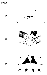
[0045] FIG. 6 illustrates a device comprised of four multiple shafts and
components arranged
in a linear array on the cortex. Fig. 6A. shows an broad top view while (B)
shows a
side view, and (C) shows a view from inside the cranium. A single small burr
hole is
used to insert all four shafts. The single burr hole is of partial thickness
because the
edges at the bottom of the partial burr hole are used to guide the tips of the
self-
drilling shafts or drill bits. Two longer shafts flank two shorter shafts
resulting in a
linear array as seen in (C) where four tips of the shafts are seen protruding
through
the inner cortex. A linear array of stimulation as shown in Figure 6 is useful
for
stimulation along a linear gyrus such as for motor cortex stimulation, where
typically
a small craniotomy is used to place a strip electrode.
[0046] FIG. 7 illustrates a device comprised of nine different shafts placed
through a single
partial small burr-hole. The overall configuration is demonstrated in the
cross section
of the skull model with three different views in (A), (B), and (C). A top view
(D) and
16
CA 02785285 2012-06-20
WO 2011/084788 PCT/US2010/061531
bottom view (E) demonstrate the arrangement of the contacts that penetrate
through
the inner cortex to affect the brain. Four shorter shafts are configured in a
"+"
configuration while four longer shafts are inserted in an "X" pattern. A
central
shortest shaft is inserted last. This configuration results in a 3 by 3 matrix
of
components that can reach the cortex. This type of configuration is useful for
epilepsy stimulation where the central electrode senses seizure activity at
the seizure
focus. This central electrode then activates its own stimulation electrode to
abort the
seizure. At the same time, the 8 surrounding ring of electrodes are activated
as well.
The activation of the ring of electrodes help to trap and cancel the spreading
wave of
seizure activity from the central epileptogenic focus. Such a configuration
would
generally necessitate a craniotomy; however this configuration is placed
through a
single partial burr hole.
[0047] FIG. 8 illustrates a shaft inserted at an axial angle that serves as a
conduit for a
guidable and steerable epidural or subdural electrode array. Fig. 8A. shows
the
drilling of a non-orthogonal hole through the cranium by a self-drilling
shaft. In (B),
an inner compartment of the shaft is unlocked and removed from the outer
threaded
portion, leaving a cylindrical conduit. This conduit allows one or more
electrode
arrays to be inserted into the epidural or subdural space (C). The angled, non-
orthogonal trajectory of the shaft allows the electrode array to safely slide
into the
epidural or subdural space at a shallow angle. In contrast if the burr hole
were
orthogonally oriented, the electrode array would have to make a 90 degree turn
after
passing through the skull. The electrode arrary can be directed similarly to
spinal
cord stimulation electrode array using mechanical turning by a small bend in
the
distal tip of the inner stylet. Alternatively, the distal inner cannular may
be
ferromagnetic allowing an external magnetic or electromagnetic field to guide
or
direct the tip of the electrode array. Lastly, a fibroptic inner cannula with
distal
camera would allow endoscopic guidance of the electrode array under direct
visualization of epidural, subdural, or intraventricular structures. The tip
of the stylet
also would allow for stereotactic image guidance by emitting signals such as
radiofrequency or sonic/ ultrasonic impulses that help localize the distal tip
in
stereotactic coordinates. Once the target and desired placement of the
electrode array
17
CA 02785285 2012-06-20
WO 2011/084788 PCT/US2010/061531
has been accomplished, the proximal end is secured to the cranial conduit/
shaft by a
locking mechanism. Alternatively, other components such as a battery,
controller,
transducer, etc. can also be placed inside the cannula, or in other
trajectories through
the cranium from the same entry site. The combination of multiple shaft
placement
through a single entry site with multiple steerable electrode arrays allow a
limitless
configuration of brain stimulation and recording through a single small burr
hole.
[0048] FIG. 9 demonstrates a simple connection system to physically link
multiple shafts and
components that are placed through a single or nearby entry sites. The
connector
shown is a multichannel connector, but any connector would suffice including
USB
or micro USB connectors. While the components can communicate wirelessly with
each other with the appropriate components included within the shaft, some
functions
are more efficient through direct physical connections.
[0049] FIG. 10 demonstrates a preconfigured head unit used to facilitate the
placement of
multiple shafts and multi-component arrays. Fig. 1OA. shows the empty head
unit
with three docking stations. Fig. IOB shows the insertion of a single shaft
into one
docking station. Two shafts are inserted into the head unit in (B), while all
three
shafts have been inserted in (C). The head unit allows direct communication
and
connection between all shafts and components of the shafts. The head unit
itself can
also contain multiple components of the overall device such as battery,
communication systems, transducers, etc. The head unit can be inserted into a
pre
made burr hole or be self-inserted by having a self-drilling and self-tapping
pointed
tip. The head unit does not need to have its own fixation to the skull as the
insertion
of shafts through the docking stations acts to lock the docking station into
the skull.
Each docking station can also have adjustable angles of insertion by having a
rotating
ball and socket mechanism as the docking station through which shafts are
inserted.
[0050] FIG. 11 shows a flow chart of a method of implanting the devices
described herein:
(I) identify the target, (II) create an incision, (III) drill a partial
thickness burrhole,
(IV) identify target and depth from partial thickness burrhole, (V) insert
device(s),
and (VI) close wound.
18
CA 02785285 2012-06-20
WO 2011/084788 PCT/US2010/061531
DETAILED DESCRIPTION OF THE INVENTION
[0051] The present invention and method of its use enables multiple effectors,
sensors, and
other components to fit through a single entry site to provide improved and/or
longer-
lasting therapeutic benefits. According to some embodiments this is
accomplished by
inserting the effectors, sensors, other components, or shafts housing any of
these
elements at different angles to permit greater subsurface reach given a small
surface
entry site. As used herein, the term "entry site" includes one or more
physically
distinct openings, holes, or incisions, within close proximity to one another
and
taking up a relatively small total area of space consistent with minimally
invasive
surgical procedures. Thus, an "entry site" may be one opening or hole but is
not
limited to such. The "entry site" may also be an entry zone, area, or region
that
encompasses two, three, four, or more distinct openings.
[0052] For each entry site, the stimulator/sensor devices may be inserted at
several different
axial angles between an axis perpendicular to the skin's surface (straight
down) and a
plane tangent to the skin's surface at the entry site. The effectors (i.e.
electrodes)
and/or sensors may also be inserted at several different radial angles around
the
periphery of an entry site in the plane of the tangent to the entry site. The
location of
the entry site, the axial (0i) and the radial (02) insertion angles determine
an unique
trajectory in the skull and in the body. Preferably, no two stimulator/sensor
devices
(comprising at least one effector or sensor as part of the device) have the
same set of
axial (0i), radial (02) angles, and entry site location so that each device
(and each
effector or sensor therein) occupies a unique position different from the
others. The
closer the first diagonal axial angle is to parallel to the skin surface, the
longer the
effector or sensor can be while still traversing substantially laterally
through the skull
without reaching the brain. Conversely, the closer the first diagonal axial
angle is to
perpendicular to the skin's surface (straight down), the shorter the effector
or sensor
must be because it is moving more closely to vertical though the skull and is
thereby
more strictly limited by the skull's vertical thickness. (See FIG. 1.)
[0053] Angled implantation allows implantation of extra components to support
or work
together with the effector or sensor (i.e. electrode) to form a longer-lasting
system or
improved bion. For example, the main device may be implanted perpendicularly
but
19
CA 02785285 2012-06-20
WO 2011/084788 PCT/US2010/061531
one or more components (i.e. extended batteries or battery packs) are
implanted at an
angle. This allows extra components that support a main electrode to be
embedded
within the skull at an angle. More supporting batteries prolongs the life of
the
electrode while effectively breaking up the overall implant into several
components
that are connected (i.e. at the top) by a connector head or connector. Other
components, in addition to batteries, can be transmitters, receivers, radio
transceivers,
heat generators, cooling devices, magnetic coils, capacitors, transformers,
ultrasonic
transducers, hypersonic emitters/receivers, electrophysiological recording
means,
sensors, iontophoresis means, optical stimulators, lasers, cameras,
address/positioning
units, etc.
[0054] As used herein, the term "component" includes effectors and sensors but
is not
limited to these categories. "Component" might also include other categories
of
auxiliary, complimentary, or supplementary elements that support an effector
or
sensor but do not themselves produce an effect on a body or sense (gather
data)
directly. For example, "component" might include a buffer solution, a physical
cushion, a catalyst, a battery, a vacuum line, etc. The present invention
includes an
implant in which at least one component is an effector or sensor. The implant
may
also include other additional components that are also effectors or sensors,
or are
neither effectors nor sensors.
[0055] The implantable devices described herein are made of biocompatible
materials. In a
self-inserting embodiment the devices need to be made of material sufficiently
durable and hard to penetrate bone without rupturing. In embodiments that rely
on
pre-drilling a hole more material options are possible and softer, more
flexible
materials may be used to encapsulate or house the device. According to a
preferred
embodiment, at least a portion of the device is made of a semi-permeable
material
that absorbs some molecules, transmits (flow through) some molecules, elutes
some
molecules, and blocks some molecules. Such a semi-permeable material may be a
mesh with openings (for example, tiny nanopores) therein that optionally also
includes key cells or molecules (that provide an auxiliary function) embedded
therein
on its surface.
CA 02785285 2012-06-20
WO 2011/084788 PCT/US2010/061531
[0056] According to a preferred embodiment, the effectors are electrodes and
supporting
components (i.e. transmitters, receivers, etc.) of the present invention are
designed to
be insertable directly or to insert themselves. By "insert themselves" or
"insertable
directly" it is meant that the components do not require burr holes to be
created in the
skull with a drill prior to implant and/or that the components do not require
expulsion
through an introducer (i.e. needle, cannula, etc.). Self inserted screws of
this type are
typically classified as self-drilling and self-tapping, in that they do not
need a pilot
hole nor does the hole need to be tapped to form the threaded tract for a
screw. This
might be accomplished by the components having distal tips that are sharp or a
housing that resembles a screw shaft with threads.
[0057] Alternatively, the cranial stimulator devices can be helical in shape
such that they
wind into the bone in a manner similar to coil anchors for sand volleyball
nets. The
distal tip of the helix enters into a small hole and the curved tail of the
device follows.
[0058] When drilling into the skull is necessary such as due to increased
resistance from
bone making self-tapping screws inadequate, a preferred system and method
involves
using a balloon along one or more sides of the stimulator device. Drilling
often
creates a hole that is slightly larger than necessary or imperfect in shape
such that
there is not a tight fit for the screw. The balloon can be filled with air and
or fluid
after insertion in a deflated condition to close the gap, reducing the
imperfect mating
between drill hole and stimulator to provide an improved friction fit that
renders the
stimulator less susceptible to internal drift / migration. The balloon can
also be used
proximally above the stimulator to push the electrode contacts on its opposite
distal
end into closer contact with the surface of the cortex.
[0059] If the effectors contain, are coated with, or are associated with
magnetic means (i.e.
coils, magnetic materials, etc.) they can be used to provide magnetic
stimulation
therapy in addition to electrical stimulation therapy. Magnetic energy can
also be
used to recharge the electrical batteries. For example, inserting a magnetic
coil inside
the skull enables one to carry out local magnetic stimulation ("intracranial
magnetic
stimulation") with a much lower intensity than that used for transcranial
magnetic
stimulation which requires a large enough magnetic field to travel through the
cranium (resulting in a diminution of signal strength in the process) and also
is not
21
CA 02785285 2012-06-20
WO 2011/084788 PCT/US2010/061531
localized. The inability to localize therapy, also known as poor selectivity,
typically
results in overbroad application that may cause damage to unintended
surrounding
regions and too weak an intensity of treatment at the target site. The ability
to
localize therapy overcomes both of these drawbacks to systemic application.
[0060] In addition to electrical and magnetic stimulation the implantable
electrode or
components associated with it can be used to generate heat or cold. Heat and
cold
have been shown to influence brain activity such that they can be used to
complement,
supplement, or as an alternative to electrical and/or magnetic stimulation.
[0061] In addition to electrical and magnetic stimulation the implantable
electrode or
components associated with it can be used to generate heat or cold. Heat and
cold
have been shown to influence brain activity such that they can be used to
complement,
supplement, or as an alternative to electrical and/or magnetic stimulation.
[0062] In different embodiments the effector batteries can be recharged inside
or outside the
body or inside the body through connection to a charging device outside the
body.
According to a preferred embodiment the effector batteries are recharged
inside the
body through a naturally occurring means including changes in heat, fluid
dynamics,
etc.. The batteries may include a thermogenerator or thermoelectric generator
that
uses local heat in situ to generate power. Or, the batteries may include a
mechanical
power generator that uses natural pulsation of the brain relative to the
cranium and
changes in cerebrospinal fluid pressure to harness and store energy.
[0063] In addition to built-in electrode batteries, the implantable sensor-
effector devices of
the present invention may be powered by any number of alternative means. In
order
to reduce their size, they may be powered from outside through a means for
receiving
energy with the means for receiving energy being smaller than a conventional
electrode battery. More specifically, they may rely upon ultrasonic,
hypersonic, or
radiofrequency energy from a source at another location in the body or outside
the
body that is absorbed and channeled through a receiving platform. These
alternative
sources of energy permit the devices to be smaller because a built-in battery
is not
required. Thus, the device may be made on the scale of microns (length, width,
height) rather than millimeters and inserted more deeply into the body, into
smaller
channels and crevices, or through intact bone and muscle for better accuracy
while
22
CA 02785285 2012-06-20
WO 2011/084788 PCT/US2010/061531
still being minimally invasive and without sacrificing anatomical structural
integrity.
Another advantage of the energy source and some of the electronic complexity
being
outside the body is that it is easier to upgrade and modify from
outside.Another
advantage of effectors radiating downward and outward from an entry site at
different
angles is that when a target region for stimulation is deeper within the brain
the
angle(s) can be set so that rays from more than one effector converge
precisely on the
deeper target. More than one entry site can be made so that several different
devices
from several different entry sites converge on the target from different
directions (see
FIG. 2). Alternatively, when there is more than one target region deep within
the
brain, effectors from a single entry site can be used to simultaneously reach
several
different regions by directing the effectors at different angles (see FIG. 3).
If the
effectors were limited to non-angled, conventional, straight-down insertion
all
effectors (even through multiple entry sites) would be pointed at the core or
center of
the brain without the ability to provide targeted therapy to intermediate
regions of the
brain between the core and the cortex.
[0064] In alternative embodiments, the effectors may have additional
characteristics that
enable them to jointly maximize length and distance within the skull. For
example,
the effectors may curve with a radius of curvature that approximately matches
the
radius of curvature or shape of the skull. Since the cranium is composed of
three
layers, a hard inner cortical layer, a hard outer cortical layer, and a softer
cancellous
middle layer, long components can be pushed through the cancellous layer being
trapped by the harder inner and outer cortical layers. Additionally, the
devices may
branch out (for example, telescopically) once inserted to form an intracranial
pathway
that provides additional battery power storage space. However, because the
branches
would have to traverse through the somewhat hard bone of the cranium these
(bifurcated, trifurcated, poly-furcated) embodiments would probably require
separate
insertion tools capable of drilling worm-like tunnels for the branched
devices.
[0065] When the effectors are electrodes the circuitry of the present
invention for all
embodiments is variable. By electronic circuitry it is meant the arrangement
and
interrelationship between electrodes, batteries, connectors, coils,
transmitters,
receivers, transceivers, capacitors, controllers/programming means, address
means,
23
CA 02785285 2012-06-20
WO 2011/084788 PCT/US2010/061531
pulse control means, sensors, etc. Any configuration of these elements that is
finctional for multiple electrodes inserted transversely through a single
entry site (at
orthogonal and/or non-orthogonal angles) is consistent with the scope of the
present
invention.
[0066] In some embodiments, the configuration of electronic circuitry may be
similar to that
of existing products and patent claims (i.e. the bion of Advanced Bionics,
Inc.).
However, the entire device is still different from conventional devices and
patent
claims. It differs by being adapted for insertion transversely through the
cranium
such as by screw-in and/or insertion at non-orthogonal angles with more than
one
element inserted through the same entry site.
[0067] In other embodiments, the configuration of electronic circuitry is
distinctly different
in one or more features from conventional products and patent claims, which
serves
to further distinguish the invention in addition to its other distinguishing
features.
[0068] As discussed previously, as neurostimulators the devices of the present
invention
have a myriad of established applications to improve pathologies (movement
disorder,
psychiatric conditions) and enhance normal functions (learning, memory) in the
neural system, particularly through direct interaction with the brain.
Additional,
potential applications include peripheral nerve stimulation and interaction
with other
biological systems to catalyze and regulate healing processes. For example,
implantable stimulators as described herein may be used at sites of bone
fracture or
disc degeneration to expedite new bone proliferation as a substitute or
supplement to
biological or chemical means (bone cement, bone graft, bone filler, bone glue,
hydroxyapatite, ground bone composition, or another bone substitute). One
specific
application is use of stimulators around pedicle screws used in pedicle screw
stabilization / fusion of adjacent vertebrae to stimulate bone regrowth over
the screws
to better camouflage the implants.
[0069] According to a preferred embodiment, the devices described herein are
used to enable
communication between two or more entities with at least one entity being a
living
organism. The other entities may be other living organisms of the same or a
different
species as the first living organism, or may be a machine including but not
limited to
24
CA 02785285 2012-06-20
WO 2011/084788 PCT/US2010/061531
a computer, a laptop, a cell phone, a personal digital assistant (PDA), a
keyboard, a
camera, a wheel chair, a bicycle, a car, etc. The communication can be one-
way,
two-way, or a multi-channel exchange amongst several different entities (group
conversation, or different entities all communicating with a centralized hub).
[0070] In this method of enabling communication between at least one living
organism and
at least one other entity a device comprising an effector and a sensor is
implanted in
the living organism. At least one additional component is implanted in the
other
entities to interact with this device. The sensor in the first entity (living
organism)
gathers data and generates a pulse that transmits the data to the other
entities. The
other entities receive the pulse through their components that read and
translate it. In
this manner the first entity (living organism) can relay information or "talk"
to the
other entities in open loop communication. In an alternative embodiment, the
device
in the first entity further comprises at least one feedback component and the
communication is closed loop with the feedback component in the first entity
verifying receipt of the pulse from the first entity by the second entity.
[0071] When receivers or transceivers are used to receive signals they may be
used alone to
receive signals directly or they may be used in conjunction with one or more
intermediary devices that relay and/ or process the signal prior to its
reception. The
intermediary device might amplify or reformat the signal and eliminate noise.
In
some embodiments, for some applications, the intermediary device could be
something similar to a bluetooth earpiece, a cell phone, a wifi router, an air
card, etc.
Likewise, when effectors are used to induce an effect in an entity (machine or
organism) they may induce the effect directly or through one or more
intermediary
devices that adjust or process the raw information and energy they provide.
[0072] The devices described herein are contemplated to be adaptable for use
with state-of-
the-art sixth sense and mind control devices. The minimally invasive implants
of the
present invention may be more convenient than headgear and may be used to read
neural states and objectives to initiate actions in the outside world rather
than relying
on hand gestures from the living organism subject or patient. As used herein
(before
and after), the term "patient" refers to any object that subjects itself or is
subjected to
a treatment incorporating the present invention. A "patient" need not be an
ill person
CA 02785285 2012-06-20
WO 2011/084788 PCT/US2010/061531
or someone with physical, emotional, or psychological impairments or
abnormalities.
In fact, a "patient" need not be a human being or even a living organism. A
"patient"
may include completely healthy, happy, and successful organisms or objects
that
choose to subject themselves to treatment or are subjected to treatment with
the
present invention in order to further their abilities and become even more
successful
or to improve certain functions.
[0073] Examples of conditions the devices of the present invention can be used
to treat
include: psychological conditions generally, genetically or biologically based
psychological conditions, depression, acute mania, bipolar disorders,
hallucinations,
obsessions, obsessive compulisive disorder, schizophrenia, catatonia, post-
traumatic
stress disorder, drug and alcohol addiction, Parkinson's disease, Alzheimer's
disease,
epilepsy, dystonia, tics, stuttering, tinnitus, spasticity, recovery of
cognitive and
motor function following stroke, pain syndromes, migraine, neuropathies, back
pain,
internal visceral diseases, urinary incontinence, etc.
[0074] Specific medical applications include using the cranial implants of the
present
invention as follows: (i) enabling a paralyzed man to send signals to operate
a
computer by "telepathically" moving a mouse, cursor, or typing on a keyboard,
improving one's ability to work; and (ii) enabling a paralyzed man to send a
signal
causing a machine or computer to speak a phrase or message for them so that
they can
communicate their needs, desires, and thoughts to others and the world.
[0075] Specific entertainment and social applications include using the
cranial implants of
the present invention as follows: (i) a person has a CranionTM implanted so
that he
can use it to control his iPhone or Wii game console without using his hands
or in
addition to hand controls; and (ii) a person has a CranionTM implanted to
communicate with one or more other persons, each with his own CranionTM
implanted to enable private "telepathic" conversations in a group of people
including
at a meeting, in church, in the courtroom, at a sporting event, and during a
card game.
[0076] Implanted devices of the present invention (especially those in the
brain) may be used
to control a projector, a camera, a laser, a bar code reader, etc. worn on the
body.
Such sixth sense and mind control devices may find application for video
games,
26
CA 02785285 2012-06-20
WO 2011/084788 PCT/US2010/061531
electronic transfers of money, trading stocks, shopping, social and
professional
networking and storage of data about people, filming, photography, etc. The
implants
could be used to read expressive conditions (facial expressions, gestures) and
emotional experiences (affective response) of the living organism in which
they are
implanted or of others with whom the patient comes in contact. The implants
could
then process and analyze this information to initiate cognitive actions in
response
thereto.
[0077] It is known that an electrical signal at the cortex of the brain looks
random across the
population for the same thought, even though it originates from the same
region of
the brain, due to a unique fold pattern of each person's brain similar to
fingerprints.
Headgear uses a mathematical algorithm to unlock the random signal to make it
consistent across the population. Alternatively, the implants of the present
invention
might be used (i) to read the signal from a source in the brain beyond the
cortex
where it is uniform without the algorithm, (ii) apply the algorithm to data
read at the
cortex, or (iii) to provide an initial equilibration process that compensates
for the
differences in signals from one person to another.
[0078] According to still other embodiments, the CranionTM has a longer
electrode lead that
passes through the skull at an angle and goes epidural to distant areas like a
spinal
cord stimulator sliding up the epidural space in the spine. This tip may then
be
steerable, for example, with a magnet.
[0079] The general method, as summarily illustrated in the flow chart of FIG.
12, in greater
detail may encompass the following sequence:
1.) Use stereotactic localization, either with a frame or frameless
stereotactic
localization to identify a target(s);
2.) Decide on a configuration. For example, either single electrode, multiple
around
the single target, single line (see FIG. 7 and 8);
3.) Single stab incision 5-10 mm;
4.) Drill 2-4mm partial thickness burrhole (this allows an "edge" so that
drills can be
angled into the corner and an off angle trajectory can be accomplished;
27
CA 02785285 2012-06-20
WO 2011/084788 PCT/US2010/061531
5.) Use stereotactic localization to identify target and depth away from the
central
partial burrhole;
6.) Plan trajectory based on the target and either drill a pilot hole or use a
self
drilling, self tapping CranionTM to insert the CranionTM device;
6a.) Drilling a pilot hole allows exact knowledge of the depth of the hole
however a cannulated CranionTM in which the sharp tip can be removed
(see FIG. 9) also allows a portal to determine whether the epidural space
has been entered.
7.) Place other CranionsTM and connect them with wires (see FIG. 9) or have
them
connect wirelessly. Or, use the head device.
8.) Add other components such as extra batteries that don't need to go all the
way out
of the skull.
9.) Close the wound
[0080] The present invention is not limited to the embodiments described
above. Various
changes and modifications can, of course, be made, without departing from the
scope
and spirit of the present invention. Additional advantages and modifications
will
readily occur to those skilled in the art. Therefore, the invention in its
broader aspects
is not limited to the specific details and representative embodiments shown
and
described herein. Accordingly, various modifications may be made without
departing
from the spirit or scope of the general inventive concept as defined by the
appended
claims and their equivalents. As used in the claims the conjunction "or" means
the
inclusive or (and/or, either element independently or any combination of the
elements
together).
28