Note: Descriptions are shown in the official language in which they were submitted.
CA 02787471 2015-01-06
COMBINED PHYSIOLOGICAL SENSOR SYSTEMS AND METHODS
Summary
The present disclosure relates to sensors and, more particularly, to combined
physiological
sensors and methods for detecting one or more physiological characteristics of
a subject. A
combined sensor (e.g., a forehead sensor) may be used to detect and/or
calculate at least one of a
pulse blood oxygen saturation level, a regional blood oxygen saturation level,
a respiration rate,
blood pressure, an electrical physiological signal (EPS), a pulse transit time
(PTT), body
temperature associated with the subject, a depth of consciousness (DOC)
measurement, any other
suitable physiological parameters, and any suitable combination thereof. The
individual sensors
within the combined sensor may be advantageously positioned in accordance with
a number of
different geometries. In addition, several techniques may be employed to
prevent or limit
interference between the individual sensors and their associated input and/or
output signals. The
combined sensor may be coupled to a monitoring device, which may receive
and/or process one or
more output signals from the individual sensors to display information about
the medical condition
of the subject.
Various embodiments of the present invention relate to a physiological sensor
device,
comprising: a flexible substrate capable of being applied to a subject; a
first electrode disposed on
the substrate for receiving a first electrical signal associated with the
subject; a second electrode
disposed on the substrate for receiving a second electrical signal associated
with the subject; and a
first optical detector disposed on the substrate at a location between the
first electrode and the
second electrode for receiving a first optical signal transmitted into the
subject; wherein the first
electrode, the second electrode and the first optical detector are disposed on
the substrate in a
geometry that positions the first electrode over the center of the subject's
forehead, the second
electrode over the subject's temple, and the first optical detector over
highly perfused tissue of the
subject, when the substrate is applied to the subject.
Various embodiments of the present invention relate to a method for processing
physiological signals, comprising: receiving a first electrical signal
associated with a subject from a
first electrode disposed on a flexible substrate; receiving a second
electrical signal associated with
the subject from a second electrode disposed on the substrate; and receiving a
first optical signal
transmitted into the subject from a first optical detector disposed on the
substrate at a location
1
CA 02787471 2015-01-06
between the first electrode and the second electrode; wherein the first
electrode, the second
electrode and the first optical detector are disposed on the substrate in a
geometry that positions the
first electrode over the center of the subject's forehead, the second
electrode over the subject's
temple, and the first optical sensor over highly perfused tissue of the
subject, when the substrate is
applied to the subject.
In an embodiment, a physiological sensor device is provided that includes a
flexible
substrate capable of being applied to a subject. A first electrode may be
disposed on the substrate
for receiving a first electrical signal associated with the subject, and a
second electrode may be
disposed on the substrate for receiving a second electrical signal associated
with the subject. An
optical detector may be disposed on the substrate at a location between the
first electrode and the
second electrode for receiving an optical signal transmitted into the subject.
The device, in some
approaches, may additionally include an optical emitter disposed on the
substrate for transmitting
an optical signal into the subject.
la
CA 02787471 2012-07-18
WO 2011/097399
PCT/US2011/023630
In an embodiment, the device includes a structure coupled to the substrate
that is also
capable of being applied to the subject. A second optical emitter may be
disposed on the
structure for transmitting a second optical signal into the subject, and a
second optical
detector may be disposed on the structure for receiving the second optical
signal. In another
embodiment, the optical detector is disposed near the optical emitter and a
second optical
detector may be disposed on the substrate at a distance from the emitter for
receiving the
optical signal transmitted into the subject.
In an embodiment, the device includes a temperature sensor disposed on the
substrate
for detecting a temperature of the subject. Moreover, the optical detector may
be disposed in
a location between the optical emitter and a second optical detector, and the
temperature
sensor may be disposed in a location between the two optical detectors. This
arrangement
may substantially isolate the temperature sensor from any heat generated by
the optical
emitter.
In some approaches, the first electrode, the second electrode, and the optical
detector
are disposed on the substrate in a geometry that positions the first electrode
over the center of
the subject's forehead, the second electrode over the subject's temple, and
the optical sensor
over highly perfused tissue of the subject, when the substrate is applied to
the subject. In an
embodiment, the device includes a third electrode disposed on the substrate
for receiving a
third electrical signal associated with the subject, and a fourth electrode
disposed on the
substrate for receiving a fourth electrical signal associated with the
subject. In this
embodiment, the first electrode, the second electrode, the third electrode,
and the fourth
electrode may be disposed on the substrate at substantially equally spaced
intervals.
In an embodiment, the output signals fi.om the first electrode, the second
electrode,
and the optical detector are routed via interconnects to a shared cable
connected to
monitoring circuitry. In another embodiment, the device includes processing
circuitry that
receives the first electrical signal, the second electrical signal, and the
optical signal, and
calculates at least one of a pulse blood oxygen saturation level, a regional
blood oxygen
saturation level, a respiration rate, blood pressure, an electrical
physiological signal, a pulse
transit time, body temperature, and a depth of consciousness measurement.
Brief Description of the Drawings
The above and other features of the present disclosure, its nature and various
advantages will be more apparent upon consideration of the following detailed
description,
taken in conjunction with the accompanying drawings in which:
2
CA 02787471 2012-07-18
WO 2011/097399
PCT/US2011/023630
FIG. 1 is a perspective view of a monitoring system, in which one or more
physiological characteristics of a subject are monitored in accordance with an
embodiment;
FIG. 2 is a block diagram of a monitoring system, such as the system depicted
in
FIG. 1, coupled to a patient in accordance with an embodiment;
FIG. 3 is a block diagram of an illustrative sensor signal measurement system
in
accordance with an embodiment;
FIGS. 4A-4D show illustrative combined physiological sensors with differing
geometries applied to a subject in accordance with some embodiments;
FIG. 5 shows a perspective view of a combined physiological sensor as applied
to
subject in accordance with an embodiment;
FIGS. 6A-6C show detailed views of various combined physiological sensors in
accordance with some embodiments;
FIGS. 7A and 7B show top and bottom views, respectively, of a combined
physiological sensor in accordance with an embodiment; and
FIG. 8 is an illustrative timing diagram for activating the assorted sensors
of a
combined physiological sensor in accordance with an embodiment.
Detailed Description
Monitoring the physiological state of a subject, for example, by determining,
estimating, and/or tracking one or more physiological parameters of the
subject, may be of
interest in a wide variety of medical and non-medical applications.
Indications of a subject's
physiological parameters obtained from sensors (e.g., photoplethysmograph
sensors,
electrical physiological signal sensors, and temperature sensors) can provide
short-term and
long-term benefits to the subject, such as early detection and/or warning of
potentially
harmful conditions, diagnosis and treatment of illnesses, and/or guidance for
preventative
medicine. Medical sensors for monitoring multiple parameters are typically
connected to one
or more devices (e.g., single parameter or multi parameter monitors). As used
herein, the
term "PPG sensor" may refer to any sensor that generates a phothplethysmograph
(PPG) or
equivalent signal, the term "BPS sensor" may refer to any sensor that
generates an electrical
physiological signal (EPS), and the term "temperature sensor" may refer to any
sensor that
generates a signal representative of a measured temperature.
One type of medical device that can be used to monitor the physiological state
of a
subject is an oximeter. An oximeter may determine the oxygen saturation of
blood. An
oximeter may include a light sensor (e.g., a PPG sensor) that is placed at a
site on a patient,
3
CA 02787471 2012-07-18
WO 2011/097399
PCT/US2011/023630
typically a fingertip, toe, forehead or earlobe, or in the case of a neonate,
across a foot. The
oximeter may pass light using a light source through blood perfused tissue and
photoelectrically sense the absorption of light in the tissue. For example,
the oximeter may
measure the intensity of light that is received at the light sensor as a
function of time. A
signal representing light intensity versus time or a mathematical manipulation
of this signal
(e.g., a scaled version thereof, a log taken thereof, a scaled version of a
log taken thereof,
etc.) may be referred to as the photoplethysmograph (PPG) signal. In addition,
the term
"PPG signal," as used herein, may also refer to an absorption signal (i.e.,
representing the
amount of light absorbed by the tissue) or any suitable mathematical
manipulation thereof.
The light intensity or the amount of light absorbed may then be used to
calculate the amount
of the blood constituent (e.g., oxyhemoglobin) being measured and other
physiological
parameters such as the pulse rate and when each individual pulse occurs.
The light passed through the tissue is selected to be of one or more
wavelengths that
are absorbed by the blood in an amount representative of the amount of the
blood constituent
present in the blood. The amount of light passed through the tissue varies in
accordance with
the changing amount of blood constituent in the tissue and the related light
absorption. Red
(RED) and infrared (IR) wavelengths may be used because it has been observed
that highly
oxygenated blood will absorb relatively less red light and more infrared light
than blood with
a lower oxygen saturation.
2 0 It will be
understood that, as used herein, the term "light" may refer to energy
produced by radiative sources and may include one or more of ultrasound,
radio, microwave,
millimeter wave, infrared, visible, ultraviolet, gamma ray or X-ray
electromagnetic radiation.
As used herein, light may also include any wavelength within the radio,
microwave, infrared,
visible, ultraviolet, or X-ray spectra, and that any suitable wavelength of
electromagnetic
radiation may be appropriate for use with the present techniques.
When the measured blood parameter is the oxygen saturation of hemoglobin, a
convenient starting point assumes a saturation calculation based on Lambert-
Beer's law. The
following notation will be used herein:
/0k,, = /6, (2) exp(¨(430 (X) + (1 ¨ s)13,. 00)1(0) (1)
where:
= wavelength;
t ¨ time;
I = intensity of light detected;
4
CA 02787471 2012-07-18
WO 2011/097399
PCT/US2011/023630
1-0 = intensity of light transmitted;
S = oxygen saturation;
130, 13r = empirically derived absorption coefficients; and
1(t) = a combination of concentration and path length from emitter to detector
as a
function of time.
One common type of oximeter is a pulse oximeter, which may indirectly measure
the
oxygen saturation of a patient's blood (as opposed to measuring oxygen
saturation directly by
analyzing a blood sample taken from the patient) and changes in blood volume
in the skin. In
pulse oximetry, by comparing the intensities of two wavelengths at different
points in the
pulse cycle, it is possible to estimate the blood oxygen saturation of
hemoglobin in arterial
blood. Ancillary to the blood oxygen saturation measurement, pulse oximeters
may also be
used to measure the pulse rate of the patient. Pulse oximeters typically
measure and display
various blood flow characteristics including, but not limited to, the oxygen
saturation of
hemoglobin in arterial blood.
For example, using a pulse oximeter, saturation may be calculated by solving
for the
"ratio of ratios" as follows.
1. First, the natural logarithm of (1) is taken ("log" will be used to
represent the natural
logarithm) for IR and Red
log / ¨ log /0-(430+(l -s) 130/ (2)
2. (2) is then differentiated with respect to time
d log/ di
dt
3. Red (3) is divided by IR (3)
d log /(2R)/ dt = s 13 0 (2,R) + (1- s) 131, (1R)
(4)
d log /(2/R) / dt s 0 (AIR) + (1- s) )81. (2 IR)
4. Solving for s
d log I (A IR)d log 1(2 )
Pr (AR ) R /31.(211?)
dt dt
s =
d log I(2
R) 00(21R)¨ fir(AIR))¨ d log /(2m ) (160(20¨ fir (2k))
dt dt
Note in discrete time
5
CA 02787471 2012-07-18
WO 2011/097399
PCT/US2011/023630
d log 1(2,0 log I (2, t2) - log / (2, )
dt
Using log A-log B = log A/B,
d log I (.1,,t) ( log1 (t2,
dt I(t /1õ)
p , )
So, (4) can be rewritten as
(
d log /(.1,R ) log (t1,)
dt I (t
__________________________________________________
(
d log /(.17R) At1,/17R) = R (5)
log
dt \i(t2,AIR)i
where R represents the "ratio of ratios." Solving (4) for s using (5) gives
fir (AR)Rfir (AIR)
s =
R(fi o(7R) ¨ r (Am)) ¨ o ('2R) + &(IR)
From (5), R can be calculated using two points (e.g., PPG maximum and
minimum), or a
family of points. One method using a family of points uses a modified version
of (5). Using
the relationship
d log / d 1 dt
dt =
(6)
now (5) b = R (7)
which defines a cluster of points whose slope of y versus x will give R where
x(t) = [i(t2, AiR )1/(ti , 2R)
y(t) = [i(t2, AR) ¨ /(ti, AR)]I(ti, A7R) (8)
y(t)= Rx(t)
Once R is determined or estimated, for example, using the techniques described
above, the
blood oxygen saturation can be determined or estimated using any suitable
technique for
relating a blood oxygen saturation value to R. For example, blood oxygen
saturation can be
determined from empirical data that may be indexed by values of R, and/or it
may be
determined from curve fitting and/or other interpolative techniques.
The foregoing is merely illustrative and any suitable processing techniques
may be
used to calculate pulse oxitnetry values. For example, Fourier transforms and
continuous
6
CA 02787471 2012-07-18
WO 2011/097399
PCT/US2011/023630
wavelet transforms may be used to process the PPG signals and derive blood
oxygen
saturation.
Another common type of oximeter is a regional oximeter, which may be used to
calculate an oxygen saturation of a patient's blood in a non-invasive manner.
In regional
oximetry, by comparing the intensities of two wavelengths of light, it is
possible to estimate
the blood oxygen saturation of hemoglobin in a region of a body. Whereas pulse
oximetry
measures blood oxygen based on changes in the volume of blood due to pulsing
tissue (e.g.,
arteries), regional oximetry typically examines blood oxygen saturation within
the venous,
arterial and capillary systems within a region of a patient. For example, a
regional oximeter
may include a sensor to be placed on a patient's forehead and may be used to
calculate the
oxygen saturation of a patient's blood within the venous, arterial and
capillary systems of a
region underlying the patient's forehead (e.g., in the cerebral cortex). The
sensor may include
two emitters (e.g., for emitting two wavelengths of light) and two detectors:
one detector that
is relatively "close" to the two emitters and another detector that is
relatively "far" from the
two emitters.
For example, if IA represents the intensity of the received/detected light
associated
with the "close" detector, /A (A., t) , may be derived using Lambert-Beer's
law, described
0 (1)
above. Similarly, if hi represents the intensity of the received/detected
light associated with
the "far" detector, I (2't) , may be derived using Lambert-Beer's law,
described above.
(1)
2 0 Light intensity of multiple wavelengths may be received at both the
"close" and the "far"
detectors. For example, if two wavelength were used, the two wavelengths may
be
contrasted at each location and the resulting signals may be contrasted to
arrive at a regional
saturation value that pertains to additional tissue through which the light
received at the "far"
detector passed (tissue in addition to the tissue through which the light
received by the
"close" detector passed, e.g,, the brain tissue), when it was transmitted
through a region of a
patient (e.g., a patient's cranium). Other methods to calculate regional blood
oxygen
saturation are well known in the art.
Pulse blood oxygen saturation and regional blood oxygen saturation may be
measured
and/or calculated, in an embodiment, simultaneously. For example, in the
configuration
described above, in which a "close" detector is disposed near two emitters (or
one emitter that
can output more than one wavelength of light) and a "far" detector is disposed
at some
distance from the two emitters, pulse oxygen saturation may be measured using
the two
7
CA 02787471 2015-01-06
emitters and the "close" detector (i.e., the intensity of light
received/detected at the "close" detector)
while regional oxygen saturation may be measured using the two emitters and
the "far" detector
(i.e., the intensity of light received/detected at the "far" detector).
Simultaneous measurement of
pulse and regional blood oxygen saturation is described further in U.S. Patent
App. Pub. No.
2011/0112387.
Another type of device that can be used to monitor the physiological state of
a subject is a
continuous non-invasive blood pressure (CNIBP) device. A CNIBP device may be
include PPG
sensors affixed to a subject that allow for the determination of the subject's
blood pressure, for
example, using CNIBP monitoring techniques. For example, some CNIBP monitoring
techniques
have been developed that involve the use of two probes or sensors (e.g., PPG
sensors) positioned at
two different locations on a subject's body. The elapsed time, T, between the
arrival of
corresponding points of a pulse signal at the two locations may then be
determined using the two
probes or sensors. The estimated blood pressure, p, may then be related to the
elapsed time, T, by
p = a + b = ln(T)
(9)
where a and b are constants that are dependent upon the nature of the subject
and the signal
detecting devices. Other blood pressure equations using elapsed time may also
be used.
Such a CNIBPB monitoring technique is described in Chen et al. U.S. Patent
No. 6,566,251. The technique described by Chen et al. may use two sensors
(e.g., ultrasound or
photoelectric pulse wave sensors) positioned at any two locations on a
subject's body where pulse
signals are readily detected. For example, sensors may be positioned on an
earlobe and a finger, an
earlobe and a toe, or a finger and a toe of a patient's body. In some
approaches, a single sensor or
probe location may be used to determine blood pressure, as described in U.S.
Patent App. Pub.
No.2009/0326386. In particular, a single PPG sensor may generate a PPG signal
that is analyzed to
compute a time difference between two or more characteristic points of the PPG
signal. This time
difference may be used to calculate blood pressure on a periodic or continuous
basis. Alternatively,
or in addition, the area under a pulse (or a portion of the pulse) in a PPG
signal may be measured
and used to calculate blood pressure.
Similar sensors or probes may also be used to determine respiration rate and
other
respiratory properties (e.g., respiratory effort). For example, as described
in more detail in Addison
etal. U.S. Patent App. Pub. No. 2006/0258921, the act of breathing may cause a
breathing band to
become present in a scalogram derived from a continuous wavelet transform of a
PPG signal. This
breathing band may occur at or about the scale having a characteristic
frequency that corresponds to
8
CA 02787471 2015-01-06
the breathing frequency. Furthermore, the features within this band (e.g., the
energy, amplitude,
phase, or modulation) or the features within other bands of the scalogram may
result from changes
in breathing rate (or breathing effort) and therefore may be correlated with
various respiratory
parameters of a patient.
Other devices and sensors may also be used to determine physiological
parameters of a
subject. For example, an electrical physiological signal (EPS) sensor may be
used to determine
such signals as electroencephalographic (EEG) signals, electrocardiography
(ECG or EKG) signals,
electromyography (EMG) signals, or any other electrical physiological signal.
Sensors may also be
used to determine a subject's body temperature, a pulse transit times (PTT),
or both. In an
embodiment, PTT may be determined by using PPG data in conjunction with EPS
data. For
example, PTT may be determined by comparing an ECG onset point with a PPG
arrival point. An
ECG signal may be processed in order to detect the QRS complex and to detect
the R wave peak.
The plethysmograph signal may be processed to detect the pulse timing. The PTT
may then be
calculated as the time between the R wave peak and the corresponding pulse
peak. Other suitable
techniques for calculating PTT are well known in the art and may also be used.
In an embodiment, sensors may be used to determine a subject's depth of
consciousness
(DOC). In particular, a DOC measurement may be determined using measured PPG
signals, EPS
signals, or a combination thereof. For example, one or more EPS signals may be
processed to
supply a consciousness index, indicating a patient's depth of consciousness on
a scale. As another
example, a PPG signal may exhibit one or more waveform features which indicate
consciousness.
Techniques for monitoring/assessing depth of consciousness using physiological
signals, such as
PPG and EPS signals, are described in detail in U.S. Provisional Patent App.
No. 61/439281.
These and other devices and sensors can be used for monitoring physiological
parameters of
a subject. The devices may be standalone devices or may be combined into
9
CA 02787471 2012-07-18
WO 2011/097399
PCT/US2011/023630
one or more multi-parameter monitoring devices. The monitoring devices may be
physically
connected to each sensor to receive signals from the sensors and perform
various processing
of the signals. The physiological parameters may, for example, be consolidated
and
displayed on a single display to provide a condensed view of the subject's
physiological state
to assist a medical professional in treating the subject. Alternatively, a
subject's physiological
parameters obtained from the sensor signals may be displayed on multiple
displays that may
be associated with one or more single parameter and multiple parameter
monitoring devices.
In accordance with the present disclosure, a combined physiological sensor is
provided to facilitate the connection and coordination of multiple sensors
with one or more
monitoring devices. In particular, a combined physiological sensor may
incorporate a
number of physiological sensors and other components embedded within, or
attached to, a
sensor structure suitable for application to a subject (e.g., a human
subject). This sensor
structure may be flexible, such that it can bend when applied to a curved
surface (e.g., a
human forehead) thereby facilitating contact between the sensor structure, or
the sensors, and
the curved surface. The sensor structure may also include an adhesive to
secure the
combined physiological sensor to the subject, further aiding contact between
the sensor
structure or sensors and the surface of application. In addition, the sensor
structure may
contain or be attached to circuitry for connecting the sensors to a cable,
wireless transmitter,
memory device or other means for storing and/or transmitting data generated by
the sensors
to a monitor. For example, the sensor structure may be a flex circuit, in
which the sensors,
interconnections, and other electronic devices may be mounted, deposited,
and/or printed
onto a flexible substrate. The composition, shape, and other details related
to the sensor
structure will be discussed further below.
Integrating multiple sensors into a combined physiological sensor in
accordance with
the present disclosure provides numerous advantages over traditional sensor
and monitoring
systems. In particular, a combined physiological sensor may require fewer
cables and
components than the traditional approach in order to support multiple sensors,
which in turn
helps reduce cost and total sensor area. In a traditional system, for example,
each sensor
applied to a patient may include its own cable connection to one or more
monitoring devices;
supporting a number of sensors may therefore involve the use of numerous
cables and/or
other supporting equipment. A combined physiological sensor, however, may
unite multiple
sensors within one or more sensor structures and may route all or some sensor
output signals
to a shared cable connected to one or more monitors. In addition to the
simplification,
minimization, and cost-reduction offered by a single-cable or reduced number
of cables
CA 02787471 2012-07-18
WO 2011/097399
PCT/US2011/023630
approach, reducing the number of cables necessary to deliver data to a
monitoring device
mitigates the potential for entanglement or human error involved in connecting
the cables
appropriately. Furthermore, a combined physiological sensor with a sensor
structure that
integrates multiple sensors in a particular geometry, may facilitate proper
placement of
sensors relative to a patient and relative to other sensors. For example, each
sensor applied to
a patient may require disposition within a specific area of the patient's
body. Moreover, when
applying multiple sensors to a patient, care may be required to avoid or
reduce interference
between the sensors and/or their respective input or output signals. As such,
in a traditional
approach, each sensor applied to a patient may require a laborious process of
sensor site
identification and application while simultaneously demanding consideration of
other sensor
locations. A combined physiological sensor, on the other hand, may integrate
multiple
sensors in a specific geometry, such that proper placement of the combined
physiological
sensor results in proper disposition of the individual sensors on the patient.
The combined
physiological sensor may also be configured, as discussed below, to reduce or
limit
interference between the individual sensors and/or their associated signals.
Sensor
integration and interference reduction, among other objects of the present
disclosure, are
more fully discussed in the description that follows in connection with FIGS.
1-8.
FIG. 1 is a perspective view of an embodiment of a monitoring system 10.
System 10
may include a sensor 12 and a monitor 14. Sensor 12 may include one or more
emitters 16
for emitting light at two or more wavelengths into a patient's tissue. One or
more detectors
18 may also be provided in sensor 12 for detecting the light originating from
emitters 16 that
either passes through the patient's tissue or reflects off one or more
surfaces of the tissue.
In accordance with the present disclosure, system 10 may include a plurality
of
sensors ¨ such as PPG sensors (e.g., oximetty and/or CNIBP sensors), BPS
sensors (e.g.,
ECG, EEG, and/or EMG sensors), and temperature sensors ¨ forming a combined
physiological sensor in lieu of single sensor 12. It should be understood that
although the
description below, in connection with FIG. 1, refers primarily to single
sensor 12, a
combined physiological sensor may be substituted to augment the capabilities
of system 10.
It should further be understood that the description of system 10, and in
particular the
description of the various embodiments and configurations thereof, is equally
applicable
when a combined physiological sensor is used in lieu of single sensor 12,
Specifically, all
interconnections, cables, components, and/or modes of communication may remain
substantially the same and/or serve substantially the same purposes. The
combined
physiological sensor may be of any configuration and composition, as discussed
herein
11
CA 02787471 2012-07-18
WO 2011/097399
PCT/US2011/023630
further below. In particular, the combined physiological sensor may be any of
those
described in connection with FIG. 4A (i.e., sensor 410), FIG. 4B (i.e., sensor
420), FIG. 4C
(i.e., sensor 430), FIG. 4D (i.e., sensor 440), FIG. 5 (i.e., sensor 510),
FIG. 6A (i.e., sensor
610), FIG. 6B (i.e., sensor 620), FIG. 6C (i.e., sensor 630), and FIG. 7A and
FIG. 7B (i.e.,
sensor 710).
Each BPS sensor of the combined physiological sensor may include a passive or
active electrode. The BPS sensors may be any suitable device for detecting
voltages,
currents, or impedances. The temperature sensors may be any suitable
temperature
measurement device, including devices that measure temperature through direct
contact and
those that measure temperature without direct contact (e.g., through detection
of thermal
radiation). Each of the PPG sensors of the combined physiological sensor may
be a
complementary metal oxide semiconductor (CMOS) sensor or a charged coupled
device
(CCD) sensor. In another embodiment, the combined physiological sensor may be
made up
of a combination of CMOS and CCD sensors. The CCD sensors may comprise a
photoactive
region and a transmission region for receiving and transmitting data whereas
the CMOS
sensors may be made up of an integrated circuit having an array of pixel
sensors. Each pixel
may have a photodetector and an active amplifier.
According to an embodiment, emitters 16 and detectors 18 of sensor 12 may be
on
opposite sides of a section of tissue (e.g., within a digit such as a finger
or toe), in which case
the light detected by detectors 18 has passed completely through the tissue.
In another
embodiment, emitters 16 and detectors 18 may be positioned on the same side of
a section of
tissue (e.g., within the forehead) so that light from emitters 16 is reflected
by the tissue into
detectors 18.
In an embodiment, sensor 12, or the sensors included in a combined
physiological
sensor, may be connected to and draw power from monitor 14 through cable 24 as
shown. In
another embodiment, the sensors may be wirelessly connected to monitor 14 and
draw power
from a battery or similar power supply (not shown). Monitor 14 may be
configured to
calculate physiological parameters based at least in part on data received
from sensor 12, or
the sensors of the combined physiological sensor, such as data relating to
light emission and
detection. In an alternative embodiment, the calculations may be performed
using circuitry
embedded in sensor 12, or in the combined physiological sensor, and the result
of the
calculations may be passed to monitor 14. Further, monitor 14 may include a
display 20
configured to display the physiological parameters or other information about
the system. In
the embodiment shown, monitor 14 may also include a speaker 22 to provide an
audible
12
CA 02787471 2012-07-18
WO 2011/097399
PCT/US2011/023630
sound that may be used in various other embodiments, such as for example,
sounding an
audible alarm in the event that a patient's physiological parameters are not
within a
predefined normal range.
In the embodiment depicted in FIG. 1, sensor 12, or the combined physiological
sensor, is communicatively coupled to monitor 14 via a cable 24. However, in
another
embodiment, a wireless transmission device (not shown) or the like may be used
instead of,
or in addition to, cable 24. In an embodiment, sensor 12, or the combined
physiological
sensor, may incorporate a storage device for recording data produced by the
sensors. The
stored data may be transmitted to monitor 14 at a later time, for example, via
cable 24 or a
wireless transmission device, or the storage device may be disconnected from
the sensor and
connected directly to monitor 14.
In the illustrated embodiment, monitoring system 10 may also include a multi-
parameter patient monitor 26. The monitor may be cathode ray tube type, a flat
panel display
(as shown) such as a liquid crystal display (LCD) or a plasma display, or any
other type of
monitor now known or later developed. Multi-parameter patient monitor 26 may
be
configured to calculate physiological parameters and to provide a display 28
for information
from monitor 14 and from other medical monitoring devices or systems (not
shown). For
example, multipararneter patient monitor 26 may be configured to display an
estimate of a
patient's pulse blood oxygen saturation (referred to as an "Sp02"
measurement), regional
blood oxygen saturation (referred to as a "regional saturation" measurement),
pulse rate,
blood pressure, respiration rate, body temperature, depth of consciousness,
and/or other
physiological information, each generated by monitor 14 or one or more other
monitoring
devices, on display 28. In an embodiment, monitor 14 may itself be configured
to provide
blood oxygen saturation, regional blood oxygen saturation, pulse rate, blood
pressure,
respiration rate, body temperature, depth of consciousness, and/or other
physiological
information on display 20 or to monitor 26.
Monitor 14 may be communicatively coupled to multi-parameter patient monitor
26
via a cable 32 or 34 that is coupled to a sensor input port or a digital
communications port,
respectively and/or may communicate wirelessly (not shown). In addition,
monitor 14 and/or
multi-parameter patient monitor 26 may be coupled to a network to enable the
sharing of
information with servers or other workstations (not shown). Monitor 14 may be
powered by
a battery (not shown) or by a conventional power source such as a wall outlet.
FIG. 2 is a block diagram of a monitoring system, such as system 10 of FIG. 1,
which may be coupled to a patient 40 in accordance with an embodiment. Certain
illustrative
13
CA 02787471 2012-07-18
WO 2011/097399
PCT/US2011/023630
components of sensor 12 and monitor 14 are illustrated in FIG. 2, Sensor 12
may include
emitters 16, detectors 18, and encoder 42. Alternatively, as discussed above
in connection
with FIG. 1, system 10 may include a plurality of sensors ¨ such as PPG
sensors (e.g.,
oximetry and/or CNIBP sensors), EPS sensors (e.g., ECG, EEG, and/or EMG
sensors), and
temperature sensors ¨ in a combined physiological sensor in lieu of single
sensor 12. It
should be understood that although the description below, in connection with
FIG. 2, refers
primarily to single sensor 12, a combined physiological sensor may be
substituted instead. In
particular, the description of the various embodiments and configurations of
system 10, as it
relates to sensor 12, may be extended to embodiments in which combined
physiological
1 0 sensor is used in lieu of sensor 12. Specifically, all
interconnections, cables, components,
and/or modes of communication may remain substantially the same and/or serve
substantially
the same purposes. The combined physiological sensor may be of any
configuration and
composition, as discussed herein further below. In particular, the combined
physiological
sensor may be any of those described in connection with FIG. 4A (i.e., sensor
410), FIG. 4B
(i.e., sensor 420), FIG. 4C (i.e., sensor 430), FIG. 4D (i.e., sensor 440),
FIG. 5 (i.e., sensor
510), FIG. 6A (i.e., sensor 610), FIG. 6B (i.e., sensor 620), FIG. 6C (i.e.,
sensor 630), and
FIG. 7A and FIG. 713 (i.e., sensor 710).
In the embodiment shown, emitters 16 may be configured to emit at least Imo
wavelengths of light (e.g., RED and IR) into a patient's tissue 40. Hence,
emitters 16 may
include a RED light emitting light source such as RED light emitting diode
(LED) 44 and an
IR light emitting light source such as IR LED 46 for emitting light into the
patient's tissue 40
at the wavelengths used to calculate the patient's physiological parameters.
In one
embodiment, the RED wavelength may be between about 600 nrn and about 700 nm,
and the
IR wavelength may be between about 700 am and about 1000 am. When multiple
light
emitting sensors are provided (e.g., in a combined physiological sensor that
includes multiple
PPG sensors), each light emitting sensor may be configured to emit one or more
wavelengths
of light. Detectors 18 may be chosen to be specifically sensitive to the
chosen targeted
energy spectrum of the emitters 16.
In an embodiment, detectors 18 may be configured to detect the intensity of
light at
both the RED and IR wavelengths. Detectors 18 may include individual detecting
elements
each configured to detect an intensity of a single wavelength. In operation,
light may enter
detectors 18 after passing through the patient's tissue 40. Detectors 18 may
convert the
intensity of the received light into an electrical signal. The light intensity
is directly related to
the absorbance and/or reflectance of light in the tissue 40. That is, in cases
where emitters 16
14
CA 02787471 2012-07-18
WO 2011/097399
PCT/US2011/023630
and detectors 18 are on opposite sides of a patient's tissue, less light of a
particular
wavelength is received by the detectors 18 when more light at the particular
wavelength is
absorbed or reflected by the tissue. Alternatively, in cases where emitters 16
and detectors 18
are on the same side of a patient's tissue, less light of a particular
wavelength is received by
the detectors 18 when more light at the particular wavelength passes through
or is absorbed
by the tissue. After converting the received light to an electrical signal,
detectors 18 may
send the signal to monitor 14, where physiological parameters may be
calculated based on the
detected intensity of the RED and IR wavelengths.
In an embodiment, encoder 42 may contain information about sensor 12, or the
combined physiological sensor, such as what type of sensor it is (e.g.,
whether the sensor is
intended for placement on a forehead or digit) and the wavelengths of light
emitted by
emitters 16. In cases of a combined physiological sensor, encoder 42 may
additionally
include information on the types and number of sensors included, as well as
identification
data enabling direct communication with one or more of the sensors. The
information
contained within encoder 42 may be used by monitor 14 to select appropriate
algorithms,
lookup tables, and/or calibration coefficients stored in monitor 14 for
calculating the patient's
physiological parameters. In addition, the information contained within
encoder 42 may be
used by monitor 14, or by processing circuitry within the combined
physiological sensor
itself, to select, configure, or communicate with one or more sensors in the
combined
physiological sensor.
Encoder 42 may contain information specific to patient 40, such as, for
example, the
patient's age, weight, and diagnosis. This information may allow monitor 14 to
determine,
for example, patient-specific threshold ranges in which the patient's
physiological parameter
measurements should fall and to enable or disable additional physiological
parameter
algorithms. Encoder 42 may, for instance, be a coded resistor which stores
values
corresponding to the type of sensor 12, or the types of each sensor in the
combined
physiological sensor, the wavelengths of light emitted by emitters 16, the
predefined
positioning of the sensors on the patient, and/or the patient's
characteristics. In another
embodiment, encoder 42 may include a memory on which one or more of the
following
information may be stored for communication to monitor 14: the type of the
sensor 12 or the
type of each sensor in the combined physiological sensor; the wavelengths of
light emitted by
emitters 16; the particular wavelength each of the detectors 18 is monitoring;
a signal
threshold for each sensor in the combined physiological sensor; the predefined
positioning of
the sensors in the combined physiological sensor on the patient, any other
suitable
CA 02787471 2012-07-18
WO 2011/097399
PCT/US2011/023630
information; or any combination thereof. Sensor type information may include
an indication
that a sensor is a PPG, BPS, temperature, or other sensor. Sensor type
information may also
specify whether a given PPG sensor is a pulse or regional blood oxygen
saturation sensor, a
CNIPB sensor, or a respiration sensor; whether a given EPS sensor is an EEG,
ECG, or EMG
sensor; and/or any other sensor identification information.
In an embodiment, signals from detectors 18 and encoder 42 may be transmitted
to
monitor 14. In the embodiment shown, monitor 14 may include a general-purpose
microprocessor 48 connected to an internal bus 50. Microprocessor 48 may be
adapted to
execute software, which may include an operating system and one or more
applications, as
part of performing the functions described herein. Also connected to bus 50
may be a read-
only memory (ROM) 52, a random access memory (RAM) 54, user inputs 56, display
20, and
speaker 22.
RAM 54 and ROM 52 are illustrated by way of example, and not limitation. Any
suitable computer-readable media may be used in the system for data storage.
Computer-
readable media are capable of storing information that can be interpreted by
microprocessor
48. This information may be data or may take the form of computer-executable
instructions,
such as software applications, that cause the microprocessor to perform
certain functions
and/or computer-implemented methods. Depending on the embodiment, such
computer-
readable media may include computer storage media and communication media.
Computer
storage media may include volatile and non-volatile, removable and non-
removable media
implemented in any method or technology for storage of information such as
computer-
readable instructions, data structures, program modules or other data.
Computer storage
media may include, but is not limited to, RAM, ROM, EPROM, EEPROM, flash
memory or
other solid state memory technology, CD-ROM, DVD, or other optical storage,
magnetic
cassettes, magnetic tape, magnetic disk storage or other magnetic storage
devices, or any
other medium which can be used to store the desired information and which can
be accessed
by components of the system.
In the embodiment shown, a time processing unit (TPU) 58 may provide timing
control signals to a light drive circuitry 60, which may control when emitters
16 are
illuminated and multiplexed timing for the RED LED 44 and the IR LED 46, TPU
58 may
also control the gating-in of signals from detectors 18 through an amplifier
62 and a
switching circuit 64. These signals are sampled at the proper time, depending
upon which
light source is illuminated. The received signals from detectors 18 may be
passed through an
amplifier 66, a low pass filter 68, and an analog-to-digital converter 70. The
digital data may
16
CA 02787471 2012-07-18
WO 2011/097399
PCT/US2011/023630
then be stored in a queued serial module (QSM) 72 (or buffer) for later
downloading to RAM
54 as QSM 72 fills up. In one embodiment, there may be multiple separate
parallel paths
having amplifier 66, filter 68, and A/D converter 70 for multiple light
wavelengths or spectra
received.
In an embodiment, microprocessor 48 may determine the patient's physiological
parameters, such as Sp02, regional oxygen saturation, pulse rate, temperature,
blood pressure,
respiration rate, and depth of consciousness using various algorithms and/or
look-up tables
based on the value of the received signals and/or data corresponding to the
received signals
(e.g., light received by detectors 18, electrical signals detected by BPS
sensors, or
temperature measured by temperature sensors). Signals corresponding to
information about
patient 40 (e.g., the intensity of light emanating from a patient's tissue
over time) may be
transmitted from encoder 42 to a decoder 74. These signals may include, for
example,
encoded information relating to patient characteristics. Decoder 74 may
translate these
signals to enable the microprocessor to determine the thresholds based on
algorithms or look-
up tables stored in ROM 52. User inputs 56 may be used to enter information
about the
patient, such as age, weight, height, diagnosis, medications, treatments, and
so forth. In an
embodiment, display 20 may exhibit a list of values which may generally apply
to the patient,
such as, for example, age ranges or medication families, which the user may
select using user
inputs 56.
An optical signal that passes through the tissue can be degraded by noise,
among
other sources. One source of noise is ambient light that reaches a light
detector. Another
source of noise is electromagnetic coupling from other electronic instruments
or other
sensors. Movement of the patient also introduces noise and affects the signal.
For example,
the contact between a detector and a patient's skin, or an emitter and the
skin, can be
temporarily disrupted when movement causes either to move away from the skin.
In
addition, because blood is a fluid, it responds differently than the
surrounding tissue to
inertial effects, thus resulting in momentary changes in volume at the point
to which an
oximeter probe, for example, is attached.
Noise (e.g., from patient movement) can degrade a pulse oximetiy signal or
other
physiological signal relied upon by a physician, without the physician's
awareness. This is
especially true if the monitoring of the patient is remote, the motion is too
small to be
observed, or the doctor is watching the instrument or other parts of the
patient, and not the
sensor site. Processing oximetry (i.e., PPG) signals, BPS signals, temperature
signals, and
other physiological signals may involve operations that reduce the amount of
noise present in
17
CA 02787471 2012-07-18
WO 2011/097399
PCT/US2011/023630
the signals or otherwise identify noise components in order to prevent them
from affecting
measurements of physiological parameters derived from the physiological
signals. Other
techniques for reducing noise in physiological signals will be discussed
below.
It will be understood that the present disclosure is applicable to any
suitable signal or
sensor, physiological or otherwise, that can be incorporated into a combined
sensor structure.
Those skilled in the art will recognize that the present disclosure has wide
applicability to
signals including, but not limited to biosignals (e.g., electrocardiogram,
electroencephalogram, electrogastrogram, electromyogram, heart rate signals,
pathological
sounds, ultrasound, or any other suitable biosignal), dynamic signals, non-
destructive testing
signals, condition monitoring signals, fluid signals, electrical signals,
sound and speech
signals, chemical signals, and/or any other suitable signal, and/or any
combination thereof.
Similarly, those skilled in the art will recognize that the present disclosure
has wide
applicability to sensors including those necessary for detecting and/or
monitoring any of the
signals identified above.
Returning to FIG. 2, in addition, or alternatively, to providing timing
control signals
to a light drive circuitry 60, TPU 58 may be used to control some or all of
the individual
sensors within a combined physiological sensor. Accordingly, when sensor 12 is
replaced
with a combined physiological sensor, TPU 58 may be connected directly, or
through other
control circuitry, to each of the sensors of the combined physiological
sensor. TPU 58 may
be configured to control or activate each individual sensor, or groups of
sensors acting in
concert. For example, TPU 58 may control or activate one or more PPG sensors
for a certain
period of time, one or more temperature sensors for another period of time,
and one or more
BPS sensors for yet another period of time. Control and activation timing of
individual
sensors incorporated into a combined physiological sensor will be discussed in
greater detail
below in connection with FIG. 8.
FIG. 3 is an illustrative sensor signal measurement system 300 in accordance
with an
embodiment. System 300 includes input signal generator 310, which generates an
input
signal 316. As illustrated, input signal generator 310 may include oximeter
320 coupled to
sensor 318, which may provide as input signal 316, a PPG signal. It will be
understood that
input signal generator 310 may include any suitable signal source, signal
generating data,
signal generating equipment, or any combination thereof to produce signal 316.
For example,
sensor 318 may be a PPG, BPS, or temperature sensor and oximeter 320 may not
be present
in the system. Accordingly, signal 316 may be any suitable signal or signals,
such as, for
18
CA 02787471 2012-07-18
WO 2011/097399
PCT/US2011/023630
example, an BPS signal, a temperature signal, and/or any other suitable
signal, and/or any
combination thereof.
In the depicted embodiment, signal 316 may be coupled to processor 312.
Processor
312 may be any suitable software, firmware, and/or hardware, and/or
combinations thereof
for processing signal 316. For example, processor 312 may include one or more
hardware
processors (e.g., integrated circuits), one or more software modules, computer-
readable
media such as memory, firmware, or any combination thereof. Processor 312 may,
for
example, be a computer or may be one or more chips (i.e., integrated
circuits). Processor 312
may perform the calculations associated with continuous wavelet transforms as
well as the
calculations associated with any suitable interrogations of the transforms.
Processor 312 may
perform any suitable signal processing (including pre-processing) of signal
316 to filter signal
316, such as any suitable band-pass filtering, adaptive filtering, closed-loop
filtering, and/or
any other suitable filtering, and/or any combination thereof.
Processor 312 may be coupled to one or more memory devices 322 or incorporate
one
or more memory devices such as any suitable volatile memory device (e.g,, RAM,
registers,
etc.), non-volatile memory device (e.g., ROM, EPROM, magnetic storage device,
optical
storage device, flash memory, etc.), or both. The memory may be used by
processor 312 to,
for example, store data corresponding to the processed input signal 316, such
as data
representing a scalogram generated using a continuous wavelet transform. In
one
embodiment, data representing the scalogram may be stored in RAM or memory
internal to
processor 312 as any suitable three-dimensional data structure such as a three-
dimensional
array that represents the scalogram as energy levels in a time-scale plane.
Any other suitable
data structure may be used to store data representing a scalogram.
Processor 312 may be coupled to output 314. Output 314 may be any suitable
output
device such as, for example, one or more medical devices (e.g., a medical
monitor that
displays various physiological parameters, a medical alarm, or any other
suitable medical
device that either displays physiological parameters or uses the output of
processor 312 as an
input), one or more display devices (e.g., monitor, PDA, mobile phone, any
other suitable
display device, or any combination thereof), one or more audio devices, one or
more memory
devices (e.g., hard disk drive, flash memory, RAM, optical disk, any other
suitable memory
device, or any combination thereof), one or more printing devices, any other
suitable output
device, or any combination thereof.
In an embodiment, sensor 318 may be a combined physiological sensor
integrating
multiple sensors, such as PPG, EPS, and temperature sensors. In one approach,
19
CA 02787471 2012-07-18
WO 2011/097399
PCT/US2011/023630
processor 312 and memory 322 may be located within the combined physiological
sensor. In
another approach, processor 312 and memory 322 may be located within output
314 (e.g.,
monitor 14 of FIG. I). In yet another approach, both the combined
physiological sensor and
the output device (i.e., output 314) may contain processing circuitry and/or a
memory device.
The combined physiological sensor and output 314 may also exchange data or
signals in
order to, among other activities, enable/disable individual sensors within the
combined
physiological sensor and process/store sensor output signals.
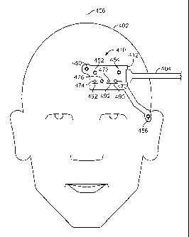
FIG. 4A shows an illustrative combined physiological sensor 410 attached to a
subject 402, in accordance with an embodiment. Combined physiological sensor
410
includes a sensor structure 412, a pulse blood oxygen saturation sensor 490, a
regional blood
oxygen saturation sensor 492, a temperature sensor 476, and a number of EEG
sensors (i.e.,
sensors 450, 452, 454, and 456). While FIGS. 4-7 and the accompanying
description refer to
EEG sensors, it should be understood that the EEG sensors may be any type of
EPS sensors,
including ECG and EMG sensors. Furthermore, while the arrangements depicted in
FIGS. 4-7 show or refer to a combined physiological sensor including four EEG
sensors, in
other arrangements, fewer or more EEG sensors may be included. Similarly, it
should be
understood that fewer or more temperature and PPG sensors (e.g., oximetry or
CNIBP
sensors) may be included in the combined physiological sensors depicted in
FIGS. 4-7.
Returning to the arrangement of FIG. 4A, cable 404 connects to combined
physiological sensor 410 to transmit data generated by the combined
physiological sensor to
a monitor or data storage device (e.g., monitor 14 of FIG. I or output 314 of
FIG. 3).
Although FIG. 4 includes cable 404, it should be understood that a wireless
transmitter (not
shown) or a memory device (e.g., memory 322 of FIG. 3) may be used instead of
a cable to
transmit or store data generated by the combined physiological sensor.
Moreover, these
components (i.e., the wireless communications device or memory device) may be
attached to
or embedded within sensor structure 412 along with the individual sensors of
combined
physiological sensor 410. Cable 404 (or a wireless receiver) may also deliver
signals
generated, for example, by a monitor or other controlling device (e.g., a
sensor hub) to
combined physiological sensor 410. Cable 404 may contain separate sets of
wires or
transmission paths for delivering signals to and receiving signals from
combined
physiological sensor 410. Alternatively, the same set of wires or transmission
paths may be
used for data delivery and reception. The wires or transmission paths in cable
404 may be
composed of conductive and/or fiber optic material. It should be understood
that while the
arrangements depicted in FIGS. 4-7 show or refer to a single cable for
delivering signals to
CA 02787471 2012-07-18
WO 2011/097399
PCT/US2011/023630
and from the combined physiological sensor, in other arrangements, additional
cables may be
provided. For example, a separate cable may be used for each individual sensor
or groups of
sensors (e.g., for each group of PPG, EPS, and temperature sensors) in the
combined
physiological sensor.
Generally speaking, sensor positioning for optimal detection of blood oxygen
saturation and EEG signals from a subject may be different, and may vary
between subjects.
In the arrangement depicted in FIG. 4A, EEG sensor 450 is positioned over the
horizontal
center of the forehead of subject 402, EEG sensors 452 and 454 are positioned
to the side of
EEG sensor 450, and EEG sensor 456 is positioned over the temple of subject
402 closest to
the other EEG sensors. In the depicted arrangement, EEG sensor 456 is shown
incorporated
into the same sensor structure (i.e., structure 412) as the other sensors. In
other arrangements,
however, EEG sensor 456 may part of a second sensor structure (e.g., flex
circuit) that
connects to sensor structure 412 via cable or other flexible connection means,
or EEG sensor
456 may communicate with a wireless receiver (not shown) on sensor structure
412, or within
cable 404, via a wireless transmitter (not shown). In yet other arrangements,
EEG sensor 456
may connect directly to the monitoring station or to an offshoot connector of
cable 404.
In the arrangement depicted in FIG. 4A, emitter 470 and detectors 472 and 474
are
positioned below the EEG sensors, above the eyebrow of subject 402. Emitter
470 and
detectors 472 and 474 may be positioned in alternative locations, although
sites of highly
perfused tissue (such as the forehead, above the eyebrow) are typically best
for measuring
blood oxygen saturation. As shown, pulse blood oxygen saturation sensor 490
includes
emitter 470 and detector 472, while regional blood oxygen saturation sensor
492 also
includes detector 474. In this arrangement, an emitter and two detectors may
be used
combinatorially to integrate the functionality of a pulse oxygen saturation
sensor (i.e., an
Sp02 sensor) and a regional oxygen saturation sensor. In particular, light
from emitter 470 is
transmitted into subject 402 where it reflects off one or more internal
substances (e.g.,
tissues). A portion of this reflected light may be received at detector 472,
and the measured
light intensity can be used to calculate pulse oxygen saturation using, for
example, the ratio
of ratios technique described above or any other suitable technique. A portion
of the
reflected light may also be received at detector 474, and the measured light
intensity can be
used along with the light intensity measured by detector 472 to calculate
regional oxygen
saturation, as described above. In this arrangement, detector 472 serves as
the "close"
detector while detector 474 serves as the "far" detector. It should be
understood that
21
CA 02787471 2012-07-18
WO 2011/097399
PCT/US2011/023630
emitter 470 may include two light emitting sources in order to emit light at
two wavelengths
(e.g., Red and IR).
Emitter 470 and detector 472 may also be used together as a CNIBP sensor in
order to
measure the blood pressure of subject 402. As described above, a single CNIBP
sensor may
be used to measure the blood pressure of subject 402, for example, by
measuring the area
under one or more portions of a pulse signal detected by the CNIBP sensor. In
other
arrangements, a second CNIBP sensor may be disposed in a different location on
subject 402
in order to calculate blood pressure using, for example, the differential
pressure pulse transit
time (DPTT) measurement technique described above. Proper placement of a
second CNIBP
sensor on subject 402, and techniques for connecting the second CNIBP sensor
to sensor
structure 412 or cable 404, is discussed in greater detail below in connection
with FIG. 5.
Combined physiological sensor 410 may also include temperature sensor 476 for
measuring the temperature of subject 402. Temperature sensor 476 may be
located at any
suitable position within sensor structure 412. For example, temperature sensor
476 may be
positioned at a distance from emitter 470, such that heat generated by emitter
470 is not
substantially included in the temperature measured by sensor 476. As another
example,
temperature sensor 476 may be positioned over a site of highly perfused
tissue, such as
directly above the eyebrow of subject 402. In the arrangement depicted in FIG.
4A,
temperature sensor 476 is positioned between detectors 472 and 474. This
arrangement may
2 0 ensure that temperature sensor 476 is sufficiently isolated from heat
generated by emitter 470,
while utilizing the unused space between detectors 472 and 474 - necessary for
measuring
regional blood oxygen saturation - advantageously. Moreover, each of these
devices may be
positioned together over the eyebrow in a highly perfused area.
It should be understood that the locations of the sensors depicted in FIG. 4A,
and the
shape of sensor structure 412, are exemplary and may differ in accordance with
other
arrangements of the sensors, as illustrated in FIGS. 4B-4D. FIG. 413 shows an
illustrative
combined physiological sensor 420 attached to subject 902, in accordance with
an
embodiment. As shown in FIG. 4B, the locations of the various sensors
incorporated into
combined physiological sensor 420 differ with respect to those depicted within
combined
physiological sensor 410 of FIG. 4A. In particular, EEG sensor 452 is
positioned to the
lower right of EEG sensor 450 and EEG sensor 454 is positioned to the lower
right of sensor
452. In order to accommodate the positioning of the EEG sensors in FIG. 413,
sensor
structure 422 is shaped to provide an area for EEG sensor 454 below and to the
right of
22
CA 02787471 2012-07-18
WO 2011/097399
PCT/US2011/023630
emitter 470. In an arrangement, all or some of EEG sensors 450, 452, 454, and
456 are
positioned to form a straight line and/or are equally spaced on subject 402.
FIG. 4C shows an illustrative combined physiological sensor 430 attached to
subject
402, in accordance with an embodiment. In addition to, or instead of,
employing emitter 470
and detector 472 as a CNIBP sensor, a separate CNIBP sensor 494 is included in
sensor
structure 432. CNIBP sensor 494 may include emitter 471 and detector 473, and
may be
positioned over tissue where pulsatility is strong over a wide variety of
perfusion conditions.
For example, as depicted, CNIBP sensor 494 may be positioned directly over the
eyebrow of
subject 402. As described above, a single CNIBP sensor (e.g., sensor 494) may
be used to
measure the blood pressure of subject 402, for example, by measuring the area
under one or
more portions of a pulse signal detected by the CNIBP sensor. As further
described above,
two CNIBP sensors at different locations on a subject's body may be used to
estimate blood
pressure by measuring the amount of time between the arrivals of corresponding
points of a
pulse signal at the two locations. In an arrangement, emitter 470 may be used
in combination
with detector 472 as one CNIBP sensor, while sensor 494 serves as the second
CNIBP
sensor. In another arrangement, combined physiological sensor 430 may include
an
additional CNIBP sensor for measuring blood pressure, as will be discussed
below in
connection with FIG. 5.
As further depicted in FIG. 4C, temperature sensor 476 may be advantageously
2 0 positioned within sensor structure 432 at a site of high perfusion. In
an arrangement,
temperature sensor 476 may be positioned as close as possible to the eyebrow,
subject to the
limitations imposed by the area and shape of sensor structure 432. In
addition, temperature
sensor 476 may be disposed in an area of sensor structure 432 at a distance
from any heat
generating devices, such as emitters 470 and 471.
FIG. 4D shows an illustrative combined physiological sensor 440 attached to
subject
402, in accordance with an embodiment. In the depicted arrangement, oximeter
sensor 490 is
located over an area of high perfusion and pulsatility. Moreover, sensor
structure 442 is
shaped to provide space for EEG sensor 454 to be positioned to the lower right
of EEG
sensor 452 (as in FIG. 4B). A vertical arrangement of emitter 470, detectors
472 and 474,
and temperature sensor 476 is shown that allows emitter 470 and detector 472
to be disposed
over highly perfused tissue. Such placement may better enable use of emitter
470 and
detector 472 as a CNIBP sensor, thereby obviating the need for an additional
CNIBP sensor
on the subject (as in FIG. 4C). Eliminating the need for an additional CNIBP
sensor reduces
cost, combined physiological sensor area, and circuit complexity. The vertical
arrangement
23
CA 02787471 2012-07-18
WO 2011/097399
PCT/US2011/023630
of the emitter, detectors, and temperature sensor may also enable a more
advantageous
arrangement of the EEG sensors. For example, in an arrangement, all or some of
EEG
sensors 450, 452, 454, and 456 are disposed in a straight line and/or are
equally spaced on
subject 402.
FIG. 5 shows a perspective view of a combined physiological sensor 508
attached to
subject 502, in accordance with an embodiment. Combined physiological sensor
508 may be
substantially similar to combined physiological sensor 420 of FIG. 4B or
combined
physiological sensor 440 of FIG. 411 Specifically, sensor structure 510 may be
substantially
similar to sensor structures 422 or 442, and EEG sensors 512 and 514 may be
substantially
similar to, and disposed in the same location as, EEG sensors 454 and 456,
respectively. In
another arrangement, combined physiological sensor 508 may be substantially
similar to
combined physiological sensor 410 of FIG. 4A or combined physiological sensor
430 of
FIG. 4C, and EEG sensor 512 may be located above the location depicted as in
FIGS. 4A
and 4C. Combined physiological sensor 508 may additionally include CNIBP
sensors 516 or
518. Although FIG. 5 depicts both sensors 516 and 518, it should be understood
that, in an
embodiment, combined physiological sensor 508 may include only one of the
sensors.
CNIBP sensor 516 includes emitter 530 and detector 532, both of which may be
incorporated
into sensor structure 510 and disposed behind the ear of subject 502, as
shown. CNIBP
sensor 516 may be used together with another CNIBP sensor (e.g., the
combination of emitter
470 and detector 472 of FIGS. 4A-41, or CNIBP sensor 494 of FIG. 4C) to
determine blood
pressure using CNIBP monitoring techniques. For example, by measuring PPG
signals with
each CNIBP sensor, the arrival of a pulse may be detected at each sensor
location. As
described above, the elapsed time between the arrivals of corresponding points
of a pulse
signal at the two locations may be used to calculate blood pressure.
CNIBP sensor 516 may detect a PPG signal by way of receiving light, produced
by
emitter 530 and reflected from one or more internal substances (e.g., tissues)
of subject 502,
with detector 532. The received light intensity may then be used to determine
a pulse signal.
Although FIG. 5 shows CNIBP sensor 516 as part of sensor structure 510, it may
alternatively be included in a second sensor structure and connected to sensor
structure 510
through a cable or wireless device. In an embodiment, CNIBP sensor 518 may be
used
instead of sensor 516 for blood pressure calculation. CNIBP sensor 518 may be
a clip
configured to attach to or clamp down on, for example, an earlobe. The clip
may include an
emitter and detector on opposite sides, such that light produced by the
emitter may pass
through an area of tissue (e.g., through the earlobe of subject 502) and be
received by the
24
CA 02787471 2012-07-18
WO 2011/097399
PCT/US2011/023630
detector on the other side. The received light intensity may then be used to
determine a pulse
signal. CNIBP sensor 518 may be connected to sensor structure 510 via cable
520, or
through wireless communication.
As described herein, CNIBP sensors are PPG sensors utilized for the purpose of
measuring blood pressure. It should therefore be understood that these same
PPG sensors
may be utilized for any physiological measurement that relies on PPG signals,
such as pulse
and regional blood oxygen saturation measurement. For example, CNIBP sensors
516 and/or
518 may be used to calculate the pulse blood oxygen saturation of the subject,
in addition to
or instead of measuring blood pressure.
in an embodiment, each of the combined physiological sensors depicted in FIGS.
4A-
4D and FIG. 5 may be applied to a subject along with a mirrored version of the
respective
combined physiological sensor. For example, a mirrored version of combined
physiological
sensor 410 of FIG. 4A (less EEG sensor 450) may be applied to subject 402 such
that an
EEG sensor is disposed in a location opposite that of EEG 452, symmetrical
about axis 406
(the horizontal center of the forehead of subject 402). Similarly, the
mirrored version of
combined physiological sensor 410 may be applied such that EEG sensors are
disposed
opposite each of EEG sensors 454 and 456, and an emitter, two detectors, and a
temperature
sensor are disposed opposite emitter 470, detectors 472 and 474, and
temperature sensor 476,
respectively, symmetrical about axis 406. A second cable (or wireless
transmitter/receiver)
may connect the mirrored combined physiological sensor to a monitor, storage
device, or to
the non-mirrored combined physiological sensor. In an embodiment, instead of a
separate
mirrored combined physiological sensor, a single sensor structure may combine
all the
sensors into one combined physiological sensor. For example, sensor structure
412 may be
expanded to incorporate duplicates of sensors 452, 454, 456, 492, and 476,
each duplicate
sensor disposed opposite the corresponding original sensor and symmetrically
about axis 406
(e.g., an EEG sensor corresponding to EEG sensor 456 is disposed on the left
temple of
subject 402). In this embodiment, cable 404 may connect to both the original
and duplicate
sensors.
FIGS. 6A-6C show detailed views of the combined physiological sensors
discussed
above in connection with FIGS. 4A-4D and FIG. 5, in accordance with some
embodiments.
FIG. 6A shows combined physiological sensor 610 including sensor structure 612
and cable
604. Although the depiction resembles that of combined physiological sensor
410 of FIG.
4A, it should be understood that combined physiological sensor 610 may
represent any of the
combined physiological sensors discussed above, or any other suitable combined
CA 02787471 2015-01-06
physiological sensor. In particular, sensor structure 612 may be substantially
similar to sensor
structures 412, 422, 432, or 442 of FIGS. 4A, 4B, 4C, and 4D, respectively,
with corresponding
disposition of the various sensors. As shown, EEG sensors 650, 652, 654, and
656 are connected to
cable 604 through interconnects 660, 662, 664, and 666, respectively.
Interconnects 660, 662, 664,
and 666 may be wires, metallic traces, or other conductive material, and may
be deposited on, or
embedded within, sensor structure 612. Furthermore, emitter 670, detectors 672
and 674, and
temperature sensor 676 are connected to cable 604 through interconnects 680,
682, 684, and 686,
respectively. Interconnects 680, 682, 684, and 686 may be wires, metallic
traces, or other
conductive material, and may be deposited on, or embedded within, sensor
structure 612. Although
shown as single lines, each of interconnects 680, 682, 684, and 686 may
include two or more
distinct interconnects, as required by the corresponding sensor. For example,
emitter 670, detectors
672 and 674, and/or temperature sensor 676 may be two-node devices requiring
two wires, traces,
or other conductive material in order for the sensors to function properly. In
an embodiment,
sensors connected in the "series" circuit topology may be connected to cable
604 through two
interconnects carrying the same current, and sensors connected in the
"parallel" circuit topology
may be connected to cable 604 through two interconnects over which a voltage
may be measured,
one of which may be electrical (or earth) ground.
In an embodiment, emitter 670 and detectors 672 and 674 may be located
remotely from
combined physiological sensor 610 (e.g., at monitor 14 of FIG. 1) and
interconnects 680, 682,
and/or 684 may be fiberoptic lines (or cables), suitable to carry light to and
from combined
physiological sensor 610. For example, light emitted by a remotely located
emitter 670 may be
transmitted through a fiberoptic channel within cable 604 to fiberoptic line
680, through which the
light may travel to an aperture that directs the light into a subject.
Reflected light may, in turn, be
received with fiberoptic lines 682 and 684 and the light may be transmitted
through one or more
fiberoptic channels within cable 604 to remotely located detectors 672 and
674, respectively. The
use of fiberoptics with PPG sensors is described in greater detail in U.S.
Patent App. Pub. No.
2011/0112382.
As depicted in FIG. 6A, the various interconnects may be disposed in a manner
that
reduces crosstalk or interference between the sensors. For example,
interconnects associated with
some or all of the EEG sensors may be disposed at a distance from the
interconnects associated
with the PPG sensors. In an arrangement, an interconnect associated with a
26
CA 02787471 2012-07-18
WO 2011/097399
PCT/US2011/023630
temperature sensor may be located between the interconnects associated with
the EEG
sensors and the interconnects associated with the PPG sensors. In another
arrangement, a
ground line (e.g., wire or metallic trace) may be run between, and thus
divide, the
aforementioned interconnects. In the arrangement shown in FIG. 6A,
interconnects 660,
662, and 664 are disposed in the upper area of sensor structure 612, while
interconnects 680,
682, and 684 are disposed in the lower area of sensor structure 612.
Interconnect 686 is
disposed in the center of sensor structure 612 and separates interconnects
660, 662, and 664
from interconnects 680, 682, and 684. Interconnect 666 is disposed at the
bottom of sensor
structure 612 and is arranged such that a minimum amount of the interconnect
is in proximity
of interconnect 684. Although not shown, ground plates, lines, or traces may
be located
anywhere on or within sensor structure 612, such as between any two
interconnects, to help
mitigate crosstalk and interference.
FIG. 6B shows combined physiological sensor 620 including sensor structure 622
and
cable 604. The shape of sensor structure 622 may be substantially similar to
sensor structure
612 of FIG. 6A, with substantially the same arrangement of sensors. In order
to further
isolate the EEG sensor signals from the PPG sensor signals, conductive or
nonconductive flex
circuit material may be removed from sensor structure 622 such that structure
area 624 is
revealed. Area 624 may be left exposed, such that an internal layer of sensor
structure 622 is
revealed. For example, the top of a foam layer on the underside of sensor
structure 622 may
be exposed. Alternatively, in an embodiment, area 624 may be filled with
electrically
conductive, non-conductive, or isolating material. For example, area 624 may
be filled with a
conductive material connected to electrical ground (e.g., via a ground wire
provided within
cable 604). As shown, area 624 is positioned between the EEG interconnects
(i.e.,
interconnects 660, 662, and 664) and the PPG sensor and temperature sensor
interconnects
(i.e., interconnects 680, 682, 684, and 686). It should be understood,
however, that area 624
may be located in any suitable location for reducing noise and crosstalk
between sensor
signals, and that multiple areas with removed flex circuit material may be
present on sensor
structure 622. For example, flex circuit material may be removed in the area
between
interconnects 684 and 666 in order to isolate the signal associated with
detector 674 from the
signal associated with EEG sensor 656. Moreover, it should be understood that
area 624, and
other similar areas, may be of any suitable shape and size. For instance, as
depicted in FIG.
6B, area 624 may not extend all the way to cable 604, thus ensuring continuity
of the flex
Circuit material of sensor structure 622. Any area of sensor structure 612 not
stripped of flex
circuit material may be used for routing conductive traces or wires (or fiber
optic lines), or to
27
CA 02787471 2012-07-18
WO 2011/097399
PCT/US2011/023630
mount devices. For example, a cable connector for linking cable 604 to the
sensors and
interconnects of the combined physiological sensor may be embedded within or
mounted on
an area of the sensor structure (e.g., structure 622) that contains flex
circuit material.
As discussed above, area 624 may be formed by scraping, cutting, or otherwise
removing flex circuit material from sensor structure 622. Alternatively, flex
circuit material
may have never been placed or deposited in the location of area 624. For
example, flex
circuit material of the shape shown in FIG. 6B (i.e., the shape of sensor
structure 622 less
area 624) may have been fabricated initially. It should be understood that any
suitable
technique for removing or preventing flex circuit material from disposition in
area 624 may
be used in accordance with the present disclosure.
FIG. 6C shows combined physiological sensor 630 including sensor structures
632
and 634, as well as Y-cable 606. In the depicted embodiment, EEG sensors 650,
652, and
654 are incorporated into sensor structure 632, while emitter 670, detectors
672 and 674, and
temperature sensor 676 are incorporated into sensor structure 634. The use of
two sensor
structures may enable a high degree of isolation between the EEG sensor
signals on the one
hand, and the PPG and temperature sensor signals on the other. In addition,
the level of
isolation may be increased or decreased by varying the gap 640 between sensor
structures
632 and 634. As shown, Y-cable 606 may include cable heads 603 and 605 for
connection to
the sensors of sensor structures 632 and 634, respectively. In particular,
cable head 603 may
receive signals from EEG sensors 650, 652, and 654 through interconnects 660,
662, and 664,
respectively, Cable head 605 may receive signals from emitter 670, detectors
672 and 674,
and temperature sensor 676 through interconnects 680, 682, 684, and 686,
respectively. In
addition to enhanced isolation, the use of multiple sensor structures further
allows targeted
placement of the sensors on a subject. For example, the EEG sensors on sensor
structure 632
may be placed in the most suitable location for EEG signal measurement (e.g.,
centered on a
subject's forehead), while the PPG sensors on sensor structure 634 may be
placed in the most
suitable location for oxygen saturation or blood pressure measurement (e.g.,
above the
eyebrow on subject's forehead, in an area of high pulsatility). EEG sensor 656
and
interconnect 666 may be incorporated into sensor structure 634 and connected
to cable head
605, as shown. It should be understood, however, that in other arrangements,
sensor 656 and
element 666 may be incorporated into sensor structure 632 and connected to
cable head 603,
or they may be incorporated into a third sensor structure connected directly
to cable 606 (e.g.,
through a third cable head).
28
CA 02787471 2012-07-18
WO 2011/097399
PCT/US2011/023630
FIGS. 7A and 7B show top and bottom views, respectively, of a combined
physiological sensor 710 in accordance with an embodiment. Although combined
physiological sensor 710, as depicted, resembles that of combined
physiological sensor 410
of FIG. 4A, it should be understood that combined physiological sensor 710 may
represent
any of the combined physiological sensors discussed above (e.g., in connection
with FIGS.
4A-4D and FIG. 5), or any other suitable combined physiological sensor. In
particular,
sensor structure 712 may be substantially similar to sensor structures 412,
422, 432, or 442 of
FIGS. 4A, 4B, 4C, and 4D, respectively, with corresponding disposition of the
various
sensors. FIG. 7A shows top surface 714 of sensor structure 712 included within
combined
physiological sensor 710. EEG sensors 750, 752, 754, and 756, emitter 770,
detectors 772
and 774, and temperature sensor 776 may be embedded within or mounted on
sensor
structure 712. In an embodiment, a portion of EEG sensors 750, 752, 754, and
756 may be
exposed and/or accessible both at top surface 714 of FIG. 7A and at bottom
surface 716 of
FIG. 7B. For example, the tops of the EEG sensors may be substantially flush
with (or may
slight protrude from) top surface 714 and/or the bottoms of the EEG sensors
may be
substantially flush with (or may slight protrude from) bottom surface 716. In
another
embodiment, the EEG sensors may be exposed and/or accessible only at bottom
surface 716
of FIG. 7B, in which case the depictions of the EEG sensors in FIG. 7A simply
indicate the
locations of the EEG sensors embedded within sensor structure 712 or mounted
on bottom
surface 716 of FIG. 7B, Emitter 770, detectors 772 and 774, and temperature
sensor 776
may be connected to cable 704 through interconnects 780, 782, 784, and 786,
respectively.
Interconnects 780, 782, 784, and 786 may be wires, metallic traces, or other
conductive
material, and may be deposited on, or embedded within, top surface 714 of
sensor structure
712. Although shown as single lines, each of interconnects 780, 782, 784, and
786 may
include two or more distinct interconnects, as required by the corresponding
sensors, one of
which may be connected to electrical (or earth) ground (e.g., via a ground
wire provided
within cable 704). Alternatively, as discussed above, interconnects to the PPG
sensors (e.g.,
interconnects 780, 782, and 784) may be fiber optic lines configured to
transfer light between
combined physiological sensor 710 and remotely located emitters and detectors
(e.g.,
remotely located emitter 770 and detectors 772 and 774).
FIG. 7B shows bottom surface 716 of sensor structure 712 included within
combined
physiological sensor 710. EEG sensors 750, 752, 754, and 756 may be connected
to cable
704 through interconnects 760, 762, 764, and 766 of FIG. 7B - which may
deposited on, or
embedded within, bottom surface 716 - and interconnects 761, 763, 765, and 767
of FIG. 7A
29
CA 02787471 2012-07-18
WO 2011/097399
PCT/US2011/023630
- which may be deposited on, or embedded within, top surface 714.
Specifically,
interconnects 760, 762, 764, and 766 may be connected, through structure 712,
to
interconnects 761, 763, 765, and 767, respectively, such that each pair of
interconnects forms
a single conductive line between an EEG sensor and cable 704. In this
arrangement,
interconnects 760, 762, 764, and 766 can be separated from interconnects 780,
782, 784, and
786, thus preventing a measure of cross-talk between the PPG and temperature
sensor signals
carried via the interconnects on top surface 714 and the EEG signals carried
via the
interconnects on bottom surface 716. As depicted, interconnects 761, 763, 765,
and 767 on
top surface 714 enable interconnects 760, 762, 764, and 766 on bottom surface
716 to access
cable 704, which may connect to sensor structure 712 by way of a cable
connector mounted
on top surface 714. Interconnects 761, 763, 765, and 767 may be wires,
metallic traces, or
other conductive material, and may be deposited on, or embedded within, top
surface 714 of
sensor structure 712. Interconnects 760, 762, 764, and 766 may be wires,
metallic traces, or
other conductive material, and may be deposited on, or embedded within, bottom
surface 716
of sensor structure 712.
As described above, disposing the interconnects associated with the EEG
sensors and
the interconnects associated with the PPG and temperature sensors on opposite
sides of
sensor structure 712 may facilitate the reduction of noise and cross-talk
affecting the sensor
signals. In addition, sensor structure 712 may include an intervening layer -
between top
surface 714 and bottom surface 716 - to further reduce interference between
the two sets of
signals. For example, the intervening layer may be a non-conductive or
dielectric material, or
the intervening layer may be connected to electrical ground (e.g., via a
ground wire provided
within cable 704). Furthermore, the length of interconnects 761, 763, 765, and
767 may be
minimized to reduce the amount of conductive material associated with the EEG
sensors
located on top surface 714. It should be understood that the techniques
illustrated in FIGS.
6A-6C and FIG. 7 are not exclusive, but intended to describe methods of
reducing noise,
interference, cross-talk, or other signal corruptions that occur as a result
of proximity between
the various sensors and interconnects. As such, modifications to the
positioning of the
individual sensors, interconnects, and the combined physiological sensor
structure that further
accomplish noise reduction are within the scope of this disclosure. For
example, referring to
FIG. 7A, interconnects 761, 763, 765, and 767 need not be disposed lengthwise
on top
surface 714, but may travel upwards from bottom surface 716 towards surface
714, up
directly beneath cable 704 (or a cable connector for attaching cable 704).
Alternatively,
interconnects 761, 763, 765, and 767 may not be present and interconnects 760,
762, 764, and
CA 02787471 2012-07-18
WO 2011/097399
PCT/US2011/023630
766 may connect directly to cable 704 (or a cable connector for attaching
cable 704) on
bottom surface 716.
FIG. 8 shows an illustrative timing diagram 800 for activating the various
sensors of
a combined physiological sensor, in accordance with an embodiment. In
particular, timing
diagram 800 shows a repeating pattern of three time intervals, wherein a
different sensor set
is active during each of the time intervals. For example, in first time
interval 812, PPG
sensors 802 may be active, including pulse and regional oxygen saturation
sensors and/or
CNIBP sensors. The sensors may include emitters and detectors, such as emitter
770 and
detectors 772 and 774 of FIG. 7. In second time interval 814, BPS sensors 804
may be
active, including EEG sensors such as sensors 750, 752, 754, and 756 of FIG.
7. In third
time interval 816, temperature sensor 806 may be active, such as sensor 776 of
FIG. 7.
Although each of time intervals 812, 814, and 816 are shown having the same
duration, it
should be understood that the duration of each may vary as suitable for each
particular set of
sensors to successfully obtain and/or transmit signal measurements.
Specifically, the
duration of each time interval may be the time required for the particular
sensors in the
corresponding sensor set to accurately process a measurement and/or to
transmit a signal.
For example, the duration of time interval 812 may be the time required for a
PPG signal to
be measured by PPG sensors 802 and/or transmitted to a data collection or
transmission
device, such as a memory chip, cable, or patient monitor. In addition,
although timing
diagram 800 includes a period of time between each time interval, in another
embodiment,
each time interval may immediately follow the previous time interval or two or
more time
intervals may overlap (e.g., partly or completely).
It should be understood that the durations, spacing or overlap, and order of
time
intervals 812, 814, and 816 in timing diagram 800 may be configured in any
manner that
reduces, prevents, or limits interference between signals generated by PPG
sensors 802, BPS
sensors 804, and temperature sensors 806. For example, time interval 816 may
occur before
time interval 814. As another example, time interval 816 may span virtually
the entire time
axis (i.e., temperature sensor 806 may be active at all times), while time
intervals 812 and
814 alternate continuously. It should also be understood that additional time
intervals may be
included during which sensors 802, 804, 806, other sensors, or a combination
thereof are
active. For example, PPG sensors 802 may be active both during time interval
812 and in
another time interval occurring between time intervals 814 and 816. As another
example, one
(or more) of PPG sensors 802 may be active during time interval 812 while
another one (or
31
CA 02787471 2012-07-18
WO 2011/097399
PCT/US2011/023630
more) of PPG sensors 802 may be active in another time interval (e.g.,
subsequent to time
interval 802 but before time interval 814).
Activation of sensors 802, 804, and 806 may be controlled by a patient monitor
or
other sensor control device (e.g., within the combined physiological sensot).
For example, as
discussed above, TPU 58, within monitor 14, of FIG. 2 may be connected
directly, or
through other control circuitry, to each of the sensors of the combined
physiological sensor.
In such an arrangement, TPU 58 may control the timing and duration of each
sensor's
activity. Specifically, TPU 58 may enable and disable sensors 802, 804, and
806 in
accordance with timing diagram 800 of FIG. 8. In an embodiment, a sensor
control device
attached to, or embedded within, the combined physiological sensor may enable
and disable
sensors 802, 804, and 806 in accordance with timing diagram 800 of FIG. 8. For
example,
combined physiological sensor 710 of FIGS. 7A and 7B may contain a sensor
control device
connected between the connection paths (e.g., paths 761-767 and 780-786) and
cable 704. As
another example, cable 704 may itself contain a sensor control device that
activates or
deactivates each of the connection paths connected to the cable. As yet
another example, a
sensor control device may reside between a cable (or wireless receiver) and a
patient monitor
to which the cable or wireless receiver) is connected. In an embodiment, both
a patient
monitor and sensor control device may be used to control sensors 802, 804, and
806 in
accordance with timing diagram 800 of FIG. 8. For example, a patient monitor
may issue
requests for various measurements from the sensors (in sequence or in
parallel), and a sensor
control device may receive, process, and/or store the requests and may
activate and deactivate
the sensors in accordance with timing diagram 800. In one case, for instance,
a patient
monitor may request PPG and BPS measurements in one instant and a temperature
measurement the next instant. The sensor control device may receive these
requests and
activate the corresponding sensors in the proper sequence and with the proper
spacing/overlap and duration, as determined by timing diagram 800.
Timing diagram 800 of FIG. 8, and in particular the order, spacing, and
durations of
time intervals 812, 814, and 816, may be predetermined and stored as data or
executable
instructions in memory (e.g., in memory 322 of FIG. 3), which may be accessed
by
processing circuitry (e.g., TPU 58 of FIG. 2 or processor 312 of FIG. 3) to
properly time the
activation of the sensors. Alternatively, the order, spacing, and durations of
time
intervals 812, 814, and 816 may be implemented directly as part of the
processing circuitry
(e.g., TPU 58 or a sensor control device). In an embodiment, timing diagram
800, and in
particular the order, spacing, and/or durations of time intervals 812, 814,
and 816, are not
32
CA 02787471 2012-07-18
WO 2011/097399
PCT/US2011/023630
predetermined. In one approach, each time interval may continue until one or
more of the
corresponding sensors successfully detects, measures, and/or processes a
signal, or until one
or more of the corresponding sensors produces a sensor output signal and/or
transmits the
output signal to a storage or other transmission device. For example, time
interval 812 may
be initiated (i.e., PPG sensors 802 may be activated) and may continue until a
signal is
successfully detected, measured, processed, and/or a sensor output signal is
transmitted by
PPG sensors 802. In some cases, there may be a maximum limit to the duration
of a time
interval so that all sets of sensors may eventually operate even if one or
more sets of sensors
fail to detect or measure a signal. For example, if PPG sensors 802 fail to
measure a PPG
signal within a certain amount of time (predetermined or calculated on the
fly) of the onset of
time interval 812, time interval 812 may end and time interval 814 may begin.
In other cases,
a time interval may be cut short by the initiation of another time interval.
For example, time
interval 814 may be configured to continuously occur at a defined frequency
and time
intervals 812 and 814 may continue (if necessary) until the onset of time
interval 814. As
another example, a patient or other operator may manually request a reading
from one of the
sensor sets and, upon reception of the request, any active sensors not within
the requested
sensor set may be disabled and the requested sensor set may be enabled. In
other words,
upon reception of a manual request, all time intervals may be ended
immediately and the time
interval corresponding to the requested sensor set may begin. For instance,
when an operator
requests an Sp02 measurement during time interval 814 or 816, the current time
interval may
end and time interval 812 may begin.
The terms activation/deactivation or enablement/disablement of sensors, as
used
herein, should be understood to mean any of providing/severing power to a
sensor,
providing/severing a data signal to a sensor, and/or allowing/disallowing
capture or analysis
of a sensor output signal. In addition, it should be understood that while the
description
above referred to enabling/disabling all sensors in a sensor set (e.g., all
PPG sensors 802 or
all BPS sensors 804), less than all the sensors in a sensor set may be
accordingly
enabled/disabled. For example, EEG sensor 756 of FIG. 7 may remain active even
when
EEG sensors 750, 752, and 754 are deactivated (e.g., insofar as EEG sensor 756
may not
interfere with any of the PPG or temperature sensors, even when active).
Lastly, it should be
understood that sensor activation/deactivation may be controlled by any
suitable system or
apparatus configured to execute machine-readable (or computer-readable)
instructions stored
on a machine-readable (or computer-readable) medium.
33
CA 02787471 2015-01-06
The foregoing is merely illustrative of the principles of this disclosure and
various
modifications may be made by those skilled in the art without departing from
the scope of the
disclosure.
34