Note: Descriptions are shown in the official language in which they were submitted.
CA 02787494 2017-01-19
TARGETED GENOMIC ALTERATION
[0001]
[0002]
TECHNICAL FIELD
[0003] The present disclosure is in the field of genomic engineering,
particularly targeted integration and/or targeted excision of one or more
exogenous
sequences into the genome of a cell.
BACKGROUND
[0004] Biotechnology has emerged as an essential tool in efforts to
meet the
challenge of increasing global demand for food production. Conventional
approaches
to improving agricultural productivity, e.g. enhanced yield or engineered pest
resistance, rely on either mutation breeding or introduction of novel genes
into the
genomes of crop species by transformation. Both processes are inherently
nonspecific
and relatively inefficient. For example, conventional plant transformation
methods
deliver exogenous DNA that integrates into the genome at random locations.
Thus, in
order to identify and isolate transgenic lines with desirable attributes, it
is necessary to
generate thousands of unique random-integration events and subsequently screen
for
the desired individuals. As a result, conventional plant trait engineering is
a
laborious, time-consuming, and unpredictable undertaking. Furthermore the
random
nature of these integrations makes it difficult to predict whether pleiotropic
effects
due to unintended genome disruption have occurred. As a result, the
generation,
isolation and characterization of plant lines with engineered genes or traits
has been
an extremely labor and cost-intensive process with a low probability of
success.
[0005] Targeted gene modification overcomes the logistical challenges
of
conventional practices in plant systems, and as such has been a long-standing
but
elusive goal in both basic plant biology research and agricultural
biotechnology.
However, with the exception of "gene targeting" via positive-negative drug
selection
in rice or the use of pre-engineered restriction sites, targeted genome
modification in
all plant species, both model and crop, has until recently proven very
difficult. Terada
et al. (2002) Nat Biotechnol 20(10):1030; Terada et al. (2007) Plant Physiol
144(2):846; D'Halluin et al. (2008) Plant Biotechnology J. 6(1):93.
1
[0006] In mammalian cells, stable transgenesis and targeted gene
insertion
have many potential applications in both gene therapy and cell engineering.
However, current strategies are often inefficient and non-specifically insert
the
transgene into genomic DNA. The inability to control the location of genome
insertion can lead to highly variable levels of transgene expression
throughout the
population due to position effects within the genome. Additionally, current
methods
of stable transgenesis and amplification of transgenes often result in
physical loss of
the transgene, transgene silencing over time, insertional mutagenesis by the
integration of a gene and autonomous promoter inside or adjacent to an
endogenous
gene, the creation of chromosomal abnormalities and expression of rearranged
gene
products (comprised of endogenous genes, the inserted transgene, or both),
and/or the
creation of vector-related toxicities or immunogenicity in vivo from vector-
derived
genes that are expressed permanently due to the need for long-term persistence
of the
vector to provide stable transgene expression.
[0007] Recently, methods and compositions for targeted cleavage of genomic
DNA have been described. Such targeted cleavage events can be used, for
example,
to induce targeted mutagenesis, induce targeted deletions of cellular DNA
sequences,
and facilitate targeted recombination at a predetermined chromosomal locus.
See, for
example, United States Patent Publications 20030232410; 20050208489;
20050026157; 20050064474; and 20060188987, and International Publication
WO 2007/014275. U.S. Patent Publication No. 20080182332 describes use of non-
canonical zinc finger nucleases (ZFNs) for targeted modification of plant
genomes
and U.S. Patent Publication No. 20090205083 describes ZFN-mediated targeted
modification of a plant EPSPS locus. In addition, Moehle et al. (2007) Proc.
Natl.
Acad, Sci. USA 104(9): 3055-3060) describe using designed ZFNs for targeted
gene
addition at a specified locus.
[0008] However, there remain needs for compositions and methods for
targeted integration, including for targeted integration into plants for
establishing
stable, heritable genetic modifications in the plant and its progeny, and for
target
integration into mammalian cells for gene therapy and cell line development
purposes.
2
CA 2787494 2017-12-28
CA 02787494 2017-01-19
SUMMARY
[0008a] Certain exemplary embodiments provide an isolated cell or cell
line
comprising an endogenous genome and an exogenous nucleic acid integrated into
the
endogenous genome, the exogenous nucleic acid comprising a non-coding sequence
comprising three or more different paired target sites for one or more pairs
of zinc
finger nucleases, wherein the paired target sites are not present in the
endogenous
genome and further wherein upon cleavage by a pair of zinc finger nucleases
that
binds to one of the paired target sites, a donor sequence is integrated into
the genome
in place of the paired target site.
[0008b] Other exemplary embodiments provide use of an isolated cell or cell
line comprising an endogenous genome and an exogenous nucleic acid integrated
into
the endogenous genome, the exogenous nucleic acid comprising a non-coding
sequence comprising three or more different paired target sites for one or
more pairs
of zinc finger nucleases, wherein the paired target sites are not present in
the
endogenous genome and further wherein upon cleavage by a pair of zinc finger
nucleases that binds to one of the paired target sites, a donor sequence is
integrated
into the genome in place of the paired target site.
[0009] The present disclosure provides methods and compositions for
expressing one or more products of an exogenous nucleic acid sequence (i.e. a
protein
or a RNA molecule) that has been integrated into a multiple insertion site
integrated
into a cell genome. The cell can be a eukaryotic cell, for example a plant,
yeast or
mammalian cell.
[0010] Integration of exogenous nucleic acid sequences is facilitated
by
genomic integration of a polynucleotide sequence comprising multiple target
sites for
one or more nucleases, for example zinc finger nucleases (ZFNs) into the
cell's
genome. The polynucleotides (also referred to herein as a multiple insertion
site)
allows for specific, targeted double-strand cleavage within the cell's genome,
which
double-stranded cleavage in turn results in integration of the exogenous
sequence(s)
through both homology-dependent and homology-independent mechanisms.
[0011] Thus, in one aspect, disclosed herein are nucleic acid molecules,
also
known as multiple insertion sites, comprising one or more target sites for
nucleases
such as zinc finger nucleases (ZFNs). In certain embodiments, the target sites
are not
present in the endogenous genome into which the multiple insertion site is
integrated.
The multiple insertion site may include one, two, three, four, five, six,
seven or more
3
CA 02787494 2017-01-19
target sites for nucleases. In certain embodiments, dimerization of the
cleavage-half
domains of two binding DNA-binding proteins that bind to adjacent target sites
(paired target sites) is required for cleavage (e.g., a pair of nucleases, one
binding to
each site, is required for cleavage). In any of the multiple insertion sites
described
herein, one target site of each pair of target sites may comprise the same
sequence.
See, e.g., Figure 1. In certain embodiments, the target sites of at least one
pair are the
same. In other embodiments, at least one pair of target sites comprises
individual
target sequences from different targets (e.g., different genes and/or genes
from
different organisms). In certain embodiments, at least one of the paired
target sites
3a
CA 02787494 2012-07-18
WO 2011/090804
PCT/US2011/000125
comprise a sequence selected from the group consisting of SEQ ID NOs: 1-20. In
certain embodiments, the multiple insertion site may include one more coding
sequences, for example a plant transcription unit (PTU) comprising a
phosphinothricin acetyl transferase (PAT) coding sequence, or a screening
marker for
use with mammalian cells.
[0012] The multiple insertion sites are integrated into the genome of
a cell
(e.g., plant or mammalian cell) to provide genomic targets for the nucleases
(e.g.,
ZFNs). In certain embodiments, the target sites are situated such that one or
more
pairs of the zinc finger nucleases bind and cleave as homodimers. In other
embodiments, the target sites are situated such that one or more pairs of the
zinc
finger nucleases bind and cleave as heterodimers.
[0013] In another aspect, disclosed herein are plants or seeds
comprising one
or more multiple insertion sites as described herein and/or one or more
exogenous
sequences integrated into the multiple insertion site. In certain embodiments,
the
multiple insertion site and/or exogenous sequence(s) is(are) integrated into
the
gametophyte of a maize plant.
[0014] In certain aspects, provided herein are modified mammalian cell
lines,
modified primary cells, modified stem cells and/or transgenic animals
comprising one
or more multiple insertion sites as described herein and/or one or more
exogenous
sequences integrated into the multiple insertion site.
[0015] In another aspect, provided herein is a method for integrating
an
exogenous sequence into the multiple insertion site integrated into the genome
of a
cell (e.g., plant or mammalian cell), the method comprising: (a) integrating a
multiple
insertion site polynucleotide comprising one or more target sites for
nucleases into the
genome of the cell; (b) providing and/or expressing one or more nucleases that
bind to
a first target site in the multiple insertion site polynucleotide, such that
binding of the
nuclease(s) to their target sites cleaves the genome of the cell; and (c)
contacting the
cell with a polynucleotide comprising an exogenous nucleic acid sequence,
thereby
resulting in homology dependent integration of the exogenous sequence into the
genome of the cell within the multiple insertion site polynucleotide.
[0016] In another aspect, provided herein is a method for integrating
multiple
exogenous sequences into the genome of a cell (e.g., a plant or mammalian
cell), the
method comprising: (a) integrating a first multiple insertion site
polynucleotide
comprising one or more target sites for nucleases into the genome of the cell,
wherein
4
CA 02787494 2012-07-18
WO 2011/090804
PCT/US2011/000125
the first multiple insertion site polynucleotide comprises at least one first
gene flanked
by target sites for first and second nucleases; and (b) expressing the first
or second
nuclease in the cell in the presence of a second multiple insertion site
polynucleotide
comprising at least one second gene flanked by target sites for third and
fourth
nucleases, thereby resulting in integration of the first and second genes into
the
genome of the cell. In certain embodiments, the method further comprises
repeating,
one or more times, the step of expressing the appropriate nucleases present on
the
inserted multiple insertion sites to integrate additional exogenous sequences,
including coding sequences and/or nuclease sites. The nucleases may be
heterodimeric ZFNs and there may be one monomer in common as between one or
more of the nucleases. In some embodiments, the exogenous DNA sequence for
insertion may comprise a ZFN half target site such that upon integration of
the
exogenous sequence, a novel ZFN target site is created comprising the half
target site
associated with the donor DNA, and a half target site associated with the
genomic
DNA. This novel ZFN target site can serve as a target site for a similarly
novel
heterodimeric ZFN.
[0017] In another aspect, disclosed herein is a method for expressing
the
product of one or more exogenous nucleic acid sequences in a cell (e.g., plant
or
mammalian cell), the method comprising: integrating one or more exogenous
nucleic
acid sequences according to any of the methods described herein, such that the
exogenous sequence is integrated into the genome of the cell in the integrated
nucleic
acid molecule and the product of the exogenous sequence is expressed.
[0018] Also provided is a method of deleting one or more genes
inserted into
the genome of a cell, the method comprising, integrating a plurality of
exogenous
sequences by any of the methods described herein and expressing the
appropriate
nucleases in the cell such that one or more of the exogenous sequences are
deleted
from the genome. In certain embodiments, the exogenous sequences deleted are
marker genes. In certain embodiments, the deletion of the exogenous sequence
and
the subsequent re-joining of the ends within the genome creates a functional
gene or
sequence in the genomic location, e.g. the creation of an expressible
screening
marker.
[0019] In yet another aspect, a method of providing a genomically
altered cell
is provided, the method comprising integrating ancUor excising one or more
exogenous nucleic acid sequences in a first cell according to any of the
methods
5
CA 02787494 2012-07-18
WO 2011/090804
PCT/US2011/000125
described herein, allowing the first cell to develop into a first sexually
mature
organism, crossing the organism with a second organism comprising genomic
alterations at an allelic position to generate a second cell with the genomic
alterations
of first and second organisms. In certain embodiments, the organism(s) is(are)
plants.
In other embodiments, the organism(s) is/are transgenic animals.
[0020] In any of the methods described herein, the methods may be
used in
combination with other methods of genomic alteration, including targeted
integration
and/or targeted inactivation at one or more endogenous loci. Furthermore, in
any of
the methods described herein, the nuclease may comprise one or more fusion
proteins
comprising a zinc finger binding domain and a cleavage half-domain, wherein
the
zinc finger binding domain has been engineered to bind to a target site in the
multiple
insertion site. Furthermore, in any of these methods, the exogenous nucleic
acid
sequence comprises one or more sequences that is (are) homologous to the
sequences
in multiple insertion site and/or endogenous sequences in the region where the
.. multiple insertion site is integrated.
[0021] In any of the methods described herein, the one or more
multiple
insertion sites may be integrated into the genome by any suitable method, for
example, by targeted integration via a nuclease (e.g., ZFN) using ZFNs that
target the
endogenous gene into which insertion is desired. Alternatively, the one or
more
multiple insertion sites may be randomly integrated into the cell's genome,
using
standard techniques.
[0022] The exogenous nucleic acid sequence may comprise a sequence
encoding one or more functional polypeptides (e.g., a cDNA), with or without
one or
more promoters and/or may produce one or more RNA sequences (e.g., via one or
more shRNA expression cassettes), which impart desirable traits to the
organism.
Such traits in plants include, but are not limited to, herbicide resistance or
tolerance;
insect resistance or tolerance; disease resistance or tolerance (viral,
bacterial, fungal,
nematode); stress tolerance and/or resistance, as exemplified by resistance or
tolerance to drought, heat, chilling, freezing, excessive moisture, salt
stress; oxidative
stress; increased yields; food content and makeup; physical appearance; male
sterility;
drydown; standability; prolificacy; starch quantity and quality; oil quantity
and
quality; protein quality and quantity; amino acid composition; and the like.
Of
course, any two or more exogenous nucleic acids of any description, such as
those
conferring herbicide, insect, disease (viral, bacterial, fungal, nematode) or
drought
6
CA 02787494 2012-07-18
WO 2011/090804
PCT/US2011/000125
resistance, male sterility, drydown, standability, prolificacy, starch
properties, oil
. quantity and quality, or those increasing yield or nutritional quality
may be employed
as desired. In certain embodiments, the exogenous nucleic acid sequence
comprises a
sequence encoding a herbicide resistance protein (e.g., the AAD
(aryloxyalkanoate
dioxygenase) gene) and/or functional fragments thereof. Expression of the
integrated
sequence can be driven by a promoter operably linked to the integrated
sequence.
Alternatively, the integrated sequence is promotorless and transcription is
driven by
the endogenous promoter in the region of insertion of the multiple insertion
site
polynucleotide. In other embodiments, the cleavage and imprecise repair of a
binding
site may inactivate or activate genes of interest. In certain embodiments, the
polynucleotide is a plasmid. In other embodiments, the polynucleotide is a
linear
DNA molecule.
[0023] In mammalian cells, the methods and compositions of the
invention
may be used for cell line construction, e.g. for the construction of cell
lines expressing
multimeric polypeptides such as antibodies. In some embodiments, the cell
lines may
be used fol research purposes, e.g. for the construction of cell lines
expressing
members of a pathway of interest. In some embodiments, primary cells or stem
cells
may be used to express multimeric proteins of interest for cell therapeutic
purposes.
[0024] In another aspect, provided herein are methods of measuring
zinc
finger nuclease activity. In certain embodiments, the methods comprise: (a)
providing
at least one zinc finger nuclease and a nucleic acid molecule as described
herein,
wherein each of the paired target sites comprises two zinc finger nuclease
half target
sites to which the zinc finger nuclease binds, and a cut site that is cut by
the bound
zinc finger nuclease, which cut site is interposed between the half target
sites; (b)
combining the zinc finger nuclease with the nucleic acid such that the zinc
finger
nuclease cleaves the paired target site at least within the cut site;(c)
sequencing at
least the cut site to generate sequence data; and (d) comparing in the
sequence data
the number and length of base pair deletions within the cut site to the number
and
length of base pair deletions within the cut site in the absence of the zinc
finger
nuclease, to thereby measure the zinc finger nuclease activity at the paired
target sites.
In certain embodiments, a deletion of more than one base pair indicates
increased
activity of the zinc finger nuclease(s).
[0025] In yet other embodiments, provided herein are methods for
optimizing
zinc finger nuclease activity at a paired target site. In certain embodiments,
the
7
CA 02787494 2012-07-18
WO 2011/090804
PCT/US2011/000125
methods comprise (a) providing at least one zinc finger nuclease and a nucleic
acid
molecule as described herein, wherein each of the paired target sites
comprises two
zinc finger nuclease half target sites to which the zinc finger nuclease
binds, and a cut
site that is cut by the bound zinc finger nuclease, which cut site is
interposed between
the half target sites; (b) combining the one or more zinc finger nucleases
with the
nucleic acid such that the zinc finger nuclease cleaves the paired target site
at least
within the cut site; (c) determining the zinc finger nuclease activity level
at the cut
site; (d) varying the number of base pairs in the cut site; (e) repeating
steps (b)-(d) a
plurality of times; and (f) selecting the cut site for incorporation into the
nucleic acid,
which comprises the number of base pairs providing the highest level of zinc
finger
nuclease activity, thereby optimizing zinc finger nuclease activity at the
paired target
site.
[0026] In any of the methods described herein involving zinc finger
nucleases,
the first and second cleavage half-domains are from a Type IIS restriction
endonuclease, for example, Fokl or StsI. Furthermore, in any of the methods
described herein, at least one of the fusion proteins may comprise an
alteration in the
amino acid sequence of the dimerization interface of the cleavage half-domain,
for
example such that obligate heterodimers of the cleavage half-domains are
formed.
[0027] In any of the methods described herein, the plant cell can
comprise a
monocotyledonous or dicotyledonous plant cell. In certain embodiments, the
plant
cell is a crop plant, for example maize. In certain embodiments, the cell can
comprise
a mammalian cell such as a primary cell, a cell line, or a stem cell. In some
embodiments, the mammalian cell line can be used for the production of
polypeptides
of interest.
BRIEF DESCRIPTION OF THE DRAWINGS
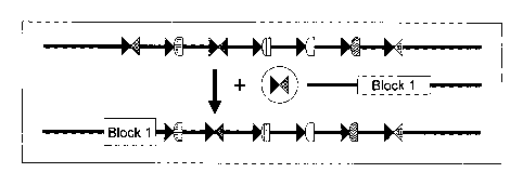
[0028] Figure 1 is a schematic depicting exemplary multiple insertion
site as
described herein. Figure 1 shows a multiple insertion site made up of 7 ZFN
target
sites. The ZFN pairs that bind to the target sites are depicted as geometric
figures.
"Block 1" is an exogenous sequence that is integrated into the multiple
insertion site
in the presence of the appropriate ZFN pair, while maintaining the ZFN target
sites
(shaded and checkered triangles). Figure 1 shows integration of "Block 1" into
a
multiple insertion site in the presence of the appropriate ZFN pair in place
of the ZFN
target sites.
8
CA 02787494 2012-07-18
WO 2011/090804
PCT/US2011/000125
[0029] Figure 2 is a schematic depicting the exemplary multiple
insertion site
as shown in Figure 1 in which "Block 2" is an exogenous sequence that is
integrated
into the multiple insertion site in the presence of the appropriate ZFN pair.
[0030] Figure 3 is a schematic of inter-allelic recombination
enhanced by
ZFNs. Two inserts at an identical genomic location, but are displaced from
each
other, can undergo homologous recombination or strand exchange after double-
stranded cleavage by a ZFN. The ZFN pair (with both ZFN monomers expressed
together) can be provided by crossing a plant expressing the ZFN pair with
plants
comprising both alleles together or by introducing the two ZFN monomers from
both
sides of a cross with plants containing a single allele.
[0031] Figure 4 is a schematic depicting the use of heterodimeric ZFN
"left"
and "right" target domains. The top line depicts the genome with the left and
right
target ZFN domains (shaded triangle and checkerboard triangle). When the
appropriate ZFN pair is added in the presence of an exogenous molecule
including a
gene flanked by different heterodimeric pairs, the gene and flanking nuclease
sites are
,inserted into the genome as shown.
[0032] Figure 5 is a schematic depicting integration and excision of
exogenous sequences (depicted as "genes") on either side of a genomically-
integrated
sequence. The added genes are flanked by regions of homology to direct the
gene
cassettes into the appropriate site. The two halves of the ZFN target site
used for
insertion are re-combined by creating two new combinations in the inserted
DNA.
Excision of a gene cassette is accomplished by binding the appropriate ZFN
pairs to
cleave at the flanking ZFN target sites. Excision may require a template
containing
homology arms to prevent deletions of desired DNA sequence. Each "gene" can
include one or more sequences, for example one or more coding sequences.
[0033] Figure 6 is a schematic depicting excision and "recycling" of
inserted
marker genes using ZFNs heterodimers (depicted as triangles with different
shadings).
[0034] Figure 7 is a plasmid map of pDAB105900.
[0035] Figure 8 is a plasmid map of pDAB105908.
[0036] Figure 9 is a diagram of the Zinc Finger Nuclease Homodimer
expression cassette.
[0037] Figure 10 is a diagram of the Zinc Finger Nuclease Heterodimer
expression cassette.
9
CA 02787494 2012-07-18
WO 2011/090804
PCT/US2011/000125
[0038] Figure 11 shows eZFN cleavage activity in maize as determined
by
the frequency of deletions resulting from non-homologous end joining after
cleavage.
[0039] Figure 12 shows eZFN cleavage activity in tobacco as
determined by
the frequency of deletions resulting from non-homologous end-joining after
cleavage.
[0040] Figure 13 is a schematic of two transgenic inserts into the same
genetic locus. The top line shows random sequence labeled MIS for multiple
insertion site (also referred to herein as a landing pad) containing eZFN
binding sites
required for the homologous recombination at the locus and Blockl comprising a
kanamycin selectable marker gene and a GUS screenable marker gene. The middle
line depicts the same multiple insertion site (MIS) as in the top DNA together
with
Block2 comprising a hygromycin resistance selectable marker gene and a yellow
fluorescence protein screenable marker gene. (HPT/YFP). The bottom line
depicts
the locus following the recombination.
[0041] Figure 14 shows homologous recombination at an allelic
position by
ZFNs and the generation of the two different DNA inserts at the same genetic
locus
described in Figure 13. A construct including Blockl(comprising the kanamycin
and
GUC markers, GUS/NPT), a multiple insertion site (MIS or landing pad) and
Block2
(comprising the hygomycin and yellow fluorescence markers, HPT/YFP) is
transformed into Arabidopsis. To generate each block alone together with the
multiple insertion site in separate plants, Block2 is excised from the
integrated site to
generate a Blockl only configuration or Blockl is excised from the integrated
site to
generate a Block2 only configuration. The removal of gene blocks is
accomplished
by crossing plants containing the original transgenic event with plants
expressing
ZFNs which cleave at eZFN binding sites that flank each of the gene blocks.
The
recovered single block plants are crossed to bring the two configurations
together in a
single plant and that plant is crossed to a plant expressing a meiosis-
specific promoter
to affect the exchange of DNA between the two Blockl and Block2 alleles.
[0042] Figure 15 is a schematic flowchart depicting the steps to
obtain
recombination between two DNA sequences located at the same genetic locus by
ZFN
cleavage at an intermediate site between the two sequences. The construct
described
in Figure 16 is transformed into Arabidopsis. One of the two gene blocks
(described
in Figure 14) is removed by crossing with plants expressing eZFNs whose
binding
sites flank the blocks, resulting in plants containing either Blockl or
Block2.
CA 02787494 2012-07-18
WO 2011/090804
PCT/US2011/000125
[0043] Figure 16 is a schematic of the plasmid used for introducing
the
Exchange Locus into Arabidopsis. It contains Blocks 1 and 2 as described in
Figure
14 and the multiple insertion site sequence. The eZFN binding sites are
indicated and
flank Blocks 1 and 2 (Blockl: eZFN1 and 8; Block2: eZFNs 3 and 6) or are
centrally
located in the multiple insertion site (eZFNs 4 and 7) to facilitate
homologous
recombination.
DETAILED DESCRIPTION
[0044] The present disclosure relates to methods and compositions for
targeted integration (TI) into a genome, for example a crop plant such as
maize or a
mammalian cell. A multiple insertion site containing multiple target sites for
one or
more nucleases (e.g., ZFNs) is integrated into the genome. Following
integration of
the multiple insertion site into the genome, the appropriate nucleases are
introduced
into the cell along with an exogenous sequence to be inserted.
[0045] In certain embodiments, the nuclease(s) comprise one or more ZFNs.
ZFNs typically comprise a cleavage domain (or a cleavage half-domain) and a
zinc
finger binding domain and may be introduced as proteins, as polynucleotides
encoding these proteins or as combinations of polypeptides and polypeptide-
encoding
polynucleotides. Zinc finger nucleases typically function as dimeric proteins
following dimerization of the cleavage half-domains. Obligate heterodimeric
ZFNs,
in which the ZFN monomers bind to the "left" and "right" recognition domains
can
associate to form an active nuclease have been described. See, e.g., U.S.
Patent
Publication No. 2008/0131962. Thus, given the appropriate target sites, a
"left"
monomer could form an active ZF nuclease with any "right" monomer. This
significantly increases the number of useful nuclease sites based on proven
left and
right domains that can be used in various combinations. For example,
recombining
the binding sites of 4 homodimeric ZF nucleases yields an additional 12
heterodimeric
ZF nucleases. More importantly, it enables a systematic approach to transgenic
design such that every new introduced sequence becomes flanked with a unique
ZFN
.. site that can be used to excise the gene back out or to target additional
genes next to it.
Additionally, this method can simplify strategies of stacking into a single
locus that is
driven by ZFN-dependent double-strand breaks
[0046] A zinc finger binding domain can be a canonical (C2H2) zinc
finger or
a non-canonical (e.g., C3H) zinc finger. Furthermore, the zinc finger binding
domain
11
CA 02787494 2012-07-18
WO 2011/090804 PCT/US2011/000125
can comprise one or more zinc fingers (e.g., 2, 3, 4, 5, 6, 7, 8, 9 or more
zinc fingers),
and can be engineered to bind to any sequence within the multiple insertion
site. The
presence of such a fusion protein (or proteins) in a cell results in binding
of the fusion
protein(s) to its (their) binding site(s) and cleavage within the multiple
insertion site,
which results in integration of the exogenous sequence(s).
General
[0047] Practice of the methods, as well as preparation and use of the
compositions disclosed herein employ, unless otherwise indicated, conventional
techniques in molecular biology, biochemistry, chromatin structure and
analysis,
computational chemistry, cell culture, recombinant DNA and related fields as
are
within the skill of the art. These techniques are fully explained in the
literature. See,
for example, Sambrook et al. MOLECULAR CLONING: A LABORATORY MANUAL,
Second edition, Cold Spring Harbor Laboratory Press, 1989 and Third edition,
2001;
Ausubel et al., CURRENT PROTOCOLS IN MOLECULAR BIOLOGY, John Wiley & Sons,
New York, 1987 and periodic updates; the series METHODS IN ENZYMOLOGY,
Academic Press, San Diego; Wolffe, CHROMATIN STRUCTURE AND FUNCTION, Third
edition, Academic Press, San Diego, 1998; METHODS IN ENZYMOLOGY, Vol. 304,
"Chromatin" (P.M. Wassarman and A. P. Wolffe, eds.), Academic Press, San
Diego,
1999; and METHODS IN MOLECULAR BIOLOGY, Vol. 119, "Chromatin Protocols"
(P.B. Becker, ed.) Humana Press, Totowa, 1999.
Definitions
[0048] The terms "nucleic acid," "polynucleotide," and
"oligonucleotide" are used
interchangeably and refer to a deoxyribonucleotide or ribonucleotide polymer,
in linear or
circular conformation, and in either single- or double-stranded form. For the
purposes of
the present disclosure, these terms are not to be construed as limiting with
respect to the
length of a polymer. The terms can encompass known analogues of natural
nucleotides, as
well as nucleotides that are modified in the base, sugar and/or phosphate
moieties (e.g.,
phosphorothioate backbones). In general, an analogue of a particular
nucleotide has the
same base-pairing specificity; i.e., an analogue of A will base-pair with T.
[0049] The terms "polypeptide," "peptide" and "protein" are used
interchangeably
to refer to a polymer of amino acid residues. The term also applies to amino
acid polymers
12
CA 02787494 2012-07-18
WO 2011/090804 PCT/US2011/000125
in which one or more amino acids are chemical analogues or modified
derivatives of a
corresponding naturally-occurring amino acids.
[0050] "Binding" refers to a sequence-specific, non-covalent
interaction
between macromolecules (e.g., between a protein and a nucleic acid). Not all
components of a binding interaction need be sequence-specific (e.g., contacts
with
phosphate residues in a DNA backbone), as long as the interaction as a whole
is
sequence-specific. Such interactions are generally characterized by a
dissociation
constant (Kd) of 10-6 M-1 or lower. "Affinity" refers to the strength of
binding:
increased binding affinity being correlated with a lower K.
[0051] A "binding protein" is a protein that is able to bind to another
molecule. A
binding protein can bind to, for example, a DNA molecule (a DNA-binding
protein), an
RNA molecule (an RNA-binding protein) and/or a protein molecule (a protein-
binding
protein). In the case of a protein-binding protein, it can bind to itself (to
form
homodimers, homotrimers, etc.) and/or it can bind to one or more molecules of
a different
protein or proteins. A binding protein can have more than one type of binding
activity.
For example, zinc finger proteins have DNA-binding, RNA-binding and protein-
binding
activity.
[0052] A "zinc finger DNA binding protein" (or binding domain) is a
protein, or a
domain within a larger protein, that binds DNA in a sequence-specific manner
through one
or more zinc fingers, which are regions of amino acid sequence within the
binding domain
whose structure is stabilized through coordination of a zinc ion. The term
zinc finger
DNA binding protein is often abbreviated as zinc finger protein or ZFP.
[0053] Zinc finger binding domains can be "engineered" to bind to a
predetermined nucleotide sequence. Non-limiting examples of methods for
engineering zinc finger proteins are design and selection. A designed zinc
finger
protein is a protein not occurring in nature whose design/composition results
principally from rational criteria. Rational criteria for design include
application of
substitution rules and computerized algorithms for processing information in a
database storing information of existing ZFP designs and binding data. See,
for
example, US Patents 6,140,081; 6,453,242; and 6,534,261; see also WO 98/53058;
WO 98/53059; WO 98/53060; WO 02/016536 and WO 03/016496.
[0054] A "selected" zinc finger protein is a protein not found in
nature whose
production results primarily from an empirical process such as phage display,
interaction
trap or hybrid selection. See e.g., US 5,789,538; US 5,925,523; US 6,007,988;
13
CA 02787494 2012-07-18
WO 2011/090804
PCT/US2011/000125
US 6,013,453; US 6,200,759; WO 95/19431; WO 96/06166; WO 98/53057;
WO 98/54311; WO 00/27878; WO 01/60970 WO 01/88197 and WO 02/099084.
[0055] The term "sequence" refers to a nucleotide sequence of any
length,
which can be DNA or RNA; can be linear, circular or branched and can be either
single-stranded or double stranded. The term "donor sequence" refers to a
nucleotide
sequence that is inserted into a genome. A donor sequence can be of any
length, for
example between 2 and 10,000 nucleotides in length (or any integer value
therebetween or thereabove), preferably between about 100 and 1,000
nucleotides in
length (or any integer therebetween), more preferably between about 200 and
500
nucleotides in length.
[0056] A "homologous, non-identical sequence" refers to a first
sequence
which shares a degree of sequence identity with a second sequence, but whose
sequence is not identical to that of the second sequence. For example, a
polynucleotide comprising the wild-type sequence of a mutant gene is
homologous
and non-identical to the sequence of the mutant gene. In certain embodiments,
the
degree of homology between the two sequences is sufficient to allow homologous
recombination therebetween, utilizing normal cellular mechanisms. Two
homologous
non-identical sequences can be any length and their degree of non-homology can
be
as small as a single nucleotide (e.g., for correction of a genomic point
mutation by
targeted homologous recombination) or as large as 10 or more kilobases (e.g.,
for
insertion of a gene at a predetermined ectopic site in a chromosome). Two
polynucleotides comprising the homologous non-identical sequences need not be
the
same length. For example, an exogenous polynucleotide (i.e., donor
polynucleotide)
of between 20 and 10,000 nucleotides or nucleotide pairs can be used.
[0057] Techniques for determining nucleic acid and amino acid sequence
identity are known in the art. Typically, such techniques include determining
the
nucleotide sequence of the mRNA for a gene and/or determining the amino acid
sequence encoded thereby, and comparing these sequences to a second nucleotide
or
amino acid sequence. Genomic sequences can also be determined and compared in
this fashion. In general, identity refers to an exact nucleotide-to-nucleotide
or amino
acid-to-amino acid correspondence of two polynucleotides or polypeptide
sequences,
respectively. Two or more sequences (polynucleotide or amino acid) can be
compared by determining their percent identity. The percent identity of two
sequences, whether nucleic acid or amino acid sequences, is the number of
exact
14
CA 02787494 2012-07-18
WO 2011/090804
PCT/US2011/000125
matches between two aligned sequences divided by the length of the shorter
sequences and multiplied by 100. An approximate alignment for nucleic acid
sequences is provided by the local homology algorithm of Smith and Waterman,
Advances in Applied Mathematics 2:482-489 (1981). This algorithm can be
applied
to amino acid sequences by using the scoring matrix developed by Dayhoff,
Atlas of
Protein Sequences and Structure, M.O. Dayhoff ed., 5 suppl. 3:353-358,
National
Biomedical Research Foundation, Washington, D.C., USA, and normalized by
Gribskov, Nucl. Acids Res. 14(6):6745-6763 (1986). An exemplary implementation
of this algorithm to determine percent identity of a sequence is provided by
the
.. Genetics Computer Group (Madison, WI) in the "BestFit" utility application.
Suitable programs for calculating the percent identity or similarity between
sequences
are generally known in the art, for example, another alignment program is
BLAST,
used with default parameters. For example, BLASTN and BLASTP can be used
using the following default parameters: genetic code = standard; filter =
none; strand
= both; cutoff= 60; expect = 10; Matrix = BLOSUM62; Descriptions = 50
sequences;
sort by = HIGH SCORE; Databases = non-redundant, GenBank + EMBL + DDBJ +
PDB + GenBanlc CDS translations + Swiss protein + Spupdate + KR. Details of
these programs can be found on the intemet. With respect to sequences
described
herein, the range of desired degrees of sequence identity is approximately 80%
to
100% and any integer value therebetween. Typically the percent identities
between
sequences are at least 70-75%, preferably 80-82%, more preferably 85-90%, even
more preferably 92%, still more preferably 95%, and most preferably 98%
sequence
identity.
[0058] Alternatively, the degree of sequence similarity between
polynucleotides can be determined by hybridization of polynucleotides under
conditions that allow formation of stable duplexes between homologous regions,
followed by digestion with single-stranded-specific nuclease(s), and size
determination of the digested fragments. Two nucleic acid, or two polypeptide
sequences are substantially homologous to each other when the sequences
exhibit at
least about 70%-75%, preferably 80%-82%, more preferably 85%-90%, even more
preferably 92%, still more preferably 95%, and most preferably 98% sequence
identity over a defined length of the molecules, as determined using the
methods
above. As used herein, substantially homologous also refers to sequences
showing
complete identity to a specified DNA or polypeptide sequence. DNA sequences
that
CA 02787494 2012-07-18
WO 2011/090804
PCT/US2011/000125
are substantially homologous can be identified in a Southern hybridization
experiment
under, for example, stringent conditions, as defined for that particular
system.
Defining appropriate hybridization conditions is within the skill of the art.
See, e.g.,
Sambrook et al., supra; Nucleic Acid Hybridization: A Practical Approach,
editors
B.D. Hames and S.J. Higgins, (1985) Oxford; Washington, DC; IRL Press).
[0059] Selective hybridization of two nucleic acid fragments can be
determined as follows. The degree of sequence identity between two nucleic
acid
molecules affects the efficiency and strength of hybridization events between
such
molecules. A partially identical nucleic acid sequence will at least partially
inhibit the
hybridization of a completely identical sequence to a target molecule.
Inhibition of
hybridization of the completely identical sequence can be assessed using
hybridization assays that are well known in the art (e.g., Southern (DNA)
blot,
Northern (RNA) blot, solution hybridization, or the like, see Sambrook, et
al.,
Molecular Cloning: A Laboratory Manual, Second Edition, (1989) Cold Spring
Harbor, N.Y.). Such assays can be conducted using varying degrees of
selectivity, for
example, using conditions varying from low to high stringency. If conditions
of low
stringency are employed, the absence of non-specific binding can be assessed
using a
secondary probe that lacks even a partial degree of sequence identity (for
example, a
probe having less than about 30% sequence identity with the target molecule),
such
that, in the absence of non-specific binding events, the secondary probe will
not
hybridize to the target.
[0060] When utilizing a hybridization-based detection system, a
nucleic acid
probe is chosen that is complementary to a reference nucleic acid sequence,
and then
by selection of appropriate conditions the probe and the reference sequence
selectively hybridize, or bind, to each other to form a duplex molecule. A
nucleic
acid molecule that is capable of hybridizing selectively to a reference
sequence under
moderately stringent hybridization conditions typically hybridizes under
conditions
that allow detection of a target nucleic acid sequence of at least about 10-14
nucleotides in length having at least approximately 70% sequence identity with
the
sequence of the selected nucleic acid probe. Stringent hybridization
conditions
typically allow detection of target nucleic acid sequences of at least about
10-14
nucleotides in length having a sequence identity of greater than about 90-95%
with
the sequence of the selected nucleic acid probe. Hybridization conditions
useful for
probe/reference sequence hybridization, where the probe and reference sequence
have
16
CA 02787494 2012-07-18
WO 2011/090804
PCT/US2011/000125
a specific degree of sequence identity, can be determined as is known in the
art (see,
for example, Nucleic Acid Hybridization: A Practical Approach, editors B.D.
Hames
and S.J. Higgins, (1985) Oxford; Washington, DC; IRL Press).
[0061] Conditions for hybridization are well-known to those of skill
in the art.
Hybridization stringency refers to the degree to which hybridization
conditions
disfavor the formation of hybrids containing mismatched nucleotides, with
higher
stringency correlated with a lower tolerance for mismatched hybrids. Factors
that
affect the stringency of hybridization are well-known to those of skill in the
art and
include, but are not limited to, temperature, pH, ionic strength, and
concentration of
organic solvents such as, for example, formamide and dimethylsulfoxide. As is
known to those of skill in the art, hybridization stringency is increased by
higher
temperatures, lower ionic strength and lower solvent concentrations.
[0062] With respect to stringency conditions for hybridization, it is
well
known in the art that numerous equivalent conditions can be employed to
establish a
particular stringency by varying, for example, the following factors: the
length and
nature of the sequences, base composition of the various sequences,
concentrations of
salts and other hybridization solution components, the presence or absence of
blocking agents in the hybridization solutions (e.g., dextran sulfate, and
polyethylene
glycol), hybridization reaction temperature and time parameters, as well as,
varying
wash conditions. The selection of a particular set of hybridization conditions
is
selected following standard methods in the art (see, for example, Sambrook, et
al.,
Molecular Cloning: A Laboratory Manual, Second Edition, (1989) Cold Spring
= Harbor, N.Y.).
[0063] "Recombination" refers to a process of exchange of genetic
information between two polynucleotides. For the purposes of this disclosure,
"homologous recombination (HR)" refers to the specialized form of such
exchange
that takes place, for example, during repair of double-strand breaks in cells.
This
process requires nucleotide sequence homology, uses a "donor" molecule to
template
repair of a "target" molecule (i.e., the one that experienced the double-
strand break),
and is variously known as "non-crossover gene conversion" or "short tract gene
conversion," because it leads to the transfer of genetic information from the
donor to
the target. Without wishing to be bound by any particular theory, such
transfer can
involve mismatch correction of heteroduplex DNA that forms between the broken
target and the donor, and/or "synthesis-dependent strand annealing," in which
the
17
CA 02787494 2012-07-18
WO 2011/090804
PCT/US2011/000125
donor is used to resynthesize genetic information that will become part of the
target,
and/or related processes. Such specialized HR often results in an alteration
of the
sequence of the target molecule such that part or all of the sequence of the
donor
polynucleotide is incorporated into the target polynucleotide.
. 5 [0064] "Cleavage" refers to the breakage of the covalent backbone
of a DNA
molecule. Cleavage can be initiated by a variety of methods including, but not
limited
to, enzymatic or chemical hydrolysis of a phosphodiester bond. Both single-
stranded
cleavage and double-stranded cleavage are possible, and double-stranded
cleavage
can occur as a result of two distinct single-stranded cleavage events. DNA
cleavage
can result in the production of either blunt ends or staggered ends. In
certain
embodiments, fusion polypeptides are used for targeted double-stranded DNA
cleavage.
[0065] A "cleavage domain" comprises one or more polypeptide
sequences
which possesses catalytic activity for DNA cleavage. A cleavage domain can be
contained in a single polypeptide chain or cleavage activity can result from
the
association of two (or more) polypeptides.
[0066] A "cleavage half-domain" is a polypeptide sequence which, in
conjunction with a second polypeptide (either identical or different) forms a
complex
having cleavage activity (preferably double-strand cleavage activity).
[0067] "Chromatin" is the nucleoprotein structure comprising the cellular
genome. Cellular chromatin comprises nucleic acid, primarily DNA, and protein,
including histones and non-histone chromosomal proteins. The majority of
eukaryotic cellular chromatin exists in the form of nucleosomes, wherein a
nucleosome core comprises approximately 150 base pairs of DNA associated with
an
octamer comprising two each of histones H2A, H2B, H3 and H4; and linker DNA
(of
variable length depending on the organism) extends between nucleosome cores. A
molecule of histone H1 is generally associated with the linker DNA. For the
purposes
of the present disclosure, the term "chromatin" is meant to encompass all
types of
cellular nucleoprotein, both prokaryotic and eukaryotic. Cellular chromatin
includes
both chromosomal and episomal chromatin.
[0068] A "chromosome," is a chromatin complex comprising all or a
portion
of the genome of a cell. The genome of a cell is often characterized by its
karyotype,
which is the collection of all the chromosomes that comprise the genome of the
cell.
The genome of a cell can comprise one or more chromosomes.
18
CA 02787494 2012-07-18
WO 2011/090804
PCT/US2011/000125
[0069] An "episome" is a replicating nucleic acid, nucleoprotein
complex or
other structure comprising a nucleic acid that is not part of the chromosomal
karyotype of a cell. Examples of episomes include plasmids and certain viral
genomes.
[0070] An "accessible region" is a site in cellular chromatin in which a
target
site present in the nucleic acid can be bound by an exogenous molecule which
recognizes the target site. Without wishing to be bound by any particular
theory, it is
believed that an accessible region is one that is not packaged into a
nucleosomal
structure. The distinct structure of an accessible region can often be
detected by its
sensitivity to chemical and enzymatic probes, for example, nucleases.
[0071] A "target site" or "target sequence" is a nucleic acid sequence
that
defines a portion of a nucleic acid to which a binding molecule will bind,
provided
sufficient conditions for binding exist. For example, the sequence 5'-GAATTC-
3' is
a target site for the Eco RI restriction endonuclease.
[0072] An "exogenous" molecule is a molecule that is not normally present
in
a cell, but can be introduced into a cell by one or more genetic, biochemical
or other
methods. "Normal presence in the cell" is determined with respect to the
particular
developmental stage and environmental conditions of the cell. Thus, for
example, a
molecule that is present only during embryonic development of muscle is an
exogenous molecule with respect to an adult muscle cell. Similarly, a molecule
induced by heat shock is an exogenous molecule with respect to a non-heat-
shocked
cell. An exogenous molecule can comprise, for example, a coding sequence for
any
polypeptide or fragment thereof, a functioning version of a malfunctioning
endogenous molecule or a malfunctioning version of a normally-functioning
.. endogenous molecule. Additionally, an exogenous molecule can comprise a
coding
sequence from another species that is an ortholog of an endogenous gene in the
host
cell.
[0073] An exogenous molecule can be, among other things, a small
molecule,
such as is generated by a combinatorial chemistry process, or a macromolecule
such
as a protein, nucleic acid, carbohydrate, lipid, glycoprotein, lipoprotein,
polysaccharide, any modified derivative of the above molecules, or any complex
comprising one or more of the above molecules. Nucleic acids include DNA and
RNA, can be single- or double-stranded; can be linear, branched or circular;
and can
be of any length. Nucleic acids include those capable of forming duplexes, as
well as
19
CA 02787494 2012-07-18
WO 2011/090804
PCT/US2011/000125
triplex-forming nucleic acids. See, for example, U.S. Patent Nos. 5,176,996
and
5,422,251. Proteins include, but are not limited to, DNA-binding proteins,
transcription factors, chromatin remodeling factors, methylated DNA binding
proteins, polymerases, methylases, demethylases, acetylases, deacetylases,
kinases,
phosphatases, integrases, recombinases, ligases, topoisomerases, gyrases and
helicases.
[0074] An exogenous molecule can be the same type of molecule as an
endogenous molecule, e.g., an exogenous protein or nucleic acid. For example,
an
exogenous nucleic acid can comprise an infecting viral genome, a plasmid or
episome
introduced into a cell, or a chromosome that is not normally present in the
cell.
Methods for the introduction of exogenous molecules into cells are known to
those of
skill in the art and include, but are not limited to, lipid-mediated transfer
(i.e.,
liposomes, including neutral and cationic lipids), electroporation, direct
injection, cell
fusion, particle bombardment, calcium phosphate co-precipitation, DEAE-dextran-
mediated transfer and viral vector-mediated transfer.
[0075] .By contrast, an "endogenous" molecule is one that is normally
present
in a particular cell at a particular developmental stage under particular
environmental
conditions. For example, an endogenous nucleic acid can comprise a chromosome,
the genome of a mitochondrion, chlorop last or other organelle, or a naturally-
occurring episomal nucleic acid. Additional endogenous molecules can include
proteins, for example, transcription factors and enzymes.
[0076] As used herein, the term "product of an exogenous nucleic
acid"
includes both polynucleotide and polypeptide products, for example,
transcription
products (polynucleotides such as RNA) and translation products
(polypeptides).
[0077] A "fusion" molecule is a molecule in which two or more subunit
molecules are linked, preferably covalently. The subunit molecules can be the
same
chemical type of molecule, or can be different chemical types of molecules.
Examples of the first type of fusion molecule include, but are not limited to,
fusion
proteins (for example, a fusion between a ZFP DNA-binding domain and a
cleavage
domain) and fusion nucleic acids (for example, a nucleic acid encoding the
fusion
protein described supra). Examples of the second type of fusion molecule
include,
but are not limited to, a fusion between a triplex-forming nucleic acid and a
polypeptide, and a fusion between a minor groove binder and a nucleic acid.
CA 02787494 2012-07-18
WO 2011/090804
PCT/US2011/000125
[0078] Expression of a fusion protein in a cell can result from
delivery of the
fusion protein to the cell or by delivery of a polynucleotide encoding the
fusion
protein to a cell, wherein the polynucleotide is transcribed, and the
transcript is
translated, to generate the fusion protein. Trans-splicing, polypeptide
cleavage and
polypeptide ligation can also be involved in expression of a protein in a
cell. Methods
for polynucleotide and polypeptide delivery to cells are presented elsewhere
in this
disclosure. .
[0079] A "gene," for the purposes of the present disclosure, includes
a DNA
region encoding a gene product (see infra), as well as all DNA regions which
regulate
the production of the gene product, whether or not such regulatory sequences
are
adjacent to coding and/or transcribed sequences. Accordingly, a gene includes,
but is
not necessarily limited to, promoter sequences, terminators, translational
regulatory
sequences such as ribosome binding sites and internal ribosome entry sites,
enhancers,
silencers, insulators, boundary elements, replication origins, matrix
attachment sites
and locus control regions.
[0080] "Gene expression" refers to the conversion of the information,
contained in a gene, into a gene product. A gene product can be the direct
transcriptional product of a gene (e.g., mRNA, tRNA, rRNA, antisense RNA,
ribozyme, structural RNA or any other type of RNA) or a protein produced by
translation of a mRNA. Gene products also include RNAs which are modified, by
processes such as capping, polyadenylation, methylation, and editing, and
proteins
modified by, for example, methylation, acetylation, phosphorylation,
ubiquitination,
ADP-ribosylation, myristilation, and glycosylation.
[0081] "Modulation" of gene expression refers to a change in the
activity of a
gene. Modulation of expression can include, but is not limited to, gene
activation and
gene repression.
[0082] "Plant"
cells include, but are not limited to, cells of monocotyledonous
(monocots) or dicotyledonous (dicots) plants. Non-limiting examples of
monocots
include cereal plants such as maize, rice, barley, oats, wheat, sorghum, rye,
sugarcane,
pineapple, onion, banana, and coconut. Non-limiting examples of dicots include
tobacco, tomato, sunflower, cotton, sugarbeet, potato, lettuce, melon,
soybean, canola
(rapeseed), and alfalfa. Plant cells may be from any part of the plant and/or
from any
stage of plant development.
21
CA 02787494 2012-07-18
WO 2011/090804 PCT/US2011/000125
100831 A "region of interest" is any region of cellular chromatin,
such as, for
example, a gene or a non-coding sequence within or adjacent to a gene, in
which it is
desirable to bind an exogenous molecule. Binding can be for the purposes of
targeted
DNA cleavage and/or targeted recombination. A region of interest can be
present in a =
chromosome, an episome, an organellar genome (e.g., mitochondrial,
chloroplast), or
an infecting viral genome, for example. A region of interest can be within the
coding
region of a gene, within transcribed non-coding regions such as, for example,
leader
sequences, trailer sequences or introns, or within non-transcribed regions,
either
upstream or downstream of the coding region. A region of interest can be as
small as
a single nucleotide pair or up to 2,000 nucleotide pairs in length, or any
integral value
of nucleotide pairs.
[0084] The terms "operative linkage" and "operatively linked" (or
"operably
linked") are used interchangeably with reference to a juxtaposition of two or
more
components (such as sequence elements), in which the components are arranged
such
that both components function normally and allow the possibility that at least
one of
the components can mediate a function that is exerted upon at least one of the
other
components. By way of illustration, a transcriptional regulatory sequence,
such as a
promoter, is operatively linked to a coding sequence if the transcriptional
regulatory
sequence controls the level of transcription of the coding sequence in
response to the
presence or absence of one or more transcriptional regulatory factors. A
transcriptional regulatory sequence is generally operatively linked in cis
with a coding
sequence, but need not be directly adjacent to it. For example, an enhancer is
a
transcriptional regulatory sequence that is operatively linked to a coding
sequence,
even though they are not contiguous.
[0085] With respect to fusion polypeptides, the term "operatively linked"
can
refer to the fact that each of the components performs the same function in
linkage to
the other component as it would if it were not so linked. For example, with
respect to
a fusion polypeptide in which a ZFP DNA-binding domain is fused to a cleavage
domain, the ZFP DNA-binding domain and the cleavage domain are in operative
linkage if, in the fusion polypeptide, the ZFP DNA-binding domain portion is
able to
bind its target site and/or its binding site, while the cleavage domain is
able to cleave
DNA in the vicinity of the target site.
[0086] A "functional fragment" of a protein, polypeptide or nucleic
acid is a
protein, polypeptide or nucleic acid whose sequence is not identical to the
full-length
22
CA 02787494 2012-07-18
WO 2011/090804
PCT/US2011/000125
protein, polypeptide or nucleic acid, yet retains the same function as the
full-length
protein, polypeptide or nucleic acid. A functional fragment can possess more,
fewer,
or the same number of residues as the corresponding native molecule, and/or
can
contain one ore more amino acid or nucleotide substitutions. Methods for
determining the function of a nucleic acid (e.g., coding function, ability to
hybridize
to another nucleic acid) are well-known in the art. Similarly, methods for
determining
protein function are well-known. For example, the DNA-binding function of a
polypeptide can be determined, for example, by filter-binding, electrophoretic
mobility-shift, or immunoprecipitation assays. DNA cleavage can be assayed by
gel
electrophoresis. See Ausubel etal., supra. The ability of a protein to
interact with
another protein can be determined, for example, by co-immunoprecipitation, two-
hybrid assays or complementation, both genetic and biochemical. See, for
example,
Fields etal. (1989) Nature 340:245-246; U.S. Patent No. 5,585,245 and PCT WO
98/44350.
Multiple Insertion Sites
[0087] Disclosed herein are multiple insertion sites, namely
polynucleotides
comprising a plurality of zinc finger nuclease (ZFN) binding sites such that,
upon
binding of the appropriate ZFN pair, the multiple insertion site is cleaved
between the
target sites of the ZFN pair.
[0088] The target sites included on the multiple insertion site
preferably are
not found in the genome of the cell into which it is integrated. As such, the
occurrence of unwanted cleavage within the genome is reduced or eliminated.
Any
number of target sites can be included in the multiple insertion site
polynucleotide, for
example 1-50 (or any number therebetween), preferably between 2 and 30 (or any
number therebetween, and even more preferably between 5 and 20 (or any number
therebetween). For zinc finger nucleases the target sites are typically in
pairs such
that the zinc finger nucleases form homo- or hetero-dimers to cleave at the
appropriate site.
[0089] Furthermore, as shown in Figure 1, one target site of each pair of
the
target site (the shaded triangle Figure 1) may be the same across the entire
multiple
insertion site. Alternatively, the heterodimeric pairs may be different as
between
sites.
23
CA 02787494 2012-07-18
WO 2011/090804
PCT/US2011/000125
[0090] The multiple insertion site may include targets sites bound by
only
homodimers, target sites bound by only heterodimers, or a combination of
target sites
bound by homo- and hetero-dimers. Target sites bound by homodimers may be
preferred in some cases for one or more of the following reasons: delivery of
one ZFN
may be more efficient than two, homodimerization reduces the issue of unequal
stoichiometry due to unequal expression of ZFNs; toxicity from cleavage at off-
target
sites may be reduced; the homodimer is half as likely to be disrupted by when
using
CCHC (non-canonical) zinc finger domains; and/or the total number of unique
targetable sites can be expanded. Alternatively, heterodimers may be preferred
in
other cases since they allow for mixing and matching of different target
sites, and thus
a potential increase in targetable sites for ZFN pairs. Also, heterodimers may
allow
for sequential addition of donors as needed by the practioner. Heterodimeric
combinations can also allow for the specific deletion of any desired sections
of a
donor through the use of novel ZFN pairs.
[0091] It will be apparent that is not necessary for a target site to be a
multiple
of three nucleotides for zinc finger nucleases. For example, in cases in which
cross-
strand interactions occur (see, e.g., US Patent 6,453,242 and WO 02/077227),
one or
more of the individual zinc fingers of a multi-finger binding domain can bind
to
overlapping quadruplet subsites. As a result, a three-finger protein can bind
a 10-
nucleotide sequence, wherein the tenth nucleotide is part of a quadruplet
bound by a
terminal finger, a four-finger protein can bind a 13-nucleotide sequence,
wherein the
thirteenth nucleotide is part of a quadruplet bound by a terminal finger, etc.
[0092] The length and nature of amino acid linker sequences between
individual zinc fingers in a multi-finger binding domain also affects binding
to a
target sequence. For example, the presence of a so-called "non-canonical
linker,"
"long linker" or "structured linker" between adjacent zinc fingers in a multi-
finger
binding domain can allow those fingers to bind subsites which are not
immediately
adjacent. Non-limiting examples of such linkers are described, for example, in
US
Patent No. 6,479,626 and WO 01/53480. Accordingly, one or more subsites, in a
target site for a zinc finger binding domain, can be separated from each other
by 1, 2,
3, 4, 5 or more nucleotides. To provide but one example, a four-finger binding
domain can bind to a 13-nucleotide target site comprising, in sequence, two
contiguous 3-nucleotide subsites, an intervening nucleotide, and two
contiguous
triplet subsites.
24
CA 02787494 2012-07-18
WO 2011/090804
PCT/US2011/000125
[0093] Distance between sequences (e.g., target sites) refers to the
number of
nucleotides or nucleotide pairs intervening between two sequences, as measured
from
the edges of the sequences nearest each other.
[0094] In certain embodiments in which cleavage depends on the
binding of
two zinc finger domain/cleavage half-domain fusion molecules to separate
target
sites, the two target sites can be on opposite DNA strands. In other
embodiments,
both target sites are on the same DNA strand.
[0095] The multiple insertion site can be integrated anywhere in the
plant
genome. In certain embodiments, the multiple insertion site is integrated into
a Zp15
in maize genome, which as described in U.S. Application No. 12/653,735 is a
desirable site for targeted integration of exogenous sequences.
DNA-binding domains
[0096] Any DNA-binding domain can be used in the methods disclosed
herein. In certain embodiments, the DNA binding domain comprises a zinc finger
protein. A zinc finger binding domain comprises one or more zinc fingers.
Miller et
al. (1985) EMBO ,I. 4:1609-1614; Rhodes (1993) Scientific American Feb.:56-65;
US
Patent No. 6,453,242. The zinc finger binding domains described herein
generally
include 2, 3, 4, 5, 6 or even more zinc fingers.
[0097] Typically, a single zinc finger domain is about 30 amino acids in
length. Structural studies have demonstrated that each zinc finger domain
(motif)
contains two beta sheets (held in a beta turn which contains the two invariant
cysteine
residues) and an alpha helix (containing the two invariant histidine
residues), which
are held in a particular conformation through coordination of a zinc atom by
the two
cysteines and the two histidines.
[0098] Zinc fingers include both canonical C2H2 zinc fingers (i.e.,
those in
which the zinc ion is coordinated by two cysteine and two histidine residues)
and non-
canonical zinc fingers such as, for example, C3H zinc fingers (those in which
the zinc
ion is coordinated by three cysteine residues and one histidine residue) and
C4 zinc
fingers (those in which the zinc ion is coordinated by four cysteine
residues). See also
WO 02/057293 and also U.S. Patent Publication No. 20080182332 regarding non-
canonical ZFPs for use in plants.
[0099] An engineered zinc finger binding domain can have a novel
binding
specificity, compared to a naturally-occurring zinc finger protein.
Engineering
CA 02787494 2012-07-18
WO 2011/090804
PCT/US2011/000125
methods include, but are not limited to, rational design and various types of
selection.
Rational design includes, for example, using databases comprising triplet (or
quadruplet) nucleotide sequences and individual zinc finger amino acid
sequences, in
which each triplet or quadruplet nucleotide sequence is associated with one or
more
amino acid sequences of zinc fingers which bind the particular triplet or
quadruplet
sequence
=
26
CA 02787494 2012-07-18
WO 2011/090804
PCT/US2011/000125
[0100] Exemplary selection methods, including phage display and two-
hybrid
systems, are disclosed in US Patents 5,789,538; 5,925,523; 6,007,988;
6,013,453;
6,410,248; 6,140,466; 6,200,759; and 6,242,568; as well as WO 98/37186; WO
98/53057; WO 00/27878; WO 01/88197 and GB 2,338,237.
[0101] Enhancement of binding specificity for zinc finger binding domains
has been described, for example, in co-owned WO 02/077227.
[0102] Since an individual zinc finger binds to a three-nucleotide
(i.e., triplet)
sequence (or a four-nucleotide sequence which can overlap, by one nucleotide,
with
the four-nucleotide binding site of an adjacent zinc finger), the length of a
sequence to
which a zinc finger binding domain is engineered to bind (e.g., a target
sequence) will
determine the number of zinc fingers in an engineered zinc finger binding
domain.
For example, for ZFPs in which the finger motifs do not bind to overlapping
subsites,
a six-nucleotide target sequence is bound by a two-finger binding domain; a
nine-
nucleotide target sequence is bound by a three-finger binding domain, etc. As
noted
herein, binding sites for individual zinc fingers (i.e., sub sites) in a
target site need not
be contiguous, but can be separated by one or several nucleotides, depending
on the
length and nature of the amino acids sequences between the zinc fingers (i.e.,
the
inter-finger linkers) in a multi-finger binding domain.
[0103] In a multi-finger zinc finger binding domain, adjacent zinc
fingers can
be separated by amino acid linker sequences of approximately 5 amino acids (so-
called "canonical" inter-finger linkers) or, alternatively, by one or more non-
canonical
linkers. See, e.g., co-owned US Patent Nos. 6,453,242 and 6,534,261. For
engineered zinc finger binding domains comprising more than three fingers,
insertion
of longer ("non-canonical") inter-finger linkers between certain of the zinc
fingers
may be desirable in some instances as it may increase the affinity and/or
specificity of
binding by the binding domain. See, for example, U.S. Patent No. 6,479,626 and
WO 01/53480. Accordingly, multi-finger zinc finger binding domains can also be
characterized with respect to the presence and location of non-canonical inter-
finger
linkers. For example, a six-finger zinc finger binding domain comprising three
fingers (joined by two canonical inter-finger linkers), a long linker and
three
additional fingers (joined by two canonical inter-finger linkers) is denoted a
2x3
configuration. Similarly, a binding domain comprising two fingers (with a
canonical
linker therebetween), a long linker and two additional fingers (joined by a
canonical
linker) is denoted a 2x2 configuration. A protein comprising three two-finger
units
27
CA 02787494 2012-07-18
WO 2011/090804
PCT/US2011/000125
(in each of which the two fingers are joined by a canonical linker), and in
which each
two-finger unit is joined to the adjacent two finger unit by a long linker, is
referred to
as a 3x2 configuration.
[0104] The presence of a long or non-canonical inter-finger linker
between
two adjacent zinc fingers in a multi-finger binding domain often allows the
two
fingers to bind to subsites which are not immediately contiguous in the target
sequence. Accordingly, there can be gaps of one or more nucleotides between
subsites in a target site; i.e., a target site can contain one or more
nucleotides that are
not contacted by a zinc finger. For example, a 2x2 zinc finger binding domain
can
bind to two six-nucleotide sequences separated by one nucleotide, i.e., it
binds to a
13-nucleotide target site. See also Moore et al. (2001a) Proc. Natl. Acad.
Sci. USA
98:1432-1436; Moore etal. (2001b) Proc. Natl. Acad. Sci. USA 98:1437-1441 and
WO 01/53480.
[0105] As mentioned previously, a target subsite is a three- or four-
nucleotide
sequence that is bound by a single zinc finger. For certain purposes, a two-
finger unit
is denoted a "binding module." A binding module can be obtained by, for
example,
selecting for two adjacent fingers in the context of a multi-finger protein
(generally
three fingers) which bind a particular six-nucleotide target sequence.
Alternatively,
modules can be constructed by assembly of individual zinc fingers. See also
WO 98/53057 and WO 01/53480.
[0106] Alternatively, the DNA-binding domain may be derived from a
nuclease. For example, the recognition sequences of homing endonucleases and
meganucleases such as I-SceI,I-CeuI,PI-PspI,PI-Sce,I-SceIV ,I-CsmI,I-PanI, I-
I-SceIII, I-CreI,I-TevI, I-TevII and I-TevIII are known. See also U.S.
Patent No. 5,420,032; U.S. Patent No. 6,833,252; Belfort etal. (1997) Nucleic
Acids
Res. 25:3379-3388; Dujon etal. (1989) Gene 82:115-118; Perler et al. (1994)
Nucleic Acids Res. 22, 1125-1127; Jasin (1996) Trends Genet. 12:224-228;
Gimble
et al. (1996) J Mol. Biol. 263:163-180; Argast etal. (1998) J. Mol. Biol.
280:345-
353 and the New England Biolabs catalogue. In addition, the DNA-binding
specificity of homing endonucleases and meganucleases can be engineered to
bind
non-natural target sites. See, for example, Chevalier et al. (2002) Molec.
Cell 10:895-
905; Epinat et al. (2003) Nucleic Acids Res. 31:2952-2962; Ashworth et al.
(2006)
Nature 441:656-659; Paques etal. (2007) Current Gene Therapy 7:49-66; U.S.
Patent Publication No. 20070117128.
28
CA 02787494 2012-07-18
WO 2011/090804
PCT/US2011/000125
[0107] As another alternative, the DNA-binding domain may be derived
from
a leucine zipper protein. Leucine zippers are a class of proteins that are
involved in
protein-protein interactions in many eukaryotic regulatory proteins that are
important
transcriptional factors associated with gene expression. The leucine zipper
refers to a
common structural motif shared in these transcriptional factors across several
kingdoms including animals, plants, yeasts, etc. The leucine zipper is formed
by two
polypeptides (homodimer or heterodimer) that bind to specific DNA sequences in
a
manner where the leucine residues are evenly spaced through an a-helix, such
that the
leucine residues of the two polypeptides end up on the same face of the helix.
The
DNA binding specificity of leucine zippers can be utilized in the DNA-binding
domains disclosed herein.
[0108] In some embodiments, the DNA binding domain is an engineered
domain from a TAL effector derived from the plant pathogen Xanthomonas (see,
Miller et al. (2010) Nature Biotechnology, Dec 22 [Epub ahead of print]; Boch
et al,
(2009) Science 29 Oct 2009 (10.1126/science.117881) and Moscou and Bogdanove,
(2009) Science 29 Oct 2009 (10.1126/science.1178817).
Cleavage Domains
[0109] As noted above, the DNA-binding domain may be associated with a
cleavage (nuclease) domain. For example, homing endonucleases may be modified
in
their DNA-binding specificity while retaining nuclease function. In addition,
zinc
finger proteins may also be fused to a cleavage domain to form a zinc finger
nuclease
(ZFN). The cleavage domain portion of the fusion proteins disclosed herein can
be
obtained from any endonuclease or exonuclease. Exemplary endonucleases from
which a cleavage domain can be derived include, but are not limited to,
restriction
endonucleases and homing endonucleases. See, for example, 2002-2003 Catalogue,
New England Biolabs, Beverly, MA; and Belfort et al. (1997) Nucleic Acids Res.
25:3379-3388. Additional enzymes which cleave DNA are known (e.g., Si
Nuclease;
mung bean nuclease; pancreatic DNase I; micrococcal nuclease; yeast HO
endonuclease; see also Linn et al. (eds.) Nucleases, Cold Spring Harbor
Laboratory
Press,1993). Non limiting examples of homing endonucleases and meganucleases
include I-SceI, I-CeuI, PI-PspI, PT-See, I-SceIV, I-CsmI, I-PanI, I-SceII, I-
PpoI, I-
SceIII, I-CreI, I-TevI, I-TevII and I-TevIII are known. See also U.S. Patent
No.
5,420,032; U.S. Patent No. 6,833,252; Belfort et al. (1997) Nucleic Acids Res.
29
CA 02787494 2012-07-18
WO 2011/090804
PCT/US2011/000125
25:3379-3388; Dujon et al. (1989) Gene 82:115-118; Perler et al. (1994)
Nucleic
Acids Res. 22, 1125-1127; Jasin (1996) Trends Genet. 12:224-228; Gimble etal.
(1996) J. Mol. Biol. 263:163-180; Argast et al. (1998) J. Mol. Biol. 280:345-
353 and
the New England Biolabs catalogue. One or more of these enzymes (or functional
fragments thereof) can be used as a source of cleavage domains and cleavage
half-
domains.
101101 Restriction endonucleases (restriction enzymes) are present in
many
species and are capable of sequence-specific binding to DNA (at a recognition
site),
and cleaving DNA at or near the site of binding. Certain restriction enzymes
(e.g.,
Type ITS) cleave DNA at sites removed from the recognition site and have
separable
binding and cleavage domains. For example, the Type IIS enzyme Fokl catalyzes
double-stranded cleavage of DNA, at 9 nucleotides from its recognition site on
one
strand and 13 nucleotides from its recognition site on the other. See, for
example, US
Patents 5,356,802; 5,436,150 and 5,487,994; as well as Li etal. (1992) Proc.
Natl.
Acad. Sci. USA 89:4275-4279; Li etal. (1993) Proc. NatL Acad. Sci. USA 90:2764-
2768; Kim etal. (1994a) Proc. Natl. Acad. Sci. USA 91:883-887; Kim etal.
(1994b)
J. Biol. Chem. 269:31,978-31,982. Thus, in one embodiment, fusion proteins
comprise the cleavage domain (or cleavage half-domain) from at least one Type
IIS
restriction enzyme and one or more zinc finger binding domains, which may or
may
not be engineered.
101111 An exemplary Type IIS restriction enzyme, whose cleavage
domain is
separable from the binding domain, is Fokl. This particular enzyme is active
as a
dimer. Bitinaite etal. (1998) Proc. Natl. Acad. Sci. USA 95: 10,570-10,575.
Accordingly, for the purposes of the present disclosure, the portion of the
Fokl
enzyme used in the disclosed fusion proteins is considered a cleavage half-
domain.
Thus, for targeted double-stranded cleavage and/or targeted replacement of
cellular
sequences using zinc finger-Fokl fusions, two fusion proteins, each comprising
a Fokl
cleavage half-domain, can be used to reconstitute a catalytically active
cleavage
domain. Alternatively, a single polypeptide molecule containing a zinc finger
binding
domain and two Fokl cleavage half-domains can also be used. Parameters for
targeted cleavage and targeted sequence alteration using zinc finger-Fokl
fusions are
provided elsewhere in this disclosure.
[0112] A cleavage domain or cleavage half-domain can be any portion of
a
protein that retains cleavage activity, or that retains the ability to
multimerize (e.g.,
dimerize) to form a functional cleavage domain.
101131 Exemplary Type IIS restriction enzymes are described in co-
owned
International Publication WO 2007/014275.
[0114] To enhance cleavage specificity, cleavage domains may also be
modified. In certain embodiments, variants of the cleavage half-domain are
employed
these variants minimize or prevent homodimerization of the cleavage half-
domains.
Non-limiting examples of such modified cleavage half-domains are described in
detail
in WO 2007/014275 to which a person of ordinary skill in the art can refer to
for
further information. See, also, Examples. In certain embodiments, the cleavage
domain comprises an engineered cleavage half-domain (also referred to as
dimerization domain mutants) that minimize or prevent homodimerization are
known
to those of skill the art and described for example in U.S. Patent Publication
Nos.
20050064474 and 20060188987. Amino acid residues at positions 446, 447. 479,
483, 484, 486, 487, 490, 491, 496, 498, 499, 500, 531, 534, 537, and 538 of
Fokl are
all targets for influencing dimerization of the Fokl cleavage half-domains.
See, e.g.,
U.S. Patent Publication Nos. 20050064474 and 20060188987; International Patent
Publication WO 07/139898; Miller et al. (2007) Nut. Biotechnol. 25(7):778-785;
and
Doyon et al (2011) Nature Methods 8(1):74-79.
[0115] Additional engineered cleavage half-domains of Fokl that form
obligate heterodimers can also be used in the ZFNs described herein. In one
embodiment, the first cleavage half-domain includes mutations at amino acid
residues
at positions 490 and 538 of Fokl and the second cleavage half-domain includes
mutations at amino acid residues 486 and 499.
[0116] In certain embodiments, the cleavage domain comprises two
cleavage
half-domains, both of which are part of a single polypeptide comprising a
binding
domain, a first cleavage half-domain and a second cleavage half-domain. The
cleavage half-domains can have the same amino acid sequence or different amino
acid
sequences, so long as they function to cleave the DNA.
[0117] In general, two fusion proteins are required for cleavage if
the fusion
proteins comprise cleavage half-domains. Alternatively, a single protein
comprising
two cleavage half-domains can be used. The two cleavage half-domains can be
derived from the same endonuclease (or functional fragments thereof), or each
cleavage half-domain can be derived from a different endonuclease (or
functional
31
CA 2787494 2017-12-28
CA 02787494 2017-01-19
fragments thereof). In addition, the target sites for the two fusion proteins
are
preferably disposed, with respect to each other, such that binding of the two
fusion
proteins to their respective target sites places the cleavage half-domains in
a spatial
orientation to each other that allows the cleavage half-domains to form a
functional
cleavage domain, e.g., by dimerizing. Thus, in certain embodiments, the near
edges
of the target sites are separated by 5-8 nucleotides or by 15-18 nucleotides.
However
any integral number of nucleotides or nucleotide pairs can intervene between
two
target sites (e.g., from 2 to 50 nucleotides or more). In general, the point
of cleavage
lies between the target sites.
Fusion proteins
[0118] Methods for design and construction of fusion proteins (and
polynucleotides encoding same) are known to those of skill in the art. For
example,
methods for the design and construction of fusion proteins comprising DNA-
binding
domains (e.g., zinc finger domains) and regulatory or cleavage domains (or
cleavage
half-domains), and polynucleotides encoding such fusion proteins, are
described in
co-owned U.S. Patents 6,453,242 and 6,534,261 and U.S. Patent Application
Publications 2007/0134796 and 2005/0064474. In certain embodiments,
polynucleotides encoding the fusion proteins are constructed. These
polynucleotides
can be inserted into a vector and the vector can be introduced into a cell
(see below
for additional disclosure regarding vectors and methods for introducing
polynucleotides into cells).
[0119] In certain embodiments of the methods described herein, a zinc
finger
nuclease comprises a fusion protein comprising a zinc finger binding domain
and a
cleavage half-domain from the FokI restriction enzyme, and two such fusion
proteins
are expressed in a cell. Expression of two fusion proteins in a cell can
result from
delivery of the two proteins to the cell; delivery of one protein and one
nucleic acid
encoding one of the proteins to the cell; delivery of two nucleic acids, each
encoding
one of the proteins, to the cell; or by delivery of a single nucleic acid,
encoding both
proteins, to the cell. In additional embodiments, a fusion protein comprises a
single
polypeptide chain comprising two cleavage half domains and a zinc finger
binding
domain. In this case, a single fusion protein is expressed in a cell and,
without
32
CA 02787494 2012-07-18
WO 2011/090804
PCT/US2011/000125
wishing to be bound by theory, is believed to cleave DNA as a result of
formation of
an intramolecular dimer of the cleavage half-domains.
[0120] In certain embodiments, the components of the fusion proteins
(e.g.,
ZFP-FokI fusions) are arranged such that the zinc finger domain is nearest the
amino
terminus of the fusion protein, and the cleavage half-domain is nearest the
carboxy-
terminus. This mirrors the relative orientation of the cleavage domain in
naturally-
occurring dimerizing cleavage domains such as those derived from the Fokl
enzyme,
in which the DNA-binding domain is nearest the amino terminus and the cleavage
half-domain is nearest the carboxy terminus. In these embodiments,
dimerization of
the cleavage half-domains to form a functional nuclease is brought about by
binding
of the fusion proteins to sites on opposite DNA strands, with the 5' ends of
the
binding sites being proximal to each other.
[0121] In additional embodiments, the components of the fusion
proteins (e.g.,
ZFP-FokI fusions) are arranged such that the cleavage half-domain is nearest
the
amino terminus of the fusion protein, and the zinc finger domain is nearest
the
carboxy-terminus. In these embodiments, dimerization of the cleavage half-
domains
to form a functional nuclease is brought about by binding of the fusion
proteins to
sites on opposite DNA strands, with the 3' ends of the binding sites being
proximal to
each other.
[0122] In yet additional embodiments, a first fusion protein contains the =
cleavage half-domain nearest the amino terminus of the fusion protein, and the
zinc
finger domain nearest the carboxy-terminus, and a second fusion protein is
arranged
such that the zinc finger domain is nearest the amino terminus of the fusion
protein,
and the cleavage half-domain is nearest the carboxy-terminus. In these
embodiments,
both fusion proteins bind to the same DNA strand, with the binding site of the
first
fusion protein containing the zinc finger domain nearest the carboxy terminus
located
to the 5' side of the binding site of the second fusion protein containing the
zinc finger
domain nearest the amino terminus.
[0123] In certain embodiments of the disclosed fusion proteins, the
amino acid
sequence between the zinc finger domain and the cleavage domain (or cleavage
half-
domain) is denoted the "ZC linker." The ZC linker is to be distinguished from
the
inter-finger linkers discussed above. See, e.g., U.S. Patent Publications
20050064474A1 and 20030232410, and International Patent Publication
W005/084190, for details on obtaining ZC linkers that optimize cleavage.
33
CA 02787494 2012-07-18
WO 2011/090804
PCT/US2011/000125
[0124] In one embodiment, the disclosure provides a ZFN comprising a
zinc
finger protein having one or more of the recognition helix amino acid
sequences
shown in Table 1. In another embodiment, provided herein is a ZFP expression
vector comprising a nucleotide sequence encoding a ZFP having one or more
recognition helices shown in Table 1.
Targeted Integration
[0125] The disclosed methods and compositions can be used to cleave
DNA
in any cell genome into which a multiple insertion site has been integrated,
which
facilitates the stable, targeted integration of an exogenous sequence into the
multiple
insertion site and/or excision of exogenous sequences in the presence of the
appropriate ZFN pairs. See, Figures 1 and 2.
[0126] Also described herein are methods in which ZFN insertion
sites, as part
of an exogenous sequence, are introduced into the cell's genome in series.
See,
Figures 4 and 5. For example, an exogenous sequence flanked by different
combination of heterodimeric nuclease sites is inserted in the genome.
Subsequently
a ZFN pair that cleaves at one of the appropriate flanking ZFN sites is
introduced into
the cell in the presence of another exogenous sequence, which again includes
different
combinations of heterodimeric nuclease sites. The process can be repeated as
desired
to insert exogenous sequences. In addition, in the presence of the appropriate
ZFN
pairs, one or more exogenous sequences may be excised from the genome.
[0127] Figure 6 shows another embodiment in which the exogenous
sequence
comprises a marker gene and a gene of interest. Both the marker gene and gene
of
interest are flanked by different ZFN binding sites (depicted as triangles
with different
shadings), so that the marker gene can be deleted as appropriate, for example
when
inserting additional genes. In organisms such as plants where there are a
limited
number of effective selectable markers, this allows the use of as few as one
selectable
marker gene, greatly facilitating the potential to stack genes of interest. In
certain
embodiments, for example depending on efficiency of homology-directed DNA
repair, a "split" selectable marker may be used. Correct integration of a
donor DNA
sequence using a split-selectable marker creates an expressible selectable
marker
gene. Selectable markers can be excised from an integrated DNA sequence and
can
therefore be recycled. In another embodiment, the exogenous sequence for
removal is
flanked in the genome by partial sequences of a split marker gene. Upon
excision, the
34
CA 02787494 2012-07-18
WO 2011/090804
PCT/US2011/000125
marker gene is re-constructed, resulting in the creation of a functional
marker gene.
Use of selectable marker excision limits the number of selectable markers
needed to
two or possibly only one.
[0128] For targeted integration into an integrated multiple insertion
site as
described herein, one or more DNA-binding domains (e.g., ZFPs) are engineered
to
bind a target site at or near the predetermined cleavage site, and a fusion
protein
comprising the engineered DNA-binding domain and a cleavage domain is
expressed
in a cell. Upon binding of the DNA-binding (e.g., zinc finger) portion of the
fusion
protein to the target site, the DNA is cleaved, preferably via a double-
stranded break,
near the target site by the cleavage domain.
[0129] The presence of a double-stranded break in the multiple
insertion site
facilitates integration of exogenous sequences via homologous recombination.
In
certain embodiments, the polynucleotide comprising the exogenous sequence to
be
inserted into the multiple insertion site will include one or more regions of
homology
with the multiple insertion site polynucleotide and/or the surrounding genome
to
facilitate homologous recombination. Approximately 25, 50, 100, 200, 500, 750,
1,000, 1,500, 2,000 nucleotides or more of sequence homology between a donor
and a
genomic sequence (or any integral value between 10 and 2,000 nucleotides, or
more)
will support homologous recombination therebetween. In certain embodiments,
the
homology arms are less than 1,000 basepairs in length. In other embodiments,
the
homology arms are less than 750 basepairs in length. See, also, U.S.
Provisional
Patent Application No. 61/124,047, which is incorporated herein by reference.
A
donor molecule (exogenous sequence) can contain several, discontinuous regions
of
homology to cellular chromatin. For example, for targeted insertion of
sequences not
normally present in a region of interest, said sequences can be present in a
donor
nucleic acid molecule and flanked by regions of homology to a gene sequence in
the
region of interest.
[0130] Any sequence of interest (exogenous sequence) can be introduced
into
or excised from a multiple insertion site as described herein. Exemplary
exogenous
sequences include, but are not limited to any polypeptide coding sequence
(e.g.,
cDNAs), promoter, enhancer and other regulatory sequences (e.g., interfering
RNA
sequences, shRNA expression cassettes, epitope tags, marker genes, cleavage
enzyme
recognition sites and various types of expression constructs. Such sequences
can be
readily obtained using standard molecular biological techniques (cloning,
synthesis,
CA 02787494 2012-07-18
WO 2011/090804
PCT/US2011/000125
=
etc.) and/or are commercially available. The exogenous sequence can be
introduced
into the cell prior to, concurrently with, or subsequent to, expression of the
fusion
protein(s).
[0131] The donor polynucleotide can be DNA or RNA, single-stranded or
double-stranded and can be introduced into a cell in linear or circular form.
If
introduced in linear form, the ends of the donor sequence can be protected
(e.g., from
exonucleolytic degradation) by methods known to those of skill in the art. For
example, one or more dideoxynucleotide residues are added to the 3' terminus
of a
linear molecule and/or self-complementary oligonucleotides are ligated to one
or both
ends. See, for example, Chang etal. (1987) Proc. Natl. Acad. Sci. USA 84:4959-
4963; Nehls et al. (1996) Science 272:886-889. Additional methods for
protecting
exogenous polynucleotides from degradation include, but are not limited to,
addition
of terminal amino group(s) and the use of modified internucleotide linkages
such as,
for example, phosphorothioates, phosphoramidates, and 0-methyl ribose or
deoxyribose residues.
[0132] A polynucleotide can be introduced into a cell as part of a
vector
molecule having additional sequences such as, for example, replication
origins,
promoters and genes encoding antibiotic resistance. Moreover, donor
polynucleotides
can be introduced as naked nucleic acid, as nucleic acid complexed with an
agent
such as a nanoparticle, liposome or poloxamer, or can be delivered to plant
cells by
bacteria or viruses (e.g., Agro bacterium, Rhizobium sp. NGR234,
Sinorhizoboium
meliloti, Mesorhizobium loti, tobacco mosaic virus, potato virus X,
cauliflower
mosaic virus and cassava vein mosaic virus. See, e.g., Chung et al. (2006)
Trends
Plant Sci. 11(1):1-4.
[0133] As detailed above, the binding sites on the multiple insertion site
for
two fusion proteins (homodimers or heterodimers), each comprising a zinc
finger
binding domain and a cleavage half-domain, can be located 5-8 or 15-18
nucleotides
apart, as measured from the edge of each binding site nearest the other
binding site,
and cleavage occurs between the binding sites. Whether cleavage occurs at a
single
site or at multiple sites between the binding sites is immaterial, since the
cleaved
genomic sequences are replaced by the donor sequences. Thus, for efficient
alteration
of the sequence of a single nucleotide pair by targeted recombination, the
midpoint of
the region between the binding sites is within 10,000 nucleotides of that
nucleotide
pair, preferably within 1,000 nucleotides, or 500 nucleotides, or 200
nucleotides, or
36
CA 02787494 2012-07-18
WO 2011/090804
PCT/US2011/000125
100 nucleotides, or 50 nucleotides, or 20 nucleotides, or 10 nucleotides, or 5
nucleotide, or 2 nucleotides, or one nucleotide, or at the nucleotide pair of
interest.
[0134] Methods and compositions are also provided that may enhance
levels
of targeted recombination including, but not limited to, the use of additional
ZFP-
functional domain fusions to activate expression of genes involved in
homologous
recombination, such as, for example, plant genes of the RAD54 epistasis group
(e.g.,
AtRad54, AtRad51), and genes whose products interact with the aforementioned
gene
products. See, e.g., Klutstein etal. Genetics. 2008 Apr;178(4):2389-97.
[0135] Similarly ZFP-functional domain fusions can be used, in
combination
with the methods and compositions disclosed herein, to repress expression of
genes
involved in non-homologous end joining (e.g., Ku70/80, XRCC4, poly(ADP ribose)
polymerase, DNA ligase 4). See, for example, Riha et al. (2002) EMBO 21:2819-
2826; Freisner etal. (2003) Plant J. 34:427-440; Chen etal. (1994) European
Journal
of Biochemistry 224:135-142. Methods for activation and repression of gene
expression using fusions between a zinc finger binding domain and a functional
domain are disclosed, for example, in co-owned US Patents 6,534,261; 6,824,978
and
6,933,113. Additional repression methods include the use of antisense
oligonucleotides and/or small interfering RNA (siRNA or RNAi) or shRNAs
targeted
to the sequence of the gene to be repressed.
[0136] Further increases in efficiency of targeted recombination, in cells
comprising a zinc finger/nuclease fusion molecule and a donor DNA molecule,
are
achieved by blocking the cells in the G2 phase of the cell cycle, when
homology-
driven repair processes are maximally active. Such arrest can be achieved in a
number of ways. For example, cells can be treated with e.g., drugs, compounds
and/or small molecules which influence cell-cycle progression so as to arrest
cells in
G2 phase. Exemplary molecules of this type include, but are not limited to,
compounds which affect microtubule polymerization (e.g., vinblastine,
nocodazole,
Taxol), compounds that interact with DNA (e.g., cis-platinum(II) diamine
dichloride,
Cisplatin, doxorubicin) and/or compounds that affect DNA synthesis (e.g.,
thyrnidine,
hydroxyurea, L-mimosine, etoposide, 5-fluorouracil). Additional increases in
recombination efficiency are achieved by the use of histone deacetylase (HDAC)
inhibitors (e.g., sodium butyrate, trichostatin A) which alter chromatin
structure to
make genomic DNA more accessible to the cellular recombination machinery.
37
CA 02787494 2012-07-18
WO 2011/090804
PCT/US2011/000125
[0137] Additional methods for cell-cycle arrest include overexpression
of
proteins which inhibit the activity of the CDK cell-cycle kinases, for
example, by
introducing a cDNA encoding the protein into the cell or by introducing into
the cell
an engineered ZFP which activates expression of the gene encoding the protein.
Cell-
.. cycle arrest is also achieved by inhibiting the activity of cyclins and
CDKs, for
example, using RNAi methods (e.g.,U U.S. Patent No. 6,506,559) or by
introducing
into the cell an engineered ZFP which represses expression of one or more
genes
involved in cell-cycle progression such as, for example, cyclin and/or CDK
genes.
See, e.g., co- owned U.S. Patent No. 6,534,261 for methods for the synthesis
of
engineered zinc finger proteins for regulation of gene expression.
[0138] Alternatively, in certain cases, targeted cleavage is conducted
in the
absence of a donor polynucleotide (preferably in S or G2 phase), and
recombination
occurs between homologous chromosomes.
Expression vectors
[0139] A nucleic acid encoding one or more fusion proteins (e.g.,
ZFNs) as
described herein can be cloned into a vector for transformation into
prokaryotic or
eukaryotic cells for replication and/or expression. Vectors can be prokaryotic
vectors,
e.g., plasmids, or shuttle vectors, insect vectors, or eukaryotic vectors. A
nucleic acid
.. encoding a fusion protein can also be cloned into an expression vector, for
administration to a cell.
[0140] To express the fusion proteins (e.g., ZFNs), sequences encoding
the
fusion proteins are typically subcloned into an expression vector that
contains a
promoter to direct transcription. Suitable bacterial and eukaryotic promoters
are well
known in the, art and described, e.g., in Sambrook et al., Molecular Cloning,
A
Laboratory Manual (2nd ed. 1989; 3rd ed., 2001); Kriegler, Gene Transfer and
Expression: A Laboratory Manual (1990); and Current Protocols in Molecular
Biology (Ausubel et al., supra. Bacterial expression systems for expressing
the ZFP
are available in, e.g., E. coli, Bacillus sp., and Salmonella (Palva et al.,
Gene 22:229-
235 (1983)). Kits for such expression systems are commercially available.
Eukaryotic expression systems for mammalian cells, yeast, and insect cells are
well
known by those of skill in the art and are also commercially available.
[0141] The promoter used to direct expression of a fusion protein-
encoding
nucleic acid depends on the particular application. For example, a strong
constitutive
38
CA 02787494 2012-07-18
WO 2011/090804
PCT/US2011/000125
promoter suited to the host cell is typically used for expression and
purification of
fusion proteins.
[0142] In contrast, when a fusion protein is administered in vivo for
regulation
of a plant gene (see, "Nucleic Acid Delivery to Plant Cells" section below),
either a
constitutive, regulated (e.g., during development, by tissue or cell type, or
by the
environment) or an inducible promoter is used, depending on the particular use
of the
fusion protein. Non-limiting examples of plant promoters include promoter
sequences derived from A. thaliana ubiquitin-3 (ubi-3) (Callis, et al., 1990,
J. Biol.
Chem. 265-12486-12493); A. tumifaciens mannopine synthase (Amu) (Petolino
etal.,
U.S. Patent No. 6,730,824); and/or Cassava Vein Mosaic Virus (CsVMV)
(Verdaguer
etal., 1996, Plant Molecular Biology 31:1129-1139). See, also, Examples.
[0143] In addition to the promoter, the expression vector typically
contains a
transcription unit or expression cassette that contains all the additional
elements
required for the expression of the nucleic acid in host cells, either
prokaryotic or
eukaryotic. A typical expression cassette thus contains a promoter operably
linked,
e.g., to a nucleic acid sequence encoding the fusion protein, and signals
required, e.g.,
for efficient polyadenylation of the transcript, transcriptional termination,
ribosome
binding sites, or translation termination. Additional elements of the cassette
may
include, e.g., enhancers, heterologous splicing signals, and/or a nuclear
localization
signal (NLS).
[0144] The particular expression vector used to transport the genetic
information into the cell is selected with regard to the intended use of the
fusion
proteins, e.g., expression in plants, animals, bacteria, fungus, protozoa,
etc. (see
expression vectors described below). Standard bacterial and animal expression
vectors are known in the art and are described in detail, for example, U.S.
Patent
Publication 20050064474A1 and International Patent Publications W005/084190,
W005/014791 and W003/080809.
[0145] Standard transfection methods can be used to produce
bacterial,
mammalian, yeast or insect cell lines that express large quantities of
protein, which
can then be purified using standard techniques (see, e.g., Colley et al., J.
Biol. Chem.
264:17619-17622 (1989); Guide to Protein Purification, in Methods in
Enzymology,
vol. 182 (Deutscher, ed., 1990)). Transformation of eukaryotic and prokaryotic
cells
are performed according to standard techniques (see, e.g., Morrison, J. Bact.
132:349-
39
CA 02787494 2017-01-19
351 (1977); Clark-Curtiss & Curtiss, Methods in Enzymology 101:347-362 (Wu et
al.,
eds., 1983).
[0146] Any of the well known procedures for introducing foreign
nucleotide
sequences into such host cells may be used. These include the use of calcium
phosphate transfection, polybrene, protoplast fusion, electroporation,
ultrasonic
methods (e.g., sonoporation), liposomes, microinjection, naked DNA, plasmid
vectors, viral vectors, both episomal and integrative, and any of the other
well known
methods for introducing cloned genomic DNA, cDNA, synthetic DNA or other
foreign genetic material into a host cell (see, e.g., Sambrook et al., supra).
It is only
necessary that the particular genetic engineering procedure used be capable of
successfully introducing at least one gene into the host cell capable of
expressing the
protein of choice.
Nucleic Acid Delivery to Plant Cells
[0147] As noted above, DNA constructs may be introduced into (e.g., into
the
genome of) a desired plant host by a variety of conventional techniques. For
reviews
of such techniques see, for example, Weissbach & Weissbach Methods for Plant
Molecular Biology (1988, Academic Press, N.Y.) Section VIII, pp. 421-463; and
Grierson & Corey, Plant Molecular Biology (1988, 2d Ed.), Blackie, London, Ch.
7-9.
[0148] For example, the DNA construct may be introduced directly into the
genomic DNA of the plant cell using techniques such as electroporation and
microinjection of plant cell protoplasts, or the DNA constructs can be
introduced
directly to plant tissue using biolistic methods, such as DNA particle
bombardment
(see, e.g., Klein et al. (1987) Nature 327:70-73). Alternatively, the DNA
construct
can be introduced into the plant cell via nanoparticle transformation (see,
e.g., US
Patent Application No. 12/245,685). Alternatively, the DNA constructs may be
combined with suitable T-DNA border/flanking regions and introduced into a
conventional Agrobacterium tumefaciens host vector. Agrobacterium
tumefaciens-mediated transformation techniques, including disarming and use of
binary vectors, are well described in the scientific literature. See, for
example Horsch
et al. (1984) Science 233:496-498, and Fraley etal. (1983) Proc. Nat'l. Acad.
Sci.
USA 80:4803.
[0149] In addition, gene transfer may be achieved using non-
Agrobacterium
bacteria or viruses such as Rhizobium sp. NGR234, Sinorhizoboium meliloti,
CA 02787494 2012-07-18
WO 2011/090804
PCT/US2011/000125
Mesorhizobium loll, potato virus X, cauliflower mosaic virus and cassava vein
mosaic
virus and/or tobacco mosaic virus, See, e.g., Chung et al. (2006) Trends Plant
Sci.
11(1):1-4.
[0150] The virulence functions of the Agrobacterium tumefaciens host
will
direct the insertion of a T-strand containing the construct and adjacent
marker into the
plant cell DNA when the cell is infected by the bacteria using binary T DNA
vector
(Bevan (1984) Nuc. Acid Res. 12:8711-8721) or the co-cultivation procedure
(Horsch
etal. (1985) Science 227:1229-1231). Generally, the Agrobacterium
transformation
system is used to engineer dicotyledonous plants (Bevan et al. (1982) Ann.
Rev. Genet
16:357-384; Rogers et al. (1986) Methods Enzymol. 118:627-641). The
Agrobacterium transformation system may also be used to transform, as well as
transfer, DNA to monocotyledonous plants and plant cells. See U.S. Patent No.
5,
591,616; Hernalsteen etal. (1984) EMBO J3:3039-3041; Hooykass-Van Slogteren et
al. (1984) Nature 311:763-764; Grimsley et al. (1987) Nature 325:1677-179;
Boulton
etal. (1989) Plant Mol. Biol. 12:31-40; and Gould etal. (1991) Plant Physiol.
95:426-434.
[0151] Alternative gene transfer and transformation methods include,
but are
not limited to, protoplast transformation through calcium-, polyethylene
glycol
(PEG)- or electroporation-mediated uptake of naked DNA (see Paszkowski et al.
(1984) EMBO J3:2717-2722, Potrykus etal. (1985) Molec. Gen. Genet.
199:169-177; Fromm etal. (1985) Proc. Nat. Acad. Sci. USA 82:5824-5828; and
Shimamoto (1989) Nature 338:274-276) and electroporation of plant tissues
(D'Halluin etal. (1992) Plant Cell 4:1495-1505). Additional methods for plant
cell
transformation include microinjection, silicon carbide mediated DNA uptake
(Kaeppler et al. (1990) Plant Cell Reporter 9:415-418), and microprojectile
bombardment (see Klein etal. (1988) Proc. Nat. Acad. Sci. USA 85:4305-4309;
and
Gordon-Kamm et al. (1990) Plant Cell 2:603-618).
101521 The disclosed methods and compositions can be used to insert
exogenous sequences into the multiple insertion site that has been inserted
into the
genome of a plant cell. This is useful inasmuch as expression of an introduced
transgene into a plant genome depends critically on its integration site.
Accordingly,
genes encoding, e.g., herbicide tolerance, insect resistance, nutrients,
antibiotics or
therapeutic molecules can be inserted, by targeted recombination, into regions
of a
plant genome favorable to their expression.
41
CA 02787494 2012-07-18
WO 2011/090804
PCT/US2011/000125
[0153] Transformed plant cells which are produced by any of the above
transformation techniques can be cultured to regenerate a whole plant which
possesses the transformed genotype and thus the desired phenotype. Such
regeneration techniques rely on manipulation of certain phytohormones in a
tissue
.. culture growth medium, typically relying on a biocide and/or herbicide
marker which
has been introduced together with the desired nucleotide sequences. Plant
regeneration from cultured protoplasts is described in Evans, et al.,
"Protoplasts
Isolation and Culture" in Handbook of Plant Cell Culture, pp. 124-176,
Macmillian
Publishing Company, New York, 1983; and Binding, Regeneration of Plants, Plant
Protoplasts, pp. 21-73, CRC Press, Boca Raton, 1985. Regeneration can also be
obtained from plant callus, explants, organs, pollens, embryos or parts
thereof. Such
regeneration techniques are described generally in Klee etal. (1987) Ann. Rev.
of
Plant Phys. 38:467-486.
[0154] Nucleic acids introduced into a plant cell can be used to
confer desired
traits on essentially any plant. A wide variety of plants and plant cell
systems may be
engineered for the desired physiological and agronomic characteristics
described
herein using the nucleic acid constructs of the present disclosure and the
various
transformation methods mentioned above. In preferred embodiments, target
plants
and plant cells for engineering include, but are not limited to, those
monocotyledonous and dicotyledonous plants, such as crops including grain
crops
(e.g., wheat, maize, rice, millet, barley), fruit crops (e.g., tomato, apple,
pear,
strawberry, orange), forage crops (e.g., alfalfa), root vegetable crops (e.g.,
carrot,
potato, sugar beets, yam), leafy vegetable crops (e.g., lettuce, spinach);
flowering
plants (e.g., petunia, rose, chrysanthemum), conifers and pine trees (e.g.,
pine fir,
spruce); plants used in phytoremediation (e.g., heavy metal accumulating
plants); oil
crops (e.g., sunflower, rape seed) and plants used for experimental purposes
(e.g.,
Arabidopsis). Thus, the disclosed methods and compositions have use over a
broad
range of plants, including, but not limited to, species from the genera
Asparagus,
Avena, Brassica, Citrus, Citrullus, Capsicum, Cucurbita, Daucus, Erigeron,
Glycine,
Gossypium, Hordeum, Lactuca, Lolium, Lycopersicon, Malus, Manihot, Nicotiana,
Orychophragmus, Oryza, Persea, Phaseolus, Pisum, Pyrus, Prunus, Raphanus,
Secale,
Solanum, Sorghum, Triticum, Vitis, Vigna, and Zea.
[0155] One of skill in the art will recognize that after the
exogenous sequence
is stably incorporated in transgenic plants and confirmed to be operable, it
can be
42
CA 02787494 2012-07-18
WO 2011/090804
PCT/US2011/000125
introduced into other plants by sexual crossing. Any of a number of standard
breeding techniques can be used, depending upon the species to be crossed.
[0156] A transformed plant cell, callus, tissue or plant may be
identified and
isolated by selecting or screening the engineered plant material for traits
encoded by
the marker genes present on the transforming DNA. For instance, selection can
be
performed by growing the engineered plant material on media containing an
inhibitory amount of the antibiotic or herbicide to which the transforming
gene
construct confers resistance. Further, transformed plants and plant cells can
also be
identified by screening for the activities of any visible marker genes (e.g.,
the
P-glucuronidase, luciferase, B or Cl genes) that may be present on the
recombinant
nucleic acid constructs. Such selection and screening methodologies are well
known
to those skilled in the art.
[0157] Physical and biochemical methods also may be used to identify
plant
or plant cell transformants containing inserted gene constructs. These methods
include but are not limited to: 1) Southern analysis or PCR amplification for
detecting
and determining the structure of the recombinant DNA insert; 2) Northern blot,
Si
RNase protection, primer-extension or reverse transcriptase-PCR amplification
for
detecting and examining RNA transcripts of the gene constructs; 3) enzymatic
assays
for detecting enzyme or ribozyme activity, where such gene products are
encoded by
the gene construct; 4) protein gel electrophoresis, Western blot techniques,
immunoprecipitation, or enzyme-linked immunoassays (ELISA), where the gene
construct products are proteins. Additional techniques, such as in situ
hybridization,
enzyme staining, and immunostaining, also may be used to detect the presence
or
expression of the recombinant construct in specific plant organs and tissues.
The
methods for doing all these assays are well known to those skilled in the art.
[0158] Effects of gene manipulation using the methods disclosed
herein can
be observed by, for example, northern blots of the RNA (e.g., mRNA) isolated
from
the tissues of interest. Typically, if the mRNA is present or the amount of
mRNA has
increased, it can be assumed that the corresponding transgene is being
expressed.
Other methods of measuring gene and/or encoded polypeptide activity can be
used.
Different types of enzymatic assays can be used, depending on the substrate
used and
the method of detecting the increase or decrease of a reaction product or by-
product.
In addition, the levels of polypeptide expressed can be measured
immunochemically,
i.e., ELISA, RIA, EIA and other antibody based assays well known to those of
skill in
43
CA 02787494 2012-07-18
WO 2011/090804
PCT/US2011/000125
the art, such as by electrophoretic detection assays (either with staining or
western
blotting). As one non-limiting example, the detection of the AAD-1 and PAT
proteins
using an ELISA assay is described in U.S. Patent Application No. 11/587,893
which
reference is hereby incorporated by reference in its entirety herein. The
transgene may
be selectively expressed in some tissues of the plant or at some developmental
stages,
or the transgene may be expressed in substantially all plant tissues,
substantially along
its entire life cycle. However, any combinatorial expression mode is also
applicable.
[0159] The present disclosure also encompasses seeds of the transgenic
plants
described above wherein the seed has the transgene or gene construct. The
present
disclosure further encompasses the progeny, clones, cell lines or cells of the
transgenic plants described above wherein said progeny, clone, cell line or
cell has the
transgene or gene construct.
[0160] Fusion proteins (e.g., ZFNs) and expression vectors encoding
fusion
proteins can be administered directly to the plant for gene regulation,
targeted
cleavage, and/or recombination. In certain embodiments, the plant contains
multiple
paralogous target genes. Thus, one or more different fusion proteins or
expression
vectors encoding fusion proteins may be administered to a plant in order to
target one
or more of these paralogous genes (e.g. Zp15, see PCT patent publication
W02010077319) genes in the plant.
[0161] Administration of effective amounts is by any of the routes normally
used for introducing fusion proteins into ultimate contact with the plant cell
to be
treated. The ZFPs are administered in any suitable manner, preferably with
acceptable carriers. Suitable methods of administering such modulators are
available
and well known to those of skill in the art, and, although more than one route
can be
used to administer a particular composition, a particular route can often
provide a
more immediate and more effective reaction than another route.
[0162] Carriers may also be used and are determined in part by the
particular
composition being administered, as well as by the particular method used to
administer the composition. Accordingly, there is a wide variety of suitable
formulations of carriers that are available.
44
CA 02787494 2017-01-19
Delivery To Mammalian Cells
[0163] The ZFNs described herein may be delivered to a target
mammalian
cell by any suitable means, including, for example, by injection of ZFN mRNA.
See,
Hammerschmidt et al. (1999) Methods Cell Biol. 59:87-115
[0164] Methods of delivering proteins comprising zinc-fingers are
described,
for example, in U.S. Patent Nos. 6,453,242; 6,503,717; 6,534,261; 6,599,692;
6,607,882; 6,689,558; 6,824,978; 6,933,113; 6,979,539; 7,013,219; and
7,163,824.
[0165] ZFNs as described herein may also be delivered using vectors
containing sequences encoding one or more of the ZFNs. Any vector systems may
be
-- used including, but not limited to, plasmid vectors, retroviral vectors,
lentiviral
vectors, adenovirus vectors, poxvirus vectors; herpesvirus vectors and adeno-
associated virus vectors, etc. See, also, U.S. Patent Nos. 6,534,261;
6,607,882;
6,824,978; 6,933,113; 6,979,539; 7,013,219; and 7,163,824. Furthermore, it
will be
apparent that any of these vectors may comprise one or more ZFN encoding
-- sequences. Thus, when one or more pairs of ZFNs are introduced into the
cell, the
ZFNs may be carried on the same vector or on different vectors. When multiple
vectors are used, each vector may comprise a sequence encoding one or multiple
ZFNs.
[0166] Conventional viral and non-viral based gene transfer methods
can be
-- used to introduce nucleic acids encoding engineered ZFPs into mammalian
cells.
Such methods can also be used to administer nucleic acids encoding ZFPs to
mammalian cells in vitro. In certain embodiments, nucleic acids encoding ZFPs
are
administered for in vivo or ex vivo uses.
[0167] Non-viral vector delivery systems include electroporation,
lipofection,
-- microinjection, biolistics, virosomes, liposomes, immunoliposomes,
polycation or
lipid:nucleic acid conjugates, naked DNA, artificial virions, and agent-
enhanced
uptake of DNA. Sonoporation using, e.g., the Sonitron 2000 system (Rich-Mar)
can
also be used for delivery of nucleic acids. Viral vector delivery systems
include DNA
and RNA viruses, which have either episomal or integrated genomes after
delivery to
-- the cell. Additional exemplary nucleic acid delivery systems include those
provided
by Amaxa Biosystems (Cologne, Germany), Maxcyte, Inc. (Rockville, Maryland),
BTX Molecular Delivery Systems (Holliston, MA) and Copernicus Therapeutics
Inc,
(see for example US6008336). Lipofection is described in e.g., US 5,049,386,
US
CA 02787494 2012-07-18
WO 2011/090804
PCT/US2011/000125
4,946,787; and US 4,897,355) and lipofection reagents are sold commercially
(e.g.,
TRANSFECTAMTm and LTPOFECTINT'"). Cationic and neutral lipids that are
suitable for efficient receptor-recognition lipofection of polynucleotides
include those
of Feigner, WO 91/17424, WO 91/16024. Delivery can be to cells (ex vivo
administration) or target tissues (in vivo administration). The preparation of
lipid:nucleic acid complexes, including targeted liposomes such as immunolipid
complexes, is well known to one of skill in the art (see, e.g., Crystal,
Science 270:404-
410 (1995); Blaese et al., Cancer Gene Ther. 2:291-297 (1995); Behr etal.,
Bioconjugate Chem. 5:382-389 (1994); Remy et al., Bioconjugate Chem. 5:647-654
(1994); Gao etal., Gene Therapy 2:710-722 (1995); Ahmad etal., Cancer Res.
52:4817-4820 (1992); U.S. Pat. Nos. 4,186,183, 4,217,344, 4,235,871,
4,261,975,
4,485,054, 4,501,728, 4,774,085, 4,837,028, and 4,946,787).
101681 As noted above, the disclosed methods and compositions can
be used
in any type of mammalian cell. The proteins (e.g., ZFPs), polynucleotides
encoding
same and compositions comprising the proteins and/or polynucleotides described
herein may be delivered to a target cell by any suitable means. Suitable cells
include
but are not limited to eukaryotic and prokaryotic cells and/or cell lines. Non-
limiting
examples of such cells or cell lines generated from such cells include COS,
CHO
(e.g., CHO-S, CHO-K1, CHO-DG44, CHO-DUXB11, CHO-DUKX, CHOK1SV),
VERO, MDCK, W138, V79, B14AF28-G3, BHK, HaK, NSO, SP2/0-Ag14, HeLa,
HEK293 (e.g., HEK293-F, HEK293-H, HEK293-T), and perC6 cells as well as insect
cells such as Spodoptera fupperda (SO, or fungal cells such as Saccharomyces,
Pichia and Schizosaccharomyces. In certain embodiments, the cell line is a CHO-
K1,
MDCK or HEK293 cell line. Suitable primary cells include peripheral blood
mononuclear cells (PBMC), and other blood cell subsets such as, but not
limited to,
CD4+ T cells or CD8+ T cells. Suitable cells also include stem cells such as,
by way
of example, embryonic stem cells, induced pluripotent stem cells,
hematopoietic stern
cells, neuronal stem cells and mesenchymal stem cells.
= 30
46
CA 02787494 2012-07-18
WO 2011/090804
PCT/US2011/000125
EXAMPLES
Example 1: Plasmid Designs
Example 1.1: eZFN Binding Sites
[0169] Eight engineered zinc finger nuclease (eZFN) binding sites
(CL:AR ¨
SEQ ID NO:1, RL:PR ¨ SEQ ID NO:2, AL:PR ¨ SEQ ID NO:3, PL:AR ¨ SEQ ID
NO:4, CL:RR ¨ SEQ ID NO:5, RL:CR ¨ SEQ ID NO:6, CL:PR ¨ SEQ ID NO:7,
RL:AR ¨ SEQ ID NO:8) were combined into a single DNA fragment (multi-eZFN
binding site) with flanking PCR primer sites unique to each of the eZFN
binding sites.
In addition, other eZFN binding sites have been designed and shown to cleave
at high
levels in yeast (see, e.g., U.S. Patent Publication No. 2009/0111119),
including:
PL:RR ¨ SEQ ID NO:9, AL:RR ¨ SEQ ID NO:10, AL:CR ¨ SEQ ID NO:11, PL:CR
¨ SEQ ID NO:12 and Homodimer eZFN's RR:RR ¨ SEQ ID NO:13, RL:RL ¨ SEQ
ID NO:14, PR:PR ¨ SEQ ID NO:15, PL:PL ¨ SEQ ID NO:16, CL:CL ¨ SEQ ID
NO:17, CR:CR ¨ SEQ ID NO:18, AR:AR ¨ SEQ ID NO:19, and AL:AL ¨ SEQ ID
NO:20. "CL" and "CR" refer, respectively, to the "left" and "right" hand zinc
finger
designs for the CCR5 receptor designated 8266 and 8196, which have the
sequences
and bind to the target sites shown in U.S. Patent Publication No.
2008/0159996.
"AL" and "AR" refer, respectively, to the "left" and "right" hand zinc finger
designs
for the AAVS1 locus designated 15556 and 15590 and have the recognition helix
sequences and bind to the target sites shown in U.S. Patent Publication No.
2008/0299580. The recognition helix sequences and target sites for the "PL"
and
"PR" designs, as well as the "RL" and "RR" designs are listed below in Tables
1 and
2. PL and PR both refer to the "left" and "right" hand zinc finger designs for
ZFNs
specific for the human PRMT1 gene, while "RL" and "RR" refer to the "left" and
"right" hand zinc finger designs for ZFNs specific for the mouse Rosa26 locus.
[0170] None of these target sites are present in the maize genome as
gauged
by bioinformatic analysis. The PCR primer sites were included for evaluation
of
NHEJ resulting from double-strand cleavage of the chromosomally-localized DNA
fragment by the eZFNs.
47
CA 02787494 2012-07-18
WO 2011/090804
PCT/US2011/000125
Table 1: ZFN Designs
ZFN Name Fl F2 F3 F4 F5 F6
(gene)
ZFN 19353 DRSNLSR RSDALTQ TSGNLTR TSGSLTR TSGHLSR
(PRMT) "PL" (SEQ ID (SEQ ID (SEQ ID (SEQ ID (SEQ ID
N/A
NO:27) NO:28) NO:29) N030) NO:31)
RSANLSV DRANLSR RSDNLRE ERGTLAR TSSNRKT
ZFN 19354
(PRMT) "PR" (SEQ ID (SEQ ID (SEQ ID (SEQ ID (SEQ ID
N/A
NO:32) NO:33) NO:34) NO:35) NO:36)
DRSARTR QSGHLSR RSDDLSK RNDHRKN
ZFN 18473
(mRosa26) "RL" (SEQ ID (SEQ ID (SEQ ID (SEQ ID N/A N/A
NO:37) NO:38) NO:39) NO:40)
QSGDLTR TSGSLTR QSGHLAR QSSDLTR RSDNLSE QNAHRKT
ZFN 18477
(mRosa26) "RR" (SEQ ID (SEQ ID (SEQ ID (SEQ ID (SEQ
ID (SEQ ID
NO:41) NO:42) NO:43) NO:44) NO:45) NO:46)
Table 2: ZFN target binding sites
ZFN Name (gene) Target Binding Site
acGGTGTTGAGcATGGACtcgtagaaga
ZFN 19353 (PRMT) "PL"
(SEQ ID NO:47)
tcTATGCCCGGGACAAGtggctggtgag
ZFN 19354 (PRMT) "PR"
(SEQ ID NO:48)
ZFN 18473 (mRosa26) "RL" gaTGGGCGGGAGTCttctgggcaggctt
(SEQ ID NO:49)
ctAGAAAGACTGGAGTTGCAgatcacga
ZFN 18477 (mRosa26) "RR"
(SEQ ID NO:50)
[0171] Att sites were included in the synthesized DNA fragment and the
fragment cloned into a plasmid using TOPO cloning (Invitrogen, Carlsbad, CA).
The
Gateway LR CLONASETM (Invitrogen) reaction was used to transfer this fragment
into pDAB101834 and pDAB101849. These vectors contain selectable markers
suitable for tobacco and maize, respectively. pDAB101834 is comprised of the
Cassava Vein Mosaic Virus promoter (CsVMV; promoter and 5' untranslated region
derived from the cassava vein mosaic virus; Verdaguer et al., (1996) Plant
Molecular
Biology, 31(6) 1129-1139), the phosphinothricin acetyl transferase gene (PAT;
Wohlleben et al., (1988) Gene 70(1), 25-37) and the AtuORF1 3' UTR (3'
untranslated region (UTR) comprising the transcriptional terminator and
polyadenylation site of open reading frame 1 (ORF1) of Agrobacterium
tumefaciens
pTi15955; Barker et al., (1983) Plant Molecular Biology, 2(6), 335-50). The
maize
pDAB101849 vector contains the selectable marker cassette including the rice
actin 1
gene promoter (OsActl; promoter, 5' untranslated region (UTR) and intron
derived
48
CA 02787494 2012-07-18
WO 2011/090804
PCT/US2011/000125
from the Oryza sativa actin 1 (Actl) gene; McElroy et al., (1990) Plant Cell
2(2):163-
71) and the ZmLip 3' UTR (3' untranslated region (UTR) comprising the
transcriptional terminator and polyadenylation site of the Zea mays LIP gene;
GenBank accession L35913).
[0172] The resultant tobacco vector, pDAB105900 (Figure 7), was transferred
into Agrobacterium tumefaciens using electroporation. After restriction enzyme
validation, the Agrobacterium was stored as glycerol stocks until used. The
maize
vector, pDAB105908 (Figure 8), was bulked and purified using the Qiagen
QIAfilter
Plasmid Giga kit (Qiagen, Valencia, CA) according to the manufacturer's
protocol.
Example 1.2: Vectors for expressing eZFNs
[0173] ZEN vectors expressing the appropriate recognition helices in
either a
canonical (C2H2) or non-canonical (C3H) backbone were prepared essentially as
described in U.S. Patent Publication Nos. 2008/0182332 and 2008/0159996.
[0174] The function of the ZFNs was tested on the eZFN multiple insertion
site as described in Example 1.1 inserted into a yeast ZEN screening system
(see, U.S.
Patent Publication No. 2009/0111119). All ZFN pairs tested were active in the
yeast
system.
[0175] Eight eZFNs are cloned into vectors which contain the
regulatory
sequences necessary for expression in plant cells. The cloning strategies
deployed for
the constructions are as essentially described in U.S. Patent Publication Nos.
2009/0111188A1 and 20100199389. Figures 9 and 10 show schematics of
generalized
eZFN expression cassettes.
Example 2: Evaluation of eZFNs in Maize
Example 2.1: WHISKERSTm-Mediated DNA Delivery
[0176] Embryogenic Hi-II cell cultures of maize were produced, and
were
used as the source of living plant cells in which integration was
demonstrated. One
skilled in the art may envision the utilization of cell cultures derived from
a variety of
plant species, or differentiated plant tissues derived from a variety of plant
species, as
the source of living plant cells in which integration was demonstrated.
[0177] In this example, a plasmid (pDAB105908) containing a PAT plant
selectable marker cassette and the multi-eZFN binding site insert sequence was
used
=
49
CA 02787494 2012-07-18
WO 2011/090804
PCT/US2011/000125
to generate transgenic events. The transgenic isolates were transformed with
eZFNs
to evaluate double strand cleavage.
[0178] In particular, 12 ml packed cell volume (PCV) from a previously
cryo-
preserved cell line plus 28 ml of conditioned medium was subcultured into 80
ml of
GN6 liquid medium (N6 medium (Chu et al. (1975) Scientia Sin 18:659-668), 2.0
mg/L 2, 4-D, 30 g/L sucrose, pH 5.8) in a 500 ml Erlenmeyer flask, and placed
on a
shaker at 125 rpm at 28 C. This step was repeated 2 times using the same cell
line
such that a total of 36 ml PCV was distributed across 3 flasks. After 24 hours
the
GN6 liquid media was removed and replaced with 72 ml GN6 S/M osmotic medium
(N6 Medium, 2.0 mg/L 2,4-D, 30 g/L sucrose, 45.5 g/L sorbitol, 45.5 g/L
mannitol,
100 mg/L myo-inositol, pH 6.0). The flask was incubated in the dark for 30-35
minutes at 28 C with Moderate agitation (125 rpm). During the incubation
period, a
50 mg/ml suspension of silicon carbide whiskers (Advanced Composite Materials,
LLC, Greer, SC) was prepared by adding 8.1 ml of GN6 S/M liquid medium to 405
mg of sterile, silicon carbide whiskers.
[0179] Following incubation in GN6 S/M osmotic medium, the contents of
each flask were pooled into a 250 ml centrifuge bottle. After all cells in the
flask
settle to the bottom, content volume in excess of approximately 14 ml of GN6
S/M
liquid was drawn off and collected in a sterile 1-L flask for future use. The
pre-
wetted suspension of whiskers was mixed at maximum speed on a vortex for 60
seconds, and then added to the centrifuge bottle.
[0180] In this example, 170 ptg of purified fragment from pDA13105908
plasmid DNA was added to each bottle. Once DNA was added, the bottle was
immediately placed in a modified Red Devil 5400 commercial paint mixer (Red
Devil
Equipment Co., Plymouth, MN) and agitated for 10 seconds. Following agitation,
the
cocktail of cells, media, whiskers and DNA was added to the contents of a 1-L
flask
along with 125 ml fresh GN6 liquid medium to reduce the osmoticant. The cells
were
allowed to recover on a shaker set at 125 rpm for 2 hours. Six mL of dispersed
suspension was filtered onto Whatman #4 filter paper (5.5 cm) using a glass
cell
collector unit connected to a house vacuum line such that 60 filters were
obtained per
bottle. Filters were placed onto 60 x 20 mm plates of GN6 solid medium (same
as
GN6 liquid medium except with 2.5 g/L Gelrite gelling agent) and cultured at
28 C
under dark conditions for 1 week.
=
CA 02787494 2012-07-18
WO 2011/090804
PCT/US2011/000125
Example 2.2: Identification and Isolation of Putative Transgenic Events
[0181] One week post-DNA delivery, filter papers were transferred to
60x20
mm plates of GN6 (1H) selection medium (N6 Medium, 2.0 mg/L 2, 4-D, 30 g/L
sucrose, 100 mg/L myo-inositol, 2.5 g/L Gelrite, pH 5.8) containing a
selective agent.
These selection plates were incubated at 28 C for one week in the dark.
Following
one week of selection in the dark, the tissue was embedded onto fresh media by
scraping half the cells from each plate into a tube containing 3.0 mL of GN6
agarose
medium held at 37-38 C (N6 medium, 2.0 mg/L 2, 4-D, 30 g/L sucrose, 100 mg/L
myo-inositol, 7 g/L SeaPlaque agarose, pH 5.8, autoclaved for only 10 minutes
at
121 C).
[0182] The agarose/tissue mixture was broken up with a spatula, and
subsequently 3 mL of agarose/tissue mixture was evenly poured onto the surface
of a
100 x 15 mm petri dish containing GN6 (1H) medium. This process was repeated
for
both halves of each plate. Once all the tissue was embedded, plates were
individually
sealed with NESCOFILMO or PARAFILM MO, and cultured at 28 C under dark
conditions for up to 10 weeks.
[0183] Putatively transformed isolates that grow under these selection
conditions were removed from the embedded plates and transferred to fresh
selection
medium in 60 x 20 mm plates. If sustained growth was evident after
approximately 2
weeks, an event was deemed to be resistant to the applied herbicide (selective
agent)
and an aliquot of cells was subsequently harvested for genotype analysis.
Example 2.3: Genomic DNA Extraction
[0184] Genomic DNA (gDNA) was extracted from isolated maize cells as
described in Example 2.2 and utilized as template for PCR genotyping
experiments.
gDNA was extracted from approximately 100-300 pl packed cell volume (PCV) of
Hi-II callus that were isolated as described above according to the
manufacturer's
protocols detailed in the DNeasy 96 Plant Kit (QIAGEN Inc., Valencia, CA).
Genomic DNA was eluted in 100 ill of kit-supplied elution buffer yielding
final
concentrations of 20-200 ng/ 1 and subsequently analyzed via PCR-based
genotyping
methods outlined below.
51
CA 02787494 2012-07-18
WO 2011/090804
PCT/US2011/000125
Example 2.4: Molecular Analysis of Copy Number
[0185] TAQMANe assays were performed to screen samples of herbicide
resistant callus to identify those that contained single copy integration of
the
pDAB105908 transgene. Detailed analysis was conducted using primers and probes
specific to gene expression cassettes. Single copy events were identified for
additional analysis.
[0186] Custom TAQMAN assays were developed for PAT gene analysis in
Hi-II callus by Third Wave Technologies (Madison, WI). The genomic DNA samples
were first denatured in 96-well plate format by incubation at 95 C and then
cooled to
ambient temperature. Next, master mix (containing probe mix for PAT and an
internal reference gene, in addition to buffer) was added to each well and the
samples
were overlaid with mineral oil. Plates were sealed and incubated in a BioRad
TETRAD thermocycler. Plates were cooled to ambient temperature before being
read on a fluorescence plate reader. All plates contained 1 copy, 2 copy and 4
copy
standards as well as wild-type control samples and blank wells containing no
sample.
Readings were collected and compared to the fold over zero (i.e. background)
for
each channel was determined for each sample by the sample raw signal divided
by no
template raw signal.
[0187] From this data a standard curve was constructed and the best
fit
determined by linear regression analysis. Using the parameters identified from
this fit,
the apparent PAT copy number was then estimated for each sample.
Example 2.5: Primer Design for PCR Genotyping
[0188] In this example, PCR genotyping was understood to include, but
not be
limited to, polyrnerase-chain reaction (PCR) amplification of genomic DNA
derived
from isolated maize callus tissue predicted to contain donor DNA embedded in
the
genome, followed by standard cloning and sequence analysis of PCR
amplification
products. Methods of PCR genotyping have been well described (for example,
Rios,
G. et al. (2002) Plant 1 32:243-253) and may be applied to genomic DNA derived
from any plant species or tissue type, including cell cultures.
[0189] One skilled in the art may devise strategies for PCR-
genotyping that
include (but are not limited to) amplification of specific sequences in the
plant
genome, amplification of multiple specific sequences in the plant genome,
amplification of non-specific sequences in the plant genome, or combinations
thereof.
52
CA 02787494 2012-07-18
WO 2011/090804
PCT/US2011/000125
Amplification may be followed by cloning and sequencing, as described in this
example, or by direct sequence analysis of amplification products. One skilled
in the
art might envision alternative methods for analysis of the amplification
products
generated herein. In one embodiment described herein, oligonucleotide primers
specific for the gene target are employed in PCR amplifications.
[0190] In the examples presented here, an oligonucleotide primer is
synthesized, e.g., by Integrated DNA Technologies, Inc. (Coralville, IA),
under
conditions of standard desalting and diluted with water to a concentration of
100 M.
The oligonucleotide primer was designed to anneal to the flanking regions of
the
DNA insert. The primers were tested using dilutions of the plasmid DNA in the
presence of DNA isolated from non-transgenic plants. The pDAB105908 transgene
was PCR amplified from genomic DNA of the putative events using the primers.
The
resulting fragment was cloned into a plasmid vector and sequenced to confirm
that the
multi-eZFN binding site sequence was completely integrated into the plant
genome
during the transformation.
Example 2.6: Selection of Transgenic Events with the Target DNA
[0191] Low copy (1-2) events were screened by PCR for intact multi-
eZFN
binding site sequence and for the PAT gene. Copy number was confirmed by
Southern analysis using standard methods with a PAT gene probe. Callus from
selected transgenic events harboring single copy, intact inserts were
maintained for
subsequent evaluation with transiently expressed eZFNs.
Example 3: eZFN DNA Delivery into Plant Cells
[0192] In order to enable eZFN-mediated double-strand cleavage, it is
understood that delivery of eZFN-encoding DNA followed by expression of
functional eZFN protein in the plant cell is required. One skilled in the art
may
envision that expression of functional ZFN protein may be achieved by several
methods, including, but not limited to transgenesis of the ZFN-encoding
construction,
.. or transient expression of the ZFN-encoding construction.
[0193] In the examples cited herein, methods are described for the
delivery of
eZFN-encoding DNA into plant cells. One skilled in the art can use any of a
variety
of DNA-delivery methods appropriate for plant cells, including, but not
limited to,
Agrobacterium-mediated transformation, biolistics-based DNA delivery or
53
CA 02787494 2012-07-18
WO 2011/090804
PCT/US2011/000125
WHISKERSTm-mediated DNA delivery. In one embodiment described herein,
biolistics-mediated DNA delivery experiments were carried out using various
eZFN-
encoding DNA constructions.
Example 3.1: Biolistic-Mediated DNA Delivery
101941 As described above, embryogenic Hi-II cell cultures of maize
were
produced, and were used as the source of living plant for evaluating eZFN
function.
One skilled in the art may envision the utilization of cell cultures derived
from a
variety of plant species, or differentiated plant tissues derived from a
variety of plant
species, as the source of living plant cells in which targeted integration is
demonstrated.
[0195] Plasmids expressing one of eight eZFNs that bind at a specific
target
sequence on the multi-eZFN binding site, together with an internal control
(IPK-1),
were bombarded into a pool of callus from 5-10 transgenic isolates.
[0196] The transgenic Hi-II maize callus events were subcultured weekly on
GN6 (1H) medium. Seven days post culture, approximately 400 mg of cells were
thinly spread in a circle 2.5 cm in diameter over the center of a 100x15 mm
petri dish
containing GN6 S/M media solidified with 2.5 g/L gelrite. The cells were
cultured
under dark conditions for 4 hours. To coat the biolistic particles with DNA, 3
mg of
0.6 micron diameter gold particles were washed once with 100% ethanol, twice
with
sterile distilled water and resuspended in 50 Al water in a siliconized
Eppendorf tube.
A total of 5 g of plasmid DNA, 20 1 spermidine (0.1 M) and 50 Al calcium
chloride
(2.5 M) were added separately to the gold suspension and gently mixed on a
vortex.
The mixture was incubated at room temperature for 10 min, pelleted at 10,000
rpm in
a benchtop microcentrifuge for 10 seconds, resuspended in 60 1 cold 100%
ethanol,
and 8-9 Al was distributed onto each macrocarrier.
[0197] Bombardment was performed using the Biolistic PDS1000/HETM
system (Bio-Rad Laboratories, Hercules, CA). Plates containing the cells were
placed
on the middle shelf under conditions of 1100 psi and 27 inches of Hg vacuum,
and
were bombarded following the operational manual. Twenty four hours post-
bombardment, the tissue was transferred in small clumps to GN6 solid medium.
54
CA 02787494 2012-07-18
WO 2011/090804
PCT/US2011/000125
Example 4: Solexa Sequencing and Analysis
Example 4.1: Sample Preparation
[0198] Seventy two hours after bombardment with the eZFNs and a
control
IPK1-ZFN (Shukla et al. (1990) Nature 459, 437-441), tissue was collected in 2
mL
microfuge tubes and lyophilized for at least 48 hrs. Genomic DNA was extracted
from lyophilized tissue using a QIAGEN gDNA extraction kit according to
manufacturer's specifications. Finally, DNA was resuspended in 200 1.1 of
water and
concentration was determined using a Nanodrop spectrophotometer (Thermo
Scientific, Wilmington, DE). Integrity of the DNA was estimated by running all
samples on 0.8% agarose E-gels (Invitrogen, Carlsbad, CA). All samples were
normalized (25 ng/ul) for PCR amplification to generate amplicons for Solexa
sequencing.
[0199] PCR primers for amplification of regions encompassing each of
the
eZFN cleavage sites as well as the IPK1-ZFN target site from targeted (ZFN-
treated)
and control samples were purchased from IDT (Integrated DNA Technologies, San
Jose, CA). Optimum amplification conditions for these primers were identified
by
gradient PCR using 0.2 j.tM appropriate primers, the Accuprime Pfx Supermix
(1.1X,
Invitrogen, Carlsbad, CA) and 100 ng of template genomic DNA in a 23.5 1.tL,
reaction. Cycling parameters include an initial denaturation at 95 (5 min)
followed
by 35 cycles of denaturation (95*C, 15 sec), annealing [55-72 C, 30 sec],
extension
(68 C, 1 min) and a final extension (72 C, 7 min). Amplification products were
analyzed on 3.5% TAE agarose gels. After identifying an optimum annealing
temperature, preparative PCR reactions were carried out to validate each set
of PCR
primers and for generating the Solexa amplicon. Oligonucleotides used for
amplification of eZFN targeting regions in maize and tobacco are shown in
Table 3
below. IPK1 targeting regions were amplified using the primers (SEQ ID NO: 27
GCAGTGCATGTTATGAGC (forward primer) and SEQ 11) NO: 28
CAGGACATAAATGAACTGAATC (reverse primer)).
55
CA 02787494 2012-07-18
WO 2011/090804 PCT/US2011/000125
Table 3: Primer Sequences Used to Amplify the eZFN Cleavage Sites.
Primer Seq ID NO: Sequence Primer Seq ID Sequence
Name Name NO:
SEQ ID GGCACAGAGTAAGA 'SEQ ID GCAGTGCTCTGTGG
SP/AL:PR NO:29 GGAAAA ASP/AL:PR NO:38 GGTC
SEQ ID AGGGACCCAGGTAT SEQ ID CCTGGACAGTTGTC
SP/CL:AR NO:30 ACATTT ASP/CL:AR NO:39 AAAATT
SEQ ID CATTCCGCCCTTGC SEQ ID GTGAACTTATTATC
SP/CL:PR NO:31 CAGC ASP/CL:PR NO:40 CATCTGTCC
SEQ ID GACAATGCCTGACT SEQ ID CACTCAGACACCAG
SP/CL:RR NO:33 CCCG ASP/CL:RR NO:41 GGTTT
SEQ ID CAAGGAATGAATGA SEQ ID AGCCGGGAGATGAG
SP/PL:AR NO:34 AACCG ASP/PL:AR NO:42 GAAG
SEQ ID NO: CTGCAGGAGACAGG SEQ ID CCTGGGCTGCTTCA
SP/RL:AR 35 TGCC ASP/RL:AR NO:43 CAAC
SEQ ID CAATCCCCACCCAA SEQ ID AGGAGGGTGATGGT
SP/RL:CR NO:36 CACT ASP/RL:CR NO:44 GAGG
SEQ ID CCTGGGGAGTAGCA SEQ ID TGTGATTACTACCC
SP/RL:PR NO:37 GTGTT ASP/RL:PR NO:45 TGCCC
[0200] For preparative PCR, 8-individual small scale PCR reactions
were
completed for each template using conditions described above and the products
were
pooled together and gel purified on 3.5% agarose gels using Qiagen MinEluteTM
gel
purification kit. Concentrations of the gel purified amplicons were determined
using
a Nanodrop spectrophotometer, and Solexa samples were prepared by pooling
approximately 100 ng of amplicons from eZFN targeted and corresponding wild
type
controls as well as the normalizing IPK-1 targeted and wild type controls.
From the
eZFN+IPK-1 targeted samples, TPK-1 targeted sample and wild type controls,
four
final Solexa samples comprising amplicons were generated and sequenced. The
amplicons were cloned into PCR-Blunt II-TOPO (Invitrogen) and submitted for
sequencing to validate the primers prior to Solexa sequencing.
Example 4.2: Solexa Sequencing and Analysis
[0201] Solexa sequencing resulted in the production of thousands of
sequences. Sequences were analyzed using DAS Next Generation Sequence (NGS)
analysis scripts. Low quality sequences (sequences with a quality score cut
off <5)
were filtered out. The sequences were then aligned with the reference sequence
and
scored for insertions/deletions (Indels) at the ZFN cleavage site caused by
the ZFN-
56
CA 02787494 2012-07-18
WO 2011/090804
PCT/US2011/000125
mediated cleavage and NHEJ mediated repair, which often causes indels that are
indicative of ZFN activity. Editing activity was determined by the number of
deletions greater than one bp within the "gap" sequence between the binding
sites for
the ZFN proteins after subtracting the background activity. The activity for
each
eZFN in the study was calculated compared to the wild type control and
normalized to
the IPK-1 ZFN activity. Normalized activities for each eZFN were then compared
to
rank the eZFNs used in the study. Activity was also assessed at the sequence
alignment level (reference as compared to Solexa output) by the presence of
indels at
the eZFN cleavage site.
[0202] As shown in Figure 11, seven out of eight eZFNs show editing
activity
in maize.
Example 5: Evaluation of eZFNs in Tobacco
Example 5.1: Stable Integration of Multi-eZFN Binding Site Sequence
[0203] To make transgenic plant events with an integrated copy of the multi-
eZFN binding site sequence described hereinabove, leaf discs (1 cm2) cut from
Petit
Havana tobacco plants (e.g., event 1585-10 containing a previously integrated
ZFN-
IL1 binding site), aseptically grown on MS medium (Phytotechnology Labs;
Shawnee
Mission, KS) and 30 g/L sucrose in PhytaTrays (Sigma, St. Louis, MO), were
floated
on an overnight culture of Agrobacterium LBA4404 harboring plasmid pDAB105900
grown to 0D600 ¨1.2, blotted dry on sterile filter paper and then placed onto
the same
medium with the addition of 1 mg/L indoleacetic acid and 1 mg/L benzyamino
purine
in 60 x 20 mm dishes (5 discs per dish). Following 72 hours of co-cultivation,
leaf
discs were transferred to the same medium with 250 mg/L cephotaxime and 5 mg/L
BASTA . After 3-4 weeks, plantlets were transferred to MS medium with 250 mg/L
cephotaxime and 10 mg/L BASTA in PhytaTrays for an additional 2-3 weeks prior
to leaf harvest and molecular analysis.
Example 5.2: Copy Number and PTU Analysis of Multi-eZFN Binding Site Sequence
Transgenic Events
[0204] DNA Isolation. Transgenic tobacco plant tissue was harvested
from
BASTA -resistant plantlets and lyophilized for at least 2 days in 96-well
collection
plates. DNA was then isolated using the DNEASYTM 96 well extraction kit
(Qiagen,
57
CA 02787494 2012-07-18
WO 2011/090804
PCT/US2011/000125
Valencia, CA) following the manufacturer's instructions. A Model 2-96A Kleco
tissue pulverizer (Garcia Manufacturing, Visalia CA) was used for tissue
disruption.
[0205] DNA Quantification. Resulting genomic DNA was quantified using
a
QUANT-IT Pico Green DNA assay kit (Molecular Probes, Invitrogen, Carlsbad,
CA). Five pre¨quantified DNA standards ranging from 20 ng/AL to 1.25 ng/ L
(serially diluted) were used for standard curve generation. Unknown samples
were
first diluted 1:10 or 1:20 dilutions to be within the linear range of the
assay. 5 I, of
diluted samples and standards were mixed with 100 j.tL of diluted Pico Green
substrate (1:200) and incubated for ten minutes in the dark. Fluorescence was
then
recorded using a Synergy2 plate reader (Biotek, Winooski, VT). Genomic DNA
concentration were estimated from the standard curve calculated after
background
fluorescence corrections. Using TE or water, DNA was then diluted to a common
concentration of 10 ng/ L using a Biorobot3000 automated liquid handler
(Qiagen).
[0206] Copy Number Estimation. Putative transgenic events were
analyzed
for integration complexity using multiplexed DNA hydrolysis probe assays which
is
analogous to TAQMAN assays. Copy number of the multi-site construct was
estimated using sequence specific primers and probes for both the PAT
transgene and
an endogenous tobacco reference gene, PAL. Assays for both genes were designed
using LIGHTCYCLER Probe Design Software 2.0 Real time PCR for both genes
was evaluated using the LIGHTCYCLER 480 system (Roche Applied Science,
Indianapolis, IN). For amplification, LIGHTCYCLER 480 Probes Master mix was
prepared at 1X final concentration in a 10 L volume multiplex reaction
containing
0.4 M of each primer and 0.2 M of each probe (Table 4 below). A two step
amplification reaction is performed with an extension at 58 C for 38 seconds
with
fluorescence acquisition. All samples were run in triplicate and the averaged
Ct values
were used for analysis of each sample. Analysis of real time PCR data was
performed
using LIGHTCYCLER software using the relative quant module and was based on
the AACt method. For this, a sample of gDNA from a single copy calibrator was
included to normalize results. The single copy calibrator event was identified
by
Southern analysis and was confirmed to have a single insert of the PAT gene.
58
CA 02787494 2012-07-18
WO 2011/090804 PCT/US2011/000125
Table 4: Primers and probes used in PAT and PAL hydrolysis probe assays
NAME Sequence (5'-3') Type Probe
TQPATS
(SEQ ID NO:9) ACAAGAGTGGATTGATGATCTAGAGAGGT Primer NA
TQPATA
(SEQ ID NO:10) CTTTGATGCCTATGTGACACGTAAACAGT Primer NA
TQPATFQ CY5-GGTGTTGTGGCTGGTATTGCTTACGCTGG-
(SEQ ID NO:11) BHQ2 Probe Cy5
TQPALS
(SEQ ID NO:12) TACTATGACTTGATGTTGTGTGGTGACTGA Primer NA
TQPALA
(SEQ ID NO:13) GAGCGGTCTAAATTCCGACCCTTATTTC Primer NA
6FAM-
TQPALFQ AAACGATGGCAGGAGTGCCCTTTTTCTATCAAT-
(SEQ JD NO:14) BHQ1 Probe 6FAM
[0207] PCR. Low copy (1-2) events were subsequently screened by PCR
for
intact plant transcriptional unit (PTU) for the PAT gene and an intact multi-
eZFN
binding site.
Example 6: Testing eZFN Cleavage at the Multi-eZFN Binding Site Sequence
[0208] For testing the ability of eZFNs to facilitate targeted
cleavage at the
integrated multi-eZFN binding site sequence, a transient assay was used based
on
transient expression of eZFN-constructs via Agrobacterium co-cultivation of
transgenic tobacco leaf discs. Leaf discs (1 cm2) cut from transgenic events
containing a single, full-length copy of the multi-eZFN binding site sequence-
containing construct (as well as a single, full-length copy of an ZFN-IL1
construct),
were floated on an overnight culture of Agrobacterium grown to 0D600 ¨1.2,
blotted
dry on sterile filter paper and then placed onto the same medium with the
addition of
1 mg/L indoleacetic acid and 1 mg/L benzyamino purine. For each eZFN tested,
three
treatments were used: pDAB1601 (negative control ¨ PAT only), pDAB4346 only
(positive control ¨ ZFN-IL1 only) and pDAB4346 + pDABeZFN-X (ZFN-IL1 +
eZFN to be tested) with twenty leaf discs per treatment.
Example 6.1: Sequence Analysis
[0209] Genomic DNA was isolated from Agrobacterium-treated, transgenic
tobacco leaf discs using a Qiagen DNA extraction kit. All treatments were in
duplicate and genomic DNA from all samples was re-suspended in 100 L of water
59
CA 02787494 2012-07-18
WO 2011/090804
PCT/US2011/000125
and concentrations were determined by the Nanodrop. Equal amounts of genomic
DNA from each replicate for individual treatments was pooled together and was
used
as a starting template for Solexa amplicon generation.
[0210] PCR primers for amplification of regions encompassing the multi-
eZFN binding site sequence and cleavage site from targeted (eZFN-treated) and
control samples were from Integrated DNA Technologies (Coralville, IA) and
were
HPLC purified. Optimum amplification conditions were identified by gradient
PCR
using 0.2 [tM appropriate primers, Accuprime Pfx Supermix (1.1x, Invitrogen,
Carlsbad, CA) and 100 ng of template genomic DNA in a 23.5 p.1_, reaction.
Cycling
parameters were initial denaturation at 95 (5 mm) followed by 35 cycles of
denaturation (95 C, 15 sec), annealing [55-72 C, 30 sec], extension (68 C, 1
min) and
a final extension (72 C, 7 min). Amplification products were analyzed on 3.5%
TAE
agarose gels. After identifying an optimum annealing temperature (56.1 C),
preparative PCR reactions were carried out to validate each set of PCR primers
and
for generating the Solexa amplicon.
[0211] For preparative PCR, 8-individual small scale PCR reactions
were
done for each template using conditions described above and the products were
pooled together and gel purified on 3.5% agarose gels using Qiagen MinElute
gel
purification kit. Concentrations of the gel purified amplicons were determined
by
using a Nanodrop spectrophotometer and approximately 200 ng of each amplicon
was
pooled together to prepare the final Solexa sequencing sample (800 ng total
sample).
The amplicons were also cloned into PCR-Blunt II-TOPO and submitted for normal
sequencing to validate the primers prior to Solexa sequencing. Solexa analysis
(Shendure et al. (2008) Nat. Biotechnology, 26: 1135-1145) was performed and
sequences were analyzed.
Example 6.2: Solexa sequencing and analysis
[0212] Solexa sequencing was performed resulting in the production of
thousands of sequences. Sequences were analyzed using DAS NGS analysis
scripts.
Low quality sequences (sequences with a quality score cut off <5) were
filtered out.
The sequences were then aligned with the reference sequence (pDAB105900
containing the multi-eZFN binding site) and scored for insertions/deletions
(Indels) at
the cleavage site. Editing activity (%NHEJ) for each eZFN and untreated
controls
was calculated (number of high quality sequences with indels/ total number of
high
CA 02787494 2017-01-19
quality sequences x 100) and are shown in the Figure 12 below. Activity of the
8-
e
ZFNs in two transgenic tobacco events (105900/#33 and 105900/#45) was
demonstrated (Figure 12). Three of the eight eZFNs were active in the two
transgenic
tobacco events tested. Activity was also assessed at the sequence alignment
level
(reference vs solexa output) by the presence of indels at the eZFN cleavage
site in
eZFN treated samples.
[0213] All combinations of ZFN monomers ("right" and "left")
halves were
active in the yeast assay. The data described for the maize and tobacco
experiments
demonstrate that the some or most of the combinations are active in plants,
supporting
the possibility of using a significant number of the permutations of the two
ZFN
monomers from the four original ZFNs selected for the study.
Example 7: Intra-allelic Recombination
[0214] Intra-allelic recombination allows the development and
optimization of
two independent blocks of transgenes, which can then be stacked together at
one locus
by recombination. To enhance the level of recombination between the two
blocks,
double-strand cleavage initiates DNA exchange by gene conversion or chromatid
exchange.
[0215] To demonstrate this concept in plants, transgenic
inserts illustrated in
Figure 13 are made in Arabidopsis thaliana. The constructs include gene blocks
which contain a selectable marker (neomycin phosphotransferase (NPTII) or
hygromycin phosphotransferase (HPT) and a scorable marker (13-glucuronidase
(GUS)
or yellow fluorescent protein (YFP)). These gene blocks are at the identical
genomic
location, but displaced approximately 2 kb from each other. Recombination
between
the two blocks is accomplished by combining chromosomes carrying each of the
two
blocks into a single plant by crossing and then re-crossing the progeny to
plants
expressing a ZFN that cleave at a location central between the two blocks
(black bar
above MIS in Figure 13). The ZFN are expressed using a meiosis
specific/preferred
promoter. Landing pad sequences that are used include those described in
International Publication No. WO 2011/091317 published on July 28, 2011.
[0216] To generate independent blocks at an identical genomic
location, a
construct was made comprising both blocks in a contiguous arrangement (Figure
14).
To create plants which carry the independent blocks alone, each block is
excised in
separate crosses using ZFNs designed to cut DNA on either side of the
respective
61
CA 02787494 2012-07-18
WO 2011/090804
PCT/US2011/000125
block at the corresponding ZFN binding sites (red and blue bars). Figure 15
illustrates that the blocks are excised, generating single block inserts,
after crossing
with appropriate lines (Arabidopsis expressing ZFNs). These lines carry the
PAT
gene as the selectable marker. The recovery of plants with the expected
phenotypes
(HygR+, KanR-, PAT+, YFP+ or KanR+, HygR-, PAT+, GUS+) are confirmed via
phenotype screening (herbicide resistance for the HygR, KanR and PAT genes or
scorable marker gene expression of GUS and YFP) or by molecular analyses such
as
PCR and Southems. Plants carrying one of the two different blocks are crossed
to
generate HygR+, KanR+, PAT-, GUS+, YFP+ progeny.
[0217] After molecular characterization of the resultant plants, plants
with the
confirmed insert are crossed with the lines that express a ZFN whose binding
site is
located between the two blocks using a meiotic-specific promoter to effect the
exchange of DNA. This results in stacking of the two blocks together at one
DNA
location. The final stacked genes plants carry the HygR+, KanR+, GUS+, YFP+
configuration as a single, segregating locus. Alternatively, plants containing
one of
the blocks are crossed with one of the two monomers comprising the meiosis
promoter/ZFN constructs, plants homozygous for the two inserts obtained and
then
crossed together.
Example 7.1: DNA construction
[0218] The cloning strategies deployed for the constructions of the
ZFN
constructs were as essentially described in U.S. Patent Publication Nos.
2009/0111188A1 and 2010/0199389. Figure 9 depicts an exemplary eZFN
expression cassette. ZFN coding sequences were expressed using the ZmUbil
promoter (promoter, 5' untranslated region (UTR) and intron derived from the
Zea
mays ubiquitin 1 (Ubi-1) gene; Christensen et al. (1992) Plant Molec. Biol.
18(4),
675-89). These were subsequently cloned into a binary GATEWAYTm destination
vector containing a rice actin] promoter driving the expression of the PAT
gene. The
resultant plasmids pDAB105951 (ZFN1; CL:AR), 105954 (ZFN8; RL:AR), 105952
(ZFN3; AL:PR), 105953 (ZFN6; CL:RR) designated as Blockl Excisor (eZFN1,8) or
Block2 Excisor (eZFN3,6) constructs, respectively, were transferred to
Agrobacterium strain DA2552recA.
[0219] The Agrobacterium DA2552 strain was made competent for
electroporation by preparing a starter culture by inoculating DA2552 strain
from a
62
CA 02787494 2012-07-18
WO 2011/090804
PCT/US2011/000125
glycerol stock into 10 ml of YEP containing spectinomycin (spec) (100 ttg/mL)
and
erythromycin (ery) (150 ilg/mL). The 10 ml culture was incubated overnight at
28 C
at 200 rpm. Five milliliters of the starter culture was used to inoculate 500
ml of YEP
with appropriate antibiotics in an appropriately labeled 1.5 L Erlenmeyer
flask. The
culture was incubated overnight at 28 C at 200 rpm. After overnight
incubation, the
culture was chilled by placing it in a wet ice-water bath and swirling gently.
The cells
were kept at 4 C for all further steps. The cells were pelleted by
centrifuging at 4000
x g for 10 mm. at 4 C in a labeled sterile centrifuge bottle in a prechilled
rotor. The
supernatant was poured off and discarded, then 5 to 10 mL of ice-cold sterile
double-
distilled water was added, and the cells were pip eted gently up and down
until no
clumps remained. The suspension volume was adjusted to approximately 500 ml
with
ice-cold sterile double-distilled water. The cells were pelleted by
centrifuging at 4000
x g for 10 mm. at 4 C in a prechilled rotor. The supernatant was discarded and
5 to 10
ml of ice-cold sterile double-distilled water was added; then a sterile wide-
bore
pipette was used to pipette the cells gently up and down until no clumps
remained.
The suspension volume was adjusted to approximately 250 ml with ice-cold
sterile
double-distilled water and the cells were pelleted again by centrifuging at
4000 x g for
10 min. at 4 C in a prechilled rotor. The supernatant was discarded and 5 to
10 ml of
ice-cold sterile double-distilled water added, the pellet gently resuspended
and final
volume adjusted to 50 ml with ice-cold sterile double-distilled water. Cells
were
pelleted by centrifuging at 4000 x g for 10 mm. at 4 C in a prechilled rotor.
Cells
were re-suspended in a final volume of 5 ml of 10% (v/v) ice-cold, sterile
glycerol.
Cells were dispensed into 50 1 aliquots in sterile 0.5 ml microfuge tubes and
frozen
in liquid nitrogen.
[0220] Twenty microliters of competent DA2552 cells were electroporated
with 50 ng of plasmid DNA using a GENE PULSER XCELL Electroporation
System (BioRad Hercules, CA.) according to the manufacture's pre-set settings
and
protocols for Agrobacterium electroporation. The cells recovered for 2 hours
in SOC
at 28 C and then plated on YEP spec/cry agar plates and grown for 48 h at 28
C.
Example 7.2: Exchange Locus Construct
[0221] The Exchange Locus DNA construct was prepared from GATE WAY
entry vectors including vector 1: AtAct2 promoter (AtAct2 promoter v2
(promoter, 5'
untranslated region and intron from an Arabidopsis thaliana actin gene (ACT2);
An et
63
CA 02787494 2012-07-18
WO 2011/090804
PCT/US2011/000125
al. (1996) Plant J. 10, 107-121))/GUS (Jefferson, (1987) EMBO J. 6, 3901-
3907)/AtuORF23 3' UTR (3' untranslated region (UTR) comprising the
transcriptional terminator and polyadenylation site of open reading frame 23
(0RF23)
of Agrobacterium tumefaciens pTi15955; Barker et at., (1983) Plant Molec.
Biol.
2(6):335-50):: AtAct2 promoter/NPTII (Bevan et al. (1983) Nature 304, 184-
187)/AtuORF23 3' UTR, flanked by eZFNs 1 and 8, vector 2: synthetic 2 kb
region
with eZFN 4 and 7 in the center of the sequence, and vector 3: CsVMV
promoter/HPT (Kaster etal. (1983) Nucleic Acids Res. 11(19), 6895-6911
(1983))/AtuORF23 3' UTR::AtUbil0 promoter (promoter, 5' untranslated region
and
intron from the Arabidopsis thaliana polyubiquitin 10 (UBQ10) gene; Norris et
al.
(1993) Plant Molecular Biology 21(5):895-906)/PhiYFP (Shagin et al., (2004)
Molecular Biol. Evol. 21:841-850)/AtuORF23 3' UTR, flanked by eZFNs 3 and 6.
The destination vector was prepared by inserting two 1 kb randomized synthetic
DNA
sequences into a Agrobacterium binary vector backbone, with restriction sites
included between them to clone a GATEWAY"' ccdB negative selectable marker
cassette. The entry vectors were cloned into the destination vector by an LR
Clonase
reaction. The resultant vector, pDAB100646 (Figure 16) was transferred to
Agrobacterium as described above.
Example 7.3: Arabidopsis transformation
[0222] All transformations into Arabidopsis were done following the
methods
described by Clough & Bent (1998 Plant J., 16, 735-743).
Excisor lines
[0223] "Excisor" line constructs possess the phosphinotricin
acetyltransferase
(PAT) gene that conveys resistance to gluphosinate. Seven, ten and thirteen
days after
planting Ti plants were sprayed with a 284 mg/L solution of Liberty herbicide
(200
grams of active ingredient per liter (g ai/L) glufosinate, Bayer Crop
Sciences, Kansas
City, MO) at a spray volume of 10 ml/tray (703 L/ha) using a DeVilbiss
compressed
air spray tip to deliver an effective rate of 200 g ai/ha glufosinate per
application.
Survivors (plants actively growing) were identified 4-7 days after the final
spraying
and transplanted individually into 3-inch pots prepared with potting media
(Metro
Mix '360).
64
CA 02787494 2012-07-18
WO 2011/090804
PCT/US2011/000125
[0224] Expression of the eZFNs in the Excisor events is determined by
reverse transcriptase PCR (RT PCR) and copy number determined by qPCR as
described herein of the PAT gene and confirmed by Southern analysis. Three low
copy events expressing the ZFNs at a high level are crossed to Exchange Locus
events.
The Exchange Locus lines
[0225] Exchange Locus lines are generated in Arabidopsis following
the
methods described by Clough & Bent (1998 Plant J., 16, 735-743), including
selection on media containing hygromycin or kanamycin.
Example 7.4: Arabidopsis crossing and progeny recovery
[0226] Crossing of the Exchange Locus events with the two sets of
Blockl
and Block2 Excisor lines are done using standard methods.
[0227] Seed from the crosses are grown on hygromycin (Block1 deletion) or
kanamycin (Block2 deletion) and resistant plants analyzed for GUS expression
(Blockl deletion) or YFP expression (Block2 deletion). GUS activity is
determined
with a histochemical assay (Jefferson etal. (1987) Plant Mol. Biol. Rep 5, 387-
405)
and YFP using fluorescent microscopy. Plants with the desired phenotypes
(Blockl
positive: GUS+,NPT+,HPT-,YFP-; Block2 positive: GUS-,NPT-,HPT+,YFP+) is
analyzed by PCR and Southerns to confirm the desired gene configuration.
Leaves
from the selected plants are painted with a bialaphos-solution to assess which
are
PAT+.
[0228] Plants containing the Blockl and Block2 gene cassettes are
crossed
and progeny selected on hygromycin/kanamycin plates. HygR/KanR plants are
analyzed for the presence of all genes by PCR and phenotype screening. Fl
plants
with the desired phenotype are grown and crossed with meiosis promoter/ZFN
plants
to achieve recombination between Blockl and Block2. The resultant progeny are
grown on hygromycin/kanamycin plates. Plants surviving the selection are
screened
for GUS and YFP. Confirmation and characterization of the recombinants are
done
using PCR, Southerns, sequencing and segregation analyses.
CA 02787494 2012-07-18
WO 2011/090804
PCT/US2011/000125
Example 8: Gene Stacking at eZFN Sites
[0229] The strategies shown in Figures 1, 2, 4, 5 and 6 can be
accomplished
using the following methods.
Construct Design
[0230] Various combinations of heterodimeric eZFN sites can be assembled
as a concatemer in a plasmid vector suitable for plant transformation. Figure
1, Figure
2, and Figure 4 illustrate various versions of heterodimeric eZFN sites which
can be
incoroporated into a vector and transformed into the chromosome of a plant.
WHISKERSTM Transformation
[0231] Embryogenic Hi-II cell cultures of maize are produced as
described in
U.S. Pat. No. 7,179,902, and are used as the source of living plant cells in
which
targeted integration is exemplified. DNA Fragments containing the
heterodimeric
eZFN sites linked to a plant selectable marker cassette are used to generate
transgenic
events. Transgenic events are isolated and characterized.
[0232] Twelve mL of packed cell volume (PCV) from a previously cryo-
preserved cell line plus 28 mL of conditioned medium is subcultured into 80 mL
of
GN6 liquid medium (N6 medium (Chu etal., (1975) Sci Sin. 18:659-668), 2.0 mg/L
2, 4-D, 30 g/L sucrose, pH 5.8) in a 500 mL Erlenmeyer flask, and placed on a
shaker
at 125 rpm at 28 C. This step is repeated two times using the same cell line,
such that
a total of 36 mL PCV is distributed across three flasks.
[0233] After 24 hours, the GN6 liquid media is removed and replaced
with 72
mL GN6 S/M osmotic medium (N6 Medium, 2.0 mg/L 2,4-D, 30 g/L sucrose, 45.5
g/L sorbitol, 45.5 g/L mannitol, 100 mg/L myo-inositol, pH 6.0). The flask is
incubated in the dark for 30-35 minutes at 28 C with moderate agitation (125
rpm).
During the incubation period, a 50 mg/mL (w/v) suspension of silicon carbide
whiskers (Advanced Composite Materials, LLC, Greer, SC) is prepared by adding
8.1
mL of GN6 S/M liquid medium to 405 mg of sterile, silicon carbide whiskers.
Following incubation in GN6 S/M osmotic medium, the contents of each flask are
pooled into a 250 mL centrifuge bottle. After all cells in the flask settle to
the bottom,
content volume in excess of approximately 14 mL of GN6 S/M liquid is drawn off
and collected in a sterile 1-L flask for future use. The pre-wetted suspension
of
whiskers is mixed at maximum speed on a vortex for 60 seconds, and then added
to
the centrifuge bottle.
66
CA 02787494 2012-07-18
WO 2011/090804
PCT/US2011/000125
[0234] An aliquot of 85 p.g of purified DNA fragment are added to each
bottle. Once DNA is added, the bottle is immediately placed in a modified Red
Devil
5400 commercial paint mixer (Red Devil Equipment Co., Plymouth, MN), and
agitated for 10 seconds. Following agitation, the cocktail of cells, media,
whiskers
and DNA are added to the contents of a 1-L flask along with 125 mL fresh GN6
liquid medium to reduce the osmoticant. The cells are allowed to recover on a
shaker
set at 125 rpm for 2 hours. 6 mL of dispersed suspension is filtered onto
Whatman #4
filter paper (5.5 cm) using a glass cell collector unit connected to a house
vacuum line
such that 60 filters are obtained per bottle. Filters are placed onto 60 x 20
mm plates
of GN6 solid medium (same as GN6 liquid medium except with 2.5 g/L Gelrite
gelling agent) and cultured at 28 C under dark conditions for 1 week.
Identification and isolation of putative targeted integration transgenic
events
[0235] One week post-DNA delivery, filter papers are transferred to
60x20
mm plates of GN6 (1H) selection medium (N6 Medium, 2.0 mg/L 2, 4-D, 30 g/L
sucrose, 100 mg/L myo-inositol, 2.5 g/L Gelrite, pH 5.8) containing a
selective agent.
These selection plates are incubated at 28 C for one week in the dark.
Following 1
week of selection in the dark, the tissue is embedded onto fresh media by
scraping 1/2
the cells from each plate into a tube containing 3.0 mL of GN6 agarose medium
held
at 37-38 C (N6 medium, 2.0 mg/L 2, 4-D, 30 g/L sucrose, 100 mg/L myo-
inositol, 7
g/L SeaPlaquee) agarose, pH 5.8, autoclaved for 10 minutes at 121 C).
[0236] The agarose/tissue mixture is broken up with a spatula and,
subsequently, 3 mL of agarose/tissue mixture is evenly poured onto the surface
of a
100 x 25 mm dish containing GN6 (1H) medium. This process is repeated for both
halves of each plate. Once all the tissue is embedded, plates are cultured at
28 C
under dark conditions for up to 10 weeks. Putatively transformed isolates that
grew
under these selection conditions are removed from the embedded plates and
transferred to fresh selection medium in 60 x 20 mm plates. If sustained
growth is
evident after approximately 2 weeks, an event is deemed to be resistant to the
applied
herbicide (selective agent) and an aliquot of cells is subsequently harvested
for
genotype analysis. Stable plant-transformation produces single copy integrants
that
are used for stacking experiments.
67
CA 02787494 2012-07-18
WO 2011/090804
PCT/US2011/000125
Molecular characterization of events
[0237] Genomic DNA (gDNA) is extracted from isolated maize cells
described and utilized as template for PCR genotyping experiments. gDNA is
extracted from approximately 100-300 pl packed cell volume (PCV) of Hi-II
callus
that is isolated according to the manufacturer's protocols detailed in the
DNEASY
96 Plant Kit (QIAGEN Inc., Valencia, CA). Genomic DNA is eluted in 100 1 of
kit-
supplied elution buffer yielding final concentrations of 20-200 ng/AL, and
subsequently analyzed via PCR-based genotyping methods.
Molecular analysis of copy number
[0238] INVADER or hydrolysis probe assays are performed to screen
samples of herbicide resistant callus to identify those that contain single
copy
integration of the T-strand DNA. Detailed analysis is conducted using primers
and
probes specific to gene expression cassettes. Single copy events are
identified for
additional analysis.
[0239] Custom INVADER assays are developed for the selectable marker
gene analysis in Hi-II callus by Third Wave Technologies (Madison, WI). The
genomic samples are amplified using the INVADER assay kit and readings are
collected. From these readings the fold-over zero (i.e., background) for each
channel
is determined for each sample by the sample raw signal divided by no template
raw
signal. From this data, a standard curve is constructed and the best fit
determined by
linear regression analysis. Using the parameters identified from this fit, the
apparent
selectable marker copy number is then estimated for each sample.
Selection of trans genie events with target DNA
[0240] Low copy (1-2 copies of the transgene) events are screened by
PCR, as
described above, for an intact plant transcriptional unit (PTU) containing the
selectable marker gene cassette and intact eZFN site. Copy number is further
confirmed by Southern analysis using standard methods with the selectable
marker
gene. Callus from selected transgenic events harboring single copy, intact
inserts are
maintained.
68
CA 02787494 2012-07-18
WO 2011/090804
PCT/US2011/000125
Biolistic-mediated DNA delivery into plant cells containinzan eZFN
[0241] As described above, embryogenic Hi-II cell cultures of maize
are
produced, and are used as the source of living plant cells in which targeted
integration
is demonstrated. Embryogenic suspensions of maize are subcultured into GN6
liquid
medium approximately 24 hours prior to experimentation as described, supra.
The
excess liquid medium is removed and approximately 0.4 mL PCV of cells are
thinly
spread in a circle 2.5 cm in diameter over the center of a 100x15 mm petri
dish
containing GN6 S/M media solidified with 2.5 g/L gelrite.
[0242] The cells are cultured under dark conditions for 4 hours. To
coat the
biolistic particles with DNA containing a Donor DNA fragment (Block 1 in
Figure 1,
Block 2 in Figure 2, or Gene 1 in Figure 4), 3 mg of 1.0 micron diameter gold
particles were washed once with 100% ethanol, twice with sterile distilled
water, and
resuspended in 50 1 water in a siliconized Eppendorf tube. A total of 5 ptg
of
plasmid DNA (containing in a single vector or in separate vectors nucleic acid
molecules encoding the eZFN and Donor DNA fragment), 20 Al spermidine (0.1 M)
and 50 1 calcium chloride (2.5 M) are added separately to the gold suspension
and
mixed on a vortex. The mixture is incubated at room temperature for 10 min,
pelleted
at 10,000 rpm in a benchtop microcentrifuge for 10 seconds, resuspended in 60
Al
cold 100% ethanol, and 8-9 Al is distributed onto each macrocarrier.
[0243] Bombardment is performed using the Biolistic PDS1000/HETM system
(Bio-Rad Laboratories, Hercules, CA). Plates containing the cells are placed
on the
middle shelf under conditions of 1,100 psi and 27 inches of Hg vacuum, and are
bombarded following the operational manual. Sixteen hours post-bombardment,
the
tissue is transferred in small clumps to GN6 (1H) medium and cultured for 2-3
weeks
at 28 C under dark conditions. Transfers continue every 2-4 weeks until
putative
transgenic isolates resulting from integration of donor DNA begin to appear.
The
bialaphos-resistant colonies are generally analyzed by PCR and Southern
blotting
using methods detailed above for generating the isolates containing the target
sequences.
Screening for targeted integration events via PCR genotyping
[0244] PCR reactions are performed to investigate the presence of an
intact
copy of the donor DNA. Additional reactions focus on the 5'-boundary between
target and donor and the 3'-boundary between donor and target. Amplified
fragments
69
CA 02787494 2012-07-18
WO 2011/090804
PCT/US2011/000125
are gel-excised and purified according to standard protocols. Purified
fragments are
subsequently cloned into pCR2.1 plasmid using TOPO TA CLONING Kit (with
pCR2.1 vector) and ONE SHOT TOP10 Chemically competent E. coli cells
(Invitrogen Life Technologies, Carlsbad, CA) according to manufacturer's
protocol.
[0245] Individual colonies are selected and confirmed to contain the
amplified
PCR fragment. Double-stranded sequencing reactions of plasmid clones are
performed to confirm that the PCR amplified genomic sequence contains the
integrated donor. Events identified to contain the donor fragment represent a
target in
which homology-driven repair of a ZFN-mediated double-stranded break and
targeted
integration of a donor DNA at a specific gene target.
Specific Application of Gene Stacking Using eZFN Sites
[0246] Figure 1 shows variations of multiple insertion sites made up
of seven
(7) eZFN target sites stably transformed into the chromosome of a plant. The
eZFN
pairs that bind to the target sites are depicted as geometric figures. "Block
1" is an
exogenous polynucleotide sequence that can be integrated into the multiple
insertion
site of the appropriate eZFN pair when transformed with an eZFN designed to
cleave
a specific eZFN site. The co-transformation of the eZFN and "Block 1" donor
DNA
sequence can be achieved using a biolistic transformation method, previously
described above. The fidelity of the various other eZFN sites are maintained
as the
eZFN transformed into the plant cell does not cleave at these other sites.
"Block 1"
integrates into the plant chromosome via homologous recombination resulting in
plant
cells which contain the sequence of "Block 1." The resulting plant cells can
be grown
into mature plants and screened for the presence of "Block 1" using analytical
molecular biology methods known in the art such as Southern Blotting, Taqman
assay, or Invader assay.
[0247] Figure 2 illustrates another variation of Figure 1, wherein a
different
eZFN binding site is targeted with a polynucleotide donor sequence "Block 2."
The
resulting integration of the DNA fragment produces a stable plant containing
"Block
2" within the chromosome.
[0248] Figure 4 illustrates the use of eZFN "left" and "right"
domains. The
top line depicts the genome of a plant transformed with the left and right
eZFN
domains (shaded triangle and checkerboard triangle). When the appropriate eZFN
is
added in the presence of an exogenous molecule including "Gene 1" flanked by
new
CA 02787494 2017-01-19
and different heterodimeric eZFN sites, the "Gene 1" and flanking eZFN sites
are
inserted into the genome. The resulting progeny which contain "Gene 1" and
flanking
eZFN sites are identified and these plants can be subsequently retargeted
using new
heterodimeric eZFN sites that were not present within the parent plant (i.e.
eZFN sites
containing the shaded triangle and checkerboard triangle).
[0249] Figure 5 and Figure 6 illustrate how the eZFN sites can be used
to
stack new transgenes into a chromosomal location. Moreover, this strategy
allows for
the excision of other gene expression cassettes. In some instances a gene
expression
cassette can be completely removed (Figure 5), in other scenarios the gene
expression
cassette can be removed in a specific generation of plants and eventually be
reintroduced to the progeny of those plants, thereby allowing for the
recycling of a
gene expression cassette. A deleted marker (Figure 6) sequence can be
reintroduced
via homologous recombination mediated gene targeting using the protocol
described
above. Gene targeting into the heterodimeric eZFN sites is completed using the
protocol described above. In this example, eZFN binding sites are used to
enable in
planta deletion of any transgene, including selectable marker genes, from a
transformed plant. See International Publication No. WO 2011/091311 published
on
July 28, 2011.
102501 All patents, patent applications and publications mentioned
herein can
be referred to for information purposes.
102511 Although disclosure has been provided in some detail by way of
illustration and example for the purposes of clarity of understanding, it will
be
apparent to those skilled in the art that various changes and modifications
can be
practiced without departing from the scope of the disclosure. Accordingly, the
foregoing descriptions and examples should not be construed as limiting.
71