Note: Descriptions are shown in the official language in which they were submitted.
CA 02787504 2012-07-18
WO 2012/048227 PCT/US2011/055376
USE OF DETECTOR RESPONSE CURVES TO OPTIMIZE SETTINGS FOR MASS
SPECTROMETRY
CROSS REFERENCE TO RELATED APPLICATIONS
[0001] This application depends from and claims priority to U.S. Provisional
Application
No. 61/390,910 filed October 7, 2010, the entire contents of which are
incorporated herein by
reference.
GOVERNMENT INTEREST
[0002] The invention described herein may be manufactured, used, and licensed
by or for
the United States Government.
FIELD OF THE INVENTION
[0003] The invention relates generally to mass spectrometry, and in particular
to methods
for surface enhanced laser desorption/ionization time-of-flight mass
spectrometry (SELDI)
signal preprocessing for improved relevant peak detection and reproducibility.
BACKGROUND OF THE INVENTION
[0004] Surface enhanced laser desorption/ionization (SELDI) time-of-flight
mass
spectrometry is a useful technology for high throughput proteomics. While
SELDI is user
friendly compared to other mass spectrometry techniques, the reproducibility
of peak detection
has known limitations. SELDI and matrix assisted laser desorption/ionization
(MALDI) mass
spectrometry are technologies used to search for molecular targets that could
be used for the
early detection of diseases such as cervical cancer. This process is generally
referred to as
biomarker discovery. One critical step of this process is the optimization of
experiment and
machine settings to ensure the best possible reproducibility of results, as
measured by the
coefficient of variation (CV). The cost of this procedure is considerable man
hours spent
optimizing the machine, opportunity cost, materials used, and spent biological
samples used in
the optimization process. The reproducibility of peaks in SELDI mass
spectrometry has been
problematic. This has led to several important research articles studying
experimental pre-
analytic and analytic factors affecting reproducibility (1-4). Recently,
several studies have been
performed studying post-analytic factors of reproducibility, namely, the
preprocessing of the data
(5-8). These studies suggest that the choice of prior preprocessing algorithms
leads to
significantly different results with respect to the quality of the peaks found
in the data.
CA 02787504 2012-07-18
WO 2012/048227 PCT/US2011/055376
2
[0005] Preprocessing methods could be improved by incorporating
characteristics of the
measurement process. Thus, there exists a need for an improved method of
signal preprocessing
for improved reproducibility in mass spectrometry platforms such as SELDI and
MALDI.
SUMMARY OF THE INVENTION
[0006] The following summary of the invention is provided to facilitate an
understanding
of some of the innovative features unique to the present invention and is not
intended to be a full
description. A full appreciation of the various aspects of the invention can
be gained by taking
the entire specification, claims, drawings, and abstract as a whole.
[0007] A process is provided that is useful for identification of optimum mass
spectrometer
instrument settings, for the identification of biomarkers, and for improving
relevant peak
detection that is rapid, reproducible, and robust. A process includes
subjecting a sample to
SELDI or MALDI mass spectrometry to produce a first mass data set, performing
a fit of at least
a portion of the first data set to a quadratic variance model to obtain a
first quadratic variance
function, obtaining a first coefficient of variation function from the first
quadratic variance
function, and identifying a first objective function in said coefficient of
variation function. By
repeating the process using the same sample set but by varying one or more
instrument settings,
one then is capable of determining a minimum of the first objective function
and a second
objective function, wherein the instrument detection parameters used at the
minimum represent
optimized instrument detection parameters. The process is repeated any number
of times at any
desired number of different instrument settings. The mass spectrometer is then
adjustable to the
identified optimum instrument settings for subsequent or simultaneous use for
test samples or
regions. Various regions of the data set(s) are operable to identify optimum
instrument settings
such as data between sample peaks within the data set, control background
samples, or
combinations thereof. The resulting quadratic variance functions are
optionally proteinaceous.
[0008] Also provided are processes for performing SELDI or MALDI comprising
mass
spectrometry including subjecting a sample to SELDI or MALDI mass
spectrometry, obtaining a
mass spectrum comprising detection data from the sample, subjecting the data
to quadratic
variance preprocessing to create preprocessed data, and generating a
preprocessed mass
spectrum from the step of subjecting.
[0009] The processes are optionally used for identifying the presence or
absence of a
biomarker in a test sample. The preprocessed mass spectrum or preprocessed
data set are then
used for reliable peak detection where the presence or absence of peaks
identifies the presence or
absence of a biomarker in the sample. It is appreciated that a biomarker is
any identifiable
CA 02787504 2012-07-18
WO 2012/048227 PCT/US2011/055376
3
biomarker including protein, lipid, molecules typically with a molecular
weight in excess of 1
kD, or other known biomarker type.
BRIEF DESCRIPTION OF THE DRAWINGS
[0010] FIG. 1 illustrates quadratic variance functions that fit SELDI data
using differing
buffer samples;
[0011] FIG. 2 is a plot of variance against mean intensity where the gray
circles indicate
mean/variance points estimated from regions in between peaks in the spectra;
the solid black line
is the best fit quadratic variance function; and while the dashed black lines
indicate plus/minus
one standard error;
[0012] FIG. 3 illustrates the number of predicted peaks at the 80% or more
level found
using LibSELDI and Ciphergen Express as shown by box-plots with the y-axis
indicating
number of peaks predicted in a QC spectrum;
[0013] FIG. 4 illustrates mean peak heights and peak height variances of peaks
where the
circles indicate the mean/variance pairs from non-peak regions used to
estimate the model; the
dark gray plus symbols correspond to peaks occurring in at least 80% of QC
spectra; while the
light gray plus symbols indicate peaks occurring in 50% to 80% of QC spectra;
the dashed and
dotted lines indicate one and two standard errors from the mean, respectively;
[0014] FIG. 5 illustrates one experimental SELDI result demonstrating mean
peak heights
and peak height variances for very large mean height values are not consistent
with the quadratic
variance model for intensities greater than 12,000 ion counts;
[0015] FIG. 6 illustrates that observed CV% values of peaks are consistent
with the
quadratic variance model for peak intensities between 3,000 and 12,000 ion
counts;
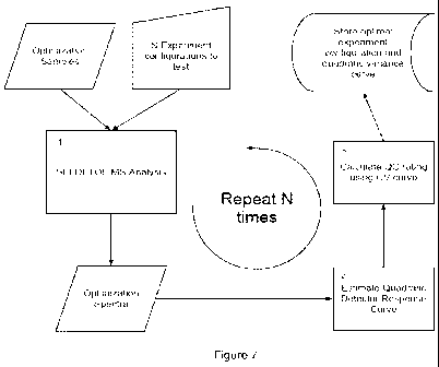
[0016] FIG. 7 is a flow diagram illustrating one embodiment of a process for
identifying
optimal experimental conditions such as instrument settings or sample
preparation; and
[0017] FIG. 8 is a flow diagram illustrating one embodiment of a process for
generating
preprocessed data.
DETAILED DESCRIPTION OF EMBODIMENTS OF THE INVENTION
[0018] The following description of particular embodiment(s) is merely
exemplary in nature
and is in no way intended to limit the scope of the invention, its
application, or uses, which may,
of course, vary. The invention is described with relation to the non-limiting
definitions and
terminology included herein. These definitions and terminology are not
designed to function as
a limitation on the scope or practice of the invention but are presented for
illustrative and
CA 02787504 2012-07-18
WO 2012/048227 PCT/US2011/055376
4
descriptive purposes only. While the processes are described as an order of
individual steps or
using specific materials, it is appreciated that described steps or materials
may be
interchangeable such that the description of the invention includes multiple
parts or steps
arranged in many ways as is readily appreciated by one of skill in the art.
[0019] By default machine settings, a SELDI spectrum is the result of
pooling/summing
numerous single-shot spectra. Skold et. al. studied the acquisition of single
shot spectra and
proposed a statistical framework for pooling the single shot spectra (10).
They introduced an
expectation-maximization algorithm for combining the spectra that results in
improved peak
heights in the pooled spectrum. Malyarenko et. al. (11) introduced a charge-
decay model for the
baseline in a SELDI spectrum and used time-series methods for the common
preprocessing
tasks. The inventors of the processes described herein and their equivalents
identify a quadratic
variance model for the response of a detector used for MALDI or SELDI, which
optionally leads
to preprocessing methods showing improved performance as described herein and
additionally at
(12).
[0020] The present invention has utility as a method for identifying optimum
mass
spectrometer detector, laser, pressure, or other setting parameter for
improved detection or
confidence in detected peaks in a test mass spectrum. The invention further
provides unique
preprocessing of mass spectrometry spectra generated by SELDI or MALDI methods
that
provide improved reproducibility and confidence in peak detection. While the
description is
primarily directed to data generated by SELDI mass spectrometry, the processes
are equally
applicable to other mass spectrometry platforms such as MALDI, among others
known in the art.
[0021] A quadratic variance model is provided that successfully explains the
variation in
SELDI spectra generated from samples such that reproducibility is improved.
The detector
response curve idea can be used to optimize the coefficient of variation (CV)
with the following
advantages over conventional methods: 1) no need to use biological samples to
determine
machine settings and model parameters to apply to actual data; 2) fewer
materials used in the
process; 3) improved CV and thus more reproducible results; 4) fewer man hours
required to find
good machine settings; and 5) optional full-automation of the process of
optimizing CV. The
inventive algorithms for peak detection based on the quadratic variance model
are used in some
embodiments to analyze SELDI spectra from multiple aliquots of a single pooled
cervical
mucous sample used as quality control (QC) for SELDI. These inventive results
are optionally
compared to peak detection with the vendor supplied Ciphergen software (13)
and found
favorable. As each spectrum is a replicate of one sample, all should have the
same number of
proteins and thus yield reproducible peaks. From this point of view,
increasing the number of
CA 02787504 2012-07-18
WO 2012/048227 PCT/US2011/055376
peaks found consistently indicates improved performance of a preprocessing
technique.
[0022] The following abbreviations are used throughout the specification:
Surface-
enhanced laser desorption/ionization time-of-flight mass spectrometry (SELDI-
TOF MS or
SELDI), Matrix-assisted laser desorption/ionization (MALDI), quadratic
variance function
5 (QVF), mean intensity ( ), variance (V), kiloDalton (kDa), microliter (pL),
liquid
chromatography/tandem mass spectrometry (LC-MS/MS).
[0023] Some embodiments of an inventive process include subjecting a first
sample to
SELDI or MALDI mass spectrometry and obtaining a mass data and/or a mass
spectrum from
the first sample. A fit of at least a portion of said mass spectrum to a
quadratic variance model is
performed to obtain a quadratic variance function (QVF). A process may also
include
converting the parameters of the QVF to obtain a coefficient of variation (CV)
for each peak.
The QVF can also be converted to a coefficient of variation function. An
objective function of
the coefficient of variation function is used to calculate a performance
metric that represents the
utility of the instrument detection parameters used. Then the optimal settings
can be selected by
choosing the parameters that minimize the objective function. Examples of
useful objective
functions/performance metrics are the maximum CV in a specified input
intensity interval (a
minimax risk approach), the area under the CV curve in a specified interval
normalized by the
length of the interval (an average risk approach), and the asymptotic "large"
signal value of the
CV function. Analyzing the coefficient of variation function or the objective
function then
allows for identifying an optimal machine parameter or set of parameters.
[0024] As used herein, the term "sample" is defined as a sample obtained from
a biological
organism, a tissue, cell, cell culture medium, or any medium suitable for
mimicking biological
conditions, or from the environment. Non-limiting examples include, saliva,
gingival secretions,
cerebrospinal fluid, gastrointestinal fluid, mucous, urogenital secretions,
synovial fluid,
cerebrospinal fluid, blood, serum, plasma, urine, cystic fluid, lymph fluid,
ascites, pleural
effusion, interstitial fluid, intracellular fluid, ocular fluids, seminal
fluid, mammary secretions,
vitreal fluid, nasal secretions, water, air, gas, powder, soil, biological
waste, feces, cell culture
media, cytoplasm, cell releasate, cell lysate, buffers, or any other fluid or
solid media. A sample
is optionally a buffer alone, water alone, or other non-protein containing
material. A sample is
optionally pooled from a plurality of subjects.
[0025] A "subject" as used herein illustratively includes any organism capable
of producing
a proteinaceous sample. A subject is illustratively a human, non-human
primate, horse, goat,
cow, sheep, pig, dog, cat, rodent, insect, or cell.
[0026] A sample is subjected to analysis by mass spectrometry. Mass
spectrometry is
CA 02787504 2012-07-18
WO 2012/048227 PCT/US2011/055376
6
optionally any spectrometry that requires desorption of a sample, or portion
thereof, from a surface
or from a fluidic sample. Illustratively, mass spectrometry is performed by
laser desorbtion.
Illustrative examples of mass spectrometry that use laser desorbtion include
MALDI or SELDI.
Methods of MALDI and SELDI are well known in the art. Illustratively, methods
of SELDI can be
found at Emanuele, V. A. and Gurbaxani, B. M., BMC Bioinformatics, 2010;
11:512. Methods
of subjecting a sample to MALDI are illustratively found in Gould, WR, et al.,
J Biol Chem,
2004; 279(4):2383-93 and references cited therein.
[0027] A mass data set and, optionally a representative mass spectrum, is
optionally obtained
from the first sample. A mass data set represents the relative abundance of
material in a sample as
defined by intensity as a function mass/charge ratio. A mass data set is
illustratively presented
graphically (e.g. mass spectrum), or as a collection of data points. The mass
data set is fit to a
quadratic equation as follows:
V (P) = uo + u1 + v2 2. (Eq. I)
[0028] with p being the mean of the intensity at a particular mass/charge
ratio (X), V(u) the
variance, and vo, v1, v2 constants, some of which may be zero. The fit of the
mass spectrum to
Equation 1 provides values for the constants vo, vi, and v2. It is observed
that different
experimental conditions provide different quadratic variance functions as
illustrated in FIG. 1 for
background spectra from two different buffer conditions. Different quadratic
variance functions
are also observed for differing instrument settings providing a basis for
instrument optimization
processes.
[0029] The obtained quadratic variance function is then optionally used to
obtain a
coefficient of variation function as defined by: ,
CV% =100=lul= 100= )
= 100 VI-2 + v1 -1 + u2 (Eq.2)
[0030] It is recognized that Equation 2 has a plurality of objective functions
each of which are
be readily identified by methods known in the art. For example, varying
machine settings provide
the minimum area under the CV curve in a specified interval normalized by the
length of the
interval (an average risk approach). This can then be used to identify mass
spectrometer settings
that produce optimal results.
[0031] FIG. 2 illustrates observed variance as a function of mean intensity
for the gaps
between peaks in QC spectra (circles) obtained from pooled cervical samples,
and the quadratic
variance function fit (using Equation 1) to the same (solid line), plus or
minus I standard error
(dashed lines). Very few points fall outside of 1 standard error. This
confirms that the area
CA 02787504 2012-07-18
WO 2012/048227 PCT/US2011/055376
7
interspersed between peaks follow the quadratic variance model.
[0032] In some embodiments, a sample is a proteinaceous sample. As an
illustration, a
proteinaceous sample produces one or more mass spectra that are used to obtain
a quadratic
variance function with a variance that is constant for a peak with a mean
intensity at or below a
lower threshold value. A quadratic variance function optionally has a
quadratic dependence of
variance as a function of mean intensity above the lower threshold value. In
some embodiments,
a quadratic variance function has an upper threshold value at or above which
the variance is
constant as a function of mean intensity. In some embodiments, a lower
threshold value is 3,700
ion counts. An upper threshold value is optionally 12,000 ion counts. A lower
threshold value
and an upper threshold value are appreciated to vary depending -on the
instrument used,
instrument settings, sample type, matrix type, or background type. It is
further appreciated that
one of skill in the art can readily determine the value of a lower threshold
value and an upper
threshold value by mathematical analysis of the quadratic variance function.
Illustratively, a
threshold value (either lower or upper) is identified by taking the first
derivative of the quadratic
variance function, and noting when that derivative becomes a constant (equal
to zero at a lower
threshold or some positive constant at an upper threshold).
[0033] In some embodiments, a plurality of mass data sets are obtained from a
single sample,
or from a plurality of samples. The plurality of mass data sets are optionally
obtained at different
mass spectrometer settings. Illustratively, an operator may alter or otherwise
adjust parameters
including laser intensity, detector sensitivity, ion mode, extraction delay,
flight tube length,
pressure, temperature, laboratory protocols that affect the preparation of the
sample on the chip,
other parameter, or combinations thereof.
[0034] A process optionally further includes adjusting mass spectrometer
detection settings
to said optimal detection parameters. Adjusting mass spectrometer settings is
optionally
performed by a user or automatically on the instrument itself. Illustratively,
a user identifies the
objective function minimum from one or a plurality of coefficient of variation
functions
optionally obtained at varying mass spectrometer settings. The mass
spectrometer settings used
at the objective function minimum represents optimal instrument detection
parameters for the
plate or sample conditions.
[0035] In some embodiments, a mass spectrometer is programmed to automatically
identify
a minimum in the objective function measure of the coefficient of variation
function obtained
from one or a plurality of mass data sets. As an example, a first sample, or a
plurality of samples
are subjected to mass spectrometry analysis. For each sample, a quadratic
variance function is
obtained by a fit of at least a portion of the mass data set generated. The
fit is optionally
CA 02787504 2012-07-18
WO 2012/048227 PCT/US2011/055376
8
performed on a general purpose computer that is separate from or associated
with the mass
spectrometer. The fit is then used to obtain one or a plurality of coefficient
of variation functions
that each may be evaluated for merit via the chosen objective functional. The
lowest minimum
of the objective function of one or plurality of coefficient of variation
functions represents the
optimal instrument detection parameters. This is readily identified by the
program of the
instrument. The instrument detection parameters are then automatically
adjusted by the
instrument for subsequent subjecting of the first sample, a second sample, or
one or more other
samples to mass spectrometry analysis.
[0036] In some embodiments, a process includes subjecting data generated in a
mass
spectrometer to quadratic variance preprocessing to create preprocessed data.
The preprocessed
data are then used for reliable peak detection, to generate a mass spectrum
from the preprocessed
data, or for other purposes recognized in the art. The process of subjecting
data to quadratic
variance preprocessing are essentially as described by Emanuele, V, and
Gurbaxani, B., BMC
Bioinformatics, 2010; 11:512. One or more mass spectra generated on a mass
spectrometer as
the result of SELDI are collected.
[0037] The inventive processes are illustrated by application to repeat
testing of a pooled
cervical mucus sample using a Protein Biology System 11-c mass spectrometer.
The invention
uses a set of MATLAB scripts (The MathWorks, Inc., Natick, MA) for
preprocessing SELDI
spectra termed by the inventors as LibSELDI. Spectra from blank, control, or
test samples
generated are preprocessed with LibSELDI, based on a quadratic variance model,
and optionally
compared to the other peak detection systems, illustratively, Ciphergen
Express (Bio-Rad
Laboratories, Inc., Hercules, CA. Peak predictions from both algorithms are
gathered into
homogenous clusters and peak prevalences and CV% of peak heights are
calculated and
compared with predictions from the quadratic variance model.
[0038] In one test embodiment, the inventive quadratic variance based
algorithm finds 84
peaks occurring in at least 80% of the spectra from pooled cervical mucus
sample while
Ciphergen finds only 18 such peaks (FIG. 2). The predictions of the quadratic
variance model
match the observed peak height variances and peak height CV%. The inventive
pre-processing
approach (synonymously referred to herein as "LibSELDI") based on the
quadratic variance
model finds four times as many reproducible peaks in the pooled cervical
mucous samples as
Ciphergen Express. Also, the model successfully assesses the CV% likely to be
observed by
making measurements of blank spectra giving rise to new ways to optimize
machine parameters.
Thus, the inventive quadratic variance model based approach detects peaks more
reproducibly
thereby increasing the utility of SELDI.
CA 02787504 2012-07-18
WO 2012/048227 PCT/US2011/055376
9
[0039] Reproducible peaks show peak height variances that are consistent with
the quadratic
variance model. This provides an indication of how the noise varies with
proteins with different
abundances. Analysis are optionally restricted to peaks appearing in at least
50% of the spectra
(guaranteeing at least n=16 for sample means and variances). This is
illustrated for the range of
intensity values encompassing most of the peaks in FIG. 4. For the few cases
with peaks of very
high mean intensity (such as those lying above an upper threshold value e.g. >
12,000 ions
counts for SELDI, which may vary for a different instrument such as a MALDI
instrument,
occurring'in the spectra, the quadratic function becomes substantially linear.
This is illustrated in
FIG. 5.
[0040] The CV% of peak height intensity for the reproducible peaks -agree with
the
quadratic variance model, showing which ranges of abundances give the best and
worst CV% for
these machine settings, as illustrated in FIG. 6. Similar to FIG. 5, the model
becomes constant
for peaks at very high mean intensity (e.g. above 12,000 ion counts for SELDI
in this
embodiment), which are a small minority of observations. However, the
predictions are still
bounded below the large CV approximation predicted by the model in Eq. (3).
[0041] Using the LibSELDI algorithm for pre-processing based on the quadratic
variance
model to explain the variation in SELDI signal detection results in
significantly improved peak
detection and reproducibility of peak detection compared to the Ciphergen
algorithm. The
affinity for finding peaks occurring in more than 80% of the spectra is
impressive- finding more
than four times as many as Ciphergen (84 peaks versus 18). The higher number
of peaks is
consistent with direct measures on the same sample using, 2-D and 1D LC-MS/MS
gel, which
despite limited sensitivity, is able to detect 49 proteins in the mass range
of 8.6 - 30 kDa (15).
Several other studies doing proteomic analysis on a similar sample type,
cervicovaginal fluid,
have also shown it to be a complex sample with total number of proteins
ranging from 59 - 685
(17-21).
[0042] The protein estimates/peaks found by the model have mean peak heights,
variances,
and CVs that are consistent with what is predicted. Thus, in simple terms, the
quadratic variance
function estimate predicts peak reproducibility as a function of intensity in
advance of an
experimental run optionally using "blank" regions of the spectra (between
visible peaks), buffer
alone, or modeled spectral data to derive parameters for the algorithm. This
allows the algorithm
to be adjusted for changing noise/background characteristics encountered with
each set of
experimental conditions. This also allows for identification of optimal
instrument settings with
minimized CV objective function optionally based on blank spectra prior to
running samples.
[0043] In some embodiments, using proteinaceous samples as typically obtained
from a
CA 02787504 2012-07-18
WO 2012/048227 PCT/US2011/055376
biological sample, the quadratic variance model of measurement for SELDI shows
a constant
variance for mean intensities below 3,700 ion counts, quadratic between 3,700
and 12,000 ion
counts, and transitioning to non-quadratic variance for very high intensities
above 12,000 ion
counts. The constant variance is optionally determined by calculating the fist
derivative at each
5 portion of the curve. When the first derivative is zero or constant, a
constant variance is
identified at that point in the curve. Fortunately, most peak heights from
exemplary pooled
mucous QC samples are observed in the quadratic variance region.
[0044] The inventive algorithm is particularly advantageous in analyzing or
identifying
proteins, peptides, or other compositions with a molecular mass near 2.5kDa,
optionally
10 anywhere from 1 kDa to 30kDa, where the baseline hits a maximum due to non-
linearities
introduced by the detector saturating.
[0045] The use of the detector response curve (i.e. the value of the objective
function as a
function of instrument setting, illustratively in the case of SELDI) and its
link to the coefficient-
of-variation (CV) has many potential commercial applications. This invention
is operative to
design a MALDUSELDI mass spectrometer that automatically optimizes itself
before a
biomarker discovery experiment (or any other experiment using this
technology). This invention
is also operative to use the detector response curve as part of a quality
control (QC) technique.
For this application, experimental data is compared on a computer to the
typical measurements
expected from the detector response curve and suspicious data can be
automatically flagged for
further inspection. This increases the reliability of the data coming from
these instruments.
Another potential use of the detector response curve is to tune the machine to
pre-specified
protein concentrations. For example, machine settings are set so that low,
medium, or high
intensity proteins show the best CV. This is useful in situations where one
knows in advance the
characteristics of the molecular target being searching for. The idea of a
detector response curve
is useful to a manufacturer of electron-multiplier detectors for MALDUSELDI to
assess which
detector designs are superior for biomarker discovery studies.
EXAMPLES
[0046] The present invention is further detailed in the following examples
that are not
intended to limit the scope of the claimed invention and instead provide
specific working
embodiments.
Example 1 SAMPLE COLLECTION AND PROCESSING
[0047] Cervical mucous is collected from women enrolled as part of an ongoing
study of
CA 02787504 2012-07-18
WO 2012/048227 PCT/US2011/055376
11
cervical neoplasia (/4). At the time of colposcopy, two Weck-Cel @ sponges
(Xomed Surgical
Products, Jacksonville, FL) are placed, one at a time, into the cervical os to
absorb cervical
secretions (15). The wicks are immediately placed on dry ice and stored at -80
C until
processed. Preparation of the pooled quality control (QC) sample is described
(15). Briefly, 40
Weck-Cel sponges with no visual blood contamination from 25 randomly selected
subjects are
extracted using M-PER buffer (Thermo Fisher Scientific, Rockford, IL)
containing lx protease
inhibitor (Roche, Indianapolis, IN). The 40 extracts are combined, aliquoted
and stored at -80 C
until assayed. Total protein content is measured using the Coomasie PIusTM kit
(Thermo Fisher
Scientific) as per the manufacturer's protocol.
10_
Example 2 SELDI-TOF MASS SPECTROMETRY
[0048] A Protein Biological System II-cTM mass spectrometer, with Protein Chip
software
(version 3.2) (Ciphergen Biosystems, Fremont, CA) is used to perform SELDI-TOF
MS. The
mass calibration standard (All-in-one protein standard, Ciphergen) spotted on
the NP-20 (normal
phase) chip surface (Ciphergen) is run weekly, following manufacturer's
instructions. Pooled
cervical mucous is spotted on chips intermittently as part of a QC step in the
experiment design.
Protein chip surface preparation, sample application and application of matrix
are performed
using the Biomek @ 2000 laboratory automation workstation (Beckman Coulter
Inc., Fullerton,
CA) according to the manufacturer's (Ciphergen) instructions.
[0049] The CM 10 chips evaluated are incubated with the sample for I h at room
temperature (24 C 2) and washed three times at 5 min intervals with the CM 10
low stringency
binding buffer, followed by a final wash with ddH2O. In the case of NP-20
arrays, the surface is
prepared with 3 l ddH2O, and ddH2O is used for all washing steps. Chips are
air-dried 30 min
prior to the application of sinnapinic acid (SPA) matrix. The chips are
analyzed on the SELDI-
TOF instrument within 4 h of application of the matrix.
[0050] Buffer-only spectra were generated by interspersing buffer only samples
with protein
samples from subjects (e.g. serum samples) and with pooled subject samples on
the same chip.
The buffer-only samples were spotted with wash buffer that was either PBS
(phosphate buffered
saline with various concentrations of phosphate and NaCI) based or
acetonitrile + TFA
(triflouroacetic acid) based, as manufacturer recommended per chip type. These
buffer only
samples were processed with the same washing steps as the subject samples, and
then SPA
matrix was applied to all spots.
[0051] The instrument settings are determined separately for the low mass and
high mass
range of the protein profile. Data collection is set to 150 kDa optimized for
m/z between 3-30
CA 02787504 2012-07-18
WO 2012/048227 PCT/US2011/055376
12
kDa for the low mass range and 30-100 kDa for the high mass range. For the low
mass range,
the laser intensities are set at 185 with a detector sensitivity of 8 and
number of shots averaged at
180 per spot for each sample. Two warming shots are fired at each position
with the selected
laser intensity +10. These are not included in the data collection. Data
collection from start to
finish took 2 weeks and included a total of 31 spectra.
Example 3 DETECTOR RESPONSE CURVE ESTIMATION
[0052] The quadratic variance model is used to characterize the measurement of
the
intensity values registered at the ion detector in response to a wide range of
signal levels. The
variance of the detector response is quadratic with respect to the mean
intensity level as observed
in a repeated experiment. To show this, we used data taken from buffer, matrix-
only spectra
containing no biological signal or protein content as described (12).
Extending this idea to our
current study, we estimated the detector response curve by using hand selected
regions where
peaks are visibly absent in all of the QC spectra. An illustrative process is
presented in FIG. 7.
A sample is subjected to SELDI analysis as in Example 2 (block 1). As
represented in block 2 of
FIG. 7, the quadratic variance model implies that the mean intensity of
repeated measurements
and corresponding variance V ( ) have the relationship
VW = vo + v1 + v2 2. (Eq.1)
with being the mean of X, V(u) the variance, and vo, vi, v2 constants, some
of which may be
zero. The variance V(u) is best estimated for the range of intensities used to
estimate the curve,
but this extrapolates well to values outside this range.
[0053] The quadratic variance function for the detector response is used to
predict how peak
intensities will behave in the spectra of a repeated SELDI experiment. One
subtle aspect of Eq.
(1) is that it predicts what the CV of such measurements will be (represented
as block 3 of FIG.
7),
CV% = 100 = I ~ = 100. Z
= 100 vo -2 + v1 -1 + v2 (Eq.2)
z 100 ( large). (Eq.3)
Equation 3 merely states that when the mean signal intensity is large, the
coefficient of variation
is approximately constant since the other terms dependent on becoming
negligible. Altogether,
equations 1-3 provide intuition and are sufficient to make predictions about
optimal instrument
detection parameters for the same or other experimental runs. As an example,
data between
peaks is used for a determination of the values for Eq. 1. This provides
simultaneous test data
CA 02787504 2012-07-18
WO 2012/048227 PCT/US2011/055376
13
acquisition and allows determination of the v0, v1, and u2 coefficients for
the experimental
conditions (sample, chip and instrument settings), and therefore the mean
heights and variances,
as well as the CV's, of peaks for the experiment. For very large peaks (e.g.
high intensity >
12,000), the CV% of peak heights is approximated by 100 = as demonstrated in
FIG. 7.
Example 4 PRE-PROCESSING WITH LIBSELDI
[0054] The LibSELD1 preprocessing package is developed in MATLAB (The
Mathworks,
Natick, MA) and takes into account a quadratic variance form of the
measurement error. The
details of the algorithms used by LibSELDI are described by Emanuele, V. A.
and Gurbaxani, B.
M., BMC Bioinformatics. 2010; 11: 512. LibSELDI is used to process the data
adhering to the
following protocols: A single quadratic variance function (QVF) is estimated
representing all 31
QC spectra; The QVF is estimated according to the procedure described in
Example 4;
Preprocessing is performed on each spectrum individually rather than the mean
spectrum. A
flowchart of the steps involved in preprocessing are illustrated in FIG. 8.
[0055] Multiple spectra considerations.
[0056] Rather than observe a single spectrum, the typical biomarker discovery
approach is
to generate at least one spectrum for each of n samples from an approximately
homogeneous
population. For example, the homogeneous population of Example 2 is studies.
As the samples
are run on the same SELDI machine with the same operating conditions, we have
X1 (r) , ..., X),(t) cc NEF-QVF (V (}u (t))) -(Eq. 4)
[0057] The X1, ... Xõ represents the optimization spectra for a single
experiment/machine
setup. A second, and optionally plurality of data sets are obtained under
diffefent instrument
settings and the process is repeated.
[0058] The assumption that all n patients have the same underlying p(t) is
equivalent to
assuming that the underlying biological condition being observed in each
patient is
approximately the same. Thus, underlying commonality p(t) related to the
biology of their
condition expressed through the SELDI signal is estimated. Some of the effects
of the QVF are
mitigated by optionally forming a mean spectrum (first introduced by 22).
1 11
X. (t) YXk-_ (t) .
n k=1 (Eq.5)
[0059] therefore
CA 02787504 2012-07-18
WO 2012/048227 PCT/US2011/055376
[ 14
E lX. (t) 1=11 (t)(Eq. 6)
VarX.t =1V(ct).
n (Eq. 7)
[0060] Modified Antoniadis-Sapatinas denoising.
[0061] For generation of a preprocessed mass spectra, the data obtained as in
Example 2 are
subjected to modified Antoniadis-Sapatinas denoising represented as block 1 of
FIG. 8. p(t)
from the mean spectrum obtained by a fit of the means spectrum to Eq. 5 Since
the Xk(t) are
sampled on a discrete time grid (and thus X.), a vector notation is
introduced.
1v.= [X. (tl), ...,X. (till)]
~~-[u (tl) , ...,~1 (t11Z)~ (Eq. 8)
[0062] or any estimate it (X.)of, p we measure its fitness using the mean-
squared-error
(MSE).
M S E(.~.) ii) -E J ir(-v.) } ' Eq.
(9)
[0063] For denoising, we use the orthogonal discrete wavelet transform with
respect to the
Symmlet 8 basis. The transform is represented by an m x m orthogonal matrix W,
w=W:X..(Eq. 10)
[0064] Where h is a length m vector with entries taking values between 0 and
1. Let H =
diag (h) be the m x m matrix defined by placing the entries of h along the
main diagonal, all
other entries 0. The class of estimators for (=V.) take the form
u (.x.) =W~Hw
r
=W HW.x=. (Eq. 11)
[0065] This is the typical wavelet denoising scenario where each wavelet
coefficient is left
alone or shrunk towards zero according to some criterion, and is completely
defined by the
vector h. Antoniadis and Sapatinas showed that a good estimator for data from
the NEF-QVF
family is given by choosing:
CA 02787504 2012-07-18
WO 2012/048227 PCT/US2011/055376
- [tivii-cry (i +
IV(i)2 M
:, >0
01 z< 0. (Eq. 12)
[0066] where the term his estimated as
ff~ 1
= (6 7 = W) V (x.) .
1 +U2 (Eq. 13)
[0067] where V(x.) is the vector constructed by applying the QVF from (1) to
each term of
5 x.. (W = W) is the matrix whose i, j element is the square of the i, j
element of W. The parameters
00, V1, u2 in Eq. 1 are measured from the background regions, buffer only
spectra, or prior test
sample data as in Example 3.
[0068] An intuitive modification is made to Eq. 13 to obtain:
ff~ -1+ ?4 (W = W) V' (x.) .
V'r (x. (i)) =inax (V (x. (i)) ) uo} '(Eq. 14)
10 [0069] Thus, the modified Antoniadis and Sapatinas estimator h uses LT- in
Eq. 12 rather
1-11)
than cr. The modification was introduced to account for cases when Eq. 13 may
underestimate
the noise when low amounts of observed signal are detected. Define
tiv(iJZ-~-(i1 +
h-
i/=diagQ).
(Eq. 15)
[0070] then, the modified Antoniadis-Sapatinas estimate ofp is defined as
/-
15 P=W HWx..(Eq.16)
[0071] Peak detection/baseline removal.
[0072] For peak detection and baseline removal the two preprocessing steps of
baseline
removal and peak detection typically performed separately are consolidated
into a single step.
These processes are represented in block 2 of FIG. 8. It is assumed that the
underlying p(t)
shown in Eq. 6 is the superposition of protein ions, s(t), and energy-
absorbing matrix ions, b(t)
CA 02787504 2012-07-18
WO 2012/048227 PCT/US2011/055376
16
striking the detector. The distribution of the isotopes in the analyte of
interest gives rise to a
roughly Gaussian peak shape. Thus, it is proposed that
Ii (t) _s (t) +b (t)(Eq. 17)
S (t) = (1j93 j (ti, cj)
1 (Eq. 18)
[00731 where, a (ti ' j)
] denotes a Gaussian kernel function centered at tj with standard
deviation aj and zero outside the interval [tj - a, tj + a].
[0074] Typically, s(t) is very sparse in the sense that it is mostly zero over
the domain of the
observed signal. Therefore, the local minima of the estimated baseline + noise
signal Ti are
points that may be assumed to touch the baseline. From this point of view,
once all the local
minima in P are detected, the baseline curve estimation reduces to an
interpolation amongst
these points. For this purpose, piecewise cubic Hermite interpolating
polynomials (as performed
in ref. 23) are excellent interpolation functions.
[0075] The minima and maxima in P are found in one pass using the extrema
function
downloadable from MATLAB central file exchange (finds all locations where the
first
derivative of P = 0). The maxima are the peaks in the mean spectrum
potentially indicating
proteins represented in the sample population of Example 2 while the minima
correspond to
samples from the baseline signal.
[0076] Normalization of block 3 of FIG. 3 is achieved by any standard
normalization
method known in the art. Illustratively, the normalization method is that of
Meuleman et al.,
BMC Bioinformatics 2008;9:88.
[0077] Each detected peak is quantified using peak area and a threshold is
chosen based on
the peak area measurement to generate the final prediction set as represented
in blocks 4 and 5 of
FIG. 8.
Example 5: PRE-PROCESSING WITH CIPHERGEN
[0078] All SELDI spectra of Example 2 are processed using Ciphergen Express
Client
software (version 3.0). Pre-processing of the spectra is performed as
previously described (16).
Briefly, baseline correction, external calibration using protein standards,
normalization using
total ion current, and mass alignment are applied to all spectra. Peak
detection is performed on
CA 02787504 2012-07-18
WO 2012/048227 PCT/US2011/055376
17
this pre-processed data. Peaks from 2.5-30 kDa are detected by centroid mass,
with first pass
settings of signal to noise ratio (S/N) = 5, valley depth = 3, second pass
settings of S/N = 3 and
valley depth = 2, and a mass window of 0.3%.
Example 6 PEAK MATCHING
[0079] When peak predictions are made in a repeated experiment, it is useful
to group peaks
from distinct spectra that are close enough in m/z value to be assumed to be
generated from the
same underlying analyte. This allows one to assess the reproducibility of a
peak in terms of its
prevalence (% of times it appears across spectra) and CV (of both peak m/z and
peak intensity).
This process is referred to as peak matching or peak clustering.
[0080] A fair comparison of reproducibility of peak predictions requires that
the same peak
matching algorithm be used for each method. Otherwise, one could not ascertain
whether the
core preprocessing algorithms (denoising, baseline removal, peak detection) or
the peak
matching algorithms contributed most to conclusions about the superiority of
one preprocessing
approach versus another. LibSELDI and Ciphergen use different peak matching
techniques,
with the Ciphergen approach being an unpublished, proprietary method. For this
reason,
LibSELDI's peak matching algorithm is used to assess prevalence and CV's for
both
preprocessing programs' peak predictions. Since the peak matching algorithm is
completely
independent of the methodology used in the core preprocessing steps of both
Ciphergen and
LibSELDI, there is no reason to believe it would give either algorithm an
advantage in this
comparison. The results are presented in FIG. 3 demonstrating improved
reproducible peak
detection by the LibSELDI process.
[0081]
Example 7: ESTIMATION OF PARAMETERS FOR PEAK CLUSTERS
[0082] For each peak in a peak cluster, the analyte mass is estimated using
the detected peak
m/z location of the smoothed, processed spectrum obtained as in Example 4 and
is illustrated as
block 6 in FIG. 7. The peak height is measured as the maximum intensity value
observed in a
window centered around the peak m/z value. The peak area is measured as the
sum of intensity
values observed in a window centered around the peak m/z value. The mean,
variance, and CV
of peak heights and peak areas are then calculated for each peak cluster. Note
that, this is
slightly different from measuring mean and variances from the peak-free
regions. For the peak-
free regions mean and variance of intensity are calculated for each fixed m/z
value.
Example 8: OPTIMIZATION OF DETECTOR SETTINGS
CA 02787504 2012-07-18
WO 2012/048227 PCT/US2011/055376
18
[0083] Thirty buffer only samples are prepared on sample plates and combined
with SPA
matrix as in Example 2. The buffer only samples are subjected to ionization in
a SELDI mass
spectrometer as described in Example 2 with varying detector sensitivity
settings ranging from 5 to
9. Ten different detector sensitivities are studied using three spot per
sensitivity setting. The
resulting data sets are used to generate mass spectra and for identification
of a quadratic variance
function representing the data set, produce a resulting coefficient of
variation function, and are
processed to obtain an objective function as in Example 3. The objective
function used in these
studies is an area under the coefficient of variation function analysis for
intensities ranging from
4,000 to 6,000. The minimum value for area under the curve from the 10
different settings is then
chosen. The detector settings producing the minimum objective function value
represent optimal
instrument detector sensitivity settings for the buffer/matrix samples.
[0084] The above studies are repeated by obtaining 10 spectra at each detector
sensitivity
setting but at varying laser intensity settings with laser intensity low
values ranging from 175 to
245 and laser high values ranging from 185 to 255. The data set of each
spectrum is then subjected
to the same analyses procedures. A 10 x 10 matrix or area under the curve is
obtained with the two
varying instrument settings. The minimum value in the matrix establishes the
optimum instrument
settings (laser intensity/detector sensitivity) for the buffer and matrix
combination.
[0085] The instrument is then adjusted to the identified optimum instrument
settings. Test
samples prepared in the same buffer and combined with the same matrix are then
used for analyses
under the optimum instrument settings.
Example 9: BIOMARKER DETECTION
[0086] Cervical mucus is collected from women enrolled as part of an ongoing
study of
cervical neoplasia (14) as in Example 1. Protein samples are prepared using 6
samples from
sponges with no visual blood contamination from women diagnosed with high-
grade squamous
intraepithelial lesion (HSIL) confirmed by colposcopy and/or biopsy (test
samples) and women
as a test group and 6 samples from women presenting negative Pap test and no
prior history of
abnormal cytology as a control group. Protein is extracted using M-PER buffer
(Thermo Fisher
Scientific, Rockford, IL) containing 1 x protease inhibitor (Roche,
Indianapolis, IN). Total
protein content is measured using the Coomassie P1usTM kit (Thermo Fisher
Scientific) as per the
manufacturer's protocol. The extracts are aliquoted and stored at -80 C until
assayed.
[0087] Each of the protein extracts are analyzed by SELDI using the protocol
of Example 2.
Each sample is spotted three times on the NP-20 sample plate and incubated for
I h at room
temperature (24 C 2) and washed three times at 5 min intervals with the CMIO
low stringency
CA 02787504 2012-07-18
WO 2012/048227 PCT/US2011/055376
19
binding buffer, followed by a final wash with ddH2O. Chips are air-dried 30
min prior to the
application of SPA matrix. The chips are analyzed on the SELDI-TOF instrument
within 4 h of
application of the matrix.
[0088] Data are collected using the instrument settings of Example 2. Each
spectrum is
individually analyzed as per Example 3. The detector response curves are
evaluated using data
from regions of the spectra interdispersed between visually identifiable
peaks. Each of the mass
data sets from each ionization is well described by Eq. 1. The values for each
of the parameters are
fit by least-squares analysis of each data set. The resulting quadratic
variance functions are then
used for quadratic variance preprocessing to create preprocessed data for each
spectra as
described in Example 4 and peaks are identified and matched as in Example 6.
[0089] The test samples identify several proteins with different abundances
(intensities)
relative to control samples. These proteins are identified as members of the
ovalbumin serine
proteinase inhibitors, cysteine proteinase inhibitors, and proteins involved
in cellular glycolysis,
cytokinesis, and metastasis. These results are in agreement with the proteins
identified by an
independent research group using traditional analyses (See Lema, C., et al.,
Proc Amer Assoc
Cancer Res, Volume 47, 2006, Abstract #4455), but are reached much faster and
with greater
confidence that is achievable by prior methods.
References
1. McLerran D, Grizzle WE, Feng Z, Thompson IM, Bigbee WL, Cazares LH et al.
SELDI-
TOF MS whole serum proteomic profiling with IMAC surface does not reliably
detect prostate
cancer. Clin Chem 2008;54:53-60.
2. Semmes OJ, Feng Z, Adam BL, Banez LL, Bigbee WL, Campos D et al. Evaluation
of
serum protein profiling by surface-enhanced laser desorption/ionization time-
of-flight mass
spectrometry for the detection of prostate cancer: I. Assessment of platform
reproducibility. Clin
Chem 2005;51:102-12.
3. Timms JF, rslan-Low E, Gentry-Maharaj A, Luo Z, T'Jampens D, Podust VN et
al.
Preanalytic influence of sample handling on SELDI-TOF serum protein profiles.
Clin Chem
2007;53:645-56.
4. McLerran D, Grizzle WE, Feng Z, Bigbee WL, Banez LL, Cazares LH et al.
Analytical
validation of serum proteomic profiling for diagnosis of prostate cancer:
sources of sample bias.
Clin Chem 2008;54:44-52.
5. Cruz-Marcelo A, Guerra R, Vannucci M, Li Y, Lau CC, Man TK. Comparison of
algorithms
for pre-processing of SELDI-TOF mass spectrometry data. Bioinformatics
2008;24:2129-36.
CA 02787504 2012-07-18
WO 2012/048227 PCT/US2011/055376
6. Emanuele VA, Gurbaxani BM. Benchmarking currently available SELDI-TOF MS
preprocessing techniques. Proteomics 2009;9:1754-62.
7. Meuleman W, Engwegen JY, Gast MC, Beijnen JH, Reinders MJ, Wessels LF.
Comparison
of normalisation methods for surface-enhanced laser desorption and ionisation
(SELDI) time-of-
5 flight (TOF) mass spectrometry data. BMC Bioinformatics 2008;9:88.
8. Wegdam W, Moerland PD, Buist MR, Ver Loren van TE, Bleijlevens B, Hoefsloot
HC et al.
Classification-based comparison of pre-processing methods for interpretation
of mass
spectrometry generated clinical datasets. Proteome Sci 2009;7:19.
9. Wei,. W_.,.Martin, A., Johnson, P.-J., and Ward, D. G. 10 Years of SELDI:
What Have we
10 Learnt? Current Proteomics 7[1], 15-25. 2010.
10. Skold M, Ryden T, Samuelsson V, Bratt C, Ekblad L, Olsson H, Baldetorp B.
Regression
analysis and modelling of data acquisition for SELDI-TOF mass spectrometry.
Bioinformatics
2007;23:1401-9.
15 11. Malyarenko DI, Cooke WE, Adam BL, Malik G, Chen H, Tracy ER et al.
Enhancement of
sensitivity and resolution of surface-enhanced laser desorption/ionization
time-of-flight mass
spectrometric records for serum peptides using time-series analysis
techniques. Clin Chem
2005;51:65-74.
12. Emanuele, V. A. and Gurbaxani, B. M. Quadratic Variance Models for
Adaptively
20 Preprocessing SELDI Mass Spectrometry Data. BMC Bioinformatics. 2010; l l:
512.
13. Fung ET, Enderwick C. ProteinChip clinical proteomics: computational
challenges and
solutions. Biotechniques 2002;Suppl:34-1.
14. Rajeevan MS, Swan DC, Nisenbaum R, Lee DR, Vernon SD, Ruffin MT et al.
Epidemiologic and viral factors associated with cervical neoplasia in HPV-16-
positive women.
Int J Cancer 2005;115:114-20.
15. Panicker G, Ye Y, Wang D, Unger ER. Characterization of the Human Cervical
Mucous
Proteome. Clin Proteomics 2010;6:18-28.
16. Panicker G, Lee DR, Unger ER. Optimization of SELDI-TOF protein profiling
for analysis
of cervical mucous. J Proteomics 2009;71:637-46.
17. Andersch-Bjorkman Y, Thomsson KA, Holmen Larsson JM, Ekerhovd E, Hansson
GC.
Large scale identification of proteins, mucins, and their O-glycosylation in
the endocervical
CA 02787504 2012-07-18
WO 2012/048227 PCT/US2011/055376
21
mucus during the menstrual cycle. Mol Cell Proteomics 2007;6:708-16.
18. Dasari S, Pereira L, Reddy AP, Michaels JE, Lu X, Jacob T et al.
Comprehensive proteomic
analysis of human cervical-vaginal fluid. J Proteome Res 2007;6:1258-68.
19. Pereira L, Reddy AP, Jacob T, Thomas A, Schneider KA, Dasari S et al.
Identification of
novel protein biomarkers of preterm birth in human cervical-vaginal fluid. J
Proteome Res
2007;6:1269-76.
20. Shaw JL, Smith CR, Diamandis EP. Proteomic analysis of human cervico-
vaginal fluid. J
Proteome Res 2007;6:2859-65.
21. Tang LJ, De SF, Odreman F, Venge P, Piva C, Guaschino S, Garcia RC.
Proteomic analysis
of human cervical-vaginal fluids. J Proteome Res 2007;6:2874-83.
22. Morris JS, Coombes KR, Koomen J, Baggerly KA, Kobayashi R. Feature
extraction and
quantification for mass spectrometry in biomedical applications using the mean
spectrum.
Bioinformatics. 2005;21(9):1764-1775. doi: 10.1093/bioinformatics/bti254.
23. Fritsch FN, Carlson RE. Monotone Piecewise Cubic Interpolation. SIAM j
Numerical
Analysis. 1980;17:238-246. doi: 10.1137/071702 1.
24. Gould, WR, et al., J Biol Chem, 2004; 279(4):2383-93
[0090] Various modifications of the present invention, in addition to those
shown and
described herein, will be apparent to those skilled in the art of the above
description. Such
modifications are also intended to fall within the scope of the appended
claims.
[0091] Patents and publications mentioned in the specification are indicative
of the levels of
those skilled in the art to which the invention pertains. These patents and
publications are
incorporated herein by reference to the same extent as if each individual
application or
publication is specifically and individually incorporated herein by reference.
[0092] The foregoing description is illustrative of particular embodiments of
the invention,
but is not meant to be a limitation upon the practice thereof. The following
claims, including all
equivalents thereof, are intended to define the scope of the invention.