Note: Descriptions are shown in the official language in which they were submitted.
CA 02787986 2012-07-24
WO 2011/092169 1
PCT/EP2011/051003
"Intraocular lens"
FIELD OF THE INVENTION
The present invention relates to an intraocular lens, and in
particular to an intraocular lens with a diffractive profile on an anterior or
posterior face.
STATE OF THE ART
An intraocular is a lens which may be implanted in the eye,
most often for replacing the crystalline lens after a cataract operation. It
normally includes lateral flexible supports, so-called "haptics", used for
supporting the lens in the capsular bag. An intraocular lens may be a
refractive lens, a diffractive lens, or else a refractive-diffractive lens. A
refractive lens converges light towards a focal point on the optical axis by
refraction, while a diffractive lens creates a diffraction pattern forming one
focal point on the optical axis per diffraction order. A refractive-
diffractive
lens combines the features of both of them.
The crystalline lens has some flexibility allowing, through
the action of ciliary muscles, adaptation of the eye to far or near vision.
By pulling on the edges of the crystalline lens, the ciliary muscles flatten
it, thereby displacing its focal point. However, because of weakening of
the ciliary muscles due to age, or because of the replacement of the
crystalline lens with an intraocular lens, a patient may at least partly lose
this adaptability.
In order to address this problem, several types of bi- or
multi-focal intraocular lenses have been proposed.
A bi- or multi-focal refractive intraocular lens has variable
refractive power, normally decreasing from the center of the lens towards
an outer edge. Such intraocular lenses are sold under the brands of
lolabe NuVue0, Stolz() Tru Vista , Alcon AcuraSee , loptexe, and
CA 02787986 2012-07-24
WO 2011/092169 2
PCT/EP2011/051003
AMOO ReZoom . This takes advantage of the fact that in situations
where near vision is required, such as for example for reading, one
normally has high luminosity, which causes closing of the iris, concealing
the outer portion of the lens and only keeping the more central portion
having the highest refractive power. In one alternative, the refractive
intraocular lens may have an aspherical profile, so as to correct
aspherical aberration of the cornea.
These purely refractive bi- or multi-focal lenses however
have drawbacks. Notably, their effect is very dependent on the size of the
pupil. Further, because they have several focal points, they only provide
reduced contrast and may form halos, in particular, in far vision, with
reduced luminosity.
An alternative is that provided by refractive-diffractive
intraocular lenses. Typically, these lenses provide a refractive optical
focal point of order zero for far vision, and at least one diffractive focal
point of first order for near vision. Certain refractive-diffractive
intraocular
lenses, such as for example those developed by 3M and those
developed by AMOO and distributed under the brand of Tecnis share
the light in a substantially equal way between both of these two focal
points. On the other hand, the intraocular lenses Acri.Tece Acri.lisa
366D, have asymmetrical distribution of the light, with more light directed
towards the focal point for far vision than for the one for near vision, with
the object of improving the contrast and reducing the formation of halos in
far vision.
In the article "History and development of the apodized
diffractive intraocular lens", by J.A. Davison and M.J. Simpson, published
in J. Cataract Refract. Surg. Vol. 32, 2006, pp. 849-858,
doi :10.1016/j.jcrs.2006.02.006, a refractive-diffractive intraocular lens is
described in which the diffractive profile is apodized, having decreasing
amplitude in the direction running from the optical axis towards an outer
edge of the lens. This lens, sold by Alcon under the brand ReSTORO
CA 02787986 2012-07-24
WO 2011/092169 3
PCT/EP2011/051003
thereby allows a variation of the distribution of the light between the focal
points for far vision and near vision according to the aperture of the pupil.
These refractive-diffractive intraocular lenses of the state of
the art, however, also have certain drawbacks. Notably, they are almost
purely bifocal, with a spacing between the focal point for far vision and
the one for near vision such that they may be uncomfortable in
intermediate vision.
Multi-focal refractive-diffractive lenses having at least one
intermediate focal point have also been proposed. In International Patent
Application WO 94/11765, a refractive-diffractive lens is proposed with a
focal point of order zero for intermediate vision, a focal point of order +1
for near vision, and a focal point of order -1 for far vision. This lens,
however, only allows a substantially equitable distribution of the light
between the three focal points, independently of the pupil aperture.
In International Patent Application WO 2007/092949, an
intra-ocular lens is proposed including a plurality of diffractive profiles,
each with a distinct focal point of order +1. The different profiles are
arranged on concentric areas, and the distribution of the light between
the focal points will therefore strongly depend on the pupil size, as in
refractive multi-focal intraocular lenses.
Further, all the diffractive and refractive-diffractive
intraocular lenses of the state of the art have the drawback of the loss of
a considerable portion of the light towards unusable focal points of an
order greater than 1.
SUMMARY OF THE INVENTION
A first object of the present invention is to provide an
intraocular lens having two useful diffractive focal points, with distribution
of the light between both of these focal points which does not necessarily
depend on the pupil size.
An intraocular lens according to the present invention
includes an anterior surface and a posterior surface and has a
CA 02787986 2012-07-24
WO 2011/092169 4
PCT/EP2011/051003
substantially antero-posterior optical axis. In this lens, one of these
anterior and posterior surfaces includes a first diffractive profile forming
at
least one first diffractive focal point of order +1 on said optical axis, and
a
second diffractive profile forming a second diffractive focal point of order
+1 on said optical axis which is distinct from the first diffractive focal
point
of order +1, at least one portion of said second diffractive profile being
superposed on at least one portion of the just diffractive profile so that the
order +2 of the second diffractive profile is added to the order +1 of the
first diffractive profile.
Both diffractive profiles, even superposed, continue to form
distinctive diffractive focal points. It is thus possible to obtain two
different
focal points of order +1 without the distribution of the light between them
being necessarily affected by the pupil size.
Another object of the present invention is to provide a multi-
focal intraocular lens. For this, said lens may advantageously be a
refractive-diffractive lens with, in said optical axis, a focal point of order
zero distinct from said first and second focal point of order +1. In
particular, said focal point of order zero may be a focal point for far
vision,
said first focal point of order +1 may be a focal point for near vision, and
said second focal point or order +1 a focal point for intermediate vision.
In this way, it is possible to obtain a multi-focal intraocular
lens, in particular with a focal point for far vision, a focal point for
intermediate vision and a focal point for near vision, without the
distribution of the light between at least two of these focal points, and in
particular between the focal point for near vision and the focal point for
intermediate vision, being necessarily affected by the pupil size.
Still another object of the present invention is to limit the
light losses due to refraction orders greater than +1. For this, said focal
point for near vision may also substantially coincide on the optical axis
with a focal point of higher order than 1 formed by the second diffractive
profile. In particular, said focal point of higher order may be a focal point
of order +2.
CA 02787986 2012-07-24
WO 2011/092169 5
PCT/EP2011/051003
Thus, the light directed towards said focal point of higher
order is not lost, but is used for reinforcing a focal point of order +1,
notably the focal point for near vision. In this way, the advantage of an
asymmetrical distribution of the light in favour of the focal point for near
vision relatively to the focal point for intermediate vision which is less
important, is thereby obtained.
Advantageously, said focal point for near vision is at a
distance from the focal point for far vision corresponding to between +2.5
diopters and +5 diopters, in particular between +3 diopters and +4
diopters, such as for example +3.5 diopters. This focal length allows
adequate simulation of the optimum adaptability of the crystalline lens.
The proportion of the light directed towards the diffractive
points of order +1 depends on the amplitude of the diffractive profile. For
example, in a refractive-diffractive lens with an amplitude of the diffractive
profile of one wavelength, the entirety of the light will be directed towards
the diffractive focal points, but with a decrease in the amplitude, an
increasing proportion of the light will be directed towards the refractive
focal point. With zero amplitude of the diffractive profile, the lens will, of
course, be purely refractive.
Advantageously, said second diffractive profile may have a
smaller amplitude than the first diffractive profile.
Advantageously, said first and/or second diffractive profiles
may be apodized with a decreasing amplitude from the optical axis
towards an outer edge of the lens, in particular proportionally to the cube
of the distance to the optical axis. In this way, with an increasing aperture
of the lens, the distribution of the light will vary in favor of the
refractive
focal point, i.e. the focal point for far vision, and to the detriment of the
focal points for close and intermediate vision.
Advantageously, the lens may be aspherical, so as to obtain
a greater field depth.
Advantageously, said first diffractive profile and/or said
second diffractive profile may be profiles of the kinoform type, with which
CA 02787986 2016-02-17
6
unnecessary refractive focal points notably those of negative order may be
suppressed. Even more advantageously, edges of said first and/or second
diffractive profiles may be rounded, which reduces the acute angles and
improves the quality of the image by reducing diffused light.
Another object of the present invention is to provide an
intraocular lens comprising: an anterior surface and a posterior surface and
having a substantially antero-posterior optical axis wherein one of these
anterior and posterior surface includes a first diffractive profile forming at
least
one first diffractive focal point of order +1 on said optical axis, and a
second
diffractive profile forming a second diffractive focal point of order +1 on
said
optical axis which is distinct from said first diffractive focal point of
order +1, at
least one portion of said second diffractive profile being superposed on at
least
one portion of said first diffractive profile so that an order +2 of said
second
diffractive profile is added to said order +1 of said first diffractive
profile.
DETAILED DESCRIPTION
Details relating to the embodiments of the invention are
described hereafter in an illustrative and non-restrictive way with
reference to the drawings.
Fig. 1 illustrates an exemplary intraocular lens according to an
embodiment of the invention.
Fig. 2 schematically illustrates the lens of Fig. 1 with a focal point
for far vision, a focal point for intermediate vision and a focal point for
near
vision.
Fig. 3 illustrates the radial section of the anterior surface of the
lens of Fig. 1 having two superposed diffractive profiles.
Fig. 4a illustrates a first of the two diffractive profiles of
Fig. 3.
Fig. 4b illustrates a second one of the two diffractive profiles
of Fig. 1.
Fig. 5 illustrates the distribution of light in the optical axis of the
lens of Fig. 1 for a determined pupil aperture.
Fig. 6 illustrates the variation of the distribution of light
between the three focal points depending on the pupil aperture.
CA 02787986 2016-02-17
6a
Fig. 7A compares the modulation transfer functions of the three
focal points of a lens according to an embodiment of the invention, as
compared with those of the two focal lengths of a bifocal lens of the state of
the art, with a pupil aperture of 2.0 mm.
Fig. 7b compares the modulation transfer functions of the
three focal points of the lens according to an embodiment of the
CA 02787986 2012-07-24
WO 2011/092169 7
PCT/EP2011/051003
invention, as compared with those of the two focal points of a bifocal lens
of the state of the art, with a pupil aperture of 3.0 mm
Fig. 7c compares the modulation transfer functions of three
focal points of a lens according to an embodiment of the invention, as
compared with those of the two focal points of a bifocal lens of the state
of the art, with a pupil aperture of 4.5 mm.
A general configuration of an intraocular lens 1 according to
an embodiment of the invention is illustrated in Fig. 1. As this may be
seen in the figure, the lens includes a central optical body 2 and, in this
exemplary configuration, two flexible supports 3, so-called "haptics", on
the outer edge of the lens 1 in order to support it in the capsular bag
when it is implanted in the eye of a patient. However, other alternative
configurations are known to one skilled in the art and applicable in an
intraocular lens according to the invention, such as for example a larger
number of haptics, loop-shaped haptics, etc.
In Fig. 2, the intraocular lens 1 according to the illustrated
embodiment of the invention is a lens of the refractive-diffractive type.
The central optical body 2 includes an anterior face 4 and a posterior face
5, and has a substantially antero-posterior axis 6. The anterior and/or
posterior faces 4,5 have curvatures such that the lens 1 directs a portion
of the incident light onto a refractive focal point 7, or of order zero, on
the
optical axis. This focal point 7 is a focal point for far vision. In this
particular embodiment, the lens 1 has an asphericity with an aspherical
aberration of -0.11 pm. This asphericity ensures a natural balance
between the sensitivity to the contrast and the field depth by inducing a
moderate positive spherical aberration in the eye implanted with this lens.
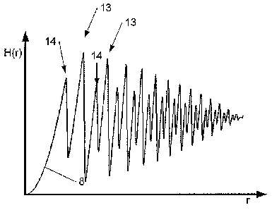
However, on its anterior face 4, the lens 1 has a relief 8
illustrated in Fig. 3 and formed by the superposition of a first diffractive
profile 9, illustrated in Fig. 4a, with a second diffractive profile 10,
illustrated in Fig. 4b. (It should be noted that in these three figures, the
height of the profiles is considerably exaggerated with respect to the
radial distance r). The relief 8 therefore generates a complex diffraction
CA 02787986 2012-07-24
WO 2011/092169 8
PCT/EP2011/051003
figure, with, on the optical axis 6, a first diffractive focal point 11 of
order
+1 corresponding to the first diffractive profile 9, and a second diffractive
focal point 11 of order +1 corresponding to the second diffractive profile
10. The first diffractive focal point 11 of order +1 is a focal point for near
vision, while the second diffractive focal point 12 of order +1 is a focal
point for intermediate vision.
The first diffractive profile 9 is a profile of the kinoform type,
approximately fitting the function:
(r3 2 ( 1 )7
H1(r)= a1¨ ¨
l mod F1 ¨ Air 2 + Fi2 )2 A ¨71- ,271-1 + z)
R3 27c
j, k,,.. n2 ¨ n, ,,
In this equation, Hi(r) is the height of the first diffractive
profile 9 as a function of the radial distance r relatively to the optical
axis,
R is the radial distance from the outer edge of the lens to the optical axis,
A is the wavelength at which the eye has greatest sensitivity (normally
550 nn"), n1 and nz are refractive indexes of the material of the lens and
of its implantation medium, al is an amplitude parameter (0.44 in the
illustrated embodiment), and F1 is the focal length of the focal point 11 of
order +1 of this first diffractive profile 9 (300 mm for +3.5 diopters in this
embodiment).
The second diffractive profile 10 is also a profile of the
kinoform type, approximately fitting the function:
r A 1
H2 (r)= a21¨-MOd[(F2 _r 2 + F2 2 )2-11- ,2H +
R , 2ff ,n ¨121 A )\_ i
In this equation Hz(r) is the height of this second diffractive
profile 10 as a function of the radial distance r with respect to the optical
axis, az is an amplitude parameter (0.27 in the illustrated embodiment)
and F2 is the focal length of the focal point 12 of order +1 of this second
diffractive profile 10 (600 mm for +1.75 diopters in this embodiment).
CA 02787986 2012-07-24
WO 2011/092169 9
PCT/EP2011/051003
It should actually be noted that, through manufacturing
constraints, the actual diffractive profiles 9, 10 may only approximately fit
these equations. In particular, the edges of these actual profiles will be
rounded, which may be simulated by a convolution as illustrated in Figs.
4a and 4b, and which has the additional advantage of reducing the
amount of diffused light to the benefit of the optical quality of the image.
The relief 8 resulting from the superposition of both of these
profiles 9, 10 therefore approximately fits the formula H(r)=H1(r)+H2(r), as
illustrated in Fig. 3. As, in this embodiment F2=2F1 , the second diffractive
profile 10 has periodicity half of the one of the first diffractive profile 9.
The relief 8 therefore has large sawteeth 13, resulting from the addition of
a step of the first profile 9 with a step of the second profile 10,
alternating
with small sawteeth 14, corresponding to one step out of two of the first
profile 10. Further, in this way the second profile 10 forms a diffractive
profile of order +2 coinciding with the focal point 11 of order +1 of the
first
profile 9. Thus, a portion of the light which would otherwise be lost is
used here for assisting near vision.
A way of estimating the optical priority of an intraocular lens
consists of determining experimentally its modulation transfer function
(MTF). The MTF of an optical system reflects the proportion of the
contrast which is transmitted through the optical system for a determined
spatial frequency. Generally, the contrast decreases with an increase in
the frequency. In Fig. 5, the curve 15 of the MTF of the lens 1 versus the
focal power D may be seen for a pupil aperture of 3.0 mm in an eye
model according to the ISO standard at 50 cycles/mm. This curve 15
shows 3 peaks 16, 17, 18 respectively corresponding to the focal point for
far vision, to the focal point 12 for intermediate vision and to the focal
point 11 for near vision. In this lens 1, with this aperture, the distribution
of the light between these three focal points is 49% for far vision, 34% for
near vision, and 17% for intermediate vision. It may also be appreciated
in this figure that very little light is directed elsewhere than on these
three
focal points.
CA 02787986 2012-07-24
WO 2011/092169 10
PCT/EP2011/051003
As this may be seen in Figs. 3, 4a and 4b, the amplitude of
the two profiles 9, 10 decreases with the cube of the radius r, according
to the equations for Hi(r) and H2(r). The relief 8 is therefore "apodized" so
as to decrease from the center of the lens 1 to its outer edge. Thus, with
increasing aperture, increasingly more light will be directed towards the
refractive focal point 7, to the detriment of the diffractive focal points 11
and 12. This may be appreciated in Fig. 6, in which the curve 19
corresponds to the percentage of incident light directed towards the focal
point 7 for far vision, the curve 20 corresponds to the one directed
towards the focal point 12 for intermediate vision, the curve 21 to the one
directed towards the focal point 11 for near vision, and the curve 22 to the
one of the light energy which is lost, as theoretically calculated according
to a pupil aperture in millimeters.
In Figs. 7a, 7b, and 7c, an exemplary intraocular lens 1
according to an embodiment of the invention was compared with a bifocal
intraocular lens Acri.Tece Acri.lisa 366D, considered as one of the best
of the state of the art. Curves 23, 24 and 25 correspond to the MTFs
versus spatial frequency for the focal point 7 for far vision, the focal point
11 for near vision, and the focal point 12 for intermediate vision,
respectively. Curves 26 and 27 correspond to the MTFs versus spatial
frequency for the focal points for far vision and near vision respectively of
a bifocal intraocular lens Acri.Tece Acri.lisa 366D, illustrated as a
comparison.
Fig. 7a corresponds to a pupil aperture of 2.0 mm. It will be
appreciated that the curve 24 corresponding to near vision, normally the
most important for a small aperture such as the latter, is very similar to
the curve 27 of the lens of the state of the art. However, the lens 1
according to this exemplary embodiment of the invention has the
advantage of also having a focal point 12 for intermediate vision. With
this aperture, the lens 1 has a theoretical distribution of light energy of
41% for far vision, 35% for near vision, and 24% for intermediate vision.
CA 02787986 2012-07-24
WO 2011/092169 11
PCT/EP2011/051003
As a comparison, the Acri.lisa lens of the state of the art has a
distribution of 65% for far vision and 35% for near vision.
Fig. 7b corresponds to a pupil aperture of 3.0 mm. In this
case, the curve 24 corresponding to near vision with the lens 1 continues
to be very similar to the curve 27 of the lens of the state of the art, while
the curve 23 for far vision is close to the reference curve 26
corresponding to far vision with the Acri.lisa lens. At this aperture, the
theoretical distribution of the light between the focal points 7, 12 and 11 is
49%134%117%, as compared with further 65%/35% for the Acri.lisa
reference lens.
Finally, Fig. 7c corresponds to a pupil aperture of 4.5 mm. In
this case, the curve 23 of MTF for far vision of the lens 1 exceeds the
corresponding curve 26 of the reference lens Acri.lisa . On the other
hand, the curve 24 for near vision remains quite close to the reference
curve 27, in particular for medium and high spatial frequencies. In this
case, the theoretical distribution of the light between the focal points 7, 12
and 11 is 67%/24%/9%, against further 65%/35% for the reference lens.
Although the present invention has been described with
reference to specific exemplary embodiments, it is obvious that
modifications and changes may be carried out on these examples without
modifying the general scope of the invention as defined by the claims.
For example, in alternative embodiments, an intraocular lens according to
the invention may have different diffractive profiles, other than kinoforms,
or else with different ratios between the period icities and distances of the
two superposed diffractive profiles. These diffractive profiles may also be
only superposed on a portion of the anterior or posterior surface of the
lens. The lens may also have different curvatures on its anterior and/or
posterior faces, or no curvature, and these curvatures may, depending on
the needs, either be aspherical or not. Therefore, the description and the
drawings should be considered in an illustrative sense rather than in a
restrictive sense.