Note: Descriptions are shown in the official language in which they were submitted.
CA 02790123 2012-08-15
WO 2011/100749 PCT/US2011/024912
METHODS AND MATERIALS FOR DETECTING
VIRAL OR MICROBIAL INFECTIONS
CROSS-REFERENCE TO RELATED APPLICATION
This application claims the benefit of priority of U.S. Provisional
Application Serial No.
61/304,784, filed February 15, 2010. The disclosure of the prior application
is considered part of
(and is incorporated by reference in) the disclosure of this application.
BACKGROUND
1. Technical Field
This document relates to methods and materials involved in detecting viral
and/or
microbial infections. For example, this document relates to methods and
materials involved in
using an enzymatic amplification cascade of restriction endonucleases to
detect nucleic acid of a
virus or microbe (e.g., a pathogen) within a sample (e.g., a biological sample
such as a nasal
swab sample) being tested, thereby assessing a mammal for a possible
infection.
2. Background
Many different viruses and microbes can infect mammals and cause harmful
infections.
For example, bacteria such as Staphylococcus, Streptococcus, and Haemophilus
species as well
as viruses such as influenza virus A and B, adenovirus 4, respiratory
syncytial virus (RSV), and
parainfluenza types 1, 2, and 3 can cause upper respiratory infections in
humans with varying
degrees of clinical symptoms. In some cases, if left undiagnosed and/or
untreated, such
infections may increase is duration and/or severity.
SUMMARY
This document provides methods and materials for detecting viral and/or
microbial
infections. For example, this document provides methods and materials related
to the use of an
enzymatic amplification cascade of restriction endonucleases to detect nucleic
acid of a virus or
microbe (e.g., a pathogen) within a sample (e.g., a biological sample such as
a blood sample,
mucus sample, or saliva sample) being tested, thereby assessing a mammal for a
possible
1
CA 02790123 2012-08-15
WO 2011/100749 PCT/US2011/024912
infection. In some cases, this document provides methods and materials for
detecting a target
microorganism's or virus' nucleic acid. For example, this document provides
methods and
materials for detecting the presence or absence of target nucleic acid (e.g.,
a target pathogen's
nucleic acid) within a sample (e.g., a biological sample), methods and
materials for detecting the
amount of target nucleic acid (e.g., a target pathogen's nucleic acid) present
within a sample
(e.g., a biological sample), kits for detecting the presence or absence of
target nucleic acid (e.g.,
a target pathogen's nucleic acid) within a sample (e.g., a biological sample),
kits for detecting the
amount of target nucleic acid (e.g., a target pathogen's nucleic acid) present
within a sample
(e.g., a biological sample), and methods for making such kits.
In general, the methods and materials provided herein can include performing
an
enzymatic amplification cascade of restriction endonucleases as described
herein to detect a
target microorganism's or virus's nucleic acid (e.g., a target pathogen's
nucleic acid) in a sample
(e.g., a biological sample) in a manner that is rapid, inexpensive, sensitive,
and specific. For
example, a biological sample can be obtained from a mammal (e.g., a human)
and/or processed
such that target microbial or viral nucleic acid (e.g., target pathogen
nucleic acid), if present
within the sample, is capable of hybridizing to probe nucleic acid of an
enzymatic amplification
cascade of restriction endonucleases described herein. In some cases, such an
obtained and/or
processed biological sample can be assessed for the presence, absence, or
amount of target
microbial or viral nucleic acid (e.g., target pathogen nucleic acid) using an
enzymatic
amplification cascade of restriction endonucleases described herein without
using a nucleic acid
amplification technique (e.g., a PCR-based nucleic acid technique). Assessing
samples (e.g.,
biological samples) for the presence, absence, or amount of target nucleic
acid using an
enzymatic amplification cascade of restriction endonucleases described herein
without using a
nucleic acid amplification technique can allow patients as well as medical,
laboratory, or
veterinarian personnel (e.g., clinicians, physicians, physician's assistants,
laboratory technicians,
research scientists, and veterinarians) to test mammals for possible
infections using a nucleic
acid-based assay without the need for potentially expensive thermal cycling
devices and
potentially time consuming thermal cycling techniques. In addition, the
methods and materials
provided herein can allow patients as well as medical, laboratory, or
veterinarian personnel to
detect an infection by any type of microbial organism (e.g., a microbial
pathogen) or virus (e.g.,
2
CA 02790123 2012-08-15
WO 2011/100749 PCT/US2011/024912
a viral pathogen) suspected of infecting a mammal. For example, the methods
and materials
provided herein can be used to detect the presence or absence of a
Staphylococcus aureus
infection in a human.
In general, one aspect of this document features a method for assessing a
mammal for an
infection. The method comprises, or consists essentially of, (a) contacting a
sample from the
mammal with a probe nucleic acid comprising an amplifying restriction
endonuclease and a
nucleotide sequence complementary to a sequence of a target nucleic acid
present within a
microorganism or virus under conditions wherein, if the target nucleic acid is
present in the
sample, at least a portion of the target nucleic acid hybridizes to at least a
portion of the probe
nucleic acid to form a double-stranded portion of nucleic acid comprising a
restriction
endonuclease cut site, (b) contacting the double-stranded portion of nucleic
acid with a
recognition restriction endonuclease having the ability to cut the double-
stranded portion of
nucleic acid at the restriction endonuclease cut site under conditions wherein
the recognition
restriction endonuclease cleaves the double-stranded portion of nucleic acid
at the restriction
endonuclease cut site, thereby separating a portion of the probe nucleic acid
comprising the
amplifying restriction endonuclease from at least another portion of the probe
nucleic acid, (c)
contacting the portion of the probe nucleic acid comprising the amplifying
restriction
endonuclease with a reporter nucleic acid comprising a double-stranded portion
of nucleic acid
comprising a restriction endonuclease cut site of the amplifying restriction
endonuclease under
conditions wherein the amplifying restriction endonuclease cleaves the
reporter nucleic acid at
the restriction endonuclease cut site of the amplifying restriction
endonuclease, thereby
separating a portion of the reporter nucleic acid from at least another
portion of the reporter
nucleic acid, and (d) determining the presence or absence of the portion of
the reporter nucleic
acid, wherein the presence of the portion of the reporter nucleic acid
indicates that the sample
contains the target nucleic acid, thereby indicating that the mammal is
infected with the
microorganism or virus, and wherein the absence of the portion of the reporter
nucleic acid
indicates that the sample does not contain the target nucleic acid, thereby
indicating that the
mammal is not infected with the microorganism or virus. The mammal can be a
human. The
mammal can be a farm animal selected from the group consisting of bovine,
porcine, and equine
species. The mammal can be a dog or cat. The infection can be a microbial
infection, and
3
CA 02790123 2012-08-15
WO 2011/100749 PCT/US2011/024912
wherein the target nucleic acid is present within a microorganism. The
infection can be a viral
infection, and the target nucleic acid can be present within a virus. The
sample can comprise a
nasal or throat swab sample. The sample can be selected from the group
consisting of nasal
samples, throat samples, sputum samples, bronchial lavage samples, tissue
samples, cellular
samples, and blood samples. Prior to step (a), the sample can be a sample that
was cultured to
enrich the population of microorganisms or viruses, if present, within the
sample. The sample
can be a sample that was cultured for at least 30 minutes in the presence of
enrichment medium.
Prior to step (a), the sample can be a sample that was processed to remove non-
nucleic acid
material from the sample, thereby increasing the concentration of nucleic
acid, if present, within
the sample. The sample can be a sample that was subjected to a nucleic acid
extraction
technique. Prior to step (a), the sample can be a sample that was subjected to
a nucleic acid
amplification technique to increase the concentration of microbial or viral
nucleic acid, if
present, within the sample. The sample can be a sample that was subjected to a
PCR-based
technique designed to amplify the target nucleic acid. Prior to step (a), the
method can comprise
culturing the sample to enrich the population of microorganisms or viruses, if
present, within the
sample. The culturing can comprise culturing the sample for at least 30
minutes in the presence
of enrichment medium. Prior to step (a), the method can comprise removing non-
nucleic acid
material from the sample, thereby increasing the concentration of nucleic
acid, if present, within
the sample. The removing can comprise performing a nucleic acid extraction
technique. Prior to
step (a), the method can comprise performing a nucleic acid amplification
technique to increase
the concentration of microbial or viral nucleic acid, if present, within the
sample. The nucleic
acid amplification technique can comprise a PCR-based technique designed to
amplify the target
nucleic acid. Prior to step (a), the method can comprise (i) culturing the
sample to enrich the
population of microorganisms or viruses, if present, within the sample and
removing non-nucleic
acid material from the sample, thereby increasing the concentration of nucleic
acid, if present,
within the sample or (ii) culturing the sample to enrich the population of
microorganisms or
viruses, if present, within the sample, removing non-nucleic acid material
from the sample,
thereby increasing the concentration of nucleic acid, if present, within the
sample, and
performing a nucleic acid amplification technique to increase the
concentration of microbial or
viral nucleic acid, if present, within the sample. The probe nucleic acid can
be single-stranded
4
CA 02790123 2012-08-15
WO 2011/100749 PCT/US2011/024912
probe nucleic acid. The probe nucleic acid can be attached to a solid support.
The probe nucleic
acid can be directly attached to a solid support. The portion of the probe
nucleic acid comprising
the amplifying restriction endonuclease can be released from the solid support
via the step (b).
Step (a) and step (b) can be performed in the same compartment, or step (a),
step (b), and step (c)
can be performed in the same compartment, or step (a), step (b), step (c), and
step (d) can be
performed in the same compartment. Step (a) and step (b) can be performed in a
first
compartment, and step (c) can be performed in a second compartment. Step (a)
and step (b) can
be performed by adding the sample to a compartment comprising the probe
nucleic acid and the
recognition restriction endonuclease. The probe nucleic acid can comprise (i)
a single-stranded
portion comprising the nucleotide sequence complementary to the sequence of
the target nucleic
acid and (ii) a double-stranded portion. The probe nucleic acid can comprise a
first nucleic acid
strand comprising the nucleotide sequence complementary to the sequence of the
target nucleic
acid hybridized to a second nucleic acid strand comprising the amplifying
restriction
endonuclease. The first nucleic acid strand can be attached to a solid
support. The first nucleic
acid strand can be directly attached to a solid support. A portion of the
second nucleic acid
strand can hybridize with the first nucleic acid strand to form the double-
stranded portion. The
portion of the probe nucleic acid comprising the amplifying restriction
endonuclease that is
separated from the at least another portion of the probe nucleic acid in step
(b) can comprise a
portion of the first nucleic acid strand and all of the second strand. The
portion of the probe
nucleic acid comprising the amplifying restriction endonuclease that is
separated from the at
least another portion of the probe nucleic acid in step (b) can comprise at
least a portion of the
target nucleic acid.
In some cases, the method can comprise using a plurality of the probe nucleic
acid in the
step (a). The method can comprise using a plurality of the reporter nucleic
acid in the step (c).
The reporter nucleic acid in the step (c) can be in molar excess of the
portion of the probe nucleic
acid comprising the amplifying restriction endonuclease from the step (b). The
number of
molecules of the portion of the probe nucleic acid comprising the amplifying
restriction
endonuclease that is separated from the at least another portion of the probe
nucleic acid in step
(b) can be in an essentially linear relationship to the number of molecules of
the target nucleic
acid present in the sample. The reporter nucleic acid can be attached to a
solid support. The
5
CA 02790123 2012-08-15
WO 2011/100749 PCT/US2011/024912
reporter nucleic acid can be directly attached to a solid support. The
reporter nucleic acid can
comprise a single-stranded portion of nucleic acid. The reporter nucleic acid
can comprise a
label. The label can be a fluorescent label, a radioactive label, an enzyme
label, or a redox label.
The portion of the reporter nucleic acid that is separated from the at least
another portion of the
reporter nucleic acid can comprise the label. The reporter nucleic acid can
comprise a first
nucleic acid strand comprising the label hybridized to a second nucleic acid
strand. The second
nucleic acid strand can be attached to a solid support. The second nucleic
acid strand can be
directly attached to a solid support. A portion of the first nucleic acid
strand can hybridize with
the second nucleic acid strand to form the double-stranded portion of nucleic
acid comprising the
restriction endonuclease cut site of the amplifying restriction endonuclease.
The reporter nucleic
acid can comprise a third nucleic acid strand. The third nucleic acid strand
can hybridize with
the second nucleic acid strand to form the double-stranded portion of nucleic
acid comprising the
restriction endonuclease cut site of the amplifying restriction endonuclease.
The reporter nucleic
acid can be attached to a solid support, and the portion of the reporter
nucleic acid that is
separated from the at least another portion of the reporter nucleic acid and
that comprises the
label can be released from the solid support via the step (c). The determining
step (d) can
comprise detecting the label. The label can be a fluorescent label, and the
determining step (d)
comprises detecting the fluorescent label. The determining step (d) can
comprise detecting the
portion of the reporter nucleic acid separated from the at least another
portion of the reporter
nucleic acid using a capillary electrophoresis technique. Steps (a), (b), and
(c) can be performed
without nucleic acid amplification, or steps (a), (b), (c), and (d) can be
performed without nucleic
acid amplification. The determining step can comprise determining the amount
of the target
nucleic acid present within the sample.
In another aspect, this document features a method for assessing a mammal for
an
infection. The method comprises, or consists essentially of, (a) contacting a
sample from the
mammal with a probe nucleic acid comprising an initial amplifying restriction
endonuclease and
a nucleotide sequence complementary to a sequence of a target nucleic acid
present within a
microorganism or virus under conditions wherein, if the target nucleic acid is
present in the
sample, at least a portion of the target nucleic acid hybridizes to at least a
portion of the probe
nucleic acid to form a double-stranded portion of nucleic acid comprising a
restriction
6
CA 02790123 2012-08-15
WO 2011/100749 PCT/US2011/024912
endonuclease cut site, (b) contacting the double-stranded portion of nucleic
acid with a
recognition restriction endonuclease having the ability to cut the double-
stranded portion of
nucleic acid at the restriction endonuclease cut site under conditions wherein
the recognition
restriction endonuclease cleaves the double-stranded portion of nucleic acid
at the restriction
endonuclease cut site, thereby separating a portion of the probe nucleic acid
comprising the
initial amplifying restriction endonuclease from at least another portion of
the probe nucleic acid,
(c) contacting the portion of the probe nucleic acid comprising the initial
amplifying restriction
endonuclease with a first nucleic acid comprising a secondary amplifying
restriction
endonuclease and a double-stranded portion of nucleic acid comprising a
restriction
endonuclease cut site of the initial amplifying restriction endonuclease under
conditions wherein
the initial amplifying restriction endonuclease cleaves the first nucleic acid
at the restriction
endonuclease cut site of the initial amplifying restriction endonuclease,
thereby separating a
portion of the first nucleic acid comprising the secondary amplifying
restriction endonuclease
from at least another portion of the first nucleic acid, (d) contacting the
portion of the first
nucleic acid comprising the secondary amplifying restriction endonuclease with
a second nucleic
acid comprising the initial amplifying restriction endonuclease and a double-
stranded portion of
nucleic acid comprising a restriction endonuclease cut site of the secondary
amplifying
restriction endonuclease under conditions wherein the secondary amplifying
restriction
endonuclease cleaves the second nucleic acid at the restriction endonuclease
cut site of the
secondary amplifying restriction endonuclease, thereby separating a portion of
the second
nucleic acid comprising the initial amplifying restriction endonuclease from
at least another
portion of the second nucleic acid, (e) contacting the portion of the second
nucleic acid
comprising the initial amplifying restriction endonuclease with a reporter
nucleic acid
comprising a double-stranded portion of nucleic acid comprising a restriction
endonuclease cut
site of the initial amplifying restriction endonuclease under conditions
wherein the initial
amplifying restriction endonuclease cleaves the reporter nucleic acid at the
restriction
endonuclease cut site of the initial amplifying restriction endonuclease,
thereby separating a
portion of the reporter nucleic acid from at least another portion of the
reporter nucleic acid, and
(f) determining the presence or absence of the portion of the reporter nucleic
acid, wherein the
presence of the portion of the reporter nucleic acid indicates that the sample
contains the target
7
CA 02790123 2012-08-15
WO 2011/100749 PCT/US2011/024912
nucleic acid, thereby indicating that the mammal is infected with the
microorganism or virus, and
wherein the absence of the portion of the reporter nucleic acid indicates that
the sample does not
contain the target nucleic acid, thereby indicating that the mammal is not
infected with the
microorganism or virus. The mammal can be a human. The mammal can be a farm
animal
selected from the group consisting of bovine, porcine, and equine species. The
mammal can be a
dog or cat. The infection can be a microbial infection, and the target nucleic
acid can be present
within a microorganism. The infection can be a viral infection, and the target
nucleic acid can be
present within a virus. The sample can comprise a nasal or throat swab sample.
The sample can
be selected from the group consisting of nasal samples, throat samples, sputum
samples,
bronchial lavage samples, tissue samples, cellular samples, and blood samples.
Prior to step (a),
the sample can be a sample that was cultured to enrich the population of
microorganisms or
viruses, if present, within the sample. The sample can be a sample that was
cultured for at least
30 minutes in the presence of enrichment medium. Prior to step (a), the sample
can be a sample
that was processed to remove non-nucleic acid material from the sample,
thereby increasing the
concentration of nucleic acid, if present, within the sample. The sample can
be a sample that was
subjected to a nucleic acid extraction technique. Prior to step (a), the
sample can be a sample
that was subjected to a nucleic acid amplification technique to increase the
concentration of
microbial or viral nucleic acid, if present, within the sample. The sample can
be a sample that
was subjected to a PCR-based technique designed to amplify the target nucleic
acid. Prior to
step (a), the method can comprise culturing the sample to enrich the
population of
microorganisms or viruses, if present, within the sample. The culturing can
comprise culturing
the sample for at least 30 minutes in the presence of enrichment medium. Prior
to step (a), the
method can comprise removing non-nucleic acid material from the sample,
thereby increasing
the concentration of nucleic acid, if present, within the sample. The removing
can comprise
performing a nucleic acid extraction technique. Prior to step (a), the method
can comprise
performing a nucleic acid amplification technique to increase the
concentration of microbial or
viral nucleic acid, if present, within the sample. The nucleic acid
amplification technique can
comprise a PCR-based technique designed to amplify the target nucleic acid.
Prior to step (a),
the method can comprise (i) culturing the sample to enrich the population of
microorganisms or
viruses, if present, within the sample and removing non-nucleic acid material
from the sample,
8
CA 02790123 2012-08-15
WO 2011/100749 PCT/US2011/024912
thereby increasing the concentration of nucleic acid, if present, within the
sample or (ii) culturing
the sample to enrich the population of microorganisms or viruses, if present,
within the sample,
removing non-nucleic acid material from the sample, thereby increasing the
concentration of
nucleic acid, if present, within the sample, and performing a nucleic acid
amplification technique
to increase the concentration of microbial or viral nucleic acid, if present,
within the sample.
The probe nucleic acid can be single-stranded probe nucleic acid. The probe
nucleic acid can be
attached to a solid support. The probe nucleic acid can be directly attached
to a solid support.
The portion of the probe nucleic acid comprising the initial amplifying
restriction endonuclease
can be released from the solid support via the step (b). Step (a) and step (b)
can be performed in
the same compartment, step (a), step (b), and step (c) can be performed in the
same
compartment, step (a), step (b), step (c), and step (d) can be performed in
the same compartment,
step (a), step (b), step (c), step (d), and step (e) can be performed in the
same compartment, or
step (a), step (b), step (c), step (d), step (e), and step (f) can be
performed in the same
compartment. Step (c) and step (d) can be performed in the same compartment.
Step (a) and
step (b) can be performed in a first compartment, and step (c) and step (d)
can be performed in a
second compartment. Step (a) and step (b) can be performed by adding the
sample to a
compartment comprising the probe nucleic acid and the recognition restriction
endonuclease.
Step (c) and step (d) can be performed by adding the portion of the probe
nucleic acid
comprising the initial amplifying restriction endonuclease to a compartment
comprising the first
nucleic acid and the second nucleic acid. The probe nucleic acid can comprise
(i) a single-
stranded portion comprising the nucleotide sequence complementary to the
sequence of the
target nucleic acid and (ii) a double-stranded portion. The probe nucleic acid
can comprise a
first nucleic acid strand comprising the nucleotide sequence complementary to
the sequence of
the target nucleic acid hybridized to a second nucleic acid strand comprising
the initial
amplifying restriction endonuclease. The first nucleic acid strand can be
attached to a solid
support. The first nucleic acid strand can be directly attached to a solid
support. A portion of the
second nucleic acid strand can hybridize with the first nucleic acid strand to
form the double-
stranded portion. The portion of the probe nucleic acid comprising the initial
amplifying
restriction endonuclease that is separated from the at least another portion
of the probe nucleic
acid in step (b) can comprise a portion of the first nucleic acid strand and
all of the second strand.
9
CA 02790123 2012-08-15
WO 2011/100749 PCT/US2011/024912
The portion of the probe nucleic acid comprising the initial amplifying
restriction endonuclease
that is separated from the at least another portion of the probe nucleic acid
in step (b) can
comprise at least a portion of the target nucleic acid.
In some cases, the method can comprise using a plurality of the probe nucleic
acid in the
step (a). The method can comprise using a plurality of the reporter nucleic
acid in the step (e).
The reporter nucleic acid in the step (e) can be in molar excess of the
portion of the probe nucleic
acid comprising the initial amplifying restriction endonuclease from the step
(b). The number of
molecules of the portion of the probe nucleic acid comprising the initial
amplifying restriction
endonuclease that is separated from the at least another portion of the probe
nucleic acid in step
(b) can be in an essentially linear relationship to the number of molecules of
the target nucleic
acid present in the sample. The first nucleic acid and the second nucleic acid
can be attached to a
solid support. The first nucleic acid and the second nucleic acid can be
directly attached to a
solid support. The first nucleic acid and the second nucleic acid can be
attached to a solid
support in the same compartment. The portion of the first nucleic acid
comprising the secondary
amplifying restriction endonuclease can be released from the solid support via
the step (c). The
portion of the second nucleic acid comprising the initial amplifying
restriction endonuclease can
be released from the solid support via the step (d). The first nucleic acid
can comprise a first
nucleic acid strand comprising the secondary amplifying restriction
endonuclease hybridized to a
second nucleic acid strand to form the double-stranded portion of nucleic acid
comprising the
restriction endonuclease cut site of the initial amplifying restriction
endonuclease. The first
nucleic acid strand can be attached to a solid support. The first nucleic acid
strand can be
directly attached to a solid support. The second nucleic acid strand can be
attached to a solid
support. The second nucleic acid strand can be directly attached to a solid
support. The second
nucleic acid can comprise a first nucleic acid strand comprising the initial
amplifying restriction
endonuclease hybridized to a second nucleic acid strand to form the double-
stranded portion of
nucleic acid comprising the restriction endonuclease cut site of the secondary
amplifying
restriction endonuclease. The first nucleic acid strand can be attached to a
solid support. The
first nucleic acid strand can be directly attached to a solid support. The
second nucleic acid
strand can be attached to a solid support. The second nucleic acid strand can
be directly attached
to a solid support. The reporter nucleic acid can be attached to a solid
support. The reporter
CA 02790123 2012-08-15
WO 2011/100749 PCT/US2011/024912
nucleic acid can be directly attached to a solid support. The reporter nucleic
acid can comprise a
single-stranded portion of nucleic acid. The reporter nucleic acid can
comprise a label. The
label can be a fluorescent label, a radioactive label, an enzyme label, or a
redox label. The
portion of the reporter nucleic acid that is separated from the at least
another portion of the
reporter nucleic acid can comprise the label. The reporter nucleic acid can
comprise a first
nucleic acid strand comprising the label hybridized to a second nucleic acid
strand. The second
nucleic acid strand can be attached to a solid support. The second nucleic
acid strand can be
directly attached to a solid support. A portion of the first nucleic acid
strand can hybridize with
the second nucleic acid strand to form the double-stranded portion of nucleic
acid comprising the
restriction endonuclease cut site of the initial amplifying restriction
endonuclease. The reporter
nucleic acid can comprise a third nucleic acid strand. The third nucleic acid
strand can hybridize
with the second nucleic acid strand to form the double-stranded portion of
nucleic acid
comprising the restriction endonuclease cut site of the initial amplifying
restriction endonuclease.
The reporter nucleic acid can be attached to a solid support, and the portion
of the reporter
nucleic acid that is separated from the at least another portion of the
reporter nucleic acid and
that comprises the label can be released from the solid support via the step
(e). The determining
step (f) can comprise detecting the label. The label can be a fluorescent
label, and the
determining step (f) can comprise detecting the fluorescent label. The
determining step (f) can
comprise detecting the portion of the reporter nucleic acid separated from the
at least another
portion of the reporter nucleic acid using a capillary electrophoresis
technique. Steps (a), (b),
(c), (d), and (e) can be performed without nucleic acid amplification, or
steps (a), (b), (c), (d),
(e), and (f) can be performed without nucleic acid amplification. The
determining step can
comprise determining the amount of the target nucleic acid present within the
sample.
In another aspect, this document features a method for assessing a mammal for
an
infection. The method comprises, or consists essentially of, (a) contacting a
sample from the
mammal with a probe nucleic acid comprising an initial amplifying restriction
endonuclease and
a nucleotide sequence complementary to a sequence of a target nucleic acid
present within a
microorganism or virus under conditions wherein, if the target nucleic acid is
present in the
sample, at least a portion of the target nucleic acid hybridizes to at least a
portion of the probe
nucleic acid to form a double-stranded portion of nucleic acid comprising a
restriction
11
CA 02790123 2012-08-15
WO 2011/100749 PCT/US2011/024912
endonuclease cut site, (b) contacting the double-stranded portion of nucleic
acid with a
recognition restriction endonuclease having the ability to cut the double-
stranded portion of
nucleic acid at the restriction endonuclease cut site under conditions wherein
the recognition
restriction endonuclease cleaves the double-stranded portion of nucleic acid
at the restriction
endonuclease cut site, thereby separating a portion of the probe nucleic acid
comprising the
initial amplifying restriction endonuclease from at least another portion of
the probe nucleic acid,
(c) contacting the portion of the probe nucleic acid comprising the initial
amplifying restriction
endonuclease with a first reporter nucleic acid comprising a secondary
amplifying restriction
endonuclease and a double-stranded portion of nucleic acid comprising a
restriction
endonuclease cut site of the initial amplifying restriction endonuclease under
conditions wherein
the initial amplifying restriction endonuclease cleaves the first reporter
nucleic acid at the
restriction endonuclease cut site of the initial amplifying restriction
endonuclease, thereby
separating a portion of the first nucleic acid comprising the secondary
amplifying restriction
endonuclease from at least another portion of the first nucleic acid, (d)
contacting the portion of
the first reporter nucleic acid comprising the secondary amplifying
restriction endonuclease with
a second reporter nucleic acid comprising the initial amplifying restriction
endonuclease and a
double-stranded portion of nucleic acid comprising a restriction endonuclease
cut site of the
secondary amplifying restriction endonuclease under conditions wherein the
initial amplifying
restriction endonuclease cleaves the second nucleic acid at the restriction
endonuclease cut site
of the secondary amplifying restriction endonuclease, thereby separating a
portion of the second
nucleic acid comprising the initial amplifying restriction endonuclease from
at least another
portion of the second nucleic acid, and (e) determining the presence or
absence of the portion of
the first reporter nucleic acid, the second reporter nucleic acid, or both the
first reporter nucleic
acid and the second reporter nucleic acid, wherein the presence indicates that
the sample contains
the target nucleic acid, thereby indicating that the mammal is infected with
the microorganism or
virus, and wherein the absence indicates that the sample does not contain the
target nucleic acid,
thereby indicating that the mammal is not infected with the microorganism or
virus. The
mammal can be a human. The mammal can be a farm animal selected from the group
consisting
of bovine, porcine, and equine species. The mammal can be a dog or cat. The
infection can be a
microbial infection, and the target nucleic acid can be present within a
microorganism. The
12
CA 02790123 2012-08-15
WO 2011/100749 PCT/US2011/024912
infection can be a viral infection, and the target nucleic acid can be present
within a virus. The
sample can comprise a nasal or throat swab sample. The sample can be selected
from the group
consisting of nasal samples, throat samples, sputum samples, bronchial lavage
samples, tissue
samples, cellular samples, and blood samples. Prior to step (a), the sample
can be a sample that
was cultured to enrich the population of microorganisms or viruses, if
present, within the sample.
The sample can be a sample that was cultured for at least 30 minutes in the
presence of
enrichment medium. Prior to step (a), the sample can be a sample that was
processed to remove
non-nucleic acid material from the sample, thereby increasing the
concentration of nucleic acid,
if present, within the sample. The sample can be a sample that was subjected
to a nucleic acid
extraction technique. Prior to step (a), the sample can be a sample that was
subjected to a
nucleic acid amplification technique to increase the concentration of
microbial or viral nucleic
acid, if present, within the sample. The sample can be a sample that was
subjected to a PCR-
based technique designed to amplify the target nucleic acid. Prior to step
(a), the method can
comprise culturing the sample to enrich the population of microorganisms or
viruses, if present,
within the sample. The culturing can comprise culturing the sample for at
least 30 minutes in the
presence of enrichment medium. Prior to step (a), the method can comprise
removing non-
nucleic acid material from the sample, thereby increasing the concentration of
nucleic acid, if
present, within the sample. The removing can comprise performing a nucleic
acid extraction
technique. Prior to step (a), the method can comprise performing a nucleic
acid amplification
technique to increase the concentration of microbial or viral nucleic acid, if
present, within the
sample. The nucleic acid amplification technique can comprise a PCR-based
technique designed
to amplify the target nucleic acid. Prior to step (a), the method can comprise
(i) culturing the
sample to enrich the population of microorganisms or viruses, if present,
within the sample and
removing non-nucleic acid material from the sample, thereby increasing the
concentration of
nucleic acid, if present, within the sample or (ii) culturing the sample to
enrich the population of
microorganisms or viruses, if present, within the sample, removing non-nucleic
acid material
from the sample, thereby increasing the concentration of nucleic acid, if
present, within the
sample, and performing a nucleic acid amplification technique to increase the
concentration of
microbial or viral nucleic acid, if present, within the sample. The probe
nucleic acid can be
single-stranded probe nucleic acid. The probe nucleic acid can be attached to
a solid support.
13
CA 02790123 2012-08-15
WO 2011/100749 PCT/US2011/024912
The probe nucleic acid can be directly attached to a solid support. The
portion of the probe
nucleic acid comprising the initial amplifying restriction endonuclease can be
released from the
solid support via the step (b). Step (a) and step (b) can be performed in the
same compartment,
step (a), step (b), and step (c) can be performed in the same compartment,
step (a), step (b), step
(c), and step (d) can be performed in the same compartment, or step (a), step
(b), step (c), step
(d), and step (e) can be performed in the same compartment. Step (c) and step
(d) can be
performed in the same compartment. Step (a) and step (b) can be performed in a
first
compartment, and step (c) and step (d) can be performed in a second
compartment. Step (a) and
step (b) can be performed by adding the sample to a compartment comprising the
probe nucleic
acid and the recognition restriction endonuclease. Step (c) and step (d) can
be performed by
adding the portion of the probe nucleic acid comprising the initial amplifying
restriction
endonuclease to a compartment comprising the first reporter nucleic acid and
the second reporter
nucleic acid. The probe nucleic acid can comprise (i) a single-stranded
portion comprising the
nucleotide sequence complementary to the sequence of the target nucleic acid
and (ii) a double-
stranded portion. The probe nucleic acid can comprise a first nucleic acid
strand comprising the
nucleotide sequence complementary to the sequence of the target nucleic acid
hybridized to a
second nucleic acid strand comprising the initial amplifying restriction
endonuclease. The first
nucleic acid strand can be attached to a solid support. The first nucleic acid
strand can be
directly attached to a solid support. A portion of the second nucleic acid
strand can hybridize
with the first nucleic acid strand to form the double-stranded portion. The
portion of the probe
nucleic acid comprising the initial amplifying restriction endonuclease that
is separated from the
at least another portion of the probe nucleic acid in step (b) can comprise a
portion of the first
nucleic acid strand and all of the second strand. The portion of the probe
nucleic acid
comprising the initial amplifying restriction endonuclease that is separated
from the at least
another portion of the probe nucleic acid in step (b) can comprise at least a
portion of the target
nucleic acid.
In some cases, the method can comprise using a plurality of the probe nucleic
acid in the
step (a). The method can comprise using a plurality of the first reporter
nucleic acid in the step
(c). The first reporter nucleic acid in the step (c) can be in molar excess of
the portion of the
probe nucleic acid comprising the initial amplifying restriction endonuclease
from the step (b).
14
CA 02790123 2012-08-15
WO 2011/100749 PCT/US2011/024912
The method can comprise using a plurality of the second reporter nucleic acid
in the step (d).
The second reporter nucleic acid in the step (d) can be in molar excess of the
portion of the probe
nucleic acid comprising the initial amplifying restriction endonuclease from
the step (b). The
number of molecules of the portion of the probe nucleic acid comprising the
initial amplifying
restriction endonuclease that is separated from the at least another portion
of the probe nucleic
acid in step (b) can be in an essentially linear relationship to the number of
molecules of the
target nucleic acid present in the sample. The first reporter nucleic acid and
the second reporter
nucleic acid can be attached to a solid support. The first reporter nucleic
acid and the second
reporter nucleic acid can be directly attached to a solid support. The first
reporter nucleic acid
and the second reporter nucleic acid can be attached to a solid support in the
same compartment.
The portion of the first reporter nucleic acid comprising the secondary
amplifying restriction
endonuclease can be released from the solid support via the step (c). The
portion of the second
reporter nucleic acid comprising the initial amplifying restriction
endonuclease can be released
from the solid support via the step (d). The first reporter nucleic acid can
comprise a label. The
label can be a fluorescent label, a radioactive label, an enzyme label, or a
redox label. The
second reporter nucleic acid can comprise a label. The label can be a
fluorescent label, a
radioactive label, an enzyme label, or a redox label. The first reporter
nucleic acid and the
second reporter nucleic acid can comprise a label. The first reporter nucleic
acid and the second
reporter nucleic acid can comprise the same label. The label can be a
fluorescent label, a
radioactive label, an enzyme label, or a redox label. The first reporter
nucleic acid can be
attached to a solid support, the portion of the first reporter nucleic acid
that is separated from the
at least another portion of the first reporter nucleic acid can comprise a
label, and the portion of
the first reporter nucleic acid that is separated from the at least another
portion of the first
reporter nucleic acid and that comprises the label can be released from the
solid support via the
step (c). The first reporter nucleic acid can comprise a first nucleic acid
strand comprising the
secondary amplifying restriction endonuclease hybridized to a second nucleic
acid strand to form
the double-stranded portion of nucleic acid comprising the restriction
endonuclease cut site of
the initial amplifying restriction endonuclease. The first nucleic acid strand
can be attached to a
solid support. The first nucleic acid strand can be directly attached to a
solid support. The
second nucleic acid strand can be attached to a solid support. The second
nucleic acid strand can
CA 02790123 2012-08-15
WO 2011/100749 PCT/US2011/024912
be directly attached to a solid support. The first nucleic acid strand can
comprise a label. The
label can be a fluorescent label, a radioactive label, an enzyme label, or a
redox label. The
second nucleic acid strand can comprise a label. The label can be a
fluorescent label, a
radioactive label, an enzyme label, or a redox label. The second reporter
nucleic acid can be
attached to a solid support, the portion of the second reporter nucleic acid
that is separated from
the at least another portion of the second reporter nucleic acid can comprise
a label, and the
portion of the second reporter nucleic acid that is separated from the at
least another portion of
the second reporter nucleic acid and that comprises the label can be released
from the solid
support via the step (d). The second reporter nucleic acid can comprise a
first nucleic acid strand
comprising the initial amplifying restriction endonuclease hybridized to a
second nucleic acid
strand to form the double-stranded portion of nucleic acid comprising the
restriction
endonuclease cut site of the secondary amplifying restriction endonuclease.
The first nucleic
acid strand can be attached to a solid support. The first nucleic acid strand
can be directly
attached to a solid support. The second nucleic acid strand can be attached to
a solid support.
The second nucleic acid strand can be directly attached to a solid support.
The first nucleic acid
strand can comprise a label. The label can be a fluorescent label, a
radioactive label, an enzyme
label, or a redox label. The second nucleic acid strand can comprise a label.
The label can be a
fluorescent label, a radioactive label, an enzyme label, or a redox label. The
portion of the first
reporter nucleic acid separated from the at least another portion of the first
reporter nucleic acid
can comprise a fluorescent label, the portion of the second reporter nucleic
acid separated from
the at least another portion of the second reporter nucleic acid can comprise
a fluorescent label,
and the determining step (e) can comprise detecting the fluorescent label. The
determining step
(e) can comprise detecting the portion of the first reporter nucleic acid
separated from the at least
another portion of the first reporter nucleic acid using a capillary
electrophoresis technique. The
determining step (e) can comprise detecting the portion of the second reporter
nucleic acid
separated from the at least another portion of the second reporter nucleic
acid using a capillary
electrophoresis technique. Steps (a), (b), (c), and (d) can be performed
without nucleic acid
amplification, or steps (a), (b), (c), (d), and (e) can be performed without
nucleic acid
amplification. The determining step can comprise determining the amount of the
target nucleic
acid present within the sample.
16
CA 02790123 2012-08-15
WO 2011/100749 PCT/US2011/024912
In another aspect, this document features a kit for assessing a mammal for an
infection.
The kit comprises, or consists essentially of, a probe nucleic acid comprising
an amplifying
restriction endonuclease and a nucleotide sequence complementary to a sequence
of a target
nucleic acid present in a microorganism or virus, wherein at least a portion
of the target nucleic
acid is capable of hybridizing to at least a portion of the probe nucleic acid
to form a double-
stranded portion of nucleic acid comprising a restriction endonuclease cut
site. The probe
nucleic acid can be single-stranded probe nucleic acid. The kit can comprise a
solid support, and
wherein the probe nucleic acid can be attached to the solid support. A portion
of the probe
nucleic acid comprising the amplifying restriction endonuclease can be
releasable from the solid
support via cleavage with a recognition restriction endonuclease having the
ability to cleave at
the restriction endonuclease cut site. The kit can further comprise the
recognition restriction
endonuclease. The probe nucleic acid can comprise (i) a single-stranded
portion comprising the
nucleotide sequence complementary to the sequence of the target nucleic acid
and (ii) a double-
stranded portion. The probe nucleic acid can comprise a first nucleic acid
strand comprising the
nucleotide sequence complementary to the sequence of the target nucleic acid
hybridized to a
second nucleic acid strand comprising the amplifying restriction endonuclease.
The kit can
further comprise a reporter nucleic acid comprising a double-stranded portion
of nucleic acid
comprising a restriction endonuclease cut site of the amplifying restriction
endonuclease. The kit
can comprise a solid support, and the reporter nucleic acid can be attached to
the solid support.
The reporter nucleic acid can be directly attached to the solid support. The
reporter nucleic acid
can comprise a single-stranded portion of nucleic acid. The reporter nucleic
acid can comprise a
label. The label can be a fluorescent label, a radioactive label, an enzyme
label, or a redox label.
A portion of the reporter nucleic acid comprising the label can be capable of
being separated
from at least another portion of the reporter nucleic acid via cleavage by the
amplifying
restriction endonuclease. The reporter nucleic acid can comprise a first
nucleic acid strand
comprising the label hybridized to a second nucleic acid strand. The kit can
further comprise: (a)
a first signal expansion nucleic acid comprising a secondary amplifying
restriction endonuclease
and a double-stranded section having a restriction endonuclease cut site for
the amplifying
restriction endonuclease, and (b) a second signal expansion nucleic acid
comprising the
amplifying restriction endonuclease and a double-stranded section having a
restriction
17
CA 02790123 2012-08-15
WO 2011/100749 PCT/US2011/024912
endonuclease cut site for the secondary amplifying restriction endonuclease.
The probe nucleic
acid can be lyophilized. All the ingredients of the kit can be lyophilized or
dry.
Unless otherwise defined, all technical and scientific terms used herein have
the same
meaning as commonly understood by one of ordinary skill in the art to which
this invention
pertains. Although methods and materials similar or equivalent to those
described herein can be
used to practice the invention, suitable methods and materials are described
below. All
publications, patent applications, patents, and other references (e.g.,
GenBank records)
mentioned herein are incorporated by reference in their entirety. In case of
conflict, the present
specification, including definitions, will control. In addition, the
materials, methods, and
examples are illustrative only and not intended to be limiting.
The details of one or more embodiments of the invention are set forth in the
accompanying drawings and the description below. Other features, objects, and
advantages of
the invention will be apparent from the description and drawings, and from the
claims.
DESCRIPTION OF THE DRAWINGS
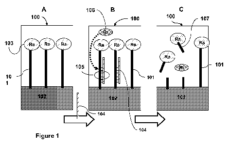
Figure 1 is a schematic depicting an exemplary method for detecting target
nucleic acid
using probe nucleic acid, a recognition restriction endonuclease, and reporter
nucleic acid.
Figure 2 is a schematic of an exemplary configuration of probe nucleic acid
that can be
used with the methods and materials provided herein for detecting target
nucleic acid.
Figure 3 is a schematic depicting an exemplary method for detecting target
nucleic acid
using probe nucleic acid, a recognition restriction endonuclease, first signal
expansion nucleic
acid, second signal expansion nucleic acid, and reporter nucleic acid.
Figure 4 is a schematic of an exemplary configuration of first signal
expansion nucleic
acid and second signal expansion nucleic acid that can be used with the
methods and materials
provided herein for detecting target nucleic acid. Such first signal expansion
nucleic acid and
second signal expansion nucleic acid can be used with or without reporter
nucleic acid. When
used without a separate reporter nucleic acid step, such signal expansion
nucleic acid can be
referred to as reporter nucleic acid.
Figure 5 is a schematic of an exemplary configuration of first signal
expansion nucleic
acid and second signal expansion nucleic acid that can be used with the
methods and materials
18
CA 02790123 2012-08-15
WO 2011/100749 PCT/US2011/024912
provided herein for detecting target nucleic acid. Such first signal expansion
nucleic acid and
second signal expansion nucleic acid can be used with or without reporter
nucleic acid. When
used without a separate reporter nucleic acid step, such signal expansion
nucleic acid can be
referred to as reporter nucleic acid.
Figure 6 contains line graphs demonstrating the effect of target
oligonucleotide
concentration (A) and recognition restriction endonuclease concentration (B)
on the cleavage of
HRP-labeled nucleic acid as detected by the formation of colored reaction
product.
Figure 7 is a schematic of an exemplary configuration for a single-use, pen-
style point of
care device.
DETAILED DESCRIPTION
This document provides methods and materials for detecting viral and/or
microbial
infections. For example, this document provides methods and materials related
to the use of an
enzymatic amplification cascade of restriction endonucleases to detect nucleic
acid of a virus or
microbe (e.g., a pathogen) within a sample (e.g., a biological sample such as
a blood sample,
mucus sample, or saliva sample) being tested, thereby assessing a mammal for a
possible
infection. In some cases, this document provides methods and materials for
detecting a target
microorganism's or virus's nucleic acid (e.g., a target pathogen's nucleic
acid). For example,
this document provides methods and materials for detecting the presence or
absence of target
nucleic acid (e.g., a target microorganism's or virus's nucleic acid) within a
sample (e.g., a
biological sample), methods and materials for detecting the amount of target
nucleic acid (e.g., a
target microorganism's or virus's nucleic acid) present within a sample (e.g.,
a biological
sample), kits for detecting the presence or absence of target nucleic acid
(e.g., a target
microorganism's or virus's nucleic acid) within a sample (e.g., a biological
sample), kits for
detecting the amount of target nucleic acid (e.g., a target microorganism's or
virus's nucleic
acid) present within a sample (e.g., a biological sample), and methods for
making such kits.
Any type of mammal can be assessed using the methods and materials provided
herein to
determine whether or not the mammal has a viral and/or microbial infection.
For example,
humans, dogs, cats, cows, horses, pigs, sheep, goats, monkeys, buffalo, bears,
whales, and
dolphins can be assessed for a viral and/or microbial infection as described
herein. Any type of
19
CA 02790123 2012-08-15
WO 2011/100749 PCT/US2011/024912
biological sample can be used with the methods and materials provided herein
to assess a
mammal for a viral and/or microbial infection. For example, nasal samples
(e.g., nasal swab
samples), throat samples (e.g., throat swab samples), sputum samples,
bronchial lavage samples,
tissue samples (e.g., tissue biopsy samples), cellular samples, and blood
samples can be collected
from a mammal and assessed to determine whether or not the mammal has a viral
or microbial
infection as described herein.
The methods and materials provided herein can be used to assess a mammal for
any type
of viral and/or microbial infection. Examples of potentially infecting viruses
include, without
limitation, influenza virus A and B, adenovirus 4, RSV, parainfluenza types 1,
2, and 3, human
coronaviruses OC43, 229E and HK, human metapneumovirus, rhinoviruses,
enteroviruses,
Hepatitis A, B, C and E viruses, rotavirus, human papillomavirus, measles
viruses, caliciviruses,
astrovirus, West Nile virus, Ebola virus, Dengue fever virus, African swine
fever, and human
immunodeficiency virus (HIV 1 and 2). Examples of potentially infecting
microorganisms
include, without limitation, bacterial microorganisms such as Staphylococcus
aureus,
Streptococcus pyogenes, Streptococcus pneumoniae, Mycoplasma pneumoniae,
Haemophilus
influenzae, Chlamydia pneumoniae, Bordetella pertussis, Mycobacterium
tuberculosis, E. coli
(e.g., enterohaemorrhagic E. coli such as 0157:H7 E. coli or enteropathogenic
E. coli),
Salmonella species (e.g., Salmonella enterica), Listeria monocytogenes,
Acinetobacter
baumanni, Klebsiella oxytoca, Giardia intestinalis, Sarcoptes scabiei, and
Treponema pallidum,
fungal microorganisms such as Aspergillus species (e.g., A. flavus, A.
fumigatus, and A. niger),
yeast (e.g., Candida norvegensis and C. albicans), Penicillium species,
Rhizopus species, and
Alternaria species, and protozoan microorganisms such as Cryptosporidium
parvum, Giardia
lamblia, and Toxoplasma gondii. In some cases, a mammal can be assessed for
one or more of
the viruses or microorganisms listed in Table 1 using the methods and
materials provided herein.
When designing a method for detecting a virus or microorganism listed in Table
1, a probe
nucleic acid can be designed that is complementary to a portion of any of the
indicated sequences
from Table 1. For example, when designing a method for detecting influenza
virus A, a probe
nucleic acid can be designed that is complementary to a portion of the
influenza A sequence set
forth in GenBank GI number 8486122.
CA 02790123 2012-08-15
WO 2011/100749 PCT/US2011/024912
Table 1. Types of infections that can be detected.
Mammal Infection Genomic sequence Sample
(GenBank gi number)
Human Upper respiratory viral infection: Nasal swab
influenza virus A Refseq: NC_002023, GenBank : or mucus
V00603
influenza virus B Refseq: NC_002209, GenBank :
J02095
adenovirus 4 Refseq: NC - 003266;
51527264
RSV NC 001781, GenBanV:
AF013254
parainfluenza type 1 Refseq: NC_003461,
GenBank : AF457102
parainfluenza type 2 Refseq: NC_003443,
GenBank : X57559
parainfluenza type 3 Refseq: NC_001796,
GenBank : ABO12132
human coronavirus OC43 Refseq: NC_012920,
GenBank : J01415
human coronavirus 229E Refseq: NC_002645,
GenBank : AF304460
human coronavirus HK Refseq: NC_012951,
GenBank : FJ938052
human metapneumovirus Refseq: NC_004148,
GenBank : AY297749
rhinoviruses Refseq: NC_001490,
GenBank : K02121
enteroviruses Refseq: NC_013115,
GenBank : AB426609
Upper respiratory microbial infection: Refseq: NZACOT00000000,
Staphylococcus aureus GenBank : ACOT00000000
Streptococcus pyogenes Refseq: NZ_AAFV00000000,
GenBank : AAFV00000000
Streptococcus pneumoniae Refseq: NZ_ACJP00000000,
GenBank : ACJP00000000
Mycoplasma pneumoniae Refseq: NC_000912,
GenBank : U00089
21
CA 02790123 2012-08-15
WO 2011/100749 PCT/US2011/024912
Haemophilus influenzae Refseq: NZ_ABWV00000000,
GenBank : ABWV00000000
Chlamydi Chlamydophila Refseq: NC_005043, GenBankR:
pneumoniae TW-183 AE009440
Bordetella pertussis Refseq: NC_008459,
GenBank : AB237782
Mycobacterium tuberculosis T46 Refseq: NZ_ACH000000000,
GenBank : ACH000000000
Human Human immunodeficiency virus (HIV) Refseq: NC_001722, Blood
GenBank : M30502 sample
Human Rabies Refseq: NC_001542,
GenBank : M13215
Human Lymes disease
Bat Rabies Refseq: NC_009528, GenBank:
EF157977
Dog Lymes disease
Cat Rabies Refseq: NC_001542,
GenBank : M13215
Cat Leptospira Refseq: NC_010846,
GenBank : CP000779
Cat Leukemia Virus (FELV)
Bovine Bovine herpesvirus 1 Refseq: NC_001847,
GenBank : AJO04801
Bovine Foot-and-Mouth Disease Refseq: NC_011452,
GenBank : AY593850
Horse Equine Encephalomyelitis (sleeping Refseq: NC_003908,
sickness) GenBank : AF214040
Horse Strangles (shipping fever) Refseq: NC_012471,
GenBank : FM204883
In some cases, nucleic acid sequences of viruses and microorganisms known to
infect the
upper respiratory tract of mammals (e.g., humans) can be used to design probe
nucleic acids for
detecting upper respiratory tract infections. For example, probe nucleic acids
having the
sequences set forth in Table 2 can be used with the indicated recognition
restriction endonuclease
to detect the indicated target nucleic acids of the indicated pathogens. In
some cases, a single kit
can be designed as described herein to detect one or more of the indicated
pathogens of Table 2.
22
CA 02790123 2012-08-15
WO 2011/100749 PCT/US2011/024912
Table 2. Target nucleic acids, recognition restriction endonucleases, and
probe nucleic acids for
detecting the indicated pathogens.
Pathogen Target Nucleic Recongition Sequence for Probe Nucleic Acid
Acid Restriction
Endonuclease
Staphylococcus gyrB (DNA EcoRV (gatatc) TGATCTAGCGAAAGCAAGATA
aureus gyrase subunit B) TCACAAAATCGTCATTATG
(SEQ ID NO:l
methicillin- mecA (penicillin Pstl (ctgcag) ATTGGCAAATCCGGTACTGCA
resistant binding protein 2) GAACTCAAAATGAAACAAG
(MRSA) (SEQ ID NO:2)
Streptococcus ply Pstl (ctgcag) AACAGAGAGGAATTTCTGCAG
pneumoniae (pneumolysin) AGCGTCCTTTGGTCTATAT
(SEQ ID NO:3)
Streptococcus speA (exotoxin BstEII ATATTTTCTTTATGAGGGTGA
pyogenes type A precursor) (ggtgacc) CCCTGTTACTCACGAGAAT
(SEQ ID NO:4)
Mycobacterium rpoB (RNA HincIl (gttgac) AACAACCCGCTGTCGGGGTTG
tuberculosis polymerase ACCCACAAGCGCCGACTGT
subunit beta) (SEQ ID NO:5)
Influenza A Ml (matrix Pstl (ctgcag) ACCGTGCCCAGTGAGCGAGGA
virus protein) CTGCAGCGTAGACGCTTTG
(SEQ ID NO:6)
Influenza B Ml (matrix HindIll (aagctt) AATGAGAAGATGTGTAAGCTT
virus protein) TCATGAAGCATTTGAAATA
(SEQ ID NO:7)
Adenovirus 4 gp 12 BglII (agatct) CCAACTCGCCGGATCGGGAAG
(E) (glycoprotein 12) ATCTTCCTTCACGCCTCGT
(SEQ ID NO:8
Respiratory M2 (matrix EcoRV (gatatc) CCATAAAAACCACATTGGATA
syncytial virus protein) TCCACAAGAGCATAACCAT
(SEQ ID NO:9)
In some cases, an enzymatic amplification cascade can be used to assess the
presence or
absence of microorganisms and viruses associated with sexually transmitted
infections (STIs).
Millions of STIs occur every year in the United States, and if untreated or
allowed to proceed to
advanced stages, they have severe consequences (e.g., infertility, blindness,
or brain damage).
Common STIs include, without limitation, gonorrhea, Chlamydia, syphilis, and
genital herpes.
Gonorrhea, Chlamydia, and syphilis are bacterial, while genital herpes is
viral. An enzymatic
amplification cascade can be used to detect, for example, N. gonorrhoeae,
Chlamydia
23
CA 02790123 2012-08-15
WO 2011/100749 PCT/US2011/024912
trachomatis, Treponema pallidum, or a herpes simplex virus such as HSV-2.
Probe nucleic acids
can be designed to contain nucleic acid sequences from the microorganism or
virus of interest, as
described herein. The type of sample used for the reaction can vary depending
on the target of
interest. For example, urine samples or urethral or endocervical swab samples
can be tested for
the presence or absence of gonorrhea and/or Chlamydia. Blood, plasma, or
lesion swab samples
can be tested for the presence or absence of syphilis, and genital sore swabs
can be tested for the
presence or absence of HSV2.
In one embodiment, a method for assessing a mammal for a viral and/or
microbial
infection can include detecting a target virus's and/or microorganism's
nucleic acid (e.g., a target
nucleic acid) within a biological sample obtained from the mammal. For
example, a biological
sample (e.g., a blood sample to be tested) can be placed in contact with probe
nucleic acid. The
probe nucleic acid can be designed to have a single-stranded portion with a
nucleotide sequence
that is complementary to at least a portion of the target nucleic acid to be
detected. In this case,
target nucleic acid present within the sample can hybridize with the
complementary sequence of
this single-stranded portion of the probe nucleic acid to form a double-
stranded section with one
strand being target nucleic acid and the other strand being probe nucleic
acid. In addition, the
single-stranded portion of the probe nucleic acid having the nucleotide
sequence that is
complementary to at least a portion of the target nucleic acid to be detected
can be designed such
that hybridization with the target nucleic acid creates a restriction
endonuclease cut site. Thus,
target nucleic acid present within the sample can hybridize with the
complementary sequence of
the single-stranded portion of the probe nucleic acid to form a double-
stranded section that
creates a cut site for a restriction endonuclease. This cut site created by
the hybridization of
target nucleic acid to probe nucleic acid can be referred to as a recognition
restriction
endonuclease cut site. In addition, a restriction endonuclease that cleaves
nucleic acid at such a
recognition restriction endonuclease cut site can be referred to as a
recognition restriction
endonuclease.
The probe nucleic acid also can be designed to contain a restriction
endonuclease. This
restriction endonuclease, which can be a component of the probe nucleic acid,
can be referred to
as an amplifying restriction endonuclease. An amplifying restriction
endonuclease is typically a
different restriction endonuclease than the restriction endonuclease that is
used as a recognition
24
CA 02790123 2012-08-15
WO 2011/100749 PCT/US2011/024912
restriction endonuclease. For example, when an EcoRI restriction endonuclease
is used as a
recognition restriction endonuclease, a restriction endonuclease other than an
EcoRI restriction
endonuclease (e.g., a Hind III restriction endonuclease) is used as an
amplifying restriction
endonuclease. Thus, in general, probe nucleic acid is designed to contain an
amplifying
restriction endonuclease and to have a nucleotide sequence such that the
target nucleic acid can
hybridize to the probe nucleic acid and create a recognition restriction
endonuclease cut site for a
recognition restriction endonuclease. In some cases, the probe nucleic acid
can be attached to a
solid support (e.g., a well of a microtiter plate). For example, the probe
nucleic acid can be
attached to a solid support such that cleavage at the recognition restriction
endonuclease cut site
via the recognition restriction endonuclease releases a portion of the probe
nucleic acid that
contains the amplifying restriction endonuclease.
After contacting the sample (e.g., a biological sample) that may or may not
contain target
nucleic acid with the probe nucleic acid that is attached to a solid support,
the target nucleic acid,
if present in the sample, can hybridize to the probe nucleic acid and create
the recognition
restriction endonuclease cut site. At this point, the recognition restriction
endonuclease, whether
added to the reaction or already present in the reaction, can cleave the probe
nucleic acid at the
recognition restriction endonuclease cut sites that are formed by the
hybridization of target
nucleic acid to the probe nucleic acid, thereby releasing the portion of the
probe nucleic acid that
contains the amplifying restriction endonuclease from the solid support. The
number of
amplifying restriction endonuclease-containing portions of the probe nucleic
acid that are
released from the solid support can be in an essentially linear relationship
(e.g., essentially a one-
for-one relationship) with the number of target nucleic acid molecules that
hybridize with the
probe nucleic acid to form the recognition restriction endonuclease cut site.
The portions of the probe nucleic acid containing the amplifying restriction
endonuclease
that were released from the solid support can be collected and placed in
contact with reporter
nucleic acid. For example, the released portions of the probe nucleic acid, if
present, can be
transferred from one well of a microtiter plate (e.g., a 96-well plate) that
contained the probe
nucleic acid to another well of a microtiter plate that contains the reporter
nucleic acid. The
reporter nucleic acid can be designed to have a double-stranded portion with a
restriction
endonuclease cut site for the amplifying restriction endonuclease of the probe
nucleic acid. This
CA 02790123 2012-08-15
WO 2011/100749 PCT/US2011/024912
restriction endonuclease cut site for the amplifying restriction endonuclease
can be referred to as
an amplifying restriction endonuclease cut site. If portions of the probe
nucleic acid containing
the amplifying restriction endonuclease are present and placed in contact with
the reporter
nucleic acid, then the reporter nucleic acid can be cleaved at the amplifying
restriction
endonuclease cut site by the amplifying restriction endonuclease. Since the
amplifying
restriction endonucleases of the released portions of the probe nucleic acid
are free to carry out
repeated cleavage events, the number of reporter nucleic acid molecules that
are cleaved can
greatly exceed the number of amplifying restriction endonucleases present in
the reaction. For
example, the number of cleaved reporter nucleic acid molecules can greatly
exceed (e.g.,
exponentially exceed) the number of amplifying restriction endonucleases
present in the reaction
and therefore can greatly exceed (e.g., exponentially exceed) the number of
target nucleic acid
molecules that were present in the sample contacted with the probe nucleic
acid. Such a greatly
expanded relationship (e.g., an exponential relationship) can allow very small
amounts of target
nucleic acid present in the sample to be readily detected.
After the released portions of the probe nucleic acid, if present, are
contacted with the
reporter nucleic acid, the presence or absence of cleaved reporter nucleic
acid can be determined.
The presence of cleaved reporter nucleic acid can indicate that the sample
contained the target
nucleic acid, thereby indicating that the sample contained the target virus or
microorganism for
which the sample is being tested, while the absence of cleaved reporter
nucleic acid can indicate
that the sample lacked the target nucleic acid, thereby indicating that the
sample lacked the target
virus or microorganism for which the sample is being tested. In some cases,
the amount of
cleaved reporter nucleic acid can be determined. In such cases, the amount of
cleaved reporter
nucleic acid can indicate the amount of target nucleic acid present in the
sample, which can
indicated the degree or level of infection by the target virus or
microorganism for which the
sample is being tested. A standard curve using known amounts of target nucleic
acid or known
amounts target viruses or microorganisms can be used to aid in the
determination of the amount
of target nucleic acid or target viruses or microorganisms present within a
sample.
In some cases, the reporter nucleic acid can contain a label to aid in the
detection of
cleaved reporter nucleic acid. For example, reporter nucleic acid can contain
a fluorescent label
and a quencher such that cleaved reporter nucleic acid provides a fluorescent
signal and
26
CA 02790123 2012-08-15
WO 2011/100749 PCT/US2011/024912
uncleaved reporter nucleic acid does not provide a fluorescent signal. In some
cases, the reporter
nucleic acid can contain a label (e.g., a colorimetric label, a fluorescent
label or an enzyme (e.g.,
a redox enzyme) such as horse radish peroxidase) and can be attached to a
solid support (e.g., a
well of a microtiter plate). For example, the reporter nucleic acid can be
attached to a solid
support such that cleavage at the amplifying restriction endonuclease cut site
by the amplifying
restriction endonuclease releases a portion of the reporter nucleic acid that
contains the label.
The resulting reaction mixture can be collected and assessed for the presence,
absence, or
amount of released portions of the reporter nucleic acid using the label. For
example, the
released portions of the reporter nucleic acid, if present, can be transferred
from one well of a
microtiter plate (e.g., a 96-well plate) that contained the reporter nucleic
acid to another well of a
microtiter plate, where the transferred material can be assessed for a signal
from the label.
One example of a method of detecting target nucleic acid that includes using
probe
nucleic acid and reporter nucleic acid is set forth in Figure 1. With
reference to Figure 1, first
reaction chamber 100 (e.g., a microtiter plate well) can contain probe nucleic
acid 101. Probe
nucleic acid 101 can be attached (e.g., immobilized) to solid support 102 and
can include
amplifying restriction endonuclease 103 (Ra). Probe nucleic acid 101 can be
attached to solid
support 102 such that amplifying restriction endonuclease 103 is released from
solid support 102
upon cleavage of a nucleic acid component of probe nucleic acid 101. Probe
nucleic acid 101
can have a single-stranded section having a nucleotide sequence that is
complementary to at least
a portion of target nucleic acid 104. Probe nucleic acid 101 can be contacted
with a sample that
may or may not contain target nucleic acid 104. If target nucleic acid 104 is
present, at least a
portion of target nucleic acid 104 and probe nucleic acid 101 can hybridize to
form a double-
stranded section of nucleic acid. Such a double-stranded section can contain
at least one
recognition restriction endonuclease cut site 105. Addition of recognition
restriction
endonuclease 106 (Rr) to first reaction chamber 100 can result in the cleave
of probe nucleic acid
101 at recognition restriction endonuclease cut site 105 formed by one strand
of probe nucleic
acid and one strand of target nucleic acid, thereby releasing portion 107 of
probe nucleic acid
101 from solid support 102. Portion 107 can include amplifying restriction
endonuclease 103.
The reaction product from first reaction chamber 100 containing released
portion 107, if
target nucleic acid 104 was present, can be transferred (e.g., manually or
automatically) to
27
CA 02790123 2012-08-15
WO 2011/100749 PCT/US2011/024912
second reaction chamber 120. Second reaction chamber 120 can contain reporter
nucleic acid
121. Reporter nucleic acid 121 can be attached (e.g., immobilized) to solid
support 122 and can
include marker (e.g., a label) 123 (M). Reporter nucleic acid 121 can be
attached to solid
support 122 such that marker 123 is released from solid support 122 upon
cleavage of a nucleic
acid component of reporter nucleic acid 121. Reporter nucleic acid 121 can
have at least one
double-stranded portion that contains at least one amplifying restriction
endonuclease cut site
124. Addition of the reaction product from first reaction chamber 100 to
second reaction
chamber 120 can result in the cleavage of reporter nucleic acid 121 at
amplifying restriction
endonuclease cut site 124 if the reaction product contains portion 107. Such
cleavage of reporter
nucleic acid 121 can result in the release of portion 127 from solid support
122. Portion 127 can
include marker 123.
The reaction product from second reaction chamber 120 can be assessed to
determine the
presence, absence, or amount of portion 127. The presence of portion 127 can
indicate that the
sample contained target nucleic acid 104, while the absence of portion 127 can
indicate that the
sample lacked target nucleic acid 104. In some cases, the amount of portion
127 can be
determined. In such cases, the amount of portion 127 can indicate the amount
of target nucleic
acid 104 present in the sample. The presence, absence, or amount of portion
127 can be
determined using marker 123, and portion 127 having marker 123 can be
distinguished from
uncleaved reporter nucleic acid 121 having marker 123 since, in this example,
portion 127 is
released from solid support 122, while uncleaved reporter nucleic acid 121
remains attached to
solid support 122. For example, in some cases, the reaction product from
second reaction
chamber 120 can be transferred to third reaction chamber where the presence or
absence of
portion 127 via marker 123 is assessed. If portion 127 is present, the amount
of portion 127
present can be quantified.
Probe nucleic acid 101 and reporter nucleic acid 121 can have various
configurations.
For example, with reference to Figure 1, probe nucleic acid 101 can be
designed to have a single
nucleic acid strand such that the entire nucleic acid component of probe
nucleic acid 101 is
single-stranded prior to contact with target nucleic acid 104. In another
example, with reference
to Figure 2, probe nucleic acid 101 can be designed to have first strand 128
and second strand
108. First strand 128 can be attached to solid support 102 and can be designed
to have a single-
28
CA 02790123 2012-08-15
WO 2011/100749 PCT/US2011/024912
stranded section having a nucleotide sequence that is complementary to at
least a portion of
target nucleic acid 104. Second strand 108 can include amplifying restriction
endonuclease 103
and can have a single-stranded section having a nucleotide sequence that can
hybridize to first
strand 128. In some cases, first strand 128 and second strand 108 can be
synthesized or obtained
separately and then mixed together to form probe nucleic acid 101. For
example, first strand 128
can be synthesized, biotinylated, and attached to a streptavidin-coated solid
support. After
synthesizing the nucleic acid component of second strand 108 and attaching
amplifying
restriction endonuclease 103 to the synthesized nucleic acid component, second
strand 108 can
be incubated with first strand 128 to form nucleic acid probe 101. In some
cases, probe nucleic
acid 101 can contain more than two strands. For example, probe nucleic acid
can include first
strand 150, second strand 152, and third strand 154. In this case, first
strand 150 can be attached
to solid support 102, second strand 152 can be hybridized to first strand 150
and can include a
single-stranded section having a nucleotide sequence that is complementary to
at least a portion
of target nucleic acid 104, and third strand 154 can be hybridized to second
strand 152 and can
be attached to amplifying restriction endonuclease 103. Similar one, two,
three, or more strand
configurations can be used to make reporter nucleic acid.
In another embodiment, a method for detecting target nucleic acid can include
contacting
a sample (e.g., a biological sample to be tested) with probe nucleic acid. The
probe nucleic acid
can be designed to have a single-stranded portion with a nucleotide sequence
that is
complementary to at least a portion of the target nucleic acid to be detected.
In this case, target
nucleic acid present within the sample can hybridize with the complementary
sequence of this
single-stranded portion of the probe nucleic acid to form a double-stranded
section with one
strand being target nucleic acid and the other strand being probe nucleic
acid. In addition, the
single-stranded portion of the probe nucleic acid having the nucleotide
sequence that is
complementary to at least a portion of the target nucleic acid to be detected
can be designed such
that hybridization with the target nucleic acid creates a recognition
restriction endonuclease cut
site. Thus, target nucleic acid present within the sample can hybridize with
the complementary
sequence of the single-stranded portion of the probe nucleic acid to form a
double-stranded
section that creates a recognition restriction endonuclease cut site for a
recognition restriction
endonuclease. The probe nucleic acid also can be designed to contain an
amplifying restriction
29
CA 02790123 2012-08-15
WO 2011/100749 PCT/US2011/024912
endonuclease. Since this method includes the use of two or more different
amplifying restriction
endonucleases, the amplifying restriction endonuclease that is a component of
the probe nucleic
acid can be referred to as a first or an initial amplifying restriction
endonuclease, with additional
amplifying restriction endonucleases being referred to as second, third, and
so on or secondary,
tertiary, and so on amplifying restriction endonucleases. This initial
amplifying restriction
endonuclease is typically a different restriction endonuclease than the
restriction endonuclease
that is used as a recognition restriction endonuclease. For example, when an
EcoRI restriction
endonuclease is used as a recognition restriction endonuclease, a restriction
endonuclease other
than an EcoRI restriction endonuclease (e.g., a Hind III restriction
endonuclease) is used as an
initial amplifying restriction endonuclease. Thus, in general, probe nucleic
acid is designed to
contain an initial amplifying restriction endonuclease and to have a
nucleotide sequence such
that the target nucleic acid can hybridize to the probe nucleic acid and
create a recognition
restriction endonuclease cut site for a recognition restriction endonuclease.
In some cases, the
probe nucleic acid can be attached to a solid support (e.g., a well of a
microtiter plate). For
example, the probe nucleic acid can be attached to a solid support such that
cleavage at the
recognition restriction endonuclease cut site via the recognition restriction
endonuclease releases
a portion of the probe nucleic acid that contains the initial amplifying
restriction endonuclease.
After contacting the sample that may or may not contain target nucleic acid
with the
probe nucleic acid that is attached to a solid support, the target nucleic
acid, if present in the
sample, can hybridize to the probe nucleic acid and create the recognition
restriction
endonuclease cut site. At this point, the recognition restriction
endonuclease, whether added to
the reaction or already present in the reaction, can cleave the probe nucleic
acid at the
recognition restriction endonuclease cut sites that are formed by the
hybridization of target
nucleic acid to the probe nucleic acid, thereby releasing the portion of the
probe nucleic acid that
contains the initial amplifying restriction endonuclease from the solid
support. The number of
initial amplifying restriction endonuclease-containing portions of the probe
nucleic acid that are
released from the solid support can be in an essentially linear relationship
(e.g., essentially a one-
for-one relationship) with the number of target nucleic acid molecules that
hybridize with the
probe nucleic acid to form the recognition restriction endonuclease cut site.
CA 02790123 2012-08-15
WO 2011/100749 PCT/US2011/024912
The portions of the probe nucleic acid containing the initial amplifying
restriction
endonuclease that were released from the solid support can be collected and
placed in contact
with first signal expansion nucleic acid and second signal expansion nucleic
acid. The first
signal expansion nucleic acid can be designed to have a double-stranded
portion with a
restriction endonuclease cut site for the initial amplifying restriction
endonuclease of the probe
nucleic acid. This restriction endonuclease cut site for the initial
amplifying restriction
endonuclease can be referred to as an initial amplifying restriction
endonuclease cut site. The
first signal expansion nucleic acid also can be designed to contain a
secondary amplifying
restriction endonuclease. The second signal expansion nucleic acid can be
designed to have a
double-stranded portion with a restriction endonuclease cut site for the
secondary amplifying
restriction endonuclease of the first signal expansion nucleic acid. This
restriction endonuclease
cut site for the secondary amplifying restriction endonuclease can be referred
to as a secondary
amplifying restriction endonuclease cut site. The second signal expansion
nucleic acid also can
be designed to contain an initial amplifying restriction endonuclease. For
example, when an
EcoRI restriction endonuclease is used as a recognition restriction
endonuclease and a Hindlll
restriction endonuclease is used as an initial amplifying restriction
endonuclease of the probe
nucleic acid, a Smal restriction endonuclease can be used as a secondary
amplifying restriction
endonuclease of the first signal expansion nucleic acid and a Hindlll
restriction endonuclease
can be used as the initial amplifying restriction endonuclease of the second
signal expansion
nucleic acid.
In some cases, the first signal expansion nucleic acid and second signal
expansion nucleic
acid can be attached to a solid support (e.g., a well of a microtiter plate).
For example, the first
signal expansion nucleic acid can be attached to a solid support such that
cleavage at the initial
amplifying restriction endonuclease cut site via the initial amplifying
restriction endonuclease
releases a portion of the first signal expansion nucleic acid that contains
the secondary
amplifying restriction endonuclease, and the second signal expansion nucleic
acid can be
attached to a solid support such that cleavage at the secondary amplifying
restriction
endonuclease cut site via the secondary amplifying restriction endonuclease
releases a portion of
the second signal expansion nucleic acid that contains the initial amplifying
restriction
endonuclease. The first signal expansion nucleic acid can be attached to the
same solid support
31
CA 02790123 2012-08-15
WO 2011/100749 PCT/US2011/024912
(e.g., two different sub-compartments of a larger compartment) that contains
the second signal
expansion nucleic acid provided that the secondary amplifying restriction
endonuclease of
uncleaved first signal expansion nucleic acid is unable to cleave the second
signal expansion
nucleic acid and provided that the initial amplifying restriction endonuclease
of uncleaved
second signal expansion nucleic acid is unable to cleave the first signal
expansion nucleic acid.
In some cases, the first signal expansion nucleic acid can be attached to the
same solid support
within a joint compartment such that the first signal expansion nucleic acid
is within a first
compartment of the joint compartment and the second signal expansion nucleic
acid is within a
second compartment of the joint compartment. In such cases, the secondary
amplifying
restriction endonuclease of uncleaved first signal expansion nucleic acid in
the first compartment
is unable to cleave the second signal expansion nucleic acid located in the
second compartment,
while the secondary amplifying restriction endonuclease of cleaved first
signal expansion nucleic
acid is capable of moving (e.g., diffusing) from the first compartment to the
second compartment
to cleave the second signal expansion nucleic acid located in the second
compartment. In
addition, the initial amplifying restriction endonuclease of uncleaved second
signal expansion
nucleic acid in the second compartment is unable to cleave the first signal
expansion nucleic acid
located in the first compartment, while the initial amplifying restriction
endonuclease of cleaved
second signal expansion nucleic acid is capable of moving (e.g., diffusing)
from the second
compartment to the first compartment to cleave the first signal expansion
nucleic acid located in
the first compartment.
If portions of the probe nucleic acid containing the initial amplifying
restriction
endonuclease are present and placed in contact with the first signal expansion
nucleic acid, then
the first signal expansion nucleic acid can be cleaved at the initial
amplifying restriction
endonuclease cut site by the initial amplifying restriction endonuclease,
thereby releasing a
portion of the first signal expansion nucleic acid that contains the secondary
amplifying
restriction endonuclease from the solid support. The released portions of the
first signal
expansion nucleic acid containing the secondary amplifying restriction
endonuclease can be free
to cleave the second signal expansion nucleic acid at the secondary amplifying
restriction
endonuclease cut site, thereby releasing a portion of the second signal
expansion nucleic acid
that contains the initial amplifying restriction endonuclease from the solid
support. Since the
32
CA 02790123 2012-08-15
WO 2011/100749 PCT/US2011/024912
initial amplifying restriction endonucleases of the released portions of the
probe nucleic acid, the
initial amplifying restriction endonucleases of the released portions of the
second signal
expansion nucleic acid, and the secondary amplifying restriction endonucleases
of the released
portions of the first signal expansion nucleic acid are free to carry out
repeated cleavage events,
the number of released portions containing the initial amplifying restriction
endonucleases is
greatly increased from the number that were released by the recognition
restriction endonuclease.
For example, the number of cleaved first signal expansion nucleic acid
molecules can greatly
exceed (e.g., exponentially exceed) the number of released portions of the
probe nucleic acid,
and the number of cleaved second signal expansion nucleic acid molecules can
greatly exceed
(e.g., exponentially exceed) the number of released portions of the probe
nucleic acid. Such a
greatly expanded relationship (e.g., an exponential relationship) can allow
very small amounts of
target nucleic acid present in the sample to be readily detected.
In some cases, this method can be performed with the first signal expansion
nucleic acid
being attached to a solid support that is different from the solid support
that contains the second
signal expansion nucleic acid. For example, the first signal expansion nucleic
acid can be
attached to one well of a microtiter plate, while the second signal expansion
nucleic acid can be
attached to a different well of a microtiter plate. In this case, the
resulting reaction material from
the well with the first signal expansion nucleic acid can be collected and
transferred to the well
containing the second signal expansion nucleic acid.
The portions of the second signal expansion nucleic acid containing the
initial amplifying
restriction endonuclease that were released from the solid support containing
the second signal
expansion nucleic acid along with any other released portions in this reaction
(e.g., the released
portions of the probe nucleic acid containing the initial amplifying
restriction endonuclease and
the released portions of the first signal expansion nucleic acid containing
the secondary
amplifying restriction endonuclease) can be collected and placed in contact
with reporter nucleic
acid. For example, the released portions, if present, can be transferred from
one well of a
microtiter plate (e.g., a 96-well plate) that contained the second signal
expansion nucleic acid to
another well of a microtiter plate that contains the reporter nucleic acid.
The reporter nucleic
acid can be designed to have a double-stranded portion with a restriction
endonuclease cut site
for the initial amplifying restriction endonuclease. If released portions
containing the initial
33
CA 02790123 2012-08-15
WO 2011/100749 PCT/US2011/024912
amplifying restriction endonuclease are present and placed in contact with the
reporter nucleic
acid, then the reporter nucleic acid can be cleaved at the initial amplifying
restriction
endonuclease cut site by the initial amplifying restriction endonuclease.
Since the initial
amplifying restriction endonucleases of the released portions are free to
carry out repeated
cleavage events, the number of reporter nucleic acid molecules that are
cleaved can greatly
exceed the number of initial amplifying restriction endonucleases present in
the reaction. For
example, the number of cleaved reporter nucleic acid molecules can greatly
exceed (e.g.,
exponentially exceed) the number of initial amplifying restriction
endonucleases present in the
reaction and therefore can greatly exceed (e.g., exponentially exceed) the
number of target
nucleic acid molecules that were present in the sample contacted with the
probe nucleic acid.
Such a greatly expanded relationship (e.g., an exponential relationship) can
allow very small
amounts of target nucleic acid present in the sample to be readily detected.
After the released portions containing the initial amplifying restriction
endonuclease, if
present, are contacted with the reporter nucleic acid, the presence or absence
of cleaved reporter
nucleic acid can be determined. The presence of cleaved reporter nucleic acid
can indicate that
the sample contained the target nucleic acid, thereby indicating that the
sample contained the
target virus or microorganism for which the sample is being tested, while the
absence of cleaved
reporter nucleic acid can indicate that the sample lacked the target nucleic
acid, thereby
indicating that the sample lacked the target virus or microorganism for which
the sample is being
tested.
In some cases, the amount of cleaved reporter nucleic acid can be determined.
In such
cases, the amount of cleaved reporter nucleic acid can indicate the amount of
target nucleic acid
present in the sample, which can indicated the degree or level of infection by
the target virus or
microorganism for which the sample is being tested. A standard curve using
known amounts of
target nucleic acid or known amounts target viruses or microorganisms can be
used to aid in the
determination of the amount of target nucleic acid or target viruses or
microorganisms present
within a sample.
In some cases, the reporter nucleic acid can contain a label to aid in the
detection of
cleaved reporter nucleic acid. For example, reporter nucleic acid can contain
a fluorescent label
and a quencher such that cleaved reporter nucleic acid provides a fluorescent
signal and
34
CA 02790123 2012-08-15
WO 2011/100749 PCT/US2011/024912
uncleaved reporter nucleic acid does not provide a fluorescent signal. In some
cases, the reporter
nucleic acid can contain a label (e.g., a colorimetric label, fluorescent
label or an enzyme such as
horse radish peroxidase) and can be attached to a solid support (e.g., a well
of a microtiter plate).
For example, the reporter nucleic acid can be attached to a solid support such
that cleavage at the
initial amplifying restriction endonuclease cut site by the initial amplifying
restriction
endonuclease releases a portion of the reporter nucleic acid that contains the
label. The resulting
reaction mixture can be collected and assessed for the presence, absence, or
amount of released
portions of the reporter nucleic acid using the label. For example, the
released portions of the
reporter nucleic acid, if present, can be transferred from one well of a
microtiter plate (e.g., a 96-
well plate) that contained the reporter nucleic acid to another well of a
microtiter plate, where the
transferred material can be assessed for a signal from the label.
In some cases, the presence or absence of cleaved first signal expansion
nucleic acid,
cleaved second signal expansion nucleic acid, or both can be determined. The
presence of such
cleaved nucleic acid can indicate that the sample contained the target nucleic
acid, thereby
indicating that the sample contained the target virus or microorganism for
which the sample is
being tested, while the absence of such cleaved nucleic acid can indicate that
the sample lacked
the target nucleic acid, thereby indicating that the sample lacked the target
virus or
microorganism for which the sample is being tested. In some cases, the amount
of cleaved first
signal expansion nucleic acid, cleaved second signal expansion nucleic acid,
or both can be
determined. In such cases, the amount of cleaved nucleic acid can indicate the
amount of target
nucleic acid present in the sample, which can indicate the degree or level of
infection by the
target virus or microorganism for which the sample is being tested. In these
cases, the use of
cleaved first signal expansion nucleic acid, cleaved second signal expansion
nucleic acid, or both
to assess the sample for target nucleic acid can be in addition to the use of
a separate reporter
nucleic acid step or can replace the use of a separate reporter nucleic acid
step. In some cases,
the first signal expansion nucleic acid, the second signal expansion nucleic
acid, or both can be
labeled in a manner similar to that described herein for the reporter nucleic
acid to aid in
detection. When the presence, absence, or amount of cleaved first signal
expansion nucleic acid,
cleaved second signal expansion nucleic acid, or both are determined to assess
the sample for
target nucleic acid, the first signal expansion nucleic acid can be referred
to as a first reporter
CA 02790123 2012-08-15
WO 2011/100749 PCT/US2011/024912
nucleic acid and the second signal expansion nucleic acid can be referred to
as a second reporter
nucleic acid even though they include amplifying restriction endonucleases. A
standard curve
using known amounts of target nucleic acid or known amounts of target viruses
or
microorganisms can be used to aid in the determination of the amount of target
nucleic acid or
target viruses or microorganisms present within a sample.
Examples of a method of detecting target nucleic acid that includes using
probe nucleic
acid, first signal expansion nucleic acid, second signal expansion nucleic
acid, and reporter
nucleic acid are set forth in Figures 3-5. With reference to Figure 3, first
reaction chamber 200
(e.g., a microtiter plate well) can contain probe nucleic acid 201. Probe
nucleic acid 201 can be
attached (e.g., immobilized) to solid support 202 and can include initial
amplifying restriction
endonuclease 203 (Ra). Probe nucleic acid 201 can be attached to solid support
202 such that
initial amplifying restriction endonuclease 203 is released from solid support
202 upon cleavage
of a nucleic acid component of probe nucleic acid 201. Probe nucleic acid 201
can have a
single-stranded section having a nucleotide sequence that is complementary to
at least a portion
of target nucleic acid 204. Probe nucleic acid 201 can be contacted with a
sample that may or
may not contain target nucleic acid 204. If target nucleic acid 204 is
present, at least a portion of
target nucleic acid 204 and probe nucleic acid 201 can hybridize to form a
double-stranded
section of nucleic acid. Such a double-stranded section can contain at least
one recognition
restriction endonuclease cut site 205. Addition of recognition restriction
endonuclease 206 (Rr)
to first reaction chamber 200 can result in the cleavage of probe nucleic acid
201 at recognition
restriction endonuclease cut site 205 formed by one strand of probe nucleic
acid and one strand
of target nucleic acid, thereby releasing portion 207 of probe nucleic acid
201 from solid support
202. Portion 207 can include initial amplifying restriction endonuclease 203.
The reaction product from first reaction chamber 200 containing released
portion 207, if
target nucleic acid 204 was present, can be transferred (e.g., manually or
automatically) to
second reaction chamber 220. Second reaction chamber 220 can contain first
signal expansion
nucleic acid 226 and second signal expansion nucleic acid 225. First signal
expansion nucleic
acid 226 can have at least one double-stranded portion that contains at least
one initial
amplifying restriction endonuclease cut site 230. First signal expansion
nucleic acid 226 can be
attached (e.g., immobilized) to solid support 222 and can include secondary
amplifying
36
CA 02790123 2012-08-15
WO 2011/100749 PCT/US2011/024912
restriction endonuclease 223 (Rb). First signal expansion nucleic acid 226 can
be attached to
solid support 222 such that portion 234 containing secondary amplifying
restriction
endonuclease 223 is released from solid support 222 upon cleavage of first
signal expansion
nucleic acid 226 at initial amplifying restriction endonuclease cut site 230.
For clarity, frame E
of Figure 3 omits depicting one strand from the cleaved versions of first
signal expansion nucleic
acid 226 and second signal expansion nucleic acid 225.
Second signal expansion nucleic acid 225 can have at least one double-stranded
portion
that contains at least one secondary amplifying restriction endonuclease cut
site 232. Second
signal expansion nucleic acid 225 can be attached (e.g., immobilized) to solid
support 222 and
can include initial amplifying restriction endonuclease 224. Second signal
expansion nucleic
acid 225 can be attached to solid support 222 such that portion 236 containing
initial amplifying
restriction endonuclease 224 is released from solid support 222 upon cleavage
of second signal
expansion nucleic acid 225 at secondary amplifying restriction endonuclease
cut site 232. Initial
amplifying restriction endonuclease 203 of probe nucleic acid 201 and initial
amplifying
restriction endonuclease 224 of second signal expansion nucleic acid 225 can
be the same
restriction endonuclease. For example, both can be an EcoRl restriction
endonuclease.
Addition of the reaction product from first reaction chamber 200 to second
reaction
chamber 220 can result in the cleavage of first signal expansion nucleic acid
226 at initial
amplifying restriction endonuclease cut site 230 if the reaction product
contains portion 207.
Such cleavage of first signal expansion nucleic acid 226 can result in the
release of portion 234
from solid support 222. Portion 234, which can include secondary amplifying
restriction
endonuclease 223, can result in the cleavage of second signal expansion
nucleic acid 225 at
secondary amplifying restriction endonuclease cut site 232. Such cleavage of
second signal
expansion nucleic acid 225 can result in the release of portion 236 from solid
support 222. Thus,
this reaction can result in the accumulation of released portions 234 and 236.
The reaction product from second reaction chamber 220 containing released
portion 207,
released portion 234, and released portion 236, if target nucleic acid 204 was
present, can be
transferred (e.g., manually or automatically) to third reaction chamber 240.
Third reaction
chamber 240 can contain reporter nucleic acid 241. Reporter nucleic acid 241
can be attached
(e.g., immobilized) to solid support 242 and can include marker (e.g., a
label) 243 (M). Reporter
37
CA 02790123 2012-08-15
WO 2011/100749 PCT/US2011/024912
nucleic acid 241 can be attached to solid support 242 such that marker 243 is
released from solid
support 242 upon cleavage of a nucleic acid component of reporter nucleic acid
241. Reporter
nucleic acid 241 can have at least one double-stranded portion that contains
at least one initial
amplifying restriction endonuclease cut site 246. Addition of the reaction
product from second
reaction chamber 220 to third reaction chamber 240 can result in the cleavage
of reporter nucleic
acid 241 at initial amplifying restriction endonuclease cut site 246 if the
reaction product
contains portion 207 and portion 236. In some cases, reporter nucleic acid 241
can include at
least one double-stranded portion that contains at least one cut site for
secondary amplifying
restriction endonuclease 223. In such cases, addition of the reaction product
from second
reaction chamber 220 to third reaction chamber 240 can result in the cleavage
of reporter nucleic
acid 241 at the cut site for secondary amplifying restriction endonuclease 223
if the reaction
product contains portion 234. Cleavage of reporter nucleic acid 241 can result
in the release of
portion 247 from solid support 242. Portion 247 can include marker 243.
The reaction product from third reaction chamber 240 can be assessed to
determine the
presence, absence, or amount of portion 247. The presence of portion 247 can
indicate that the
sample contained target nucleic acid 204, while the absence of portion 247 can
indicate that the
sample lacked target nucleic acid 204. In some cases, the amount of portion
247 can be
determined. In such cases, the amount of portion 247 can indicate the amount
of target nucleic
acid 204 present in the sample. The presence, absence, or amount of portion
247 can be
determined using marker 243, and portion 247 having marker 243 can be
distinguished from
uncleaved reporter nucleic acid 241 having marker 243 since, in this example,
portion 247 is
released from solid support 242, while uncleaved reporter nucleic acid 241
remains attached to
solid support 242. For example, in some cases, the reaction product from third
reaction chamber
24 can be transferred to fourth reaction chamber where the presence or absence
of portion 247
via marker 243 is assessed. If portion 347 is present, the amount of portion
247 present can be
quantified.
In some cases and with reference to Figures 4 and 5, first signal expansion
nucleic acid
226 can include marker (e.g., a label) 243 (M) and second signal expansion
nucleic acid 225 can
include marker (e.g., a label) 243 (M). In such cases, cleavage of first
signal expansion nucleic
acid 226 and cleavage of second signal expansion nucleic acid 225 can be
assessed using marker
38
CA 02790123 2012-08-15
WO 2011/100749 PCT/US2011/024912
243 to determine the presence, absence, or amount of target nucleic acid
within a sample. For
example, detector 250 can be used to detect marker 243 released from solid
support 222.
Probe nucleic acid 201, first signal expansion nucleic acid 226, second signal
expansion
nucleic acid 225, and reporter nucleic acid 241 can have various
configurations. For example,
with reference to Figure 3, probe nucleic acid 201 can be designed to have a
single nucleic acid
strand such that the entire nucleic acid component of probe nucleic acid 201
is single-stranded
prior to contact with target nucleic acid 204. In another example, probe
nucleic acid 201 can be
designed in a manner like probe nucleic acid 101 to have two or more strands.
See, e.g., Figure
2. For example, probe nucleic acid 201 can have a first strand and a second
strand. The first
strand can be attached to a solid support and can be designed to have a single-
stranded section
having a nucleotide sequence that is complementary to at least a portion of
target nucleic acid.
The second strand can include an initial amplifying restriction endonuclease
and can have a
single-stranded section having a nucleotide sequence that can hybridize to the
first strand. In
some cases, the first strand and second strand can be synthesized or obtained
separately and then
mixed together to form probe nucleic acid 201. For example, the first strand
can be synthesized,
biotinylated, and attached to a streptavidin-coated solid support. After
synthesizing the nucleic
acid component of the second strand and attaching an initial amplifying
restriction endonuclease
to the synthesized nucleic acid component, the second strand can be incubated
with the first
strand to form nucleic acid probe 201. In some cases, probe nucleic acid 201
can contain more
than two strands. For example, probe nucleic acid can include a first strand,
a second strand, and
a third strand. In this case, the first strand can be attached to a solid
support, the second strand
can be hybridized to the first strand and can include a single-stranded
section having a nucleotide
sequence that is complementary to at least a portion of target nucleic acid,
and the third strand
can be hybridized to the second strand and can be attached to an initial
amplifying restriction
endonuclease. Similar one, two, three, or more strand configurations can be
used to make first
signal expansion nucleic acid, second signal expansion nucleic acid, or
reporter nucleic acid. For
example, first signal expansion nucleic acid and second signal expansion
nucleic acid can be
designed to have a configuration as shown in Figure 4 or 5.
Probe nucleic acid described herein typically includes at least one single-
stranded DNA
section that is designed to hybridize with a desired target nucleic acid and
thereby create a
39
CA 02790123 2012-08-15
WO 2011/100749 PCT/US2011/024912
recognition restriction endonuclease cut site. The other portions of the probe
nucleic acid can
include DNA, RNA, or other molecules. For example, probe nucleic acid can
include biotin
such that the probe nucleic acid can be attached to a streptavidin-coated
solid support. In some
cases, the single-stranded section of the probe nucleic acid that is designed
to hybridize with a
desired target nucleic acid and create a recognition restriction endonuclease
cut site can be RNA
or a nucleic acid analog (e.g., a peptide nucleic acid (PNA)) provided that
such a single-stranded
section can (i) hybridize with the desired target nucleic acid and (ii) create
a recognition
restriction endonuclease cut site with the complementary target nucleic acid
sequence that is
capable of being cleaved by the recognition restriction endonuclease. Examples
of restriction
endonucleases that can be used as recognition restriction endonucleases to
cleave a recognition
restriction endonuclease cut site that is created between an RNA section of
the probe nucleic acid
and a DNA section of the target nucleic acid include, without limitation,
Hhal, Alul, Taql,
HaeIII, EcoRl, HindII, Sall, and Mspl restriction endonucleases.
Probe nucleic acid described herein can be any length provided that the single-
stranded
section of the probe nucleic acid that is designed to hybridize with a desired
target nucleic acid is
capable of hybridizing to the target nucleic acid and provided that the
amplifying restriction
endonuclease of the probe nucleic acid is capable of cleaving its amplifying
restriction
endonuclease cut site after the probe nucleic acid is cleaved by a recognition
restriction
endonuclease. In general, the single-stranded section of the probe nucleic
acid that is designed to
hybridize with a desired target nucleic acid can be between about 10 and about
500 or more
nucleotides (e.g., between about 10 and about 400 nucleotides, between about
10 and about 300
nucleotides, between about 10 and about 200 nucleotides, between about 10 and
about 100
nucleotides, between about 10 and about 50 nucleotides, between about 10 and
about 25
nucleotides, between about 20 and about 500 nucleotides, between about 30 and
about 500
nucleotides, between about 40 and about 500 nucleotides, between about 50 and
about 500
nucleotides, between about 15 and about 50 nucleotides, between about 15 and
about 25
nucleotides, between about 20 and about 50 nucleotides, between about 18 and
about 25
nucleotides, between about 20 and about 60 nucleotides, between about 25 and
about 55
nucleotides, between about 30 and about 50 nucleotides, between about 35 and
about 45
nucleotides, or between about 38 and about 42 nucleotides) in length. The
recognition restriction
CA 02790123 2012-08-15
WO 2011/100749 PCT/US2011/024912
endonuclease cut site that will be created by the hybridization of target
nucleic acid to this
single-stranded section of the probe nucleic acid can be located at any
position alone the single-
stranded section. For example, the recognition restriction endonuclease cut
site to be created can
be towards the 5' end, towards the `3 end, or near the center of the single-
stranded section of the
probe nucleic acid. In general, the overall length of the probe nucleic acid
described herein can
be between about 10 and about 2500 or more nucleotides (e.g., between about 10
and about 2000
nucleotides, between about 10 and about 1000 nucleotides, between about 10 and
about 500
nucleotides, between about 10 and about 400 nucleotides, between about 10 and
about 300
nucleotides, between about 10 and about 200 nucleotides, between about 10 and
about 100
nucleotides, between about 10 and about 50 nucleotides, between about 10 and
about 25
nucleotides, between about 20 and about 500 nucleotides, between about 30 and
about 500
nucleotides, between about 40 and about 500 nucleotides, between about 50 and
about 500
nucleotides, between about 75 and about 500 nucleotides, between about 100 and
about 500
nucleotides, between about 150 and about 500 nucleotides, between about 15 and
about 50
nucleotides, between about 15 and about 25 nucleotides, between about 20 and
about 50
nucleotides, between about 18 and about 25 nucleotides, between about 20 and
about 60
nucleotides, between about 25 and about 55 nucleotides, between about 30 and
about 50
nucleotides, between about 35 and about 45 nucleotides, or between about 38
and about 42
nucleotides) in length.
The recognition restriction endonuclease cut site to be created by
hybridization of target
nucleic acid to the probe nucleic acid can be a cut site of any type of
restriction endonuclease. In
addition, any type of restriction endonuclease can be used as a recognition
restriction
endonuclease to cleave probe nucleic acid upon target nucleic acid
hybridization. Examples of
restriction endonucleases that can be used as recognition restriction
endonucleases include,
without limitation, EcoRI, EcoRII, BamHI, HindIll, Taql, Nod, Hinfl, Sau3A,
PovII, Smal,
HaeIII, Hgal, Alul, EcoRV, EcoP151, KpnI, Pstl, SacI, Sall, Scal, Sphl, Stul,
Xbal, Aarl, Banll,
BseGI, BspPI, CfrI, EcoNI, Hsp9211, N1aIV, Rsal, Tail, Aasl, BbsI, BseJI,
BspTI, Clal,
EcoO109I, I-Ppol, NmuCI, RsrII, Taqal, AatII, Bbul, BseLI, BsrBI, Cpol,, Kasl,
Acc651,
BbvCI, BseMI, BsrDI, Csp451, Kpn2I, Nrul, SacII, Tasl, AccB7I, BbvI, BseMII,
BsrFI, Csp6I,
Ehel, KpnI, Nsbl, Sall, Tatl, Accl, BceAI, BseNI, BsrGI, Cspl, Esp3I, KspAI,
Nsil, Sapl, and
41
CA 02790123 2012-08-15
WO 2011/100749 PCT/US2011/024912
Taul restriction endonucleases. In some cases, nucleic acid encoding a
naturally-occurring
restriction endonuclease can be genetically engineered to create a modified
restriction
endonuclease that has the ability to recognize a particular cut site. Common
computer
algorithms can be used to locate restriction endonuclease cut sites along the
nucleotide sequence
of any desired target nucleic acid. Once located, the sequence of the
restriction endonuclease cut
site along with additional flanking sequence (e.g., 5' flanking sequence, 3'
flanking sequence, or
both 5' and 3' flanking sequence) can be used to design the complementary
sequence of the
probe nucleic acid that is used to hybridize to the target nucleic acid and
create the recognition
restriction endonuclease cut site upon target nucleic acid hybridization. In
some cases, a probe
nucleic acid can be designed to have the restriction endonuclease cut site
located in the middle or
near the middle such that the restriction endonuclease cut site has both 5'
and 3' flanking
sequences that are complementary to the target nucleic acid.
In general, probe nucleic acid can be designed to have a single-stranded
section that is
designed to hybridize with desired target nucleic acid and to form a single
recognition restriction
endonuclease cut site upon target nucleic acid hybridization. In some cases,
probe nucleic acid
can be designed to have a single-stranded section that is designed to
hybridize with desired target
nucleic acid and to form more than one (e.g., two, three, four, five, six,
seven, eight, nine, ten, or
more) recognition restriction endonuclease cut site upon target nucleic acid
hybridization. When
more than one recognition restriction endonuclease cut site is used, the
multiple recognition
restriction endonuclease cut sites can be cut sites for the same restriction
endonuclease or cut
sites for different restriction endonucleases. For example, probe nucleic acid
can be designed to
have a single-stranded section that is designed to hybridize with desired
target nucleic acid and
to form one recognition restriction endonuclease cut site for an EcoRl
recognition restriction
endonuclease and one recognition restriction endonuclease cut site for an Xbal
recognition
restriction endonuclease upon target nucleic acid hybridization. In such
cases, each recognition
restriction endonuclease can be used individually or in combination (e.g., as
a mixture) to cleave
probe nucleic acid that hybridized to target nucleic acid and formed the
corresponding
recognition restriction endonuclease cut site via such hybridization.
Probe nucleic acid can be designed such that any target nucleic acid of a
target virus or
microorganism suspected of infecting a mammal (e.g., a human) can be detected.
Examples of
42
CA 02790123 2012-08-15
WO 2011/100749 PCT/US2011/024912
target nucleic acid that can be detected using the methods and materials
provided herein include,
without limitation, viral DNA or RNA, microbial DNA or RNA (e.g., bacterial,
fungal, or
protozoan DNA or RNA), and methylated microbial DNA. In some cases such as
those
involving assessing a biological sample for an RNA virus, the target nucleic
acid can be an RNA
or a cDNA generated from an RNA. When detecting an RNA target nucleic acid,
restriction
endonucleases having the ability to cleave a recognition restriction
endonuclease cut site that is
created between a DNA section of the probe nucleic acid and the RNA target
nucleic acid can be
used as recognition restriction endonucleases. Examples of such restriction
endonucleases
include, without limitation, Hhal, Alul, Taql, HaeIII, EcoRl, HindII, Sall,
and Mspl restriction
endonucleases. When detecting methylated target nucleic acid (e.g., a
methylated target nucleic
acid of a bacterial organism), restriction endonucleases having the ability to
cleave a recognition
restriction endonuclease cut site that includes a methylated nucleotide to be
assessed can be used
as recognition restriction endonucleases. Examples of restriction
endonucleases having the
ability to recognize methylated nucleotides include, without limitation, DpnI,
Glal, HpaII, Mspl,
Acil, Hhal, and Sssl restriction endonucleases. In such cases, a control can
include detecting the
same target nucleic acid without the methylated nucleotide. In some cases, a
combination of
methylation insensitive and methylation sensitive restriction endonucleases
can be used to assess
a sample for methylated target nucleic acid. For example, similar generation
of cleavage
products using both methylation insensitive and methylation sensitive
restriction endonucleases
designed for the same site can indicate that the target nucleic acid lacks
methylation at that site,
while an increased level of cleavage products using a methylation insensitive
restriction
endonuclease as compared to the level generated using a methylation sensitive
restriction
endonuclease designed for the same site can indicate that the target nucleic
acid is methylated at
that site.
The nucleotide sequence of target nucleic acid to be detected can be obtained
from, for
example, common nucleic acid sequence databases such as GenBank . A portion of
target
nucleic acid sequence can be selected using a computer-based program. For
example, a
computer-based program can be used to detect restriction endonuclease cut
sites within a portion
of target nucleic acid. Such information can be used to design probe nucleic
acid such that the
single-stranded section creates at least one recognition restriction
endonuclease cut site upon
43
CA 02790123 2012-08-15
WO 2011/100749 PCT/US2011/024912
hybridization of the target nucleic acid. In some cases, bioinformatics
computer-based programs
and tools can be used to assist in the design of probe nucleic acid. For
example, computer
programs (e.g., BLAST and alignment programs) and computer databases (e.g.,
GenBank ) can
be used to indentify nucleic acid sequences from particular viruses or
microorganisms and can be
used to identify regions of high sequence similarity among various strains or
variants of
particular viruses or microorganisms. In addition, computer programs such as
CLC Workbench
or Vector NTI (Invitrogen) can be used to identify the location of restriction
endonuclease cut
sites within a particular nucleic acid sequence. In some cases, sequence
analysis computer
programs can be used to identify sequences with limited or an absence of
repeats, a presence of
high sequence complexity of a potential recognition restriction endonuclease
cut site, and/or
limited or an absence of hairpin structures. Identification of such sequences
can help reduce the
risk of probe self-hybridization and potentially unintended cutting by a
recognition
endonuclease.
Any appropriate method can be used to obtain the nucleic acid component of the
probe
nucleic acid. For example, common molecular cloning and chemical nucleic acid
synthesis
techniques can be used to obtain the nucleic acid component of the probe
nucleic acid. In some
cases, the nucleic acid component of the probe nucleic acid can be synthesized
using
commercially available automated oligonucleotide synthesizers such as those
available from
Applied Biosystems (Foster City, CA). In some cases, probe nucleic acids can
be synthesized de
novo using any of a number of procedures widely available in the art. Examples
of such methods
of synthesis include, without limitation, the (3-cyanoethyl phosphoramidite
method (Beaucage et
at., Tet. Let., 22:1859-1862 (1981)) and the nucleoside H-phosphonate method
(Garegg et al.,
Tet. Let., 27:4051-4054 (1986); Froehler et at., Nucl. Acid Res., 14:5399-5407
(1986); Garegg et
at., Tet. Let., 27:4055-4058 (1986); and Gaffney et at., Tet. Let., 29:2619-
2622 (1988)). These
methods can be performed by a variety of commercially-available automated
oligonucleotide
synthesizers. In some cases, recombinant nucleic acid techniques such as PCR
and those that
include using restriction enzyme digestion and ligation of existing nucleic
acid sequences (e.g.,
genomic DNA or cDNA) can be used to obtain the nucleic acid component of the
probe nucleic
acid.
44
CA 02790123 2012-08-15
WO 2011/100749 PCT/US2011/024912
Probe nucleic acid described herein can be attached to a solid support.
Examples of solid
supports include, without limitation, a well of a microtiter plate (e.g., a 96-
well microtiter plate
or ELISA plate), beads (e.g., magnetic, glass, plastic, or gold-coated beads),
slides (e.g., glass or
gold-coated slides), micro- or nano-particles (e.g., carbon nanotubes),
platinum solid supports,
palladium solid supports, and a surface of a chamber or channel within a
microfluidic device. In
some cases, a solid support can be a silicon oxide-based solid support, a
plastic polymer-based
solid support (e.g., a nylon, nitrocellulose, or polyvinylidene fluoride-based
solid support), or a
biopolymer-based (e.g., a cross-linked dextran or cellulose-based solid
support) solid support.
Probe nucleic acid can be directly or indirectly attached to a solid support.
For example, biotin
can be a component of the probe nucleic acid, and the probe nucleic acid
containing biotin can be
indirectly attached to a solid support that is coated with streptavidin via a
biotin-streptavidin
interaction. In some cases, probe nucleic acid can be attached to a solid
support via a covalent or
non-covalent interaction. For example, probe nucleic acid can be covalently
attached to
magnetic beads as described elsewhere (Albretsen et at., Anal. Biochem.,
189(1):40-50 (1990)).
Probe nucleic acid can be designed to contain any type of restriction
endonuclease as an
amplifying restriction endonuclease. In general, an amplifying restriction
endonuclease of the
probe nucleic acid is typically a different restriction endonuclease than the
restriction
endonuclease that is used as a recognition restriction endonuclease. For
example, when an
EcoRl restriction endonuclease is used as a recognition restriction
endonuclease, a restriction
endonuclease other than an EcoRl restriction endonuclease (e.g., a HindIll
restriction
endonuclease) is used as an amplifying restriction endonuclease. Examples of
restriction
endonucleases that can be used as amplifying restriction endonucleases
include, without
limitation, EcoRl, EcoRII, BamHI, HindIll, Taql, Nod, Hinfl, Sau3A, PovII,
Smal, HaeIII,
Hgal, Alul, EcoRV, EcoP151, KpnI, Pstl, SacI, Sall, Scal, Sphl, Stul, Xbal,
Aarl, Banll, BseGI,
BspPI, CfrI, EcoNI, Hsp9211, N1aIV, Rsal, Tail, Aasl, BbsI, BseJI, BspTI,
Clal, EcoO109I, I-
Ppol, NmuCI, RsrII, Taqal, AatII, Bbul, BseLI, BsrBI, Cpol, Kasl, Acc651,
BbvCI, BseMI,
BsrDI, Csp451, Kpn2I, Nrul, SacII, Tasl, AccB7I, BbvI, BseMII, BsrFI, Csp6I,
Ehel, KpnI,
Nsbl, Sall, Tatl, Accl, BceAI, BseNI, BsrGI, Cspl, Esp3I, KspAI, Nsil, Sapl,
and Taul
restriction endonucleases. Any number of molecules of the same amplifying
restriction
endonuclease can be attached to one probe nucleic acid molecule. For example,
a single probe
CA 02790123 2012-08-15
WO 2011/100749 PCT/US2011/024912
nucleic acid molecule can contain one, two, three, four, five, or more EcoRI
amplifying
restriction endonuclease molecules. In some cases, a single probe nucleic acid
molecule can
contain two or more (e.g., two, three, four, five, or more) different types of
amplifying restriction
endonucleases. For example, a single probe nucleic acid molecule can contain
three EcoRI
amplifying restriction endonuclease molecules and two BanII amplifying
restriction
endonuclease molecules.
Any appropriate method can be used to attach an amplifying restriction
endonuclease to a
nucleic acid component of the probe nucleic acid. In some cases, an amplifying
restriction
endonuclease can be attached by an ionic or covalent attachment. For example,
covalent bonds
such as amide bonds, disulfide bonds, and thioether bonds, or bonds formed by
crosslinking
agents can be used. In some cases, a non-covalent linkage can be used. The
attachment can be a
direct attachment or an indirect attachment. For example, a linker can be used
to attach an
amplifying restriction endonuclease to a nucleic acid component of the probe
nucleic acid. In
some cases, nucleic acid can include a thiol modification, and a restriction
endonuclease can be
conjugated to the thiol-containing nucleic acid based on succinimidyl 4-[N-
maleimidomethyl]cyclohexane-l-carboxylate (SMCC) using techniques similar to
those
described elsewhere (Dill et at., Biosensors and Bioelectronics, 20:736-742
(2004)). In some
cases, a biotinylated nucleic acid and a streptavidin-containing restriction
endonuclease can be
attached to one another via a biotin-streptavidin interaction. A restriction
endonuclease can be
conjugated with streptavidin using, for example, sulfosuccinimidyl 6-(3'-[2-
pyridyldithio]-
propionamido)hexanoate. An amplifying restriction endonuclease can be attached
at any
location of a nucleic acid component of the probe nucleic acid. For example,
an amplifying
restriction endonuclease can be attached at an end (e.g., a 5' end or 3' end)
of a nucleic acid
component, in the middle of a nucleic acid component, or at any position along
the length of a
nucleic acid component.
Signal expansion nucleic acid (e.g., first signal expansion nucleic acid and
second signal
expansion nucleic acid) and reporter nucleic acid described herein typically
include at least one
double-stranded DNA section that includes an amplifying restriction
endonuclease cut site (e.g.,
an initial amplifying restriction endonuclease cut site, a secondary
amplifying restriction
endonuclease cut site, or a tertiary amplifying restriction endonuclease cut
site). The other
46
CA 02790123 2012-08-15
WO 2011/100749 PCT/US2011/024912
portions of the signal expansion nucleic acid or reporter nucleic acid can
include DNA, RNA, or
other molecules. For example, reporter nucleic acid can include biotin such
that the reporter
nucleic acid can be attached to a streptavidin-coated solid support. In some
cases, one or both
strands of the double-stranded section of the signal expansion nucleic acid or
the reporter nucleic
acid that contains an amplifying restriction endonuclease cut site can be RNA
or a nucleic acid
analog (e.g., a peptide nucleic acid (PNA)) provided that such a double-
stranded section is
capable of being cleaved by the amplifying restriction endonuclease. Examples
of restriction
endonucleases that can be used as amplifying restriction endonucleases to
cleave a DNA:RNA
hybrid section of signal expansion nucleic acid or reporter nucleic acid
include, without
limitation, Hhal, Alul, Taql, HaeIII, EcoRI, HindII, Sall, and Mspl
restriction endonucleases.
Signal expansion nucleic acid or reporter nucleic acid described herein can be
any length
provided that the double-stranded section that contains the amplifying
restriction endonuclease
cut site is capable of being cleaved by the amplifying restriction
endonuclease. In general, the
double-stranded section of signal expansion nucleic acid or reporter nucleic
acid can be between
about 10 and about 500 or more nucleotides (e.g., between about 10 and about
400 nucleotides,
between about 10 and about 300 nucleotides, between about 10 and about 200
nucleotides,
between about 10 and about 100 nucleotides, between about 10 and about 50
nucleotides,
between about 10 and about 25 nucleotides, between about 20 and about 500
nucleotides,
between about 30 and about 500 nucleotides, between about 40 and about 500
nucleotides,
between about 50 and about 500 nucleotides, between about 15 and about 50
nucleotides,
between about 15 and about 25 nucleotides, between about 20 and about 50
nucleotides, or
between about 18 and about 25 nucleotides, between about 20 and about 60
nucleotides, between
about 25 and about 55 nucleotides, between about 30 and about 50 nucleotides,
between about
35 and about 45 nucleotides, or between about 38 and about 42 nucleotides) in
length. In some
cases, the double-stranded section of signal expansion nucleic acid or
reporter nucleic acid can
be between 5 and 50 nucleotides in length. The amplifying restriction
endonuclease cut site of
the signal expansion nucleic acid or the reporter nucleic acid can be located
at any position alone
the double-stranded section. For example, the amplifying restriction
endonuclease cut site can
be towards the 5' end, towards the `3 end, or near the center of the double-
stranded section of the
signal expansion nucleic acid or the reporter nucleic acid. In general, the
overall length of signal
47
CA 02790123 2012-08-15
WO 2011/100749 PCT/US2011/024912
expansion nucleic acid or reporter nucleic acid described herein can be
between about 10 and
about 2500 or more nucleotides (e.g., between about 10 and about 2000
nucleotides, between
about 10 and about 1000 nucleotides, between about 10 and about 500
nucleotides, between
about 10 and about 400 nucleotides, between about 10 and about 300
nucleotides, between about
10 and about 200 nucleotides, between about 10 and about 100 nucleotides,
between about 10
and about 50 nucleotides, between about 10 and about 25 nucleotides, between
about 20 and
about 500 nucleotides, between about 30 and about 500 nucleotides, between
about 40 and about
500 nucleotides, between about 50 and about 500 nucleotides, between about 75
and about 500
nucleotides, between about 100 and about 500 nucleotides, between about 150
and about 500
nucleotides, between about 15 and about 50 nucleotides, between about 15 and
about 25
nucleotides, between about 20 and about 50 nucleotides, between about 18 and
about 25
nucleotides, between about 20 and about 60 nucleotides, between about 25 and
about 55
nucleotides, between about 30 and about 50 nucleotides, between about 35 and
about 45
nucleotides, or between about 38 and about 42 nucleotides) in length.
The amplifying restriction endonuclease cut site of signal expansion nucleic
acid or
reporter nucleic acid described herein can be a cut site of any type of
restriction endonuclease.
In addition, any type of restriction endonuclease can be used as an amplifying
restriction
endonuclease to cleave signal expansion nucleic acid or reporter nucleic acid.
Examples of
restriction endonucleases that can be used as amplifying restriction
endonucleases include,
without limitation, EcoRI, EcoRII, BamHI, HindIll, Taql, Nod, Hinfl, Sau3A,
PovII, Smal,
HaeIII, Hgal, Alul, EcoRV, EcoP151, KpnI, Pstl, SacI, Sall, Scal, Sphl, Stul,
Xbal, Aarl, Banll,
BseGI, BspPI, CfrI, EcoNI, Hsp9211, N1aIV, Rsal, Tail, Aasl, BbsI, BseJI,
BspTI, Clal,
EcoO109I, I-Ppol, NmuCI, RsrII, Taqal, AatII, Bbul, BseLI, BsrBI, Cpol, Kasl,
Acc651, BbvCI,
BseMI, BsrDI, Csp451, Kpn2I, Nrul, SacII, Tasl, AccB7I, BbvI, BseMII, BsrFI,
Csp6I, Ehel,
KpnI, Nsbl, Sall, Tatl, Accl, BceAI, BseNI, BsrGI, Cspl, Esp3I, KspAI, Nsil,
Sapl, and Taul
restriction endonucleases.
In general, signal expansion nucleic acid or reporter nucleic acid can be
designed to have
a double-stranded section that contains a single amplifying restriction
endonuclease cut site. In
some cases, signal expansion nucleic acid or reporter nucleic acid provided
herein can be
designed to have a double-stranded section that contains more than one (e.g.,
two, three, four,
48
CA 02790123 2012-08-15
WO 2011/100749 PCT/US2011/024912
five, six, seven, eight, nine, ten, or more) amplifying restriction
endonuclease cut site. When
more than one amplifying restriction endonuclease cut site is used, the
multiple amplifying
restriction endonuclease cut sites can be cut sites for the same restriction
endonuclease or cut
sites for different restriction endonucleases. For example, reporter nucleic
acid can be designed
to have a double-stranded section that contains one initial amplifying
restriction endonuclease
cut site for an EcoRI initial amplifying restriction endonuclease and one
secondary amplifying
restriction endonuclease cut site for an Xbal secondary amplifying restriction
endonuclease.
Any appropriate method can be used to obtain the nucleic acid component of
signal
expansion nucleic acid or reporter nucleic acid. For example, common molecular
cloning and
chemical nucleic acid synthesis techniques can be used to obtain the nucleic
acid component of
signal expansion nucleic acid or reporter nucleic acid. In some cases, the
nucleic acid
component of signal expansion nucleic acid or reporter nucleic acid can be
synthesized using
commercially available automated oligonucleotide synthesizers such as those
available from
Applied Biosystems (Foster City, CA). In some cases, signal expansion nucleic
acid or reporter
nucleic acid can be synthesized de novo using any of a number of procedures
widely available in
the art. Examples of such methods of synthesis include, without limitation,
the (3-cyanoethyl
phosphoramidite method (Beaucage et at., Tet. Let., 22:1859-1862 (1981)) and
the nucleoside H-
phosphonate method (Garegg et al., Tet. Let., 27:4051-4054 (1986); Froehler et
al., Nucl. Acid
Res., 14:5399-5407 (1986); Garegg et at., Tet. Let., 27:4055-4058 (1986); and
Gaffney et at.,
Tet. Let., 29:2619-2622 (1988)). These methods can be performed by a variety
of commercially-
available automated oligonucleotide synthesizers. In some cases, recombinant
nucleic acid
techniques such as PCR and those that include using restriction enzyme
digestion and ligation of
existing nucleic acid sequences (e.g., genomic DNA or cDNA) can be used to
obtain the nucleic
acid component of signal expansion nucleic acid or reporter nucleic acid.
Signal expansion nucleic acid or reporter nucleic acid described herein can be
attached to
a solid support. Examples of solid supports include, without limitation, a
well of a microtiter
plate (e.g., a 96-well microtiter plate or ELISA plate), beads (e.g.,
magnetic, glass, plastic, or
gold-coated beads), slides (e.g., glass or gold-coated slides), micro- or nano-
particles (e.g.,
carbon nanotubes), platinum solid supports, palladium solid supports, and a
surface of a chamber
or channel within a microfluidic device. In some cases, a solid support can be
a silicon oxide-
49
CA 02790123 2012-08-15
WO 2011/100749 PCT/US2011/024912
based solid support, a plastic polymer-based solid support (e.g., a nylon,
nitrocellulose, or
polyvinylidene fluoride-based solid support) or a biopolymer-based (e.g., a
cross-linked dextran
or cellulose-based solid support) solid support.
Signal expansion nucleic acid or reporter nucleic acid can be directly or
indirectly
attached to a solid support. For example, biotin can be a component of signal
expansion nucleic
acid or reporter nucleic acid, and the signal expansion nucleic acid or the
reporter nucleic acid
containing biotin can be indirectly attached to a solid support that is coated
with streptavidin via
a biotin-streptavidin interaction. In some cases, signal expansion nucleic
acid or reporter nucleic
acid can be attached to a solid support via a covalent or non-covalent
interaction. For example,
signal expansion nucleic acid or reporter nucleic acid can be covalently
attached to magnetic
beads as described elsewhere (Albretsen et at., Anal. Biochem., 189(1):40-50
(1990)).
Signal expansion nucleic acid can be designed to contain any type of
restriction
endonuclease as an amplifying restriction endonuclease (e.g., an initial
amplifying restriction
endonuclease, a secondary amplifying restriction endonuclease, or a tertiary
amplifying
restriction endonuclease). In general, an amplifying restriction endonuclease
of signal expansion
nucleic acid is typically a different restriction endonuclease than the
restriction endonuclease that
is used as a recognition restriction endonuclease. For example, when an EcoRl
restriction
endonuclease is used as a recognition restriction endonuclease, a restriction
endonuclease other
than an EcoRl restriction endonuclease (e.g., a HeaIII restriction
endonuclease) is used as an
amplifying restriction endonuclease. Examples of restriction endonucleases
that can be used as
amplifying restriction endonucleases include, without limitation, EcoRl,
EcoRII, BamHI,
HindIll, Taql, Nod, Hinfl, Sau3A, PovII, Smal, HaeIII, Hgal, Alul, EcoRV,
EcoP151, KpnI,
Pstl, SacI, Sall, Scal, Sphl, Stul, Xbal, Aarl, Banll, BseGI, BspPI, CfrI,
EcoNI, Hsp9211, N1aIV,
Rsal, Tail, Aasl, BbsI, BseJI, BspTI, Clal, EcoO109I, I-Ppol, NmuCI, RsrII,
Taqal, AatII, Bbul,
BseLI, BsrBI, Cpol, Kasl, Acc651, BbvCI, BseMI, BsrDI, Csp451, Kpn2I, Nrul,
SacII, Tasl,
AccB7I, BbvI, BseMII, BsrFI, Csp6I, Ehel, KpnI, Nsbl, Sall, Tatl, Accl, BceAI,
BseNI, BsrGI,
Cspl, Esp3I, KspAI, Nsil, Sapl, and Taul restriction endonucleases. Any number
of molecules
of the same amplifying restriction endonuclease can be attached to one signal
expansion nucleic
acid molecule. For example, a single signal expansion nucleic acid molecule
can contain one,
two, three, four, five, or more EcoRl amplifying restriction endonuclease
molecules. In some
CA 02790123 2012-08-15
WO 2011/100749 PCT/US2011/024912
cases, a single signal expansion nucleic acid molecule can contain two or more
(e.g., two, three,
four, five, or more) different types of amplifying restriction endonucleases.
For example, a
single signal expansion nucleic acid molecule can contain three BanII
amplifying restriction
endonuclease molecules and two SacII amplifying restriction endonuclease
molecules.
Reporter nucleic acid can be designed to contain a label to aid in the
detection of cleaved
reporter nucleic acid. In some cases, signal expansion nucleic acid can be
designed to contain a
label. In such cases, signal expansion nucleic acid containing a label can be
used in addition to
reporter nucleic acid or in place of reporter nucleic acid to detect target
nucleic acid. Examples
of labels that can be a component of reporter nucleic acid or signal expansion
nucleic acid
include, without limitation, fluorescent labels (with or without the use of
quenchers), dyes,
antibodies, radioactive material, enzymes (e.g., horse radish peroxidase,
alkaline phosphatase,
laccase, galactosidase, or luciferase), redox labels (e.g., ferrocene redox
labels), metallic particles
(e.g., gold nanoparticles), and green fluorescent protein-based labels. In
some cases, for a redox
label, such as ferrocene, the detector can be an electrode for amperometric
assay of redox
molecules. For example, if the redox label is present in a reduced form of
ferrocene, then the
electrode at high electrode potential can provide an oxidation of the reduced
form of ferrocene,
thereby converting it to an oxidized form of ferrocene. The generated current
can be
proportional to the concentration of ferrocene label in the solution.
In one embodiment, reporter nucleic acid or signal expansion nucleic acid can
contain a
fluorescent label and a quencher such that cleaved reporter nucleic acid
provides a fluorescent
signal and uncleaved reporter nucleic acid does not provide a fluorescent
signal. In some cases,
the reporter nucleic acid or signal expansion nucleic acid can contain a label
(e.g., a fluorescent
label or an enzyme such as horse radish peroxidase) and can be attached to a
solid support (e.g.,
a well of a microtiter plate). For example, the reporter nucleic acid or
signal expansion nucleic
acid can be attached to a solid support such that cleavage at the amplifying
restriction
endonuclease cut site by the amplifying restriction endonuclease releases a
portion of the
reporter nucleic acid or the signal expansion nucleic acid that contains the
label. The resulting
reaction mixture can be collected and assessed for the presence, absence, or
amount of released
portions of the reporter nucleic acid or signal expansion nucleic acid using
the label. For
example, the released portions of the reporter nucleic acid or the signal
expansion nucleic acid, if
51
CA 02790123 2012-08-15
WO 2011/100749 PCT/US2011/024912
present, can be transferred from one well of a microtiter plate (e.g., a 96-
well plate) that
contained the reporter nucleic acid or the signal expansion nucleic acid to
another well of a
microtiter plate, where the transferred material can be assessed for a signal
from the label. Any
number of molecules of a label can be attached to one reporter nucleic acid
molecule or one
signal expansion nucleic acid molecule. For example, a reporter nucleic acid
molecule or a
single signal expansion nucleic acid molecule can contain one, two, three,
four, five, or more
fluorescent molecules.
Any appropriate method can be used to attach a label to a nucleic acid
component of
reporter nucleic acid or signal expansion nucleic acid. In some cases, a label
can be attached by
an ionic or covalent attachment. For example, covalent bonds such as amide
bonds, disulfide
bonds, and thioether bonds, or bonds formed by crosslinking agents can be
used. In some cases,
a non-covalent linkage can be used. The attachment can be a direct attachment
or an indirect
attachment. For example, a linker can be used to attach a label to a nucleic
acid component of
reporter nucleic acid or signal expansion nucleic acid. In some cases, nucleic
acid can include a
thiol modification, and a label can be conjugated to the thiol-containing
nucleic acid based on
succinimidyl 4-[N-maleimidomethyl]cyclo-hexane-l-carboxylate (SMCC) using
techniques
similar to those described elsewhere (Dill et at., Biosensors and
Bioelectronics, 20:736-742
(2004)). In some cases, a biotinylated nucleic acid and a streptavidin-
containing label can be
attached to one another via a biotin-streptavidin interaction. A label can be
conjugated with
streptavidin using, for example, sulfosuccinimidyl 6-(3'-[2-pyridyldithio]-
propionamido)hexanoate. A label can be attached at any location of a nucleic
acid component of
reporter nucleic acid or signal expansion nucleic acid. For example, a label
can be attached at an
end (e.g., a 5' end or 3' end) of a nucleic acid component, in the middle of a
nucleic acid
component, or at any position along the length of a nucleic acid component of
reporter nucleic
acid or signal expansion nucleic acid.
As described herein, the methods and materials provided herein can be used to
detect
target nucleic acid of a target virus or microorganism in any type of sample
(e.g., a biological
sample). For example, a nasal or mucus sample can be collected from a mammal
and assessed
for target nucleic acid to determined if the mammal has an infection. Once
obtained, a sample to
be assessed can be processed to obtain nucleic acid. For example, a nucleic
acid extraction can
52
CA 02790123 2012-08-15
WO 2011/100749 PCT/US2011/024912
be performed on a blood sample to obtain a sample that is enriched for nucleic
acid. In some
cases, a sample can be heated or treated with a cell lysis agent to release
nucleic acid from cells
present in the sample.
As described herein, a sample (e.g., a biological sample) can be assessed for
the presence,
absence, or amount of target viral and/or microbial nucleic acid (e.g., target
pathogen nucleic
acid) using an enzymatic amplification cascade of restriction endonucleases
described herein
without using a nucleic acid amplification technique (e.g., a PCR-based
nucleic acid technique).
Assessing samples (e.g., biological samples) for the presence, absence, or
amount of target
nucleic acid using an enzymatic amplification cascade of restriction
endonucleases described
herein without using a nucleic acid amplification technique can allow patients
as well as medical,
laboratory, or veterinarian personnel (e.g., clinicians, physicians,
physician's assistants,
laboratory technicians, research scientists, and veterinarians) to test for an
infection without the
need for potentially expensive thermal cycling devices and potentially time
consuming thermal
cycling techniques. In some cases, the methods and materials provided herein
can be used in
combination with a PCR-based nucleic acid technique. For example, a PCR-based
nucleic acid
technique can be performed to amplify nucleic acid (e.g., a target pathogen's
nucleic acid)
present within a biological sample, and the resulting amplification material
can be assessed using
an enzymatic amplification cascade of restriction endonucleases described
herein to detect the
presence, absence, or amount of a particular nucleic acid (e.g., a target
pathogen's nucleic acid).
In some cases, a limited PCR-based nucleic acid technique can be performed to
amplify a target
nucleic acid to a point where the amount of amplified target nucleic acid is
increased only
slightly over the amount of target nucleic acid originally present within the
biological sample.
For example, a two to twelve cycle PCR technique (e.g., a 2, 3, 4, 5, 6, 7, 8,
9, 10, 11, or 12 cycle
PCR technique) can be performed to slightly increase the amount of amplified
target nucleic acid
as compared to the amount of unamplified target nucleic acid originally
present within the
biological sample. Such limited PCR-based nucleic acid techniques, when used
in combination
with an enzymatic amplification cascade of restriction endonucleases described
herein, can allow
medical, laboratory, or veterinarian personnel to test mammals with a
potentially increased level
of sensitivity and/or specificity without the potentially lengthy time
involved in thermal cycling
techniques that include a greater number of cycles. This increased level of
sensitivity and/or
53
CA 02790123 2012-08-15
WO 2011/100749 PCT/US2011/024912
specificity can be over the high level of sensitivity and specificity of a
comparable testing
procedure that includes an enzymatic amplification cascade of restriction
endonucleases
described herein without the limited PCR-based nucleic acid technique. In some
cases, the PCR-
based nucleic acid technique can be performed to amplify a target nucleic acid
to a point where
the amount of amplified target nucleic acid is easily detectable (e.g.,
visually detectable using gel
electrophoresis and ethidium bromide staining). For example, a 15 or more
cycle PCR technique
(e.g., a 20 cycle PCR technique) can be performed to produce at least ng
amounts (e.g., greater
than 1 ng, 10 ng, 100 ng, 1 g, 10 g, or more) of amplified nucleic acid.
Such PCR-based
nucleic acid techniques, when used in combination with an enzymatic
amplification cascade of
restriction endonucleases described herein, can allow medical, laboratory, or
veterinarian
personnel to test mammals with a potentially increased level of sensitivity
and/or specificity.
This increased level of sensitivity and/or specificity can be over the high
level of sensitivity and
specificity of a comparable testing procedure that includes an enzymatic
amplification cascade of
restriction endonucleases described herein without the PCR-based nucleic acid
technique.
In some cases, a sample (e.g. a biological sample) can be obtained and
subjected to a
culturing technique. For example, a mucus sample can be collected and cultured
with medium
(e.g., enrichment medium or broth with or without the ability to promote
selective growth) to
enrich the sample such that the number of viruses or microorganisms present in
the sample can
increase. Examples of enrichment media include, without limitation, blood
agar, selenite-cystine
broth, and tetrathionate. In some cases, the culture medium can contain a
nutrient, ingredient, or
drug that prevents certain microbial species or strains from replicating while
allowing other
microbial species or strains to replicate. In some cases, the culturing
technique can include
incubating a sample at an appropriate temperature (e.g. between 15 C and 45 C,
between 20 C
and 45 C, between 25 C and 45 C, between 30 C and 45 C, between 30 C and 40 C,
between
35 C and 45 C, or between 35 C and 40 C) for an appropriate period of time
(e.g., between
about 0.5 hours and 48 hours, between about 0.5 hours and 36 hours, between
about 0.5 hours
and 24 hours, between about 0.5 hours and 12 hours, between about 0.5 hours
and 8 hours,
between about 0.5 hours and 6 hours, between about 0.5 hours and 5 hours,
between about 0.5
hours and 4 hours, between about 0.5 hours and 3 hours, between about 0.5
hours and 2 hours,
between about 1 hour and 4 hours, or between about 2 hours and 4 hours). For
example, a
54
CA 02790123 2012-08-15
WO 2011/100749 PCT/US2011/024912
sample can be obtained and cultured in enrichment medium at 37 C for 2 to 6
hours. Examples
of culture techniques that can be used as described herein include, without
limitation, those
described elsewhere (Schrank et at., Vet. Micro., 82:45-53 (2001); and Black,
J.G. Microbiology.
Principles and Applications, Third Edition, 144-148 (1996)).
In some cases, a sample, obtained and subjected to a culturing technique or
not, can be
processed, for example, to remove non-nucleic acid material, to disrupt cell
membranes to
release nucleic acid, and/or to collect or extract nucleic acid, such that
nucleic acid of the sample,
if present within the sample, is available for hybridization to probe nucleic
acid. For example, a
blood or nasal swab sample can be treated with a lysis buffer and subjected to
nucleic acid
extraction such that a major component of the sample is nucleic acid. In some
cases, a sample
can be homogenized and treated to disrupt cells including microbial cells that
are present in the
sample. For example, a mucus sample can be subjected to high speed mechanical
homogenization with glass/silica/zirconium/stainless steel beads, can be
subjected to high
temperature (e.g., boiling or autoclaving), can be subjected to chemical lysis
with detergents
and/or surfactants (e.g., sodium dodecyl sulfate, cetyltrimethylammonium
bromide, or sodium
lauroyl sarcosin), can be subjected to one or more freeze-thaw cycles using,
e.g., liquid nitrogen
or dry ice, can be subjected to sonication, or can be subjected to
combinations thereof. The
resulting sample can be subjected to a standard nucleic acid extraction
technique such as those
described elsewhere (e.g., Sambrook and Russell, (2001) Molecular Cloning: A
Laboratory
Manual, Third Edition, Cold Spring Harbor Press) or a nucleic acid extraction
technique that
includes the use of magnetic beads or selective DNA-binding membranes (see,
e.g., QIAGEN
DNeasy Blood & Tissue Kit, or Mo Bio PowerFoodTM Microbial DNA Isolation
Kit). For
example, the blood sample can be contacted with magnetic beads that bind
nucleic acid, the
beads can be removed, and bound nucleic acid can be eluted into an appropriate
buffer to form a
processed sample for further analysis using the methods and materials provide
herein. Such a
process can be carried out using a variety of kits including, without
limitation, Qiagen BioSprint
96 One-For-All Vet Kit (a rapid and economical automated purification of viral
nucleic acid
and/or bacterial nucleic acid from samples based on magnetic beads) and
Chemicell geneMAG-
PCR cleanup kit.
CA 02790123 2012-08-15
WO 2011/100749 PCT/US2011/024912
In some cases, a sample can be processed in a manner designed to fragment any
nucleic
acid present within the sample. For example, genomic or large pieces of
nucleic acid present
within a sample can be subjected to a sonication technique and/or restriction
digestion with a
restriction endonuclease such as DpnII or CviJI to generate nucleic acid
fragments. Such
fragmentation can be performed using restriction endonucleases that are
different from those
used as recognition or amplifying restriction endonucleases to assess the
sample as described
herein.
In some cases, the sample can be treated such that any double-stranded nucleic
acid
present within the sample is separated. For example, a biological sample can
be heated and then
snap-cooled or can be subjected to chemical (e.g., sodium hydroxide)
denaturation. In some
cases, when the sample is subjected to a PCR-based technique, certain primer
or reaction
modifications can be used to generate preferentially single-stranded product.
For example,
unidirectional DNA polymerase reaction can be performed with a single specific
primer. In
some cases, the strands of nucleic acid can be separated, and the strand of
interest can be
enrichment using specific biotinylated primers and streptavidin-conjugated
magnetic beads. In
some cases, selective digestion of one of the strands can be accomplished
using lambda
exonucleases.
As described herein, a sample (e.g., a biological sample) can be subjected to
a nucleic
acid amplification technique. For example, a tissue sample containing
extracted nucleic acid can
be subjected to a quick PCR-based amplification of one or more specific
targets (e.g., 1 hour,
end-point PCR) or to a whole genome amplification technique (e.g., Qiagen
REPLI-g Screening
Kit for high-throughput manual or automated whole genome amplification).
Once obtained, a sample to be assessed, whether subjected to a PCR-based
nucleic acid
technique or not, can be contacted with a probe nucleic acid as described
herein. This contacting
step can be carried out for any period of time and at any temperature that
allows target nucleic
acid to hybridize with probe nucleic acid. For example, this step can be
performed between 10
seconds and 24 hours (e.g., between 30 seconds and 12 hours, between 30
seconds and 8 hours,
between 30 seconds and 4 hours, between 30 seconds and 2 hours, between 30
seconds and 1
hour, between 1 minute and 24 hours, between 1 minute and 12 hours, between 1
minute and 8
hours, between 1 minute and 4 hours, between 1 minute and 2 hours, between 1
minute and 1
56
CA 02790123 2012-08-15
WO 2011/100749 PCT/US2011/024912
hour, between 5 minutes and 1 hour, between 10 minutes and 1 hour, between 15
minutes and 1
hour, or between 30 minutes and 1 hour). The initial temperature can be
between 15 C and
100 C (e.g., between 23 C and 98 C, between 23 C and 90 C, between 23 C and 85
C, between
23 C and 75 C, between 23 C and 65 C, between 23 C and 55 C, between 23 C and
45 C,
between 23 C and 35 C, between 30 C and 95 C, between 30 C and 85 C, between
30 C and
75 C, between 30 C and 65 C, between 30 C and 55 C, between 30 C and 45 C,
between 20 C
and 40 C, between 20 C and 30 C, and between 25 C and 35 C). The temperature
during this
contacting step can remain constant or can be increased or decreased. For
example, the initial
temperature can be between about 40 C and about 85 C, and then the temperature
can be
allowed to decrease to room temperature over a period of about 30 seconds to
about 30 minutes
(e.g., between about 30 seconds and about 15 minutes, between about 30 seconds
and about 10
minutes, between about 1 minute and about 30 minutes, between about 1 minute
and about 15
minutes, or between about 1 minute and about 5 minutes).
Contact of the sample (e.g., a biological sample to be tested) with probe
nucleic acid can
occur in the presence of the recognition restriction endonucleases, or a
separate step of adding
the recognition restriction endonucleases to the reaction can be performed.
The recognition
restriction endonuclease step can be carried out for any period of time and at
any temperature
that allows the recognition restriction endonuclease to cleave recognition
restriction
endonuclease cut sites formed by the hybridization of target nucleic acid to
the probe nucleic
acid. For example, this step can be performed between one second and 24 hours
(e.g., between
one second and 30 minutes, between one second and one hour, between five
seconds and one
hour, between 30 seconds and 24 hours, between 30 seconds and 12 hours,
between 30 seconds
and 8 hours, between 30 seconds and 4 hours, between 30 seconds and 2 hours,
between 30
seconds and 1 hour, between 1 minute and 24 hours, between 1 minute and 12
hours, between 1
minute and 8 hours, between 1 minute and 4 hours, between 1 minute and 2
hours, between 1
minute and 1 hour, between 5 minutes and 1 hour, between 10 minutes and 1
hour, between 15
minutes and 1 hour, or between 30 minutes and 1 hour). The temperature can be
between 15 C
and 75 C (e.g., between 15 C and 75 C, between 15 C and 65 C, between 15 C and
55 C,
between 15 C and 45 C, between 15 C and 35 C, between 15 C and 30 C, between
23 C and
55 C, between 23 C and 45 C, between 30 C and 65 C, between 30 C and 55 C,
between 30 C
57
CA 02790123 2012-08-15
WO 2011/100749 PCT/US2011/024912
and 45 C, between 30 C and 40 C, between 35 C and 40 C, and between 36 C and
38 C). Any
appropriate concentration of recognition restriction endonuclease can be used.
For example,
between about 0.001 units and 1000 units (e.g., between about 0.001 units and
750 units,
between about 0.001 units and 500 units, between about 0.001 units and 250
units, between
about 0.001 units and 200 units, between about 0.001 units and 150 units,
between about 0.001
units and 100 units, between about 0.001 units and 50 units, between about
0.001 units and 25
units, between about 0.001 units and 10 units, between about 0.001 units and 1
unit, between
about 0.001 units and 0.1 units, between about 0.01 units and 1000 units,
between about 0.1 units
and 1000 units, between about 1 unit and 1000 units, between about 10 units
and 1000 units,
between about 50 units and 1000 units, between about 0.5 units and 100 units,
or between about
1 unit and 100 units) of restriction endonuclease can be used. Other
restriction endonuclease
reaction conditions such as salt conditions can be used according to
manufacture's instructions.
When one step of a method provided herein is completed, the resulting reaction
product
containing cleaved nucleic acid can be used in the next step. For example,
cleaved nucleic acid
of a reaction product can be removed from uncleaved nucleic acid and used in
the next step of
the method. For example, when probe nucleic acid is attached to a solid
support, the released
portions of probe nucleic acid that contain an amplifying restriction
endonuclease can be
collected and placed in contact with reporter nucleic acid or signal expansion
nucleic acid as
described herein. The resulting reaction products of a particular step can be
manually or
automatically (e.g., robotically) transferred to a location containing nucleic
acid for the next step
(e.g., reporter nucleic acid or signal expansion nucleic acid), which nucleic
acid can be attached
or not attached to a solid support. In some cases, one reaction of a method
described herein can
be carried out at one location (e.g., a chamber) of a microfluidic device or
blister package device,
and the reaction products that are generated can be moved to another location
(e.g., another
chamber) that contains nucleic acid for the next step (e.g., reporter nucleic
acid or signal
expansion nucleic acid) via a channel. In some cases, cleaved nucleic acid of
a reaction product
can be used in the next step of the method by removing the uncleaved nucleic
acid from the
reaction product. For example, when magnetic beads are used as a solid
support, a magnetic
force can be used to remove the magnetic beads and any attached uncleaved
nucleic acid from
the reaction product. In some cases, two or more reactions of a method
provided herein can be
58
CA 02790123 2012-08-15
WO 2011/100749 PCT/US2011/024912
carried out at one location (e.g., a single well of a microtiter plate or a
single chamber of a
microfluidic device). For example, a single compartment can have one region
that contains
immobilized probe nucleic acid and another region that contains immobilized
reporter nucleic
acid provided that the amplifying restriction endonuclease of the immobilized
probe nucleic acid
is not capable of cleaving the amplifying restriction endonuclease cut site of
the reporter nucleic
acid unless target nucleic acid hybridizes to the probe nucleic acid and the
recognition restriction
endonuclease cleaves the probe nucleic acid, thereby releasing a portion of
the probe nucleic acid
that contains the amplifying restriction endonuclease so that it is capable of
cleaving the reporter
nucleic acid. In another example, a single compartment can have one region
that contains
immobilized probe nucleic acid, other regions that contain immobilized signal
expansion nucleic
acid (e.g., one region that contains a first signal expansion nucleic acid and
another region that
contains a second signal expansion nucleic acid), and another region that
contains immobilized
reporter nucleic acid provided that the amplifying restriction endonucleases
of immobilized
probe nucleic acid and signal expansion nucleic acid are not capable of
cleaving their intended
amplifying restriction endonuclease cut sites until they are released as
described herein. Such
single compartments can be made using partitions or sub-compartments within
the single
compartment. For example, a sample to be tested can be placed into a single
well of a microtiter
plate that contains probe nucleic acid, recognition restriction endonucleases,
first and second
signal expansion nucleic acid, and reporter nucleic acid such that cleaved
reporter nucleic acid
and/or signal expansion nucleic acid is produced as described herein when
target nucleic acid is
present in the sample being tested and little or no cleaved reporter nucleic
acid and/or signal
expansion nucleic acid is produced when target nucleic acid is not present in
the sample being
tested.
Any appropriate method can be used to detect cleaved reporter nucleic acid
and/or signal
expansion nucleic acid to determine the presence, absence, or amount of target
nucleic acid in a
sample, which can indicate the presence, absence, or amount of a target virus
or microorganism.
For example, size separation techniques can be used to assess reaction
products for cleaved
reporter nucleic acid and/or signal expansion nucleic acid. Examples of such
size separation
techniques include, without limitation, gel electrophoresis and capillary
electrophoresis
techniques. In some cases, a melt curve analysis can be performed to assess
reaction products
59
CA 02790123 2012-08-15
WO 2011/100749 PCT/US2011/024912
for cleaved reporter nucleic acid and/or signal expansion nucleic acid. As
described herein, a
label can be used to aid in the detection of cleaved nucleic acid (e.g.,
reporter nucleic acid and/or
signal expansion nucleic acid). Examples of labels that can be used include,
without limitation,
fluorescent labels (with or without the use of quenchers), dyes, antibodies,
radioactive material,
enzymes (e.g., horse radish peroxidase, alkaline phosphatase, laccase,
galactosidase, or
luciferase), redox labels (e.g., ferrocene redox labels), metallic particles
(e.g., gold
nanoparticles), and green fluorescent protein based labels. For example, the
release of
fluorescently labeled portions of reporter nucleic acid and/or signal
expansion nucleic acid from
a solid support can be assessed using common fluorescent label detectors. In
some cases,
cleaved reporter nucleic acid and/or signal expansion nucleic acid can be
detected
electrochemically. For electrochemical detection, the reporter nucleic acid
and/or signal
expansion nucleic acid can include a ferrocene redox label. Reporter nucleic
acid and/or signal
expansion nucleic acid containing ferrocene can be obtained by coupling
ferrocene carboxylic
acid with an amino-modified oligonucleotide using the carbodiimide reaction in
the presence of
an excess of ferrocene carboxylic acid. In one embodiment, for a redox label,
such as ferrocene,
the detector can be an electrode for amperometric assay of redox molecules.
For example, if the
redox label is present in a reduced form of ferrocene, then the electrode at
high electrode
potential can provide an oxidation of the reduced form of ferrocene, thereby
converting it to an
oxidized form of ferrocene. The generated current can be proportional to the
concentration of
ferrocene label in the solution.
The methods and materials provided herein can be used to assess one or more
samples for
target nucleic acid in real-time. For example, a fluorescent label/quencher
system or an
electrochemical redox label system can be used to detect cleavage of reporter
nucleic acid and/or
signal expansion nucleic acid in real time.
The methods and materials provided herein can be used to assess one or more
samples
(e.g., two, three, four, five, six, seven, eight, nine, ten, 20, 50, 100, 500,
1000, or more) for a
single type of target nucleic acid. For example, 100s of tissue samples (e.g.,
tissue biopsy
samples) can be assessed for a target nucleic acid present in particular virus
or microbe. In some
case, the methods and materials provided herein can be used in a multiplex
manner to assess one
or more samples for more than one (e.g., two, three, four, five, six, seven,
eight, nine, ten, 20, 50,
CA 02790123 2012-08-15
WO 2011/100749 PCT/US2011/024912
100, 500, 1000, or more) type of target nucleic acid. For example, target
nucleic acid for ten
different sequences (e.g., ten different sequences from a single bacterial
species or strain, or a
different sequence from ten different bacterial species or strains) can be
used to design ten
different probe nucleic acid molecules. In such cases, a different label can
be used to correspond
to each probe nucleic acid such that the detected signals can indicate which
of the ten target
nucleic acids are being detected. In some cases, the methods and materials
provided herein can
be used in a multiplex manner to assess samples for an infection by a
particular bacterial species
that can exist in nature as a heterogeneous species. For example, the methods
and materials
provided herein can be used in a multiplex manner to assess blood samples for
infection by any
one of a group of different E. coli strains that exist in nature. In such
cases, many different target
nucleic acids can be designed and included in a particular testing protocol or
device such that the
presence of any one of the group of different E. coli strains are detected.
This document also provides kits for performing the methods described herein.
For
example, a kit provided herein can include probe nucleic acid with or without
being attached to a
solid support and/or reporter nucleic acid with or without being attached to a
solid support. In
some cases, such a kit can include a recognition restriction endonuclease,
first signal expansion
nucleic acid, second signal expansion nucleic acid, or a combination thereof.
In some cases, a kit
can be configured into a microfluidic device that allows for the movement of
probe nucleic acid,
first signal expansion nucleic acid, second signal expansion nucleic acid,
reporter nucleic acid, or
recognition restriction endonucleases (or any combination thereof) as well as
a cleaved portion
of any such nucleic acid in a manner that allows a detection method provided
herein to be carried
out with or without the nucleic acid being attached to a solid support. For
example, a kit
provided herein can be a microfluidic device capable of receiving a sample and
contacting that
sample with probe nucleic acid. The probe nucleic acid can be designed to
include a length of
nucleotides followed by the sequence complementary to the target nucleic acid,
which can create
a recognition restriction endonuclease cut site, followed by an amplifying
restriction
endonuclease. The distance from the recognition restriction endonuclease cut
site to the
amplifying restriction endonuclease can be relatively short (e.g., 100, 50,
25, 10, or less
nucleotides), while the distance from the recognition restriction endonuclease
cut site to the
beginning of the length of nucleotides can be relatively long (e.g., 50, 100,
150, 200, 500, 1000,
61
CA 02790123 2012-08-15
WO 2011/100749 PCT/US2011/024912
2000, or more). In such cases, cleavage of the probe nucleic acid at the
recognition restriction
endonuclease cut site can result in a relatively small portion that contains
the amplifying
restriction endonuclease and is capable of travelling faster than the larger
uncleaved probe
nucleic acid. This difference can allow the cleaved portion containing the
amplifying restriction
endonuclease to reach an area of the microfluidic device containing signal
expansion nucleic
acid or reporter nucleic acid so that the next reaction can be carried out
without the presence of
uncleaved probe nucleic acid. In some cases, after the smaller portion
containing the amplifying
restriction endonuclease enters the area containing signal expansion nucleic
acid or reporter
nucleic acid, a valve can be used to prevent the larger uncleaved probe
nucleic acid from
entering. In some cases, a filter can be used to limit the ability of larger
uncleaved probe nucleic
acid from proceeding to the next reaction location. Similar approaches can be
used during other
steps of a method provided herein to separate cleaved nucleic acid from
uncleaved nucleic acid.
In some cases, a kit provided herein can be a portable or self-contained
device, packet,
vessel, or container that can be used, for example, in point of care
applications (e.g., in a clinic,
hospital, or other health care facility). For example, such a kit can be
configured to allow a
patient or physician's assistant to insert a sample for analysis. In some
cases, a kit can be
designed for use in a home setting or any other setting. Once inserted, the
sample can be heated
(e.g., heated to about 25, 26, 27, 28, 29, 30, 31, 32, 33, 34, 35, 36, 37, 38,
39, 40, 41, 42, 43, 44,
45, 46, 47, 48, 49, 50, 51, 52, 53, 54, 55, 56, 57, 58, 59, 60, 61, 62, 63,
64, 65, 66, 67, 68, 69, 70,
75, 80, 85, 90, 95, or more C) and/or cooled by a heating or cooling
mechanism located within
the kit. For example, an exothermic or endothermic chemical reaction can be
initiated within the
kit to increase, decrease, or maintain the temperature. Such exothermic or
endothermic chemical
reactions can be carried out within the kit without being in fluid
communication with the
reactions of the target nucleic acid detection method. An iron oxidation
reaction is an example
of an exothermic chemical reaction that can be used to heat a kit provided
herein. An
endothermic chemical reaction that can be used to cool a kit provided herein
can be a reaction
that includes the use of ammonium chloride and water, potassium chloride and
water, or sodium
carbonate and ethanoic acid. In general, when detecting DNA target nucleic
acid, the kit can be
designed to generate, if needed, enough heat to denature double stranded DNA
present within the
sample. The kit also can be designed to generate appropriate heating and
cooling temperatures to
62
CA 02790123 2012-08-15
WO 2011/100749 PCT/US2011/024912
carry out each step of a detection method provided herein. In some cases, a
kit provided herein
can include a temperature indicator (e.g., color indicator or thermometer) to
allows a user to
assess temperature.
In some cases, a kit can be designed to provide a user with a "yes" or "no"
indication
about the presence of target nucleic acid within a tested sample. For example,
a label having the
ability to generate a change in pH can be used, and a visual indicator (e.g.,
a pH-based color
indicator) can be used to inform the user of the presence of target nucleic
acid based on a change
in pH.
In some cases, a point of care or home use device can be designed to carry out
the
reactions described herein. For example, point of care or home use device can
be designed to
include a series of adjacent chambers. In a relatively simple configuration,
for example, a first
"sample" chamber can be configured for sample insertion, and can contain
reagents (e.g., in dry
or liquid form) to effect generation of single stranded nucleic acid
fragments. A second
"recognition" chamber can be configured to receive single stranded nucleic
acid fragments from
the first chamber, and can contain probe nucleic acid and recognition
restriction endonuclease
(e.g., in dry or liquid form). A third "amplification" chamber can be
configured to receive
cleaved portions of probe nucleic acid from the second chamber, and can
contain reporter nucleic
acid (e.g., in dry or liquid form). A fourth "detection" chamber can be
configured to receive
cleaved portions of marker nucleic acid from the third chamber, and can
contain a reagent (e.g.,
in dry or liquid form) that serves as an indicator of whether or not target
nucleic acid was present
in the sample. It is noted that one or more additional "signal expansion"
chambers can be
present between the "recognition" chamber and the "amplification" chamber.
In some cases, a point of care or home use device can be configured such the
chambers
are separated from each other by membranes that can provide controlled passage
of reaction
materials. For example, chambers can be separated by membranes that are
subject to
degradation by particular reagents or solutions. In such cases, a reaction can
be confined to a
particular chamber until the membrane separating it from the adjacent chamber
degrades,
permitting passage of reaction components there between.
In some cases, a point of care or home use device can be adapted for automatic
transfer of
the reaction mixture between chambers. For example, insertion of a sample into
the first
63
CA 02790123 2012-08-15
WO 2011/100749 PCT/US2011/024912
chamber can trigger a reaction or provide a reagent that gradually degrades
the membrane
separating the first chamber from the second chamber. Movement of all or a
portion of the
reaction mixture into the second chamber can in turn provide a reagent or
trigger a reaction that
gradually degrades the membrane separating the second chamber from the third
chamber. For
example, if the sample reaction mixture in the first chamber is an aqueous
solution, the reagents
in the second chamber are dry, and the membrane in the second chamber is
degraded by water,
movement of the aqueous reaction mixture into the second chamber can trigger
degradation of
the membrane therein.
In some cases, a point of care or home use device can be adapted for automatic
controlled
flow transfer of reaction mixture between chambers. For example, insertion of
a sample into the
first chamber can trigger a reaction or provide a reagent that allows
controlled flow movement of
the sample through absorption media. Movement of all or a portion of the
reaction mixture into
the second chamber can in turn provide a reagent or trigger a reaction that
allows controlled flow
movement of the sample through absorption media to a third chamber. In such
cases, a reaction
can be confined to a particular chamber until the media separating it from the
adjacent chamber
absorbs and permits passage of reaction components there between.
In some cases, a point of care or home use device can be adapted for automatic
controlled
flow transfer of reaction mixture between chambers. For example, insertion of
a sample into the
first chamber can trigger a reaction or provide a reagent that allows
controlled capillary flow
movement of the sample through micro-fluidic channels. Movement of all or a
portion of the
reaction mixture into the second chamber can in turn provide a reagent or
trigger a reaction that
allows controlled flow movement of the sample through micro-fluidic channels
to a third
chamber. In such cases, a reaction can be confined to a particular chamber
until the microfluidic
channel permits passage of reaction components there between.
In some cases, a point of care or home use device can be adapted for automatic
controlled
flow transfer of reaction mixture without chambers. For example, insertion of
a sample into the
device can trigger a reaction or provide a reagent that allows controlled
capillary flow movement
of the sample through microfluidic channels. Movement of all or a portion of
the reaction
mixture in the microfluidic channel can trigger a reaction that allows
reagents to enter the
reaction mixture in a continuous flow-through manner with no specific chamber
for a reaction.
64
CA 02790123 2012-08-15
WO 2011/100749 PCT/US2011/024912
In such cases, a reaction does not need to be confined to a particular section
of the microfluidic
channel.
In some cases, transfer of a reaction mixture from one chamber to the next can
be
controlled by a user. An exemplary user-controlled, pen-style point of care or
home use device
is depicted in Figure 7. Device 300 can include sample collector 310 and
reaction unit 320.
Sample collector 310 can have cap 312 with screw threads 314, shaft 316, and
swabber 318.
Swabber 318 can be smooth or rough, and in some cases can have bristles (e.g.,
smooth or rough
bristles) or a matted texture to facilitate sample collection from, for
example, the inside cheek,
throat, or skin of an individual to be tested.
Reaction unit 320 can include tube 322, open end 324 reversibly closed by
safety cap
326, and closed end 328. Open end 324 can have internal screw threads, and cap
326 can have
external screw threads 329. Screw threads 329 of safety cap 326, as well as
screw threads 314 of
sample collector cap 312, can be adapted to mate with the internal screw
threads at open end
324, such that either safety cap 326 or sample collector 310 can be screwed
into open end 324.
Tube 322 can contain several chambers, such as lysing and isolation chamber
330,
recognition and amplification chamber 360, and detection chamber 390. As
described herein, the
chambers can be separated from one another to prevent premature mixing of
reaction
components. Tube 322 and the chambers contained therein can be made from, for
example, clear
plastic (e.g., polycarbonate, acrylic, nylon, or PVC). Tube 322 also can
contain first and second
safety bands 340 and 370, and first and second spring returns 350 and 380.
Lysing and isolation chamber 330 can be positioned proximal to open end 324.
Lysing
and isolation chamber 330 can have proximal end 332, distal end 334, proximal
membrane 336,
distal membrane 337, and reaction completion indicator 338. Proximal membrane
336 can be
located adjacent to proximal end 332, and distal membrane 337 can be located
adjacent to distal
end 334. Membranes 336 and 337 can be made from, for example, synthetic
rubber, natural
latex rubber, or silicone. Chamber 330 can contain reagents for lysing cells
as well as reagents
for cleaving and denaturing cellular nucleic acids. Reaction completion
indicator 338 can be, for
example, a built in timer or stop watch, a built in pH indicator, a built in
color change reagent, or
a conductivity probe, and can indicate when cell lysis and nucleic acid sample
generation are
sufficient to proceed to the next step.
CA 02790123 2012-08-15
WO 2011/100749 PCT/US2011/024912
First safety band 340 can be positioned distal to lysing and isolation chamber
330 within
tube 322, and first spring return 350 can be positioned distal to first safety
band 340. First safety
band 340 can be, for example, connected to a tab or strap, and can be moved or
removed from
reaction unit 320 by pulling on the tab or strap. First spring return 350 can
be made from a shape
memory material that can be compressed and then automatically return to or
toward its original
configuration.
The safety band 340 can be attached to the tube as a secured ring that can be,
for
example, over molded as a soft rubber component or inserted as a spring like
split ring
component. The safety band 340 can lock the position of the lysing and
isolation tube chamber
340, preventing linear sliding of the lysing and isolation chamber 330 to that
of the recognition
and amplification chamber 360. Upon removal of safety band 340, the user can
actuate linear
movement of the entire device 300 by holding the proximal end firm and
pressing the distal
closed end 328 such that both distal chambers recognition and amplification
360 and detection
chamber 390 are moved toward the lysing and isolation chamber 330. The needle
and sample
collector 362 can pierce membrane 337 and enter the lysing and isolation
chamber 330. The user
can release a firm hold on the assembly and spring return 350 can draw the
sample into
recognition and amplification chamber 360. After completion of the reaction,
the user can
remove safety band 370, and the user can actuate linear movement of the
assembly by holding
the recognition and amplification chamber 360 firm and pressing the distal
closed end 328 such
that the detection chamber 390 moves toward the recognition and amplification
chamber 360.
The needle and sample collector 392 can pierce membrane 366. The user can
release the firm
hold on the assembly, and spring return 380 can draw the sample into detection
chamber 390.
Recognition and amplification chamber 360 can be positioned distal to first
spring return
350. Chamber 360 can have proximal end 361, which in turn can have piercing
needle and
sample collector 362, distal end 364, membrane 366, and reaction completion
indicator 368.
Recognition and amplification chamber 360 can contain, for example, probe
nucleic acid and
reporter nucleic acid and restriction endonucleases for use in enzymatic
amplification cascades
as described herein. Piercing needle and sample collector 362 can have a
pointed, beveled, or
barbed tip. In addition, the interior of piercing needle and sample collector
362 can be in fluid
communication with the interior of recognition and amplification chamber 360,
such that a
66
CA 02790123 2012-08-15
WO 2011/100749 PCT/US2011/024912
nucleic acid test sample can be collected from lysing and isolation chamber
330 and transferred
to recognition and amplification chamber 360 via collector 362. Membrane 364
can be located
adjacent to distal end 364, and can be made from, for example, synthetic
rubber, natural latex
rubber, or silicone. Reaction completion indicator 368 can be, for example, a
built in timer or
stop watch, a built in pH indicator, a built in color change reagent, or a
conductivity probe, and
can indicate when cell lysis and nucleic acid sample generation are sufficient
to proceed to the
next step.
Second safety band 370 can be positioned distal to recognition and
amplification chamber
360 within tube 322, and second spring return 380 can be positioned distal to
second safety band
370. Second safety band 370 can be, for example, connected to a tab or strap,
and can be moved
or removed from reaction unit 320 by pulling on the tab or strap. Second
spring return 380 can
be made from a shape memory material (e.g., spring steel, plastic, or rubber)
that can be
compressed and then automatically return to or toward its original
configuration.
Detection chamber 390 can be positioned distal to second spring return 380,
adjacent to
closed end 328 of tube 322. Detection chamber 390 can have proximal end 391,
which in turn
can have piercing needle and sample collector 392, and distal end 394.
Piercing needle and
sample collector 392 can have a pointed, beveled, or barbed tip. In addition,
the interior of
piercing needle and sample collector 392 can be in fluid communication with
the interior of
detection chamber 390, such that a reaction sample can be collected from
recognition and
amplification chamber 360 and transferred to detection chamber 390 via
collector 392.
Detection chamber 390 can contain a substrate for an enzyme marker such as a
substrate for
horseradish peroxidase (HRP) or alkaline phosphate (AP).
Sample collector 310 and reaction unit 320 can be packaged together and sold
as a kit. In
use, the sample collector 310 can be removed from the package, and a swab can
be obtained
from, for example, a subject's body. Cap 326 can be removed from open end 324
of tube 322,
and sample collector 310 can be screwed into open end 324 such that all or a
portion of swabber
318 extends through proximal membrane 336 and into the interior of lysing and
isolation
chamber 330. The sample can be mixed (e.g., by shaking), and the lysing and
nucleic acid
preparation can proceed for a particular length of time, or until reaction
completion indicator 338
indicates that the user can proceed to the next reaction step.
67
CA 02790123 2012-08-15
WO 2011/100749 PCT/US2011/024912
When the nucleic acid sample is ready, the user can remove first safety band
350 from
reaction unit 320, and can actuate reaction unit 320 such that piercing needle
and sample
collector 362 moves proximally to penetrate distal membrane 337 of lysing and
isolation
chamber 330, collects a sample from chamber 330, and, by virtue of first
spring return 350,
moves distally to its original position. The sample can again be mixed, and
the recognition and
amplification steps can proceed for a particular length of time, or until
reaction completion
indicator 368 indicates that the user can proceed to the next reaction step.
When the reaction sample is ready, the user can remove second safety band 380
from
reaction unit 320, and can actuate reaction unit 320 such that piercing needle
and sample
collector 392 moves proximally to penetrate membrane 366 of recognition and
amplification
chamber 360, collects a sample from chamber 360, and, by virtue of second
spring return 380,
moves distally to its original position. The sample can again be mixed, and
marker released
during the amplification step can be detected (e.g., colorimetrically or
fluorescently). In some
cases, the outer surface of tube 322 can have a color code printed thereon, so
a user can compare
the color of detection chamber 390 with the color code to determine whether or
not the tested
sample contains target nucleic acid.
Device 300 can have any suitable dimensions. For example, the size of device
300 can
approximate that of a pen or a marker, which can make it particularly
convenient to transport. In
some cases, device 300 can have a diameter at its widest point of about 0.25
to about 2 cm (e.g.,
0.25, 0.3, 0.4, 0.5, 0.6, 0.75, 0.8, 0.9, 1, 1.1, 1.2, 1.3, 1.4, 1.5, 1.6,
1.7, 1.8, 1.9, or 2 cm), and a
length of about 5 cm to about 200 cm (e.g., 5, 10, 15, 20, 25, 30, 40, 50, 60,
70, 80, 90, 100, 110,
120, 130, 140, 150, 160, 170, 180, 190, or 200 cm).
The invention will be further described in the following examples, which do
not limit the
scope of the invention described in the claims.
EXAMPLES
Example 1 - Formation and Cleavage of Target-Probe Hybrids
An oligonucleotide probe (5'-thiol-GGT AGT GCG AAA TGC CAT TGC TAG TTG
TTT-biotin-3'; SEQ ID NO: 10) that was modified with a thiol group at the 5'
end and a biotin
molecule at the 3' end was conjugated to horseradish peroxidase (HRP).
Conjugation was
68
CA 02790123 2012-08-15
WO 2011/100749 PCT/US2011/024912
performed using the SMCC reagent according to a technique modified from Dill
et at.
(Biosensors and Bioelectronics, 20:736-742 (2004)). The HRP conjugate solution
was incubated
with a streptavidin-coated ELISA plate to immobilize the HRP-oligonucleotide
probe to the
surface via a biotin-streptavidin interaction. The ELISA plate was then
incubated with different
concentrations of a target oligonucleotide (5'-AAA CAA CTA GCA ATG GCA TTT-3';
SEQ
ID NO: 11). The target oligonucleotide sequence was reverse-complementary to
the probe
sequence to form a double-stranded hybrid molecule. After washing, the plate
was incubated in
a solution containing the restriction endonuclease Bfal. Bfal specifically
recognizes the
sequence 5'-CTAG-3' and cleaves the double-stranded, target-probe hybrids to
release the HRP-
oligonucleotide into the reaction solution. After a two-hour incubation at 37
C, the reaction
solution was transferred to a new ELISA plate. The cleaved HRP-oligonucleotide
was contacted
to 3,3',5,5'-tetramethyl benzidine (TMB) to form a colored reaction product.
When the restriction endonuclease Bfal was added in excess to the reaction
mixture, a
clear direct dependence between the amount of released HRP-probe and the
concentration of
oligonucleotide target was observed (Figure 6A). The detectable target
concentration was
approximately 1 nM. This detection limit was obtained by direct measurement
without any
secondary signal amplification. The addition of a restriction endonuclease
signal amplification
cascade as described herein can further improve the detection limit by several
orders of
magnitude.
When the HRP-oligonucleotide probes were pre-incubated with an excess of
target
oligonucleotide (500 nM), the amount of cleaved HRP-oligonucleotide probe was
limited by the
amount of recognition restriction endonuclease Bfal (Figure 6B). Taken
together, these data
demonstrate that recognition restriction endonucleases can be used to initiate
the restriction
endonuclease cascades described herein.
Example 2 - Detecting Target Nucleic Acid using Pobe
Nucleic Acid and Reporter Nucleic Acid
A target nucleic acid is selected. Once selected, target nucleic acid is
analyzed using a
common genetic database such as GenBank and/or a computer-based sequence
analysis
program to identify a portion of the target nucleic acid that contains a cut
site for a restriction
69
CA 02790123 2012-08-15
WO 2011/100749 PCT/US2011/024912
endonuclease. Probe nucleic acid is designed to be complementary to at least a
portion of target
nucleic acid that contains a cut site. Once designed and obtained by standard
oligonucleotide
synthesis methods, probe nucleic acid is conjugated to an amplifying
restriction endonuclease
and immobilized to the surface of a first well of a microtiter plate. A sample
to be tested is
incubated in the first well. If target nucleic acid is present in the sample,
at least a portion of the
target nucleic acid hybridizes to the probe nucleic acid, and thereby forms a
recognition
restriction endonuclease cut site. The recognition restriction endonuclease is
added to the first
well having the sample and probe nucleic acid. The microtiter plate is
incubated at 37 C for an
appropriate length of time for the cleavage reaction to proceed.
Upon cleavage of probe nucleic acid by the recognition restriction
endonuclease, the
reaction solution containing the released portion of the probe nucleic acid is
transferred into a
second well. The second well contains reporter nucleic acid that is
immobilized to the surface
and contains at least one double-stranded portion having an amplifying
restriction endonuclease
cut site. Reporter nucleic acid also has a fluorescent label. Upon transfer to
the second chamber,
the amplifying restriction endonuclease bound to the released portion of the
probe nucleic acid
contacts the reporter nucleic acid. The amplifying restriction endonuclease
cleaves reporter
nucleic acid at the double-stranded amplifying restriction endonuclease cut
site to form at least
two portions. The liberated portion of the reporter nucleic acid having the
fluorescent label is
moved to a third microtiter plate well, and a standard fluorescent reader is
used to measure any
fluorescent signal.
A standard curve of known amounts of target nucleic acid is used to quantify
the amount
of target nucleic acid in the tested sample.
Example 3 - Detecting Target Nucleic Acid using Probe Nucleic Acid,
First Signal _ pansion Nucleic Acid, Second Signal _ pansion Nucleic Acid, and
Reporter
Nucleic Acid
Once selected, target nucleic acid is analyzed using a common genetic database
such as
GenBank and/or a computer-based sequence analysis program to identify a
portion of target
nucleic acid that contains a cut site for a restriction endonuclease. Probe
nucleic acid is designed
based on the desired target nucleic acid as described herein. Standard
oligonucleotide synthesis
CA 02790123 2012-08-15
WO 2011/100749 PCT/US2011/024912
methods are used to make the probe nucleic acid, which is then conjugated to
an initial
amplifying restriction endonuclease and immobilized to the surface of a first
well of a microtiter
plate. A sample to be tested for the target nucleic acid is incubated in the
first well. If target
nucleic acid is present in the sample, at least a portion of target nucleic
acid hybridizes to probe
nucleic acid and thereby forms a recognition restriction endonuclease cut
site. Recognition
restriction endonuclease is added to the first well having the sample and
probe nucleic acid. The
microtiter plate is incubated at 37 C for an appropriate length of time for
the cleavage reaction to
proceed.
After cleavage of the probe nucleic acid:target nucleic acid hybrid by the
recognition
restriction endonuclease, the reaction solution containing the free portion of
probe nucleic acid is
transferred to another well that includes first signal expansion nucleic acid
and second signal
expansion nucleic acid. The first signal expansion nucleic acid and second
signal expansion
nucleic acid creates a positive feedback loop that causes an exponential
acceleration of release of
initial amplifying restriction enzymes. The reaction product from this well is
transferred to
another well containing reporter nucleic acid, and cleavage of the reporter
nucleic acid is used to
determine the presence, absence, or amount of target nucleic acid in the
sample. A standard
curve of known amounts of target nucleic acid is used to quantify the amount
of target nucleic
acid in the tested sample.
Example 4 - Detecting the Presence or Absence of Bacteria
The presence or absence of methicillin-resistant Staphylococcus aureus (MRSA)
in a
sample is detected using an enzymatic amplification cascade. A MRSA-specific
target nucleic
acid is analyzed using a common genetic database such as GenBank and/or a
computer-based
sequence analysis program to identify a portion of target nucleic acid that
contains a cut site for a
restriction endonuclease. Probe nucleic acid is designed to be complementary
to at least a
portion of the selected target nucleic acid. Once designed and obtained by
standard
oligonucleotide synthesis methods, probe nucleic acid is conjugated to an
amplifying restriction
endonuclease and immobilized to the surface of a first well of a microtiter
plate. A biological
sample (e.g., tissue sample or nasal swab) that is suspected of having MRSA is
obtained, and the
nucleic acid from that sample is incubated in the first well. If MRSA is
present in the sample, at
71
CA 02790123 2012-08-15
WO 2011/100749 PCT/US2011/024912
least a portion of the MRSA-specific nucleic acid hybridizes to the probe
nucleic acid and
thereby forms a recognition restriction endonuclease cut site. Recognition
restriction
endonuclease is added to the first well having the sample and probe nucleic
acid. The microtiter
plate is incubated at 37 C for an appropriate length of time for the cleavage
reaction to proceed.
After cleavage of the probe nucleic acid:target nucleic acid hybrid by the
recognition
restriction endonuclease, the reaction solution in the first well is
transferred to a second well
containing reporter nucleic acid that is immobilized to the surface of the
second well and that has
at least one double-stranded portion having an amplifying restriction
endonuclease cut site. The
reporter nucleic acid also has a fluorescent label. In some cases, first
signal expansion nucleic
acid and second signal expansion nucleic acid are used prior to the report
nucleic acid step to
increase the level of target nucleic acid detection. The first signal
expansion nucleic acid and
second signal expansion nucleic acid can include labels in which case they can
be used together
with reporter nucleic acid or in place of reporter nucleic acid.
After transferring the reaction mixture to the second chamber, the amplifying
restriction
endonucleases of the released portions of probe nucleic acid contact reporter
nucleic acid, and
the microtiter plate is incubated at an appropriate temperature (e.g., at 37
C) for an appropriate
length of time for the cleavage reaction to proceed. The amplifying
restriction endonucleases
cleave reporter nucleic acid at the double-stranded amplifying restriction
endonuclease cut site to
form at least two portions. The reaction solution of the second well is
transferred to a third well
for fluorescence detection using a fluorescent microtiter plate reader.
Fluorescence in the third
well is indicative of MRSA-specific nucleic acid present in the sample. If the
sample was
obtained from a patient, such a result is indicative of a MRSA infection in
that patient. If no
fluorescence is detected in the third well, such a result is indicative of the
absence of MRSA in
the sample.
Example 5 - Detecting the Presence or
Absence of Staphylococcus aureus in a Human Nasal Swab Sample
The presence or absence of Staphylococcus aureus in a human nasal swab sample
is
detected using an enzymatic amplification cascade. A Staphylococcus aureus
nucleic acid
(GenBank Accession No. NCO 13450; GenBank GI No. 269201690) was analyzed
using the
72
CA 02790123 2012-08-15
WO 2011/100749 PCT/US2011/024912
GenBank genetic database and CLC DNA Workbench software to identify a portion
of target
Staphylococcus aureus nucleic acid that contains a DNA gyrase subunit B gene
(GenBank GI
No. 269201690:5034-6968) with a cut site for the EcoRV restriction
endonuclease, which
cleaves at the 6 bp nucleotide sequence 5'-GATATC-3'. A 40 nt probe nucleic
acid (5'-
TGATCTAGCGAAAGCAAGATATCACAAAATCGTCATTATG-3'; SEQ ID NO:12) was
designed to be complementary to nucleotides 5340 to 5379 of the selected
target nucleic acid.
Once designed and obtained by standard oligonucleotide synthesis methods,
probe
nucleic acid is conjugated to an amplifying restriction endonuclease and
immobilized to the
surface of a first well of a microtiter plate. A human nasal swab sample to be
tested is obtained,
and the nucleic acid of that sample is incubated in the first well. If
Staphylococcus aureus is
present in the sample, at least a portion of the Staphylococcus aureus nucleic
acid hybridizes to
the probe nucleic acid and thereby forms an EcoRV cut site. EcoRV recognition
restriction
endonuclease, which is present within the first well or which is added to the
first well, is allowed
to cleave any formed recognition restriction endonuclease cut sites by
incubating the microtiter
plate at 37 C for an appropriate length of time (e.g., 1 minute to 2 hours)
for the cleavage
reaction to proceed.
After cleavage of the probe nucleic acid:target nucleic acid hybrid by EcoRV,
the
reaction solution in the first well is transferred to a second well containing
reporter nucleic acid
that is immobilized to the surface of the second well and that has at least
one double-stranded
portion having an amplifying restriction endonuclease Ncol cut site. The
reporter nucleic acid
can be a double-stranded nucleic acid having a first strand (e.g., 5'-
CATTGCTAGTTGTTT-
CCATGGGGTAGTGCGAAATGC-3'; SEQ ID NO:13) and a second strand (e.g., 5'-GCAT-
TTCGCACTACCCCATGGAAACAACTAGCAATG-3'; SEQ ID NO:14). The reporter nucleic
acid also has a fluorescent label. In some cases, first signal expansion
nucleic acid and second
signal expansion nucleic acid are used prior to the reporter nucleic acid step
to increase the level
of target nucleic acid detection. The first signal expansion nucleic acid and
second signal
expansion nucleic acid can include labels, in which case they can be used
together with reporter
nucleic acid or in place of reporter nucleic acid.
After transferring the reaction mixture to the second chamber, the amplifying
restriction
endonucleases of the released portions of probe nucleic acid contact reporter
nucleic acid, and
73
CA 02790123 2012-08-15
WO 2011/100749 PCT/US2011/024912
the microtiter plate is incubated at an appropriate temperature (e.g., at 37
C) for an appropriate
length of time (e.g., 1 minute to 2 hours) for the cleavage reaction to
proceed. The amplifying
restriction endonucleases cleave reporter nucleic acid at the double-stranded
amplifying
restriction endonuclease cut site to form at least two portions. The reaction
solution of the
second well is transferred to a third well for fluorescence detection using a
fluorescent microtiter
plate reader. Fluorescence in the third well is indicative of Staphylococcus
aureus nucleic acid
present in the sample.
Example 6 - Detecting the Presence or
Absence of MRSA in a Human Skin Swab Sample
The presence or absence of MRSA in a human skin swab sample is detected using
an
enzymatic amplification cascade. A MRSA target nucleic acid (GenBank
Accession No. NC_
002952; GenBank GI No. 49482253) was analyzed using the GenBank genetic
database and
CLC DNA Workbench software to identify a portion of target MRSA nucleic acid
that contains a
penicillin-binding protein 2 (mecA) gene (GenBank GI No. 49482253:c46925-
44919) with a
cut site for the Pstl restriction endonuclease, which cleaves at the 6 bp
nucleotide sequence 5'-
CTGCAG-3'. A 40 nt probe nucleic acid (5'-ATTGGCAAATCCGGTACTGCAGAACT-
CAAAATGAAACAAG-3'; SEQ ID NO: 15) was designed to be complementary to
nucleotides
46702 to 46741 of the selected target nucleic acid.
Once designed and obtained by standard oligonucleotide synthesis methods,
probe
nucleic acid is conjugated to an amplifying restriction endonuclease and
immobilized to the
surface of a first well of a microtiter plate. A human skin swab sample to be
tested is obtained,
and the nucleic acid of that sample is incubated in the first well. If MRSA is
present in the
sample, at least a portion of the MRSA nucleic acid hybridizes to the probe
nucleic acid and
thereby forms a Pstl site. Pstl recognition restriction endonuclease, which is
present within the
first well or which is added to the first well, is allowed to cleave any
formed recognition
restriction endonuclease cut sites by incubating the microtiter plate at 37 C
for an appropriate
length of time (e.g., 1 minute to 2 hours) for the cleavage reaction to
proceed.
After cleavage of the probe nucleic acid:target nucleic acid hybrid by PstI,
the reaction
solution in the first well is transferred to a second well containing reporter
nucleic acid that is
74
CA 02790123 2012-08-15
WO 2011/100749 PCT/US2011/024912
immobilized to the surface of the second well and that has at least one double-
stranded portion
having an amplifying restriction endonuclease Ncol cut site. The reporter
nucleic acid can be a
double-stranded nucleic acid having a first strand (e.g., 5'-
CATTGCTAGTTGTTTCCATGGG-
GTAGTGCGAAATGC-3'; SEQ ID NO:13) and a second strand (e.g., 5'-GCATTTCGCAC-
TACCCCATGGAAACAACTAGCAATG-3'; SEQ ID NO:14). The reporter nucleic acid also
has a fluorescent label. In some cases, first signal expansion nucleic acid
and second signal
expansion nucleic acid are used prior to the reporter nucleic acid step to
increase the level of
target nucleic acid detection. The first signal expansion nucleic acid and
second signal
expansion nucleic acid can include labels, in which case they can be used
together with reporter
nucleic acid or in place of reporter nucleic acid.
After transferring the reaction mixture to the second chamber, the amplifying
restriction
endonucleases of the released portions of probe nucleic acid contact reporter
nucleic acid, and
the microtiter plate is incubated at an appropriate temperature (e.g., at 37
C) for an appropriate
length of time (e.g., 1 minute to 2 hours) for the cleavage reaction to
proceed. The amplifying
restriction endonucleases cleave reporter nucleic acid at the double-stranded
amplifying
restriction endonuclease cut site to form at least two portions. The reaction
solution of the
second well is transferred to a third well for fluorescence detection using a
fluorescent microtiter
plate reader. Fluorescence in the third well is indicative of MRSA nucleic
acid present in the
sample.
Example 7 - Detecting the Presence or
Absence of Streptococcus pneumoniae in a Human Sputum Sample
The presence or absence of Streptococcus pneumoniae in a sputum sample
collected from
a human is detected using an enzymatic amplification cascade. A Streptococcus
pneumoniae
nucleic acid (GenBank Accession No. NC-008533; GenBank GI No. 116515308) was
analyzed using the GenBank genetic database and CLC DNA Workbench software to
identify a
portion of target Streptococcus pneumoniae nucleic acid that contains a
pneumolysin (ply) gene
(GenBank GI No. 116515308:c1722872-1721457) with a cut site for the Pstl
restriction
endonuclease, which cleaves at the 6 bp nucleotide sequence 5'-CTGCAG-3'. A 40
nt probe
CA 02790123 2012-08-15
WO 2011/100749 PCT/US2011/024912
nucleic acid (5'-AACAGAGAGGAATTTCTGCAGAGCGTCCTTTGGTCTATAT-3'; SEQ ID
NO: 16) was designed from positions 1722127 to 1722166 of the selected target
nucleic acid.
Once designed and obtained by standard oligonucleotide synthesis methods,
probe
nucleic acid is conjugated to an amplifying restriction endonuclease and
immobilized to the
surface of a first well of a microtiter plate. A human sputum sample to be
tested is obtained, and
the nucleic acid of that sample is incubated in the first well. If
Streptococcus pneumoniae is
present in the sample, at least a portion of the Streptococcus pneumoniae
nucleic acid hybridizes
to the probe nucleic acid and thereby forms a Pstl cut site. Pstl recognition
restriction
endonuclease, which is present within the first well or which is added to the
first well, is allowed
to cleave any formed recognition restriction endonuclease cut sites by
incubating the microtiter
plate at 37 C for an appropriate length of time (e.g., 1 minute to 2 hours)
for the cleavage
reaction to proceed.
After cleavage of the probe nucleic acid:target nucleic acid hybrid by PstI,
the reaction
solution in the first well is transferred to a second well containing reporter
nucleic acid that is
immobilized to the surface of the second well and that has at least one double-
stranded portion
having an amplifying restriction endonuclease Ncol cut site. The reporter
nucleic acid can be a
double-stranded nucleic acid having a first strand (e.g., 5'-
CATTGCTAGTTGTTTCCATGGG-
GTAGTGCGAAATGC-3'; SEQ ID NO:13) and a second strand (e.g., 5'-GCATTTCGCA-
CTACCCCATGGAAACAACTAGCAATG-3'; SEQ ID NO:14). The reporter nucleic acid also
has a fluorescent label. In some cases, first signal expansion nucleic acid
and second signal
expansion nucleic acid are used prior to the reporter nucleic acid step to
increase the level of
target nucleic acid detection. The first signal expansion nucleic acid and
second signal
expansion nucleic acid can include labels, in which case they can be used
together with reporter
nucleic acid or in place of reporter nucleic acid.
After transferring the reaction mixture to the second chamber, the amplifying
restriction
endonucleases of the released portions of probe nucleic acid contact reporter
nucleic acid, and
the microtiter plate is incubated at an appropriate temperature (e.g., at 37
C) for an appropriate
length of time (e.g., 1 minute to 2 hours) for the cleavage reaction to
proceed. The amplifying
restriction endonucleases cleave reporter nucleic acid at the double-stranded
amplifying
restriction endonuclease cut site to form at least two portions. The reaction
solution of the
76
CA 02790123 2012-08-15
WO 2011/100749 PCT/US2011/024912
second well is transferred to a third well for fluorescence detection using a
fluorescent microtiter
plate reader. Fluorescence in the third well is indicative of Streptococcus
pneumoniae nucleic
acid present in the sample.
Example 8 - Detecting the Presence or Absence of Streptococcus yyogenes
in a Human Sputum or Blood Sample
The presence or absence of Streptococcus pyogenes in a sputum or blood sample
collected from a human is detected using an enzymatic amplification cascade. A
Streptococcus
pyogenes nucleic acid (GenBank Accession No. NC_003485; GenBank GI No.
19745201)
was analyzed using the GenBank genetic database and CLC DNA Workbench
software to
identify a portion of target Streptococcus pyogenes nucleic acid that contains
an exotoxin type A
precursor (speA) gene (GenBank GI No. 19745201:c332312-331557) with a cut
site for the
BstEII restriction endonuclease, which cleaves at the 7 bp nucleotide sequence
5'-GGTGACC-
3'. A 40 nt probe nucleic acid (5'-ATATTTTCTTTATGAGGGTGACCCTGTTAC-
TCACGAGAAT-3'; SEQ ID NO:17) was designed from positions 331712 to 331751 of
the
selected target nucleic acid.
Once designed and obtained by standard oligonucleotide synthesis methods,
probe
nucleic acid is conjugated to an amplifying restriction endonuclease and
immobilized to the
surface of a first well of a microtiter plate. A sputum or blood sample to be
tested is obtained,
and the nucleic acid of that sample is incubated in the first well. If
Streptococcus pyogenes is
present in the sample, at least a portion of the Streptococcus pyogenes
nucleic acid hybridizes to
the probe nucleic acid and thereby forms a BstEII restriction endonuclease cut
site. BstEII
restriction endonuclease, which is present within the first well or which is
added to the first well,
is allowed to cleave any formed recognition restriction endonuclease cut sites
by incubating the
microtiter plate at 37 C for an appropriate length of time (e.g., 1 minute to
2 hours) for the
cleavage reaction to proceed.
After cleavage of the probe nucleic acid:target nucleic acid hybrid by BstEII
restriction
endonuclease, the reaction solution in the first well is transferred to a
second well containing
reporter nucleic acid that is immobilized to the surface of the second well
and that has at least
one double-stranded portion having an amplifying restriction endonuclease Ncol
cut site. The
77
CA 02790123 2012-08-15
WO 2011/100749 PCT/US2011/024912
reporter nucleic acid can be a double-stranded nucleic acid having a first
strand (e.g., 5'-CAT-
TGCTAGTTGTTTCCATGGGGTAGTGCGAAATGC-3'; SEQ ID NO:13) and a second strand
(e.g., 5'-GCATTTCGCACTACCCCATGGAAACAACTAGCAATG-3'; SEQ ID NO:14). The
reporter nucleic acid also has a fluorescent label. In some cases, first
signal expansion nucleic
acid and second signal expansion nucleic acid are used prior to the reporter
nucleic acid step to
increase the level of target nucleic acid detection. The first signal
expansion nucleic acid and
second signal expansion nucleic acid can include labels, in which case they
can be used together
with reporter nucleic acid or in place of reporter nucleic acid.
After transferring the reaction mixture to the second chamber, the amplifying
restriction
endonucleases of the released portions of probe nucleic acid contact reporter
nucleic acid, and
the microtiter plate is incubated at an appropriate temperature (e.g., at 37
C) for an appropriate
length of time (e.g., 1 minute to 2 hours) for the cleavage reaction to
proceed. The amplifying
restriction endonucleases cleave reporter nucleic acid at the double-stranded
amplifying
restriction endonuclease cut site to form at least two portions. The reaction
solution of the
second well is transferred to a third well for fluorescence detection using a
fluorescent microtiter
plate reader. Fluorescence in the third well is indicative of Streptococcus
pyogenes nucleic acid
present in the sample.
Example 9 - Detecting the Presence or
Absence of Mycobacterium tuberculosis in a Human Sputum Sample
The presence or absence of Mycobacterium tuberculosis in a sputum sample
collected
from a human is detected using an enzymatic amplification cascade. A
Mycobacterium
tuberculosis nucleic acid (GenBank Accession No. NC000962; GenBank GI No.
57116681)
was analyzed using the GenBank genetic database and CLC DNA Workbench
software to
identify a portion of target Mycobacterium tuberculosis nucleic acid that
contains an RNA
polymerase subunit beta (rpoB) gene (GenBank GI No. 57116681:759807-763325)
with a cut
site for the HincIl restriction endonuclease, which cleaves at the 6 bp
nucleotide sequence 5'-
GTTGAC-3'. A 40 nt probe nucleic acid (5'-AACAACCCGCTGTCGGGGTTGACCCACAA-
GCGCCGACTGT-3'; SEQ ID NO:18) was designed from positions 761115 to 761154 of
the
selected target nucleic acid.
78
CA 02790123 2012-08-15
WO 2011/100749 PCT/US2011/024912
Once designed and obtained by standard oligonucleotide synthesis methods,
probe
nucleic acid is conjugated to an amplifying restriction endonuclease and
immobilized to the
surface of a first well of a microtiter plate. A sputum sample to be tested is
obtained, and the
nucleic acid of that sample is incubated in the first well. If Mycobacterium
tuberculosis is
present in the sample, at least a portion of the Mycobacterium tuberculosis
nucleic acid
hybridizes to the probe nucleic acid and thereby forms a Hincll restriction
endonuclease cut site.
Hincll restriction endonuclease, which is present within the first well or
which is added to the
first well, is allowed to cleave any formed recognition restriction
endonuclease cut sites by
incubating the microtiter plate at 37 C for an appropriate length of time
(e.g., 1 minute to 2
hours) for the cleavage reaction to proceed.
After cleavage of the probe nucleic acid:target nucleic acid hybrid by Hincll
restriction
endonuclease, the reaction solution in the first well is transferred to a
second well containing
reporter nucleic acid that is immobilized to the surface of the second well
and that has at least
one double-stranded portion having an amplifying restriction endonuclease Ncol
cut site. The
reporter nucleic acid can be a double-stranded nucleic acid having a first
strand (e.g., 5'-
CATTGCTAGTTGTTTCCATGGGGTAGTGCGAAATGC-3'; SEQ ID NO:13) and a second
strand (e.g., 5'-GCATTTCGCACTACCCCATGGAAACAACTAGCAATG-3'; SEQ ID
NO: 14). The reporter nucleic acid also has a fluorescent label. In some
cases, first signal
expansion nucleic acid and second signal expansion nucleic acid are used prior
to the reporter
nucleic acid step to increase the level of target nucleic acid detection. The
first signal expansion
nucleic acid and second signal expansion nucleic acid can include labels, in
which case they can
be used together with reporter nucleic acid or in place of reporter nucleic
acid.
After transferring the reaction mixture to the second chamber, the amplifying
restriction
endonucleases of the released portions of probe nucleic acid contact reporter
nucleic acid, and
the microtiter plate is incubated at an appropriate temperature (e.g., at 37
C) for an appropriate
length of time (e.g., 1 minute to 2 hours) for the cleavage reaction to
proceed. The amplifying
restriction endonucleases cleave reporter nucleic acid at the double-stranded
amplifying
restriction endonuclease cut site to form at least two portions. The reaction
solution of the
second well is transferred to a third well for fluorescence detection using a
fluorescent microtiter
79
CA 02790123 2012-08-15
WO 2011/100749 PCT/US2011/024912
plate reader. Fluorescence in the third well is indicative of Mycobacterium
tuberculosis nucleic
acid present in the sample.
Example 10 - Detecting the Presence or
Absence of Influenza A Virus in a Human Nasal Swab Sample
The presence or absence of influenza A virus in a nasal swab sample collected
from a
human is detected using an enzymatic amplification cascade. An influenza A
virus nucleic acid
(GenBank Accession No. NC-002016; GenBank GI No. 8486122) was analyzed using
the
GenBank genetic database and CLC DNA Workbench software to identify a portion
of target
influenza A virus nucleic acid that contains a matrix protein 1 (Ml) gene
(GenBank GI No.
8486122:26-784) with a cut site for the Pstl restriction endonuclease, which
cleaves at the 6 bp
nucleotide sequence 5'-CTGCAG-3'. A 40 nt probe nucleic acid (5'-ACCGTGCCCAGTG-
AGCGAGGACTGCAGCGTAGACGCTTTG -3'; SEQ ID NO:19) was designed from positions
225 to 264 of the selected target nucleic acid.
Once designed and obtained by standard oligonucleotide synthesis methods,
probe
nucleic acid is conjugated to an amplifying restriction endonuclease and
immobilized to the
surface of a first well of a microtiter plate. A nasal swab sample to be
tested is obtained, and the
nucleic acid of that sample is incubated in the first well. If influenza A
virus is present in the
sample, at least a portion of the influenza A virus nucleic acid hybridizes to
the probe nucleic
acid and thereby forms a Pstl restriction endonuclease cut site. Pstl
restriction endonuclease,
which is present within the first well or which is added to the first well, is
allowed to cleave any
formed recognition restriction endonuclease cut sites by incubating the
microtiter plate at 37 C
for an appropriate length of time (e.g., 1 minute to 2 hours) for the cleavage
reaction to proceed.
After cleavage of the probe nucleic acid:target nucleic acid hybrid by Pstl
restriction
endonuclease, the reaction solution in the first well is transferred to a
second well containing
reporter nucleic acid that is immobilized to the surface of the second well
and that has at least
one double-stranded portion having an amplifying restriction endonuclease Ncol
cut site. The
reporter nucleic acid can be a double-stranded nucleic acid having a first
strand (e.g., 5'-CATT-
GCTAGTTGTTTCCATGGGGTAGTGCGAAATGC-3'; SEQ ID NO:13) and a second strand
(e.g., 5'-GCATTTCGCACTACCCCATGGAAACAACTAGCAATG-3'; SEQ ID NO:14). The
CA 02790123 2012-08-15
WO 2011/100749 PCT/US2011/024912
reporter nucleic acid also has a fluorescent label. In some cases, first
signal expansion nucleic
acid and second signal expansion nucleic acid are used prior to the reporter
nucleic acid step to
increase the level of target nucleic acid detection. The first signal
expansion nucleic acid and
second signal expansion nucleic acid can include labels, in which case they
can be used together
with reporter nucleic acid or in place of reporter nucleic acid.
After transferring the reaction mixture to the second chamber, the amplifying
restriction
endonucleases of the released portions of probe nucleic acid contact reporter
nucleic acid, and
the microtiter plate is incubated at an appropriate temperature (e.g., at 37
C) for an appropriate
length of time (e.g., 1 minute to 2 hours) for the cleavage reaction to
proceed. The amplifying
restriction endonucleases cleave reporter nucleic acid at the double-stranded
amplifying
restriction endonuclease cut site to form at least two portions. The reaction
solution of the
second well is transferred to a third well for fluorescence detection using a
fluorescent microtiter
plate reader. Fluorescence in the third well is indicative of influenza A
virus nucleic acid present
in the sample.
Example 11 - Detecting the Presence or
Absence of Adenovirus 4 in a Human Nasal Swab Sample
The presence or absence of adenovirus 4 in a nasal swab sample collected from
a human
is detected using an enzymatic amplification cascade. An adenovirus 4 nucleic
acid (GenBank
Accession No. NC003266; GenBank GI No. 51527264) was analyzed using the
GenBank
genetic database and CLC DNA Workbench software to identify a portion of
target adenovirus 4
nucleic acid that contains a glycoprotein 12 (gp 12) gene (GenBank GI No.
51527264:26686-
31469) with a cut site for the BglII restriction endonuclease, which cleaves
at the 6 bp nucleotide
sequence 5'-AGATCT-3'. A 40 nt probe nucleic acid (5'-CCAACTCGCCGGATCGGGAAGA-
TCTTCCTTCACGCCTCGT-3'; SEQ ID NO:20) was designed from positions 26776 to
26815
of the selected target nucleic acid.
Once designed and obtained by standard oligonucleotide synthesis methods,
probe
nucleic acid is conjugated to an amplifying restriction endonuclease and
immobilized to the
surface of a first well of a microtiter plate. A nasal swab sample to be
tested is obtained, and the
nucleic acid from that sample is incubated in the first well. If adenovirus 4
is present in the
81
CA 02790123 2012-08-15
WO 2011/100749 PCT/US2011/024912
sample, at least a portion of the Adenovirus 4 nucleic acid hybridizes to the
probe nucleic acid
and thereby forms a BglII restriction endonuclease cut site. BglII restriction
endonuclease,
which is present within the first well or which is added to the first well, is
allowed to cleave any
formed recognition restriction endonuclease cut sites by incubating the
microtiter plate at 37 C
for an appropriate length of time (e.g., 1 minute to 2 hours) for the cleavage
reaction to proceed.
After cleavage of the probe nucleic acid:target nucleic acid hybrid by BglII
restriction
endonuclease, the reaction solution in the first well is transferred to a
second well containing
reporter nucleic acid that is immobilized to the surface of the second well
and that has at least
one double-stranded portion having an amplifying restriction endonuclease Ncol
cut site. The
reporter nucleic acid can be a double-stranded nucleic acid having a first
strand (e.g., 5'-CATT-
GCTAGTTGTTTCCATGGGGTAGTGCGAAATGC-3'; SEQ ID NO:13) and a second strand
(e.g., 5'-GCATTTCGCACTACCCCATGGAAACAACTAGCAATG-3'; SEQ ID NO:14). The
reporter nucleic acid also has a fluorescent label. In some cases, first
signal expansion nucleic
acid and second signal expansion nucleic acid are used prior to the reporter
nucleic acid step to
increase the level of target nucleic acid detection. The first signal
expansion nucleic acid and
second signal expansion nucleic acid can include labels, in which case they
can be used together
with reporter nucleic acid or in place of reporter nucleic acid.
After transferring the reaction mixture to the second chamber, the amplifying
restriction
endonucleases of the released portions of probe nucleic acid contact reporter
nucleic acid, and
the microtiter plate is incubated at an appropriate temperature (e.g., at 37
C) for an appropriate
length of time (e.g., 1 minute to 2 hours) for the cleavage reaction to
proceed. The amplifying
restriction endonucleases cleave reporter nucleic acid at the double-stranded
amplifying
restriction endonuclease cut site to form at least two portions. The reaction
solution of the
second well is transferred to a third well for fluorescence detection using a
fluorescent microtiter
plate reader. Fluorescence in the third well is indicative of adenovirus 4
nucleic acid present in
the sample.
82
CA 02790123 2012-08-15
WO 2011/100749 PCT/US2011/024912
Example 12 - Detecting the Presence or
Absence of Respiratory Syncytial Virus in a Human Nasal Swab Sample
The presence or absence of respiratory syncytial virus in a nasal swab sample
collected
from a human is detected using an enzymatic amplification cascade. A
Respiratory syncytial
virus nucleic acid (GenBank Accession No. NC-001803; GenBank GI No. 9629367)
was
analyzed using the GenBank genetic database and CLC DNA Workbench software to
identify a
portion of target respiratory syncytial virus nucleic acid that contains a
matrix protein 2 (M2)
gene (GenBank GI No. 9629367:7567-8527) with a cut site for the EcoRV
restriction
endonuclease, which cleaves at the 6 bp nucleotide sequence 5'-GATATC-3'. A 40
nt probe
nucleic acid (5'-CCATAAAAACCACATTGGATATCCACAAGAGCATAACCAT-3'; SEQ ID
NO:21) was designed from positions 8054 to 8093 of the selected target nucleic
acid.
Once designed and obtained by standard oligonucleotide synthesis methods,
probe
nucleic acid is conjugated to an amplifying restriction endonuclease and
immobilized to the
surface of a first well of a microtiter plate. A nasal swab sample to be
tested is obtained, and the
nucleic acid of that sample is incubated in the first well. If respiratory
syncytial virus is present
in the sample, at least a portion of the respiratory syncytial virus nucleic
acid hybridizes to the
probe nucleic acid and thereby forms a EcoRV restriction endonuclease cut
site. EcoRV
restriction endonuclease, which is present within the first well or which is
added to the first well,
is allowed to cleave any formed recognition restriction endonuclease cut sites
by incubating the
microtiter plate at 37 C for an appropriate length of time (e.g., 1 minute to
2 hours) for the
cleavage reaction to proceed.
After cleavage of the probe nucleic acid:target nucleic acid hybrid by EcoRV
restriction
endonuclease, the reaction solution in the first well is transferred to a
second well containing
reporter nucleic acid that is immobilized to the surface of the second well
and that has at least
one double-stranded portion having an amplifying restriction endonuclease Ncol
cut site. The
reporter nucleic acid can be a double-stranded nucleic acid having a first
strand (e.g., 5'-CATT-
GCTAGTTGTTTCCATGGGGTAGTGCGAAATGC-3'; SEQ ID NO:13) and a second strand
(e.g., 5'-GCATTTCGCACTACCCCATGGAAACAACTAGCAATG-3'; SEQ ID NO:14). The
reporter nucleic acid also has a fluorescent label. In some cases, first
signal expansion nucleic
acid and second signal expansion nucleic acid are used prior to the reporter
nucleic acid step to
83
CA 02790123 2012-08-15
WO 2011/100749 PCT/US2011/024912
increase the level of target nucleic acid detection. The first signal
expansion nucleic acid and
second signal expansion nucleic acid can include labels, in which case they
can be used together
with reporter nucleic acid or in place of reporter nucleic acid.
After transferring the reaction mixture to the second chamber, the amplifying
restriction
endonucleases of the released portions of probe nucleic acid contact reporter
nucleic acid, and
the microtiter plate is incubated at an appropriate temperature (e.g., at 37
C) for an appropriate
length of time (e.g., 1 minute to 2 hours) for the cleavage reaction to
proceed. The amplifying
restriction endonucleases cleave reporter nucleic acid at the double-stranded
amplifying
restriction endonuclease cut site to form at least two portions. The reaction
solution of the
second well is transferred to a third well for fluorescence detection using a
fluorescent microtiter
plate reader. Fluorescence in the third well is indicative of Respiratory
syncytial virus nucleic
acid present in the sample.
Example 13 - Detecting the Presence or Absence of Neisseria gonorrhoeae in a
Human
Endocervical or Urethral Swab Sample
The presence or absence of Neisseria gonorrhoeae in a endocervical or urethral
swab
sample collected from a human is detected using an enzymatic amplification
cascade. A N.
gonorrhoeae target nucleic acid (GenBank Accession No. NC002946; GenBank GI
No.
59800473:1572106-1573584) was analyzed using the GenBank genetic database and
CLC
DNA Workbench software to identify a portion of target N. gonorrhoeae nucleic
acid that
contains orfl gene (gene accession number NC002946.2) with a cut site for the
EcoRl
restriction endonuclease, which cleaves at the 6 bp nucleotide sequence 5'-
GAATTC-3'. A 40 nt
probe nucleic acid (5'-CGCCGTCGAAGACGAAGAATTCGGGTTCGGGGCCGAAATA-3';
SEQ ID NO:22) was designed from positions 1573069 to 1573109 of the selected
target gene.
Once designed and obtained by standard oligonucleotide synthesis methods,
probe
nucleic acid is conjugated to an amplifying restriction endonuclease and
immobilized to the
surface of a first well of a microtiter plate. An endocervical or urethral
swab sample to be tested
is obtained, and the nucleic acid of that sample is incubated in the first
well. If N. gonorrhoeae
is present in the sample, at least a portion of the N. gonorrhoeae nucleic
acid hybridizes to the
probe nucleic acid and thereby forms a EcoRl cut site. EcoRI, which is present
within the first
84
CA 02790123 2012-08-15
WO 2011/100749 PCT/US2011/024912
well or which is added to the first well, is allowed to cleave any formed
recognition restriction
endonuclease cut sites by incubating the microtiter plate at 37 C for an
appropriate length of time
(e.g., 1 minute to 2 hours) for the cleavage reaction to proceed.
After cleavage of the probe nucleic acid:target nucleic acid hybrid by EcoRI,
the reaction
solution in the first well is transferred to a second well containing reporter
nucleic acid that is
immobilized to the surface of the second well and that has at least one double-
stranded portion
having an amplifying restriction endonuclease Ncol cut site. The reporter
nucleic acid can be a
double-stranded nucleic acid having a first strand (e.g., 5'-
CATTGCTAGTTGTTTCCATGGG
GTAGTGCGAAATGC-3'; SEQ ID NO:13) and a second strand (e.g., 5'-GCATTTCGCACTA
CCCCATGGAAACAACTAGCAATG-3'; SEQ ID NO: 14). The reporter nucleic acid also has
a
fluorescent label. In some cases, first signal expansion nucleic acid and
second signal expansion
nucleic acid are used prior to the reporter nucleic acid step to increase the
level of target nucleic
acid detection. The first signal expansion nucleic acid and second signal
expansion nucleic acid
can include labels, in which case they can be used together with reporter
nucleic acid or in place
of reporter nucleic acid.
After transferring the reaction mixture to the second chamber, the amplifying
restriction
endonucleases of the released portions of probe nucleic acid contact reporter
nucleic acid, and
the microtiter plate is incubated at an appropriate temperature (e.g., at 37
C) for an appropriate
length of time (e.g., 1 minute to 2 hours) for the cleavage reaction to
proceed. The amplifying
restriction endonucleases cleave reporter nucleic acid at the double-stranded
amplifying
restriction endonuclease cut site to form at least two portions. The reaction
solution of the
second well is transferred to a third well for fluorescence detection using a
fluorescent microtiter
plate reader. Fluorescence in the third well is indicative of N. gonorrhoeae
nucleic acid present
in the sample.
Example 14 - Detecting the Presence or Absence of Chlamydia trachomatis in a
Human
Endocervical or Urethral Swab Sample
The presence or absence of Chlamydia trachomatis in an endocervical and
urethral swab
sample collected from a human is detected using an enzymatic amplification
cascade. A C.
trachomatis target nucleic acid (GenBank Accession No. CP000052; GenBank GI
No.
CA 02790123 2012-08-15
WO 2011/100749 PCT/US2011/024912
76168153) was analyzed using the GenBank genetic database and CLC DNA
Workbench
software to identify a portion of target C. trachomatis nucleic acid that
contains plasmid pCTA
(gene accession number CP000052. 1) with a cut site for the BamHI restriction
endonuclease,
which cleaves at the 6 bp nucleotide sequence 5'-GGATCC-3'. A 40 nt probe
nucleic acid (5'-
ATTTCGTCTAACTTACGGATCCCTTGTACAATCAATTTAC-3'; SEQ ID NO:23) was
designed from positions 628 to 667 of the selected target nucleic acid.
Once designed and obtained by standard oligonucleotide synthesis methods,
probe
nucleic acid is conjugated to an amplifying restriction endonuclease and
immobilized to the
surface of a first well of a microtiter plate. An endocervical and urethral
swab sample to be
tested is obtained, and the nucleic acid of that sample is incubated in the
first well. If C.
trachomatis is present in the sample, at least a portion of the C. trachomatis
nucleic acid
hybridizes to the probe nucleic acid and thereby forms a BamHI cut site.
BamHl, which is
present within the first well or which is added to the first well, is allowed
to cleave any formed
recognition restriction endonuclease cut sites by incubating the microtiter
plate at 37 C for an
appropriate length of time (e.g., 1 minute to 2 hours) for the cleavage
reaction to proceed.
After cleavage of the probe nucleic acid:target nucleic acid hybrid by, the
reaction
solution in the first well is transferred to a second well containing reporter
nucleic acid that is
immobilized to the surface of the second well and that has at least one double-
stranded portion
having an amplifying restriction endonuclease Ncol cut site. The reporter
nucleic acid can be a
double-stranded nucleic acid having a first strand (e.g., 5'-
CATTGCTAGTTGTTTCCATGGGG
TAGTGCGAAATGC-3'; SEQ ID NO:13) and a second strand (e.g., 5'-GCATTTCGCACTA
CCCCATGGAAACAACTAGCAATG-3'; SEQ ID NO: 14). The reporter nucleic acid also has
a
fluorescent label. In some cases, first signal expansion nucleic acid and
second signal expansion
nucleic acid are used prior to the reporter nucleic acid step to increase the
level of target nucleic
acid detection. The first signal expansion nucleic acid and second signal
expansion nucleic acid
can include labels, in which case they can be used together with reporter
nucleic acid or in place
of reporter nucleic acid.
After transferring the reaction mixture to the second chamber, the amplifying
restriction
endonucleases of the released portions of probe nucleic acid contact reporter
nucleic acid, and
the microtiter plate is incubated at an appropriate temperature (e.g., at 37
C) for an appropriate
86
CA 02790123 2012-08-15
WO 2011/100749 PCT/US2011/024912
length of time (e.g., 1 minute to 2 hours) for the cleavage reaction to
proceed. The amplifying
restriction endonucleases cleave reporter nucleic acid at the double-stranded
amplifying
restriction endonuclease cut site to form at least two portions. The reaction
solution of the
second well is transferred to a third well for fluorescence detection using a
fluorescent microtiter
plate reader. Fluorescence in the third well is indicative of C. trachomatis
nucleic acid present in
the sample.
Example 15 - Detecting the Presence or Absence of Treponema pallidum in a
Human Lesion
Swab Sample
The presence or absence of Treponema pallidum in a lesion swab sample
collected from a
human is detected using an enzymatic amplification cascade. A T pallidum
target nucleic acid
(GenBank Accession No. NC010741; GenBank GI No. 189025236:115803-118796) was
analyzed using the GenBank genetic database and CLC DNA Workbench software to
identify a
portion of target T. pallidum nucleic acid that contains polA gene (gene
accession number
NC-O10741.1) with a cut site for the BamHI restriction endonuclease, which
cleaves at the 6 bp
nucleotide sequence 5'-GGATCC-3'. A 40 nt probe nucleic acid (5'-TTGCAGCTTGGTT
GCTGGATCCCGATCGCGGTACATACGG-3'; SEQ ID NO:24) was designed from positions
117359 to 117398 of the selected target nucleic acid.
Once designed and obtained by standard oligonucleotide synthesis methods,
probe
nucleic acid is conjugated to an amplifying restriction endonuclease and
immobilized to the
surface of a first well of a microtiter plate. A lesion swab sample to be
tested is obtained, and the
nucleic acid of that sample is incubated in the first well. If T pallidum is
present in the sample,
at least a portion of the T. pallidum nucleic acid hybridizes to the probe
nucleic acid and thereby
forms a BamHI cut site. BamHI, which is present within the first well or which
is added to the
first well, is allowed to cleave any formed recognition restriction
endonuclease cut sites by
incubating the microtiter plate at 37 C for an appropriate length of time
(e.g., 1 minute to 2
hours) for the cleavage reaction to proceed.
After cleavage of the probe nucleic acid:target nucleic acid hybrid by BamHI,
the
reaction solution in the first well is transferred to a second well containing
reporter nucleic acid
that is immobilized to the surface of the second well and that has at least
one double-stranded
87
CA 02790123 2012-08-15
WO 2011/100749 PCT/US2011/024912
portion having an amplifying restriction endonuclease Ncol cut site. The
reporter nucleic acid
can be a double-stranded nucleic acid having a first strand (e.g., 5'-
CATTGCTAGTTGTTT
CCATGGGGTAGTGCGAAATGC-3'; SEQ ID NO:13) and a second strand (e.g., 5'-
GCATTTCGCACTACCCCATGGAAACAACTAGCAATG-3'; SEQ ID NO:14). The reporter
nucleic acid also has a fluorescent label. In some cases, first signal
expansion nucleic acid and
second signal expansion nucleic acid are used prior to the reporter nucleic
acid step to increase
the level of target nucleic acid detection. The first signal expansion nucleic
acid and second
signal expansion nucleic acid can include labels, in which case they can be
used together with
reporter nucleic acid or in place of reporter nucleic acid.
After transferring the reaction mixture to the second chamber, the amplifying
restriction
endonucleases of the released portions of probe nucleic acid contact reporter
nucleic acid, and
the microtiter plate is incubated at an appropriate temperature (e.g., at 37
C) for an appropriate
length of time (e.g., 1 minute to 2 hours) for the cleavage reaction to
proceed. The amplifying
restriction endonucleases cleave reporter nucleic acid at the double-stranded
amplifying
restriction endonuclease cut site to form at least two portions. The reaction
solution of the
second well is transferred to a third well for fluorescence detection using a
fluorescent microtiter
plate reader. Fluorescence in the third well is indicative of T pallidum
nucleic acid present in
the sample.
Example 16 - Detecting the Presence or Absence of Herpes simplex virus 2 (HSV-
2) in a Human
Herpes Sore Sample
The presence or absence of HSV-2 in a herpes sore swab sample collected from a
human
is detected using an enzymatic amplification cascade. A HSV-2 target nucleic
acid (GenBank
Accession No. X01712.1; GenBank GI No. 59898) was analyzed using the GenBank
genetic
database and CLC DNA Workbench software to identify a portion of target HSV-2
nucleic acid
that contains thymidine kinase gene (gene accession number X01712 J02225) with
a cut site for
the BamHI restriction endonuclease, which cleaves at the 6 bp nucleotide
sequence 5'-
GGATCC-3'. A 40 nt probe nucleic acid (5'-CCTCCGAAGCCCGCGGGGATCCGGAG
CTGCCCACGCTGCT-3'; SEQ ID NO:25) was designed from positions 442 to 481 of the
selected target nucleic acid.
88
CA 02790123 2012-08-15
WO 2011/100749 PCT/US2011/024912
Once designed and obtained by standard oligonucleotide synthesis methods,
probe
nucleic acid is conjugated to an amplifying restriction endonuclease and
immobilized to the
surface of a first well of a microtiter plate. A herpes sore swab sample to be
tested is obtained,
and the nucleic acid of that sample is incubated in the first well. If HSV-2
is present in the
sample, at least a portion of the HSV-2 nucleic acid hybridizes to the probe
nucleic acid and
thereby forms a BamHI cut site. BamHI, which is present within the first well
or which is added
to the first well, is allowed to cleave any formed recognition restriction
endonuclease cut sites by
incubating the microtiter plate at 37 C for an appropriate length of time
(e.g., 1 minute to 2
hours) for the cleavage reaction to proceed.
After cleavage of the probe nucleic acid:target nucleic acid hybrid by BamHI,
the
reaction solution in the first well is transferred to a second well containing
reporter nucleic acid
that is immobilized to the surface of the second well and that has at least
one double-stranded
portion having an amplifying restriction endonuclease Ncol cut site. The
reporter nucleic acid
can be a double-stranded nucleic acid having a first strand (e.g., 5'-
CATTGCTAGTTGTT
TCCATGGGGTAGTGCGAAATGC -3'; SEQ ID NO:13) and a second strand (e.g., 5'-
GCATTTCGCACTACCCCATGGAAACAACTAGCAATG-3'; SEQ ID NO:14). The reporter
nucleic acid also has a fluorescent label. In some cases, first signal
expansion nucleic acid and
second signal expansion nucleic acid are used prior to the reporter nucleic
acid step to increase
the level of target nucleic acid detection. The first signal expansion nucleic
acid and second
signal expansion nucleic acid can include labels, in which case they can be
used together with
reporter nucleic acid or in place of reporter nucleic acid.
After transferring the reaction mixture to the second chamber, the amplifying
restriction
endonucleases of the released portions of probe nucleic acid contact reporter
nucleic acid, and
the microtiter plate is incubated at an appropriate temperature (e.g., at 37
C) for an appropriate
length of time (e.g., 1 minute to 2 hours) for the cleavage reaction to
proceed. The amplifying
restriction endonucleases cleave reporter nucleic acid at the double-stranded
amplifying
restriction endonuclease cut site to form at least two portions. The reaction
solution of the
second well is transferred to a third well for fluorescence detection using a
fluorescent microtiter
plate reader. Fluorescence in the third well is indicative of HSV-2 nucleic
acid present in the
sample.
89
CA 02790123 2012-08-15
WO 2011/100749 PCT/US2011/024912
OTHER EMBODIMENTS
It is to be understood that while the invention has been described in
conjunction with the
detailed description thereof, the foregoing description is intended to
illustrate and not limit the
scope of the invention, which is defined by the scope of the appended claims.
Other aspects,
advantages, and modifications are within the scope of the following claims.