Note: Descriptions are shown in the official language in which they were submitted.
CA 02792736 2012-09-10
WO 2011/110960
PCT/IB2011/050601
1
PROBABILISTIC REFINEMENT OF MODEL-BASED SEGMENTATION
DESCRIPTION
The present application relates to image segmentation. It finds particular
application in conjunction with medical diagnostic imaging for delineating
target
volumes, organs, and the like.
Various diagnostic imaging modalities, such as CT, MRI, PET, SPEC,
and ultrasound, generate three-dimensional images of the internal anatomy of a
patient.
Different organs, different tissues, cancerous versus non-cancerous tissues,
and the like
are typically depicted with different gray-scale levels, which gray-scale
levels can be
mapped to different colors for easier differentiation. Often, adjoining
organs, tissue
volumes, and the like have little or no significant gray-scale difference. For
example,
some soft tissue structures may be poorly contrasted in CT data. Such poor or
ambiguous
contrast makes the corresponding boundary portions only partially visible,
i.e., ambiguous
and not positively defined.
Model-based segmentation has been used to address this problem.
Typically, some regions of the boundary are well-defined and others are not.
in the prior
model-based segmentation techniques, a library of object models. e.g.,
specific organ
models, was developed. These organ models were typically registered, e.g.,
rotated,
scaled, and the like, to align with the clearly-defined segmented boundaries.
Organ
models can be generated by averaging accurately manually segmented like
objects or
organs to develop a nominal model for the object or organ.
One efficient model-based segmentation technique for fitting the model to
the boundary includes defining the models as a flexible triangular mesh and
adapting the
triangular mesh to the boundaries of the object or organ of interest. One
technique for
fitting the mesh model to the current image data includes mathematically
applying
opposing forces to the mesh model. Specifically, the technique determines an
equilibrium
between external energy attracting the mesh to the known image features, such
as edges
or boundaries in the image, and an opposing shape-preserving internal energy
which
urges the model to retain its shape.
Unfortunately, imposing constraints on the model shape can be
disadvantageous in accurately following the boundary of the structure or organ
of interest.
CA 2792736 2017-05-12
81669237
=
2
Finding the optimal balance between the two energy terms is usually not an
easy task and may
leads to ambiguous or multiple potential solutions.
The present application describes a refined approach which in many cases
achieves more accurate final segmentation results by classifying voxels
located in a band
around the adapted mesh which represents an area of segmentation uncertainty.
In accordance with one aspect, there is provided a system for segmenting
current diagnostic images comprising: one or more workstations which segment a
volume of
interest in previously generated diagnostic images of a selected volume of
interest generated
from a plurality of patients; one or more processors programmed to: register
the segmented
previously generated images, and merge the segmented previously generated
images into a
probability map which depicts a probability that each voxel represents the
volume of interest,
a probability that each voxel represents background, and a mean segmentation
boundary; a
segmentation processor which registers the probability map with a current
diagnostic image of
the volume of interest in a current patient to generate a transformed
probability map, the
segmentation processor being programmed to register the probability map with
the current
image by performing the steps of: registering the mean segmentation boundary
to the volume
of interest of one of the current image and a model registered to the current
image;
determining a transform by which the mean segmentation boundary was
transformed to be
registered to the current image or model; and transforming the probability map
with the
determined transform to generate the transformed probability map; and a
segmentation
boundary processor which determines a segmentation boundary for the volume of
interest
based on the transformed probability map.
In accordance with another aspect, a method of segmenting diagnostic images
is provided. A volume of interest in prior diagnostic images of a selected
volume of interest
generated from a plurality of patients are segmented. The segmented prior
images are
registered and the registered segmented prior images are merged into a
probability map which
depicts a probability that each voxel represents the volume of interest, a
probability that each
voxel represents background, and a mean segmentation boundary. The probability
map is
CA 2792736 2017-05-12
' 81669237
2a
requested with a current diagnostic image of the volume of interest from a
current patient to
generate a transformed probability map.
In accordance with another aspect, a probability map generated by the
foregoing method is provided.
In accordance with another aspect, a tangible computer-readable medium
carrying one or more computer programs for controlling one or more processors
to perform
the above-described method is provided.
One advantage resides in facilitating fully automated accurate segmentation.
Another advantage resides in more reliable segmentation results.
Still further advantages of the present invention will be appreciated to those
of
ordinary skill in the art upon reading and understand the following detailed
description.
CA 02792736 2012-09-10
WO 2011/110960
PCT/1B2011/050601
3
The invention may take form in various components and arrangements of
components, and in various steps and arrangements of steps. The drawings are
only for
purposes of illustrating the preferred embodiments and are not to be construed
as limiting
the invention.
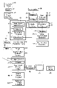
FIGURE 1 is a diagrammatic illustration of an apparatus or system for
automatically segmenting diagnostic images;
FIGURE 2 is a diagrammatic illustration of an axial slice of a probability
map of a brain stem model;
FIGURE 3 is a map which depicts voxels which certainly belong to the
brain stem, voxels which certainly belong to the background, and an area of
uncertainty;
FIGURE 4 is a flow chart which diagrammatically illustrates an automatic
method of segmenting images; and,
FIGURE 5 is a flow chart which diagrammatically illustrates an operator
assisted method of segmenting images.
With reference to FIGURE 1, a diagnostic imaging scanner 10, such as a CT
scanner, MRI scanner, PET scanner, nuclear scanner, an ultrasound scanner or
the like,
generates image data which is reconstructed by a reconstruction processor 12
to generate a
current 3D diagnostic image which is stored in a memory, memory segment, or
buffer 14.
With continuing reference to FIGURE 1 and further reference to FIGURE 2,
a memory or memory segment 20 stores a library of 3D probability maps 22. The
probability map defines a volume of interest region 24 which is known to be a
part of the
region or volume of interest, in the present example, the brain stem. A
background region
26 defines objects or tissues which are known to be background, i.e., not the
brain stem.
That is, voxels in brain stem region 24 have a 100% probability of depicting
the brain stem
and a 0% probability of depicting the background. Conversely, voxels in the
background
region 26 have a 100% probability of depicting the background and a 0%
probability of
depicting the brain stem. An uncertainty region 28 lies between the brain stem
region 24
and the background region 26. In the uncertainty region, each voxel has a
probability
between 100% and 0% that it lies in the object or organ of interest, e.g., the
brain stem, and
a probability between 0% and 100% that it lies in the background.
CA 02792736 2012-09-10
WO 2011/110960
PCT/1B2011/050601
4
To generate the probability map 22 for the brain stem or other volume of
interest, images with good contrast, i.e., accurately segmentable boundaries,
are manually
segmented at work station 30 to define binary masks in which all of the voxels
which
correspond to the volume of interest, e.g., the brain stem, are given the
maximum
probability value, e.g., one, and all of the voxels which correspond to the
background are
given a minumum probability value, e.g., zero. The boundary between the
minimum and
maximum probability regions of the binary mask defines the boundary of the
masks, i.e.,
the segmentation boundary which can be defined by a triangular mesh surface.
One or
more processors 32 has a binary mask registration computer routine 34 which is
programmed to register a plurality of the binary masks and the segmentation
boundaries.
That is, the binary masks are scaled to adjust for patients or objects of
interest of different
size, rotated, shifted, and the like, and optionally, elastically deformed to
compensate, for
example, for images generated in different motion states of the object of
interest to bring
them into alignment. The one or more processors 32 is also programmed to or
has a binary
mask merging computer routine 36 which merges the plurality of aligned binary
masks. In
the present example, background regions which are defined as background by all
binary
masks are given the probability value of zero, and brain stem regions which
are defined by
all of the binary masks as being part of the brain stem are assigned a value
of one, i.e.,
assigned to the brain stem region 24. Based on the relative location of the
boundaries in
the plurality of images, probabilities greater than zero and less than one,
are assigned to the
other voxels corresponding to the uncertainty region 28. For example, each
voxel in the
uncertainty region is given the average of its value in the binary maps. If
the voxel is
background, i.e. a value of zero in one in half the maps and in the brain stem
i.e. a value of
one in half the maps, the voxel is assigned the average or 0.5. The processor
32 is also
programmed to or has a computer routine 38 which determines a median or mean
segmentation boundary 40, i.e., a mean or median or other average location of
the
segmentation boundaries of all of the binary masks. The probabilities for each
voxel and
the mean segmentation boundary define the segmentation map 22. Typically, this
same
process is performed for a plurality of organs or volumes of interest to build
a library of
probability maps that are suitable for numerous different imaging locations or
applications.
In one embodiment, a model-based segmentation processor 50 is
programmed to include a computer routine 52 which extracts a conventional
volume or
CA 02792736 2012-09-10
WO 2011/110960
PCT/1B2011/050601
organ model from a model library 54 and fits it to the volume or organ of
interest. The
segmentation processor is further programmed to include a computer routine 56
which
registers the mean segmentation boundary 40 from the probability model with
the
conventional model and is programmed to or includes a computer routine 58
which
determines a transform that brings the mean segmentation boundary into
registration with
the registered conventional model. The segmentation processor is also
programmed to or
has a computer routine 60 which transforms the probability map in accordance
with this
determined transform to bring the probability map into registration with the
volume or
organ of interest in the current image. The transformed probability map is
stored in a
buffer or memory 62. Alternately, rather than using a conventional model for
the model-
based registration 56, the mean segmentation boundary from the probability
map, can be
used as the model.
In a fully-automated embodiment, a classifier 70, such as a processor or
computer routine, is previously trained to classify voxels of images based on
image
properties, such as intensity, gradient, texture, etc., as belonging to the
volume or organ of
interest, as belonging to the background, or a probability thereof. The
classifier has been
previously trained offline using dummy data. Any of a multiplicity of known
classifying
techniques can be used, such as nearest neighbors, support vector machines,
and the like.
The volume of interest, for example, may have a known surface properties, such
a smooth,
rounded, free of sudden transitions, or the like. The classifier operates on
the current
image from the buffer 14 to generate a probability that each voxel belongs to
the
background or the volume or organ of interest.
With reference to FIGURE 3, to save processing time, the classification
process may be performed only on voxels corresponding to the uncertainty
region 28 of the
transformed probability map. Without processing regions 24, 26 which have been
determined by the probability mask as being definitely in the organ or volume
of interest or
the background. Optionally,
a threshold circuit or processor can operate on the
transformed probability map to identify the uncertainty region 28 by
eliminating voxels
with a certainty of zero or one which represent a 100% probability of being in
the organ or
volume of interest and a 100% probability of being in the background.
Optionally, the
threshold can be set lower such that classification is performed only on
voxels which the
CA 02792736 2012-09-10
WO 2011/110960
PCT/1B2011/050601
6
probability map has determined has less than a 95%, for example, probability
of being in
the volume or organ of interest, or in the background.
A merge processor or computer routine 80 is programmed to merge, on a
voxel by voxel basis, the probabilities determined by the classifier 70 and
the probabilities
from the transformed probability map 62. The merging, in one embodiment,
averages the
classification and probability map probabilities for each voxel. Other
techniques for
combining the probabilities are also contemplated. As one example, an operator
control or
first slider 82 may he provided to adjust the relative weighting of the
classifier and
probability map probabilities. A human operator can selectively adjust the
relative
weighting to adjust the threshold boundary. Based on the merged probabilities,
the merge
processor determines whether each voxel has a higher chance of being in the
volume or
organ of interest or in the background. A determined segment boundary is
determined
from the interface between the two regions and stored in an appropriate memory
or buffer
84.
An image processor 90 is programmed to or includes a computer routine 92
which combines the current image from the memory or buffer 14 with the
determined
segmentation boundary 84 to create a segmented image. The image processor 90,
optionally, is further programmed to or has a computer routine 94 which
performs further
image processing, such as colorization, smoothing, and the like, of the
current image
combined with the segmentation boundary, i.e., the segmented current image.
The
segmented current image is stored in a patient medical database 96 as part of
the patient's
medical record. A video processor 98 extracts selected slices, 3D volume
representations,
or the like from the segmented image 94 and displays them on a human-readable
display
100, such as a video monitor.
In a semi-automated embodiment, a threshold circuit, processor, or
computer routine 110 determines whether the probability for each voxel of the
transformed
probability map exceeds a threshold. For example, the threshold may be
initially set as 0.5
in the above example, which indicates that the voxel is equally likely to be
in the volume of
interest and in the background. A processor or computer routine 112 defines
the
segmentation boundary based on the interface between the voxels which are more
likely to
be in the background and the voxcls which are more likely to be in the volume
or organ of
interest. In this embodiment, the segmentation boundary 114 is supplied to the
image
CA 02792736 2012-09-10
WO 2011/110960
PCT/1B2011/050601
7
processor routine 92 which combines the segmentation boundary with the current
image.
An operator viewing the segmented image on the display 100 uses a user input
device 116
to adjust the threshold 110, in the present example to shift the 0.5 threshold
higher towards
one or lower towards zero. As the threshold is adjusted, the interface between
the volume
or organ of interest and the background shifts as does the segmentation
boundary 114. In
one embodiment, the operator moves a slider with a mouse to select higher and
lower
threshold values until the operator is satisfied with the segmentation
displayed on the
display 100. Once the segmentation is optimized by the operator, the optimized
segmentation is stored in the patient medical database 96.
Once the image segmentation is complete, the segmented image has various
applications. For example, the segmented image can be used in a radiation
therapy system
120 to plan a radiation treatment protocol. Numerous other uses of the
segmented images
are, of course, contemplated.
In the foregoing discussion, it is to be understood that the various
processors, computer routines, and steps can be performed by one or more
computers or
processors. A single processor can perform one or more of the computer
processes or steps
and any one or more of the computer routines or steps can be distributed among
a plurality
of computer processors. Similarly, memories, memory segments, and buffers
described
above can take form in a single large memory, distributed memories, or the
like.
Moreover, a computer program for controlling one or more processors to
generate
segmented images in accordance with the above description can be carried on a
computer-readable medium, particularly a tangible medium, such as a CD or DVD,
or
other portable memory, a hard drive, resident computer memory, and the like.
The
program can also be carried by a non-tangible medium such as a digital or
analog signal or
the like.
With reference to FIGURE 4, a plurality of images of a selected region of
interest in each of a plurality of patients are generated and segmented at
step 130. At step
132, the plurality of segmented images is registered. At step 134, the
registered images are
merged generating a composite image of the region of interest with a plurality
of
superimposed segmentation boundaries. A mean segmentation boundary is
determined at
step 136. A probability that each voxel is within the volume of interest or
within the
background is determined at a step 138. For example, all voxels which are
inside of all of
CA 02792736 2012-09-10
WO 2011/110960
PCT/1B2011/050601
8
the superimposed segmentation boundaries are assigned to the volume of
interest and all
voxels which are outside of all of the superimposed segmentation boundaries
are assigned
to the background. Those voxels which are inside some of the segmentation
boundaries
and outside of others are assigned a probability in accordance with the
relative percentage
of the segmentation boundaries that the voxel is inside of or outside of. For
example, all
voxels which are within the volume of interest can be assigned the value of
one, all voxels
which are in the background can be assigned a value of zero, and all voxels
that are inside
of some of the superimposed segmentation boundaries and outside others are
assigned a
fractional value between zero and one based on the percentage of the
superimposed
segmentation boundaries that they are inside of or outside of. At step 140,
the probabilities
and the mean threshold boundary are combined to generate a probability map.
The
probability maps for each of a plurality of a volume of image can be stored in
a library to
be available for segmenting current images from a current patient.
When segmented images of a current patient are to be prepared, a plurality
of current images are generated at step 150. At step 152, an organ model is
retrieved from
memory and at step 154, the organ model is fit to the current image. At step
156, the
transform which brought the organ model into registration with the current
image is
determined. Various organ models are contemplated, such as a conventional
organ model,
the mean segmentation boundary, or the like.
At step 160, the probability map is transformed with the determined
transform to generate a transformed probability map 162 which represents a
probability
that each voxel is in the volume of interest or in the background. In parallel
in step 170,
each voxel of the current image is classified based on image properties, such
as intensity,
gradient, texture, and the like, and assigned a probability based on the image
properties that
it belongs to the volume of interest or to the background.
At a step 180, the probabilities from the transformed probability map and
the probabilities based on classification are merged on a voxel by voxel
basis. At a step
182, a segmentation boundary for the region of interest in the current image
is generated
based on the merged probabilities. For example, all voxels that are in the
volume of
interest with a probability greater than a preselected or adjustable threshold
are assigned to
the volume of interest and those which are below the threshold are assigned to
the
background. The interface represents the segmentation boundary of the volume
of interest.
CA 02792736 2012-09-10
WO 2011/110960
PCT/1B2011/050601
9
In the above example in which the volume of interest is assigned a value of
one and the
background a volume of zero, the threshold might be set for example at 0.5.
At a step 190, the segmentation boundary is combined with,
e.g., superimposed on, the current image to generate a segmented current
image. In a step
192, the segmented current image is stored in memory, for example, in a
patient medical
database. In a step 194, the segmented current image is displayed on a monitor
or other
clinician readable display.
With reference to FIGURE 5, in an operated assisted mode, the probability
map is subject to a threshold segmented at a step 200. In the above example in
which the
volume of interest has a value of one and the background a value of zero, the
threshold
might be initially set, for example, at 0.5. At a step 202, the segmentation
boundary is
defined as the interface between the voxels which are more probably in the
region of
interest and the voxels which are more probably in the background, e.g., above
or below
0.5. In step 204, the segmentation boundary is superimposed on the generated
current
image 150 to generate a segmented current image. At a step 206, the segmented
current
image is displayed to a radiologist or other technician. At a step 208, the
radiologist or
medical technician views the displayed segmented image and determines whether
the
segmentation is satisfactory. If the segmentation is satisfactory at a step
210, the
segmented current image is stored, such as in a patient medical database. If
the segmented
image is not satisfactory, the radiologist or other medical technician adjusts
the threshold at
a step 212. When the threshold is adjusted, the segmentation boundary defining
step 202
redefines the segmentation boundary, which redefine segmentation boundary is
superimposed on the current image in the step 204 and displayed in step 206.
This
adjustment process continues iteratively until the radiologist or other
medical technician is
satisfied with the segmentation.
The invention has been described with reference to the preferred
embodiments. Modifications and alterations may occur to others upon reading
and
understanding the preceding detailed description. It is intended that the
invention be
constructed as including all such modifications and alterations insofar as
they come within
the scope of the appended claims or the equivalents thereof.