Note: Descriptions are shown in the official language in which they were submitted.
CA 02794259 2012-09-24
WO 2011/119854 PCT/US2011/029826
THREE-DIMENSIONAL SURGICAL IMPLANT
BACKGROUND
Technical Field
[0001] The present disclosure relates to implants or surgical meshes and, more
particularly,
to meshes that have a grip-type knit mesh knit and a three-dimensional
structure.
Description of the Related Art
[0002] Surgical meshes formed from degradable or non-degradable materials for
use during
both open and minimally invasive surgeries are known. These meshes are
typically flat
fibrous material that a surgeon places over a defect, such as a tear in
tissue, as reinforcement.
The surgeon then secures the mesh in place with a surgical fastener, such as a
staple, clip,
tack, suture or the like.
[0003] Meshes exhibiting structures other than a planar or flat structure are
also known.
These meshes form a plug to fill the defect. In some cases, these meshes are
preformed from
permanent rigid materials with pleats to create some form of flexibility.
These permanent
meshes can also require a separate flat mesh overlay to reinforce the defect.
[0004] Surgical meshes formed from non-degradable materials can be rigid.
Rigid surgical
meshes have benefits in hernia repair, for example, a rigid hernia mesh keeps
the hernia sac
retracted, is quicker and easier to use, and is inserted using an easily
reproducible procedure.
However, the non-degradable materials result in permanent foreign material
inside a patient's
body. The heavy non-degradable materials used to form rigid meshes also have
small pore
sizes, which can inhibit tissue in-growth.
[0005] Surgical meshes formed from degradable materials may produce a soft,
pliant surgical
mesh. The level of flexibility of a pliant mesh is controlled by the materials
used to form the
mesh and the weave or knitting of the mesh. For example, a large pore mesh
formed from
CA 02794259 2012-09-24
WO 2011/119854 PCT/US2011/029826
lightweight degradable materials has enhanced tissue in-growth and reduced
inflammatory
response following implantation; it also results in less scarring than a
heavyweight, small
pore mesh. A soft, pliant mesh will form to the abdominal wall of the
patient's body and flex
more naturally with the movement of the abdominal wall following implantation.
Due to the
more natural action of a flexible, pliant mesh the patient typically
experiences less
postoperative pain and improved comfort. However, meshes made solely from
degradable
material may not be suitable for long term hernia repair.
[0006] It would be advantageous to provide a surgical mesh formed of both non-
degradable
and degradable materials so as to produce a soft, pliant mesh providing
improved comfort
and less postoperative pain for the patient. It would also be advantageous to
provide a
surgical mesh that can be formed or reformed into a three-dimensional
structure needed to fit
the defect.
[0007] In particular, it would be advantageous to provide a surgical mesh that
forms and
maintains a three-dimensional structure, exhibits the flexibility of a
degradable mesh and the
strength of a non-degradable mesh, leaves little permanent foreign material
inside a patient's
body, and secures itself within the defect.
SUMMARY
[0008] The present disclosure is directed to a three-dimensional surgical
implant. The three-
dimensional surgical implant includes a grip-type knit mesh defining pores and
including a
plurality of spiked naps extending from a surface thereof. The grip-type knit
mesh is folded
into a predetermined three-dimensional structure such that at least a portion
of the spiked
naps grip at least a portion of the pores to hold the three-dimensional
structure of the surgical
implant.
[0009] The present disclosure also is directed to a method of forming a three-
dimensional
surgical implant. The method includes: providing a grip-type knit mesh
defining pores and
2
CA 02794259 2012-09-24
WO 2011/119854 PCT/US2011/029826
including a plurality of spiked naps extending from a surface thereof; folding
the grip-type
knit mesh into a three-dimensional structure such that at least a portion of
the pores and at
least a portion of the spiked naps engage to fasten the surgical implant in
the three-
dimensional structure.
[0010] The present disclosure is also directed to a method of hernia repair.
The method
includes: providing a grip-type knit mesh defining pores and including a
plurality of spiked
naps extending from a surface thereof, folding the grip-type knit mesh into a
three-
dimensional structure such that at least a portion of the pores and at least a
portion of the
spiked naps engage to fasten the surgical implant into the three-dimensional
structure;
transferring said grip-type knit mesh into a body cavity having a hernia; and
placing the grip-type knit mesh in the hernia to repair the hernia.
[0011] The present disclosure includes three-dimensional surgical implant. The
three-
dimensional surgical implant includes a grip-type knit mesh defining pores and
including a
plurality of spiked naps extending from a surface thereof. The three-
dimensional surgical
implant is formed by folding the grip-type knit mesh into a predetermined
three-dimensional
structure such that at least a portion of the spiked naps grip at least a
portion of the pores to
hold the three-dimensional structure of the surgical implant.
BRIEF DESCRIPTION OF THE DRAWINGS
[0012] The foregoing objects and advantages of the disclosure will become more
apparent
from the reading of the following description in connection with the
accompanying drawings,
in which:
[0013] FIG. 1 is a top view of a grip-type knit mesh prior to forming a three-
dimensional
structure;
3
CA 02794259 2012-09-24
WO 2011/119854 PCT/US2011/029826
[0014] FIG. 2A and B are perspective views of the grip-type knit mesh formed
into three-
dimensional structures; and
[0015] FIG. 3A-D are side cross-sectional views showing the use of the grip-
type knit mesh
in a hernia repair.
DETAILED DESCRIPTION
[0016] The present disclosure relates to a grip-type knit mesh folded into a
three-
dimensional configuration. The grip-type knit mesh may be formed from
biodegradable
materials, non-biodegradable materials, or a combination of these. A grip-type
knit mesh
formed from a combination of biodegradable and non-biodegradable materials
produces a
semi-absorbable mesh resulting in less implanted mass while still providing a
strong rigid
support to maintain the long term integrity of the repair. A three-dimensional
design formed
with the grip portion facing outwards provides an additional means of fixation
to secure the
mesh to the tissue. The grip-type knit of the mesh also allows for formation
of a specific
shape to fit the patient's defect and the three-dimensional structure will be
maintained
without the need for stitching, gluing or pre-forming the mesh to a specific
structure.
[0017] The present disclosure relates to devices, systems, and methods for
minimally
invasive surgeries such as, endoscopic, laparoscopic, arthroscopic,
endoluminal and/or
transluminal placement of a surgical patch at a surgical site. As used herein
the term
"surgical mesh" is used to refer to any three-dimensional grip-type implant
for use in surgical
procedures, such as, for example, meshes that do not require suturing to the
abdominal wall.
Although described herein with reference to a hernia mesh, the method of the
disclosure may
be used in any surgical repair. As used herein the term "laparoscopic
deployment device" is
used to refer to a deployment device that may be used during minimally
invasive surgeries
4
CA 02794259 2012-09-24
WO 2011/119854 PCT/US2011/029826
described above. Although described herein with reference to a minimally
invasive surgery,
the surgical mesh may also be used in open surgery.
Materials
[0018] As stated above, the fibers forming the grip-type knit mesh may be made
from any
fiber-forming biocompatible polymer. The biocompatible polymer may be
synthetic or
natural. The biocompatible polymer may be biodegradable, non-biodegradable or
a
combination of biodegradable and non-biodegradable. The term "biodegradable"
as used
herein is defined to include both bioabsorbable and bioresorbable materials.
By
biodegradable, it is meant that the materials decompose, or lose structural
integrity under
body conditions (e.g., enzymatic degradation or hydrolysis) or are broken down
(physically
or chemically) under physiologic conditions in the body such that the
degradation products
are excretable or absorbable by the body.
[0019] Representative natural biodegradable polymers which may be used
include:
polysaccharides, such as alginate, dextran, chitin, hyaluronic acid,
cellulose, collagen, gelatin,
fucans, glycosaminoglycans, and chemical derivatives thereof (substitutions
and/or additions
of chemical groups, for example, alkyl, alkylene, hydroxylations, oxidations,
and other
modifications routinely made by those skilled in the art); and proteins, such
as albumin,
casein, zein, silk, and copolymers and blends thereof, alone or in combination
with synthetic
polymers.
[0020] Synthetically modified natural polymers which may be used include:
cellulose
derivatives, such as alkyl celluloses, hydroxyalkyl celluloses, cellulose
ethers, cellulose
esters, nitrocelluloses, and chitosan. Examples of suitable cellulose
derivatives include
methyl cellulose, ethyl cellulose, hydroxypropyl cellulose, hydroxypropyl
methyl cellulose,
hydroxybutyl methyl cellulose, cellulose acetate, cellulose propionate,
cellulose acetate
CA 02794259 2012-09-24
WO 2011/119854 PCT/US2011/029826
butyrate, cellulose acetate phthalate, carboxymethyl cellulose, cellulose
triacetate, and
cellulose sulfate sodium salt. These are collectively referred to herein as
"celluloses."
[0021] Representative synthetic degradable polymers suitable for use include:
polyhydroxy
acids prepared from lactone monomers, such as glycolide, lactide,
caprolactone, E-
caprolactone, valerolactone, and S-valerolactone, as well as pluronics,
carbonates (e.g.,
trimethylene carbonate, tetramethylene carbonate, and the like); dioxanones
(e.g., 1,4-
dioxanone and p-dioxanone), 1,dioxepanones (e.g., 1,4-dioxepan-2-one and 1,5-
dioxepan-2-
one), and combinations thereof. Polymers formed therefrom include:
polylactides;
poly(lactic acid); polyglycolides; poly(glycolic acid); poly(trimethylene
carbonate);
poly(dioxanone); poly(hydroxybutyric acid); poly(hydroxyvaleric acid);
poly(lactide-co-(E-
caprolactone-)); poly(glycolide-co-(c-caprolactone)); polycarbonates;
poly(pseudo amino
acids); poly(amino acids); poly(hydroxyalkanoate)s; polyalkylene oxalates;
polyoxaesters;
polyanhydrides; polyortho esters; and copolymers, block copolymers,
homopolymers, blends,
and combinations thereof.
[0022] Some non-limiting examples of suitable non-bioabsorbable materials from
which the
fibers of the grip-type knit mesh may be made include: polyolefins, such as
polyethylene and
polypropylene including atactic, isotactic, syndiotactic, and blends thereof;
polyethylene
glycols; polyethylene oxides; ultra high molecular weight polyethylene;
copolymers of
polyethylene and polypropylene; polyisobutylene and ethylene-alpha olefin
copolymers;
fluorinated polyolefins, such as fluoroethylenes, fluoropropylenes,
fluoroPEGSs, and
polytetrafluoroethylene; polyamides, such as nylon and polycaprolactam;
polyamines;
polyimines; polyesters, such as polyethylene terephthalate and polybutylene
terephthalate;
aliphatic polyesters; polyethers; polyether-esters, such as polybutester;
polytetramethylene
ether glycol; 1,4-butanediol; polyurethanes; acrylic polymers and copolymers;
modacrylics;
vinyl halide polymers and copolymers, such as polyvinyl chloride; polyvinyl
alcohols;
6
CA 02794259 2012-09-24
WO 2011/119854 PCT/US2011/029826
polyvinyl ethers, such as polyvinyl methyl ether; polyvinylidene halides, such
as
polyvinylidene fluoride and polyvinylidene chloride; polyacrylonitrile;
polyaryletherketones;
polyvinyl ketones; polyvinyl aromatics, such as polystyrene; polyvinyl esters,
such as
polyvinyl acetate; copolymers of vinyl monomers with each other and olefins,
such as
etheylene-methyl methacrylate copolymers, acrylonitrile-styrene copolymers,
ABS resins,
and ethylene-vinyl acetate copolymers; alkyd resins; polycarbonates;
polyoxymethylenes;
polyphosphazine; polyimides; epoxy resins; aramids, rayon; rayon-triacetate;
spandex;
silicones; and combinations thereof.
[0023] Rapidly biodegradable polymers, such as poly(lactide-co-glycolide)s,
polyanhydrides, and polyorthoesters, which have carboxylic groups exposed on
the external
surface as the smooth surface of the polymer erodes, may also be used. It
should of course be
understood that any combination of natural, synthetic, biodegradable and non-
biodegradable
materials may be used to form the grip-type knit mesh.
[0024] In embodiments, the naps of the grip-type knit mesh are formed from
polylactic acid
(PLA) and the mesh is formed from a monofilament polyester of polyethylene
terephthalate
(PET).
Bioactive Agents
[0025] The grip-type knit mesh may include a bioactive agent. The term
"bioactive agent"
as used herein, is used in its broadest sense and includes any substance or
mixture of
substances that have clinical use. Consequently, bioactive agents may or may
not have
pharmacological activity per se, e.g., a dye.
[0026] Alternatively, a bioactive agent could be any agent that provides a
therapeutic or
prophylactic effect, a compound that affects or participates in tissue growth,
cell growth, cell
differentiation, an anti-adhesive compound, a compound that may be able to
invoke a
biological action such as an immune response, or could play any other role in
one or more
7
CA 02794259 2012-09-24
WO 2011/119854 PCT/US2011/029826
biological processes. It is envisioned that the bioactive agent may be applied
to the implant
in any suitable form of matter, e.g., films, powders, liquids, gels and the
like.
[0027] The bioactive agent may be bound to the grip-type knit mesh covalently,
non-
covalently, i.e., electrostatically, through a thiol-mediated or peptide-
mediated bond, or using
biotin-avidin chemistries and the like.
[0028] Examples of classes of bioactive agents, which may be utilized in
accordance with
the present disclosure include, for example, anti-adhesives, antimicrobials,
analgesics,
antipyretics, anesthetics, antiepileptics, antihistamines, anti-
inflammatories, cardiovascular
drugs, diagnostic agents, sympathomimetics, cholinomimetics, antimuscarinics,
antispasmodics, hormones, growth factors, muscle relaxants, adrenergic neuron
blockers,
antineoplastics, immunogenic agents, immunosuppressants, gastrointestinal
drugs, diuretics,
steroids, lipids, lipopolysaccharides, polysaccharides, platelet activating
drugs, clotting
factors and enzymes. It is also intended that combinations of bioactive agents
may be used.
[0029] Anti-adhesive agents can be used to prevent adhesions from forming
between the
grip-type knit mesh and the surrounding tissues opposite the target tissue. In
addition, anti-
adhesive agents may be used to prevent adhesions from forming between the
coated
implantable medical device and the packaging material. Some examples of these
agents
include, but are not limited to hydrophilic polymers such as poly(vinyl
pyrrolidone),
carboxymethyl cellulose, hyaluronic acid, polyethylene oxide, poly vinyl
alcohols, and
combinations thereof.
[0030] Suitable antimicrobial agents which may be included as a bioactive
agent include, for
example, triclosan, also known as 2,4,4'-trichloro-2'-hydroxydiphenyl ether,
chlorhexidine
and its salts, including chlorhexidine acetate, chlorhexidine gluconate,
chlorhexidine
hydrochloride, and chlorhexidine sulfate, silver and its salts, including
silver acetate, silver
benzoate, silver carbonate, silver citrate, silver iodate, silver iodide,
silver lactate, silver
8
CA 02794259 2012-09-24
WO 2011/119854 PCT/US2011/029826
laurate, silver nitrate, silver oxide, silver palmitate, silver protein, and
silver sulfadiazine,
polymyxin, tetracycline, aminoglycosides, such as tobramycin and gentamicin,
rifampicin,
bacitracin, neomycin, chloramphenicol, miconazole, quinolones such as oxolinic
acid,
norfloxacin, nalidixic acid, pefloxacin, enoxacin and ciprofloxacin,
penicillins such as
oxacillin and pipracil, nonoxynol 9, fusidic acid, cephalosporins, and
combinations thereof.
In addition, antimicrobial proteins and peptides such as bovine lactoferrin
and lactoferricin B
may be included as a bioactive agent.
[0031] Other bioactive agents, which may be included include: local
anesthetics; non-
steroidal antifertility agents; parasympathomimetic agents; psychotherapeutic
agents;
tranquilizers; decongestants; sedative hypnotics; steroids; sulfonamides;
sympathomimetic
agents; vaccines; vitamins; antimalarials; anti-migraine agents; anti-
parkinson agents such as
L-dopa; anti-spasmodics; anticholinergic agents (e.g., oxybutynin);
antitussives;
bronchodilators; cardiovascular agents, such as coronary vasodilators and
nitroglycerin;
alkaloids; analgesics; narcotics such as codeine, dihydrocodeinone,
meperidine, morphine
and the like; non-narcotics, such as salicylates, aspirin, acetaminophen, d-
propoxyphene and
the like; opioid receptor antagonists, such as naltrexone and naloxone; anti-
cancer agents;
anti-convulsants; anti-emetics; antihistamines; anti-inflammatory agents, such
as hormonal
agents, hydrocortisone, prednisolone, prednisone, non-hormonal agents,
allopurinol,
indomethacin, phenylbutazone and the like; prostaglandins and cytotoxic drugs;
chemotherapeutics, estrogens; antibacterials; antibiotics; anti-fungals; anti-
virals;
anticoagulants; anticonvulsants; antidepressants; antihistamines; and
immunological agents.
[0032] Other examples of suitable bioactive agents which may be included in
the grip-type
knit mesh include, for example, viruses and cells, including stem cells;
peptides, polypeptides
and proteins, as well as analogs, muteins, and active fragments thereof;
immunoglobulins;
antibodies; cytokines (e.g., lymphokines, monokines, chemokines); blood
clotting factors;
9
CA 02794259 2012-09-24
WO 2011/119854 PCT/US2011/029826
hemopoietic factors; interleukins (IL-2, IL-3, IL-4, IL-6); interferons ((3-
IFN, a-IFN and y-
IFN); erythropoietin; nucleases; tumor necrosis factor; colony stimulating
factors (e.g.,
GCSF, GM-CSF, MCSF); insulin; anti-tumor agents and tumor suppressors; blood
proteins
such as fibrin, thrombin, fibrinogen, synthetic thrombin, synthetic fibrin,
synthetic
fibrinogen; gonadotropins (e.g., FSH, LH, CG, etc.); hormones and hormone
analogs (e.g.,
growth hormone); vaccines (e.g., tumoral, bacterial and viral antigens);
somatostatin;
antigens; blood coagulation factors; growth factors (e.g., nerve growth
factor, insulin-like
growth factor); bone morphogenic proteins; TGF-B; protein inhibitors; protein
antagonists;
protein agonists; nucleic acids, such as antisense molecules, DNA, RNA, RNAi;
oligonucleotides; polynucleotides; and ribozymes.
Mesh Structure
[0033] The knit forming the mesh may include a monofilament sheet forming, on
at least a
portion of at least one face of the knit, spiked naps which protrude with
respect to the sheet.
In embodiments, the naps each have a substantially rectilinear body and, at
the free end of
this body, a head of greater width than that of the body.
[0034] This knit can be formed using a thermofusible monofilament to form a
monofilament
sheet, forming outer loop-shaped meshes in the sheet, and then partially
fusing the
monofilament.
[0035] The length of the spiked naps is defined so as to penetrate and fasten
to the porous
textile structure of the knit in a limited manner, that is to say without
emerging from the other
face, for example when the nap portion of a knit including spiked naps is
applied against a
porous portion, of the same knit or of a different knit.
[0036] In embodiments, the monofilament forming the spiked naps can have a
diameter
from about 0.05 mm to about 0.15 mm, in embodiments a diameter of over 0.10
mm. Each
spiked nap can have a length of from about 1 mm and about 2 mm, in embodiments
a length
CA 02794259 2012-09-24
WO 2011/119854 PCT/US2011/029826
of about 1.5 mm. The density of the spiked naps can be from about 50 and about
90 naps per
square centimeter, in embodiments from about 65 and about 75 naps per square
centimeter.
Suitable grip-type knit meshes and methods for making them are disclosed in
U.S. Patent No.
7,331,199, the disclosure of which is incorporated by reference herein in its
entirety.
[0037] The textile structure of the knit may include two faces, one with the
spiked naps, and
one with open pores, which for example may have a diameter of from about 1 mm
and about
3 mm. For example, this structure can include several sheets of interlaced
yarns, which
together form a layered structure. When interlaced yarns are used, the layered
structure may
be composed, for example, of three sheets: an intermediate sheet of yarn
distributed to form a
zigzag openwork pattern between the columns of meshes; a front sheet of yarn
distributed to
form a chain stitch; and a rear sheet of monofilament placed in partial weft
under the chain
stitch and "thrown onto" the needle not forming a chain stitch, this sheet may
include the
spiked naps.
[0038] When a grip-type knit is applied, with spiked naps to the front, onto a
surface of a
porous prosthetic knit during manipulation into a three-dimensional
configuration, the spiked
naps engage into the mesh and between the multifilament yarns of the porous
knit and fasten
the grip-type knit onto the porous knit. This fastening, effective even in a
liquid
environment, is sufficient to secure the mesh in the desired three-dimensional
configuration,
and to offer mechanical resistance to tangential stresses, while at the same
time permitting
unfastening of the grip-type knit in order to adjust its position in relation
to the element lying
underneath, if desired.
[0039] In embodiments, the porous knit portion of the mesh may include size
markings.
The size markings may indicate the location into which the grip-type knit may
be secured to
the porous knit during manipulation into a three-dimensional configuration in
order to obtain
three-dimensional structures (e.g., cones) of various sizes. The markings may
be any type of
11
CA 02794259 2012-09-24
WO 2011/119854 PCT/US2011/029826
marking as is known in the art. For example, a dye or colorant may be placed
(e.g., printed)
at specific locations on the porous knit. As another example, a colored yam
may be woven
into specific locations of the porous knit. Those skilled in the art will
readily envision other
ways of applying suitable markings to the mesh.
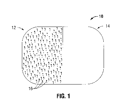
[0040] Referring now in specific detail to the drawings, in which like numbers
identify
similar or identical elements, FIG. 1 is an illustration of a grip-type knit
mesh prior to
forming a three-dimensional structure. The grip-type knit mesh 10 includes
sides 12 and 14.
Side 12 includes naps 16 which grip into the open pore structure of side 14.
Although sides
12 and 14 are each shown as covering half of the mesh, the naps 16 may cover
less or more
of the mesh. It is also envisioned that the naps can cover an entire side of a
mesh.
Three-Dimensional Structure
[0041] As stated above, the spiked naps grip onto the porous portion of the
mesh in such a
manner as to be secure yet capable of being detached and reattached as
necessary. The grip-
type knit mesh may be formed or folded into a three-dimensional structure. For
example, the
knit may be formed or folded into a cone, cylinder, triangle, square, and the
like. In
embodiments, the three-dimensional structure can be held together by using the
naps engaged
with the open pore structure wherever there is overlap.
[0042] The naps of the grip-type knit mesh may face inward or outward in
relation to the
three-dimensional structure of the mesh. When the naps face outward, they
provide a means
of affixing the mesh to the surrounding tissue. In embodiments, the three-
dimensional
structure can be formed from the grip-type knit during production, i.e.,
without the use of the
naps to hold the structure into a shape.
[0043] In embodiments, the surgeon can form the grip-type knit mesh into the
desired shape
prior to using the mesh in situ. In embodiments, markings on the grip-type
knit mesh can
provide guidance as to how to fold or form the mesh into a three-dimensional
structure.
12
CA 02794259 2012-09-24
WO 2011/119854 PCT/US2011/029826
[0044] FIGS. 2A and B show different configurations of the three-dimensional
mesh of the
present disclosure. Mesh 20, when planar, has a nap portion 22 on one side and
an open pore
portion 24 on the other side. When folded into a conical structure (FIG. 2A),
the nap portion
22 may face inward or outward. Mesh 20 may also be folded into a conical
formation (FIG.
2B) with nap portion 22 facing outward from open pore portion 24.
Methods of Use
[0045] In accordance with the present disclosure the three-dimensional grip-
type knit mesh
may be used in either minimally invasive or open surgery. A minimally invasive
method of
treating a hernia includes: making an incision in the abdominal wall close to
the herniated
area; making a subcutaneous cut, through the incision, over and surrounding
the area of the
hernia; inserting a three-dimensional grip-type knit mesh through the incision
using a
laparoscopic device; and inserting the three-dimensional grip-type knit mesh
into the hernia
[0046] Thus, a mesh according to the present disclosure can be inserted
through a small
incision (e.g., from about 1 cm to about 2 cm in length) in the abdominal
cavity. In
embodiments, a hernia region is reached using an anterior surgical approach.
The grip-type
surgical mesh is formed into a three-dimensional structure by fastening the
grip portion to the
porous portion of the mesh. The three-dimensional structure may mirror the
three-
dimensional structure of the defect. The mesh is then inserted through the
opening in the
tissue wall until the base lies flush with or slightly beyond the defect. When
the grip portions
are facing outward they will grip to the tissue securing the mesh within the
tissue. The mesh
thus conforms to the shape of the defect and adheres to the surrounding tissue
in such a way
as to secure the mesh to the tissue. It is also contemplated that a surgical
fastener is used to
attach the mesh to the surrounding tissue. In embodiments where the naps of
the grip-type
knit mesh are formed from a biodegradable material such as, for example, a
polylactic acid
(PLA) and the mesh is formed from a non-biodegradable material such as, for
example,
13
CA 02794259 2012-09-24
WO 2011/119854 PCT/US2011/029826
monofilament polyester of polyethylene terephthalate (PET), the naps of the
mesh will
degrade over time while the non-degradable portion of the mesh remains to
provide stability
to the mesh. This results in less foreign material left in the patient.
[0047] A separate flat grip-type knit mesh may also be adhered to the
surrounding tissue.
[0048] Referring now to FIGS. 3A-3D, a method of using a three-dimensional
grip-type knit
mesh to perform a surgical repair procedure is shown and described. With
reference to FIG.
3A, a hernia may involve a tear 30, in the abdominal wall 32. Abdominal wall
32 is defined
by an external side 32a and peritoneum 32b. A surface tissue 34, which covers
the external
side 32a of abdominal wall 32, may or may not be immediately affected by this
tear 30. An
internal organ 36 located below the peritoneum 32b of the abdominal wall 32
may not
protrude until some form of exertion or use of the muscle located at the
abdominal wall 32
forces the internal organ 36 into the tear 30. Depending on the size and
location of the tear
30, exertion may not be needed to cause the organ to protrude. As shown in
FIG. 3B, a
hernia occurs when internal organ 36 protrudes into the tear 30 of abdominal
wall 32.
Oftentimes the protrusion creates a bulge 38 in the surface tissue 34.
[0049] In order to correct the defect, as depicted in FIG. 3C, an incision 42
is made through
the abdominal wall 32 in close proximity to tear 30 and a three-dimensional
grip-type knit
mesh 20 is inserted using a trocar 44 or similar laparoscopic device. As shown
in FIG. 3D, a
three-dimensional grip-type knit mesh 20 is then placed in the tear 30 from
the peritoneum
32b of the abdominal wall 32. The naps 16 attach to the abdominal wall 32 and
allow the
mesh 20 to fill the tear 30 in the abdominal wall 32 and return the internal
organ 36 to its
original location.
[0050] While the above description contains many specifics, these specifics
should not be
construed as limitations on the scope of the present disclosure, but merely as
exemplifications
14
CA 02794259 2012-09-24
WO 2011/119854 PCT/US2011/029826
of preferred embodiments thereof. Those skilled in the art will envision many
other possible
variations that are within the scope and spirit of the present invention.